Article

Dysregulation of Retinal Melatonin Biosynthetic Pathway and Differential Expression of Retina-Specific Genes Following Blast-Induced Ocular Injury in Ferrets

by
Chetan Pundkar
1,†,
Rex Jeya Rajkumar Samdavid Thanapaul
1,2,†,
Manoj Govindarajulu
1,
Gaurav Phuyal
1,
Joseph B. Long
1 and
Peethambaran Arun
1,*
1
Blast-Induced Neurotrauma Branch, Center for Military Psychiatry and Neuroscience, Walter Reed Army Institute of Research, Silver Spring, MD 20910, USA
2
National Research Council (NRC) Research Associateship Programs, National Academies of Sciences, Engineering, and Medicine, Washington, DC 20001, USA
*
Author to whom correspondence should be addressed.
These authors contributed equally to this work.
Neurol. Int. 2025, 17(3), 42; https://doi.org/10.3390/neurolint17030042
Submission received: 25 January 2025 / Revised: 7 March 2025 / Accepted: 10 March 2025 / Published: 17 March 2025

Abstract

:
Background/Objectives: Blast-induced traumatic ocular injuries (bTOI) pose a significant risk to military and civilian populations, often leading to visual impairment or blindness. Retina, the innermost layer of ocular tissue consisting of photoreceptor and glial cells, is highly susceptible to blast injuries. Despite its prevalence, the molecular mechanisms underlying retinal damage following bTOI remain poorly understood, hindering the development of targeted therapies. Melatonin, a neuroprotective indoleamine with antioxidant, anti-inflammatory, and circadian regulatory properties, is synthesized in the retina and plays a crucial role in retinal health. Similarly, retina-specific genes, such as Rhodopsin, Melanopsin, and RPE65, are essential for photoreceptor function, visual signaling, and the visual cycle. However, their responses to blast exposure have not been thoroughly investigated. Methods: In this study, we utilized a ferret model of bTOI to evaluate the temporal expression of melatonin-synthesizing enzymes, such as tryptophan hydroxylase 1 and 2 (TPH1 and TPH2), Aralkylamine N-acetyltransferase (AANAT), and Acetylserotonin-O-methyltransferase (ASMT), and retina-specific genes (Rhodopsin, Melanopsin) and retinal pigment epithelium-specific 65 kDa protein (RPE65) at 4 h, 24 h, 7 days, and 28 days post-blast. Ferrets were exposed to tightly coupled blast overpressure waves using an advanced blast simulator, and retinal tissues were collected for quantitative polymerase chain reaction (qPCR) analysis. Results: The results revealed dynamic and multiphasic transcriptional responses. TPH1 and TPH2 exhibited significant upregulation at 24 h, followed by downregulation at 28 days, indicating blast-induced dysregulation of tryptophan metabolism, including melatonin synthesis. Similarly, AANAT and ASMT showed acute downregulation post-blast, with late-phase disruptions. Rhodopsin expression increased at 24 h but declined at 28 days, while Melanopsin and RPE65 demonstrated early upregulation followed by downregulation, reflecting potential disruptions in circadian regulation and the visual cycle. Conclusions: These findings highlight the complex regulatory mechanisms underlying retinal responses to bTOI, involving neuroinflammation, oxidative stress, and disruptions in melatonin synthesis and photoreceptor cell functions. The results emphasize the therapeutic potential of melatonin in mitigating retinal damage and preserving visual function.

1. Introduction

Blast-induced traumatic ocular injuries (bTOI) pose a serious risk to both military and civilian populations, and, in both, loss of vision can disrupt careers and adversely impact lifestyle [1]. Among military personnel, eye injuries rank as the fourth most prevalent battlefield injury, constituting 6–13% of all blast injuries [2]. In particular, explosions during combat significantly contribute to ocular injuries, which often result in visual impairment or permanent blindness [3,4]. The incidence and severity of these ocular injuries have been exacerbated by the advancement of weaponry with enhanced explosive capabilities [5]. Ocular trauma, primarily due to blast injuries from improvised explosive devices (IEDs), has been a leading cause of injury among U.S. service members [6]. The bTOI can be primary, secondary, tertiary, or quaternary, resulting from various sources such as blast waves, fragments, structural collapse, burns, and indirect injuries, respectively [7]. Bilateral ocular explosion injuries are common [8]. The blast impacts numerous regions throughout the eye such as eyelids, cornea, conjunctiva, retina, and optic nerve [3]. Increased light sensitivity, retinal separation, retinal edema, retinopathy, optic neuropathy, and loss of visual field are among the clinical consequences of bTOI [9]. The retina is highly susceptible to shockwaves resulting from blasts. However, the lack of understanding of the molecular mechanisms underlying retinal damage post bTOI hinders the development of targeted therapies.
Melatonin, an indoleamine molecule produced by the pineal gland from the amino acid tryptophan, regulates circadian rhythms [10]. The first and rate-limiting step for synthesizing the neurotransmitter serotonin from tryptophan is catalyzed by TPH1 and TPH2. This hydroxylation of tryptophan, followed by decarboxylation, results in the formation of 5-hydroxytryptamine (serotonin). AANAT converts serotonin to N-acetylserotonin, which is considered the rate-limiting step in synthesizing melatonin, after which ASMT catalytically converts N-acetylserotonin to melatonin [11]. Melatonin is also synthesized from tryptophan in the retina, skin, kidneys, gastrointestinal tract, and spleen through the same metabolic pathway [12]. Along with regulating circadian cycles, melatonin also plays a vital role in immunological, cardiovascular, and retinal functioning [13]. It has the potential to be a therapeutic and neuroprotective agent in neurodegenerative and age-related disorders due to its antioxidant properties and ability to protect against oxidative stress [14]. Melatonin acts via receptors that mediate its essential role in ocular angiogenesis and other significant effects on the immune system, peripheral organs, and the central nervous system. Previous studies have shown the salutary anti-inflammatory and antioxidant properties of melatonin in retinal diseases such as age-related macular degeneration (AMD) and diabetic retinopathy (DR) [15]. However, the role of melatonin has not yet been explored in the pathogenesis of bTOI.
Retina-associated photoreceptors, pigments, and enzymes play a crucial role in normal vision. Melanopsin, a photopigment in intrinsically photosensitive retinal ganglion cells (ipRGCs), regulates melatonin synthesis in the retina [16]. When activated by light, it provides information about ambient light levels to the brain, regulating the pineal gland’s melatonin production [16] and synchronizing the body’s circadian cycles with the external light–dark cycle. RPE65 maintains photoreceptors’ responsiveness to light, thereby contributing to signaling cascades involving Melanopsin-expressing ipRGCs [17]. Rhodopsin, a light-sensitive receptor protein in the retinal photoreceptive rod cells, is necessary for vision in low-light environments and helps control the body’s circadian cycles [18]. Alterations in the time-dependent expression pattern of these retina-specific genes in blast-induced retinal injury have not been previously described.
This study explored the temporal expression pattern of genes involved in retinal melatonin production and retina-specific genes in the retina after blast exposure in ferrets.

2. Materials and Methods

2.1. Animals

Adult male ferrets (Mustela putorius furo), aged 13–15 weeks and weighing around 1–1.2 kg, were purchased from Triple F Farms (Gillett, PA, USA) and were socially housed in pairs in ventilated cages at 20–22 °C on a 12:12 h light–dark cycle. Ferrets were provided with free access to food and chlorinated water ad libitum throughout the course of the study. Research using ferrets was conducted under an Institutional Animal Care and Use Committee-approved animal use protocol in an AAALAC International-accredited facility in compliance with the Animal Welfare Act and other federal statutes and regulations relating to animals and experiments involving animals and adhered to principles stated in the Guide for the Care and Use of Laboratory Animals, NRC Publication, 2011 edition. Ferrets were randomized into two groups: sham and repeated blast (BB), with each group containing a minimum of four ferrets.

2.2. Blast Exposure and Sample Collection

Ferrets were anesthetized with 5% isoflurane for 8 min and positioned in a longitudinal prone orientation (facing the oncoming shockwave) within the test section of an advanced blast simulator (ABS) [19]. Each ferret was exposed to two 19 psi blast overpressure waves, separated by a 2 min interval as described earlier [19]. Following blast exposure, the ferrets were euthanized at 4 h, 24 h, 7 days, or 28 days. Retinas from both eyes were dissected as described previously [20]. Retinal dissection involves an incision at the corneoscleral junction and removing the anterior part of the eye, including the cornea and lens. The short stalk of the optic nerve on the posterior side of the eye was secured with tweezers, and the eyeball was gently squeezed in the opposite direction to allow the retina to float out of the sclera. Retinas from both eyes were pooled and preserved in phosphate-buffered saline (PBS) and stored at −80 °C for further analysis.

2.3. RNA Extraction and Quantitative RT-PCR

Total RNA was extracted from the retina using the RNeasy Lipid Tissue Mini Kit (Cat #74104, Qiagen, Redwood City, CA, USA). RNA quantity was measured using the Nanodrop D2000 spectrophotometer (Thermo Scientific, Wilmington, DE, USA), and sample purity was assessed by the absorbance ratio at 260/280 nm. Only samples with a ratio of 1.8–2.0 were used for cDNA synthesis. RNA was reverse transcribed into cDNA using the RT2 First Strand Kit (Cat #330404, Qiagen, MD, USA), and the cDNA was stored at −20 °C until further use. Primers used in the study were procured from Eurofins Genomics (Louisville, KY, USA) and are listed in Table 1. Quantitative real-time polymerase chain reaction (qRT-PCR) was performed on the synthesized cDNA using RT2 SYBR Green qPCR Mastermix reagent, followed by amplification on an Applied QuantStudio 6 Flex qPCR system (Life Technologies, Grand Island, NY, USA). Each sample was run in triplicate, and the average critical threshold cycle (Ct) was used to calculate relative quantification by fold change and statistical significance. No-template controls were included in triplicate for each probe in every run to ensure accuracy and prevent contamination. Relative mRNA expression was normalized to the housekeeping gene 18S rRNA, and the fold changes were calculated using the 2−ΔΔCt method. The ΔΔCt calculation involved determining the difference between the ΔCt values of the blast group and the mean ΔCt value of the sham group. Fold changes in specific mRNA expression in the blast group relative to the sham group were presented as normalized fold changes.

3. Statistical Analysis

All data are expressed as the mean ± SEM and were compared using a student t-test to assess the effects of blast exposure compared to the sham group. Statistical analyses were conducted using GraphPad Prism 9 software (GraphPad Software Inc., Boston, MA, USA). Differences among the groups were considered statistically significant at p-values < 0.05.

4. Results

4.1. Effect of Blast Exposure on Expression of Melatonin-Synthesizing Enzymes

TPH exists in two isoforms, TPH1 and TPH2, with TPH2 being primarily expressed in the brain and retina, while TPH1 is mainly found in the gut and pineal gland [21]. In the retina, melatonin is predominantly synthesized by the photoreceptor cells and other retinal cell types during certain pathological conditions [22]. Studies have shown that TPH2 expression in the retina exhibits a circadian rhythm, suggesting a potential role in regulating the body’s internal clock [23,24]. To explore the impact of blast exposure on the melatonin synthesis pathway, we investigated the mRNA expression levels of TPH1 and TPH2 in the retina at various time points. The temporal expression of TPH1 and TPH2 mRNA in the retina following blast exposure was assessed at four time points (4 h, 24 h, 7 days, and 28 days). Both enzymes exhibited dynamic and biphasic expression patterns. For TPH1 (Figure 1A), no significant difference in mRNA expression was observed between the blast and sham groups at 4 h post-blast (p > 0.05). At 24 h, TPH1 mRNA expression was significantly upregulated, with a fold change of 3.80 compared to the sham group (p < 0.001), representing the peak expression level. By 7 days, TPH1 mRNA levels returned to baseline, showing no significant difference from sham controls. At 28 days, TPH1 mRNA expression was significantly downregulated compared to the sham group, with a fold change of approximately 0.5 (p < 0.01). Similarly, TPH2 mRNA followed a comparable temporal pattern (Figure 1B), with no significant changes observed at 4 h. At 24 h, TPH2 mRNA was significantly upregulated, showing a fold change of 2.12 (p < 0.001). By 7 days, TPH2 expression normalized to baseline, while at 28 days, it was significantly downregulated, with a fold change of approximately 0.63 (p < 0.001). These findings suggest that TPH1 and TPH2 are involved in acute and long-term retinal responses to blast injury, including neuroinflammation, oxidative stress, and neuroplasticity, due to potential dysregulation of melatonin synthesis, as shown below.
To explore the impact of blast exposure on melatonin synthesis, we further investigated the mRNA expression of AANAT and ASMT in the retina. The expression of AANAT and ASMT mRNA, key enzymes in melatonin synthesis, was also analyzed, revealing dynamic multiphasic responses to blast exposure. For AANAT (Figure 2A), expression at 4 h post-blast showed a non-significant upward trend compared to the sham group. At 24 h, AANAT mRNA expression was significantly downregulated (p < 0.001), reflecting an acute inhibitory effect of blast injury on melatonin synthesis. By 7 days, expression returned to baseline, showing no significant difference from sham controls. At 28 days, AANAT mRNA was significantly upregulated compared to the sham group (p < 0.05), with a fold change of 1.69, suggesting a late-phase compensatory activation for the decreased expression of TPH1&2 genes. For ASMT (Figure 2B), no significant differences were observed at 4 h post-blast. At 24 h, ASMT expression was significantly downregulated (p < 0.001), while at 7 days, a substantial upregulation with a fold change of 2.74 was observed (p < 0.01). By 28 days, ASMT mRNA was significantly downregulated again, showing a fold change of 0.47 (p < 0.001). These findings suggest blast exposure affects melatonin synthesis through complex regulatory mechanisms involving AANAT and ASMT, with implications for circadian signaling and retinal neuroprotection.

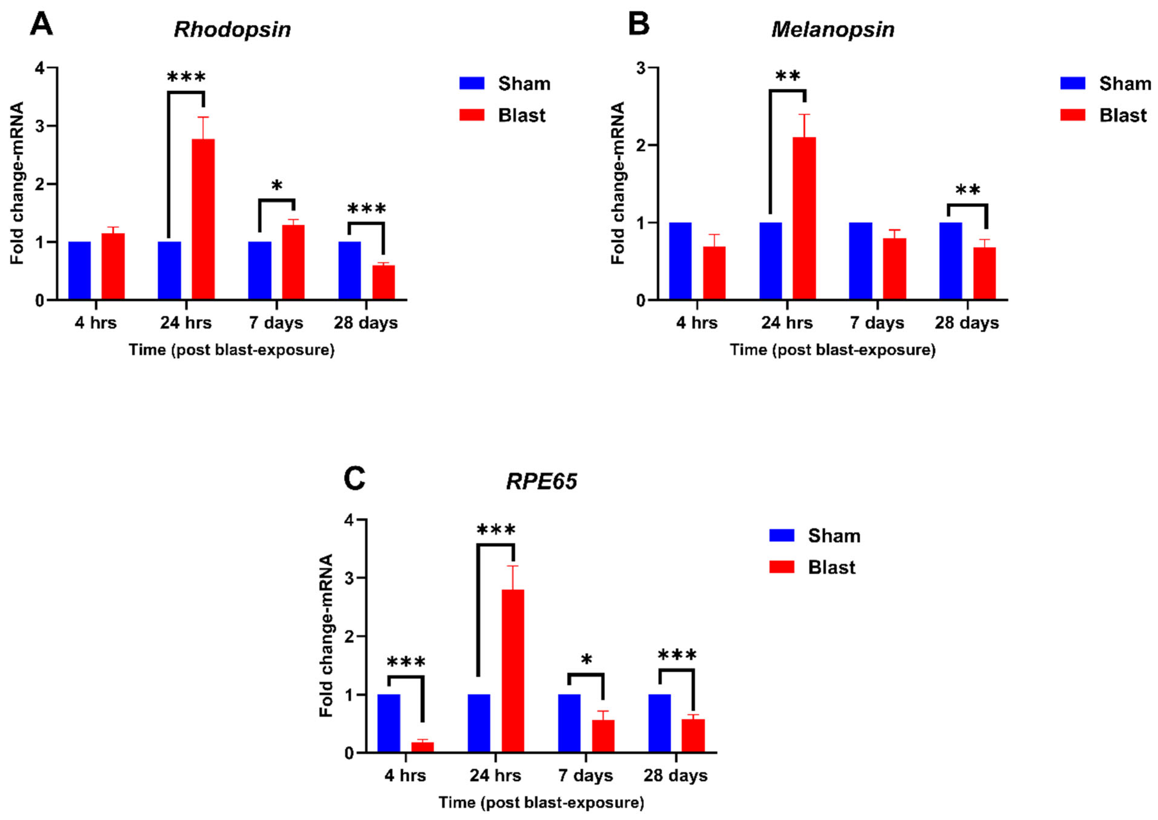
4.2. Effect of Blast Exposure on Expression of Retina-Specific Genes

Blast exposure can lead to disturbances in vision by disrupting the function of retinal cells [25,26]. However, direct research on the interaction between certain retina-specific proteins such as Rhodopsin, Melanopsin, and RPE65 and blast exposure is limited. Therefore, we investigated the effects of repeated blast exposure on the mRNA expression of these key proteins at various time points post-blast.
Retina-specific genes, Rhodopsin, Melanopsin, and RPE65, exhibited distinct transcriptional responses to blast exposure, reflecting potential disruptions in photoreceptor function, retinal signaling, and visual cycle maintenance post-blast. For Rhodopsin (Figure 3A), expression remained stable at 4 h post-blast. At 24 h, significant upregulation was observed, with a fold change of 2.76 compared to the sham group (p < 0.001). This upregulation persisted at 7 days, albeit at a lower level, with a fold change of 1.28 (p < 0.05). At 28 days, Rhodopsin expression was significantly downregulated (p < 0.001), with a fold change of 0.60, suggesting long-term disruptions in photoreceptor function. Melanopsin expression (Figure 3B) showed no significant changes at 4 h. At 24 h, Melanopsin mRNA was significantly upregulated with a fold change of 2.10 (p < 0.01), while at 7 days, it normalized to baseline. At 28 days, Melanopsin mRNA was significantly downregulated, with a fold change of 0.67 (p < 0.01), indicating potential long-term alterations in circadian regulation. RPE65, essential for the visual cycle, exhibited (Figure 3C) an acute downregulation at 4 h (p < 0.001). At 24 h, a marked upregulation of RPE65 mRNA was observed, with a fold change of 2.79 (p < 0.001), potentially reflecting a compensatory or inflammatory response. By 7 days, RPE65 expression declined (p < 0.05), and at 28 days, it was downregulated further, showing a fold change of approximately 0.58 (p < 0.001). These results suggest that Rhodopsin, Melanopsin, and RPE65 are integral to the retinal response to blast injury, with dynamic transcriptional changes indicating acute, sub-acute, and chronic effects.
Collectively, these findings demonstrate that blast exposure induces distinct temporal changes in the transcription of melatonin-synthesizing enzymes and retina-specific genes, highlighting their roles in neuroinflammation, oxidative stress, circadian signaling, and visual function. The multiphasic dysregulation observed across these genes underscores the complexity of retinal responses to blast injury and the need for further studies to elucidate their functional significance.

5. Discussion

The retina is highly susceptible to damage by the shearing forces produced by the blast waves [25,27,28], which primarily affect retinal ganglion cells, as manifested by thinning of the retinal layers and loss of retinal ganglion cell somas [29,30,31]. Furthermore, in rats, blast exposure has been shown to cause visual and retinal changes up to 8 months post-blast, mimicking some of the visual defects seen in human blast-exposed patients [32]. Previous animal studies have evaluated blast-induced changes in retinal and visual function, upregulation of proteins associated with oxidative stress, activation of inflammation, apoptosis, and cell death [25,26,33,34,35,36]. However, to date, no studies have determined the role of melatonin synthesis and signaling in the retina following bTOI. To our knowledge, this is the first study to demonstrate changes in the mRNA levels of genes involved in melatonin synthesis post-blast in the retina in an animal model of bTOI. We also report changes in the mRNA expression of retina-specific proteins, namely Rhodopsin, Melanopsin, and RPE65 after blast injury.
Recent studies indicate that melatonin synthesis in the retina shares features with that occurring in the pineal gland, including the same biochemical substrates and enzymatic pathway [37,38]. In the retina, tryptophan hydroxylase (TPH), the first enzyme involved in the biosynthesis of serotonin/melatonin, is primarily expressed in the photoreceptor cells and converts tryptophan into 5-hydroxytryptophan, the precursor to serotonin. Hence, an increase or decrease in TPH catalytic activity can lead to corresponding alterations in serotonin levels in the retina [39]. Therefore, TPH is essential for regulating the circadian rhythm of the eye, which is linked to light–dark cycles [40,41]. Our study noted decreases in TPH1 (non-significant) and TPH2 mRNA expression levels at 4 h post-blast, followed by an increase in both levels at 24 h and, interestingly, an ensuing decrease at 28 days post-blast. Though no studies have investigated the effect of blast exposure on TPH isoforms in the retina, increased mRNA levels of TPH2 in the dorsal raphe nucleus at 24 h post-blast have been noted [42,43]. Similarly, transcript levels of TPH2 were elevated in the dorsal raphe nucleus acutely post-blast and remained elevated even after one week [44]. Our previous study showed decreased TPH1 and increased TPH2 mRNA levels at 6 and 24 h post-blast injury in the pineal gland [45]. Several animal studies indicate that TPH2 gene expression is modulated by various stressors. For instance, a significant increase in TPH2 expression was noted in rat medulla following hypotensive hemorrhage [46]. Similarly, rats exposed to repeated forced swim and chronic variable stress resulted in elevated TPH2 expression in the midbrain [47]. Hence, the increased TPH1 and TPH2 levels in the retina at 24 h post-blast could be at least partly accounted for as a stress response. Interestingly, we noted a decrease in TPH1 and TPH2 levels in the retina at 28 days post-blast injury. This can be attributed to the loss of retinal ganglion cell axons [48] and subsequent retinal degeneration [25], which have been reported at 4–6 weeks post-blast injury [49].
Melatonin synthesis in the retinal photoreceptors is regulated primarily by changes in the activity of AANAT, the key enzyme that converts serotonin to N-acetylserotonin [50]. Apart from playing a role in circadian rhythm, it also plays a key role in neurotransmission and detoxification by acetylating arylalkylamines that may react with retinaldehyde [51]. We have noted a decrease in AANAT mRNA expression levels at 24 h and an increase at 28 days post-blast. Furthermore, ASMT mRNA levels decreased at 24 h and at 28 days post-blast, indicating perturbed melatonin synthesis in the retina at those time points. In contrast, the mRNA levels of ASMT showed an increase at day 7 post-blast. A previous study noted decreased AANAT gene expression in the rat frontal cortex at eight months post-blast exposure [52]. Our previous study in rats showed decreased AANAT and ASMT mRNA levels at 6 h and 24 h post-blast exposure in the pineal gland. The biological importance of time-dependent changes in the mRNA levels of enzymes involved in the melatonin synthesis pathway in the retina needs to be further evaluated.
Serotonin (5-hydroxytryptamine (5-HT)) plays a crucial role in neuroprotection within the eye, particularly in maintaining the health of retinal cells and regulating visual acuity [53,54]. Serotonin enhances retinal cell survival, decreases inflammation, and protects against oxidative damage [55,56]. It acts via serotonin receptors (HTRs) in retinal cells, particularly hydroxytryptamine receptor 1B (HTR1B) and hydroxytryptamine receptor 1D (HTR1D), and it influences the transmission of visual information to the thalamus [57]. Serotonin also regulates glutamate release and receptor activity, minimizing excitotoxicity and preventing damage to retinal neurons [54]. It influences the retinal response to hypoxia, potentially improving tissue oxygenation and decreasing damage caused by ischemia [58]. Serotonin additionally controls blood flow within the retina, regulating blood vessel constriction and dilation [59,60]. Serotonin governs circadian rhythms, the body’s innate biological clocks [61]. The suprachiasmatic nucleus (SCN) in the hypothalamus manages the circadian time system [62]. Serotonin from the raphe nuclei modulates SCN activity, effecting phase shifts and the sleep–wake cycle [63]. It can prevent light-induced phase shifts, sustaining circadian rhythms [62]. This control ensures physiological and behavioral processes are coordinated with the day-night cycle. Hence, serotonin is vital to retinal function and circadian rhythm modulation.
Rhodopsin is the most abundant protein in the retina’s rod cells and functions as the primary photoreceptor molecule mediating vision along with the maintenance of circadian rhythm [64,65]. Degeneration of these photoreceptors manifests as an outer nuclear layer thinning, a reduction in electroretinogram amplitudes, and vision loss [66]; hence, studies indicate that Rhodopsin can be considered as a biomarker associated with retinal thinning and degeneration [67,68,69]. Furthermore, changes in the Rhodopsin levels have been shown to contribute to early events of retinal degeneration [70]. We have noted an increase in mRNA levels of Rhodopsin at 24 h and then a decrease at 7 days and 28 days post-blast exposure. Our results align with another study, which showed lighter and diminished staining of Rhodopsin in the retina at 7 days and 30 days post-blast exposure [71]. Like Rhodopsin, Melanopsin is a photosensitive protein predominantly located in the retinal ganglion cells, which has also been implicated in regulating circadian rhythm. Repeated blast exposure showed an increase in mRNA levels of Melanopsin at 24 h and then a decrease at 7 days and 28 days post-blast. Though there are no reports of a direct link between blast exposure and changes in Melanopsin levels, a study by Pérez MP et al. showed high-intensity light exposure of rats induced reduction in Melanopsin positive retinal ganglionic cells (RGCs) [72]. In another study, Nadal-Nicolás et al. demonstrated the downregulation of melanopsin+ RGCs and melanopsin mRNA level after 7 days post optic nerve crush injury [73]. Moreover, a blast model of mild TBI showed significantly reduced retinal ganglion cells and increased Melanopsin immunolabeling in both eyes of C57BL/6 mice after 5 days of blast exposure [74]. Interestingly, studies have shown that Melanopsin-expressing retinal ganglion cells (mRGCs) are more resilient to damage from various retinal insults like axonal injury, chronic ocular hypertension, and neurodegeneration compared to other retinal ganglion cell types [75]. Hence, the biological impact of the mRNA expression changes in Melanopsin we have seen after blast exposure deserves further investigation.
Melatonin signaling has also been implicated in controlling the daily rhythm of RPE65 protein transcription, thus, regulating the visual cycle [76]. Mutations in the RPE65 gene are associated with Leber Congenital amaurosis, characterized by progressive retinal photoreceptor cell degeneration and eventually severe vision loss [77]. While direct evidence of the interaction between RPE65 and blast exposure is limited, blast waves can directly damage the retinal pigment epithelium (RPE), where RPE65 is primarily located, and bring about changes in the expression levels of RPE65. We have noted a decrease in RPE65 mRNA levels at 4 h, an increase at 24 h, and a decrease at 7 days and 28 days post-blast exposure.
In the retina, the melatonin signaling pathway modulates photoreceptor viability during aging and protects photoreceptors from oxidative stress and apoptosis. Furthermore, melatonin signaling has been shown to regulate retinal dopamine levels, rod/cone electrical coupling, and photopic and scotopic vision [11,37]. Since the retinal pigment epithelium lacks regenerative capacity, slight alterations in melatonin signaling may prompt significant implications for retinal health [78]. Though melatonin or its derivatives are currently not approved by the FDA for the treatment of ocular diseases, several animal studies have indicated the potential benefits of melatonin in addressing such conditions [11]. For instance, melatonin supplementation has been shown to reduce the risk of age-related macular degeneration (AMD). Furthermore, melatonin administration has been shown to protect the retina and delay AMD progression [79]. These findings strongly suggest that a deficiency in melatonin and its signaling pathway(s) may play a role in promoting photoreceptor death following blast-induced eye injury and that melatonin supplementation can be a potential treatment strategy.
In our study, we utilized ferrets as a model to assess blast injury as ferrets’ genome analysis indicates very little genetic divergence from humans. Furthermore, rodents have poor binocular vision, and their visual system is poorly developed compared to higher-order mammals. It is associated with nocturnal activity that is instead primarily dependent upon olfactory and/or auditory perception [80]. The anatomical features of the ocular and central nervous system in monkeys closely resemble those of humans; however, the large number of experimental subjects and associated cost required for experiments of this nature limit their utilization [81,82]. Since ferrets possess developed binocular vision, unlike rodents, we used ferrets as a model for blast-induced eye injury.
Some of the limitations of our study include the following: (1) Melatonin and serotonin levels in the retinal tissue following blast exposure need to be evaluated; (2) protein levels of various enzymes involved in melatonin/serotonin synthesis and retinal specific proteins need to be determined; and (3) functional changes associated with circadian rhythm in the retina post-blast need to be assessed.

6. Conclusions

Based upon the importance of melatonin’s pleiotropic effects in the retina, our results appears to demonstrate retinal pathophysiological sequelae triggered by blast exposure. Although our study shows changes in mRNA expression of melatonin synthesis enzymes and photoreceptor genes in the retina at various time points following blast exposure in ferrets, the precise mechanisms that underline the role of melatonin in the retina post-blast exposure remain unclear. Future work investigating the effect of melatonin supplementation to alleviate retinal damage and improve outcomes for individuals affected by blast exposure needs to be evaluated.

Author Contributions

C.P.: Formal analysis, Investigation, Methodology, Writing—Reviewing and Editing. R.J.R.S.T.: Investigation, Methodology, Statistical analysis, Writing—Reviewing and Editing. M.G.: Investigation, Methodology. G.P.: Investigation, Methodology. J.B.L.: Conceptualization, Funding acquisition, Project administration, Resources, Writing—Reviewing and Editing. P.A.: Conceptualization, Formal analysis, Funding acquisition, Methodology, Project administration, Resources, Supervision, Visualization, Writing—Reviewing and Editing. All authors have read and agreed to the published version of the manuscript.

Funding

This study was funded by the Combat Casualty Care Research Program at the United States Army Medical Research and Development Command (C0210037).

Institutional Review Board Statement

The animal study protocol was approved by WRAIR IACUC (Walter Reed Army Institute of Research) (protocol code 21-PN-12S and date of approval: 9 April 2021).

Informed Consent Statement

Not applicable.

Data Availability Statement

Data are within the article.

Disclaimer

Material has been reviewed by the Walter Reed Army Institute of Research. There is no objection to its presentation and/or publication. The opinions or assertions contained herein are the private views of the author, and are not to be construed as official, or as reflecting true views of the Department of the Army or the Department of Defense. The research was conducted under an approved Walter Reed Army Institute of Research (AAALAC-International-accredited) Institutional Animal Care and Use Committee (IACUC) in compliance with the Animal Welfare Act and other federal statutes and regulations relating to animals and experiments involving animals and adheres to principles stated in the Guide for the Care and Use of Laboratory Animals, NRC Publication, 2011 edition.

Acknowledgments

We acknowledge Irene Gist for assistance with general laboratory procedures.

Conflicts of Interest

The authors declare no conflicts of interest.

References

  1. National Academies of Sciences, Engineering, and Medicine; Health and Medicine Division; Board on Population Health and Public Health Practice; Committee on Public Health Approaches to Reduce Vision Impairment and Promote Eye Health; Welp, A.; Woodbury, R.B.; McCoy, M.A.; Teutsch, S.M. The Impact of Vision Loss. In Making Eye Health a Population Health Imperative: Vision for Tomorrow; National Academies Press (US): Washington, DC, USA, 2016. [Google Scholar]
  2. Ríos, J.D.; Choi, J.H.; McDaniel, J.S.; Becera, S.; Bice, L.; Johnson, P.; Cleland, J.M.; Glickman, R.D.; Reilly, M.A.; Gray, W.; et al. Altered Expression of Aquaporin 1 and Aquaporin 5 in the Cornea after Primary Blast Exposure. Mol. Vis. 2019, 25, 283–294. [Google Scholar] [PubMed]
  3. Zhang, Y.; Kang, X.; Wu, Q.; Zheng, Z.; Ying, J.; Zhang, M.-N. Explosive Eye Injuries: Characteristics, Traumatic Mechanisms, and Prognostic Factors for Poor Visual Outcomes. Mil. Med. Res. 2023, 10, 3. [Google Scholar] [CrossRef] [PubMed]
  4. Hussain, S.F.; Raza, Z.; Cash, A.T.G.; Zampieri, T.; Mazzoli, R.A.; Kardon, R.H.; Gomes, R.S.M. Traumatic Brain Injury and Sight Loss in Military and Veteran Populations—A Review. Mil. Med. Res. 2021, 8, 42. [Google Scholar] [CrossRef]
  5. McMaster, D.; Clare, G. Incidence of Ocular Blast Injuries in Modern Conflict. Eye 2021, 35, 3451–3452. [Google Scholar] [CrossRef]
  6. Lee, I.; Davis, B.; Purt, B.; DesRosiers, T. Ocular Trauma and Traumatic Brain Injury on the Battlefield: A Systematic Review After 20 Years of Fighting the Global War on Terror. Mil. Med. 2023, 188, 2916–2923. [Google Scholar] [CrossRef]
  7. Edri, I.E. Blast Injury Risks to Humans within a Military Trench. Def. Technol. 2024, 40, 91–104. [Google Scholar] [CrossRef]
  8. Erdurman, F.C.; Hurmeric, V.; Gokce, G.; Durukan, A.H.; Sobaci, G.; Altinsoy, H.I. Ocular Injuries from Improvised Explosive Devices. Eye 2011, 25, 1491–1498. [Google Scholar] [CrossRef] [PubMed]
  9. Brain Injury and Vision Loss from Blast Trauma. Available online: https://www.aao.org/eyenet/article/brain-injury-vision-loss-from-blast-trauma (accessed on 16 January 2025).
  10. Cipolla-Neto, J.; Amaral, F.G. do Melatonin as a Hormone: New Physiological and Clinical Insights. Endocr. Rev. 2018, 39, 990–1028. [Google Scholar] [CrossRef]
  11. Zhang, J.; Zhou, H.; Cai, Y.; Yoshida, S.; Li, Y.; Zhou, Y. Melatonin: Unveiling the Functions and Implications in Ocular Health. Pharmacol. Res. 2024, 205, 107253. [Google Scholar] [CrossRef]
  12. Acuña-Castroviejo, D.; Escames, G.; Venegas, C.; Díaz-Casado, M.E.; Lima-Cabello, E.; López, L.C.; Rosales-Corral, S.; Tan, D.-X.; Reiter, R.J. Extrapineal Melatonin: Sources, Regulation, and Potential Functions. Cell Mol. Life Sci. 2014, 71, 2997–3025. [Google Scholar] [CrossRef]
  13. Tordjman, S.; Chokron, S.; Delorme, R.; Charrier, A.; Bellissant, E.; Jaafari, N.; Fougerou, C. Melatonin: Pharmacology, Functions and Therapeutic Benefits. Curr. Neuropharmacol. 2017, 15, 434–443. [Google Scholar] [CrossRef] [PubMed]
  14. Pandi-Perumal, S.R.; BaHammam, A.S.; Brown, G.M.; Spence, D.W.; Bharti, V.K.; Kaur, C.; Hardeland, R.; Cardinali, D.P. Melatonin Antioxidative Defense: Therapeutical Implications for Aging and Neurodegenerative Processes. Neurotox. Res. 2013, 23, 267–300. [Google Scholar] [CrossRef] [PubMed]
  15. Yu, H.; Wang, Q.; Wu, W.; Zeng, W.; Feng, Y. Therapeutic Effects of Melatonin on Ocular Diseases: Knowledge Map and Perspective. Front. Pharmacol. 2021, 12, 721869. [Google Scholar] [CrossRef] [PubMed]
  16. La Morgia, C.; Carelli, V.; Carbonelli, M. Melanopsin Retinal Ganglion Cells and Pupil: Clinical Implications for Neuro-Ophthalmology. Front. Neurol. 2018, 9, 1047. [Google Scholar] [CrossRef]
  17. Kiser, P.D. Retinal Pigment Epithelium 65 kDa Protein (RPE65): An Update. Prog. Retin. Eye Res. 2022, 88, 101013. [Google Scholar] [CrossRef]
  18. Paul, K.N.; Saafir, T.B.; Tosini, G. The Role of Retinal Photoreceptors in the Regulation of Circadian Rhythms. Rev. Endocr. Metab. Disord. 2009, 10, 271–278. [Google Scholar] [CrossRef]
  19. Govindarajulu, M.; Patel, M.Y.; Wilder, D.M.; Krishnan, J.; LaValle, C.; Pandya, J.D.; Shear, D.A.; Hefeneider, S.H.; Long, J.B.; Arun, P. Upregulation of Multiple Toll-like Receptors in Ferret Brain after Blast Exposure: Potential Targets for Treatment. Neurosci. Lett. 2023, 810, 137364. [Google Scholar] [CrossRef]
  20. Escher, P.; Schorderet, D.F. Exploration of the Visual System: Part 1: Dissection of the Mouse Eye for RNA, Protein, and Histological Analyses. Curr. Protoc. Mouse Biol. 2011, 1, 445–462. [Google Scholar] [CrossRef]
  21. Chivite, M.; Leal, E.; Míguez, J.M.; Cerdá-Reverter, J.M. Distribution of Two Isoforms of Tryptophan Hydroxylase in the Brain of Rainbow Trout (Oncorhynchus Mykiss). An in Situ Hybridization Study. Brain Struct. Funct. 2021, 226, 2265–2278. [Google Scholar] [CrossRef]
  22. Liu, C.; Fukuhara, C.; Wessel, J.H.; Iuvone, P.M.; Tosini, G. Localization of Aa-Nat mRNA in the Rat Retina by Fluorescence in Situ Hybridization and Laser Capture Microdissection. Cell Tissue Res. 2004, 315, 197–201. [Google Scholar] [CrossRef]
  23. Chen, G.-L.; Miller, G.M. Advances in Tryptophan Hydroxylase-2 Gene Expression Regulation: New Insights into Serotonin–Stress Interaction and Clinical Implications. Am. J. Med. Genet. Part B Neuropsychiatr. Genet. 2012, 159B, 152–171. [Google Scholar] [CrossRef] [PubMed]
  24. Chen, G.-L.; Miller, G.M. Tryptophan Hydroxylase-2: An Emerging Therapeutic Target for Stress Disorders. Biochem. Pharmacol. 2013, 85, 1227–1233. [Google Scholar] [CrossRef] [PubMed]
  25. DeMar, J.; Sharrow, K.; Hill, M.; Berman, J.; Oliver, T.; Long, J. Effects of Primary Blast Overpressure on Retina and Optic Tract in Rats. Front. Neurol. 2016, 7, 59. [Google Scholar] [CrossRef]
  26. Mohan, K.; Kecova, H.; Hernandez-Merino, E.; Kardon, R.H.; Harper, M.M. Retinal Ganglion Cell Damage in an Experimental Rodent Model of Blast-Mediated Traumatic Brain Injury. Investig. Ophthalmol. Vis. Sci. 2013, 54, 3440–3450. [Google Scholar] [CrossRef]
  27. Thomas, C.N.; Courtie, E.; Bernardo-Colón, A.; Essex, G.; Rex, T.S.; Ahmed, Z.; Blanch, R.J. Assessment of Necroptosis in the Retina in a Repeated Primary Ocular Blast Injury Mouse Model. Exp. Eye Res. 2020, 197, 108102. [Google Scholar] [CrossRef]
  28. Wang, H.-C.H.; Choi, J.-H.; Greene, W.A.; Plamper, M.L.; Cortez, H.E.; Chavko, M.; Li, Y.; Dalle Lucca, J.J.; Johnson, A.J. Pathophysiology of Blast-Induced Ocular Trauma With Apoptosis in the Retina and Optic Nerve. Mil. Med. 2014, 179, 34–40. [Google Scholar] [CrossRef]
  29. Dutca, L.M.; Stasheff, S.F.; Hedberg-Buenz, A.; Rudd, D.S.; Batra, N.; Blodi, F.R.; Yorek, M.S.; Yin, T.; Shankar, M.; Herlein, J.A.; et al. Early Detection of Subclinical Visual Damage after Blast-Mediated TBI Enables Prevention of Chronic Visual Deficit by Treatment with P7C3-S243. Investig. Ophthalmol. Vis. Sci. 2014, 55, 8330–8341. [Google Scholar] [CrossRef]
  30. Harper, M.M.; Woll, A.W.; Evans, L.P.; Delcau, M.; Akurathi, A.; Hedberg-Buenz, A.; Soukup, D.A.; Boehme, N.; Hefti, M.M.; Dutca, L.M.; et al. Blast Preconditioning Protects Retinal Ganglion Cells and Reveals Targets for Prevention of Neurodegeneration Following Blast-Mediated Traumatic Brian Injury. Investig. Ophthalmol. Vis. Sci. 2019, 60, 4159–4170. [Google Scholar] [CrossRef]
  31. Harper, M.M.; Rudd, D.; Meyer, K.J.; Kanthasamy, A.G.; Anantharam, V.; Pieper, A.A.; Vázquez-Rosa, E.; Shin, M.-K.; Chaubey, K.; Koh, Y.; et al. Identification of Chronic Brain Protein Changes and Protein Targets of Serum Auto-Antibodies after Blast-Mediated Traumatic Brain Injury. Heliyon 2020, 6, e03374. [Google Scholar] [CrossRef]
  32. Allen, R.S.; Motz, C.T.; Feola, A.; Chesler, K.C.; Haider, R.; Ramachandra Rao, S.; Skelton, L.A.; Fliesler, S.J.; Pardue, M.T. Long-Term Functional and Structural Consequences of Primary Blast Overpressure to the Eye. J. Neurotrauma 2018, 35, 2104–2116. [Google Scholar] [CrossRef]
  33. Bricker-Anthony, C.; Hines-Beard, J.; D’Surney, L.; Rex, T.S. Exacerbation of Blast-Induced Ocular Trauma by an Immune Response. J. Neuroinflam. 2014, 11, 192. [Google Scholar] [CrossRef] [PubMed]
  34. Bricker-Anthony, C.; Hines-Beard, J.; Rex, T.S. Molecular Changes and Vision Loss in a Mouse Model of Closed-Globe Blast Trauma. Investig. Ophthalmol. Vis. Sci. 2014, 55, 4853–4862. [Google Scholar] [CrossRef] [PubMed]
  35. Bricker-Anthony, C.; Rex, T.S. Neurodegeneration and Vision Loss after Mild Blunt Trauma in the C57Bl/6 and DBA/2J Mouse. PLoS ONE 2015, 10, e0131921. [Google Scholar] [CrossRef] [PubMed]
  36. Zhu, Y.; Howard, J.T.; Edsall, P.R.; Morris, R.B.; Lund, B.J.; Cleland, J.M. Blast Exposure Induces Ocular Functional Changes with Increasing Blast Over-Pressures in a Rat Model. Curr. Eye Res. 2019, 44, 770–780. [Google Scholar] [CrossRef]
  37. Felder-Schmittbuhl, M.P.; Hicks, D.; Ribelayga, C.P.; Tosini, G. Melatonin in the Mammalian Retina: Synthesis, Mechanisms of Action and Neuroprotection. J. Pineal Res. 2024, 76, e12951. [Google Scholar] [CrossRef]
  38. Żmijewski, M.A.; Sweatman, T.W.; Slominski, A.T. The Melatonin-Producing System Is Fully Functional in Retinal Pigment Epithelium (ARPE-19). Mol. Cell. Endocrinol. 2009, 307, 211–216. [Google Scholar] [CrossRef]
  39. Kuhn, D.M.; Hasegawa, H. Chapter 12—Tryptophan Hydroxylase and Serotonin Synthesis Regulation. In Handbook of Behavioral Neuroscience; Müller, C.P., Cunningham, K.A., Eds.; Handbook of the Behavioral Neurobiology of Serotonin; Elsevier: Amsterdam, The Netherlands, 2020; Volume 31, pp. 239–256. [Google Scholar]
  40. Chong, N.W.; Cassone, V.M.; Bernard, M.; Klein, D.C.; Iuvone, P.M. Circadian Expression of Tryptophan Hydroxylase mRNA in the Chicken Retina1. Mol. Brain Res. 1998, 61, 243–250. [Google Scholar] [CrossRef]
  41. Green, C.B.; Besharse, J.C. Tryptophan Hydroxylase Expression Is Regulated by a Circadian Clock in Xenopus Laevis Retina. J. Neurochem. 1994, 62, 2420–2428. [Google Scholar] [CrossRef]
  42. Kawa, L.; Arborelius, U.P.; Yoshitake, T.; Kehr, J.; Hökfelt, T.; Risling, M.; Agoston, D. Neurotransmitter Systems in a Mild Blast Traumatic Brain Injury Model: Catecholamines and Serotonin. J. Neurotrauma 2015, 32, 1190–1199. [Google Scholar] [CrossRef]
  43. Kawa, L.; Kamnaksh, A.; Long, J.B.; Arborelius, U.P.; Hökfelt, T.; Agoston, D.V.; Risling, M. A Comparative Study of Two Blast-Induced Traumatic Brain Injury Models: Changes in Monoamine and Galanin Systems Following Single and Repeated Exposure. Front. Neurol. 2018, 9, 479. [Google Scholar] [CrossRef]
  44. Kawa, L. Experimental, Mild Blast-Induced Traumatic Brain Injury: Focus on the Monoamine and Galanin Systems. Ph.D. Thesis, Karolinska Institutet, Stockholm, Sweden, 2017. [Google Scholar]
  45. Govindarajulu, M.; Patel, M.Y.; Wilder, D.M.; Long, J.B.; Arun, P. Blast Exposure Dysregulates Nighttime Melatonin Synthesis and Signaling in the Pineal Gland: A Potential Mechanism of Blast-Induced Sleep Disruptions. Brain Sci. 2022, 12, 1340. [Google Scholar] [CrossRef] [PubMed]
  46. Brown, H.J.; Henderson, L.A.; Keay, K.A. Hypotensive but Not Normotensive Haemorrhage Increases Tryptophan Hydroxylase-2 mRNA in Caudal Midline Medulla. Neurosci. Lett. 2006, 398, 314–318. [Google Scholar] [CrossRef] [PubMed]
  47. Shishkina, G.T.; Kalinina, T.S.; Dygalo, N.N. Serotonergic Changes Produced by Repeated Exposure to Forced Swimming: Correlation with Behavior. Ann. N. Y. Acad. Sci. 2008, 1148, 148–153. [Google Scholar] [CrossRef] [PubMed]
  48. Boehme, N.A.; Hedberg-Buenz, A.; Tatro, N.; Bielecki, M.; Castonguay, W.C.; Scheetz, T.E.; Anderson, M.G.; Dutca, L.M. Axonopathy Precedes Cell Death in Ocular Damage Mediated by Blast Exposure. Sci. Rep. 2021, 11, 11774. [Google Scholar] [CrossRef]
  49. Ha, Y.; Palacios, E.; Luo, B.; Li, S.; Xia, F.; Liu, H.; Zhang, W. Alterations of Retinal Pathophysiology Following Repeated Blast Injury Resulting from Reproduced Open-Field Blast Environment. Investig. Ophthalmol. Vis. Sci. 2022, 63, 4138-F0375. [Google Scholar]
  50. Pomianowski, K.; Gozdowska, M.; Burzyński, A.; Kalamarz-Kubiak, H.; Sokołowska, E.; Kijewska, A.; Kulczykowska, E. A Study of Aanat and Asmt Expression in the Three-Spined Stickleback Eye and Skin: Not Only “on the Way to Melatonin”. Comp. Biochem. Physiol. Part A Mol. Integr. Physiol. 2020, 241, 110635. [Google Scholar] [CrossRef]
  51. Tosini, G.; Chaurasia, S.S.; Iuvone, P.M. Regulation of Arylalkylamine N-Acetyltransferase (AANAT) in the Retina. Chronobiol. Int. 2006, 23, 381–391. [Google Scholar] [CrossRef]
  52. Haghighi, F.; Ge, Y.; Chen, S.; Xin, Y.; Umali, M.U.; De Gasperi, R.; Gama Sosa, M.A.; Ahlers, S.T.; Elder, G.A. Neuronal DNA Methylation Profiling of Blast-Related Traumatic Brain Injury. J. Neurotrauma 2015, 32, 1200–1209. [Google Scholar] [CrossRef]
  53. Zhou, X.; Zhang, R.; Zhang, S.; Wu, J.; Sun, X. Activation of 5-HT1A Receptors Promotes Retinal Ganglion Cell Function by Inhibiting the cAMP-PKA Pathway to Modulate Presynaptic GABA Release in Chronic Glaucoma. J. Neurosci. 2019, 39, 1484–1504. [Google Scholar] [CrossRef]
  54. Zhou, X.; Li, G.; Zhang, S.; Wu, J. 5-HT1A Receptor Agonist Promotes Retinal Ganglion Cell Function by Inhibiting OFF-Type Presynaptic Glutamatergic Activity in a Chronic Glaucoma Model. Front. Cell. Neurosci. 2019, 13, 167. [Google Scholar] [CrossRef]
  55. Liang, H.; Liu, N.; Wang, R.; Zhang, Y.; Chen, J.; Dai, Z.; Yang, Y.; Wu, G.; Wu, Z. N-Acetyl Serotonin Alleviates Oxidative Damage by Activating Nuclear Factor Erythroid 2-Related Factor 2 Signaling in Porcine Enterocytes. Antioxidants 2020, 9, 303. [Google Scholar] [CrossRef] [PubMed]
  56. Biswal, M.R.; Paulson, R.J.; Vichare, R.; Lewin, A.S. Buspirone Enhances Cell Survival and Preserves Structural Integrity during Oxidative Injury to the Retinal Pigment Epithelium. Antioxidants 2023, 12, 2129. [Google Scholar] [CrossRef] [PubMed]
  57. Wirth, A.; Holst, K.; Ponimaskin, E. How Serotonin Receptors Regulate Morphogenic Signalling in Neurons. Prog. Neurobiol. 2017, 151, 35–56. [Google Scholar] [CrossRef]
  58. Masson, J. Serotonin in Retina. Biochimie 2019, 161, 51–55. [Google Scholar] [CrossRef]
  59. Hayreh, S.S.; Piegors, D.J.; Heistad, D.D. Serotonin-Induced Constriction of Ocular Arteries in Atherosclerotic Monkeys. Implications for Ischemic Disorders of the Retina and Optic Nerve Head. Arch. Ophthalmol. 1997, 115, 220–228. [Google Scholar] [CrossRef]
  60. Vanhoutte, P.M. Serotonin and the Vascular Wall. Int. J. Cardiol. 1987, 14, 189–203. [Google Scholar] [CrossRef]
  61. Maddaloni, G.; Barsotti, N.; Migliarini, S.; Giordano, M.; Nazzi, S.; Picchi, M.; Errico, F.; Usiello, A.; Pasqualetti, M. Impact of Serotonin Deficiency on Circadian Dopaminergic Rhythms. Int. J. Mol. Sci. 2024, 25, 6475. [Google Scholar] [CrossRef]
  62. Meyer-Bernstein, E.L.; Morin, L.P. Electrical Stimulation of the Median or Dorsal Raphe Nuclei Reduces Light-Induced FOS Protein in the Suprachiasmatic Nucleus and Causes Circadian Activity Rhythm Phase Shifts. Neuroscience 1999, 92, 267–279. [Google Scholar] [CrossRef]
  63. Ramkisoensing, A.; Meijer, J.H. Synchronization of Biological Clock Neurons by Light and Peripheral Feedback Systems Promotes Circadian Rhythms and Health. Front. Neurol. 2015, 6, 128. [Google Scholar] [CrossRef]
  64. Menon, S.T.; Han, M.; Sakmar, T.P. Rhodopsin: Structural Basis of Molecular Physiology. Physiol. Rev. 2001, 81, 1659–1688. [Google Scholar] [CrossRef]
  65. Okada, T.; Ernst, O.P.; Palczewski, K.; Hofmann, K.P. Activation of Rhodopsin: New Insights from Structural and Biochemical Studies. Trends Biochem. Sci. 2001, 26, 318–324. [Google Scholar] [CrossRef] [PubMed]
  66. Xiao, J.; Adil, M.Y.; Chang, K.; Yu, Z.; Yang, L.; Utheim, T.P.; Chen, D.F.; Cho, K.-S. Visual Contrast Sensitivity Correlates to the Retinal Degeneration in Rhodopsin Knockout Mice. Investig. Ophthalmol. Vis. Sci. 2019, 60, 4196–4204. [Google Scholar] [CrossRef] [PubMed]
  67. Cideciyan, A.V.; Jacobson, S.G.; Aleman, T.S.; Gu, D.; Pearce-Kelling, S.E.; Sumaroka, A.; Acland, G.M.; Aguirre, G.D. In Vivo Dynamics of Retinal Injury and Repair in the Rhodopsin Mutant Dog Model of Human Retinitis Pigmentosa. Proc. Natl. Acad. Sci. USA 2005, 102, 5233–5238. [Google Scholar] [CrossRef] [PubMed]
  68. Lenahan, C.; Sanghavi, R.; Huang, L.; Zhang, J.H. Rhodopsin: A Potential Biomarker for Neurodegenerative Diseases. Front. Neurosci. 2020, 14, 326. [Google Scholar] [CrossRef]
  69. Xiong, B.; Bellen, H.J. Rhodopsin Homeostasis and Retinal Degeneration: Lessons from the Fly. Trends Neurosci. 2013, 36, 652–660. [Google Scholar] [CrossRef]
  70. Di Pierdomenico, J.; García-Ayuso, D.; Pinilla, I.; Cuenca, N.; Vidal-Sanz, M.; Agudo-Barriuso, M.; Villegas-Pérez, M.P. Early Events in Retinal Degeneration Caused by Rhodopsin Mutation or Pigment Epithelium Malfunction: Differences and Similarities. Front. Neuroanat. 2017, 11, 14. [Google Scholar] [CrossRef]
  71. Parsons, T.; Morris, A.; Zhu, J.; Arja, R.; Arjona, K.; Wang, K.; Yang, Z. Changes In Retinal Opsins Reflect Neurological Abnormalities in Multiple Models of Traumatic Brain Injury. J. Neurotrauma 2021, 38, A76. [Google Scholar]
  72. García-Ayuso, D.; Galindo-Romero, C.; Di Pierdomenico, J.; Vidal-Sanz, M.; Agudo-Barriuso, M.; Villegas Pérez, M.P. Light-Induced Retinal Degeneration Causes a Transient Downregulation of Melanopsin in the Rat Retina. Exp. Eye Res. 2017, 161, 10–16. [Google Scholar] [CrossRef]
  73. Nadal-Nicolás, F.M.; Madeira, M.H.; Salinas-Navarro, M.; Jiménez-López, M.; Galindo-Romero, C.; Ortín-Martínez, A.; Santiago, A.R.; Vidal-Sanz, M.; Agudo-Barriuso, M. Transient Downregulation of Melanopsin Expression After Retrograde Tracing or Optic Nerve Injury in Adult Rats. Investig. Ophthalmol. Vis. Sci. 2015, 56, 4309–4323. [Google Scholar] [CrossRef]
  74. Honig, M.G.; Del Mar, N.A.; Henderson, D.L.; Ragsdale, T.D.; Doty, J.B.; Driver, J.H.; Li, C.; Fortugno, A.P.; Mitchell, W.M.; Perry, A.M.; et al. Amelioration of Visual Deficits and Visual System Pathology after Mild TBI via the Cannabinoid Type-2 Receptor Inverse Agonism of Raloxifene. Exp. Neurol. 2019, 322, 113063. [Google Scholar] [CrossRef]
  75. Cui, Q.; Ren, C.; Sollars, P.J.; Pickard, G.E.; So, K.-F. The Injury Resistant Ability of Melanopsin-Expressing Intrinsically Photosensitive Retinal Ganglion Cells. Neuroscience 2015, 284, 845–853. [Google Scholar] [CrossRef] [PubMed]
  76. Xue, Y.; Shen, S.Q.; Corbo, J.C.; Kefalov, V.J. Circadian and Light-Driven Regulation of Rod Dark Adaptation. Sci. Rep. 2015, 5, 17616. [Google Scholar] [CrossRef] [PubMed]
  77. Cai, X.; Conley, S.M.; Naash, M.I. RPE65: Role in the Visual Cycle, Human Retinal Disease, and Gene Therapy. Ophthalmic Genet. 2009, 30, 57–62. [Google Scholar] [CrossRef] [PubMed]
  78. Kaczurowski, M.I. The Pigment Epithelium of the Human Eye. Am. J. Ophthalmol. 1962, 53, 79–92. [Google Scholar] [CrossRef]
  79. Yi, C.; Pan, X.; Yan, H.; Guo, M.; Pierpaoli, W. Effects of Melatonin in Age-Related Macular Degeneration. Ann. N. Y. Acad. Sci. 2005, 1057, 384–392. [Google Scholar] [CrossRef]
  80. Fujishiro, T.; Kawasaki, H.; Aihara, M.; Saeki, T.; Ymagishi, R.; Atarashi, T.; Mayama, C.; Araie, M. Establishment of an Experimental Ferret Ocular Hypertension Model for the Analysis of Central Visual Pathway Damage. Sci. Rep. 2014, 4, 6501. [Google Scholar] [CrossRef]
  81. Ito, Y.; Shimazawa, M.; Chen, Y.-N.; Tsuruma, K.; Yamashima, T.; Araie, M.; Hara, H. Morphological Changes in the Visual Pathway Induced by Experimental Glaucoma in Japanese Monkeys. Exp. Eye Res. 2009, 89, 246–255. [Google Scholar] [CrossRef]
  82. Shimazawa, M.; Tomita, G.; Taniguchi, T.; Sasaoka, M.; Hara, H.; Kitazawa, Y.; Araie, M. Morphometric Evaluation of Changes with Time in Optic Disc Structure and Thickness of Retinal Nerve Fibre Layer in Chronic Ocular Hypertensive Monkeys. Exp. Eye Res. 2006, 82, 427–440. [Google Scholar] [CrossRef]
Figure 1. Temporal expression of TPH1 and TPH2 mRNA in the retina following blast exposure. Fold changes in mRNA levels of TPH1 and TPH2 were quantified at 4 h, 24 h, 7 days, and 28 days post-blast exposure, normalized to sham controls. (A) TPH1 mRNA expression showed a biphasic pattern, with significant upregulation at 24 h (p < 0.001) and downregulation at 28 days (p < 0.01). (B) TPH2 mRNA expression exhibited a similar biphasic trend, with significant upregulation at 24 h (p < 0.001) and downregulation at 28 days (p < 0.001). Data are presented as mean ± SEM, with statistical significance indicated as, ** p < 0.01, and *** p < 0.001.
Neurolint 17 00042 g001
Figure 2. Temporal expression of AANAT and ASMT mRNA in the retina following blast exposure. Fold changes in AANAT and ASMT mRNA levels were quantified at 4 h, 24 h, 7 days, and 28 days post-blast exposure, normalized to sham controls. (A) AANAT mRNA expression demonstrated significant downregulation at 24 h (p < 0.001) and upregulation at 28 days (p < 0.05). (B) ASMT mRNA expression showed dynamic regulation, with significant downregulation at 24 h (p < 0.001), upregulation at 7 days (p < 0.01), and downregulation again at 28 days (p < 0.001). Data are presented as mean ± SEM, with statistical significance indicated as * p < 0.05, ** p < 0.01, and *** p < 0.001.
Neurolint 17 00042 g002
Figure 3. Temporal expression of Rhodopsin, Melanopsin, and RPE65 mRNA in the retina following blast exposure. Fold changes in mRNA levels of Rhodopsin, Melanopsin, and RPE65 were quantified at 4 h, 24 h, 7 days, and 28 days post-blast exposure, normalized to sham controls. (A) Rhodopsin mRNA expression showed a biphasic pattern with significant upregulation at 24 h (p < 0.001), and 7 days (p < 0.05), and significant downregulation at 28 days (p < 0.001). (B) Melanopsin mRNA expression also exhibited a biphasic change with significant upregulation at 24 h (p < 0.01) and downregulation at 28 days (p < 0.01). (C) RPE65 mRNA expression demonstrated a triphasic change with significant downregulation at 4 h (p < 0.001), upregulation at 24 h (p < 0.001), and downregulation at 7 days (p < 0.05) and 28 days (p < 0.001). Data are presented as mean ± SEM, with statistical significance indicated as * p < 0.05, ** p < 0.01, and *** p < 0.001.
Neurolint 17 00042 g003
Table 1. List of primers.
GeneForward Primer (5′-3′)Reverse Primer (5′-3′)
TPH1GCCGATCATCCTGGCTTCAACTGCAGGCATGGGTTGGGTA
TPH2ATGCCGACCACCCAGGATTTAACACGACACCCCACGTCTT
AANATTCGAGCGCGAAGCCTTCATGGTCCCAAAGCGAACCGATG
ASMTACGACGTACCTGTGTTGGGGCGCTCACCCTCGGATCTGTA
RhodopsinGTGGTGGTGTGTAAGCCCATCCTCTGGGATGTACCTGGACC
MelanopsinTCTATACCTTCTGCAGGACCAGCTTATGGAGGCTGCTGACGA
RPE65CCTCTGAATATTGACAAGGCTGACACACGCTTAGGAAAACTCTGAA
18S rRNAGTAACCCGTTGAACCCCATTCCATCCAATCGGTAGTAGCG
Disclaimer/Publisher’s Note: The statements, opinions and data contained in all publications are solely those of the individual author(s) and contributor(s) and not of MDPI and/or the editor(s). MDPI and/or the editor(s) disclaim responsibility for any injury to people or property resulting from any ideas, methods, instructions or products referred to in the content.

