Article

Genetic Correlates of Synergy Mechanisms of Daptomycin Plus Fosfomycin in Daptomycin-Susceptible and -Resistant Methicillin-Resistant Staphylococcus aureus (MRSA)

by
Warren E. Rose
1,
Selvi C. Ersoy
2,3,
Wessam Abdelhady
2,
Alan R. Dominguez
4,
Jedidiah Ndam Muyah Manna
4,
Jorge N. Artaza
4,
Reetakshi Mishra
5,
Ahmed M. Elsayed
2,
Richard A. Proctor
6,
Sarah L. Baines
7,8,
Benjamin P. Howden
7,8 and
Nagendra N. Mishra
2,3,*
1
School of Pharmacy, University of Wisconsin-Madison, Madison, WI 53705, USA
2
Division of Infectious Diseases, The Lundquist Institute at Harbor-UCLA Medical Center, Torrance, CA 90502, USA
3
David Geffen School of Medicine, University of California, Los Angeles (UCLA), Los Angeles, CA 90095, USA
4
Department of Health and Life Sciences, Charles R. Drew University of Medicine and Science, Los Angeles, CA 90059, USA
5
Cypress High School, Cypress, CA 90630, USA
6
Departments of Medicine and Medical Microbiology & Immunology, University of Wisconsin School of Medicine and Public Health, Madison, WI 53705, USA
7
Department of Microbiology & Immunology, The University of Melbourne at the Doherty Institute for Infection and Immunity, Melbourne, VIC 3000, Australia
8
Centre for Pathogen Genomics, The University of Melbourne, Melbourne, VIC 3010, Australia
*
Author to whom correspondence should be addressed.
Microorganisms 2025, 13(7), 1532; https://doi.org/10.3390/microorganisms13071532
Submission received: 30 May 2025 / Revised: 25 June 2025 / Accepted: 26 June 2025 / Published: 30 June 2025
(This article belongs to the Section Antimicrobial Agents and Resistance)

Abstract

This study elucidates potential genetic determinants and mechanisms involved in the synergistic effects of daptomycin (DAP) + fosfomycin (FOF) combination therapy. Among 33 clinically derived DAP-susceptible (S)/DAP-resistant (R) isogenic strain pairs, mutations in the mprF gene occurred in 30/33 DAP-R strains, including polymorphisms of L826F (33%) or T345A/L/I (15%). Strain variants of DAP-S CB1483 serially passaged in vitro for 10 days in DAP +/− FOF identified a key non-synonymous mutation in mprF (L826F) only in the DAP monotherapy arm. Interestingly, passage in FOF alone or DAP + FOF prevented the emergence of this mprF mutation following 10-day passage. This L826F mprF polymorphism, associated with a “gain-in-function” phenotype, exhibited increased amounts of lysyl-phosphatidylglycerol (L-PG) in the cell membrane (CM). Transcriptomics revealed a relatively modest number (~10) of distinct genes that were significantly up- or downregulated (≥2 log fold) in both the DAP-S and DAP-R strain pairs upon DAP + FOF exposures (vs. DAP or FOF alone). Of note, DAP + FOF decreased expression of lrgAB and sdrE and increased the expression level of fosB. In a rabbit infective endocarditis (IE) model, the DAP-R CB185 strain treated with DAP +/− FOF showed significantly reduced lrgB expression in vegetations compared with DAP treatment alone. Overall, these findings indicate that DAP + FOF therapy impacts MRSA through multiple specific mechanisms, enhancing bacterial clearance.

1. Introduction

Staphylococcus aureus is a prominent human pathogen that causes severe, invasive infections, including infective endocarditis (IE) [1]. Methicillin-resistant S. aureus (MRSA) in IE reduces the treatment options wherein vancomycin remains the primary treatment, with daptomycin (DAP) or ceftobiprole as FDA-approved options [2,3,4]. DAP has rapid bactericidal activity against many gram-positive pathogens, including MRSA. While DAP has been used to treat MRSA as an essential standard-of-care antibiotic in bacteremia/IE, DAP-resistance (DAP-R) has emerged during treatment, resulting in treatment failure [4,5]. DAP-R in S. aureus is commonly associated with specific genetic mutations in the multipeptide resistance factor (mprF) gene, which mediates the gain-in-function lysinylation of phosphatidylglycerol (PG) in the cell membrane (CM), to form lysyl-PG (L-PG) [6,7,8,9,10]. This modifies the cell membrane order and surface charge of S. aureus [10,11,12,13,14,15,16]. Factors such as high bacterial inocula, reduced metabolic activity, and altered antibiotic penetration into deep-seated infection sites provide conditions for DAP-R to emerge via this mechanism during treatment [5,17,18].
Innovative approaches are needed to bolster and sustain the efficacy of antibiotics. This could involve using combination therapy strategies with DAP, which have demonstrated success in vitro, in vivo, and in clinical settings [19,20,21,22]. Recently, the combination of DAP + fosfomycin (FOF) has been explored as an experimental approach against S. aureus [23,24]. This combination has shown significant bactericidal synergy against MRSA strains in vitro and in experimental IE animal models [23,24]. FOF is a potent bactericidal antibiotic against a broad spectrum of both Gram-positive and Gram-negative pathogens, including multi-drug-resistant strains [25]. Like β-lactams, FOF targets bacterial synthesis of cell wall peptidoglycan [25]. However, it acts by inhibiting an earlier, initial step in peptidoglycan biosynthesis, particularly by blocking UDP-N-acetylglucosamine enolpyruvyl transferase (MurA) [25]. The combination of DAP + FOF was investigated in a small (n = 167 patients, 82 receiving DAP + FOF; 85 DAP alone), randomized, open-label, multicenter clinical trial of MRSA bacteremia in Spain [20]. This combination exhibited a 12% higher rate of treatment success compared with DAP alone, but it did not reach statistical significance for this outcome. Notably, DAP + FOF demonstrated significant secondary endpoints including lower microbiologic failure and lower risk of complicated bacteremia [20].
The molecular mechanisms by which the combination of DAP + FOF exhibits an enhanced impact on MRSA clearance remain largely unknown. Recently, we investigated the phenotypic mechanism by which the addition of FOF to DAP (i) synergistically kills MRSA; (ii) forestalls evolution of resistance to both antibiotics, and/or (iii) resensitizes DAP-R or FOF-R strains to susceptible phenotypes [23]. These effects are intriguing, but the genetic mechanisms of the putative interrelatedness of these three key phenotypic outcome “signatures” against MRSA remain unknown.
This current study uses clinically derived, isogenic DAP-susceptible (DAP-S)/DAP-R MRSA strain pairs to elucidate the potential genetic determinants and mechanisms involved in DAP + FOF combination effects. Integrating genomics and transcriptomics, phenotypic associations, and in vivo expression, we identify key S. aureus targets related to DAP +FOF combinatorial effects.
Note: We have used the terminology, “DAP-R”, instead of “DAP-nonsusceptibility” for a more facile presentation.
This work was presented in part at the ESCMID Meeting, Vienna, 11–15 April 2025.
This work is part of a Master’s thesis in Biomedical Sciences by Alan R. Dominguez and Jedidiah Ndam Muyah Manna at Charles R. Drew University of Medicine and Science, Los Angeles, CA, USA.

2. Materials and Methods

2.1. Bacterial Strains

Clinical MRSA bloodstream isolates of 33 DAP-S and 33 DAP-R isogenic strain pairs were used in this study and have been published elsewhere (Table 1 and Table S1) [26,27,28]. In brief, each pair was collected from a patient with MRSA bacteremia and consists of an initial isolate before DAP treatment (DAP-S) and a subsequent isolate from the same patient that evolved DAP-R [26,27,28]. We employed a well-characterized MRSA pair, CB1483 (DAP-S) and CB185 (DAP-R), for the transcriptomics analysis (Table 2 and Table S2A–F) [27]. The isogenicity of the strain pairs was confirmed by genotypic profiling (i.e., clonal complex, agr, spa, and SCC mec typing) published elsewhere [26,27].

2.2. Minimum Inhibitory Concentrations (MICs)

The DAP MIC breakpoint distinguishing DAP-S and DAP-R MRSA isolates is ≤1 mg/L for susceptible and >1 mg/L for resistance [29]. The DAP MIC was performed by Etest, as described previously [29], on Mueller-Hinton agar. FOF susceptibility was measured via broth microdilution MIC testing, with concentrations ranging from 0.25 to 256 mg/L, using cation-adjusted Mueller–Hinton broth (CAMHB) supplemented with 25 mg/mL glucose-6-phosphate (G6P, Sigma-Aldrich Chemical Co., St. Louis, MO, USA). The 5 × 10⁵ CFU/well of the initial inoculum (standardized from a McFarland unit of 0.5) was used, and all incubations were conducted at 37 °C for 16–20 h. The MIC was defined as the lowest antibiotic concentration that inhibited visible growth.

2.3. Serial In Vitro Passage Experiments

The DAP-S MRSA strain CB1483 was serially passaged for 10 days in distinct concentrations of DAP, FOF alone, and DAP + FOF to investigate the prevention of emergence of DAP-R or increase in FOF MIC. Passage was performed in CAMHB further supplemented with 50 mg/L calcium (for DAP) and G6P (for FOF) [23]. WGS, described in detail below, pre- and post-passage was performed. Initial concentrations of DAP were 0.5 × MIC (0.125 mg/L for CB1483) and were increased two-fold daily [23,24]. If growth was not observed at a particular concentration, the previous concentration that allowed growth was maintained, and the passaging process was repeated. To prevent excessive escalation of DAP MICs beyond the clinically relevant range for DAP-R S. aureus (2–4 mg/L), DAP concentrations did not exceed 4 mg/L [23,24]. For FOF, an initial concentration of 4 mg/L (0.5 × MIC for both strains) was used throughout the passage experiments and remained constant [23,24].
All passage mixtures were incubated at 37 °C for 16–20 h, with a flask-to-medium volume ratio of 7:1 and shaking at 225 rpm [23,24]. Daily passaging involved inoculating 20 µL into 2 mL of fresh medium. After each day’s passage, samples were collected and stored at (−80 °C for subsequent in vitro assays. The proportion of MRSA strains developing a DAP-R phenotype was determined by parallel plating onto Mueller–Hinton agar plates (plus 50 mg/L calcium), with and without DAP at concentrations of 2 and 4 mg/L. Colonies growing on antibiotic screening plates were further evaluated for DAP MICs using Etest [29]. To assess the stability of DAP-R strains that emerged during the 10-day passage period, resulting resistant strains were passaged for an additional 5 days in antibiotic-free CAMHB [23].

2.4. CM Phospholipid (PL) Composition

S. aureus membrane phospholipid (PL) composition was analyzed as described before [8,9,12,13,14,15,26,28,30]. Major PLs—lysylphosphatidylglycerol (LPG), phosphatidylglycerol (PG), and cardiolipin (CL)—were separated using two-dimensional thin-layer chromatography (2D-TLC) with a specified solvent system [8,9,12,13,14,15].
PL spots on TLC plates were scraped, digested with 70% perchloric acid, and spectrophotometrically quantified at an OD660 using a phosphate estimation assay. PL spot identification was confirmed by iodine vapor exposure and spraying with CuSO4 (100 mg/mL) in 8% phosphoric acid, heated at 180 °C [8,9,12,13,14,15].
A ninhydrin staining assay was performed to identify CM-LPG. Each TLC spot was identified by comparison with known positive controls of standard PLs. Data are presented as mean (±SD) percentages of the three major PLs (total LPG + PG + CL = 100%).

2.5. Genetic Profiling

To establish genetic mechanisms involved in DAP + FOF interaction dynamics in MRSA, and their relationships to signature phenotypic outcome metrics [23], we performed whole genome sequencing (WGS) and RNA sequencing (comparing pre- and post-DAP + FOF exposures vs. DAP alone or FOF alone exposures).
For whole genome sequencing (WGS), genomic DNA of the studied MRSA strain pairs was extracted prior to and then following DAP +/− FOF exposures from each experiment using the Qiagen QIAampDNA mini-Kit. WGS was performed using the Illumina Nova Seq 6000 for sequencing and reads assembled at the UW-Madison Biotechnology Center DNA Sequencing Facility. The short-read sequence data were mapped to lineage or strain-matched reference genomes for each pair using Snippy v4.5.
For RNA sequencing (RNA-seq)-based transcriptomics, genomic RNA was extracted prior to and following DAP +/− FOF exposures to CB1483 and CB185 MRSA. Strains were growth in antibiotic-free media, DAP 0.5 × MIC, FOF 0.5 × MIC, or DAP + FOF 0.5 × MIC of both antibiotics for 16–20 h. Next, 20 µL of the overnight culture was inoculated into 2 mL of medium conditions above and grown to exponential growth phase (OD600 = 0.5), when RNA was extracted using the Qiagen QIAampDNA mini-Kit. Alignment of adapter-trimmed [31,32] 2 × 150 (paired-end; PE) bp strand-specific Illumina reads to the S. aureus subsp. aureus COL ASM1204v1genome (assembly accession GCA_000012045.1) was achieved with bowtie2 software v2.4.4 [33]. Expression estimation was performed with RSEM v1.3.3 [34]. To compare expression differences among the DAP or FOF treated samples and DAP + FOF treated sample versus untreated cells, expected counts derived from RSEM were log transformed after first filtering the expression matrix from low-count genes (transcript count per million > 1 across both samples) and performing library normalization with the trimmed mean of M-values [34]. The top 100 most variable genes were selected; the expression in each condition was subtracted from the mean for each gene and a heatmap constructed with pHeatmap v1.0.12 [32]. In bioinformatics analysis, we focused on genes “significantly” up- or downregulated (>2 log2-fold, comparing control vs. antibiotic-exposed conditions) following DAP + FOF exposures (vs. DAP alone or FOF alone exposures). We used relevant programs (e.g., KEGG; Gene Ontology) to organize differentially expressed genes into the most closely aligned metabolic and other functional groupings.

2.6. In Vitro qRT-PCR Validation of the Selected Genes

Following RNA-seq analyses, we performed in vitro qRT-PCR validation of the lrgB gene selected from the overall repertoire of the differentially expressed genes. lrgB is an anti-holin locus that represses murine hydrolase secretion and yields enhanced autolysis and increased organism clearance. The qRT-PCR was performed using primers listed in Table S3. The gyrB gene was used as a housekeeping gene for transcript normalization. Because of the greater antibiotic pressure of the DAP + FOF combination on bacterial growth, bacteria were grown overnight to collect the optimum amount of bacterial cells for the RNA extraction. In brief, for in vitro RNA extraction, cells were grown overnight in brain heart infusion (BHI) in the presence of 1 × MIC of the indicated antibiotic. When cells were exposed to DAP, media was supplemented with 50 µg/mL Ca2+. Following overnight growth, cells were pelleted and RNA extracted as previously described [35,36,37]. The qRT-PCR was performed on a StepOne thermocycler (ThermoFisher, Waltham, MA, USA) and analyzed with StepOne Software. Relative gene expression was calculated using the 2−ΔΔCT method, performed in experimental triplicate for each treatment group in at least two independent runs.

2.7. Rabbit IE Model and RNA Extraction from In Vivo Rabbit Vegetations

In vivo experiments were performed to confirm the translatability of the observed activity from the in vitro gene expression studies described above. For this purpose, the well-characterized rabbit aortic valve IE model was employed validating the potential capacity of DAP + FOF combination to synergistically kill (>2 log reduction in CFU/g tissue compared to either agent alone) in various target tissues i.e., cardiac vegetations, kidney, and spleen as described before [23]. The IE studies employed the DAP and FOF dose regimens (10 mg/kg and 300 mg/kg i.v., respectively; given once daily for 4 days) in rabbits. The experimental IE employed the DAP-R CB185 MRSA strain receiving (i) no therapy; (ii) DAP alone; (iii) FOF alone; or (iv) DAP + FOF combination at the same dose-regimens as described above. Animals were sacrificed at least 12 h after the last antibiotic doses to minimize antibiotic carryover (based on the half-lives of the DAP or FOF in rabbits Cardiac vegetations, kidneys, and spleens were sterilely excised, weighed, and quantitatively cultured [23]. Vegetation samples from untreated rabbits, or those treated with either DAP, FOF, or combination (DAP + FOF) were pooled by treatment group and homogenized using a gentleMACS Dissociator (Miltenyi Biotech San Jose, CA USA) in RLT buffer (Qiagen) containing 1% β-mercaptoethanol (v/v) [36] Homogenized vegetations were centrifuged at 2000 rpm for 1 min and the supernatant was collected directly for RNA extraction as described previously [35,36,37,38]. qRT-PCR and analyses were performed as described in in vitro qRT-PCR validation.

2.8. Statistical Analysis

Statistical comparisons were performed using the unpaired Student t-test and Kruskal–Wallis ANOVA. p values < 0.05 were considered statistically significant

3. Result and Discussion

3.1. Whole Genome Sequencing (WGS)

We performed WGS for 33 DAP-S and 33 DAP-R MRSA strain pairs to establish the genetic determinants in the DAP-R development of each pair. Thirty DAP-R strains contained mutations in the mprF locus (involved in maintenance of a relative positive surface charge in S. aureus via lysinylation of CM phosphatidylglycerol [PG] to generate lysyl-PG [LPG]) [9,10,13,16]. The most common mprF single nucleotide polymorphisms (SNPs) identified resulted in either L826F or T345A/L/I substitutions, constituting 33% and 15% of strains, respectively (Table 1 and Table S1). These SNPs are well-known mprF ‘hot spot’ mutations in the open reading frame linked to DAP-R (7–15). In addition, some of the DAP-R strains had mutations among other genes involved in lipid metabolism known for mediating this phenotype in S. aureus such as cls1 and cls2 (cardiolipin biosynthesis), yycG, fabFH (maintenance of membrane homeostasis and fatty acid synthesis, respectively), and crtN (carotenoid biosynthesis, impacting membrane fluidity/rigidity) (Table 1 and Table S1) [8,11,12]. In addition, in a selected number of strains, mutations were also observed in dlt (surface charge maintenance via cell wall d-alanylation); and vraS (involved in cationic antimicrobial peptide sense-response) (Table 1 and Table S1) [35].
We next performed serial passage of a strain pair in DAP alone, FOF alone, and DAP + FOF over a 10-day period. This strain pair was selected for passage because the pre- and post-passage DAP MICs were 0.25 mg/L and 4 mg/L, respectively, consistent with typical DAP MICs of these phenotypes in the clinical setting. Also, the DAP-R strain of this pair (CB185) contained a common “hot-spot” mprF SNP in L825F, as well as a cls2 mutation, which are both noted to also be independently associated with DAP-R (Table 2). Furthermore, this strain pair was ideal because both strains exhibited the same susceptibility to FOF (8 mg/L), thereby providing an equal assessment of FOF. WGS identified a limited number of mutations in comparing the three passage groups, mostly in genes of unknown function. However, of importance, a key non-synonymous mprF mutation occurred in the DAP alone post-passage group in the synthase domain of the MprF protein (L826F) (Table 2). This replicated the same mutation that occurred in the DAP-R strain (CB185) derived in the clinical setting from DAP treatment. DAP susceptibility testing of this passage strain also found that a similar DAP-R phenotype emerged (MIC = 4 mg/L) by day 10 of passage. Notably, such mutations occur only in a limited number of key “hotspots” within mprF (such as the one identified above) and are linked to a “gain-in-function” phenotype, yielding excess amounts of CM L-PG [12,13,15,16]. Passage of the wild-type DAP-S parental strain (CB1483) in FOF alone or DAP + FOF prevented the emergence of the DAP-R phenotype (day 10 passage DAP MIC = 1 mg/L). Further, the DAP + FOF passaged strain displayed a wild-type phospholipid phenotype and did not accumulate SNPs associated with DAP-R following 10 days of passage treatment (Table 2). We previously noted the profound synergistic killing of DAP + FOF in this strain background, and here we identify the impact of FOF on preventing DAP-R. This is notable given the improved microbiologic response of this combination observed in the clinic [20].
The novel findings of above indicate that FOF can forestall the emergence of DAP-R through prevention of mutations associated with increasing outer membrane surface charge (mprF/cls). To underscore the key and correlative phenotypic impact of these findings, we quantified the proportionality of the three major CM PLs in the post-passage variants. These PL data of DAP passage alone were compared with the FOF alone and DAP + FOF combination passages. As seen in Table 2, the emergence of the mprF mutation in the DAP alone passage variant was clearly a gain-in-function SNP, with increased amounts of CM L-PG detected, coincident with reduced PG levels. In contrast, the two other passage groups (FOF alone and DAP + FOF) that prevented the emergence of this mprF mutation contained CM L-PG at parental control levels (Table 2). It is noteworthy that the ratio of the PG/L-PG correlates with S. aureus susceptibility not only to DAP but also to key innate host-defense peptides. Therefore, FOF may play a critical role with DAP in strengthening the innate immune response to improve bacterial clearance.

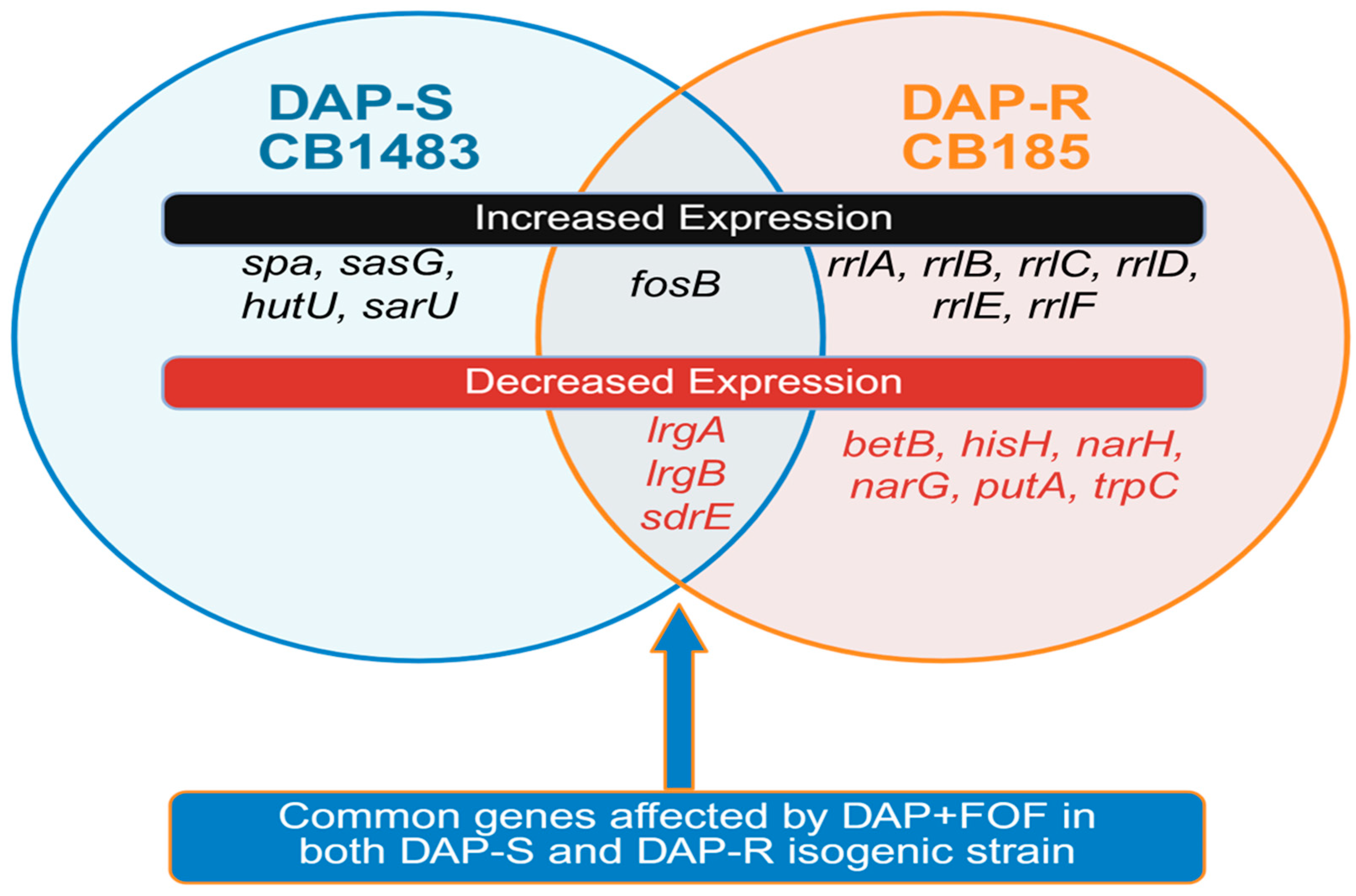
3.2. RNA Sequencing (RNA-Seq)-Based Transcriptomics

We used transcriptomic analysis to identify genes at the transcriptome level that may be mechanistically involved in the response to DAP + FOF treatment. We compared transcriptomic profiles by RNA-seq of DAP-S strain CB1483 and DAP-R strain CB185 without antibiotic, compared with DAP, FOF, or the combination of DAP + FOF at exponential growth as described (Figure 1; Table S2A–F). Transcriptomic analysis revealed differentially and selectively expressed genes in both DAP + FOF-exposed strains; there were a limited number of genes that were differentially (significantly) expressed in both strains. These included upregulation in fosB along with downregulation of lrgA, lrgB, and sdrE. The upregulation of fosB represents an expected transcriptional response in S. aureus containing this gene, as this locus is involved in the reduced FOF susceptibility phenotype [39]. Of interest, lrgA was downregulated; lrgAB is an anti-holin locus, which represses murine hydrolase secretion, suggesting that DAP + FOF-induced suppression of this locus may yield enhanced autolysis and increased organism clearance [40,41]. This locus is also involved in biofilm formation [41,42], suggesting DAP + FOF-mediated repression of lrgAB fosters less biofilm formation, and potentially better clearance of DAP-R strains by antibiotics and host defenses. This is further supported by downregulation of sdrE (responsible for adhesion, pathogenicity, and immune evasion) [43]. We are currently investigating the relationship of DAP + FOF-induced downregulation of lrgAB and sdrE, as adhesion and biofilm formation is controlled by several genetic responses in S. aureus.
Several genes-of-interest were differentially regulated in either the DAP-S strain or the DAP-R strain, but not both, following DAP + FOF exposure. In the DAP-S strain, these include upregulation of genes that potentially mediate bacterial clearance (i.e., sasG gene, encoding a cell wall surface anchor protein); virulence (i.e., spa, encoding protein A, enabling evasion of host immune responses); metabolic pathway genes (i.e., hutU, involved in histidine metabolism), and regulatory genes (i.e., sarU, a staphylococcal accessory regulator family gene; an activator of agr expression). In the DAP-R strain, there were distinct differentially expressed genes observed, including upregulation of the ribosomal locus rrlA-F and downregulation of several metabolic pathway genes, amino acid metabolic genes, and stress response genes. This is consistent with the ability of FOF to interfere with the metabolic response in DAP-R strains noted previously (Figure 1) [24].

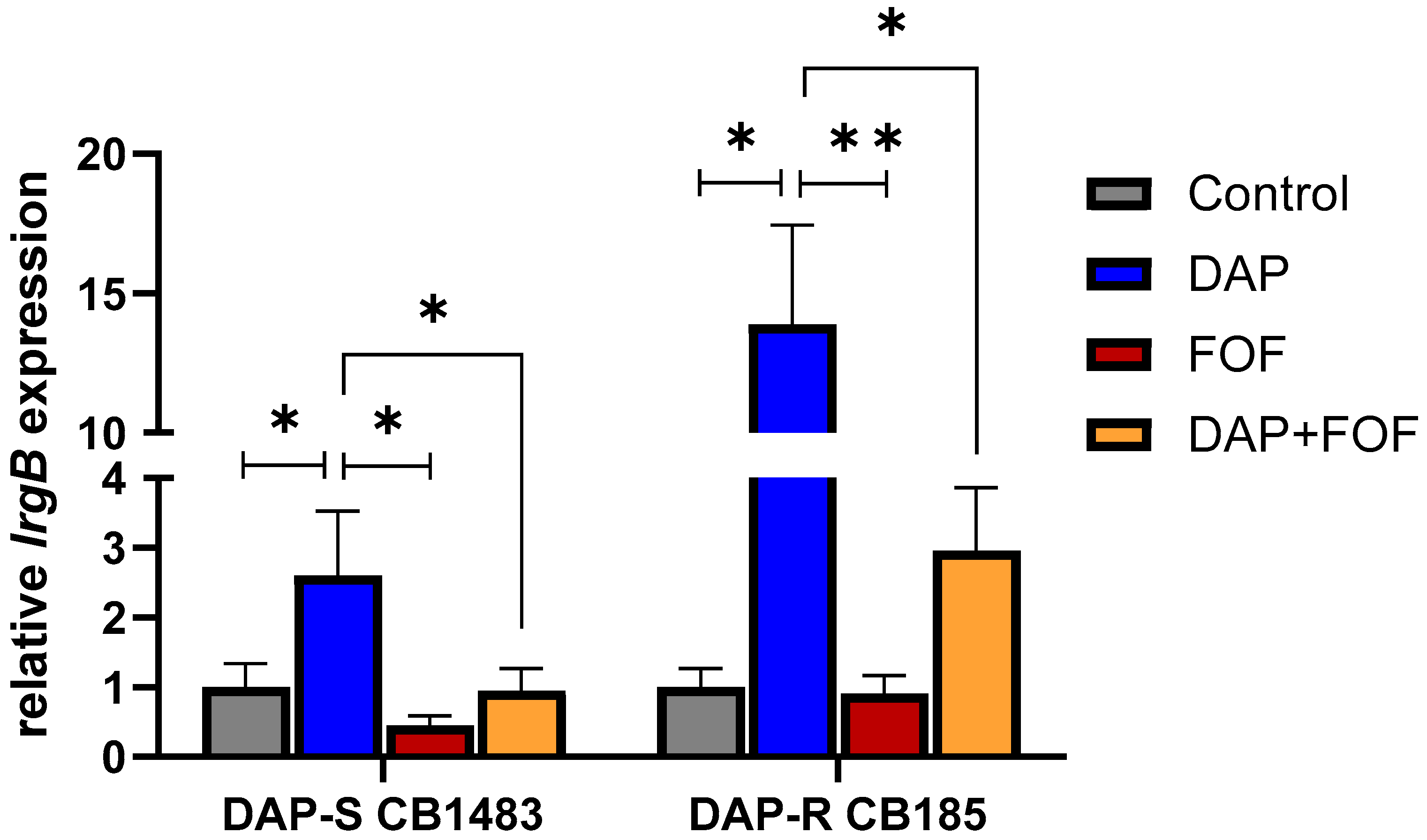
3.3. qRT-PCR Validation of the lrgB Gene

We were particularly interested in the downregulation of lrgAB, identified in transcriptomic comparison between DAP-S and DAP-R strains set during DAP + FOF treatment. Importantly, this locus is critical in MRSA virulence and pathogenesis, as well as in resistance of this organism to multiple antimicrobial and host defense clearance mechanisms [40,41,42,43]. Therefore, we performed further validation of this gene by qRT-PCR in the same strain pair (CB1483/CB185) following DAP, FOF, and DAP + FOF exposures. For these experiments, we focused on the latter co-impacted gene, lrgB, which showed decreased expression following FOF exposures in both strains. In validation studies done in vitro by qRT-PCR, both FOF alone and the DAP + FOF combination blocked DAP-induced hyper-expression of lrgB in both strains (Figure 2), consistent with our RNA-seq analyses.

3.4. Impacts of DAP-FOF Therapy upon Expression of lrgB Genes in Rabbit IE Vegetations

As noted above, FOF exposure in vitro caused repression of the anti-holin gene lrgB in both our prototype strain pairs. Therefore, in-parallel, in vivo gene expression experiments were performed to confirm the translatability of the lrgB gene expression from the in vitro studies described above. We quantified the intra-vegetation (cardiac vegetation samples isolated from rabbit IE model) expression of lrgB by qRT-PCR in rabbits with experimental IE caused by the DAP-R CB185 MRSA strain receiving (i) no therapy; (ii) DAP alone; (iii) FOF alone; or (iv) DAP + FOF combination at the same dose-regimens as reported before [23]. Similar to our in vitro RNA-seq data, FOF alone and DAP + FOF caused substantial reductions in expression of lrgB compared to DAP alone, similar to expression levels observed in untreated vegetations (Figure 3). These data suggested that the notable in vivo microbiologic efficacy of DAP + FOF in this model may relate, in part, to impacts on this key regulatory gene.

4. Conclusions

Our genetic analyses provided the validation for the central hypothesis of this investigation that DAP–FOF combinations can impact MRSA through multiple mechanisms. The studies likely reveal consensus genes/pathways most consistently related to the abilities of DAP + FOF combinations to evoke bactericidal synergistic outcomes. The genomic and transcriptomic signatures in the context of DAP + FOF synergy and/or resistance prevention mechanisms indicate that lrgAB is a key locus of interest with this combination because it represses murine hydrolase secretion and may increase autolysis. Additional DAP-S/DAP-R strain pairs along with genetic knockout and complementation studies are key to further validating this unique mechanism. These studies are imperative to translate these findings to clinical scenarios and therapeutics regimens for future clinical applications.

Supplementary Materials

The following supporting information can be downloaded at https://www.mdpi.com/article/10.3390/microorganisms13071532/s1. Table S1. Whole genome sequencing of DAP-S/DAP-R MRSA strain pairs; Table S2A. Summary of gene functional classes altered ≥ two-fold in DAP-S 1483 exposed to FOFA; Table S2B. Summary of DAP-S 1483 gene functional classes altered ≥ 2-fold in DAP-S 1483 exposed by DAPA alone; Table S2C. Summary of gene functional classes altered ≥ two-fold in DAP-S 1483 exposed by DAP–FOF vs. DAPA alone; Table S2D. Summary of gene functional classes altered ≥ two-fold in DAP-R 185 exposed by FOFA; Table S2E. Summary of gene functional classes altered ≥ two-fold in DAP-R 185 exposed by DAPA; Table S2F. Summary of gene functional classes altered ≥ two-fold in DAP-R 185 exposed by DAP + FOF vs. DAPA alone; Table S3. Primers used in this study for qRT-PCR gene expression analyses.

Author Contributions

Conceptualization, N.N.M.; methodology, N.N.M., S.C.E., W.A., W.E.R. and A.M.E.; software, W.E.R.; validation, W.E.R., S.C.E., W.A., N.N.M., R.A.P., B.P.H. and S.L.B.; formal analysis, N.N.M., W.E.R., S.C.E. and W.A.; investigation, W.E.R., S.C.E., W.A., A.M.E. and N.N.M.; resources, W.E.R. and N.N.M.; data curation, N.N.M., W.E.R., S.C.E., W.A. and A.M.E.; writing—original draft preparation, N.N.M. and W.E.R.; writing—review and editing, N.N.M., W.E.R., S.C.E., W.A., A.R.D., J.N.M.M., J.N.A., R.M., R.A.P., S.L.B. and B.P.H.; visualization, N.N.M., W.E.R., S.C.E., W.A., A.R.D., J.N.M.M., J.N.A., R.M., R.A.P., S.L.B. and B.P.H.; supervision, N.N.M. and W.E.R.; project administration, N.N.M.; funding acquisition, N.N.M. and W.E.R. All authors have read and agreed to the published version of the manuscript.

Funding

This research was supported by grants from NIH/NIAID: R01-AI132627 (to WER). NNM was supported by The Lundquist Institute at Harbor-UCLA intramural research grant (533122-01-00, 862412-00-00).

Institutional Review Board Statement

Not applicable.

Informed Consent Statement

Not applicable.

Data Availability Statement

The original contributions presented in this study are included in the article/Supplementary Materials. Further inquiries can be directed to the corresponding author.

Acknowledgments

We thank the Bioinformatic Center at University of Wisconsin Madison for gene sequencing data. We extend our heartfelt gratitude to Arnold S. Bayer, Distinguished Professor of Medicine at UCLA School of Medicine and the Lundquist Institute at Harbor UCLA Medical Center, for his invaluable support and contributions to this work. His legacy will forever inspire us and remain in our memories.

Conflicts of Interest

The authors declare no conflict of interest.

References

  1. Roch, M.; Gagetti, P.; Davis, J.; Ceriana, P.; Errecalde, L.; Corso, A.; Rosato, A.E. Daptomycin resistance in clinical MRSA strains is associated with a high biological fitness cost. Front. Microbiol. 2017, 8, 2303. [Google Scholar] [CrossRef] [PubMed]
  2. Holland, T.L.; Cosgrove, S.E.; Doernberg, S.B.; Jenkins, T.C.; Turner, N.A.; Boucher, H.W.; Pavlov, O.; Titov, I.; Kosulnykov, S.; Atanasov, B.; et al. ERADICATE Study Group. Ceftobiprole for treatment of complicated staphylococcus aureus bacteremia. N. Engl. J. Med. 2023, 389, 1390–1401. [Google Scholar] [CrossRef] [PubMed]
  3. Fowler, V.G., Jr.; Boucher, H.W.; Corey, G.R.; Abrutyn, E.; Karchmer, A.W.; Rupp, M.E.; Levine, D.P.; Chambers, H.F.; Tally, F.P.; Vigliani, G.A.; et al. Daptomycin versus standard therapy for bacteremia and endocarditis caused by Staphylococcus aureus. N. Engl. J. Med. 2006, 355, 653–665. [Google Scholar] [CrossRef] [PubMed]
  4. Rose, W.E.; Fallon, M.; Moran, J.J.; Vanderloo, J.P. Vancomycin tolerance in methicillin-resistant Staphylococcus aureus: Influence of vancomycin, daptomycin, and telavancin on differential resistance gene expression. Antimicrob. Agents Chemother. 2012, 56, 4422–4427. [Google Scholar] [CrossRef]
  5. Marty, F.M.; Yeh, W.W.; Wennersten, C.B.; Venkataraman, L.; Albano, E.; Alyea, E.P.; Gold, H.S.; Baden, L.R.; Pillai, S.K. Emergence of a clinical daptomycin-resistant Staphylococcus aureus isolate during treatment of methicillin-resistant Staphylococcus aureus bacteremia and osteomyelitis. J. Clin. Microbiol. 2006, 44, 595–597. [Google Scholar] [CrossRef]
  6. Oku, Y.; Kurokawa, K.; Ichihashi, N.; Sekimizu, K. Characterization of the Staphylococcus aureus mprF gene, involved in lysinylation of phosphatidylglycerol. Microbiology 2004, 150, 45–51. [Google Scholar] [CrossRef]
  7. Andrä, J.; Goldmann, T.; Ernst, C.M.; Peschel, A.; Gutsmann, T. Multiple peptide resistance factor (MprF)-mediated resistance of Staphylococcus aureus against antimicrobial peptides coincides with a modulated peptide interaction with artificial membranes comprising lysyl-phosphatidylglycerol. J. Biol. Chem. 2011, 286, 18692–18700. [Google Scholar] [CrossRef]
  8. Ernst, C.M.; Peschel, A. MprF-mediated daptomycin resistance. Int. J. Med. Microbiol. 2019, 309, 359–363. [Google Scholar] [CrossRef]
  9. Ernst, C.M.; Staubitz, P.; Mishra, N.N.; Yang, S.J.; Hornig, G.; Kalbacher, H.; Bayer, A.S.; Kraus, D.; Peschel, A. The bacterial defensin resistance protein MprF consists of separable domains for lipid lysinylation and antimicrobial peptide repulsion. PLoS Pathog. 2009, 5, e1000660. [Google Scholar] [CrossRef]
  10. Bayer, A.S.; Schneider, T.; Sahl, H.G. Mechanisms of daptomycin resistance in Staphylococcus aureus: Role of the cell membrane and cell wall. Ann. N. Y. Acad. Sci. 2013, 1277, 139–158. [Google Scholar] [CrossRef]
  11. de Carvalho, C.C.C.R.; Taglialegna, A.; Rosato, A.E. Impact of PrsA on membrane lipid composition during daptomycin-resistance-mediated β-lactam sensitization in clinical MRSA strains. J. Antimicrob. Chemother. 2021, 77, 135–147. [Google Scholar] [CrossRef] [PubMed]
  12. Nakamura, M.; Kawada, H.; Uchida, H.; Takagi, Y.; Obata, S.; Eda, R.; Hanaki, H.; Kitasato, H. Single nucleotide polymorphism leads to daptomycin resistance causing amino acid substitution-T345I in MprF of clinically isolated MRSA strains. PLoS ONE 2021, 16, e0245732. [Google Scholar] [CrossRef] [PubMed]
  13. Vásquez, A.; Leidy, C.; Manrique-Moreno, M. Lysyl-Phosphatidylglycerol: A Lipid Involved in the resistance of Staphylococcus aureus to antimicrobial peptide activity. Antibiotics 2025, 14, 349. [Google Scholar] [CrossRef]
  14. Chen, F.J.; Lauderdale, T.L.; Lee, C.H.; Hsu, Y.C.; Huang, I.W.; Hsu, P.C.; Yang, C.S. Effect of a point mutation in mprF on susceptibility to daptomycin, vancomycin, and oxacillin in an MRSA clinical strain. Front. Microbiol. 2018, 9, 1086. [Google Scholar] [CrossRef] [PubMed]
  15. Camargo, I.L.; Neoh, H.M.; Cui, L.; Hiramatsu, K. Serial daptomycin selection generates daptomycin-nonsusceptible Staphylococcus aureus strains with a heterogeneous vancomycin-intermediate phenotype. Antimicrob. Agents Chemother. 2008, 52, 4289–4299. [Google Scholar] [CrossRef]
  16. Nguyen, A.H.; Hood, K.S.; Mileykovskaya, E.; Miller, W.R.; Tran, T.T. Bacterial cell membranes and their role in daptomycin resistance: A review. Front. Mol. Biosci. 2022, 9, 1035574. [Google Scholar] [CrossRef]
  17. Gaupp, R.; Lei, S.; Reed, J.M.; Peisker, H.; Boyle-Vavra, S.; Bayer, A.S.; Bischoff, M.; Herrmann, M.; Daum, R.S.; Powers, R.; et al. Staphylococcus aureus metabolic adaptations during the transition from a daptomycin susceptibility phenotype to a daptomycin nonsusceptibility phenotype. Antimicrob. Agents Chemother. 2015, 59, 4226–4238. [Google Scholar] [CrossRef]
  18. Rio-Marques, L.; Hartke, A.; Bizzini, A. The effect of inoculum size on selection of in vitro resistance to vancomycin, daptomycin, and linezolid in methicillin-resistant Staphylococcus aureus. Microb. Drug Resist. 2014, 20, 539–543. [Google Scholar] [CrossRef]
  19. Jorgensen, S.C.J.; Zasowski, E.J.; Trinh, T.D.; Lagnf, A.M.; Bhatia, S.; Sabagha, N.; Abdul-Mutakabbir, J.C.; Alosaimy, S.; Mynatt, R.P.; Davis, S.L.; et al. Daptomycin plus β-Lactam combination therapy for methicillin-resistant Staphylococcus aureus bloodstream infections: A Retrospective, comparative cohort study. Clin. Infect. Dis. 2020, 71, 1–10. [Google Scholar] [CrossRef]
  20. Pujol, M.; Miro, J.M.; Shaw, E.; Aguado, J.M.; San-Juan, R.; Puig-Asensio, M.; Pigrau, C.; Calbo, E.; Montejo, M.; Rodriguez-Alvarez, R.; et al. Daptomycin plus fosfomycin versus daptomycin alone for methicillin-resistant Staphylococcus aureus bacteremia and endocarditis: A randomized clinical trial. Clin. Infect. Dis. 2020, 72, 1517–1525. [Google Scholar] [CrossRef]
  21. Geriak, M.; Haddad, F.; Rizvi, K.; Rose, W.; Kullar, R.; LaPlante, K.; Yu, M.; Vasina, L.; Ouellette, K.; Zervos, M.; et al. Clinical data on daptomycin plus ceftaroline versus standard of care monotherapy in the treatment of methicillin-resistant Staphylococcus aureus bacteremia. Antimicrob. Agents Chemother. 2019, 63, e02483-18. [Google Scholar] [CrossRef] [PubMed]
  22. Wang, C.; Ye, C.; Liao, L.; Wang, Z.; Hu, Y.; Deng, C.; Liu, L. Adjuvant β-lactam therapy combined with vancomycin or daptomycin for methicillin-resistant Staphylococcus aureus bacteremia: A systematic review and meta-analysis. Antimicrob. Agents Chemother. 2020, 64, e01377-20. [Google Scholar] [CrossRef] [PubMed]
  23. Mishra, N.N.; Lew, C.; Abdelhady, W.; Lapitan, C.K.; Proctor, R.A.; Rose, W.E.; Bayer, A.S. Synergy mechanisms of daptomycin-fosfomycin combinations in daptomycin-susceptible and -resistant methicillin-resistant Staphylococcus aureus: In vitro, ex vivo, and in vivo metrics. Antimicrob. Agents Chemother. 2022, 66, e0164921. [Google Scholar] [CrossRef] [PubMed]
  24. Reed, J.M.; Gardner, S.G.; Mishra, N.N.; Bayer, A.S.; Somerville, G.A. Metabolic interventions for the prevention and treatment of daptomycin non-susceptibility in Staphylococcus aureus. J. Antimicrob. Chemother. 2019, 74, 2274–2283. [Google Scholar] [CrossRef]
  25. Falagas, M.E.; Vouloumanou, E.K.; Samonis, G.; Vardakas, K.Z. Fosfomycin. Clin. Microbiol. Rev. 2016, 29, 321–347. [Google Scholar] [CrossRef]
  26. Bayer, A.S.; Mishra, N.N.; Chen, L.; Kreiswirth, B.N.; Rubio, A.; Yang, S.J. Frequency and distribution of single-nucleotide polymorphisms within mprf in methicillin-resistant Staphylococcus aureus clinical isolates and their role in cross-resistance to daptomycin and host defense antimicrobial peptides. Antimicrob. Agents Chemother. 2015, 59, 4930–4937. [Google Scholar] [CrossRef]
  27. Mishra, N.N.; McKinnell, J.; Yeaman, M.R.; Rubio, A.; Nast, C.C.; Chen, L.; Kreiswirth, B.N.; Bayer, A.S. In vitro cross-resistance to daptomycin and host defense cationic antimicrobial peptides in clinical methicillin-resistant Staphylococcus aureus isolates. Antimicrob. Agents Chemother. 2011, 55, 4012–4018. [Google Scholar] [CrossRef]
  28. Mishra, N.N.; Bayer, A.S.; Baines, S.L.; Hayes, A.S.; Howden, B.P.; Lapitan, C.K.; Lew, C.; Rose, W.E. Cell membrane adaptations mediate β-lactam-induced resensitization of daptomycin-resistant (DAP-R) Staphylococcus aureus in vitro. Microorganisms. 2021, 9, 1028. [Google Scholar] [CrossRef]
  29. CLSI. Performnace Standards for Antimicrobial Susceptibility Testing, 34th ed.; CLSI Supplement M100; Clinical and Laboratory Standards Institute: Wayne, PA, USA, 2024. [Google Scholar]
  30. Thitiananpakorn, K.; Aiba, Y.; Tan, X.E.; Watanabe, S.; Kiga, K.; Sato'o, Y.; Boonsiri, T.; Li, F.Y.; Sasahara, T.; Taki, Y.; et al. Association of mprF mutations with cross-resistance to daptomycin and vancomycin in methicillin-resistant Staphylococcus aureus (MRSA). Sci. Rep. 2020, 10, 16107. [Google Scholar] [CrossRef]
  31. Jiang, H.; Lei, R.; Ding, S.W.; Zhu, S. Skewer: A fast and accurate adapter trimmer for next-generation sequencing paired-end reads. BMC Bioinformatics. 2014, 15, 1–12. [Google Scholar] [CrossRef]
  32. Kolde, R. Pheatmap: Pretty Heatmaps. R Package Version 1.0.12. Available online: https://raivokolde.r-universe.dev/pheatmap (accessed on 5 June 2025).
  33. Love, M.I.; Huber, W.; Anders, S. Moderated estimation of fold change and dispersion for RNA-seq data with DESeq2. Genome Biol. 2014, 15, 1–21. [Google Scholar] [CrossRef] [PubMed]
  34. Robinson, M.D.; Oshlack, A. A scaling normalization method for differential expression analysis of RNA-seq data. Genome Biol. 2010, 11, 1–9. [Google Scholar] [CrossRef] [PubMed]
  35. Taglialegna, A.; Varela, M.C.; Rosato, R.R.; Rosato, A.E. VraSR and virulence trait modulation during daptomycin resistance in methicillin-resistant Staphylococcus aureus infection. mSphere 2019, 4, e00557-18. [Google Scholar] [CrossRef] [PubMed]
  36. Ersoy, S.C.; Abdelhady, W.; Li, L.; Chambers, H.F.; Xiong, Y.Q.; Bayer, A.S. Bicarbonate resensitization of methicillin-resistant Staphylococcus aureus to β-Lactam antibiotics. Antimicrob. Agents Chemother. 2019, 63, e00496-19. [Google Scholar] [CrossRef]
  37. Vlaeminck, J.; Lin, Q.; Xavier, B.B.; De Backer, S.; Berkell, M.; De Greve, H.; Hernalsteens, J.P.; Kumar-Singh, S.; Goossens, H.; Malhotra-Kumar, S. The dynamic transcriptome during maturation of biofilms formed by methicillin-resistant Staphylococcus aureus. Front. Microbiol. 2022, 13, 882346. [Google Scholar] [CrossRef]
  38. Elgharably, H.; Claesen, J.; Sangwan, N.; Etiwy, M.; Houghtaling, P.; Procop, G.; Shrestha, N.K.; Griffin, B.; Navia, J.L.; Svensson, L.G.; et al. In vivo virulence of Staphylococcus aureus in native versus prosthetic left-sided valve endocarditis. JTCVS Open 2024, 24, 156–169. [Google Scholar] [CrossRef]
  39. Fu, Z.; Liu, Y.; Chen, C.; Guo, Y.; Ma, Y.; Yang, Y.; Hu, F.; Xu, X.; Wang, M. Characterization of fosfomycin resistance gene, fosB, in methicillin-resistant Staphylococcus aureus isolates. PLoS ONE 2016, 11, e0154829. [Google Scholar] [CrossRef] [PubMed]
  40. Beltrame, C.O.; Côrtes, M.F.; Bonelli, R.R.; Côrrea, A.B.; Botelho, A.M.; Américo, M.A.; Fracalanzza, S.E.; Figueiredo, A.M. Inactivation of the autolysis-related genes lrgB and yycI in Staphylococcus aureus Increases cell lysis-dependent eDNA release and enhances biofilm development in vitro and in vivo. PLoS ONE 2015, 10, e0138924. [Google Scholar] [CrossRef]
  41. Ranjit, D.K.; Endres, J.L.; Bayles, K.W. Staphylococcus aureus CidA and LrgA proteins exhibit holin-like properties. J Bacteriol. 2011, 193, 2468–2476. [Google Scholar] [CrossRef]
  42. Rice, K.C.; Mann, E.E.; Endres, J.L.; Weiss, E.C.; Cassat, J.E.; Smeltzer, M.S.; Bayles, K.W. The cidA murein hydrolase regulator contributes to DNA release and biofilm development in Staphylococcus aureus. Proc. Natl. Acad. Sci. USA 2007, 104, 8113–8118. [Google Scholar] [CrossRef]
  43. Zhang, Y.; Wu, M.; Hang, T.; Wang, C.; Yang, Y.; Pan, W.; Zang, J.; Zhang, M.; Zhang, X. Staphylococcus aureus SdrE captures complement factor H’s C-terminus via a novel ‘close, dock, lock and latch’ mechanism for complement evasion. Biochem. J. 2017, 474, 1619–1631. [Google Scholar] [CrossRef]
Figure 1. Venn diagram of genes altered ≥ 2-fold by DAP + FOF vs. DAP or FOF alone exposures in DAP-S/DAP-R MRSA strain pair. Red and black fonts indicate whether expression was increased (black) or decreased (red) by DAP + FOF exposure.
Microorganisms 13 01532 g001
Figure 2. In vitro lrgB gene expression was significantly decreased in the DAP-S CB1483 and DAP-R CB185 MRSA strain pairs treated either with FOF alone or DAP + FOF vs. DAP alone treatment. * p < 0.05; ** p < 0.01 vs. DAP exposure.
Microorganisms 13 01532 g002
Figure 3. In vivo lrgB gene expression was decreased in rabbit endocarditis vegetations of DAP-R CB185 treated either with FOF alone or DAP + FOF or control (untreated) vs. DAP alone. ** p < 0.01; *** p < 0.001 vs. DAP exposure.
Microorganisms 13 01532 g003
Table 1. Whole genome sequencing of 33 DAP-S/DAP-R strains. Single nucleotide polymorphisms (SNPs) represent differences compared to the respective isogeneic parent strain (N = 33).
Polymorphisms in mprF,
Amino Acid Changes
(# of DAP-R Strains)
Polymorphisms Among Other Genes
(# of DAP-R Strains)
Leu826Phe (11); Thr345Ala/Lys/Ile (5);
Ser337Leu (2); Ser295Leu (2);
Leu341Ser (4);
Met347Arg (2); Val351Glu (1); Thr472Lys (1);
Ile420Asn (1);
Phe349_Asn352del (1), Pro314Leu (1); Leu42del (1); No mutation (1)
cls2 (5); cls1 (1);
dltD (1); yycG (3);
crtN (1); lytN (2);
vraRS (4); fabFH (2)
Parenthesis ( ) = number of DAP-R strains.
Table 2. WGS and phospholipid composition of DAP-S1483 strain passaged in DAP or FOF alone and DAP + FOF for 10 days in vitro vs. control (untreated) strain.
StrainAntibiotic Exposure for 10 DaysDAP MIC
(µg/mL)
FOF MIC
(µg/mL)
mprF or cls SNP% of PL Composition
(Mean ± SD)
L-PGPGCL
DAP-S
1483
Control ** 0.25 (S)8 (S)None14 ± 376 ± 39 ± 1
DAP4 (R)8 (S)mprF (L826F); cls2: (Leu52Phe)25 ± 6 *58 ± 9 *17 ± 6
FOF0.5 (S)>256 (R)None12 ± 376 ± 913 ± 6
DAP + FOF1 (S)8 (S)None12 ± 174 ± 1114 ± 12
* p < 0.01 vs. control. ** p < 0.003; DAP-S vs. DAP-R strain. S = susceptible; R = resistant; CL = cardiolipin.
Disclaimer/Publisher’s Note: The statements, opinions and data contained in all publications are solely those of the individual author(s) and contributor(s) and not of MDPI and/or the editor(s). MDPI and/or the editor(s) disclaim responsibility for any injury to people or property resulting from any ideas, methods, instructions or products referred to in the content.

