Article

High Diversity and Low Coinfections of Pathogens in Ticks from Ruminants in Pakistan

1
Institute of EcoHealth, School of Public Health, Shandong University, Jinan 250012, China
2
State Key Laboratory of Pathogen and Biosecurity, Academy of Military Medical Sciences, Beijing 100071, China
3
Livestock and Dairy Development (Ext.) Department, Peshawar 25000, Pakistan
*
Authors to whom correspondence should be addressed.
These authors contributed equally to this work.
Microorganisms 2025, 13(6), 1276; https://doi.org/10.3390/microorganisms13061276
Submission received: 27 April 2025 / Revised: 25 May 2025 / Accepted: 27 May 2025 / Published: 30 May 2025
(This article belongs to the Section Public Health Microbiology)

Abstract

Emerging tick-borne infections pose growing public health threats, causing global disease burdens and economic losses. In this study, tick-borne pathogens were detected in ticks collected from ruminants in 19 sites of Khyber Pakhtunkhwa Province, Pakistan, between 2023 and 2024. A total of 989 ticks, belonging to five species, i.e., Hyalomma marginatum, Rhipicephalus microplus, Rhipicephalus sanguineus, Rhipicephalus haemaphysaloides, and Haemaphysalis bispinosa, were tested by specific PCR followed by Sanger sequencing. In total, fourteen pathogens including two Anaplasma species, three Ehrlichia species, three Rickettsia species, one Babesia species, and five Theileria species were identified, with an overall infection rate of 20.2% (95% CI: 17.7–22.7%). Phylogenetic analyses revealed two undefined Ehrlichia species: Candidatus Ehrlichia hyalommae was exclusively detected in Hy. marginatum ticks, while Candidatus Ehrlichia rhipicephalis was only found in R. microplus. Additionally, an undefined Rickettsia, provisionally named Candidatus Rickettsia pakistanensis, was identified, which is phylogenetically close to R. sibirica in North Asia and R. africae in Africa, suggesting its potential pathogenicity to humans. Although coinfections of two pathogens were observed, the coinfection rates were quite low. The findings revealed a significant diversity of tick-borne pathogens in Pakistani ticks, which may pose risks to livestock and humans.

1. Introduction

Ticks as important vectors transmit a wide range of human and animal pathogens, including bacteria, protozoa, and viruses, and cause substantial disease burden and economic losses worldwide [1]. Emerging tick-borne pathogens are especially a growing global public health threat [2,3]. Pakistan is located in a subtropical zone in South Asia, most parts of which provide favorable environments for ticks, which can infest a variety of hosts and transmit diseases to humans, livestock, and companion animals. In Pakistan, ticks pose a significant challenge for both livestock and human health [4], as they are distributed all over Pakistan’s diverse geographical regions with high abundance, and carry a variety of pathogens [5,6]. Pakistan is home to over 40 distinct species of ticks, with the predominant tick species classified into three genera: Hyalomma, Haemaphysalis, and Rhipicephalus [6]. The high prevalence of Anaplasma in buffaloes throughout Khyber Pakhtunkhwa Province constitutes a significant threat to livestock health [7]. The Hyalomma and Rhipicephalus ticks, particularly Rhipicephalus microplus, act as primary vectors for Anaplasma marginale, responsible for tick fever in Pakistan [8,9,10]. Similarly, the Crimean–Congo Hemorrhagic Fever Virus (CCHFV) is transmitted by Hyalomma ticks and presents a serious risk to public health in Pakistan, resulting in severe hemorrhagic fever, elevated mortality rates, and considerable economic repercussions [11,12,13]. A study conducted from 2017 to 2021 identified Rickettsia massiliae in Rhipicephalus ticks infesting wild boars in Khyber Pakhtunkhwa Province, indicating the presence of Rickettsia in local tick populations, which are associated with adverse effects on animal productivity due to tick-borne diseases [14,15]. In Pakistan, Theileria annulata and Babesia bigemina exhibit prevalence rates of 16% and 51%, respectively, within cattle populations [9]. Furthermore, research concerning bovine ticks revealed that 31.6% were infected with either Babesia or Theileria species, with tropical theileriosis, caused by T. annulata, being one of the most significant tick-borne diseases affecting bovines [16,17,18,19,20].
Ticks in Pakistan show extensive co-infection in their natural ecosystems. Recent studies indicate that Hyalomma excavatum, Hy. isacci, and Rhipicephalus decoloratus exhibit dual-pathogen co-infections, while Hy. scupense is known to harbor three pathogens. Significantly, four different tick-borne pathogens were identified in ticks from Hyalomma, Haemaphysalis, and Rhipicephalus. These patterns of multi-pathogen presence highlight the complex transmission dynamics within tick-borne disease systems, reinforcing the need for thorough surveillance strategies in endemic areas [6,21].
Khyber Pakhtunkhwa Province has diverse geographical situations, comprising mountains, plains, and valleys. Although the region experiences hot summers and cold winters, the combination of moderate humidity, vegetation cover, and the presence of domestic and wild hosts creates microhabitats that support tick survival. These climatic and geographical characteristics can facilitate the thriving of tick populations and the transmission of tick-borne pathogens [22]. Moreover, livestock production activities, which are the major source of income for rural households and contribute over 60.5% to agricultural value and nearly 11% to the country’s GDP during 2019 and 2020 [23], further offer a favorable ecology for tick infestation. The frequent contact between farmers and livestock certainly increases the transmission opportunities of tick-borne pathogens from animals to humans. However, the situation of ticks and tick-borne infections, as well as their potential risks to public and animal health, has not been well recognized in Khyber Pakhtunkhwa Province, which has hindered the effective control of ticks and tick-borne infections in the region. In this study, we collected ticks from 19 sites of three districts in Khyber Pakhtunkhwa Province, and reported the presence, prevalence, and genetic characteristics of tick-borne agents for establishing target control measures for transmission of potential tick-borne zoonotic pathogens in this region.

2. Materials and Methods

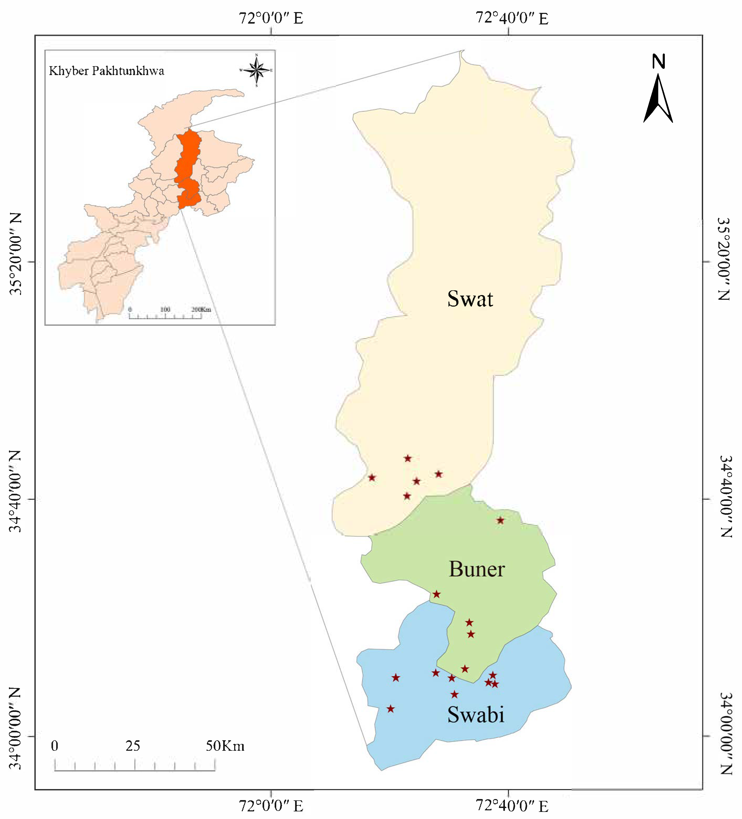
2.1. Study Areas and Sample Collection

The present study was carried out in Swabi (32.1442° N, 72.4702° E), Swat (35.2220° N, 72.4258° E), and Buner districts (34.4215° N, 72.6150° E) of Khyber Pakhtunkhwa Province in Pakistan in December 2023 to March 2024 (Figure 1). The collection sites’ latitude, longitude, and host animals were recorded during tick collection (Table S1). Tick collection was carried out with local authorities’ support and approval, including the Civil Veterinary Hospital and Department of Animal Husbandry, Peshawar. Ticks were collected from livestock such as buffaloes, goats, and sheep using approaches that ensured minimal animal stress. These three districts were chosen due to their distinct agroclimatic features. Swabi, at an elevation of approximately 345 m, has a semi-arid subtropical climate with moderate humidity and a dry season extending from November to May. In contrast, Swat and Buner lie above 900 m and experience temperate climates with cooler temperatures and higher rainfall. Livestock in these areas are raised under traditional management systems, including a mix of stall feeding and open grazing, which vary by species and location. These environmental and husbandry differences may influence tick distribution and pathogen transmission.
After immobilizing the animal, the ticks were removed using sterile forceps. The collected tick specimens were then washed with 70% ethanol followed by distilled water to remove contaminants. The ticks were identified by a senior entomologist (Yi Sun) using a stereo microscope (Olympus SZX10, Olympus Corporation, Tokyo, Japan) based on morphological criteria to determine their species and different developmental stages [24,25,26,27]. After identification, the samples were placed in centrifuge tubes, and the ticks were stored at −80 °C before DNA was extracted.

2.2. DNA Extraction and PCR Assays

Total DNA of individual ticks was extracted in a separate laboratory using TaKaRa Mini BEST Viral RNA/DNA Extraction Kit Ver. 5.0 (TaKaRa, Dalian, China). Briefly, each tick was placed in a microcentrifuge tube, and buffer VGB, proteinase K, and carrier RNA were added. Then, incubation was performed at 56 °C for cell lysis. Ethanol was added, and the samples were vortexed well and transferred to a centrifuge column, where a series of centrifugation and washing steps were performed in a rotating column according to the manufacturer’s instructions. The DNA was eluted in a final volume of 35 μL of RNase-free water, incubated for one minute at room temperature, and then centrifuged for one minute at 6000× g at room temperature. The final step was repeated with the eluent to increase the DNA yield. Extracted DNA was stored at −20 °C before testing.
All tick samples were tested for the presence of Rickettsia, Anaplasma, Ehrlichia, Borrelia, Babesia, and Theileria using specific PCR assays. The specific primers and amplification conditions used for the detection of the various tick-borne agents are shown in Table S2. Rickettsial species were identified by nested PCR using specific primers to amplify the outer membrane protein A gene (OmpA), the 17 kDa antigen gene (17 kDa), and the citrate synthase gene (gltA). The genera Anaplasma and Ehrlichia were detected using semi-nested primers out1/3–17 and out1/out2 targeting the 16S rRNA gene. Positive samples were then detected by PCR targeting citrate synthase (gltA) and heat shock protein (groEL) gene fragments. PCR targeting the 18S rRNA gene was used to detect Babesia and Theileria. Borrelia were detected by PCR targeting the 5S–23S rRNA intergenic spacer region. All the PCR amplifications were performed using a Veriti 96-Well Thermal Cycler (Applied Biosystems, Waltham, Massachusetts, USA), and all the positive PCR amplicons were confirmed by Sanger sequencing in both directions. Nuclease-free water was used as a negative control in PCR. The obtained nucleotide sequences were then proofread and assembled using CLC Main Workbench 5.0 (Qiagen, Redwood City, CA, USA).

2.3. Phylogenetic Analyses

Sequences of each amplified segment by specific PCR was aligned using the BLASTn tool in NCBI (Nucleotide BLAST: Search nucleotide databases using a nucleotide query (nih.gov)) to preliminarily assess the sequences’ similarity and origin to verify the sequencing results’ accuracy. Then, multiple sequence comparisons of the sample sequences with the reference sequences were performed using MAFFT (v7.487) software [28] to ensure the consistency and accuracy of the comparisons. Trial software was used to trim the sequences and remove low-quality regions to ensure the data quality of each fragment [29]. For constructing the joint phylogenetic tree, the results were further pruned using Gblocks comparison to remove unstable sequence regions [30]. The trimmed comparison sequences were subsequently concatenated for subsequent phylogenetic analysis. We used IQ-TREE (v2.1.4) to construct both single-gene and concatenated phylogenetic trees. The software automatically selects the best-fit substitution model and infers phylogenies using the maximum likelihood (ML) method [31]. To assess the confidence level of the phylogenetic tree, an ultra-fast bootstrap method (1000 replications) was used to verify the stability and reliability of the results through multiple random sampling. Finally, the phylogenetic tree was annotated and visualized using the iTOL online tool [32], and genetic distances were calculated by Mega 11 software [33]. The GenBank reference sequences used in the phylogenetic analysis are shown in black in Figures 2–5, and are labeled with their accession numbers, species names, host organisms, and geographic origins.

2.4. Statistical Analyses

The positive rate with its 95% confidence interval (95% CI) was estimated for each tick-borne agent in each tick species, and for overall infections of each tick species. Associations between pathogen positivity and tick species were assessed using chi-square (χ2) or Fisher’s exact tests where appropriate (i.e., expected frequency < 5). All analyses were performed in R software (v4.4.2), and a two-sided p-value < 0.05 was considered statistically significant.

2.5. Ethical Approval

The Livestock and Dairy Development Department (ext) in Peshawar, Khyber Pakhtunkhwa, Pakistan approved the ethical protocol for this study (Approval No. 3075). All animal procedures were approved by the relevant ethics committee, ensuring compliance with animal welfare standards.

3. Results

3.1. Collected Tick Samples

A total of 989 tick species were collected from the 19 sites of three districts in Khyber Pakhtunkhwa Province, Pakistan (Figure 1), comprising 867 females and 122 males from cattle, goats, and sheep (Table 1). The collected ticks belonged to five species of the family Ixodidae, including Hyalomma marginatum (157, 15.9%), Rhipicephalus microplus (753, 76.1%), Rhipicephalus sanguineus (17, 1.7%), Rhipicephalus haemaphysaloides (13, 1.3%), and Haemaphysalis bispinosa (49, 5.0%). Among them, all the above five tick species were found in Swabi district, four species except R. sanguineus were collected from Buner district, and only R. microplus was obtained in Swat district.

3.2. Detection and Phylogenetic Analysis of Anaplasma

Three gene fragments, 16S rRNA, gltA, and groEL, were detected by nested PCR assays. Although the positive samples amplified from these three gene fragments were not entirely identical, genetic homology comparison and phylogenetic analysis revealed the presence of two Anaplasma species in these tick samples: Anaplasma marginale and Anaplasma ovis (Figure 2). All the sequences of the PCR products positive for Anaplasma were deposited in GenBank, and the accession numbers are listed in Table S3. The sequences of A. marginale-positive samples were closely related to each other, with nucleotide identities of 99.5–100%, 97.2–100%, and 98.2–100% for the 16S rRNA, gltA, and groEL genes, respectively (Table S4). A. marginale was detected only in R. microplus ticks from cattle, in either Swat or Buner districts (Figure 1). In contrast, A. ovis was detected in the other four tick species Hy. marginatum, R. sanguineus, R. haemaphysaloides, and Hae. bispinosa. All the ticks positive for A. ovis were exclusively from sheep, albeit different tick species. These findings reveal a distinct pattern of vector association between the two Anaplasma species. A. marginale was exclusively associated with Rhipicephalus microplus, whereas A. ovis was predominantly found in other tick species. Such distribution patterns likely reflect underlying ecological or biological vector–pathogen specificity. This non-random association was strongly supported by statistical analysis (Fisher’s exact test, p < 0.001 for both A. marginale and A. ovis) (Table S5).

3.3. Detection and Phylogenetic Analysis of Ehrlichia

Eleven ticks were positive for both 16S rRNA and groEL genes of Ehrlichia. All the sequences of amplicons were deposited in GenBank, and the accession numbers are listed in Table S6. Phylogenetic analyses based on 16S rRNA and groEL revealed that a R. microplus tick from Swabi was positive for Ehrlichia minasensis (Figure 3), the 16S rRNA gene of which shared 99.5% identity with E. minasensis strains detected in R. microplus from Anhui, China (GenBank accession number: OQ136683.1), and the groEL gene fragments were 99.0% similar to those from R. microplus from Hubei Province, China (GenBank accession number: OQ511477.1). Three Hy. marginatum ticks were positive for an undefined Ehrlichia species, the 16S rRNA genes of which were identical with each other and shared 99.4% identity with a previously uncultured Ehrlichia species found in Turkey (GenBank accession number: OM065753.1) (Figure 3A). Meanwhile, their groEL gene fragments were 99.0–100% identical to each other, and only bore 92.0% similarity to the closest Ehrlichia sp. reported in Japan (GenBank accession number: LC385854.1) (Figure 3B). On the phylogenetic tree based on the combined 16S rRNA and groEL gene sequences (Figure 3C), the Ehrlichia species from the three positive samples formed a distinct branch, suggesting a putative new Ehrlichia species. Given that all samples infected with this Ehrlichia species were Hy. marginatum, it was provisionally named Candidatus Ehrlichia hyalommae (Figure 3).
The 16S rRNA gene sequences of the remaining seven Ehrlichia-positive samples had 98.9–100% similarity among themselves and 98.3–100% similarity to that of uncultured Ehrlichia sp detected in R. microplus from Colombia (GenBank accession number: OM475361.1) (Figure 3A). Their groEL gene sequences exhibited 99.0–100% similarity to each other and 98.0–99.0% similar to the genetically closest uncultured Ehrlichia sp. found in R. microplus in China (GenBank accession number: OQ185234.1) (Figure 3B). In the phylogenetic tree based on the combined 16S rRNA and groEL gene sequences, the seven positive samples formed a distinctively independent branch (Figure 3C), supporting the notion that it represents an undefined Ehrlichia species, which we provisionally designated as Candidatus Ehrlichia rhipicephalis because it was found only in R. microplus ticks. This Ehrlichia species was all detected in R. microplus that fed on cattle, although they were distributed in all three districts.
Statistical analyses were conducted to explore potential associations between Ehrlichia species and tick vectors. Although E. minasensis and Candidatus E. rhipicephalis were detected exclusively in R. microplus, these associations were not statistically significant (Fisher’s exact test, p = 1 and p = 0.783, respectively) (Table S7), possibly due to the limited number of positive samples. In contrast, Candidatus E. hyalommae exhibited a statistically significant association with Hy. marginatum (p = 0.022), suggesting a potential vector preference. These findings indicate species-specific trends in vector association among Ehrlichia spp., although larger sample sizes are needed to confirm these patterns with greater confidence.

3.4. Detection and Phylogenetic Analysis of Rickettsia

To screen for Rickettsia infections in ticks, we amplified fragments of the OmpA, 17 kDa, and gltA genes using nested PCR and detected six samples positive for all three genes and eight samples positive for two of three genes. All the sequences of the detected Rickettsia segments were deposited in GenBank, and the accession numbers are listed in Table S8. Although the positive samples amplified from the three genes fragments were not entirely identical, genetic homology comparison and phylogenetic trees constructed based on OmpA, 17 kDa, and gltA genes identified three Rickettsia species. Among them, Rickettsia felis was identified in an R. microplus tick, showing 99.8% identity to previously reported R. felis sequences from Ctenocephalides felis in Guatemala for the OmpA gene (GenBank accession number: JN990593.1) and 100% identity to R. felis sequences from Ctenophthalmus agyrtes in Lithuania for the 17 kDa gene (GenBank accession number: MF491767.1) (Figure 4A,B). Phylogenetic trees constructed based on the OmpA, 17 kDa, and gltA gene fragments revealed that the Rickettsia detected in R. sanguineus and R. microplus ticks clustered within the same branch as Rickettsia massiliae, indicating that they belong to the same species (Figure 4). Specifically, R. sanguineus ticks infected with R. massiliae were all from goats or sheep in Swabi district, while the positive R. microplus tick was from cattle in Buner district.
Another Rickettsia exhibited a similarity of 100%, 99.2–100%, and 98.3–100% similarity in OmpA, 17 kDa, and gltA genes sequences, respectively. In the phylogenetic trees based on OmpA and gltA genes, they were clustered with Rickettsia africae, with similarities of 99.6–99.8% and 97.4–100%, respectively (Figure 4A,C). However, in the 17 kDa phylogenetic tree, they were in the same branch as known human-derived Rickettsia sibirica strains, with 100% identity (Figure 4B). To further validate the evolutionary position of the Rickettsia, a phylogenetic tree on the basis of combined OmpA, 17 kDa, and gltA gene sequences was constructed, and revealed that the Rickettsia from positive samples in this study formed a separate branch (Figure 4D), distinctive from R. sibirica and R. africae, suggesting that they represent a new Rickettsia species. Here, we provisionally named it Candidatus Rickettsia pakistanensis, in honor of discovering this new Rickettsia species in Pakistan. Notably, Candidatus Rickettsia pakistanensis was only detected in Hy. marginatum ticks parasitizing goats or sheep in the Swabi district.

3.5. Detection and Phylogenetic Analysis of Babesia and Theilria

To screen for Babesia and Theileria infections in ticks, we amplified the 18S rRNA gene using a pair of universal primers, and proved the test results through Sanger sequencing of PCR products (Table S9). Based on similarity comparison and phylogenetic analysis of 18S rRNA gene sequences, one species of Babesia and five species of Theileria were identified, namely Babesia bigemina, Theileria annulata, Theileria buffeli, Theileria luwenshuni, Theileria orientalis, and Theileria sinensis (Figure 5). The two samples positive for B. bigemina were detected in R. microplus ticks from cattle in Swat district. The 18S rRNA gene of the 43 T. annulata sequences had 98.3–100% nucleotide identity with previously reported T. annulata sequence from Iran (GenBank accession number: HM628581.1), clustering into one branch on the phylogenetic tree. All 43 T. annulata detected in this study were in R. microplus ticks from cattle and sheep in all three districts. The 24 T. orientalis sequences shared 99.2–100% identity with each other and with known T. orientalis sequence in Myanmar (GenBank accession number: MG599099.1). Among the 24 T. orientalis-positive ticks, 22 were R. microplus from the three districts, while the other 2 positive samples included Hae. bispinosa and Hy. marginatum from Swabi district. There were 14 sequences belonging to T. luwenshuni, which shared 98.3–100% identity with each other and 98.4–100% identity with T. luwenshuni reported in India (GenBank accession number: MG194417.1). The tick species for T. luwenshuni detected in the Swabi district were Hy. marginatum (seven samples) and Hae. bispinosa (five samples), with goats (or sheep) as the hosts. T. luwenshuni-infected R. microplus ticks were all from the Buner district, indicating that T. luwenshuni infections might be distributed in different tick species of different regions. Twelve samples were detected with T. sinensis, sharing 96.9–100% identity with previously reported T. sinensis sequences found in Ixodes persulcatus (GenBank accession number: OR244360.1) from Pakistan. All T. sinensis-positive samples were from Hae. bispinosa in the Swabi district. Only one sample was detected with T. buffeli, sharing 100% similarity with the T. buffeli detected in Buffalo from India (GenBank accession number: OR067901.1).

3.6. Prevalence and Co-Infections of Tick-Borne Agents

As mentioned above, this study detected a total of 14 agents in 989 ticks across the three districts. For Anaplasma, Ehrlichia, and Rickettsia, two to three gene fragments were tested. To more accurately determine the infection rate of these tick-borne agents, a sample was considered positive only if at least two gene fragments of the same agent were detected. As a result, 200 ticks tested positive for at least one agent, with an overall positive rate of 20.2% (95% CI: 17.7–22.7%). The infection rates of Hy. marginatum, R. microplus, R. sanguineus, R. haemaphysaloides, and Hae. bispinosa were 40.1% (95% CI: 32.5–47.8%), 17.3% (95% CI: 14.6–20.0%), 47.1% (95% CI: 23.3–70.8%), 7.7% (95% CI: 3.5–11.8%), and 40.8% (95% CI: 27.1–54.6%), respectively (Table 2). R. microplus carried nine agents, followed by Hy.marginatum (seven) and Hae. bispinosa (four). T. orientalis and T. luwenshuni were detected in three tick species, and R. massiliae and T. sinensis in two tick species, respectively. The other nine tick-borne agents were found each in one tick species (Table 2 and Table 3).
A total of 22 out of 989 ticks were positive for two agents (Table 4), with an overall co-infection rate of 2.2% (95% CI: 1.31–3.14%). None of the ticks was infected by three or more agents. The co-infection of A. marginale and T. annulata in R. microplus was most commonly seen, but only observed in 1.5% ticks. Notably, the occurrence of co-infections was associated with specific tick species and districts. Co-infections of A. marginale with B. bigemina and A. marginale with T. annulata were only found in R. microplus parasitizing cattle in the Swat district. Co-infections of A. ovis with Candidatus Rickettsia pakistanensis were detected in Hy. marginatum, and co-infections of A. ovis and T. sinensis occurred in both Hy. marginatum and Hae. bispinosa in Swabi district (Table 4). To assess whether the co-infection of A. marginale and T. annulata in R. microplus occurred more frequently than expected by chance, we conducted a 2 × 2 Chi-square test. The result revealed a statistically significant association between the two pathogens (χ2 = 22.49, df = 1, p = 2.11 × 10⁻⁶), indicating that this co-infection occurred more often than expected under random distribution. The contingency table and expected frequencies are provided in Table S10.

4. Discussion

In this study, Rhipicephalus ticks were observed as the most frequently occurring, followed by the Hyalomma and Haemaphysalis ticks. This pattern of tick infestation aligns with prior studies conducted in different agro-ecological regions areas in Khyber Pakhtunkhwa Province, Pakistan [34]. Pakistan’s hot and humid climate might be conducive to the proliferation of ticks and their associated pathogens [8,35]. A total of 14 agents were identified in parasitic ticks on livestock, including 3 Rickettsia species, 2 Anaplasmsa species, 3 Ehrlichia species, 1 Babesia species, and 5 Theilria species, with an overall positive rate of 20.2%. Except the 3 undefined tick-borne agents, the other 11 are known pathogens of humans and animals. Notably, the phylogenetic association of Candidatus Rickettsia pakistanensis with R. sibirica, known to cause tick-borne typhus in North Asia, and R. africae, responsible for tick bite fever in Africa, indicates a potential zoonotic risk. The phylogenetic trees based on 16S rRNA and groEL genes revealed two undefined Ehrlichia species, Candidatus E. hyalommae and Candidatus E. rhipicephalis, each of which was situated in a distinct branch, and exclusively identified in Hy. marginatum and R. microplus, respectively. Their pathogenicity to animals and humans requires further investigation. The high diversity in tick-borne bacteria and protozoan underscores the intricate ecological network of tick-borne pathogens in the region, which can present a dual threat to both the livestock economy and human health. The proactive surveillance of ticks and tick-borne pathogens should be enhanced in the region to alleviate the risk of tick-borne diseases in humans and livestock.
The tick-borne pathogens seem to be specific to tick species. Our findings reveal species-specific associations between tick-borne pathogens and their tick vectors, suggesting ecological or biological specificity in these vector–pathogen systems. A. ovis has a broad range of tick vectors; four (Hy. marginatum, Hae. bispinosa, R. sanguineus, and R. haemaphysaloides) of five tick species in this study are positive for the pathogen, which causes diseases in sheep and goats. This pattern highlights the potential for A. ovis to circulate among diverse tick species infesting small ruminants. In contrast, A. marginale is detected exclusively in R. microplus. This strong and statistically significant association (Fisher’s exact test, p < 0.001) suggests a degree of vector–pathogen adaptation. R. microplus is an important vector of A. marginale in several tropical and subtropical countries and other regions of Pakistan [36], possibly due to the pathogen’s adaptation to host immune tolerance. Similarly, our analysis of Ehrlichia species also indicated possible species-specific vector preferences. E. minasensis, a widely distributed pathogen known to cause clinical ehrlichiosis in dogs [37], is detected exclusively in R. microplus, suggesting a potential association, although not statistically significant (p = 1), possibly due to limited sample size. Two novel Ehrlichia are identified in this study, designated as Candidatus E. hyalommae and Candidatus E. rhipicephalis. Candidatus E. hyalommae is exclusively identified in Hy. marginatum of Swabi district, and this association was statistically significant (p = 0.022), suggesting a potential ecological specialization. In contrast, Candidatus E. rhipicephalis is identified across all three districts, but only in R. microplus ticks, yet the association was not statistically significant (p = 0.783). These findings highlight the possible vector-specificity of certain Ehrlichia species and underscore the need to broaden the detection range and conduct further molecular studies to elucidate the genetic characteristics, distribution, and epidemiological significance of the two novel species.
R. massiliae is detected in both R. microplus and R. sanguineus ticks, and it can cause a disease known as Mediterranean spotted fever [38,39], emphasizing the need for enhanced surveillance among human population. The newly identified “Candidatus R. pakistanensis” is only detected it in Hy. marginatum infesting goats (or sheep) in Swabi district, and is phylogenetically related to human-pathogenic R. africae (causing African tick bite fever) and R. sibirica (causing Siberian tick typhus) [38], suggesting its potential risk for infecting humans. It is important to conduct additional surveillance studies to investigate their animal hosts and geographic range, as well as their potential zoonotic implications. One R. microplus from Swabi tested positive for E. minasensis, which has been detected in R. sanguineus and R. microplus ticks infesting stray dogs in Pakistan [40], indicating it has a wider range of vectors and animal hosts.
This study identifies one species of Babesia (B. bigemina) and five species of Theileria, illuminating the diverse range of tick-borne protozoa in this province. Although evidence of B. bigemina in cattle has been reported in Pakistan [41], this study directly confirmed its presence in tick vectors through molecular detection. Among detected Theileria species, T. annulata exhibits the highest infection rate, consistent with findings from livestock-dense areas such as Punjab, Pakistan, further confirming its role as the primary causative agent of tropical theileriosis in cattle [42,43]. Notably, the detection of T. sinensis addresses the first detection of the Asian-specific Theileria specie in Pakistan, emphasizing the necessity to reassess the distribution patterns of Theileria throughout the country. Additionally, the detection of T. luwenshuni holds particular significance, as this species is traditionally associated with sheep and goat infections [44], suggesting potential cross-species transmission risks in the region, likely linked to mixed grazing practices in agro-pastoral transition zones. This study reveals the diversity of tick-borne protozoa in Khyber Pakhtunkhwa Province; however, comparisons with other areas of Pakistan indicates significant geographical differences. For instance, the reported infection rate of T. annulata in Sindh Province reached 12–15% [43], significantly higher than the 4.3% observed in this study. This discrepancy may be attributed to variations in humidity, livestock density, and tick control measures. Furthermore, the low prevalence of T. buffeli (0.10%) stands in contrast to studies conducted in the Middle East [45], indicating the possibility of distinct limiting factors influencing ecological adaptation or host specificity of the pathogen within the context of Pakistan.
In spite of the high diversity of tick-borne agents in this study, their co-infection rates are quite low, with no co-infection of three or more agents observed. The presence of co-infection is associated with specific tick species and districts. The co-infections of Anaplasma species (A. marginale and A. ovis) and either a protozoa or Candidatus R. pakistanensis are present in R. microplus, Hy. Marginatum, and Hae. bispinosa ticks (Table 4), suggesting potential interactions between these pathogens within the three tick species. These results are similar to two previous studies in China and Thailand, where co-infection with Anaplasma and Rickettsia were observed in Dermacentor and Amblyomma ticks [46,47]. In particular, persistent infection of A. ovis is associated cyclical fluctuations in Rickettsial levels, which can alter the infection rate of ticks and thus influence transmission [48]. The low overall co-infection rate may reflect ecological segregation or differing vector and host preferences among pathogens. However, one notable exception is the statistically significant co-infection pattern observed between A. marginale and T. annulata in R. microplus. This non-random association suggests that these two pathogens may share similar ecological niches or transmission routes. The co-infection may arise from simultaneous exposure to infected cattle, or potentially through facilitative mechanisms within the tick that enhance dual colonization. While the precise biological interactions remain to be elucidated, such co-infections may affect pathogen persistence, host pathogenicity, and vector competence. These findings highlight the complexity of tick–pathogen interactions and underscore the importance of considering co-infection dynamics in surveillance and control programs. Further studies are needed to investigate the underlying mechanisms of pathogen–pathogen interactions and their epidemiological consequences.
The study has several limitations. It was geographically limited to three districts in Khyber Pakhtunkhwa Province, Pakistan, potentially missing pathogen diversity in other regions. Tick samples were collected only from livestock, excluding potential pathogens in wildlife or other animal species. The short sampling period did not account for seasonal variations in tick populations and pathogen prevalence. Additionally, PCR-based detection may have missed some pathogens, especially in coinfected ticks, leading to an underestimation of pathogen diversity.
In Pakistan, there remain major knowledge gaps regarding various ticks. Therefore, pathogen detection and genetic diversity analysis for ticks parasitizing ruminants were conducted in three districts of Khyber Pakhtunkhwa Province, Pakistan. Our findings reveal the identification of 14 tick-borne pathogens from five tick species in three genera in Pakistan, including Anaplasma, Ehrlichia, Rickettsia, Babesia, and Theileria. Notably, in addition to known pathogens, a novel Rickettsia species and two novel Ehrlichia species were detected in this study. These findings highlight the high diversity and complexity of tick-borne pathogens in the region, while differences in host adaptation and regional distribution between different pathogen species highlight the complexity of tick-borne pathogen transmission. The results of this study contribute to understanding the characteristics, molecular epidemiology, and geographical distribution of ticks and their associated pathogens in Pakistan and are important for developing targeted and effective control measures to control tick-borne diseases in livestock and humans.

5. Conclusions

This study provides critical insights into the diversity, host specificity, and geographic distribution of tick-borne pathogens in Khyber Pakhtunkhwa Province, Pakistan. A total of 14 pathogens—including bacteria and protozoa—were detected across five tick species, with notable discoveries including one novel Rickettsia and two novel Ehrlichia species. The predominance of Rhipicephalus ticks and the detection of zoonotic agents such as Candidatus Rickettsia pakistanensis underscore the potential public health implications. The species-specific associations, limited co-infection rates, and regional variations observed in this study highlight the complexity of tick–pathogen interactions and the influence of ecological factors. These findings emphasize the urgent need for enhanced, region-wide surveillance and targeted control strategies to mitigate the risk of tick-borne diseases in both livestock and human populations.

Supplementary Materials

The following supporting information can be downloaded at: https://www.mdpi.com/article/10.3390/microorganisms13061276/s1, Table S1: Geographical information of sampling sites and associated host species in Pakistan. Table S2: List of primers used to amplify target genes of tick-borne pathogens. Table S3: GenBank accession numbers of Anaplasma sequences obtained in this study. Table S4: Nucleotide identity of Anaplasma marginale positive sequences based on BLAST analysis of the gltA gene. Table S5: Association between tick species and Anaplasma based on 2 × n contingency tables, with Fisher’s exact test results. Table S6: GenBank accession numbers of Ehrlichia sequences obtained in this study. Table S7: Association between tick species and Ehrlichia based on 2 × n contingency tables, with Fisher’s exact test results. Table S8: GenBank accession numbers of Rickettsia sequences obtained in this study. Table S9: GenBank accession numbers of Babesia and Theileria sequences obtained in this study Table S10: 2 × 2 contingency table for co-infection of A. marginale and T. annulata in Rhipicephalus microplus (n = 753).

Author Contributions

Conceptualization, W.C. and L.Z.; tick species identification, Y.S.; investigation, L.J., J.J., C.L., S.S., W.T. and D.Z.; writing—original draft preparation, C.L. and Y.W.; data curation, C.L. and Y.W.; writing—review and editing, W.C. All authors reviewed and approved the final draft. All authors have read and agreed to the published version of the manuscript.

Funding

This research was funded by the National Key Research and Development Program of China [Grant Number: 2024YFC2310405].

Institutional Review Board Statement

Not applicable.

Informed Consent Statement

Not applicable.

Data Availability Statement

The datasets supporting the conclusions of this article are included within the article and Supplementary Materials.

Acknowledgments

Livestock and Dairy Development Department (Ext.) Peshawar assisted with tick collection in Pakistan. We thank them for their help and constructive comments.

Conflicts of Interest

The authors declare no conflicts of interest.

References

  1. Nasirian, H. Hard tick species parasitism levels in domestic ruminants with their distribution and role as vectors: A detailed global meta-analysis and systematic review. Acta Parasitol. 2023, 69, 1–105. [Google Scholar] [CrossRef] [PubMed]
  2. Paules, C.I.; Marston, H.D.; Bloom, M.E.; Fauci, A.S. Tickborne diseases—Confronting a growing threat. N. Engl. J. Med. 2018, 379, 701–703. [Google Scholar] [PubMed]
  3. Rochlin, I.; Toledo, A. Emerging tick-borne pathogens of public health importance: A mini-review. J. Med. Microbiol. 2020, 69, 781–791. [Google Scholar] [CrossRef]
  4. Rahman, A.; Kashif, M.; Nasir, A.; Idress, A.; Jamil, M.; Qadir, Z.A.; Qasim, M.; Khan, I.; Aziz, H.; Qazi, I.; et al. A review of tick and tick control strategies in Pakistan. Pak. J. Med. Health Sci. 2022, 16, 652–655. [Google Scholar] [CrossRef]
  5. Mushtaq, A.; Shoukat, T.; Mumtaz, T.; Qasim, M.; Ajmal, K.; Fatima, N.; Khan, A.; Kouser, M.; Hussain, N.; Khan, S.S.; et al. Tick-borne diseases in sheep and goats in Pakistan: A systematic review and meta-analysis. Acta Parasitol. 2021, 66, 1316–1325. [Google Scholar] [CrossRef]
  6. Ghafar, A.; Abbas, T.; Rehman, A.; Sandhu, Z.-U.-D.; Cabezas-Cruz, A.; Jabbar, A. Systematic review of ticks and tick-borne pathogens of small ruminants in Pakistan. Pathogens 2020, 9, 937. [Google Scholar] [CrossRef]
  7. Ashraf, Q.U.; Khan, A.U.; Khattak, R.M.; Ali, M.; Shaikh, R.S.; Ali, M.; Iqbal, F. A report on the high prevalence of Anaplasma sp. in buffaloes from two provinces in Pakistan. Ticks Tick. Borne Dis. 2013, 4, 395–398. [Google Scholar] [CrossRef]
  8. Karim, S.; Budachetri, K.; Mukherjee, N.; Williams, J.; Kausar, A.; Hassan, M.J.; Adamson, S.; Dowd, S.E.; Apanskevich, D.; Arijo, A.; et al. A study of ticks and tick-borne livestock pathogens in Pakistan. PLoS Negl. Trop. Dis. 2017, 11, e0005681. [Google Scholar] [CrossRef] [PubMed]
  9. Hasan, M.; Roohi, N.; Rashid, M.I.; Ali, S.; Ul-Rehman, Z. Occurrence of ticks and tick-borne mixed parasitic microbiota in cross-bred cattle in District Lahore, Pakistan. Braz. J. Biol. 2022, 82, e266721. [Google Scholar]
  10. Zulfiqar, S.; Shahnawaz, S.; Ali, M. Detection of Babesia bovis in blood samples and its effect on the hematological and serum biochemical profile in large ruminants from Southern Punjab. Asian Pac. J. Trop. Biomed. 2012, 2, 104–108. [Google Scholar] [CrossRef]
  11. Ammar, M.; Moaaz, M.; Yue, C.; Fang, Y.; Zhang, Y.; Shen, S.; Deng, F. Emerging arboviral diseases in Pakistan: Epidemiology and public health implications. Viruses 2025, 17, 232. [Google Scholar] [CrossRef] [PubMed]
  12. Aslam, M.; Abbas, R.Z.; Alsayeqh, A. Distribution pattern of Crimean-Congo Hemorrhagic Fever in Asia and the Middle East. Front. Public. Health 2023, 11, 1093817. [Google Scholar] [CrossRef]
  13. Yasmeen, N.; Jabbar, A.; Shah, T. One Health Paradigm to Confront Zoonotic Health Threats: A Pakistan Prospective. Front. Microbiol. 2022, 12, 719334. [Google Scholar] [CrossRef]
  14. Ullah, S.; Alouffi, A.; Almutairi, M.M. First Report of Rickettsia conorii in Hyalomma kumari Ticks. Animal 2023, 13, 1488. [Google Scholar] [CrossRef]
  15. Seidi, S.; Omidi, A.H.; Esmaeili, S. Distribution of different Rickettsia species in countries of the WHO Eastern Mediterranean (WHO-EMRO) region: An overview. Travel. Med. Infect. Dis. 2024, 58, 102695. [Google Scholar]
  16. Gebrekidan, H.; Abbas, T.; Wajid, M.; Ali, A.; Gasser, R.B.; Jabbar, A. Molecular characterisation of Theileria orientalis in imported and native bovines from Pakistan. Infect. Genet. Evol. 2017, 47, 19–25. [Google Scholar] [CrossRef]
  17. Nasreen; Khan, A.; Niaz, S. Molecular detection of small ruminant piroplasmosis and first report of Theileria luwenshuni (Apicomplexa: Theileridae) in small ruminants of Pakistan. Exp. Parasitol. 2020, 212, 107872. [Google Scholar] [CrossRef] [PubMed]
  18. El Imam, A.H.; Hassan, S.M.; Gameel, A.A.; El Hussein, A.M.; Taha, K.M.; Oosthuizen, M.C. Molecular identification of different Theileria and Babesia species infecting sheep in Sudan. Ann. Parasitol. 2016, 62, 47–54. [Google Scholar] [PubMed]
  19. Zeb, J.; Shams, S.; Din, I.U. Molecular epidemiology and associated risk factors of Anaplasma marginale and Theileria annulata in cattle from North-western Pakistan. Vet. Parasitol. 2020, 279, 109044. [Google Scholar] [CrossRef]
  20. Khan, M.; Islam, N.; Khan, A. New records of Amblyomma gervaisi from Pakistan, with detection of a reptile-associated Borrelia sp. Ticks Tick. Borne Dis. 2022, 13, 102047. [Google Scholar] [CrossRef]
  21. Zeb, J.; Song, B.; Khan, M.A.; Senbill, H.; Aziz, M.U.; Hussain, S.; Sánchez, A.A.D.; Cabezas-Cruz, A.; Alzahrani, A.; Alshehri, M.; et al. Genetic diversity of tick-borne zoonotic pathogens in ixodid ticks collected from small ruminants in Northern Pakistan. Infect. Genet. Evol. 2024, 124, 105663. [Google Scholar] [CrossRef] [PubMed]
  22. Ashraf, A.; Darzi, M.M.; Wani, B.M.; Shah, S.A.; Shabir, M.; Shafi, M. Climate change and infectious diseases of animals: A review. J. Entomol. Zool. Stud. 2017, 5, 1470–1477. [Google Scholar]
  23. Government of Pakistan (GOP). Pakistan Economic Survey 2019–2020. Ministry of Finance. Available online: https://www.finance.gov.pk/updates.html (accessed on 11 June 2020).
  24. Apanaskevich, D.A.; Horak, I.G. The genus Hyalomma Koch, 1844: V. re-evaluation of the taxonomic rank of taxa comprising the H. (Euhyalomma) marginatum koch complex of species (Acari: Ixodidae) with redescription of all parasitic stages and notes on biology. Int. J. Acarol. 2008, 34, 13–42. [Google Scholar] [CrossRef]
  25. Brahma, R.K.; Dixit, V.; Sangwan, A.K.; Doley, R. Identification and characterization of Rhipicephalus (Boophilus) microplus and Haemaphysalis bispinosa ticks (Acari: Ixodidae) of northeast India by ITS2 and 16S rDNA sequences and morphological analysis. Exp. Appl. Acarol. 2014, 62, 253–265. [Google Scholar] [CrossRef] [PubMed]
  26. Nava, S.; Beati, L.; Venzal, J.M.; Labruna, M.B.; Szabó, M.P.J.; Petney, T.; Saracho-Bottero, M.N.; Tarragona, E.L.; Dantas-Torres, F.; Silva, M.M.S.; et al. Rhipicephalus sanguineus (Latreille, 1806): Neotype designation, morphological re-description of all parasitic stages and molecular characterization. Ticks Tick. Borne Dis. 2018, 9, 1573–1585. [Google Scholar] [CrossRef]
  27. Intirach, J.; Lv, X.; Han, Q.; Lv, Z.Y.; Chen, T. Morphological and molecular identification of hard Ticks in Hainan Island, China. Genes 2023, 14, 1592. [Google Scholar] [CrossRef]
  28. Katoh, K.; Standley, D.M. MAFFT multiple sequence alignment software version 7: Improvements in performance and usability. Mol. Biol. Evol. 2013, 30, 772–780. [Google Scholar] [CrossRef]
  29. Capella-Gutiérrez, S.; Silla-Martínez, J.M.; Gabaldón, T. trimAl: A tool for automated alignment trimming in large-scale phylogenetic analyses. Bioinformatics 2009, 25, 1972–1973. [Google Scholar] [CrossRef]
  30. Talavera, G.; Castresana, J.; Kjer, K.; Page, R.; Sullivan, J. Improvement of phylogenies after removing divergent and ambiguously aligned blocks from protein sequence alignments. Syst. Biol. 2007, 56, 564–577. [Google Scholar] [CrossRef]
  31. Minh, B.Q.; Schmidt, H.A.; Chernomor, O.; Schrempf, D.; Woodhams, M.D.; von Haeseler, A.; Lanfear, R. IQ-TREE 2: New models and efficient methods for phylogenetic inference in the genomic era. Mol. Biol. Evol. 2020, 37, 1530–1534. [Google Scholar] [CrossRef]
  32. Letunic, I.; Bork, P. Interactive Tree of Life (iTOL) v6: Recent updates to the phylogenetic tree display and annotation tool. Nucl. Acids Res. 2024, 52, W78–W82. [Google Scholar] [CrossRef]
  33. Tamura, K.; Stecher, G.; Kumar, S.; Battistuzzi, F.U. MEGA11: Molecular evolutionary genetics analysis version 11. Mol. Biol. Evol. 2021, 38, 3022–3027. [Google Scholar] [CrossRef] [PubMed]
  34. Ali, A.; Khan, M.A.; Zahid, H.; Yaseen, P.M.; Khan, M.Q.; Nawab, J.; Rehman, Z.U.; Ateeq, M.; Khan, S.; Ibrahim, M.; et al. Seasonal dynamics, record of ticks infesting humans, wild and domestic animals and molecular phylogeny of Rhipicephalus microplus in Khyber Pakhtunkhwa Pakistan. Front. Physiol. 2019, 10, 793. [Google Scholar] [CrossRef] [PubMed]
  35. Ali, A.; Mulenga, A.; Vaz, I.S. Editorial: Tick and tick-borne pathogens: Molecular and immune targets for control strategies. Front. Physiol. 2020, 11, 744. [Google Scholar] [CrossRef] [PubMed]
  36. Khan, Z.; Shehla, S.; Alouffi, A.; Obaid, M.K.; Khan, A.Z.; Almutairi, M.M.; Numan, M.; Aiman, O.; Alam, S.; Ullah, S.; et al. Molecular survey and genetic characterization of Anaplasma marginale in ticks collected from livestock hosts in Pakistan. Animals 2022, 12, 1708. [Google Scholar] [CrossRef]
  37. Moura de Aguiar, D.; Pessoa Araújo Junior, J.; Nakazato, L.; Bard, E.; Aguilar-Bultet, L.; Vorimore, F.; Leonidovich Popov, V.; Moleta Colodel, E.; Cabezas-Cruz, A. Isolation and characterization of a novel pathogenic strain of Ehrlichia minasensis. Microorganisms 2019, 7, 528. [Google Scholar] [CrossRef]
  38. Parola, P.; Paddock, C.D.; Socolovschi, C.; Labruna, M.B.; Mediannikov, O.; Kernif, T.; Abdad, M.Y.; Stenos, J.; Bitam, I.; Fournier, P.-E.; et al. Update on tick-borne rickettsioses around the world: A geographic approach. Clin. Microbiol. Rev. 2013, 26, 657–702. [Google Scholar] [CrossRef] [PubMed]
  39. Vitale, G.; Mansuelo, S.; Rolain, J.M.; Raoult, D. Rickettsia massiliae human isolation. Emerg. Infect. Dis. 2006, 12, 174–175. [Google Scholar] [CrossRef]
  40. Ali, A.; Ullah, S.; Numan, M.; Almutairi, M.M.; Alouffi, A.; Tanaka, T. First report on tick-borne pathogens detected in ticks infesting stray dogs near butcher shops. Front. Vet. Sci. 2023, 10, 1246871. [Google Scholar] [CrossRef]
  41. Khan, M.; Khan, M.; Khan, M.; Batool, S.; Tanaka, T.; Aloufi, A.; Almutairi, M.M.; Ali, A. Babesia bigemina and Theileria annulata infections in cattle: Molecular detection, phylogenetic analysis, and assessment of risk factors. Trop. Anim. Health Prod. 2024, 56, 282. [Google Scholar] [CrossRef]
  42. Gharbi, M.; Sassi, L.; Dorchies, P.; Darghouth, M.A. Infection of calves with Theileria annulata in Tunisia: Economic analysis and evaluation of the potential benefit of vaccination. Vet. Parasitol. 2006, 137, 231–241. [Google Scholar] [CrossRef] [PubMed]
  43. Asif, M.; Said, M.B.; Parveen, A.; Ejaz, A.; Ikram, M.; Awais, M.M.; Ozubek, S.; Aktas, M.; Baber, M.; Iqbal, F. Seasonal survey, risk factor’s analysis and genotyping of Theileria annulata infecting cattle in Punjab province, Pakistan. Acta Trop. 2022, 234, 106587. [Google Scholar] [CrossRef]
  44. Roy, B.C.; Krücken, J.; Ahmed, J.S.; Majumder, S.; Baumann, M.P.; Clausen, P.H.; Nijhof, A.M. Molecular identification of tick-borne pathogens infecting cattle in Mymensingh district of Bangladesh reveals emerging species of Anaplasma and Babesia. Transbound. Emerg. Dis. 2018, 65, e231–e242. [Google Scholar] [CrossRef] [PubMed]
  45. Al-Hamidhi, S.; HTageldin, M.; Weir, W.; Al-Fahdi, A.; Johnson, E.H.; Bobade, P.; Alqamashoui, B.; Beja-Pereira, A.; Thompson, J.; Kinnaird, J.; et al. Genetic diversity and population structure of Theileria annulata in Oman. PLoS ONE 2015, 10, e0139581. [Google Scholar] [CrossRef]
  46. Zhu, W.-J.; Ye, R.-Z.; Tian, D.; Wang, N.; Gao, W.-Y.; Wang, B.-H.; Lin, Z.-T.; Liu, Y.-T.; Wang, Y.-F.; Zhu, D.-Y.; et al. The first direct detection of spotted fever group Rickettsia spp. diversity in ticks from Ningxia, northwestern China. PLoS Negl. Trop. Dis. 2025, 19, e0012729. [Google Scholar] [CrossRef] [PubMed]
  47. Nooroong, P.; Trinachartvanit, W.; Baimai, V.; Ahantarig, A. Phylogenetic studies of bacteria (Rickettsia, Coxiella, and Anaplasma) in Amblyomma and Dermacentor ticks in Thailand and their co-infection. Ticks Tick. Borne Dis. 2018, 9, 963–971. [Google Scholar] [CrossRef]
  48. Hoffmann, A.; Müller, T.; Fingerle, V.; Silaghi, C.; Noll, M. Co-infection of potential tick-borne pathogens of the order Rickettsiales and Borrelia burgdorferi s. l. and their link to season and area in Germany. Microorganisms 2023, 11, 157. [Google Scholar] [CrossRef]
Figure 2. Phylogenetic trees of Anaplasma were constructed using the maximum likelihood (ML) method with 1000 bootstrap replicates based on (A) 16S rRNA, (B) gltA, and (C) groEL genes. The evolutionary positions of samples that tested positive for at least two of the three gene fragments are highlighted in red text.
Microorganisms 13 01276 g002
Figure 3. Phylogenetic trees of Ehrlichia were constructed using the maximum likelihood (ML) method with 1000 bootstrap replicates. Separate trees were generated for (A) 16S rRNA and (B) groEL genes. (C) A concatenated tree was built based on 16S rRNA and groEL nucleotide sequences. The evolutionary positions of samples that tested positive for all two gene fragments are highlighted in red text.
Microorganisms 13 01276 g003
Figure 4. Phylogenetic trees of Rickettsia were constructed using the maximum likelihood (ML) method with 1000 bootstrap replicates. Separate trees were generated for (A) OmpA, (B) 17 kDa, and (C) gltA genes. (D) A concatenated tree was built based on OmpA, 17 kDa, and gltA nucleotide sequences. The evolutionary positions of samples that tested positive for at least two of the three gene fragments are highlighted in red text.
Microorganisms 13 01276 g004
Figure 5. Phylogenetic trees of Theileria and Babesia strains based on their 18S rRNA gene sequences. Sequences obtained in this study are highlighted in red text.
Microorganisms 13 01276 g005
Figure 1. Distribution of tick samples in Khyber Pakhtunkhwa Province, Pakistan. The red stars indicated in the diagram designate 19 specific sampling sites located across three districts within the Khyber Pakhtunkhwa Province of Pakistan.
Microorganisms 13 01276 g001
Table 1. Information on tick samples collected from different hosts in three districts of Khyber Pakhtunkhwa Province, Pakistan.
DistrictHostTick SpeciesTotal
RhipicephalusHaemaphysalisHyalomma
Rhipicephalus
microplus
Rhipicephalus sanguineusRhipicephalus haemaphysaloidesHaemaphysalis bispinosaHyalomma
marginatum
SwabiCattle156 (F122, M34) *--3 (M3)155 (F105, M50)314
Goat/Sheep-17 (F9, M8)9 (F9)45 (F40, M5)-71
BunerCattle114 (F114)----114
Sheep101 (F95, M6)-4 (M4)1 (M1)2 (F2)108
SwatCattle382 (F371, M11)----382
TotalSheep/Goat/Cattle753171349157989
* F means female; M means male.
Table 2. Positive rate of tick-borne bacteria in ticks from ruminants, Pakistan.
Tick SpeciesNo. of Tested TicksPositive Rate % (95% CI)
AnaplasmaEhrlichiaRickettsia
A.
marginale
A.
ovis
E.
minasensis
C. E. hyalommaeC. E. rhipicephalisR.
felis
R.
massiliae
C. R. pakistanensis
Hyalomma marginatum157026.1
(19.2–32.9)
0.64
(0.02–3.5)
1.92
(0.4–5.5)
0004.5
(1.2–7.7)
Rhipicephalus microplus7536.8
(5.0–8.6)
0000.93
(0.2–1.6)
0.1
(0.0–0.7)
0.1
(0.0–0.7)
0
Rhipicephalus sanguineus17017.6
(4.1–41.9)
000029.4
(10.3–55.8)
0
Rhipicephalus haemaphysaloides1307.7
(0.2–35.8)
000000
Haemaphysalis bispinosa49010.2
(3.4–22.2)
000000
Total9895.2
(3.7–6.5)
5.1
(3.7–6.4)
0.1
(0.0–0.6)
0.3
(0.1–0.8)
0.7
(0.2–1.2)
0.1
(0.0–0.6)
0.6
(0.1–1.1)
0.7
(0.2–1.2)
Table 3. Positive rate of tick-borne protozoans in ticks from ruminants, Pakistan.
Tick SpeciesNo. of Tested TicksPositive Rate % (95% CI)
BabesiaTheileria
B.
bigemina
T.
annulata
T.
buffeli
T.
luwenshuni
T.
orientalis
T.
sinensis
Hyalomma marginatum1570004.5 (1.2–7.7)0.64 (0.02–3.5)1.92 (0.4–5.5)
Rhipicephalus microplus7530.3 (0.0–0.9)5.7 (4.0–7.4)0.1 (0.0–0.7)0.3 (0.0–0.9)3.0 (1.7–4.1)0
Rhipicephalus sanguineus17000000
Rhipicephalus haemaphysaloides13000000
Haemaphysalis bispinosa4900010.2 (3.4–22.2)2.0 (0.0–11.0)18.8 (7.6–29.2)
Total9890.2 (0.0–0.5)4.3 (3.0–5.6)0.1 (0.0–0.6)1.4 (0.7–2.2)2.4 (1.5–3.4)1.2 (0.5–1.9)
Table 4. Co-infect rate of pathogens in ticks from ruminants, Pakistan.
Co-Infection PathogensTick SpeciesHostDistrictNumberPositive Rate % (95% CI)
A. marginale + B. bigeminaR. microplusCattleSwat20.2 (0.0–0.5)
A. marginale + T. annulataR. microplusCattleSwat111.1 (0.5–1.7)
A. ovis + C. R. pakistanensisHy. marginatumGoat/SheepSwabi10.1 (0.0–0.6)
A. ovis + T. luwenshuniHy. marginatumGoat/SheepSwabi10.1 (0.0–0.6)
A. ovis + T. sinensisHae. bispinosaGoat/SheepSwabi30.3 (0.1–0.8)
Hy. marginatumGoat/SheepSwabi30.3 (0.1–0.8)
C. R. pakistanensis + T. luwenshuniHy. marginatumGoat/SheepSwabi10.1 (0.0–0.6)
Disclaimer/Publisher’s Note: The statements, opinions and data contained in all publications are solely those of the individual author(s) and contributor(s) and not of MDPI and/or the editor(s). MDPI and/or the editor(s) disclaim responsibility for any injury to people or property resulting from any ideas, methods, instructions or products referred to in the content.

