
The Medical Relevance of Toxoplasma Infections in Terms of the Safety of Blood Recipients under Immunosuppression—A Meta-Analysis


Abstract
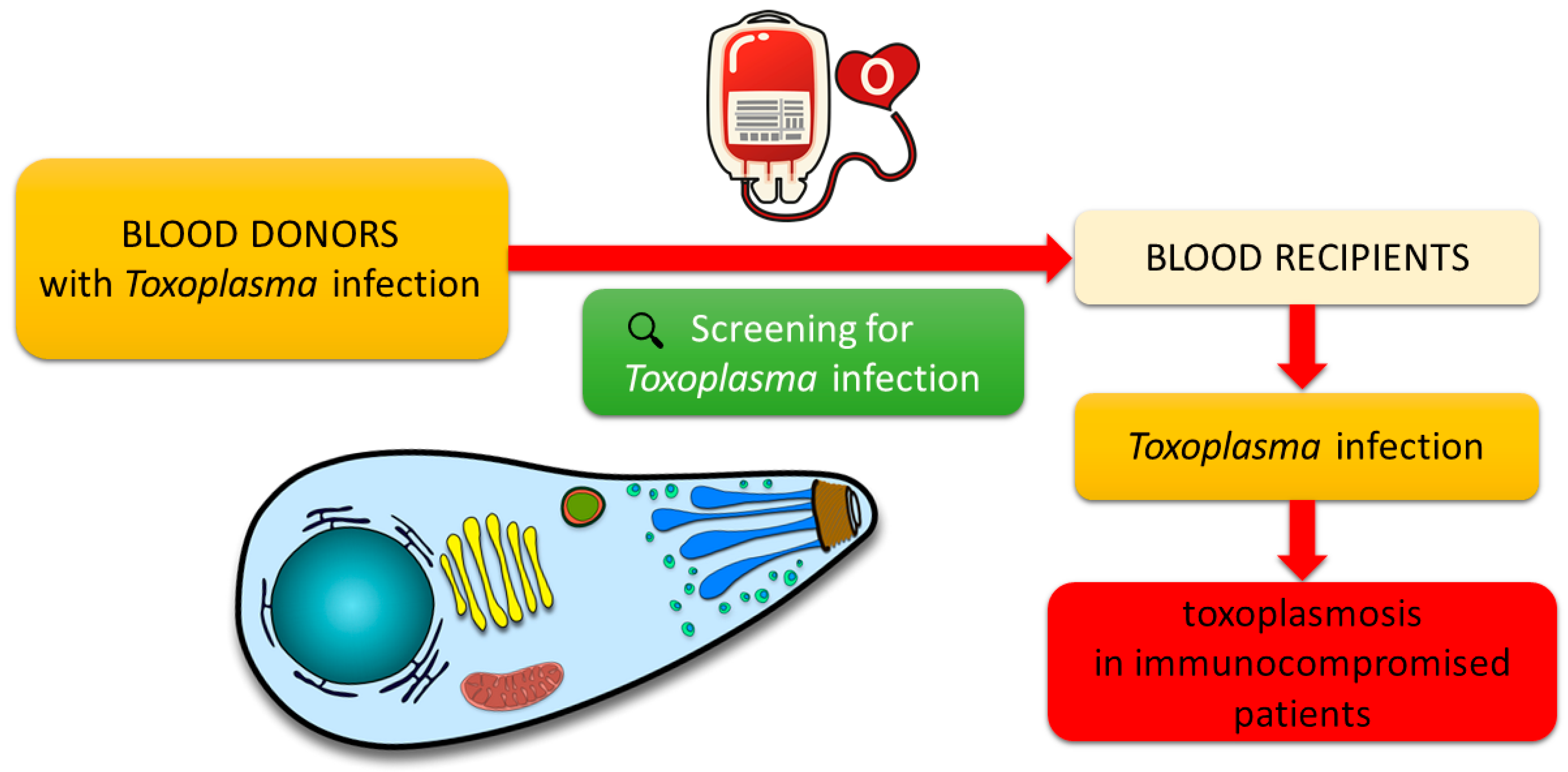
:1. Introduction
2. Methods
3. Results
3.1. The Classical Course of Toxoplasma Infection and Toxoplasmosis
3.2. Prevalence of Toxoplasma Infections in Blood Donors

| Size of the Study Group | Analyzed Parameter | Method | Results | Refs. |
|---|---|---|---|---|
| n = 790 |
| ECLIA | 213 (24.2%)–only IgG (+) 8 (0.9%)–only IgM (+) 12 (1.4%)–both IgM (+) and IgG (+) | [8] |
| n = 385 |
| ELISA, LAMP | 146 (37.9%)–only IgG (+) 4 (1.03%)–only IgM (+) 6 (1.56%)–both IgM (+) and IgG (+) 6 (1.56%)–T. gondii DNA (+) | [33] |
| n = 1347 |
| LAT | 618 (45.9%)–IgM (+) and/or IgG (+) | [26] |
| n = 375 |
| ELISA | 94 (25.1%)–only IgG (+) | [34] |
| n = 800 |
| IFA, ELISA | 352 (44%)–only IgG (+) 3 (0.4%)–both IgM (+) and IgG (+) | [12] |
| n = 103 |
| ELISA | 46 (47.7%)–only IgG (+) | [28] |
| n = 462 |
| ELISA, PCR (529 bp) | 150 (32.5%)–only IgG (+) 7 (1.5%)–only IgM (+) 9 (2%)–both IgM (+) and IgG (+) 30 (18%) of 166 seropositive donors–T. gondii DNA (+) | [10] |
| n = 400 |
| ELISA, nested-PCR multilocus, nested-PCR-RFLP | 294 (73.5%)–only IgG (+) 9 (2.2%)–only IgM (+) 7 (1.8%)–both IgM (+) and IgG (+) 7 (1.8%)–T. gondii DNA (+) | [19] |
| n = 46 |
| ELISA | 11 (23.9%)–only IgG (+) 1 (2.2%)–only IgM (+) | [35] |
| n = 380 |
| ELISA | 131 (34.47%)–only IgG (+) 2 (0.5%)–only IgM (+) 11 (2.9%)–both IgM (+) and IgG (+) | [36] |
| n = 520 |
| MAT | 198 (38.1%)–IgG (+) | [21] |
| n = 510 |
| ELC, nested-PCR, qPCR | 223 (43.7%)–only IgG (+) 8 (1.6%)–both IgM (+) and IgG (+) all samples–T. gondii DNA (−) | [37] |
| n = 750 |
| ELISA, nested-PCR (B1 gene) | 335 (44.7%)–only IgG (+) 5 (0.6%)–only IgM (+) 21 (2.8%)–both IgM (+) and IgG (+) 38 (10.8%) of IgG (+)–T. gondii DNA (+) | [13] |
| n = 150 |
| ELISA, real-time PCR (B1 gene) | 98 (65.3%)–only IgG (+) 15 (10%)–T. gondii DNA (+) | [38] |
| n = 1783 |
| ELISA, real-time PCR | 161 (9.3%)–only IgG (+) 5 (0.28%)–both IgM (+) and IgG (+) all samples–T. gondii DNA (−) | [39] |
| n = 207 |
| ELISA | 46 (22.2%)–IgM (+) | [30] |
| n = 500 |
| ELISA, real-time PCR | 144 (28.8%)–only IgG (+) 11 (2.2%)–only IgM (+) 5 (1%)–both IgM (+) and IgG (+) 1 (9%) of IgM (+)–T. gondii DNA (+) | [31] |
| n = 194 |
| CLIA | 75 (38.66%)–only IgG (+) 2 (1.03%)–only IgM (+) | [32] |
| n = 1480 |
| ELISA, nested-PCR (B1 gene) | 182 (12.3%)–only IgG (+) 81 (5.47%)–only IgM (+) 23 (1.6%)–both IgM (+) and IgG (+) 2 (1.9%) of IgM (+)–T. gondii DNA (+) | [40] |
3.3. Prevalence of Toxoplasma Infections in Blood and Transplant Recipients
3.4. Laboratory Diagnosis of Toxoplasma Infections in Terms of the Safety of Blood and Transplant Recipients
4. Conclusions
Author Contributions
Funding
Data Availability Statement
Conflicts of Interest
References
- Szewczyk-Golec, K.; Pawłowska, M.; Wesołowski, R.; Wróblewski, M.; Mila-Kierzenkowska, C. Oxidative Stress as a Possible Target in the Treatment of Toxoplasmosis: Perspectives and Ambiguities. Int. J. Mol. Sci. 2021, 22, 5705. [Google Scholar] [CrossRef] [PubMed]
- Dupont, D.; Fricker-Hidalgo, H.; Brenier-Pinchart, M.-P.; Garnaud, C.; Wallon, M.; Pelloux, H. Serology for Toxoplasma in Immunocompromised Patients: Still Useful? Trends Parasitol. 2021, 37, 205–213. [Google Scholar] [CrossRef] [PubMed]
- Wesołowski, R.; Pawłowska, M.; Smoguła, M.; Szewczyk-Golec, K. Advances and Challenges in Diagnostics of Toxoplasmosis in HIV-Infected Patients. Pathogens 2023, 12, 110. [Google Scholar] [CrossRef]
- De La Mata Navazo, S.; Slöcker Barrio, M.; García-Morín, M.; Beléndez, C.; Escobar Fernández, L.; Rincón-López, E.M.; Aguilera Alonso, D.; Guinea, J.; Marín, M.; Butragueño-Laiseca, L.; et al. Case Report: Severe ARDS in a Pediatric Hematopoietic Stem-Cell Transplantation Recipient Caused by Disseminated Toxoplasmosis. Front. Pediatr. 2022, 9, 810718. [Google Scholar] [CrossRef] [PubMed]
- Adekunle, R.O.; Sherman, A.; Spicer, J.O.; Messina, J.A.; Steinbrink, J.M.; Sexton, M.E.; Lyon, G.M.; Mehta, A.K.; Phadke, V.K.; Woodworth, M.H. Clinical Characteristics and Outcomes of Toxoplasmosis among Transplant Recipients at Two US Academic Medical Centers. Transpl. Infect. Dis. 2021, 23, e13636. [Google Scholar] [CrossRef] [PubMed]
- La Hoz, R.M.; Morris, M.I. The Infectious Diseases Community of Practice of the American Society of Transplantation Tissue and Blood Protozoa Including Toxoplasmosis, Chagas Disease, Leishmaniasis, Babesia, Acanthamoeba, Balamuthia, and Naegleria in Solid Organ Transplant Recipients—Guidelines from the American Society of Transplantation Infectious Diseases Community of Practice. Clin. Transpl. 2019, 33, e13546. [Google Scholar] [CrossRef]
- Amoo, A.; Njaanake, K.; Dada-Adegbola, H.; Omosa-Manyonyi, G. Toxoplasmosis among Blood Donors: Unsafe Blood Transfusion in Ibadan, Southwest Nigeria. J. Appl. Hematol. 2019, 10, 120. [Google Scholar] [CrossRef]
- Yılmaz, A.; Yazıcı, E.; Turk, C. Assessment of Seroprevalence of Toxoplasma gondii in Blood Donors Applied to the Blood Center of Gazi University Hospital. Iran. J. Microbiol. 2021, 13, 243–247. [Google Scholar] [CrossRef]
- Stopić, M.; Štajner, T.; Marković-Denić, L.; Nikolić, V.; Djilas, I.; Srzentić, S.J.; Djurković-Djaković, O.; Bobić, B. Epidemiology of Toxoplasmosis in SERBIA: A Cross-Sectional Study on Blood Donors. Microorganisms 2022, 10, 492. [Google Scholar] [CrossRef]
- Asfaram, S.; Rezaei, R.; Fakhar, M.; Ghezelbash, B.; Nakhaei, M.; Hezarjaribi, H.Z.; Mardani, A.; Teshnizi, S.H. High Occurrence of Toxoplasma gondii Infection among Blood Donors in Ardabil Province as Main Focus of Zoonotic Visceral Leishmaniosis, Northwestern Iran. Ann. Parasitol. 2021, 67, 611–617. [Google Scholar] [CrossRef]
- Elmore, S.A.; Jones, J.L.; Conrad, P.A.; Patton, S.; Lindsay, D.S.; Dubey, J.P. Toxoplasma gondii: Epidemiology, Feline Clinical Aspects, and Prevention. Trends Parasitol. 2010, 26, 190–196. [Google Scholar] [CrossRef] [PubMed] [Green Version]
- Lachkhem, A.; Lahmar, I.; Galal, L.; Babba, O.; Mezhoud, H.; Hassine, M.; Lachkhem, A.; Dardé, M.-L.; Mercier, A.; Babba, H. Seroprevalence of Toxoplasma gondii among Healthy Blood Donors in Two Locations in Tunisia and Associated Risk Factors. Parasite 2020, 27, 51. [Google Scholar] [CrossRef] [PubMed]
- Nakashima, F.; Pardo, V.S.; Miola, M.P.; Murata, F.H.A.; Paduan, N.; Longo, S.M.; Brandão De Mattos, C.C.; Pereira-Chioccola, V.L.; Ricci, O.; De Mattos, L.C. Serum IgG Anti-Toxoplasma gondii Antibody Concentrations Do Not Correlate Nested PCR Results in Blood Donors. Front. Cell. Infect. Microbiol. 2020, 9, 461. [Google Scholar] [CrossRef] [Green Version]
- Lima, T.S.; Lodoen, M.B. Mechanisms of Human Innate Immune Evasion by Toxoplasma gondii. Front. Cell. Infect. Microbiol. 2019, 9, 103. [Google Scholar] [CrossRef] [Green Version]
- Lüder, C.G.K.; Rahman, T. Impact of the Host on Toxoplasma Stage Differentiation. Microb. Cell 2017, 4, 203–211. [Google Scholar] [CrossRef]
- Pleyer, U.; Groß, U.; Schlüter, D.; Wilking, H.; Seeber, F. Toxoplasmosis in Germany: Epidemiology, Diagnosis, Risk Factors, and Treatment. Dtsch. Ärzteb. Int. 2019, 116, 435–444. [Google Scholar] [CrossRef]
- Rauwolf, K.K.; Floeth, M.; Kerl, K.; Schaumburg, F.; Groll, A.H. Toxoplasmosis after Allogeneic Haematopoietic Cell Transplantation—Disease Burden and Approaches to Diagnosis, Prevention and Management in Adults and Children. Clin. Microbiol. Infect. 2021, 27, 378–388. [Google Scholar] [CrossRef]
- Blanchard, N.; Dunay, I.R.; Schlüter, D. Persistence of Toxoplasma gondii in the Central Nervous System: A Fine-Tuned Balance between the Parasite, the Brain and the Immune System. Parasite Immunol. 2015, 37, 150–158. [Google Scholar] [CrossRef] [PubMed]
- Hosseini, S.A.; Golchin, E.; Sharif, M.; Sarvi, S.; Ahmadpour, E.; Rostamian, A.; Gholami, S.; Amouei, A.; Daryani, A. A Serological Investigation and Genotyping of Toxoplasma gondii among Iranian Blood Donors Indicates Threat to Health of Blood Recipients. Transfus. Apher. Sci. 2020, 59, 102723. [Google Scholar] [CrossRef] [PubMed]
- Alvarado-Esquivel, C.; Mercado-Suarez, M.F.; Rodríguez-Briones, A.; Fallad-Torres, L.; Ayala-Ayala, J.O.; Nevarez-Piedra, L.J.; Duran-Morales, E.; Estrada-Martínez, S.; Liesenfeld, O.; Márquez-Conde, J.Á.; et al. Seroepidemiology of Infection with Toxoplasma gondii in Healthy Blood Donors of Durango, Mexico. BMC Infect. Dis. 2007, 7, 75. [Google Scholar] [CrossRef] [PubMed] [Green Version]
- Rodrigues, F.T.; Sousa, A.P.; Escoval, M.A.; Condeço, J.; Cardoso, L.; Lopes, A.P. Seroepidemiology of Toxoplasma gondii in Blood Donors in Portugal. Transfus. Apher. Sci. 2020, 59, 102777. [Google Scholar] [CrossRef] [PubMed]
- Mardani, A. Prevention Strategies of Transfusion-Transmitted Parasitic Infections (TTPIs): Strengths and Challenges of Current Approaches, and Evaluation of the Strategies Implemented in Iran. Parasite Epidemiol. Control 2020, 9, e00141. [Google Scholar] [CrossRef] [PubMed]
- Pawełczyk, A.; Bednarska, M.; Caraballo Cortés, K.; Glamkowska-Sady, M.; Kowalska, J.; Uszyńska-Kałuża, B.; Radkowski, M.; Welc-Falęciak, R. Seronegative Infection with Toxoplasma gondii in Asymptomatic Human Immunodeficiency Virus Type 1 (HIV-1)-Infected Patients and in Blood Donors. J. Clin. Med. 2022, 11, 638. [Google Scholar] [CrossRef] [PubMed]
- Rozporządzenie Ministra Zdrowia z Dnia 11 Września 2017 r. w Sprawie Warunków Pobierania Krwi Od Kandydatów Na Dawców Krwi i Dawców Krwi. Regulation of the Minister of Health of September 11, 2017 on the Conditions for Collecting Blood from Candidates for Blood Donors and Blood Donors. Available online: https://isap.sejm.gov.pl/isap.nsf/DocDetails.xsp?id=WDU20170001741 (accessed on 24 June 2023).
- Botein, E.F.; Darwish, A.; El-Tantawy, N.L.; EL-baz, R.; Eid, M.I.; Shaltot, A.M. Serological and Molecular Screening of Umbilical Cord Blood for Toxoplasma gondii Infection. Transpl. Infect. Dis. 2019, 21, e13117. [Google Scholar] [CrossRef]
- Lupu, M.A.; Lighezan, R.; Paduraru, A.A.; Dragomir, A.; Pavel, R.; Grada, S.; Mihu, A.G.; Ursoniu, S.; Olariu, T.R. Seroepidemiology of Toxoplasma gondii Infection in Blood Donors from Western Romania. Microorganisms 2022, 10, 973. [Google Scholar] [CrossRef]
- Foroutan-Rad, M.; Majidiani, H.; Dalvand, S.; Daryani, A.; Kooti, W.; Saki, J.; Hedayati-Rad, F.; Ahmadpour, E. Toxoplasmosis in Blood Donors: A Systematic Review and Meta-Analysis. Transfus. Med. Rev. 2016, 30, 116–122. [Google Scholar] [CrossRef]
- Belkacemi, M.; Heddi, B. Toxoplasmosis Immunity Status of Blood Donors in Sidi Bel Abbès, West Algeria. Cureus 2022, 14, e28826. [Google Scholar] [CrossRef]
- El-Sayed, N.M. Recent Updates in Transfusion Transmitted Parasitic Diseases. Aperito J. Bacteriol. Virol. Parasitol. 2015, 2, 110. [Google Scholar] [CrossRef]
- Saraswathi, R.; Anupriya, A.; Lalithambigai, J.; Prabhusaran, N.; Velayutharaj, A.; Uma, A. Seroprevalence of Toxoplasmosis among Blood Donors in a Tertiary Care Teaching Hospital in South India. Int. J. Med. Sci. Clin. Invent. 2017, 4, 2846–2849. [Google Scholar] [CrossRef] [Green Version]
- Mahmoudvand, H.; Saedi Dezaki, E.; Soleimani, S.; Baneshi, M.R.; Kheirandish, F.; Ezatpour, B.; Zia-ali, N. Seroprevalence and Risk Factors of Toxoplasma gondii Infection among Healthy Blood Donors in South-East of Iran. Parasite Immunol. 2015, 37, 362–367. [Google Scholar] [CrossRef]
- Bahhaj, R.; Ahmadpour, E.; Mahami-Oskouei, M.; Fallah, E.; Shamsasenjan, K.; Safaiyan, A. Toxoplasma gondii Infection and Related Risk Factors Among Blood Donors in Northwest Iran. Arch. Clin. Infect. Dis. 2017, 12, e62005. [Google Scholar] [CrossRef]
- Manouchehri Naeini, K.; Heidari Soureshjani, E.; Jafari, M.; Parchami, S.; Karimi, G.; Abdizade, R. Prevalence of Toxoplasma gondii Infection in Healthy Volunteer Blood Donors Using Serological and Molecular Methods from Chaharmahal and Bakhtiari Province, Southwest Iran. Jundishapur J. Microbiol. 2019, 12, e91042. [Google Scholar] [CrossRef] [Green Version]
- Jafari Modrek, M.; Mousavi, M.; Saravani, R. Toxoplasma gondii Seroprevalence Among Blood Donors in Zahedan, Southeastern Iran. Int. J. Infect. 2014, 1, e21111. [Google Scholar] [CrossRef]
- Yıldız, S.; Esen, R.; Karaman, K.; Beyhan, Y.E. Seropositivity of Toxoplasma gondii Among Blood Donors and Patients with Hematologic Malignity. East J. Med. 2023, 28, 291–295. [Google Scholar] [CrossRef]
- Saki, J.; Foroutan, M.; Khodkar, I.; Khodadadi, A.; Nazari, L. Seroprevalence and Molecular Detection of Toxoplasma gondii in Healthy Blood Donors in Southwest Iran. Transfus. Apher. Sci. 2019, 58, 79–82. [Google Scholar] [CrossRef] [PubMed] [Green Version]
- Paraboni, M.L.R.; Commodaro, A.G.; Campi-Azevedo, A.C.; Brito-de-Sousa, J.P.; Gonçalves, I.L.; Da Costa, D.F.; Ribeiro, K.S.; Garcia, J.L.; Silveira, C.; Martins-Filho, O.A.; et al. Seroprevalence and Systemic Immune Biomarkers Associated with Toxoplasma gondii Infection in Blood Donors from Southern Brazil. Immunobiology 2022, 227, 152294. [Google Scholar] [CrossRef] [PubMed]
- El-Geddawi, O.; El-Sayad, M.; Sadek, N.; Hussien, N.; Ahmed, M. Detection of T. Gondii Infection in Blood Donors in Alexandria, Egypt, Using Serological and Molecular Strategies. Parasitol. United J. 2016, 9, 24. [Google Scholar] [CrossRef]
- Chiang, T.-Y.; Hsieh, H.-H.; Kuo, M.-C.; Chiu, K.-T.; Lin, W.-C.; Fan, C.-K.; Fang, C.-T.; Ji, D.-D. Seroepidemiology of Toxoplasma gondii Infection among Healthy Blood Donors in Taiwan. PLoS ONE 2012, 7, e48139. [Google Scholar] [CrossRef] [Green Version]
- Sarkari, B.; Shafiei, R.; Zare, M.; Sohrabpour, S.; Kasraian, L. Seroprevalence and Molecular Diagnosis of Toxoplasma gondii Infection among Blood Donors in Southern Iran. J. Infect. Dev. Ctries 2014, 8, 543–547. [Google Scholar] [CrossRef] [Green Version]
- Siegel, S.E.; Lunde, M.N.; Gelderman, A.H.; Halterman, R.H.; Brown, J.A.; Levine, A.S.; Graw, R.G., Jr. Transmission of Toxoplasmosis by Leukocyte Transfusion. Blood 1971, 37, 388–394. [Google Scholar] [CrossRef]
- Edvinsson, B.; Lappalainen, M.; Evengård, B. Real-time PCR targeting a 529-bp repeat element for diagnosis of toxoplasmosis. Clin. Microbiol. Infect. 2006, 12, 131–136. [Google Scholar] [CrossRef] [PubMed] [Green Version]
- Wang, T.; Han, Y.; Pan, Z.; Wang, H.; Yuan, M.; Lin, H. Seroprevalence of Toxoplasma gondii Infection in Blood Donors in Mainland China: A Systematic Review and Meta-Analysis. Parasite 2018, 25, 36. [Google Scholar] [CrossRef] [PubMed] [Green Version]
- Rousseau, A.; La Carbona, S.; Dumètre, A.; Robertson, L.J.; Gargala, G.; Escotte-Binet, S.; Favennec, L.; Villena, I.; Gérard, C.; Aubert, D. Assessing Viability and Infectivity of Foodborne and Waterborne Stages (Cysts/Oocysts) of Giardia duodenalis, Cryptosporidium spp., and Toxoplasma gondii: A Review of Methods. Parasite 2018, 25, 14. [Google Scholar] [CrossRef] [PubMed] [Green Version]

Disclaimer/Publisher’s Note: The statements, opinions and data contained in all publications are solely those of the individual author(s) and contributor(s) and not of MDPI and/or the editor(s). MDPI and/or the editor(s) disclaim responsibility for any injury to people or property resulting from any ideas, methods, instructions or products referred to in the content. |
© 2023 by the authors. Licensee MDPI, Basel, Switzerland. This article is an open access article distributed under the terms and conditions of the Creative Commons Attribution (CC BY) license (https://creativecommons.org/licenses/by/4.0/).
Share and Cite
Wesołowski, R.; Pawłowska, M.; Mila-Kierzenkowska, C. The Medical Relevance of Toxoplasma Infections in Terms of the Safety of Blood Recipients under Immunosuppression—A Meta-Analysis. Microorganisms 2023, 11, 1980. https://doi.org/10.3390/microorganisms11081980
Wesołowski R, Pawłowska M, Mila-Kierzenkowska C. The Medical Relevance of Toxoplasma Infections in Terms of the Safety of Blood Recipients under Immunosuppression—A Meta-Analysis. Microorganisms. 2023; 11(8):1980. https://doi.org/10.3390/microorganisms11081980
Chicago/Turabian StyleWesołowski, Roland, Marta Pawłowska, and Celestyna Mila-Kierzenkowska. 2023. "The Medical Relevance of Toxoplasma Infections in Terms of the Safety of Blood Recipients under Immunosuppression—A Meta-Analysis" Microorganisms 11, no. 8: 1980. https://doi.org/10.3390/microorganisms11081980
APA StyleWesołowski, R., Pawłowska, M., & Mila-Kierzenkowska, C. (2023). The Medical Relevance of Toxoplasma Infections in Terms of the Safety of Blood Recipients under Immunosuppression—A Meta-Analysis. Microorganisms, 11(8), 1980. https://doi.org/10.3390/microorganisms11081980