
Real-Time Heart Rate Detection Method Based on 77 GHz FMCW Radar
Abstract
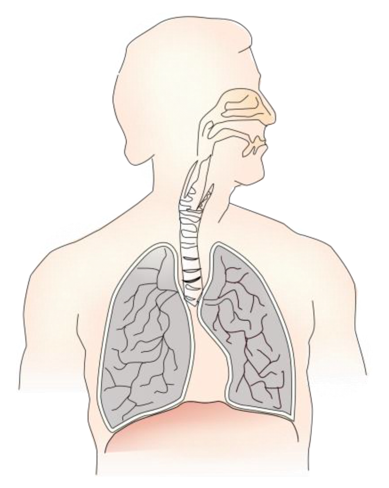
:1. Introduction
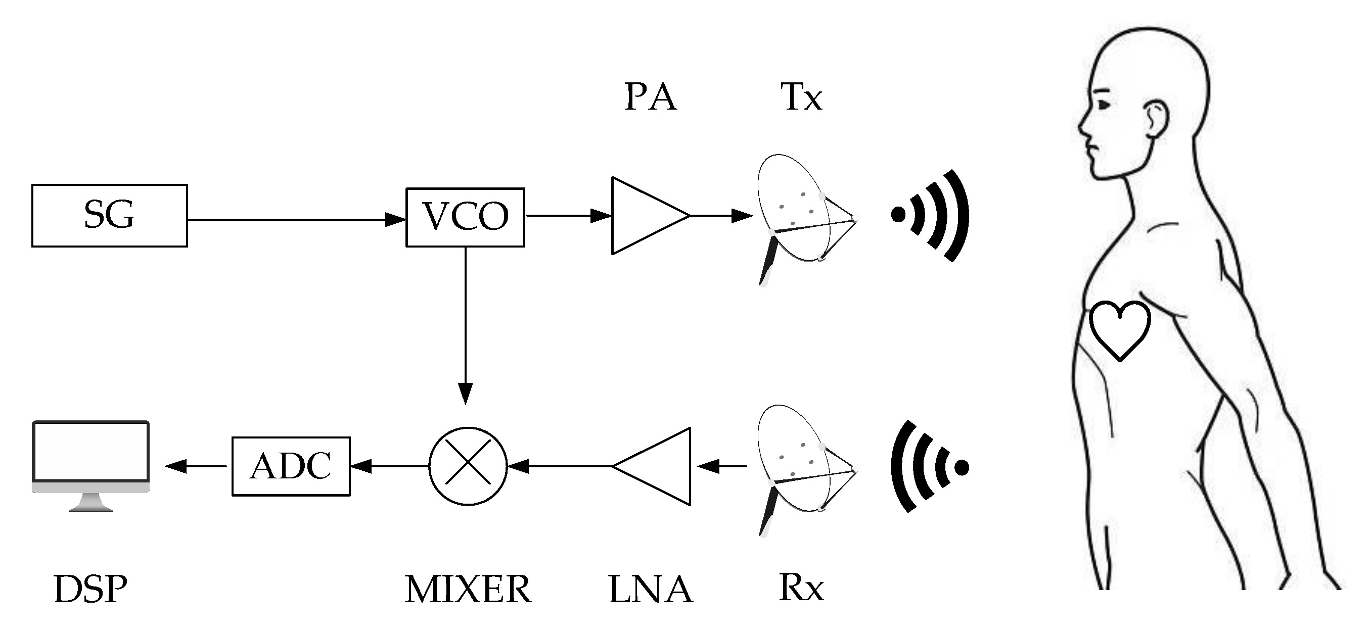
2. Theory
3. Methods
3.1. New Motion Model
3.2. Acquisition of Vital Signs
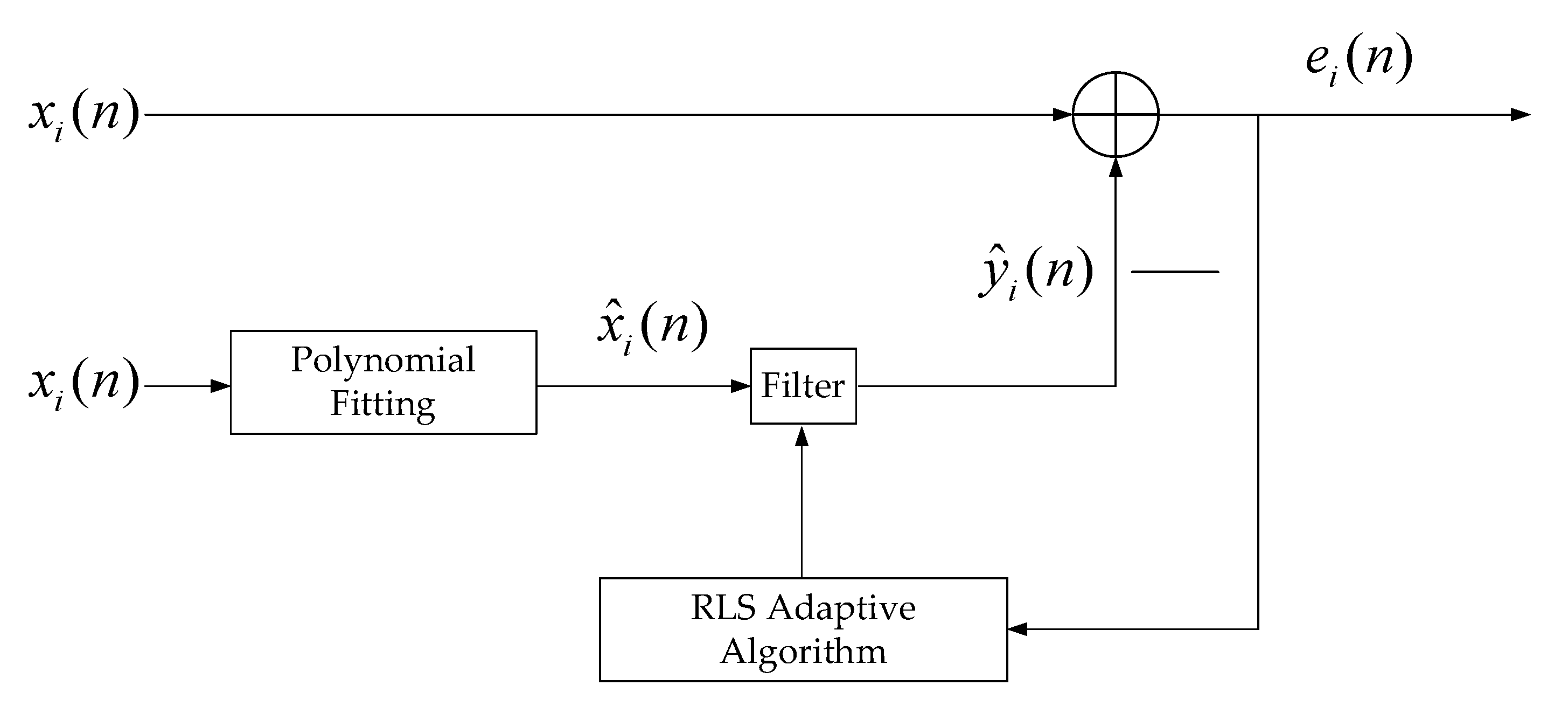
3.3. Elimination of the RBM Signal
3.4. Elimination of Respiratory Harmonics
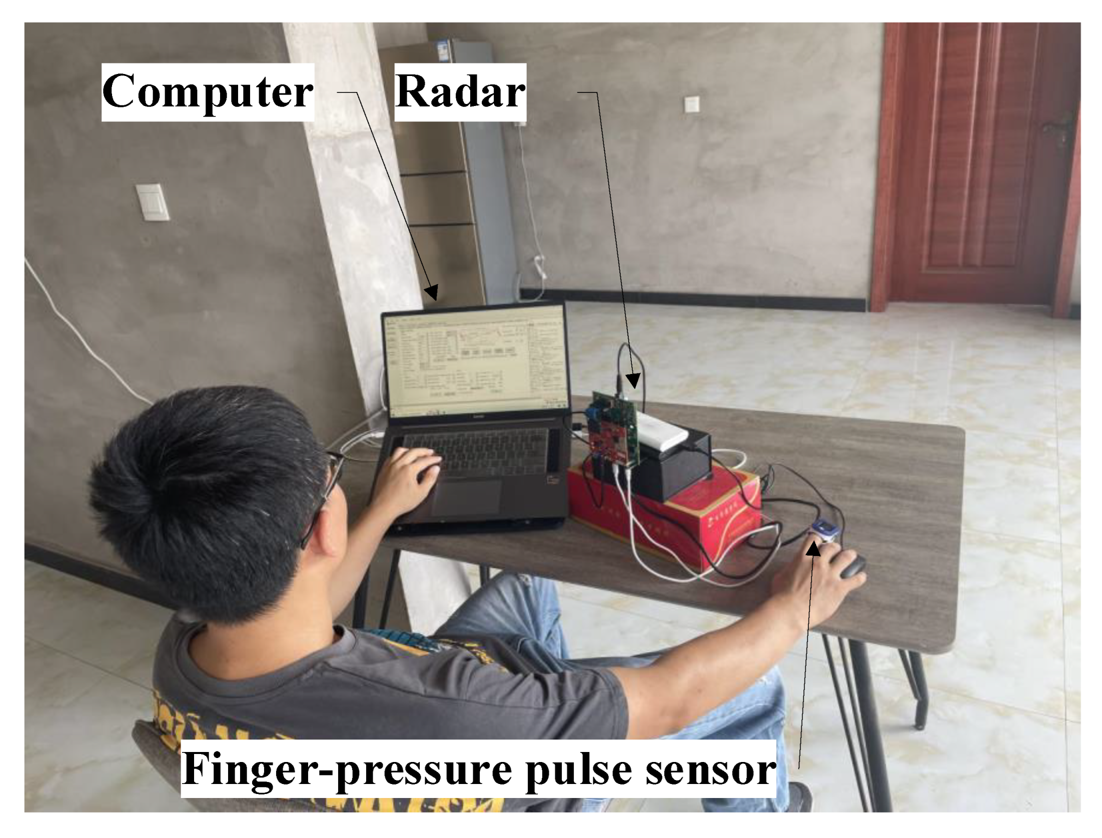
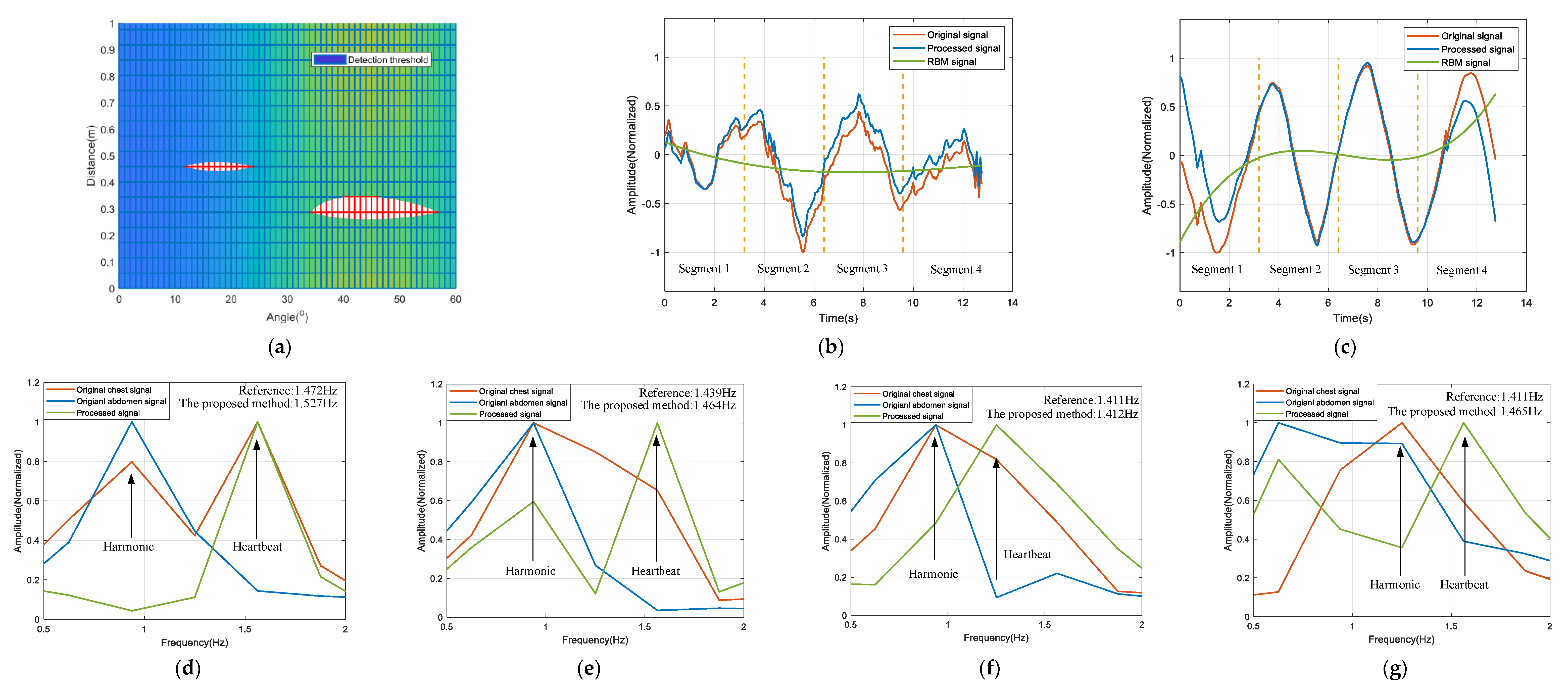
4. Results and Discussion
5. Conclusions
Author Contributions
Funding
Institutional Review Board Statement
Informed Consent Statement
Data Availability Statement
Conflicts of Interest
References
- Cavagnaro, M.; Pittella, E.; Pisa, S. UWB pulse propagation into human tissues. Phys. Med. Biol. 2013, 58, 8689–8707. [Google Scholar] [CrossRef]
- Pimentel, M.A.; Redfern, O.C.; Hatch, R.; Young, J.D.; Tarassenko, L.; Watkinson, P.J. Trajectories of vital signs in patients with COVID-19. Resuscitation 2020, 156, 99–106. [Google Scholar] [CrossRef] [PubMed]
- Khan, A.A.; Lip, G.Y.; Shantsila, A. Heart rate variability in atrial fibrillation: The balance between sympathetic and parasympathetic nervous system. Eur. J. Clin. Investig. 2019, 49, e13174. [Google Scholar] [CrossRef] [PubMed] [Green Version]
- Sun, L.; Hong, H.; Li, Y.; Gu, C.; Xi, F.; Li, C.; Zhu, X. Noncontact Vital Sign Detection based on Stepwise Atomic Norm Minimization. IEEE Signal Process. Lett. 2015, 22, 2479–2483. [Google Scholar] [CrossRef]
- Li, M.; Lin, J. Wavelet-Transform-Based Data-Length-Variation Technique for Fast Heart Rate Detection Using 5.8-GHz CW Doppler Radar. IEEE Trans. Microw. Theory Tech. 2018, 66, 568–576. [Google Scholar] [CrossRef]
- Nosrati, M.; Tavassolian, N. High-Accuracy Heart Rate Variability Monitoring Using Doppler Radar Based on Gaussian Pulse Train Modeling and FTPR Algorithm. IEEE Trans. Microw. Theory Tech. 2018, 66, 556–567. [Google Scholar] [CrossRef]
- Yamamoto, K.; Toyoda, K.; Ohtsuki, T. Spectrogram-Based Non-Contact RRI Estimation by Accurate Peak Detection Algorithm. IEEE Access 2018, 6, 60369–60379. [Google Scholar] [CrossRef]
- Li, C.; Lin, J. Random Body Movement Cancellation in Doppler Radar Vital Sign Detection. IEEE Trans. Microw. Theory Tech. 2008, 56, 3143–3152. [Google Scholar]
- Massagram, W.; Lubecke, V.M.; Host-Madsen, A.; Boric-Lubecke, O. Assessment of Heart Rate Variability and Respiratory Sinus Arrhythmia via Doppler Radar. IEEE Trans. Microw. Theory Tech. 2009, 57, 2542–2549. [Google Scholar] [CrossRef]
- Wang, K.F.; Horng, T.S.; Peng, K.C.; Jau, J.K.; Li, J.Y.; Chen, C.C. Single-Antenna Doppler Radars Using Self and Mutual Injection Locking for Vital Sign Detection With Random Body Movement Cancellation. IEEE Trans. Microw. Theory Tech. 2011, 59, 3577–3587. [Google Scholar] [CrossRef]
- Gu, C.; Wang, G.; Li, Y.; Lnoue, T.; Li, C. A Hybrid Radar-Camera Sensing System With Phase Compensation for Random Body Movement Cancellation in Doppler Vital Sign Detection. IEEE Trans. Microw. Theory Tech. 2013, 61, 4678–4688. [Google Scholar] [CrossRef]
- Tang, M.C.; Kuo, C.Y.; Wun, D.C.; Wang, F.K.; Horng, T.S. A Self- and Mutually Injection-Locked Radar System for Monitoring Vital Signs in Real Time With Random Body Movement Cancellation. IEEE Trans. Microw. Theory Tech. 2016, 64, 4812–4822. [Google Scholar] [CrossRef]
- Gu, C.; Wang, J.; Lien, J. Deep Neural Network based Body Movement Cancellation for Doppler Radar Vital Sign Detection. In Proceedings of the 2019 IEEE MTT-S International Wireless Symposium (IWS), Guangzhou, China, 19–22 May 2019; pp. 1–3. [Google Scholar]
- Yang, Z.K.; Shi, H.; Zhao, S.; Huang, X.D. Vital sign detection during large-scale and fast body movements based on an adaptive noise cancellation algorithm using a single Doppler radar sensor. Sensors 2020, 20, 4183. [Google Scholar] [CrossRef] [PubMed]
- Ti, J.; Lin, J. Respiration harmonics cancellation for Accurate Heart Rate measurement in non-contact vital sign detection. In Proceedings of the 2013 IEEE MTT-S International Microwave Symposium Digest (MTT), Seattle, WA, USA, 2–7 June 2013; pp. 1–3. [Google Scholar]
- Huang, T.Y.; Hayward, L.F.; Lin, J. Adaptive harmonics comb notch digital filter for measuring heart rate of laboratory rat using a 60-GHz radar. In Proceedings of the 2016 IEEE MTT-S International Microwave Symposium (IMS), San Francisco, CA, USA, 22–27 May 2016; pp. 1–4. [Google Scholar]
- Nosrati, M.; Tavassolian, N. Accurate Doppler Radar-Based Cardiopulmonary Sensing Using Chest-Wall Acceleration. IEEE J. Electromagn. RF Microw. Med. Biol. 2019, 3, 41–47. [Google Scholar] [CrossRef]
- Yang, Z.K.; Zhao, S.; Huang, X.D.; Lv, W. Accurate Doppler radar-based heart rate measurement using matched filter. IEICE Electron. Express 2020, 17, 20200062. [Google Scholar] [CrossRef] [Green Version]
- Lee, C.; Yoon, C.; Kong, H.J.; Kim, H.C.; Kim, Y. Heart Rate Tracking Using a Doppler Radar With the Reassigned Joint Time-Frequency Transform. IEEE Antennas Wirel. Propag. Lett. 2011, 10, 1096–1099. [Google Scholar]
- Hu, W.; Zhao, Z.; Wang, Y.; Zhang, H.; Lin, F. Noncontact Accurate Measurement of Cardiopulmonary Activity Using a Compact Quadrature Doppler Radar Sensor. IEEE Trans. Biomed. Eng. 2014, 61, 725–735. [Google Scholar] [CrossRef]
- Tu, J.; Lin, J. Fast Acquisition of Heart Rate in Noncontact Vital Sign Radar Measurement Using Time-Window-Variation Technique. IEEE Trans. Instrum. Meas. 2015, 65, 112–122. [Google Scholar] [CrossRef]
- Park, J.; Ham, J.W.; Park, S.; Kim, D.H.; Park, S.J.; Kang, H.; Park, S.O. Polyphase-Basis Discrete Cosine Transform for Real-Time Measurement of Heart Rate With CW Doppler Radar. IEEE Trans. Microw. Theory Tech. 2018, 66, 1644–1659. [Google Scholar] [CrossRef]
- Ye, C.; Toyoda, K.; Ohtsuki, T. A stochastic gradient approach for robust heartbeat detection with Doppler radar using time-window-variation technique. IEEE Trans. Biomed. Eng. 2019, 66, 1730–1741. [Google Scholar] [CrossRef]
- Lv, W.; Zhao, Y.; Zhang, W.; Liu, W.; Hu, A.; Miao, J. Remote Measurement of Short-Term Heart Rate With Narrow Beam Millimeter Wave Radar. IEEE Access 2021, 9, 165049–165058. [Google Scholar] [CrossRef]
- Gao, Z.; Ali, L.; Wang, C.; Liu, R.; Wang, C.; Qian, C.; Sung, H.; Meng, F. Real-Time Non-Contact Millimeter Wave Radar-Based Vital Sign Detection. Sensors 2022, 22, 7560. [Google Scholar] [CrossRef] [PubMed]
- Ding, Y.; Yu, X.; Lei, C.; Sun, Y.; Xu, x.; Zhang, J. A Novel Real-Time Human Heart Rate Estimation Method for Noncontact Vital Sign Radar Detection. IEEE Access 2020, 8, 88689–88699. [Google Scholar] [CrossRef]


| Segment 1 (3.2 s) | Segment 2 (3.2 s) | Segment 3 (3.2 s) | Segment 4 (3.2 s) | |||||||||
|---|---|---|---|---|---|---|---|---|---|---|---|---|
| 1 | 1.5124 | 1.5722 | 3.80 | 1.6625 | 1.5833 | 5.00 | 1.4808 | 1.5222 | 2.72 | 1.4584 | 1.4583 | 0.01 |
| 2 | 1.4452 | 1.4778 | 2.21 | 1.4461 | 1.4500 | 0.27 | 1.4387 | 1.4167 | 1.56 | 1.4293 | 1.4167 | 0.89 |
| 3 | 1.6998 | 1.6333 | 4.07 | 1.6437 | 1.5944 | 3.09 | 1.5625 | 1.5222 | 2.65 | 1.4323 | 1.4250 | 0.51 |
| 4 | 1.4176 | 1.3500 | 5.01 | 1.3715 | 1.3222 | 3.29 | 1.4168 | 1.3278 | 6.70 | 1.3697 | 1.3611 | 0.61 |
| 5 | 1.5276 | 1.4667 | 4.15 | 1.4638 | 1.4389 | 1.73 | 1.4123 | 1.4111 | 0.08 | 1.4650 | 1.4056 | 3.82 |
| 6 | 1.3385 | 1.3222 | 1.23 | 1.4446 | 1.3222 | 9.36 | 1.4305 | 1.3167 | 8.65 | 1.3753 | 1.3500 | 1.87 |
| 7 | 1.4252 | 1.4417 | 1.14 | 1.4235 | 1.4167 | 0.48 | 1.3939 | 1.4000 | 0.44 | 1.4058 | 1.3722 | 2.45 |
| 8 | 1.4366 | 1.3833 | 3.85 | 1.3347 | 1.3833 | 3.52 | 1.4048 | 1.3944 | 0.74 | 1.3410 | 1.3833 | 3.06 |
| 9 | 1.4153 | 1.4111 | 0.29 | 1.4159 | 1.4500 | 2.35 | 1.4047 | 1.3792 | 1.85 | 1.3329 | 1.3125 | 1.55 |
| 10 | 1.6660 | 1.5778 | 5.59 | 1.6342 | 1.5667 | 4.31 | 1.4657 | 1.4917 | 1.74 | 1.3995 | 1.4444 | 3.11 |
| 11 | 1.3705 | 1.4500 | 5.48 | 1.4312 | 1.4111 | 1.42 | 1.3589 | 1.3667 | 0.57 | 1.3481 | 1.3722 | 1.76 |
| 12 | 1.4006 | 1.4056 | 0.35 | 1.4104 | 1.4444 | 2.80 | 1.3733 | 1.3889 | 1.28 | 1.4198 | 1.3400 | 5.96 |
| 13 | 1.4215 | 1.4667 | 3.08 | 1.4167 | 1.4500 | 2.30 | 1.3647 | 1.3889 | 1.74 | 1.3864 | 1.3667 | 1.44 |
| 14 | 1.3649 | 1.3000 | 4.99 | 1.3854 | 1.3000 | 6.57 | 1.3966 | 1.3056 | 6.97 | 1.3611 | 1.2944 | 5.15 |
| 15 | 1.4124 | 1.3944 | 1.29 | 1.3930 | 1.3389 | 4.04 | 1.3668 | 1.3333 | 2.51 | 1.3897 | 1.3222 | 5.10 |
| /% | 3.10 | /% | 3.37 | /% | 2.68 | /% | 2.55 | |||||
| Ref. No. | Detection Distance/m | Acquisition Time/s | Time Window Length/s | |
|---|---|---|---|---|
| [19] | 0.5 | 75 | 5 | 5 |
| [20] | 0.5 | 240 | 15 | <3 |
| [21] | Not mentioned | 30 | 2–5 | 3.4 |
| [5] | Not mentioned | 60 | 3–5 | 3.5 |
| [22] | 1–1.1 | 80–90 | 3, 2, 1.5 | 3.75, 5.58, 7.58 |
| [23] | 0.8 | 120 | 8 | 4.2 |
| [24] | 1 | 100 | 3 | 4.38 |
| [25] | <0.5 | Not mentioned | Not mentioned | 3.65 |
| This work | 0.28–0.70 | 12.8 | 3.2 | 2.925 |
Publisher’s Note: MDPI stays neutral with regard to jurisdictional claims in published maps and institutional affiliations. |
© 2022 by the authors. Licensee MDPI, Basel, Switzerland. This article is an open access article distributed under the terms and conditions of the Creative Commons Attribution (CC BY) license (https://creativecommons.org/licenses/by/4.0/).
Share and Cite
Huang, X.; Ju, Z.; Zhang, R. Real-Time Heart Rate Detection Method Based on 77 GHz FMCW Radar. Micromachines 2022, 13, 1960. https://doi.org/10.3390/mi13111960
Huang X, Ju Z, Zhang R. Real-Time Heart Rate Detection Method Based on 77 GHz FMCW Radar. Micromachines. 2022; 13(11):1960. https://doi.org/10.3390/mi13111960
Chicago/Turabian StyleHuang, Xiaohong, Zedong Ju, and Rundong Zhang. 2022. "Real-Time Heart Rate Detection Method Based on 77 GHz FMCW Radar" Micromachines 13, no. 11: 1960. https://doi.org/10.3390/mi13111960
APA StyleHuang, X., Ju, Z., & Zhang, R. (2022). Real-Time Heart Rate Detection Method Based on 77 GHz FMCW Radar. Micromachines, 13(11), 1960. https://doi.org/10.3390/mi13111960