Article

LRRK2 in Drosophila Melanogaster Model: Insights into Cellular Dysfunction and Neuroinflammation in Parkinson’s Disease

1
Department of Biomedical Sciences, University of Sassari, via Francesco Muroni 25, 07100 Sassari, Italy
2
Department Pharmacy and Biotechnology, University of Bologna, 40126 Bologna, Italy
3
Department of Agricultural Sciences, University of Sassari, 07100 Sassari, Italy
*
Author to whom correspondence should be addressed.
Int. J. Mol. Sci. 2025, 26(5), 2093; https://doi.org/10.3390/ijms26052093
Submission received: 22 January 2025 / Revised: 24 February 2025 / Accepted: 25 February 2025 / Published: 27 February 2025

Abstract

Parkinson’s disease (PD) is a fatal neurodegenerative disease for which there are no still effective treatments able to stop or slow down neurodegeneration. To date, pathological mutations in the leucine-rich repeat kinase 2 (LRRK2) gene have been identified as the major genetic cause of PD, although the molecular mechanism responsible for the loss of dopaminergic neurons is still cryptic. In this review, we explore the contribution of Drosophila models to the elucidation of LRRK2 function in different cellular pathways in either neurons or glial cells. Importantly, recent studies have shown that LRRK2 is highly expressed in immunocompetent cells, including astrocytes and microglia in the brain, compared to neuronal expression. LRRK2 mutations are also strongly associated with the development of inflammatory diseases and the production of inflammatory molecules. Using Drosophila models, this paper shows that a genetic reduction of the inflammatory response protects flies from the neurodegeneration induced by LRRK2 pathological mutant expression.

1. Introduction

Parkinson’s disease (PD) is the second most common neurological disease and the most common movement disorder worldwide. PD clinical manifestations include resting tremor, muscle rigidity, bradykinesia, and postural instability. Aging remains the biggest risk factor for developing idiopathic PD and in fact, the global burden of PD is increasing with socioeconomic progress and the aging of the world’s population.
The main pathological sign of PD is the loss of dopaminergic neurons in the Substantia Nigra pars compacta (SNpc) that project to the dorsal putamen of the striatum, resulting in the progressive impairment of voluntary motor control. Another neuropathological feature of PD is the accumulation in neurons of cytoplasmic inclusions called Lewy bodies (LB), mainly composed of misfolded α-synuclein [1]. This was also the first gene mutation associated with familial PD [1]. Importantly, no current treatments/therapies are able to stop or delay dopaminergic neuronal degeneration in PD patients, only relieve motor symptoms. PD is generally believed to be the result of a combination of age, genetics, and environmental factors. Currently, monogenic PD accounts for approximately 5% of total cases while the other 95% fall into the category of idiopathic PD. In familial PD cases, pathological mutations in the leucine-rich repeat kinase 2 (LRRK2) gene are the most common genetic cause, reaching up to 40% in some ethnic groups, such as Ashkenazi Jewish and North African Arab Berbers and, in addition, LRRK2 is also found mutated in approximately 1–3% of idiopathic PD cases [2,3]. LRRK2 pathological mutations are autosomal dominant and, importantly, PD patients carrying LRRK2 mutations are clinically and neuropathologically indistinguishable from idiopathic patients, although they exhibit a prevalence of tau aggregates [4], supporting the idea that the elucidation of the physiological and pathological role of LRRK2 may strongly contribute to the discovery of the molecular mechanisms of neurodegeneration in PD, possibly leading to new therapeutical approaches.
The LRRK2 gene, located on chromosome 12 (12q12 area) and composed of 51 exons, encodes a large protein of 288kDa belonging to the Roco family. Roco proteins are characterized by the presence of kinase domain associated with an ROC (Ras of complex proteins) domain and a COR (C-terminal of ROC) domain [5] (Figure 1). Moreover, LRRK2 is a multifunctional protein containing additional domains, including armadillo, ankyrin, and leucine-rich repeat motifs in the N-terminus and the WD40 domain in the C-terminus of the protein (Figure 1). These domains are likely involved in the interaction with different proteins and regulate the physiological function of LRRK2 [6].
The phosphorylation activity of the LRRK2 protein is considered a key feature of its function. LRRK2 is a serine-threonine kinase capable of both autophosphorylation at its serine-1292 residue and phosphorylation of target substrates, including Rab GTPases [7]. In addition, ROC and COR domains modulate the kinase domain controlling protein dimerization, kinase activity, and interactions with other regulatory proteins [8].
LRRK2 gene mutations were first discovered to be associated with PD in 2004 [9,10]. The most prevalent mutation is G2019S, which is highly prevalent in North African and Middle Eastern ethnic groups, including Ashkenazi Jews and Arabs (27). Interestingly, all pathological mutations identified to date are located in the two catalytic domains of the proteins and increase the kinase activity: G2019S and I2020T are located in the kinase domain, R1441C/G/H are in the ROC domain, and Y1699C is in the COR domain [6]. LRRK2 is expressed in the brain but also in peripheral organs including the liver, kidneys, heart, and lungs, with different levels of expression in different cell types. In the mammalian brain, LRRK2 mRNA and protein are highly expressed in brain areas innervated by dopamine neurons; in fact, compared to other PD-associated genes, there is very low LRRK2 mRNA and protein level in the dopamine neurons themselves [11]. In addition, LRRK2 expression is greater in microglia and astrocytes compared to neurons, suggesting a possible non-cell autonomous mechanism of toxicity [11].
LRRK2 protein (with its multiple enzymatic activities and domains with different functions) has been implicated in many different biological processes, although most experimental results converge on a critical role in the control of vesicle trafficking [12]. In fact, a sub-set of Rab proteins are considered the most likely substrates of LRRK2 kinase activity [13]. Steger and colleagues were the first to demonstrate that LRRK2 phosphorylates a subset of Rab GTPases on a conserved residue in their switch-II domains [14]. About 70 Rab GTPase family members are expressed in humans and are considered critical regulators of intracellular vesicle trafficking. Most of the Rabs are ubiquitously expressed proteins and have been implicated in vesicle formation, vesicle motility/delivery along cytoskeleton elements, and finally vesicle docking/fusion at target membranes through the recruitment of several effectors that may positively or negatively regulate the Rab function [13].
Neurobiological alterations associated with PD pathogenesis focus on altered protein balance, mitochondrial dysfunction, oxidative stress, and, not least, neuroinflammation [11]. In fact, neuroinflammation is one of the common features of several neurodegenerative diseases, including PD. Interestingly, according to high LRRK2 expression in immune competent cells, genetic variations in the LRRK2 gene have been implicated in susceptibility to infectious agents such as Mycobacterium Leprae [15] and Mycobacterium Tuberculosis [16] and associated with inflammatory diseases including Systemic Lupus Erythematosus [17] and Inflammatory Bowel Disorders (IBD) [18]; patients with IBD have a higher increased risk of PD.
Importantly, in the brain, the expression of pathological LRRK2 mutations leads to an increase in the production of inflammatory cytokines and chemokines in response to different stimuli, both in microglia and astrocytes [19,20,21,22]. In contrast, LRRK2 knock-out [23,24] or pharmacological LRRK2 inhibition [23,25,26,27] determine a reduction of inflammatory mediators, further supporting the critical role of LRRK2 in the control of inflammation in general and, in particular, in the brain. Interestingly, inflammation has been extensively implicated in the development of PD [28] and other parkinsonisms [29]. Indeed, reactive microglia and activated complement components are evident in affected brain regions of PD patients and this correlates with several markers of chronic inflammation [28]. In addition to inflammatory molecules, other neuromodulators secreted by non-dopaminergic cells may affect the physiology or survival of dopaminergic neurons [30]. For example, the neuropeptide orexin, produced by orexigenic neurons in the lateral parts of the hypothalamus, is involved in the modulation of motor control and has a neuroprotective effect on the dopaminergic neurons highlighted in preclinical models of PD [31].
Based on the previous considerations, the physiological and pathological function of LRRK2 is still cryptic. Unfortunately, LRRK2 transgenic rodent models have not contributed significantly to a clear definition of LRRK2 function, as most transgenic rodents, collectively, do not show overt nigrostriatal neurodegeneration or consistent alterations in the physiology of dopaminergic neurons [32]. Rather, LRRK2 rodent models appear to be a useful tool for testing the dual-hit or multiple-hit hypothesis of PD. Non-rodent models have been widely used to study LRRK2 function and disease mechanism [33]. The expression of human LRRK2 variants in non-rodent organisms has made it possible to directly observe the effects of mutations on behavior, neurodegeneration, and alteration of specific cellular pathways. For example, zebrafish, Drosophila, or C. elegans expressing mutated forms of LRRK2 often show reductions in longevity, alterations in movement, and neuronal degeneration similar to those observed in patients with PD [33]. A particular advantage of using simpler organisms as a model is the possibility of conducting large-scale genetic screenings to evaluate gene pathway interaction and easily test possible therapeutic options. These approaches can identify other genes or chemical compounds that modify the effects of LRRK2 mutations, providing potential targets for new therapeutic treatments. Furthermore, with the advanced imaging techniques and genetic tools available in simpler models, it is possible to examine in detail the cellular and molecular changes induced by LRRK2 mutations. For various reasons, the role of LRRK2 in neurodegeneration in non-rodent models has been mainly analyzed using Drosophila models, with few research articles on other in vivo models, such as zebrafish and C. elegans. Moreover, we include original experimental data suggesting an important contribution of inflammatory molecules in the neurodegeneration in LRRK2 Drosophila models.

2. Drosophila LRRK2 Models

Compared with higher eukaryote model organisms, such as rodents, the Drosophila melanogaster model offers some advantages to investigate brain diseases: low-cost maintenance, fast reproduction, and short experimental cycle, large availability of genetic tools for gene manipulation and gene interaction studies [34,35]. Obviously, Drosophila and higher eukaryotes differ in terms of neuronal structures, organization, ratio between neuronal, and glial cells, making the higher eukaryotes a more appropriate model to investigate the physiology of dopaminergic neurons and to translate the experimental findings to human patients. However, between Drosophila and humans, a large number of genes have high levels of conservation, meaning that both species share common gene functions/pathways. Recently, both the whole-brain connectome of an adult fly, containing over 130,000 neurons and millions of synaptic connections [36,37] and network statistics of the whole-brain connectome have become available [38], opening new important scenarios to brain studies. In Drosophila, a sole LRRK ortholog (CG5483) has been identified by genome sequence analysis, designated as dLRRK, while in human and high eukaryotes, two members of the leucine-rich repeat kinase family exist (LRRK1 and LRRK2). Drosophilia dLRRK shares 24% identity and 38% similarity to the human aminoacidic sequence. Obviously, the kinase domain shows a higher identity and similarity between dLRRK and hLRRK2, and importantly, analogous critical amino acids for LRRK2 kinase function, including proton acceptor (D1994) and ATP binding site (K1906) and the most common pathological mutations (R1441C, Y1699C, and G2019S) are highly conserved [39] (Figure 1). Similarly to human expression data, dLRRK is largely ubiquitously expressed in both the head and body tissues of Drosophila [40].

2.1. Loss of Function Mutants

Different dLRRK deletion mutants have been generated (summarised in Table 1). In 2007, Lee et al. [40] first showed that the dLRRK loss-of-function mutants exhibit a severely impaired locomotor activity as early as 3 days after eclosion and becoming more severe with age. Moreover, this locomotor phenotype is associated with a significant reduction in tyrosine hydroxylase immunostaining and shrunken morphology of DA neurons [40]. However, another study by Imai et al. [41] in 2008 showed no significant change in TH-positive neurons in dLRRK (−/−) flies, although an increase in DA content was observed, likely due to changes of dopamine transmission, storage, or metabolism. Those dLRRK (−/−) animals are viable, despite the females having reduced fertility. Moreover, KO animals appear to be more resistant to oxidative stress inducers such as paraquat or H2O2 [41]. A dispensable role in terms of life span and survival of dopaminergic neurons was confirmed by other independent groups using different Drosophila mutants [39,42], while the role of dLRRK in the control of oxidative stress is still debated. For instance, a truncated dLRRK variant lacking C-terminal kinase appear more sensitive to H2O2-induced stress but not to paraquat, rotenone, and beta-mercaptoethanol [39]. It is possible that the different genetic backgrounds, large deletion in dLRR genomic regions that may affect the expression of neighboring genes or different nutrient conditions, may account for the divergent results related to oxidative stress.

2.2. Gain of Function Mutants

In human, LRRK2 pathological mutants are autosomal dominant, strongly suggesting a LRRK2 gain of function. In flies, different LRRK2 isoforms have been expressed to model human disease phenotype under the control of different cell-type specific GAL4 drivers (Table 1). In general, results from several laboratories agree that when the LRRK2 isoforms are expressed in dopaminergic neurons, either by Tyrosine Hydroxylase (TH)- or DOPA decarboxylase (Ddc)-GAL4 drivers, a significant pathological phenotype, including climbing defects and a decrease in dopaminergic neuron number, is detected. Liu et al. first showed that the expression of either LRRK2 WT or LRRK2-G2019S in neurons resulted in a selective loss of dopaminergic neurons paralleled by locomotor dysfunction and early mortality. Expression of the G2019S mutant caused a more severe parkinsonism-like phenotype compared to equivalent expression levels of wild-type LRRK2. Treatment with L-DOPA improved mutant LRRK2-induced locomotor dysfunction but was ineffective in preventing the loss of TH-positive neurons [43]. The late-onset loss of dopaminergic (DA) neurons in selected clusters accompanied by locomotion deficits has been confirmed by several independent groups [44,45,46]. Interestingly, this phenotype is also observed when expressing dLRRK isoforms mutated in the same critical amino acids [41] and specific Rab phosphorylation is detected in Drosophila over-expressing LRRK2 isoforms comparable to high eukaryotic systems, further validating the fly models [50,51].
More controversial is the toxicity in flies when LRRK2 is driven by pan-neuronal expression ranging from a less severe phonotype compared to specific expression in DA neurons [43] to an absence of a significant phenotype [48]. This paradox may be easily explained by the lower expression of LRRK2 proteins in DA neurons under the control of a pan-neuronal-GAL4, such as the elav-GAL4 driver. In our case, we could detect a strong and consistent pathological phenotype using ubiquitous promoter either tubulin or actin. This could be particularly relevant considering that, both in human and Drosophila, LRRK is ubiquitously expressed and, importantly, mammalian studies have shown that LRRK2 expression occurs at lower level in dopaminergic neurons compared to other brain areas. In addition, in human LRRK2 is highly enriched in immune competent cells, including microglia and astrocytes, strongly suggesting a possible non-cell autonomous mechanism of neuronal toxicity. Interestingly, in our Drosophila model, the ubiquitous expression of LRRK2 leads to a strong increase of inflammatory signals that may partially mediate the neuronal toxicity [48]. Moreover, in Drosophila, pan-glial LRRK2 expression by the repo-GAL4 driver leads to both inflammation and locomotor defects that parallels neurodegeneration [48]; endogenous dLRRK depletion by dLRRK-RNAi under the control of the repo-GAL4 driver causes locomotion deficits, glial apoptosis, and DA neurodegeneration in an age-dependent manner [49]. Furthermore, neuron-glial bone morphogenic protein (BMP) signaling cascade is critical for mediating age-dependent neurodegeneration in two different LRRK2 Drosophila models [47].
Taken together, these findings demonstrate a functional role of hLRRK2/dLRRK in Drosophila glial cells, suggesting a possible contribution to neurodegeneration.

3. Cellular Dysfunction in LRRK2 Drosophila Models

3.1. LRRK2 and Vesicle Trafficking

To date, the physiological and pathological function of LRRK2 is still cryptic, although most of experimental results converge on the critical role of LRRK2 in the regulation of vesicle trafficking [12]. In this regard, experimental Drosophila models recapitulate many of the alterations in vesicular trafficking that have been observed in higher eukaryotes (Table 2). For instance, in flies expressing the R1441C LRRK2 mutant, many synaptic vesicle proteins are differentially expressed, including synaptotagmin-1, syntaxin-1A, and RAB3 [52]. In addition, a phosphoproteome approach reveals an enhanced phosphorylation of several SV proteins, including synaptojanin-1 [52]. Finally, a protein-protein interaction screen in the fly brain confirms that LRRK2 robustly interacts with numerous SV proteins, including synaptojanin-1 and EndophilinA (EndoA) [52]. In high eukaryotes, LRRK2 directly phosphorylates synaptojanin1 in vitro, resulting in the disruption of endophilin-synaptojanin1 interaction and leading to a deregulation of SV trafficking in ventral midbrain neurons [53]. Furthermore, LRRK2 phosphorylates the EndoA BAR domain both in flies and eukaryotic cells; this phosphorylation impairs synaptic vesicle endocytosis [54]. Moreover, EndoA phosphoswitch mediated by LRRK2 seems critical for macroautophagy at presynaptic terminals [54]. The postsynaptic overexpression of either the fly or human LRRK2 transgene induces a retrograde enhancement of presynaptic neurotransmitter release by increasing the vesicle size of the readily releasable pool, while the postsynaptic knockdown of the dLRRK shows an opposite effect [55]. LRRK2 modulates the phosphorylation of the μ2 subunit of the adaptor protein AP2 (AP2M1), a core component of the clathrin-mediated endocytic machinery both in human SH-SY5Y cells and mouse neurons and tissues [56]. Moreover, in Drosophila, the LRRK2-dependent phosphorylation of AP2M1 mediates dopaminergic neurodegeneration [56]. Similarly, in mouse models or high eukaryotic cells, the expression of LRRK2 modifies the recruitment and phosphorylation of AP2. Furthermore, the phosphorylation of AP2 mediated by LRRK2 R1441C pathogenic mutation results in an impaired clathrin-mediated endocytosis [57].
Recently, experiments in high eukaryotic models demonstrate that the VPS35 pathological mutant (D620N) enhances the LRRK2 kinase activity towards the phosphorylation of different Rabs under basal conditions, strongly suggesting that LRRK2 and VP35 share common pathological pathways [58,59,60]. The genetic interaction between LRRK2 and VPS35 has been first demonstrated in Drosophila models. In fact, Linhart et al. in 2014 demonstrated that overexpression of VPS35 or VPS26 significantly protects against the LRRK2 pathological phenotype, both in terms of locomotion deficits and shortened lifespan, when expressed by Ddc-GAL4 drivers [61]. Similar results were obtained by analyzing the LRRK2 pathological phenotype in the eyes when the transgenes are both expressed by Glass Multiple Reporter (GMR)-GAL4 drivers [61]. Mechanistically, experimental evidence in Drosophila indicates that LRRK2 and VPS35 proteins cooperate in synaptic vesicle trafficking endocytosis through the regulation of the endosomal pathway [62].
Importantly, vesicle trafficking plays a prominent role in the regulation of autophagic processes and autophagic alterations likely lead to synaptic dysfunction and neurodegeneration [63]. Lysosome generation or fusion and lysosomal protein trafficking relay in vesicle dynamics. In Drosophila, dLRRK physically interacts with RAB7 and inhibits the RAB7-dependent perinuclear lysosome clustering [64]. On the contrary, the dLRRK G1914S mutant (analogous to LRRK2 G2019S) promotes lysosome clustering, suggesting an endo-lysosomal dysfunction in the LRRK2 mutant pathogenesis [64]. In Drosophila and high eukaryotic experimental models, the LRRK2-dependent EndoA phosphorylation plays a critical role in autophagy [54]. Mechanistically, phosphorylated EndoA creates curved membrane-protein docking sites that are capable of recruiting Atg3 [54]. Moreover, Sergio Hernandez-Diaz et al. have demonstrated, using Drosophila models, that also Endophilin-B (EndoB) is required for autophagosome biogenesis at the synapse, that EndoB functions downstream of EndoA and, finally, that the loss of EndoB blocks the autophagy induction promoted by the PD mutation LRRK2 G2019S [65]. Interestingly, LRRK2 G2019S-induced neurodegeneration in Drosophila is highly dependent on dietary amino acid content, since it can be alleviated by both lower and higher amino acid diets [66]. The protective effect of a moderately high amino acid diet in dopamine neuron loss and motor deficits seems to involve the stress-responsive activation of 5′-AMP-activated protein kinase (AMPK) and consequently neuroprotection induction of autophagy [66].
Different experimental evidence underlines a significant LRRK2 localization at Endoplasmic Reticulum (ER) exit sites [67]. ER exit sites are key hubs in the regulation of many physiological processes and organelle biogenesis, including lysosomes and mitochondria. For instance, in high eukaryotes, the endoplasmic reticulum contributes to lysosomal tubulation/sorting driven by LRRK2 pathological mutants [68] and, in Drosophila, altered ER-mitochondria contact impacts mitochondria calcium homeostasis and contributes to neurodegeneration in LRRK2 models [69].
A possible explanation of the complex phenotype associated with LRRK2 pathological mutant expression is that any alteration in vesicle trafficking may modulate several neuronal and not neuronal structures/functions. These range from the addition of membranes and proteins during neurite outgrowth or at mature synapses, to the release of inflammatory proteins by immune cells or neurotransmitters by differentiated neurons, and, not least, organelle biogenesis and function, such as lysosomes and mitochondria.
Table 2. LRRK2 and Vesicle Trafficking in Drosophila models.
Pathway/MechanismModel SystemEffect/OutcomeRefs.
Synaptic protein alterationTransgenic expression in DA neurons:
UAS LRRK2 R1441C/Ddc-GAL4
Altered expression of synaptotagmin-1, syntaxin-1A, RAB3; enhanced phosphorylation of different SV proteins; LRRK2 interacts with synaptojanin-1 and EndoA.[52]
EndocytosisLrrk loss-of-function mutants: (LrrkP1[e03680] and LrrkEX2).
Ubiquity transgenic expression:
UAS-LRRK2 WT or mutant/Da-GAL4
LRRK2 phosphorylates EndoA at BAR domain; impairs synaptic vesicle endocytosis; affects macroautophagy at synapses.[54]
Synaptic FunctionLrrk loss-of-function mutants: (LrrkP1[e03680]) or UAS-dLRRK-RNAi in different tissue.
Transgenic expression in motor neurons or muscle of
UAS-LRRK2 WT or mutants.
LRRK2 expression increased vesicle size of the readily releasable pool.
LRRK2 knockdown shows opposite effect
[55]
Clathrin-mediated EndocytosisLrrk loss-of-function mutants:
LrrkP1[e03680] and LrrkEX2
dLRRK KO leads to impairments in clathrin-mediated synaptic vesicle endocytosis and neurotransmission[54]
VPS35 InteractionTransgenic expression in dopaminergic neurons or eyes:
UAS-LRRK2 mut or UAS-dVPS35-VPS26 lines/Ddc-GAL4 or GMR-GAL4.
dLRRK2−/− flies crossed with different dVPS35+/− and dVPS35−/− genetic backgrounds or expressing UAS-dVPS35 WT or mutant in different tissues.
VPS35 expression protects from LRRK2 pathology: improves locomotor deficits and lifespan.
LRRK2 and VPS35 cooperate in endosomal pathway.
[61,62]
Lysosomal FunctionLrrk loss-of-function mutants: LrrkP1[e03680].
Transgenic expression in follicle cells: UAS-dLRRK G1914S/CY2-GAL4
LRRK2 interacts with RAB7 and affects lysosome clustering[64]
AutophagyLrrk loss-of-function mutants: LrrkP1[e03680] and LrrkEX2.
Ubiquity transgenic expression:
UAS-LRRK2 WT or mutant/Da-GAL4
Lrrk loss-of-function mutants:
LrrkP1[e03680] mutant.
Transgenic expression in motor neurons or eyes:
UAS-hLRRK2-G2019S/D42-GAL4 or GMR-GAL4
EndoA phosphorylation by LRRK2 is crucial for autophagy
EndoB is required for autophagosome formation
Loss of EndoB blocks G2019S-induced autophagy
[54,65]
ER FunctionTransgenic expression in DA neurons:
UAS-hLRRK2-G2019S/TH-GAL4
LRRK2 affects ER-mitochondria contact.
LRRK2 impacts calcium homeostasis
[69]

3.2. LRRK2 and Protein Translation

The first evidence that LRRK2 might be implicated in the regulation of protein translation was provided by Imai et al. in 2008 using a Drosophila model [41]. Both dLRRK and hLRRK2 modulate the phosphorylation of the eukaryotic initiation factor 4E (eIF4E)-binding protein (4E-BP), thereby increasing the protein translation both in vivo and in vitro [41] (Table 3). By combining Drosophila and human neuron PD models, Martin et al. were able to demonstrate that LRRK2 could directly phosphorylate the ribosomal protein s15. Pathological LRRK2 mutants stimulate both cap-dependent and cap-independent mRNA translation, leading to a bulk increase in protein synthesis in Drosophila, which can be prevented by phosphodeficient T136A ribosomal protein s15 mutant [70].
As previously mentioned, neurodegeneration driven by the LRRK2 G2019S mutation in Drosophila is critically modulated by dietary amino acid content, with both amino acid restriction and supplementation effectively attenuating its progression [66]. The protective effect of a low amino acid diet on the LRRK2 pathological mutant phenotype seems to involve a significant reduction in aberrant protein synthesis, both in Drosophila and rat primary neurons [66]. The LRRK2 effect on cap-dependent translation in vitro and in vivo was confirmed by another study focused on the ability of LRRK2 to impact the translation of specific mRNAs that contain highly complex 5′-UTRs. In particular, the authors identified Furin 1 as one of the main LRRK2 translational target, which is specifically required to modulate the synaptic function of LRRK2 [55].
The results obtained in Drosophila were afterwards confirmed in human dopaminergic neurons differentiated from patient-derived induced pluripotent stem cells (iPSCs) where the altered translation due to G2019S LRRK2 expression leads to an increased expression of genes responsible for calcium influx [71]. In agreement with this result, an increase or decrease in mRNA translation was observed in G2019S LRRK2 and LRRK2 knock-out mouse brains, respectively [72]. Another group identified S6 kinase as an important gene modifier of LRRK2-induced neurotoxicity [73]. Phosphorylation of S6 stimulates protein synthesis at the ribosome, and the RNAi for S6 kinase significantly ameliorates the LRRK2 G2019S pathogenic phenotype [73]. Of course, the results should be treated with caution, since S6 kinase is able to phosphorylate several important substrates other than ribosomal proteins. Finally, an RNA-sequencing using mRNA isolated from dopaminergic neurons of different LRRK2 Drosophila transgenic animals identified genes involved in actin cytoskeleton reorganization and elongation of protein translation [74].
Table 3. LRRK2 and Protein Translation in Drosophila models.
Process/PathwayModel SystemEffect/OutcomeRefs.
LRRK2 phosphorylates 4E-BPTransgenic expression in neurons or only in DA neurons:
UAS-dLRRK WT or Y1383C or I1915T/Elav-GAL4 or Ddc-GAL4
Increased protein translation[41]
LRRK2 phosphorylates ribosomal protein s15Lrrk loss-of-function mutants: LrrkP1[e03680]
Transgenic expression in DA neurons
UAS-hLRRK2 WT or mutants/Ddc-GAL4
Increased cap-dependent and cap-independent translation
Bulk increase in protein synthesis
[70]
G2019S mutation & amino acid dietTransgenic expression in neurons or only in DA neurons
UAS-hLRRK2-G2019S/Ddc-GAL4 or elav-GAL4
Both amino acid restriction and supplementation attenuate LRRK2 neurodegeneration.
Low amino acid diet reduces aberrant LRRK2-dependent protein synthesis.
[66]
LRRK2 impacts the translation of specific mRNAsLrrk loss-of-function mutants: LrrkP1[e03680]
Transgenic expression in muscle
UAS hLRRK2/G14-GAL4
Affects translation of mRNAs with complex 5′-UTRs.
Furin 1 was identified as main translational target.
[55]
S6 kinase as gene modifierTransgenic UAS-hLRRK2-G2019S in dopaminergic neurons or muscle (Ddc-Gal4 or 24B-GAL4)RNAi for S6 kinase ameliorates G2019S phenotype[73]

3.3. LRRK2 Pathology and Therapeutic Drugs

LRRK2 pathogenic mutations are autosomal dominant and determine an increase in LRRK2 kinase activity. Consequently, the development and use of LRRK2 kinase inhibitors represent a primary strategy for therapeutic intervention (Table 4). To date, highly specific LRRK2 kinase inhibitors have been developed. These are able to cross the blood-brain barrier and to significantly reduce the LRRK2 kinase activity [75]. The major problem seems related to the side effects of these inhibitors, mainly in pneumocytes, likely due to the LRRK2 ubiquitous expression in the different tissues, including the lungs [75]. LRRK2 kinase inhibitors have been tested also in Drosophila models. Collectively, these experiments demonstrate that LRRK2 kinase inhibitors protect LRRK2 Drosophila models against LRRK2 neurotoxicity in terms of locomotion deficits and decrease in the number of dopaminergic neurons [76,77,78].
Interestingly, the LRRK2 kinase inhibitor PF-06447475 can also bind with high affinity to dLRRK and protects Drosophila against paraquat-induced locomotor impairment, life span reduction, and oxidative stress [79]. Further validating these results, PF-06447475 does not show any protective effect on phenotypes due to dLRRK RNAi [79]. Interestingly, parallel to the human condition, L-DOPA treatment in Drosophila improves LRRK2-induced locomotor impairment without affecting the decrease in TH-positive neurons [43].
Drosophila is a very convenient organism for a rapid evaluation of specific neuroprotective compounds or for a large chemical or genetic screening of potential neuroprotective agents. For instance, screening of a 640 compound FDA-approved drug library that could suppress neurite degeneration in LRRK2-G2019S Drosophila models led to the identification of Lovastatin as a compound able to restore motor disability and dopaminergic neuron loss in aged LRRK2-G2019S flies [44]. Mechanistically, Lovastatin acts by regulating the Akt/NRF2 pathway and, in particular, inhibits the downstream GSK3β activity by increasing the GSK3β (Ser9) phosphorylation [44].
The neuroprotective effects of compounds capable of reducing LRRK2 mRNA levels have been evaluated using Drosophila models. The AICAr compound regulates LRRK2 levels through ARE-mediated mRNA decay modulated by AUF1 binding and importantly AICAr treatment suppresses LRRK2 expression and rescues LRRK2-induced dopaminergic neurodegeneration and neuroinflammation in PD Drosophila and mouse models [80]. AICAr is the substrate of the protein encoded by ATIC gene and, in line with the previous findings, ATIC over-expression (that leads to a AICAr decrease) or KO (that leads to an AICAr increase) can reduce or increase the LRRK2 mRNA levels, respectively, in various experimental models, including yeast [80]. In the attempt to block LRRK2, inhibitors not targeting the LRRK2 kinase domain have also been developed or identified [81]. For instance, 5′-deoxyadenosylcobalamin (AdoCbl), a physiological form of the essential micronutrient vitamin B12, directly binds LRRK2, leading to the alterations of protein conformation, ATP binding, and disrupting LRRK2 dimerization [82]. Treatment with AdoCbl inhibits LRRK2 kinase activity in cultured cells and brain tissues, and, importantly, has a neuroprotective effect in cultured primary rodent neurons as well as in in vivo LRRK2 models, including Drosophila [82].
In Ddc-GAL4/LRRK2-G2019S-expressing flies, treatment with epigallocatechin gallate (EGCG), a green tea-derived catechin, ameliorates the loss of DA neurons and mitochondrial dysfunction due to LRRK2 G2019S expression [83]. Interestingly, the EGCG neuroprotective effect was abolished when AMP-activated protein kinase (AMPK) was genetically inactivated, suggesting that EGCG-mediated neuroprotection requires AMPK. Consistent with this finding, the EGCG protective effect was mimicked by pharmacological or genetic activation of AMPK [83]. Finally, we demonstrated that the Levetiracetam, a compound largely used in human epilepsy treatment, can significantly rescue the LRRK2 R1441C pathological effect in eucaryotic cells and Drosophila [48].
Table 4. LRRK2 pathology and therapeutic approaches tested in Drosophila models.
CompoundTherapeutic TargetModel SystemEffectsRefs.
LRRK2 kinase inhibitorsLRRK2 pathwaydLRRK RNAi in DA neurons
UAS-Lrrk-RNAi/TH-GAL4
Different LRRK2 transgenes:
UAS-LRRK2 isoforms/TH or Ddc-GAL4
Reduces loss of dopaminergic neurons.
Protects against paraquat toxic effects.
Improves locomotor impairment.
Reduces oxidative stress.
[76,77,78,79]
Lovastatin
(FDA-approved drug as oral antilipemic agent)
Increases the GSK3β (Ser9) phosphorylation modulating the Akt/Nrf signalling
pathway
Transgenic expression in DA neurons:
UAS-LRRK2-G2019S/Ddc-GAL4
Restores motor disability.
Prevents dopaminergic neuron loss.
[44]
AICArARE-mediated mRNA decayTransgenic expression in DA neurons:
UAS-LRRK2-G2019S/Ddc-GAL4
Suppresses LRRK2 expression.
Rescues neurodegeneration.
Reduces neuroinflammation.
[79]
AdoCblLRRK2Transgenic expression in DA neurons
UAS-LRRK2-G2019S/TH-GAL4
Lrrk loss-of-function mutants LrrkP1[e03680]
Binds directly LRRK2.
Disrupts LRRK2 dimerization.
Shows neuroprotective effects.
[82]
EGCG
(Green tea derivative)
AMP-activated protein kinase (AMPK)Transgenic expression in neurons, DA neurons or muscle:
UAS-hLRRK2-G2019S/Elav-GAL4, ddc-GAL4 or 24B-GAL4
Ameliorates DA neuron loss.
Improves mitochondrial function.
[83]
Levetiracetam
(FDA-approved antiseizure drug)
SV2A and other neuronal targetsUbiquity transgenic expression:
UAS-LRRK2 R1441C/Actin-GAL4
Ameliorates DA neuron loss.
Improves motor functions.
[48]

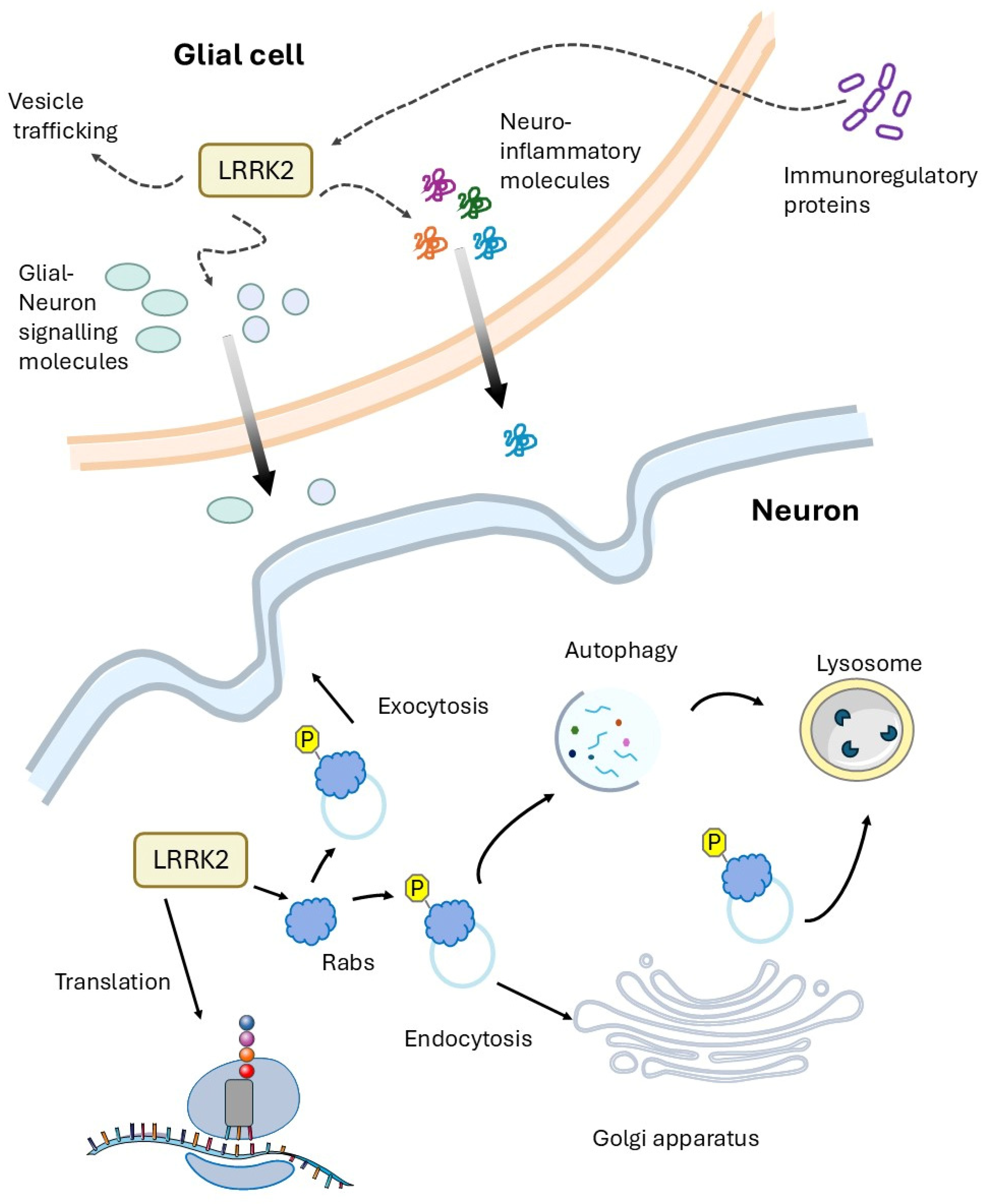
4. Is the PD LRRK2-Related Disease a Cell Autonomous Disease?

As discussed in the introduction, LRRK2 is expressed at low levels in dopaminergic neurons compared to other neuronal districts and is expressed at high levels in cells of the immune system, including microglia and astrocytes. Furthermore, the involvement of LRRK2 in some autoimmune diseases strongly suggests a prominent role of LRRK2 in inflammation and probably neuroinflammation. Growing evidence supports the idea that pathological interplay between peripheral and CNS innate immunity significantly contributes to the LRRK2 dependent neurotoxicity [84]. In the CNS, LRRK2 directly modules microglia/astrocyte physiological function while at peripheral level could, for instance, affect the gut-brain axis.
Compared to high eukaryotes, Drosophila show a simpler innate immune response that involves both humoral and cell-mediated response upon microbial infections. The humoral response relies in the massive production (up to 300 μM) of antimicrobial peptides (AMPs) mainly from fat bodies, a functional equivalent to the mammalian liver [85]. The cellular immune response of Drosophila involves the three subpopulation of circulating hemocytes: plasmatocytes, lamellocytes, and crystal cells. Plasmatocytes, the most abundant circulating hemocytes, are equivalent to mammalian macrophages, acting as professional phagocytes and releasing a large range of cytokines and AMPs. Interestingly, accumulating experimental evidence suggests a potential alternative role of AMPs as signaling molecules regulating different biological function in various tissues independently of their classical antimicrobial function [86]. For instance, as in humans, in Drosophila the AMPs tend to gradually and significantly increase during the aging [46,48,50,51] and, importantly, overexpression of certain AMPs (including Attacin-A) may lead to cognitive decline, neurotoxicity, and a decrease in lifespan [87,88].
We have previously demonstrated that ubiquitous expression of LRRK2 R1441C (by actin-GAL4 driver) leads to a toxic phenotype in Drosophila [48] that, at least in our case, was not observed by pan-neuronal expression (either by elav- or nSyb-GAL4 drivers). LRRK2 R1441C expressing flies show a strong inflammatory status in older animals monitored by inflammatory peptide (AMPs) and, in particular, two AMPs (Attacin-A and Drosocin) are already over-expressed at 7 days after eclosion, when no motor defects or neurodegeneration signs are detected in transgenic flies [35].
In an attempt to evaluate the inflammatory contribution of AttA inflammatory peptide in LRRK2 neuronal toxicity, we generated flies having both UAS-LRRK2 R1441C and an UAS-AttA RNAi. The flies carrying the double transgenes and flies carrying the single transgene as controls were crossed with the actin-GAL4 drivers. One week after eclosion, the LRRK2 expression and RAB10 phosphorylation were analyzed in the different lines by western blot. As illustrated in Figure 2A, the presence of two transgenes does not alter either LRRK2 expression or the increase in RAB10 phosphorylation due to LRRK2 R1441C expression. As expected, the presence of AttA RNAi decrease the AttA expression in the transgenic flies (Figure 2B) analyzed by Real-Time PCR. At 45 days after eclosion, the different flies were analyzed for locomotor activity by climbing assay. The concomitant expression of AttA RNAi is able to significantly rescue the climbing defects due to LRRK2 R1441C expression (Figure 2C). Finally, in 45-day-old flies, dopaminergic neurons in the Protocerebral Posterior Lateral 1 (PPL1) cluster were analyzed by immunofluorescence, using an antibody against Tyrosine Hydroxylase (TH) (Figure 2D,E). Of course, although the single UAS-AttA RNAi line has been characterized so far, we may not exclude the possibility that the RNA interference may affect other unrelated cellular pathways.

5. Discussion

In studies on Parkinson’s disease, Drosophila LRRK2 models largely confirm, validate, or sometimes anticipate, the findings arising from higher eukaryote experimental models. Expression of human or Drosophila LRRK2 variants in Drosophila has allowed the direct observation of the effects of mutations on behavior, neurodegeneration, and alterations in various cellular processes. Interestingly, flies expressing mutant forms of LRRK2 often show reduced lifespan, altered movement, and neuronal degeneration similar to that observed in patients with Parkinson’s disease. A particular advantage of using Drosophila as an experimental model is the availability of a large number of genetic tools both in terms of GAL4 drivers (to express the gene of interest in specific cell types) and in terms of fly lines that over- or hypo-express specific genes (to analyze the crosstalk between different signaling pathways).
Recent studies, in human and experimental models, have shown that LRRK2 is expressed at higher levels in immunocompetent cells compared to other cell types, including neurons, making it a possible key regulator of immune functions. Indeed, LRRK2 is strongly expressed in cells of the innate immune system such as neutrophils, macrophages, monocytes, and in the brain in astrocytes and microglia cells [84], both of which are considered part of the innate immune system based on the production of specific immune modulators and the expression of specific cell membrane receptors [89]. Interestingly, in the brain cells, there is a consistent convergence that LRRK2 exerts a pro-inflammatory effect, whereas in systemic immune cells the LRRK2 role on inflammatory cytokines/chemokines generation is still under debate [90].
Several reports underline a significant increase in neuroinflammation in PD patients. For example, post-mortem analysis of affected brain regions of PD patients shows activated complement components and reactive microglia [28]. Accordingly, different studies have reported elevated levels of inflammatory cytokines such as TNF, IL-1b, and IL-6 in both serum and CSF [91]. In addition, higher levels of inflammatory biomarkers appear to correlate with worse motor function and cognitive impairment in PD patients [92]. Interestingly, evidence from human and animal experimental models suggests that inflammation, due to an initial stimulus, may persist for many years and that chronic inflammation may be detrimental to neurons. All these findings point to chronic inflammation as a possible cause or contributing factor in the development of PD. In animal models, a number of anti-inflammatory compounds offer neuroprotection, while in PD patients, although epidemiological and preclinical studies suggest a possible beneficial effect, clinical trials have failed to provide robust evidence of their efficacy in the symptomatic phase of the disease [93]. It may be worthwhile to evaluate the anti-inflammatory effect in the pre-symptomatic phase to inhibit the initial pathological mechanisms before the likely irreversible neuronal decline in the symptomatic phase.
Several lines of evidence from human samples and animal experimental models suggest that pathogenic PD signs (e.g., alpha-synuclein misfolding) begin in the gastrointestinal tract and subsequently spread to vulnerable brain regions [94]. Experimental models suggest that the interplay between the gut microbiome or exogenous toxicants and intestinal immune system, may act as triggers or modifiers of neuronal pathology, further supporting the contribution of immunity, and in particular, the innate immune system to PD onset or progression [94]. LRRK2 is highly expressed in the innate immune cells and significantly upregulated by inflammatory modulators such as IFN-γ [95,96], a central cytokine in the control of the immune response. In turn, LRRK2 enhances the NF-κB-dependent transcription of inflammatory molecules in response to various stimuli [95,96], strongly suggesting a possible potential role in the communication between gut, immune, and nervous system [84].
In Drosophila, we have detected a strong increase in inflammatory markers when LRRK2 pathological mutants are expressed either in all tissues (via actin-GAL4 driver) or in glial cells (via repo-GAL4 driver). Interestingly, here we show that the reduction in the expression of AttA inflammatory peptides by RNAi significantly rescues the LRRK2 R1441C phenotype in terms of motor performance and neurodegeneration. Figure 3 shows the potential role of LRRK2 in neuronal and glial cells, as suggested by experimental evidence from the Drosophila and higher eukaryotic models previously described. Taken together, these experimental results suggest that a non-cell-autonomous mechanism may contribute to PD LRRK2-related disease.
In conclusion, LRRK2 is an important gene in the context of neurodegenerative diseases, and non-rodents provide a highly useful model for exploring the pathogenetic mechanisms associated with this gene. The combination of non-rodent genetics and molecular biology may significantly contribute to the understanding of the role of LRRK2 in Parkinson’s disease and pave the way for new therapeutic possibilities, including compounds capable of reducing neuroinflammation. Up to the present, a general approach of anti-inflammatory therapy, although producing promising findings in pre-clinical experimental models, failed to translate into a clinical therapy. Likely, the identification of some certain inflammatory actors that play a key role in gut-immune-nerve communication could become specific therapeutic targets with a greater chance of success, especially if used in the pre-symptomatic phase of the disease.

6. Materials and Methods

6.1. Drosophila Lines

All fly stocks were maintained in our laboratory on standard cornmeal medium at 25 °C on a 12 h:12 h light:dark cycle. The Actin-GAL4 driver (BDSC #4414) and UAS-AttA RNAi (BDSC #56904) were from Bloomington Stock Center (Bloomington, IN, USA). The UAS-LRRK2-R1441C was a generous gift from prof. Cheng-Ting Chien (National Taiwan University Hospital Yun-Lin Branch, Taipei, Taiwan).

6.2. Evaluation of mRNA Expression by RT-PCR

Total RNA was extracted from Drosophila heads (5 male and 5 female) for each genotype in 500 µL of TRI-ZOL solution according to the manufacturer’s instruction (Life Technology, Thermo Fischer Scientific, Waltham, MA, USA). The RNA (0.5 µg) was converted to cDNA by AMV reverse transcriptase (Promega, Madison, WI, USA) at 37° for 1 h. Diluted (1:10) cDNAs were amplified by PCR (94° 30 s, 55° 30 s, 72° 30 s) in the presence of SYBR green (Biorad, Hercules, CA, USA) using the primers indicated in [97] (for RpL32 GACGCTTCAAGGGACAGTATCTG and AAACGCGGTTCTGCATGAG; for AttA CCCGGAGTGAAGGATG and GTTGCTGTGCGTCAAG) by Rotor-Gene Q Thermocycler. The delta-delta Ct method (2−∆∆Ct) normalized by machine internal software was used to evaluate the RNA content between the different genotypes.

6.3. Western Blot Analysis

Western blot analysis was performed as previously described [51]. Briefly, total body protein extracts were prepared by direct lysis in Laemmli buffer, resolved by standard sodium dodecyl sulphate-polyacrylamide gel electrophoresis, and subsequently transferred into nitrocellulose membrane (Life Technology). The membranes were incubated with 3% low-fat milk in 1X PBS-Tween 0.05% solution with the following antibody: anti-LRRK2 (1:5000 MJFF2 c41-2 Epitomics, Burlingame, CA, USA), anti-phospo-RAB10 (T73) (1:1000 MJF-R21 Abcam, Cambridge, UK), anti-alpha-tubulin (1:1000 12G10 DSHB, Iowa City, IA, USA), for 16 h at 4 °C. Goat anti-mouse immunoglobulin G (IgG) peroxidase-conjugated antibody (1:2500 Millipore Corporation, Burlington, MA, USA) or goat anti-rabbit IgG peroxidase-conjugated antibody (1:5000 Millipore Corporation) were used to identify immunocomplexes by enhanced chemiluminescence (Millipore Corporation).

6.4. Whole-Mount Immunostaining of the Adult Drosophila Brains

Fluorescent immunostaining was performed as previously described [51]. Briefly, whole adult brains were dissected. Cohorts of 6 to 10 flies per genotype were used each time. Brains were fixed for 20 min using 4% paraformaldehyde in phosphate-buffered saline (PBS), pH 7.4. After fixation, brains were permeabilized with 0.3% Triton X-100 in PBS and incubated in blocking buffer (5% normal goat serum in PBS 1X-0.3% Triton X-100) for 1 h at RT. Subsequently, primary anti-body anti-TH (1:500 AB152, Millipore) diluted in blocking buffer was incubated for 48 h at 4 °C. After extensive washing, the brains were incubated by secondary antibody Alexa Fluor® 546 (Life Technology) diluted 1:1000 in blocking solution for 48 h at 4 °C. Finally, the brains were mounted using Mowiol mounting medium and fluorescence was revealed with a Leica TCS SP5 confocal microscope with LAS lite 170 image software (LAS AF 2019). DA neurons located in the PPL1 were counted in both hemispheres after Z-stack image acquisition to visualize all TH-positive neurons. A minimum of 10 brains were analyzed for each genotype in every experiment. Three independent experiments have been performed. Statistical analysis was performed with One-way ANOVA followed by a Bonferroni post hoc test.

6.5. Climbing Assay

Flies were age and sex matched, randomly selected, anesthetized with ice, and placed in conical tubes with a diameter of 2 cm. After 15 min of recovery at 25 °C, the flies were tapped to the bottom of the tube, and their subsequent climbing activity was quantified as the percentage of flies that reached 5 cm in 10 s. Any experimental sample was performed in duplicate (each with 15 flies) and the assay was repeated three or four times.

6.6. Statistical Analysis

The results are presented as means ± SEM of independent experiments as indicated. Statistical evaluation was conducted by One-way ANOVA and Bonferroni’s multiple comparison post-test. Values significantly different from the relative control are indicated with one, two, or three asterisks when p < 0.05, p < 0.005, and p < 0.001, respectively.

Author Contributions

Conceived and designed the experiments: C.I. and C.C. (Claudia Crosio). Performed the experiments: C.C. (Cristina Ciampelli), G.G., M.P., P.M. and M.G. Analysed the data: C.C. (Cristina Ciampelli), C.C. (Claudia Crosio), M.P. and P.M. Contributed reagents/materials/analysis tools: M.G., R.B. and D.A. Wrote the paper: C.I. Edit the paper: C.C. (Claudia Crosio), C.C. (Cristina Ciampelli) and R.B. All authors have read and agreed to the published version of the manuscript.

Funding

This research was supported with grants to C.I: Bando competitivo Fondazione di Sardegna 2017-Iaccarino, Fondazione di Sardegna Annualità 2022–2023 Progetti di ricerca di base dipartimentali, Fondo di Ateneo per la ricerca 2020 (FAR2020), Bando interno per la ricerca collaborativa tra Ateneo di Sassari e Ateneo di Cagliari-Formato, to C. Crosio: Fondazione di Sardegna Annualità 2022–2023 Progetti di ricerca di base dipartimentali, Fondo di Ateneo per la ricerca 2020 (FAR2020), Bando interno per la ricerca collaborativa tra Ateneo di Sassari e Ateneo di Cagliari DM 737/2021, Risorse 2022–2023-Formato and to G. G: Progetti di ricerca interdisciplinare DM 737/2021, Risorse 2021–2022, NextGenerationEU. C. Ciampelli is supported by a PhD fellowship (Borsa di Ateneo, Università di Sassari, 2021).

Institutional Review Board Statement

“Not applicable” for studies not involving humans or animals.

Informed Consent Statement

Not applicable.

Data Availability Statement

The data underlying the findings presented in this study are available upon reasonable request.

Acknowledgments

We would like to acknowledge all the people from the laboratory that critically read the manuscript. Cheng-Ting Chien (Department of Neurology, National Taiwan University Hospital Yun-Lin Branch, Taipei, Taiwan) for LRRK2 Drosophila lines.

Conflicts of Interest

The authors declare no conflicts of interest.

References

  1. Dauer, W.; Przedborski, S. Parkinson’s Disease: Mechanisms and Models. Neuron 2003, 39, 889–909. [Google Scholar] [CrossRef] [PubMed]
  2. Healy, D.G.; Falchi, M.; O’Sullivan, S.S.; Bonifati, V.; Durr, A.; Bressman, S.; Brice, A.; Aasly, J.; Zabetian, C.P.; Goldwurm, S.; et al. Phenotype, Genotype, and Worldwide Genetic Penetrance of LRRK2-Associated Parkinson’s Disease: A Case-Control Study. Lancet Neurol. 2008, 7, 583–590. [Google Scholar] [CrossRef] [PubMed]
  3. El Otmani, H.; Daghi, M.; Tahiri Jouti, N.; Lesage, S. An Overview of the Worldwide Distribution of LRRK2 Mutations in Parkinson’s Disease. Neurodegener. Dis. Manag. 2023, 13, 335–350. [Google Scholar] [CrossRef] [PubMed]
  4. Henderson, M.X.; Sengupta, M.; Trojanowski, J.Q.; Lee, V.M.Y. Alzheimer’s Disease Tau Is a Prominent Pathology in LRRK2 Parkinson’s Disease. Acta Neuropathol. Commun. 2019, 7, 183. [Google Scholar] [CrossRef]
  5. Marin, I.; van Egmond, W.N.; van Haastert, P.J. The Roco Protein Family: A Functional Perspective. FASEB J. 2008, 22, 3103–3110. [Google Scholar] [CrossRef]
  6. Bonet-Ponce, L.; Cookson, M.R. LRRK2 Recruitment, Activity, and Function in Organelles. FEBS J. 2022, 289, 6871–6890. [Google Scholar] [CrossRef]
  7. Taymans, J.M. Regulation of LRRK2 by Phosphatases. Adv. Neurobiol. 2017, 14, 145–160. [Google Scholar] [CrossRef]
  8. Cogo, S.; Ho, F.Y.; Tosoni, E.; Tomkins, J.E.; Tessari, I.; Iannotta, L.; Montine, T.J.; Manzoni, C.; Lewis, P.A.; Bubacco, L.; et al. The Roc Domain of LRRK2 as a Hub for Protein-Protein Interactions: A Focus on PAK6 and Its Impact on RAB Phosphorylation. Brain Res. 2022, 1778, 147781. [Google Scholar] [CrossRef]
  9. Paisan-Ruiz, C.; Jain, S.; Evans, E.W.; Gilks, W.P.; Simon, J.; van der Brug, M.; Lopez de Munain, A.; Aparicio, S.; Gil, A.M.; Khan, N.; et al. Cloning of the Gene Containing Mutations That Cause PARK8-Linked Parkinson’s Disease. Neuron 2004, 44, 595–600. [Google Scholar] [CrossRef]
  10. Zimprich, A.; Biskup, S.; Leitner, P.; Lichtner, P.; Farrer, M.; Lincoln, S.; Kachergus, J.; Hulihan, M.; Uitti, R.J.; Calne, D.B.; et al. Mutations in LRRK2 Cause Autosomal-Dominant Parkinsonism with Pleomorphic Pathology. Neuron 2004, 44, 601–607. [Google Scholar] [CrossRef]
  11. Panicker, N.; Ge, P.; Dawson, V.L.; Dawson, T.M. The Cell Biology of Parkinson’s Disease. J. Cell Biol. 2021, 220, e202012095. [Google Scholar] [CrossRef] [PubMed]
  12. Harvey, K.; Outeiro, T.F. The Role of LRRK2 in Cell Signalling. Biochem. Soc. Trans. 2019, 47, 197–207. [Google Scholar] [CrossRef] [PubMed]
  13. Pfeffer, S.R. LRRK2 Phosphorylation of Rab GTPases in Parkinson’s Disease. FEBS Lett. 2022, 597, 811–818. [Google Scholar] [CrossRef] [PubMed]
  14. Steger, M.; Tonelli, F.; Ito, G.; Davies, P.; Trost, M.; Vetter, M.; Wachter, S.; Lorentzen, E.; Duddy, G.; Wilson, S.; et al. Phosphoproteomics Reveals That Parkinson’s Disease Kinase LRRK2 Regulates a Subset of Rab GTPases. eLife 2016, 5, e12813. [Google Scholar] [CrossRef]
  15. Zhang, F.R.; Huang, W.; Chen, S.M.; Sun, L.D.; Liu, H.; Li, Y.; Cui, Y.; Yan, X.X.; Yang, H.T.; Yang, R.D.; et al. Genomewide Association Study of Leprosy. N. Engl. J. Med. 2009, 361, 2609–2618. [Google Scholar] [CrossRef]
  16. Härtlova, A.; Herbst, S.; Peltier, J.; Rodgers, A.; Bilkei-Gorzo, O.; Fearns, A.; Dill, B.D.; Lee, H.; Flynn, R.; Cowley, S.A.; et al. LRRK2 Is a Negative Regulator of Mycobacterium Tuberculosis Phagosome Maturation in Macrophages. EMBO J. 2018, 37, e98694. [Google Scholar] [CrossRef]
  17. Zhang, M.; Yao, C.; Cai, J.; Liu, S.; Liu, X.-N.; Chen, Y.; Wang, S.; Ji, P.; Pan, M.; Kang, Z.; et al. LRRK2 Is Involved in the Pathogenesis of System Lupus Erythematosus through Promoting Pathogenic Antibody Production. J. Transl. Med. 2019, 17, 37. [Google Scholar] [CrossRef]
  18. Hui, K.Y.; Fernandez-Hernandez, H.; Hu, J.; Schaffner, A.; Pankratz, N.; Hsu, N.-Y.; Chuang, L.-S.; Carmi, S.; Villaverde, N.; Li, X.; et al. Functional Variants in the LRRK2 Gene Confer Shared Effects on Risk for Crohn’s Disease and Parkinson’s Disease. Sci. Transl. Med. 2018, 10, eaai7795. [Google Scholar] [CrossRef]
  19. Gillardon, F.; Schmid, R.; Draheim, H. Parkinson’s Disease-Linked Leucine-Rich Repeat Kinase 2(R1441G) Mutation Increases Proinflammatory Cytokine Release from Activated Primary Microglial Cells and Resultant Neurotoxicity. Neuroscience 2012, 208, 21–48. [Google Scholar] [CrossRef]
  20. Russo, I.; Di Benedetto, G.; Kaganovich, A.; Ding, J.; Mercatelli, D.; Morari, M.; Cookson, M.R.; Bubacco, L.; Greggio, E. Leucine-Rich Repeat Kinase 2 Controls Protein Kinase A Activation State through Phosphodiesterase 4. J. Neuroinflamm. 2018, 15, 297. [Google Scholar] [CrossRef]
  21. Sonninen, T.-M.; Hämäläinen, R.H.; Koskuvi, M.; Oksanen, M.; Shakirzyanova, A.; Wojciechowski, S.; Puttonen, K.; Naumenko, N.; Goldsteins, G.; Laham-Karam, N.; et al. Metabolic Alterations in Parkinson’s Disease Astrocytes. Sci. Rep. 2020, 10, 14474. [Google Scholar] [CrossRef] [PubMed]
  22. Caesar, M.; Felk, S.; Zach, S.; Brønstad, G.; Aasly, J.O.; Gasser, T.; Gillardon, F. Changes in Matrix Metalloprotease Activity and Progranulin Levels May Contribute to the Pathophysiological Function of Mutant Leucine-Rich Repeat Kinase 2. Glia 2014, 62, 1075–1092. [Google Scholar] [CrossRef] [PubMed]
  23. Kim, J.; Pajarillo, E.; Rizor, A.; Son, D.-S.; Lee, J.; Aschner, M.; Lee, E. LRRK2 Kinase Plays a Critical Role in Manganese-Induced Inflammation and Apoptosis in Microglia. PLoS ONE 2019, 14, e0210248. [Google Scholar] [CrossRef]
  24. Russo, I.; Kaganovich, A.; Ding, J.; Landeck, N.; Mamais, A.; Varanita, T.; Biosa, A.; Tessari, I.; Bubacco, L.; Greggio, E.; et al. Transcriptome Analysis of LRRK2 Knock-out Microglia Cells Reveals Alterations of Inflammatory- and Oxidative Stress-Related Pathways upon Treatment with α-Synuclein Fibrils. Neurobiol. Dis. 2019, 129, 67–78. [Google Scholar] [CrossRef]
  25. Moehle, M.S.; Webber, P.J.; Tse, T.; Sukar, N.; Standaert, D.G.; DeSilva, T.M.; Cowell, R.M.; West, A.B. LRRK2 Inhibition Attenuates Microglial Inflammatory Responses. J. Neurosci. 2012, 32, 1602–1611. [Google Scholar] [CrossRef]
  26. Munoz, L.; Kavanagh, M.E.; Phoa, A.F.; Heng, B.; Dzamko, N.; Chen, E.-J.; Doddareddy, M.R.; Guillemin, G.J.; Kassiou, M. Optimisation of LRRK2 Inhibitors and Assessment of Functional Efficacy in Cell-Based Models of Neuroinflammation. Eur. J. Med. Chem. 2015, 95, 29–34. [Google Scholar] [CrossRef]
  27. Russo, I.; Berti, G.; Plotegher, N.; Bernardo, G.; Filograna, R.; Bubacco, L.; Greggio, E. Leucine-Rich Repeat Kinase 2 Positively Regulates Inflammation and down-Regulates NF-κB P50 Signaling in Cultured Microglia Cells. J. Neuroinflamm. 2015, 12, 230. [Google Scholar] [CrossRef]
  28. McGeer, P.L.; McGeer, E.G. Inflammation and Neurodegeneration in Parkinson’s Disease. Parkinsonism Relat. Disord. 2004, 10 (Suppl. S1), S3–S7. [Google Scholar] [CrossRef]
  29. Leńska-Mieciek, M.; Madetko-Alster, N.; Alster, P.; Królicki, L.; Fiszer, U.; Koziorowski, D. Inflammation in Multiple System Atrophy. Front. Immunol. 2023, 14, 1214677. [Google Scholar] [CrossRef]
  30. Thakur, J.; Godad, A. Deciphering the Role of Neuropeptides as Biomarkers for Early Diagnosis of Parkinson’s Disease. Life Sci. 2025, 363, 123376. [Google Scholar] [CrossRef]
  31. Alster, P.; Madetko-Alster, N.; Otto-Ślusarczyk, D.; Migda, A.; Migda, B.; Struga, M.; Friedman, A. Role of Orexin in Pathogenesis of Neurodegenerative Parkinsonisms. Neurol. Neurochir. Pol. 2023, 57, 335–343. [Google Scholar] [CrossRef] [PubMed]
  32. Domenicale, C.; Magnabosco, S.; Morari, M. Modeling Parkinson’s Disease in LRRK2 Rodents. Neuronal Signal. 2023, 7, NS20220040. [Google Scholar] [CrossRef] [PubMed]
  33. Seegobin, S.P.; Heaton, G.R.; Liang, D.; Choi, I.; Blanca Ramirez, M.; Tang, B.; Yue, Z. Progress in LRRK2-Associated Parkinson’s Disease Animal Models. Front. Neurosci. 2020, 14, 674. [Google Scholar] [CrossRef] [PubMed]
  34. Jeibmann, A.; Paulus, W. Drosophila Melanogaster as a Model Organism of Brain Diseases. Int. J. Mol. Sci. 2009, 10, 407–440. [Google Scholar] [CrossRef]
  35. Venken, K.J.T.; Simpson, J.H.; Bellen, H.J. Genetic Manipulation of Genes and Cells in the Nervous System of the Fruit Fly. Neuron 2011, 72, 202–230. [Google Scholar] [CrossRef]
  36. Dorkenwald, S.; McKellar, C.E.; Macrina, T.; Kemnitz, N.; Lee, K.; Lu, R.; Wu, J.; Popovych, S.; Mitchell, E.; Nehoran, B.; et al. FlyWire: Online Community for Whole-Brain Connectomics. Nat. Methods 2022, 19, 119–128. [Google Scholar] [CrossRef]
  37. Dorkenwald, S.; Matsliah, A.; Sterling, A.R.; Schlegel, P.; Yu, S.-C.; McKellar, C.E.; Lin, A.; Costa, M.; Eichler, K.; Yin, Y.; et al. Neuronal Wiring Diagram of an Adult Brain. Nature 2024, 634, 124–138. [Google Scholar] [CrossRef]
  38. Lin, A.; Yang, R.; Dorkenwald, S.; Matsliah, A.; Sterling, A.R.; Schlegel, P.; Yu, S.-C.; McKellar, C.E.; Costa, M.; Eichler, K.; et al. Network Statistics of the Whole-Brain Connectome of Drosophila. Nature 2024, 634, 153–165. [Google Scholar] [CrossRef]
  39. Wang, D.; Tang, B.; Zhao, G.; Pan, Q.; Xia, K.; Bodmer, R.; Zhang, Z. Dispensable Role of Drosophila Ortholog of LRRK2 Kinase Activity in Survival of Dopaminergic Neurons. Mol. Neurodegener. 2008, 3, 3. [Google Scholar] [CrossRef]
  40. Lee, S.B.; Kim, W.; Lee, S.; Chung, J. Loss of LRRK2/PARK8 Induces Degeneration of Dopaminergic Neurons in Drosophila. Biochem. Biophys. Res. Commun. 2007, 358, 534–539. [Google Scholar] [CrossRef]
  41. Imai, Y.; Gehrke, S.; Wang, H.Q.; Takahashi, R.; Hasegawa, K.; Oota, E.; Lu, B. Phosphorylation of 4E-BP by LRRK2 Affects the Maintenance of Dopaminergic Neurons in Drosophila. EMBO J. 2008, 27, 2432–2443. [Google Scholar] [CrossRef]
  42. Tain, L.S.; Mortiboys, H.; Tao, R.N.; Ziviani, E.; Bandmann, O.; Whitworth, A.J. Rapamycin Activation of 4E-BP Prevents Parkinsonian Dopaminergic Neuron Loss. Nat. Neurosci. 2009, 12, 1129–1135. [Google Scholar] [CrossRef]
  43. Liu, Z.; Wang, X.; Yu, Y.; Li, X.; Wang, T.; Jiang, H.; Ren, Q.; Jiao, Y.; Sawa, A.; Moran, T.; et al. A Drosophila Model for LRRK2-Linked Parkinsonism. Proc. Natl. Acad. Sci. USA 2008, 105, 2693–2698. [Google Scholar] [CrossRef] [PubMed]
  44. Lin, C.-H.; Lin, H.-I.; Chen, M.-L.; Lai, T.-T.; Cao, L.-P.; Farrer, M.J.; Wu, R.-M.; Chien, C.-T. Lovastatin Protects Neurite Degeneration in LRRK2-G2019S Parkinsonism through Activating the Akt/Nrf Pathway and Inhibiting GSK3β Activity. Hum. Mol. Genet. 2016, 25, 1965–1978. [Google Scholar] [CrossRef]
  45. Ng, C.-H.; Mok, S.Z.S.; Koh, C.; Ouyang, X.; Fivaz, M.L.; Tan, E.-K.; Dawson, V.L.; Dawson, T.M.; Yu, F.; Lim, K.-L. Parkin Protects against LRRK2 G2019S Mutant-Induced Dopaminergic Neurodegeneration in Drosophila. J. Neurosci. 2009, 29, 11257–11262. [Google Scholar] [CrossRef] [PubMed]
  46. Xiong, Y.; Yuan, C.; Chen, R.; Dawson, T.M.; Dawson, V.L. ArfGAP1 Is a GTPase Activating Protein for LRRK2: Reciprocal Regulation of ArfGAP1 by LRRK2. J. Neurosci. 2012, 32, 3877–3886. [Google Scholar] [CrossRef] [PubMed]
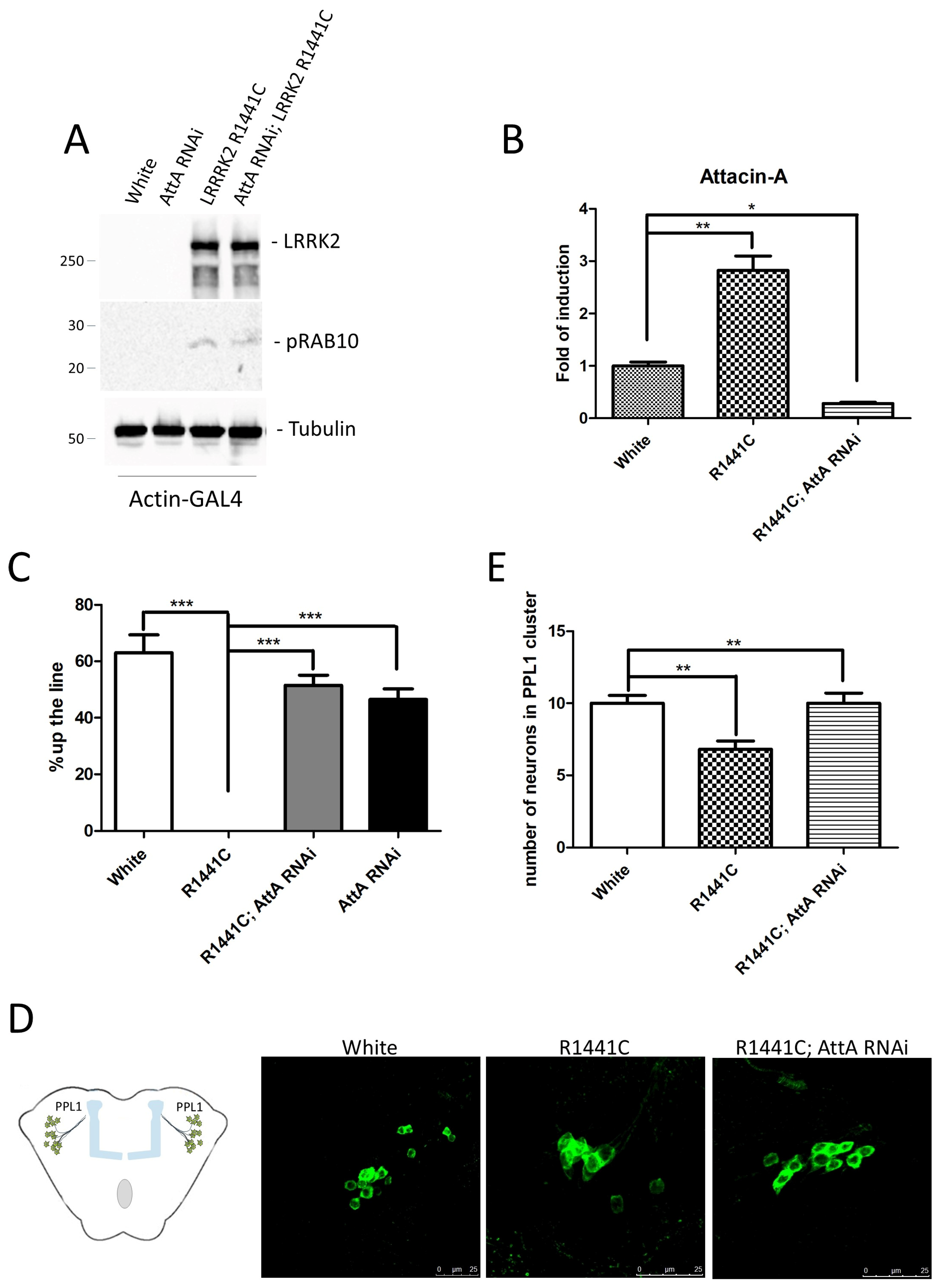
  47. Maksoud, E.; Liao, E.H.; Haghighi, A.P. A Neuron-Glial Trans-Signaling Cascade Mediates LRRK2-Induced Neurodegeneration. Cell Rep. 2019, 26, 1774–1786.e4. [Google Scholar] [CrossRef]
  48. Nguyen, H.N.; Galleri, G.; Rassu, A.; Ciampelli, C.; Bernardoni, R.; Galioto, M.; Albani, D.; Crosio, C.; Iaccarino, C. Evaluation of Neuroinflammatory Contribution to Neurodegeneration in LRRK2 Drosophila Models. Biomedicines 2024, 12, 1555. [Google Scholar] [CrossRef]
  49. Wang, L.; Wang, H.; Yi, S.; Zhang, S.; Ho, M.S. A LRRK2/dLRRK-Mediated Lysosomal Pathway That Contributes to Glial Cell Death and DA Neuron Survival. Traffic 2022, 23, 506–520. [Google Scholar] [CrossRef]
  50. Fellgett, A.; Middleton, C.A.; Munns, J.; Ugbode, C.; Jaciuch, D.; Wilson, L.G.; Chawla, S.; Elliott, C.J.H. Multiple Pathways of LRRK2-G2019S/Rab10 Interaction in Dopaminergic Neurons. J. Park. Dis. 2021, 11, 1805–1820. [Google Scholar] [CrossRef]
  51. Ciampelli, C.; Galleri, G.; Puggioni, S.; Fais, M.; Iannotta, L.; Galioto, M.; Becciu, M.; Greggio, E.; Bernardoni, R.; Crosio, C.; et al. Inhibition of the Exocyst Complex Attenuates the LRRK2 Pathological Effects. Int. J. Mol. Sci. 2023, 24, 12656. [Google Scholar] [CrossRef]
  52. Islam, M.S.; Nolte, H.; Jacob, W.; Ziegler, A.B.; Pütz, S.; Grosjean, Y.; Szczepanowska, K.; Trifunovic, A.; Braun, T.; Heumann, H.; et al. Human R1441C LRRK2 Regulates the Synaptic Vesicle Proteome and Phosphoproteome in a Drosophila Model of Parkinson’s Disease. Hum. Mol. Genet. 2016, 25, 5365–5382. [Google Scholar] [CrossRef] [PubMed]
  53. Pan, P.Y.; Li, X.; Wang, J.; Powell, J.; Wang, Q.; Zhang, Y.; Chen, Z.; Wicinski, B.; Hof, P.; Ryan, T.A.; et al. Parkinson’s Disease-Associated LRRK2 Hyperactive Kinase Mutant Disrupts Synaptic Vesicle Trafficking in Ventral Midbrain Neurons. J. Neurosci. 2017, 37, 11366–11376. [Google Scholar] [CrossRef] [PubMed]
  54. Matta, S.; Van Kolen, K.; da Cunha, R.; van den Bogaart, G.; Mandemakers, W.; Miskiewicz, K.; De Bock, P.J.; Morais, V.A.; Vilain, S.; Haddad, D.; et al. LRRK2 Controls an EndoA Phosphorylation Cycle in Synaptic Endocytosis. Neuron 2012, 75, 1008–1021. [Google Scholar] [CrossRef]
  55. Penney, J.; Tsurudome, K.; Liao, E.H.; Kauwe, G.; Gray, L.; Yanagiya, A.; Calderon, M.R.; Sonenberg, N.; Haghighi, A.P. LRRK2 Regulates Retrograde Synaptic Compensation at the Drosophila Neuromuscular Junction. Nat. Commun. 2016, 7, 12188. [Google Scholar] [CrossRef] [PubMed]
  56. Liu, Q.; Bautista-Gomez, J.; Higgins, D.A.; Yu, J.; Xiong, Y. Dysregulation of the AP2M1 Phosphorylation Cycle by LRRK2 Impairs Endocytosis and Leads to Dopaminergic Neurodegeneration. Sci. Signal. 2021, 14, eabg3555. [Google Scholar] [CrossRef]
  57. Heaton, G.R.; Landeck, N.; Mamais, A.; Nalls, M.A.; Nixon-Abell, J.; Kumaran, R.; Beilina, A.; Pellegrini, L.; Li, Y.; International Parkinson Disease Genomics Consortium (IPDGC); et al. Sequential Screening Nominates the Parkinson’s Disease Associated Kinase LRRK2 as a Regulator of Clathrin-Mediated Endocytosis. Neurobiol. Dis. 2020, 141, 104948. [Google Scholar] [CrossRef]
  58. Mir, R.; Tonelli, F.; Lis, P.; Macartney, T.; Polinski, N.K.; Martinez, T.N.; Chou, M.-Y.; Howden, A.J.M.; König, T.; Hotzy, C.; et al. The Parkinson’s Disease VPS35[D620N] Mutation Enhances LRRK2-Mediated Rab Protein Phosphorylation in Mouse and Human. Biochem. J. 2018, 475, 1861–1883. [Google Scholar] [CrossRef]
  59. Kadgien, C.A.; Kamesh, A.; Milnerwood, A.J. Endosomal Traffic and Glutamate Synapse Activity Are Increased in VPS35 D620N Mutant Knock-in Mouse Neurons, and Resistant to LRRK2 Kinase Inhibition. Mol. Brain 2021, 14, 143. [Google Scholar] [CrossRef]
  60. McCarron, K.R.; Elcocks, H.; Mortiboys, H.; Urbé, S.; Clague, M.J. The Parkinson’s Disease Related Mutant VPS35 (D620N) Amplifies the LRRK2 Response to Endolysosomal Stress. Biochem. J. 2024, 481, 265–278. [Google Scholar] [CrossRef]
  61. Linhart, R.; Wong, S.A.; Cao, J.; Tran, M.; Huynh, A.; Ardrey, C.; Park, J.M.; Hsu, C.; Taha, S.; Peterson, R.; et al. Vacuolar Protein Sorting 35 (Vps35) Rescues Locomotor Deficits and Shortened Lifespan in Drosophila Expressing a Parkinson’s Disease Mutant of Leucine-Rich Repeat Kinase 2 (LRRK2). Mol. Neurodegener. 2014, 9, 23. [Google Scholar] [CrossRef] [PubMed]
  62. Inoshita, T.; Arano, T.; Hosaka, Y.; Meng, H.; Umezaki, Y.; Kosugi, S.; Morimoto, T.; Koike, M.; Chang, H.-Y.; Imai, Y.; et al. Vps35 in Cooperation with LRRK2 Regulates Synaptic Vesicle Endocytosis through the Endosomal Pathway in Drosophila. Hum. Mol. Genet. 2017, 26, 2933–2948. [Google Scholar] [CrossRef] [PubMed]
  63. Khan, S.; Upadhyay, S.; Hassan, M.I. Novel Prospects in Targeting Neurodegenerative Disorders via Autophagy. Eur. J. Pharmacol. 2024, 984, 177060. [Google Scholar] [CrossRef] [PubMed]
  64. Dodson, M.W.; Zhang, T.; Jiang, C.; Chen, S.; Guo, M. Roles of the Drosophila LRRK2 Homolog in Rab7-Dependent Lysosomal Positioning. Hum. Mol. Genet. 2012, 21, 1350–1363. [Google Scholar] [CrossRef] [PubMed]
  65. Hernandez-Diaz, S.; Ghimire, S.; Sanchez-Mirasierra, I.; Montecinos-Oliva, C.; Swerts, J.; Kuenen, S.; Verstreken, P.; Soukup, S.-F. Endophilin-B Regulates Autophagy during Synapse Development and Neurodegeneration. Neurobiol. Dis. 2022, 163, 105595. [Google Scholar] [CrossRef]
  66. Chittoor-Vinod, V.G.; Villalobos-Cantor, S.; Roshak, H.; Shea, K.; Abalde-Atristain, L.; Martin, I. Dietary Amino Acids Impact LRRK2-Induced Neurodegeneration in Parkinson’s Disease Models. J. Neurosci. 2020, 40, 6234–6249. [Google Scholar] [CrossRef]
  67. Cho, H.J.; Yu, J.; Xie, C.; Rudrabhatla, P.; Chen, X.; Wu, J.; Parisiadou, L.; Liu, G.; Sun, L.; Ma, B.; et al. Leucine-Rich Repeat Kinase 2 Regulates Sec16A at ER Exit Sites to Allow ER-Golgi Export. EMBO J. 2014, 33, 2314–2331. [Google Scholar] [CrossRef]
  68. Bonet-Ponce, L.; Cookson, M.R. The Endoplasmic Reticulum Contributes to Lysosomal Tubulation/Sorting Driven by LRRK2. Mol. Biol. Cell 2022, 33, ar124. [Google Scholar] [CrossRef]
  69. Lee, K.-S.; Huh, S.; Lee, S.; Wu, Z.; Kim, A.-K.; Kang, H.-Y.; Lu, B. Altered ER-Mitochondria Contact Impacts Mitochondria Calcium Homeostasis and Contributes to Neurodegeneration in Vivo in Disease Models. Proc. Natl. Acad. Sci. USA 2018, 115, E8844–E8853. [Google Scholar] [CrossRef]
  70. Martin, I.; Kim, J.W.; Lee, B.D.; Kang, H.C.; Xu, J.-C.; Jia, H.; Stankowski, J.; Kim, M.-S.; Zhong, J.; Kumar, M.; et al. Ribosomal Protein S15 Phosphorylation Mediates LRRK2 Neurodegeneration in Parkinson’s Disease. Cell 2014, 157, 472–485. [Google Scholar] [CrossRef]
  71. Kim, J.W.; Yin, X.; Jhaldiyal, A.; Khan, M.R.; Martin, I.; Xie, Z.; Perez-Rosello, T.; Kumar, M.; Abalde-Atristain, L.; Xu, J.; et al. Defects in mRNA Translation in LRRK2-Mutant hiPSC-Derived Dopaminergic Neurons Lead to Dysregulated Calcium Homeostasis. Cell Stem Cell 2020, 27, 633–645.e7. [Google Scholar] [CrossRef] [PubMed]
  72. Kim, J.W.; Yin, X.; Martin, I.; Xiong, Y.; Eacker, S.M.; Ingolia, N.T.; Dawson, T.M.; Dawson, V.L. Dysregulated mRNA Translation in the G2019S LRRK2 and LRRK2 Knock-Out Mouse Brains. eNeuro 2021, 8, ENEURO.0310-21.2021. [Google Scholar] [CrossRef] [PubMed]
  73. Sim, J.P.L.; Ziyin, W.; Basil, A.H.; Lin, S.; Chen, Z.; Zhang, C.; Zeng, L.; Cai, Y.; Lim, K.-L. Identification of PP2A and S6 Kinase as Modifiers of Leucine-Rich Repeat Kinase-Induced Neurotoxicity. Neuromolecular Med. 2020, 22, 218–226. [Google Scholar] [CrossRef]
  74. Toh, J.; Chua, L.L.; Ho, P.; Sandanaraj, E.; Tang, C.; Wang, H.; Tan, E.K. Identification of Targets from LRRK2 Rescue Phenotypes. Cells 2021, 10, 76. [Google Scholar] [CrossRef]
  75. Morez, M.; Lara Ordóñez, A.J.; Melnyk, P.; Liberelle, M.; Lebègue, N.; Taymans, J.-M. Leucine-Rich Repeat Kinase 2 (LRRK2) Inhibitors for Parkinson’s Disease: A Patent Review of the Literature to Date. Expert. Opin. Ther. Pat. 2024, 34, 773–788. [Google Scholar] [CrossRef]
  76. Arranz, A.M.; Delbroek, L.; Van Kolen, K.; Guimaraes, M.R.; Mandemakers, W.; Daneels, G.; Matta, S.; Calafate, S.; Shaban, H.; Baatsen, P.; et al. LRRK2 Functions in Synaptic Vesicle Endocytosis through a Kinase-Dependent Mechanism. J. Cell Sci. 2015, 128, 541–552. [Google Scholar] [CrossRef]
  77. Yang, D.; Li, T.; Liu, Z.; Arbez, N.; Yan, J.; Moran, T.H.; Ross, C.A.; Smith, W.W. LRRK2 Kinase Activity Mediates Toxic Interactions between Genetic Mutation and Oxidative Stress in a Drosophila Model: Suppression by Curcumin. Neurobiol. Dis. 2012, 47, 385–392. [Google Scholar] [CrossRef]
  78. Cording, A.C.; Shiaelis, N.; Petridi, S.; Middleton, C.A.; Wilson, L.G.; Elliott, C.J.H. Targeted Kinase Inhibition Relieves Slowness and Tremor in a Drosophila Model of LRRK2 Parkinson’s Disease. npj Parkinson’s Dis. 2017, 3, 34. [Google Scholar] [CrossRef]
  79. Quintero-Espinosa, D.A.; Jimenez-Del-Rio, M.; Velez-Pardo, C. LRRK2 Kinase Inhibitor PF-06447475 Protects Drosophila Melanogaster against Paraquat-Induced Locomotor Impairment, Life Span Reduction, and Oxidative Stress. Neurochem. Res. 2024, 49, 2440–2452. [Google Scholar] [CrossRef]
  80. Liu, Q.; Zhu, D.; Li, N.; Chen, S.; Hu, L.; Yu, J.; Xiong, Y. Regulation of LRRK2 mRNA Stability by ATIC and Its Substrate AICAR through ARE-Mediated mRNA Decay in Parkinson’s Disease. EMBO J. 2023, 42, e113410. [Google Scholar] [CrossRef]
  81. Cao, R.; Chen, C.; Wen, J.; Zhao, W.; Zhang, C.; Sun, L.; Yuan, L.; Wu, C.; Shan, L.; Xi, M.; et al. Recent Advances in Targeting Leucine-Rich Repeat Kinase 2 as a Potential Strategy for the Treatment of Parkinson’s Disease. Bioorg. Chem. 2023, 141, 106906. [Google Scholar] [CrossRef] [PubMed]
  82. Schaffner, A.; Li, X.; Gomez-Llorente, Y.; Leandrou, E.; Memou, A.; Clemente, N.; Yao, C.; Afsari, F.; Zhi, L.; Pan, N.; et al. Vitamin B12 Modulates Parkinson’s Disease LRRK2 Kinase Activity through Allosteric Regulation and Confers Neuroprotection. Cell Res. 2019, 29, 313–329. [Google Scholar] [CrossRef] [PubMed]
  83. Ng, C.-H.; Guan, M.S.H.; Koh, C.; Ouyang, X.; Yu, F.; Tan, E.-K.; O’Neill, S.P.; Zhang, X.; Chung, J.; Lim, K.-L. AMP Kinase Activation Mitigates Dopaminergic Dysfunction and Mitochondrial Abnormalities in Drosophila Models of Parkinson’s Disease. J. Neurosci. 2012, 32, 14311–14317. [Google Scholar] [CrossRef]
  84. Wallings, R.L.; Herrick, M.K.; Tansey, M.G. LRRK2 at the Interface Between Peripheral and Central Immune Function in Parkinson’s. Front. Neurosci. 2020, 14, 443. [Google Scholar] [CrossRef] [PubMed]
  85. Lemaitre, B.; Hoffmann, J. The Host Defense of Drosophila Melanogaster. Annu. Rev. Immunol. 2007, 25, 697–743. [Google Scholar] [CrossRef]
  86. Hanson, M.A.; Lemaitre, B. New Insights on Drosophila Antimicrobial Peptide Function in Host Defense and Beyond. Curr. Opin. Immunol. 2020, 62, 22–30. [Google Scholar] [CrossRef]
  87. Kounatidis, I.; Chtarbanova, S.; Cao, Y.; Hayne, M.; Jayanth, D.; Ganetzky, B.; Ligoxygakis, P. NF-κB Immunity in the Brain Determines Fly Lifespan in Healthy Aging and Age-Related Neurodegeneration. Cell Rep. 2017, 19, 836–848. [Google Scholar] [CrossRef]
  88. Badinloo, M.; Nguyen, E.; Suh, W.; Alzahrani, F.; Castellanos, J.; Klichko, V.I.; Orr, W.C.; Radyuk, S.N. Overexpression of Antimicrobial Peptides Contributes to Aging through Cytotoxic Effects in Drosophila Tissues. Arch. Insect Biochem. Physiol. 2018, 98, e21464. [Google Scholar] [CrossRef]
  89. Matejuk, A.; Ransohoff, R.M. Crosstalk Between Astrocytes and Microglia: An Overview. Front. Immunol. 2020, 11, 1416. [Google Scholar] [CrossRef]
  90. Russo, I.; Bubacco, L.; Greggio, E. LRRK2 as a Target for Modulating Immune System Responses. Neurobiol. Dis. 2022, 169, 105724. [Google Scholar] [CrossRef]
  91. Kim, M.E.; Lee, J.S. Mechanisms and Emerging Regulators of Neuroinflammation: Exploring New Therapeutic Strategies for Neurological Disorders. Curr. Issues Mol. Biol. 2024, 47, 8. [Google Scholar] [CrossRef] [PubMed]
  92. Hall, S.; Janelidze, S.; Surova, Y.; Widner, H.; Zetterberg, H.; Hansson, O. Cerebrospinal Fluid Concentrations of Inflammatory Markers in Parkinson’s Disease and Atypical Parkinsonian Disorders. Sci. Rep. 2018, 8, 13276. [Google Scholar] [CrossRef] [PubMed]
  93. Arbo, B.D.; Schimith, L.E.; Goulart Dos Santos, M.; Hort, M.A. Repositioning and Development of New Treatments for Neurodegenerative Diseases: Focus on Neuroinflammation. Eur. J. Pharmacol. 2022, 919, 174800. [Google Scholar] [CrossRef]
  94. Sampson, T.R.; Tansey, M.G.; West, A.B.; Liddle, R.A. Lewy Body Diseases and the Gut. Mol. Neurodegener. 2025, 20, 14. [Google Scholar] [CrossRef] [PubMed]
  95. Ahmadi Rastegar, D.; Hughes, L.P.; Perera, G.; Keshiya, S.; Zhong, S.; Gao, J.; Halliday, G.M.; Schüle, B.; Dzamko, N. Effect of LRRK2 Protein and Activity on Stimulated Cytokines in Human Monocytes and Macrophages. npj Parkinson’s Dis. 2022, 8, 34. [Google Scholar] [CrossRef] [PubMed]
  96. Panagiotakopoulou, V.; Ivanyuk, D.; De Cicco, S.; Haq, W.; Arsić, A.; Yu, C.; Messelodi, D.; Oldrati, M.; Schöndorf, D.C.; Perez, M.-J.; et al. Interferon-γ Signaling Synergizes with LRRK2 in Neurons and Microglia Derived from Human Induced Pluripotent Stem Cells. Nat. Commun. 2020, 11, 5163. [Google Scholar] [CrossRef]
  97. Neyen, C.; Bretscher, A.J.; Binggeli, O.; Lemaitre, B. Methods to Study Drosophila Immunity. Methods 2014, 68, 116–128. [Google Scholar] [CrossRef]
Figure 1. Schematic representation of the functional domains of human LRRK2 (hLRRK2) compared to Drosophila LRRK (dLRRK). The key functional domains are highly conserved between human and Drosophila, including the aminoacidic residues mutated in the human pathological variants (R114C, Y1669C, and G2019S). Armadillo (ARM), Ankyrin (ANK), Leucine-rich-repeat (LRR), Ras of complex proteins (ROC), C-terminal of ROC (COR), Kinase, and WD40 domains.
Ijms 26 02093 g001
Figure 2. Analysis of AttA RNAi expression on the toxic phenotype due to LRRK2 R1441C expression under the control of actin-GAL4 driver. Transgenic lines used: Actin-GAL4-White (White), Actin-GAL4-UAS-LRRK2 R1441C (R1441C), Actin-GAL4-UAS AttA RNAi (AttA RNAi), Actin-GAL4-UAS-LRRK2 R1441C; UAS-AttA RNAi (R1441C; AttA RNAi). (A) Evaluation of LRRK2 and phospho-RAB10 expression by western blot analysis in head protein extracts of the indicated genotypes in 7-day-old animals. Anti-Tubulin antibody was used to normalize the different protein samples. (B) Evaluation of AttA expression by Real-Time PCR using RNA extracted from the heads of the different genotypes in 7-day-old animals. L32 was used as control to normalize the different samples. * p < 0.05 and ** p < 0.005. (C) Evaluation by climbing assay of locomotor activity of the different 45-day-old Drosophila lines. *** p < 0.001. (D) Schematic representation of PPL1 cluster localization in Drosophila brain and staining of dopaminergic neurons in PPL1 cluster of 45-day-old Drosophila lines of the different genotypes by immunofluorescence on whole brains using anti-TH antibody. (E) Quantification of dopaminergic neuronal number in the PPL1 cluster of the different genotypes. ** p < 0.005.
Ijms 26 02093 g002
Figure 3. LRRK2 is highly expressed in glial cells compared to neurons. Several experimental results, in neurons, indicate an important role for LRRK2 in vesicle trafficking, autophagy, lysosome function, and protein translation. In glial cells, in addition to alterations in these molecular pathways, an alteration in the production of inflammatory molecules or molecules/proteins involved in neuronal-glial cell communication has been demonstrated. In addition, LRRK2 is significantly induced by immunoregulatory proteins such as by interferon-γ, further supporting the role of LRRK2 at the interface between neuronal and non-neuronal cells.
Ijms 26 02093 g003
Table 1. LRRK2 Drosophila models.
Genetic ModificationExpression PhenotypeRefs.
dLRRK loss-of-function:
LRRKex1, generated by imprecise excision of the EP-element in the G7459 line
No expressionSeverely impaired locomotor activity at 3 days after eclosion.
Reduced TH-positive neurons
[40]
dLRRK loss-of-function
P-element insertion line
No expression Viable; reduced female fertility; increased DA content; more resistant to oxidative stress (paraquat/H2O2).
No significant change in TH-positive neurons
[41]
Truncated dLRRK:
P-element insertion in dLRRK gene
causing the C-terminal kinase deletion
Endogenous truncated dLRRMore sensitive to H2O2 stress
No sensitivity to paraquat, rotenone, beta-mercaptoethanol.
No significant change in TH-positive neurons
[39]
Transgenic expression in dopaminergic neurons:
UAS-dLRRK WT or Y1383C or I1915T/TH-GAL4 or Ddc-GAL4
DA neuronsSelective loss of dopaminergic neurons
Locomotor dysfunction
Early mortality
[41]
Transgenic expression
UAS-LRRK2/TH-GAL4 or Ddc-GAL4
DA neuronsSelective loss of dopaminergic neurons
Locomotor dysfunction
Early mortality
[43]
Transgenic expression
UAS-LRRK2-G2019S/TH-GAL4 or Ddc-GAL4
DA neurons Loss of TH-positive neurons
Locomotor dysfunction (improved with L-DOPA)
[43,44,45,46]
Transgenic expression
UAS-LRRK2-G2019S/Ddc-GAL4
DA neuronsLoss of TH-positive neurons
Neuron-glial BMP-signaling cascade is critical for mediating age-dependent neurodegeneration.
[47]
Transgenic expression
UAS-LRRK2 WT or mutant/elav-GAL4
Pan-neuronal Less severe or absent phenotype compared to DA neuron expression[43,48]
RNA interference
UAS-dLRRK-RNAi/repo-GAL4
Pan-glialLocomotor deficits
Glial apoptosis
DA neurodegeneration
[49]
Transgenic expression
UAS-LRRK R1441C/repo-GAL4
Pan-glialLocomotor deficits
DA neurodegeneration
[48]
Transgenic expression
UAS-LRRK2-G2019S/Actin-GAL4
Ubiquitous Loss of TH-positive neurons
Locomotor deficits
Increased inflammatory signals
[48]
Disclaimer/Publisher’s Note: The statements, opinions and data contained in all publications are solely those of the individual author(s) and contributor(s) and not of MDPI and/or the editor(s). MDPI and/or the editor(s) disclaim responsibility for any injury to people or property resulting from any ideas, methods, instructions or products referred to in the content.

