Review

Understanding the Role of Perilipin 5 in Non-Alcoholic Fatty Liver Disease and Its Role in Hepatocellular Carcinoma: A Review of Novel Insights

by
Paola Berenice Mass Sanchez
,
Marinela Krizanac
,
Ralf Weiskirchen
* and
Anastasia Asimakopoulos
*
Institute of Molecular Pathobiochemistry, Experimental Gene Therapy and Clinical Chemistry (IFMPEGKC), RWTH University Hospital Aachen, D-52074 Aachen, Germany
*
Authors to whom correspondence should be addressed.
Int. J. Mol. Sci. 2021, 22(10), 5284; https://doi.org/10.3390/ijms22105284
Submission received: 24 March 2021 / Revised: 7 May 2021 / Accepted: 16 May 2021 / Published: 17 May 2021
(This article belongs to the Section Molecular Oncology)

Abstract

:
Consumption of high-calorie foods, such as diets rich in fats, is an important factor leading to the development of steatohepatitis. Several studies have suggested how lipid accumulation creates a lipotoxic microenvironment for cells, leading cells to deregulate their transcriptional and translational activity. This deregulation induces the development of liver diseases such as non-alcoholic fatty liver disease (NAFLD) and subsequently also the appearance of hepatocellular carcinoma (HCC) which is one of the deadliest types of cancers worldwide. Understanding its pathology and studying new biomarkers with better specificity in predicting disease prognosis can help in the personalized treatment of the disease. In this setting, understanding the link between NAFLD and HCC progression, the differentiation of each stage in between as well as the mechanisms underlying this process, are vital for development of new treatments and in exploring new therapeutic targets. Perilipins are a family of five closely related proteins expressed on the surface of lipid droplets (LD) in several tissues acting in several pathways involved in lipid metabolism. Recent studies have shown that Plin5 depletion acts protectively in the pathogenesis of liver injury underpinning the importance of pathways associated with PLIN5. PLIN5 expression is involved in pro-inflammatory cytokine regulation and mitochondrial damage, as well as endoplasmic reticulum (ER) stress, making it critical target of the NAFLD-HCC studies. The aim of this review is to dissect the recent findings and functions of PLIN5 in lipid metabolism, metabolic disorders, and NAFLD as well as the progression of NAFLD to HCC.

1. Introduction

Lipid accumulation in the liver plays a pivotal role in the pathogenesis of NAFLD that might result from dysfunction of cellular lipid trafficking and lipid droplet formation. The accepted model of LD formation is based on the biosynthesis of neutral lipids (e.g., triacylglycerides, TAGs) by esterification of fatty acids (FA) into diacylglycerol or sterols [1,2]. The enzymes involved in this biosynthesis of sterols are acetyl-CoA acetyltransferases 1 and 2 (ACAT1 and ACAT2), whereas TAGs are the product of diacylglycerol acyltransferases (DGAT1 and DGAT2). Once TAG accumulates in the ER, a vesicle of lipids is formed in the ER membrane and LDs are released. Typically, the LD surface is covered with a monolayer of phospholipids and proteins [1,2]. Several enzymes are located in ER, helping the formation, stabilization, and degradation of LDs. These proteins belong to the perilipin/PAT family, the Ras superfamily of GTPases (Rab), and ADP-ribosylation factor1-coat protein complex I (Arf-COPI) [3]. In this context, the general structure of LD consists of a central core of TAG and cholesterol esters, while a peripheral monolayer is composed of phospholipids with attached proteins with structural and functional roles as depicted in Figure 1 [1].
Perilipins were initially localized in the phospholipid monolayer of LDs in the 1990s [4,5,6] and it was not until 2010 that the “perilipin family protein: PAT protein” was suggested to unify the name of this family [7]. Perilipins are a family comprising five distinct proteins, whose expression varies depending on the tissue. PLIN1 presents its major expression in white adipocyte tissue (WAT). It can even be expressed in brown adipocyte tissue (BAT) or cardiac muscle and its functions include hormone-induced lipolysis and large LD stabilization [8,9]. PLIN2 is mainly expressed in liver and pre-adipocytes and is involved in adipocyte differentiation, small LD formation and stabilization [9,10]. PLIN3 expression is ubiquitous and the protein functions as a LD stabilizer and is necessary for intracellular lipid trafficking [9]. The fourth member, PLIN4, is expressed in WAT and skeletal muscle and is involved in human adipocyte differentiation [9,11]. PLIN5 is expressed in oxidative tissues such as cardiac muscle, BAT, skeletal muscle, and liver and is functionally linked to LD stabilization and FA supply to mitochondria [9].
NAFLD is characterized by augmented accumulation of hepatic LD, which is in line with increased PLIN5 expression [12]. Recently, alterations in lipid metabolism have also been proposed as a new hallmark of cancer [13,14,15]. Moreover, several cellular processes such as oxidative stress, ER stress, and inflammation have been related to non-alcoholic steatohepatitis (NASH) the progressed form of NAFLD [16,17] as well as to cancer development and progression [18,19]. Meanwhile, PLIN5 has indeed been implicated in the regulation of lipid and glucose homeostasis and many cellular processes such as oxidative stress, ER stress, inflammation, and autophagy [12,20,21,22,23,24,25,26,27,28,29,30]. This evidence once again supports the notion that PLIN5 could play a direct role in transition from NAFLD to HCC. Therefore, it was suggested that knowing precisely how the regulation of PLIN5 in the NAFLD-HCC pathogenesis occurs, will potentially help to identify new targets for the diagnosis and prognosis of respective pathologies.

2. An Overview of Plin5 Transcriptional Regulation and Interactions

In humans and mice, PLIN5 is a protein consisting of 463 residues. In humans, PLIN5 maps to chromosome 19p13.3 and in mice to chromosome 17. The gene is composed of eight exons in humans and of nine exons in mice, while the loci span approximately 12.9 kbp and 5.9 kbp, respectively, both encoding transcripts of 1.9 kb in size [31]. PLIN5 was independently discovered by three groups [31,32,33]. In one study, PLIN5 was named myocardial LD protein (MLDP) due to its cardial expression [32]. The other two groups reported the expression of PLIN5 in oxidative tissues-enriched PAT protein (OXPAT) and lipid storage droplet protein 5 (LSDP5), a protein not exclusively expressed in the heart, but highly expressed in oxidative tissues, such as muscle and liver [31,33]. Peroxisome proliferator-activated receptor (PPAR) was shown to regulate the transcription of Plin5 since the first intron of the gene contains a PPAR regulatory element (PPRE) [31]. All three groups showed that Plin5 is induced in the liver under fasting conditions in a PPARα-dependent fashion [31,32,33]. Furthermore, the expression of PLIN5 was found to be induced by PPARα agonists in liver, skeletal, and cardiac muscle and in WAT [34]. In line with this, a study performed in porcine kidneys showed that the transcriptional factor CCAAT/enhancer-binding protein alpha (C/EBPα) binds to the promoter region of Plin5, inducing its expression under the fasting state [35]. It has been also reported that Jun proto-oncogene (JUN), activating transcription factor (ATF)1, ATF3, and ATF4 can bind to the Plin5 promoter and induce its expression in hepatocytes [26].
Different enzymes, such as adipose triglyceride lipase (ATGL), hormone-sensitive lipase (HSL), and monoglyceride lipase (MGL) regulate lipid hydrolysis. TAG hydrolysis is catalyzed by ATGL, followed by cleavage of one molecule of FA forming diaglycerides by HSL and finally hydrolysis is completed by MGL [1]. The interaction between PLIN5 and ATGL and its protein activator, α-β-hydrolase domain-containing 5 (ABHD5), results in decreased lipolysis [36,37]. In this scenario, different lipid droplet proteins play a key role in regulating lipid metabolism and TAG storage in cells.
These findings elucidate the participation of PLIN5 in lipid metabolism. Several studies have tested different approaches in order to have a better understanding of how PLIN5 exerts its functions in a cell-specific manner. Under this premise, the question arises of how perilipins, specifically PLIN5, are involved in disorders such as NAFLD in which LD formation is deregulated.

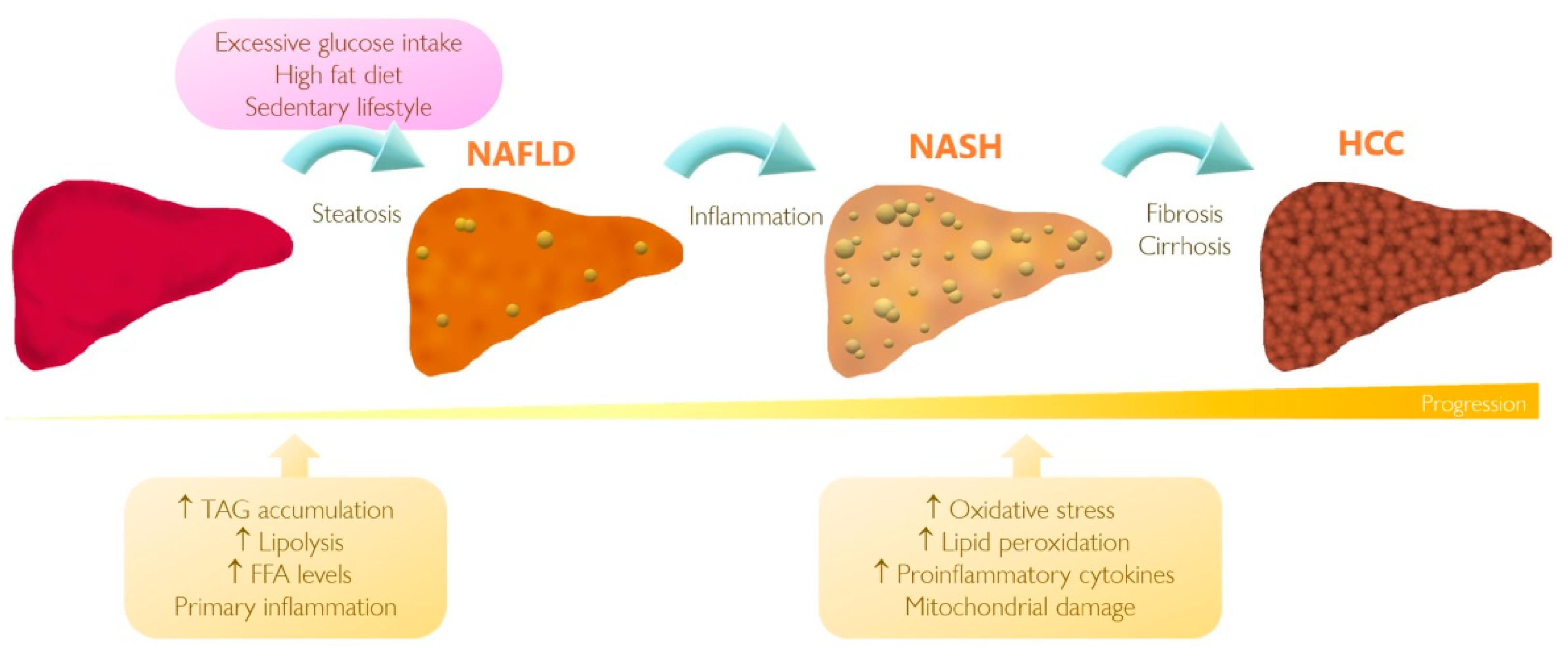
3. Understanding NAFLD and Its Progression to HCC

The progression of NAFLD towards the development of HCC and how risk factors can promote its appearance is well documented [38,39]. Historically, NAFLD has been described as a spectrum of chronic liver diseases characterized by hepatic lipid accumulation as well as excess of TAG in the cytoplasm [40]. Diverse factors are involved in the appearance of these pathologies. These include the excessive caloric intake that potentially leads to obesity and diabetes as well as the consumption of foods rich in fats and sucrose accompanying a sedentary lifestyle. Moreover, genetic modifications such as single-nucleotide polymorphisms (SNPs), epigenetic modifications, and viral infections (e.g., hepatitis C) are further critical risk factors for the development of HCC [41,42,43,44].
A “two-hit theory” has been proposed to describe the NASH pathogenesis. The first hit is steatosis, the accumulation of fat in the liver, followed by the second hit which is increased susceptibility of hepatocytes to secondary injuries such as oxidative stress, mitochondrial dysfunction, and release of pro-inflammatory cytokines [45,46,47,48,49]. Figure 2 illustrates the main events that take place during progress of NAFLD to NASH and eventually HCC.
According to histopathological features, steatosis is characterized by increased storage of TAGs in hepatocytes [40]. A remarkable characteristic of this stage is the ballooning of hepatocytes, which is currently considered as a hallmark of NAFLD [40]. Steatosis is often associated with increased lipolysis and insulin resistance resulting in elevation of serum free fatty acid (FFA) levels, followed by TAG relocation from the liver to peripheral organs. When inflammation and hepatocyte injury occur, the pathogenesis shifts to NASH [40,50]. In this scenario, a source of free radicals, induced by TAG accumulation, is capable of inducing oxidative stress, initiating lipid peroxidation accompanied by the release of pro-inflammatory cytokines, mitochondrial damage, and ER stress aggravating the condition of NAFLD [51]. Once hepatic fibrosis and cirrhosis occur, NASH can lead to the development of HCC, which is the most severe manifestation of hepatic injury [40,50]. Fibrosis is characterized by the deposition of high density extracellular matrix (ECM) proteins in diseased tissue, formation of regenerative nodules, and impaired wound-healing response, thus leading to cirrhosis [40,50,52]. While this “two-hit” approach allows us to understand how NAFLD could progress, a more accurate model has been suggested in recent years. This “multiple parallel hits” theory suggests that not only steatosis, insulin resistance and oxidative stress can lead to NASH, but instead multiple factors including primary inflammation inducing secondary steatosis, changes in gut microbiota, and the release of serum cytokines may act in concert to contribute to different disease stages [53,54].
Currently the “gold standard” for the identification of NAFLD is histological staining of liver biopsies, which is an invasive method accompanied with further risks for the health of the patient. Unfortunately, the lack of specific biomarkers able to differentiate the progress stages of the disease presently makes proper diagnosis and treatment uncertain [51,52]. Moreover, nowadays there is not an approved drug available for the treatment of NAFLD and NASH. Presently, intensive lifestyle changes which take into account a balanced, low-caloric diet and increased physical activity are the general recommendation to avoid disease progression [55,56]. Therapy for patients with metabolic syndrome and HCC, is focused on defeating cancer. In this regard, there is still a long way to go to identify new therapeutically effective drugs or specific biomarkers helpful in management of this complex pathology [52].

4. The Role of PLIN5 in Lipid Metabolism and NAFLD

As mentioned before, NAFLD is characterized by increased accumulation of LD in the liver as well as increased expression of PLIN5 [12]. This has been previously reported in patients with severe NAFLD as well as in mice fed with a high fat diet (HFD) inducing NAFLD [12,23]. Interestingly, compared to PLIN5 protein levels, mRNA levels are only slightly increased, suggesting that elevated PLIN5 activity is regulated post-transcriptionally [12].
One of the first studies to elucidate the role of PLIN5 in hepatocytes demonstrated that PLIN5 expression is induced by FFA in a time-independent manner [57]. Additionally, a study performed in Plin5-deficient mouse hepatocyte AML-12 cells showed a decrease in cellular TAG content as well as an increase of mitochondrial oxidation [58]. To gain a better understanding of consequences resulting from Plin5 depletion in vivo, several knockout (KO) models in mice have been developed. Table 1 contains key points of the major models reported as well as the feeding conditions evaluated in each of these, underpinning the notion that the role of PLIN5 in metabolism seems to be tissue-specific.
Mouse models based on HFD and Plin5 disruption (Plin5−/−) help to understand the role of PLIN5 in steatohepatitis and beyond the phenotype observed, they have provided critical information about molecular signaling and regulation of this protein. For instance, research in Plin5−/− mice showed an improvement of glucose tolerance in liver and insulin resistance in muscle [20]. This can be explained by enhanced lipolysis derived from FA, turning into sphingolipids, and consequently causing insulin resistance. In this scenario, Plin5−/− mice present a phenotype characterized by insulin resistance due to impairment in glucose disposal in skeletal muscle and WAT, but not in the liver [20]. In line with this, specific hepatic knockdown of Notch1 in mice resulted in an upregulated expression of glucose-6-phosphatase (G6P) and Plin5, leading to a predisposition of a diabetic phenotype through development of insulin resistance [62]. These reports indicate the possible cross-talk between these genes in glucose response.
Another HFD model revealed a decrease in hepatic TAG content and a decrease in expression of enzymes related to FA synthesis in Plin5−/− mice. Expression of pro-inflammatory and inflammatory biomarkers was increased in Plin5−/− mice and enhanced under a HFD. ER stress-induced genes, hepatic injury markers, and mitochondrial markers were upregulated in Plin5−/− animals, indicating the importance of PLIN5 in maintaining proper functions of hepatic cells [23]. Similarly, a study conducted in hepatocyte-specific Plin5−/− mice revealed a decrease in TAG secretion and impairment in glucose metabolism that led to systemic insulin resistance. In the same study, Plin5 ablation activated the c-Jun N-terminal kinase (JNK) signaling pathway and modulated insulin response [21].
A steatosis-like model of primary mouse and human hepatocytes, showed that statins decrease the levels of PLIN5, but not other LD-associated genes via the sterol regulatory element-binding protein 2 (SREBP2) [63]. Statins are commonly used in patients with hypercholesterolemia to reduce hepatic TAG content, but their mode of action is yet unknown. However, this finding provides a possible explanation on how the effects of PLIN5 regulation mimic the use of statins as a therapeutic drug during steatosis and shows the importance of PLIN5 as a possible target in this pathology.
Recently, it was reported that LD formation after stimulation with oleic acid (OA) leads to activation of PLIN5 in human liver cancer HepG2 cells via the PI3K/PPARα pathway [64]. In the respective study, it was demonstrated that the PPAR α inhibitor GW6471 decreased the levels of PLIN5 in a dose-dependent manner. Moreover, the phosphatidylinositol-3-kinase (PI3K) inhibitor LY294002 diminished the levels of PLIN5 mRNA in HepG2 cells. Thus the combination of OA treatment and inhibition of PI3K or PPARα activities showed a decrease in LDs, revealing the partial role of PLIN5 in the control of LD formation [64]. In this context, the PI3K pathway is also involved in ER stress. Insulin’s protective role during lipotoxicity induced by saturated fatty acids activated the PI3K/Akt/p53 pathway and ameliorated ER stress [65]. This evidence links PLIN5 as a possible mediator in response to ER stress.
The role of PLIN5 was also investigated in a high-glucose-induced podocyte injury model [30]. Feng and colleagues demonstrated the interplay among Akt/GSK-3β/Nrf2 and PLIN5 to reduce apoptosis, reactive oxygen species (ROS) production, and inflammatory response. Overexpression of PLIN5 alleviated the release of pro-inflammatory markers such as tumor necrosis factor-α (TNF-α), interleukin-6 (IL-6), and interleukin-1β (IL-1β) and upregulated the expression of nuclear factor erythroid 2–related factor 2 (Nrf2) and its targeted heme oxygenase-1 (HO-1) and NAD(P)H quinone dehydrogenase 1 (NQO1). In addition, protein kinase B (AKT) phosphorylating its target, glycogen synthase kinase 3-β (GSK-3β), led to Nrf2/ARE inactivation. In line with this study, PLIN5 was involved in oxidative stress via AMPK/Nrf2 [24]. Plin5 expression was suppressed under HFD in mouse hepatic stellate cells (HSC) which surprisingly led to HSC activation. In chronic liver damage, HSC are known as the key drivers of liver fibrosis as their activation and transdifferentiation to myofibroblasts leads to deposition of collagen and extracellular matrix in the tissue. Exogenous overexpression of PLIN5 restored the formation of LDs and induced 5′ AMP-activated protein kinase (AMPK) expression leading to increase of endogenous PLIN5 and to the inhibition of HSC activation. The induction of PLIN5 via AMPK induced the transcription activity of Nrf2 and led to reduced ROS and increased levels of glutathione, indicating how PLIN5 attenuates oxidative stress in HSC.
Interestingly, emerging evidence suggests that PLIN5 can act as a co-regulator during lipolysis stimulated by catecholamine. The results indicate that phosphorylation of PLIN5 by protein kinase A (PKA) triggers PLIN5 nuclear translocation during fasting and lipolysis conditions when PLIN5 binds to the promoter region of peroxisome proliferator-activated receptor gamma coactivator 1-alpha (PGC-1α) [66]. Glucagon, a PKA activator, stimulated the phosphorylation of PLIN5 and inhibited its interaction with CGI-58 promoting lipolysis and reduced TG accumulation in the liver [67].
PLIN5 also regulates the complex sirtuin 1-deleted in breast cancer-1 (SIRT1-DBC1), favoring the deacetylase activity of SIRT1 to its target PGC-1α, leading to transcription of their targets, and promoting FA oxidation and mitochondrial efficiency [66,68]. Moreover, hepatic PKA activity is increased under a chronic HFD, while interestingly this effect was enhanced in female mice, suggesting that the hormonal status plays a pivotal role in its regulation [69]. Nevertheless, it has been shown that phosphorylation of PLIN5 at serine 155 (S155) has important effects in hepatic lipid metabolism and glycemic control by allowing PLIN5 interaction with the lipase ATGL regulating further lipolysis [36]. Furthermore, it was recently shown that the loss of S155 phosphorylation of PLIN5 in liver impacts whole-body glucose tolerance and decreases levels of FA oxidation derived from TAGs [70].
Similarly, PLIN5 plays a role in autophagy by diminishing inflammation through deacetylase activity of SIRT1 in hepatocytes under fasting conditions. SIRT1 deacetylates nuclear factor-kappa B (NF-κB), thus inflammation is attenuated by the decreased levels of target genes of NF-κB such as Il-6, Il-1β, and Tnf-α [71]. Furthermore, PLIN5 acts as a scaffold in chaperone-mediated autophagy in HepG2 cells demonstrating that co-localization of heat shock protein 70 (Hsp70) and PLIN5 serves as a substrate allowing degradation of LD in cells under FA stimuli, which mimics NAFLD [12]. The SIRT1–PLIN5 axis has also been studied in vitro and in vivo as a target for glycycoumarin (GCM), a compound effective against liver diseases. The respective report showed that, GCM treatment increased SIRT1 expression in palmitic acid-derived lipotoxicity in AML-12 cells, which in consequence led to decreased expression of inflammatory genes such as Tnf-α and Il-6. Thus depletion of Plin5 or Sirt1 decreases GCM effectivity, revealing the coupled effect of these proteins to balance inflammatory response and lipoapoptosis [71].

5. PLIN5 Is Responsive to Cellular Processes Altered in NAFLD

NAFLD is a condition characterized of dyslipidemia, increased oxidative stress, ROS production, and mitochondrial alterations provoking inflammatory responses [17]. There are several reports analyzing how PLIN5 participates in each of these processes. In one study, the depletion of Plin5 in an in vivo mouse model under fasting conditions was associated with decreased LD formation and TAG content in the heart [59]. The same effect was observed in soleus muscle, BAT, and WAT. Additionally, in the heart, PLIN5 induces ROS levels while it blocks the activity of lipase ATGL suggesting that this mechanism maintains cardial LDs. In the absence of Plin5, FA are oxidized in mitochondria in the heart and not retained as TAG, which explains the decreased levels of TAG in this tissue [59].
In muscle, ER stress, as well as inflammation or oxidative stress, was not observed in mice lacking Plin5 [20]. This is opposite to the liver, in which several studies have found a protective role of Plin5 deletion in this tissue [21,23,27]. The mechanisms that regulate these tissue-specific functions remain unclear, but a few studies linked fibroblast growth factor 21 (Fgf21) as a critical mediator of the effects observed in skeletal muscle. Overexpression of Plin5 in skeletal muscle in mice protected the liver from inflammation during HFD feeding [61]. Interestingly, PLIN5 overexpression in skeletal muscle was associated with reduced hepatic expression of inflammatory markers such as IL-6, TNF-α, and monocyte chemoattractant protein-1 (MCP1) [61]. This model also suggested that PLIN5 drives FGF21 expression in muscle, which partially is metabolically protective [61]. On the contrary, Plin5−/− skeletal muscles showed decreased levels of FGF21 in liver and muscle tissues, leading to lower levels of the secreted protein in plasma and reduced ER stress [25]. FGF21 is a stress-inducible hepatokine, which regulates energy balance and glucose and lipid homeostasis and regulates ER stress via ATF4-CHOP [72,73]. This evidence suggests an important cross-talk among tissues as well cell-autonomous regulation, which balances metabolic responses.
Another study reported that overexpression of PLIN5 in the heart induces changes in the transcriptome of cells, especially in the signaling pathways related to mitochondrial bioenergetic processes, lipid metabolism, and cytoskeleton reorganization where interestingly, overexpression of PLIN5 led to an augment of Nrf2 targets genes [22]. This suggests that PLIN5 overexpression triggers oxidative stress and induces upregulation of the NRF2 pathway, possibly as a compensatory mechanism against LD formation in heart [22].
In the same line, recent evidence indicates that PLIN5 can modulate oxidative stress in pancreatic β-cells via Nrf2 [29]. In a palmitate-triggered lipotoxicity model in INS-1 pancreatic β-cells, Plin5 activated the Nrf2-ARE system by stimulating PI3K/Akt and ERK signaling pathways, while phosphorylation of JNK and p38 were decreased, indicating that these pathways are involved in the Plin5-mediated oxidative responses [29]. Again, this study points to a protective role of PLIN5 against oxidative stress.
Experiments in HepG2 cells showed that PLIN5 overexpression exerts a protective role during lipopolysaccharide (LPS)- or H2O2-induced oxidative stress by increasing cellular LD content and promoting contact between LD and mitochondria [26]. In this study, PLIN5 reduced ROS levels and augmented the expression of key genes in mitochondrial function including cytochrome C oxidase subunit 2 and 4 (COX2 and COX4) and citrate synthase (CS), which regulate the mitochondrial respiratory chain as well as glutathione peroxidase 2 (GPX2) and catalase (CAT), genes related to antioxidant response [26]. PLIN5 expression was also upregulated by the JNK-p38-ATF pathway, potentially by binding of JUN, ATF1, ATF3, and ATF4 to specific binding elements within the PLIN5 promoter [26]. It has been previously reported that this pathway is involved in the response of ROS activation [74,75]. Therefore, PLIN5 can potentially act as a co-regulator in oxidative response.
In livers of Plin5−/− mice, induction of inflammation via LPS failed to trigger expression of proteins such as NF-κB, NLR family pyrin domain-containing 3 (NLRP3), and the inflammatory protein lipocalin 2 (LCN2), indicating that PLIN5 acts as a vital protein protecting the liver from injury [27]. In this study, mitochondria of Plin5−/− mice presented a larger size and a closer localization to the ER in an HFD model as well as increased levels of mitochondrial markers such as OPA1 mitochondrial dynamin-like GTPase (OPA1), sigma 1 receptor (SIG1R), and voltage-dependent anion-selective channel 1 (VDAC1) involved in mitochondrial structure formation and trafficking. Moreover, increased levels of mitofusin 1 (MFN1) and mitofusin 2 (MFN2) were observed, genes which participate in mitochondrial fusion, possibly explaining the observed enlargement of these organelles [27]. This phenomenon of enlarged mitochondria was also reported in Plin5-S115A mutant mice [76]. However, in contrast to the previous report, mitochondrial fusion markers such as Mfn1, fission protein 1 (Fis1), and dynamin-related protein 1 (DRP1) were downregulated in Plin5-S115A mutant mice, whereas MFN2 levels were unchanged, demonstrating that normal mitochondrial activity is highly regulated by functional PLIN5 [76]. This evidence suggests that PLIN5 plays a pleiotropic role in cells from orchestrating lipid metabolism to regulating inflammation and mitochondrial function.
PLIN5 showed implications in the inflammatory response in different models, and strikingly, LCN2, a well-known acute pro-inflammatory protein and critical regulator of lipid uptake, has been shown to act as a regulator of PLIN5 activity in a NASH model based in a methionine- and choline-deficient diet [77]. This report supports that LCN2 impacts PLIN5 expression in a PPAR-independent manner, since its stimulation with an PPAR-γ agonist failed to suppress its expression [77].
Furthermore, studies on Plin5−/− mice have revealed a close local connection of mitochondria and LDs [27,78]. In the respective model, the oxidative capacity of mitochondria in cardiac muscle was impaired due to changes in lipid composition of the membrane and its polarization [78]. Super-resolution microscopy-based approaches in skeletal muscle have shown that under basal conditions there are three pools of PLIN5, namely in the cytosol, in close proximity of mitochondria, and surrounding LDs in the mitochondria–LD interface [79]. Moreover, it was observed that increased FA levels led to an augment of PLIN5 in the mitochondria–LD interface pool, suggesting that this relocation dynamic of PLIN5 might facilitate flux of FA from LDs to mitochondria [79,80]. All these data demonstrate the fine-tuned regulation of PLIN5 in lipotoxicity, inflammation response, and mitochondrial dynamics, processes that are all closely related in the development of NAFLD. The major biological processes in which PLIN5 participates are illustrated in Figure 3.

6. Perilipins in Cancer: The Unrevealed Role of PLIN5

Liver cancer represents the sixth most common type of cancer and the third highest cause of cancer death in 2020 according to International Agency for Research on Cancer (https://gco.iarc.fr/, last accessed 7 May 2021). Primary liver cancer includes HCC, cholangiocarcinoma, and other rarer types. Several risk factors are associated with the development of cancer such as chronic hepatitis B virus (HBV) and hepatitis C virus (HCV) infections, alcohol intake, obesity, type 2 diabetes, and NAFLD [81,82].
The tumor microenvironment (TME) is a complex net involving cancer cells, immune cells (B cells, T cells, and NK cells), and stromal cells (blood and lymphatic endothelial cells and cancer associated fibroblasts). Moreover, diverse molecular components including chemokines, cytokines, extracellular matrix compounds, and soluble immunosuppressive molecules act coordinately to maintain chronic inflammation and immune suppression leading to tumorigenesis [83]. In HCC, a heterogenous cell population consisting of progenitor cells, hepatocytes, and adult stems cells present significant transcriptional changes, thereby leading to chronic inflammation, giving place to cancer stem cells and promoting aberrant differentiation which ultimately leads to tumor progression and metastasis [13]. In order to identify these cell populations with stem cells properties, some surface markers such as epithelial cell adhesion molecule (EpCAM), cluster of differentiation (CD)44, CD90, and CD33 have been proposed [13].
Nowadays, there is a considerable knowledge on how metabolic changes in lipid homeostasis contribute to aberrant cell growth and cancer progression. For instance, the accumulation of saturated or unsaturated FA levels triggers cellular stress and altered cascades of signaling of lipid metabolism [14,83]. Proper homeostasis of intracellular FA is required for biosynthesis of biological membrane lipids, signaling molecules, and post-translational modifications of proteins. Impairment in this balance is taken advantage of by cancer cells which use it to benefit their proliferation rate by acquiring FA of exogenous sources (FA accumulation sites) or by endogenous synthesis. Two of the mechanisms altered in order to guarantee cancer cell survival are de novo lipogenesis and β-oxidation processes, suggesting that alterations in lipid metabolism can act as a general hallmark of cancer [13,14,15]. Indeed, samples of HCC revealed the upregulation of the transcriptional factor sterol regulatory element-binding factor 1 (SREBF1) whose expression was correlated with high mortality [84]. A recent meta-analysis study revealed a positive correlation between the overexpression of SREBP1, fatty acid synthase (FASN), and stearoyl-CoA desaturase-1 (SCD1) in HCC, while a poor prognosis was associated with increased levels of these markers [85]. The above findings suggest that activation of the lipogenic pathways is fundamental in the pathogenesis of HCC.
Advanced technologies have been useful to detect novel targets and biomarkers for estimating prognosis in cancer. Perilipins have also recently come into focus of such studies. For example, RNA-seq and microarray analysis revealed that PLIN1 can participate in breast cancer development, while patients with upregulated levels of PLIN1 mRNA present lower overall survival rates [86]. Immunohistochemical validation of these findings in human specimens confirmed these findings as well as in MCF-7 and MDA-MB-231 cell lines, where overexpression of PLIN1 suppressed cellular proliferation, migration and invasion, possibly indicating a role of PLIN1 as tumor suppressor gene [86]. A similar approach performed in clear cell renal cell carcinoma (ccRCC) revealed that PLIN2 upregulation is associated with a good disease prognosis, while PLIN2 knockdown in human kidney carcinoma A-498 cells favors proliferation, migration, and invasion of ccRCC cells [87]. In prostate cancer, PLIN3 upregulation has been associated with poor prognosis in patients [88]. Moreover, elevated PLIN3 expression has recently been related to poor prognosis of lung adenocarcinoma, whereas ablation of PLIN3 inhibited the invasion ability of lung adenocarcinoma cells [89].
Information contained in the Human Protein Atlas indicates PLIN5 level alterations in some types of cancer (http://www.proteinatlas.org/, last accessed 24 March 2021). A first study about participation of perilipins in neoplastic steatogenesis was reported by Straub and colleagues. They suggested that PLIN1, PLIN2, and PLIN3 may be involved in cancers such as hepatocellular adenoma and carcinoma, sebaceous adenoma and carcinoma, and lipoma tumors, while PLIN5 was excluded as not sufficient proof was given [90].
Little is understood so far about how PLIN5 is involved in the development or progress of cancer, but a novel transcriptome analysis of lung cancer, predicted that higher expression of the guanine nucleotide-binding protein α-inhibiting activity polypeptide 3 (GNAI3) and the lncRNA AC087521.1 was associated with a short survival time, while the NHL repeat-containing protein 2 (NHLRC2) and PLIN5 correlated with longer survival times [91].
Regarding HCC, recent reports have shown increased expression of PLIN5 in HCC, ccRCC, and lipo-, rhabdo- and leiomyosarcoma by immunohistochemical analysis [92]. More recently, perilipins were reported as markers of liposarcoma and liposarcoma subtypes. Using a set of 245 samples and analyzing tissue microarray and immunohistochemistry expression, the results showed that PLIN2, PLIN3, and PLIN5 were commonly expressed in liposarcomas, rhabdomyosarcomas, leiomyosarcomas, dermatofibrosarcoma protuberans, undifferentiated sarcomas, fibrosarcomas, Ewing’s sarcomas, and epithelioid sarcomas [93]. Opposite to these reports, Plin5 expression is downregulated by promoter methylation in ovarian cancer. Furthermore, an in vivo model, revealed the possible role of PLIN5 as a tumor repressor gene that abrogates cell proliferation, migration, and invasion, thus promoting apoptosis [94]. These findings underpin the pleiotropic activity of PLIN5 in cancer.
Information about PLIN5 implication in HCC is still limited. It was first shown in 2019 that PLIN5 expression is upregulated in HCC tumoral areas in murine HCC models and human HCC biopsy specimens as assessed by Western blot analysis and quantitative real time PCR [95]. RNA-seq and splicing factor (SF) data of 390 HCC cases deposited in the Cancer Genome Atlas (TCGA) data portal (https://tcgadata.nci.nih.gov/tcga/, last accessed 24 March 2021) indicate that PLIN5-46808-AT was found more common in primary sites of HCC with metastasis than primary HCC [96]. It is evident that more studies elucidating HCC development are necessary to clarify the actual function of PLIN5 in HCC.

7. Conclusions

This review discusses PLIN5 as a key regulator of pathways involved in pathogenesis of NAFLD and cancer. Since the discovery of PLIN5 in 2006, its fundamental role in lipid metabolism has been extensively studied. PLIN5 regulates lipid metabolism, reduces oxidative stress and ER stress, and promotes autophagy, making this member of the perilipin protein family an interesting therapeutic target. However, the precise mechanisms by which PLIN5 protects the liver from injury are still poorly understood. Depending on the tissue studied, effects of Plin5 depletion can be contradictory, possibly reflecting its fine-tuned tissue-specific regulation and activity. Broadening the network of PLIN5 targets and cofactors in distinct stages of NAFLD-HCC pathogenesis will help to characterize more specific roles of PLIN5, potentially allowing the use of this protein as a tool for diagnosis or therapeutic targeting. Hot topics are currently the mitochondrial functions affected by PLIN5 and its participation in HCC initiation and progression.

Author Contributions

P.B.M.S. and A.A. wrote the original draft of this review. R.W. and M.K. revised the first draft. A.A., P.B.M.S. and R.W. edited the final version. All authors have read and agreed to the published version of the manuscript.

Funding

A.A. is supported by the START-Program of the Faculty of Medicine of the RWTH Aachen University and by the Wilhelm-Sander Stiftung für Krebsforschung (Grant No. 2020.002.1). R.W. is supported by grants from the German Research Foundation (projects WE2554/13-1 and W2554/15-1). None of the funders had a role in the design of this review or in the decision to publish it.

Institutional Review Board Statement

This review article does not require approval by Institutional Review Board.

Informed Consent Statement

Not applicable.

Data Availability Statement

This review includes no original data.

Conflicts of Interest

The authors declare no conflict of interest.

Abbreviations

ABHD5α-β-hydrolase domain-containing 5
ACAA2Acetyl-CoA acyltransferase 2
ACAT1Acetyl-CoA acetyltransferase 1
ACAT2Acetyl-CoA acetyltransferase 2
AKTProtein kinase B
ALPAlkaline phosphatase
ALTAlanine aminotransferase
AMPK5’-AMP-activated protein kinase
Arf-COPIADP-ribosylation factor1-coat protein complex I
ASTAspartate aminotransferase
ATFActivating transcription factor
ATGLAdipose triglyceride lipase
BATBrown adipocyte tissue
C/EBPαFactor CCAAT/enhancer-binding protein alpha
CATCatalase
ccRCCClear cell renal cell carcinoma
CDCluster of differentiation
CtrDControl diet
CGI-58Comparative gene identification-58
CHOPC/EBP homologous protein
CM-Plin5Cardiac muscle-specific overexpression of Plin5
COX2Cytochrome c oxidase subunit 2
COX4Cytochrome c oxidase subunit 4
CSCitrate synthase
DBC1Deleted in breast cancer-1
DGAT1Diacylglycerol acyltransferase1
DGAT2Diacylglycerol acyltransferase 2
DRP1Dynamin related protein 1
ECMExtracellualr matrix
EDLExtensor digitorum longus (muscle)
EpCAMEpithelial cell adhesion molecule
EREndoplasmic reticulum
ERKExtracellular-regulated kinase
FAFatty acid
FASNFatty acid synthase
FFAFree fatty acid
FGF21Fibroblast growth factor 21
FSN-1Fission protein 1
G6PGucose-6-phosphatase
GMCGlycycoumarin
GNAI3G Protein subunit alpha I3
GPX2Glutathione Peroxidase 2
GSK-3βGlycogen synthase kinase 3 beta
HBVHepatitis B virus
HCCHepatocellular carcinoma
HCVHepatitis C virus
HFDHigh fat diet
HO-1Heme oxygenase-1
HSCHepatic stellate cell(s)
HSLHormone-sensitive lipase
HSP70Heat shock protein 70
IL-1βInterleukin-1 beta
IL-6Interleukin-6
JNKc-Jun N-terminal kinase
JUNJun proto-oncogene
KOKnockout
LCN2Lipocalin 2
LDLipid droplets
LPSLipopolysaccharide
LSDP5Lipid storage droplet protein 5
MCK-Plin5Skeletal muscle-specific overexpression of Plin5
MCP1Monocyte chemoattractant protein-1
MFN1Mitofusin 1
MFN2Mitofusin 2
MGLMonoglyceride lipase
MKOMuscle-specific Plin5 knockout
MLDPMyocardial LD protein
NAFLDNon-alcoholic fatty liver disease
NASHNon-alcoholic steatohepatitis
NF-κBNuclear factor-kappa B
NHLRC2NHL repeat containing 2
NLRP3NLR family pyrin domain containing 3
NOTCH1Notch homolog 1
NQO1NAD(P)H quinone dehydrogenase 1
NRF2Nuclear factor erythroid 2-related factor 2
OAOleic acid
OPA1Mitochondrial dynamin-like GTPase
OXPATOxidative tissues-enriched PAT protein
p38p38 mitogen-activated protein kinase
p53Tumor protein P53
PGC-1αPeroxisome proliferator-activated receptor gamma coactivator 1-alpha
PI3KPhosphatidylinositol-3-kinase
PKAProtein kinase A
Plin5−/−Plin5 deficient
PLIN5Perilipin 5
PPARPeroxisome proliferator-activated receptor
PPARαPeroxisome proliferator-activated receptor alpha
PPREPPAR regulatory element
RABRas superfamily of GTPases
ROSReactive oxygen species
SCD1Stearoyl-CoA desaturase-1
SFSplicing factor
SIG1RSigma 1 receptor
SIRT1Sirtuin-1
SNPsSingle-nucleotide polymorphisms
SREBF1Sterol regulatory element binding factor 1
SREBP1Sterol regulatory element-binding protein 1
SREBP2Sterol regulatory element-binding protein 2
TAGTriacylglycerides
TCGAThe cancer genome atlas
TMETumor microenvironment
TNF-αTumor necorsis factor-alpha
VDAC1Voltage-dependent anion-selective channel 1
WATWhite adipocyte tissue

References

  1. Minehira, K.; Gual, P. Role of Lipid Droplet Proteins in the Development of NAFLD and Hepatic Insulin Resistance. Non-Alcoholic Fat Liver Disease; IntechOpen: London, UK, 2018; pp. 55–77. [Google Scholar] [CrossRef] [Green Version]
  2. Olzmann, J.A.; Carvalho, P. Dynamics and functions of lipid droplets. Nat. Rev. Mol. Cell Biol. 2019, 20, 137–155. [Google Scholar] [CrossRef]
  3. Sahini, N.; Borlak, J. Recent insights into the molecular pathophysiology of lipid droplet formation in hepatocytes. Prog. Lipid Res. 2014, 54, 86–112. [Google Scholar] [CrossRef] [PubMed]
  4. Greenberg, A.S.; Egan, J.J.; Wek, S.A.; Garty, N.B.; Blanchette-Mackie, E.J.; Londos, C. Perilipin, a major hormonally regulated adipocyte-specific phosphoprotein associated with the periphery of lipid storage droplets. J. Biol. Chem. 1991, 266, 11341–11346. [Google Scholar] [CrossRef]
  5. Jiang, H.P.; Serrero, G. Isolation and characterization of a full-length cDNA coding for an adipose differentiation-related protein. Proc. Natl. Acad. Sci. USA 1992, 89, 7856–7860. [Google Scholar] [CrossRef] [Green Version]
  6. Díaz, E.; Pfeffer, S.R. Tip47: A cargo selection device for mannose 6-phosphate receptor trafficking. Cell 1998, 93, 433–443. [Google Scholar] [CrossRef] [Green Version]
  7. Kimmel, A.R.; Brasaemle, D.L.; McAndrews-Hill, M.; Sztalryd, C.; Londos, C. Adoption of PERILIPIN as a unifying nomenclature for the mammalian PAT-family of intracellular lipid storage droplet proteins. J. Lipid Res. 2010, 51, 468–471. [Google Scholar] [CrossRef] [PubMed] [Green Version]
  8. Brasaemle, D.L.; Subramanian, V.; Garcia, A.; Marcinkiewicz, A.; Rothenberg, A. Perilipin A and the control of triacylglycerol metabolism. Mol. Cell. Biochem. 2009, 326, 15–21. [Google Scholar] [CrossRef] [PubMed]
  9. Itabe, H.; Yamaguchi, T.; Nimura, S.; Sasabe, N. Perilipins: A diversity of intracellular lipid droplet proteins. Lipids Health Dis. 2017, 16, 1–11. [Google Scholar] [CrossRef] [PubMed] [Green Version]
  10. Listenberger, L.L.; Ostermeyer-Fay, A.G.; Goldberg, E.B.; Brown, W.J.; Brown, D.A. Adipocyte differentiation-related protein reduces the lipid droplet association of adipose triglyceride lipase and slows triacylglycerol turnover. J. Lipid Res. 2007, 48, 2751–2761. [Google Scholar] [CrossRef] [PubMed] [Green Version]
  11. Wolins, N.E.; Skinner, J.R.; Schoenfish, M.J.; Tzekov, A.; Bensch, K.G.; Bickel, P.E. Adipocyte protein S3-12 coats nascent lipid droplets. J. Biol. Chem. 2003, 278, 37713–37721. [Google Scholar] [CrossRef] [Green Version]
  12. Ma, S.Y.; Sun, K.S.; Zhang, M.; Zhou, X.; Zheng, X.H.; Tian, S.Y.; Liu, Y.S.; Chen, L.; Gao, X.; Ye, J.; et al. Disruption of Plin5 degradation by CMA causes lipid homeostasis imbalance in NAFLD. Liver Int. 2020, 40, 2427–2438. [Google Scholar] [CrossRef] [PubMed]
  13. Hayes, C.N.; Zhang, P.; Chayama, K. The role of lipids in hepatocellular carcinoma. In Hepatocellular Carcinoma; Tirnitz-Parker, J.E.E., Ed.; Codon Publications: Brisbane, Australia, 2019; Chapter 5. [Google Scholar] [CrossRef] [Green Version]
  14. Corn, K.C.; Windham, M.A.; Rafat, M. Lipids in the tumor microenvironment: From cancer progression to treatment. Prog. Lipid Res. 2020, 80. [Google Scholar] [CrossRef] [PubMed]
  15. Calvisi, D.F.; Wang, C.; Ho, C.; Ladu, S.; Lee, S.A.; Mattu, S.; Destefanis, G.; Delogu, S.; Zimmermann, A.; Ericsson, J.; et al. Increased lipogenesis, induced by AKT-mTORC1-RPS6 signaling, promotes development of human hepatocellular carcinoma. Gastroenterology 2011, 140, 1071–1083. [Google Scholar] [CrossRef] [PubMed] [Green Version]
  16. Ashraf, N.U.; Sheikh, T.A. Endoplasmic reticulum stress and Oxidative stress in the pathogenesis of Non-alcoholic fatty liver disease. Free Radic. Res. 2015, 49, 1405–1418. [Google Scholar] [CrossRef]
  17. Chen, Z.; Tian, R.; She, Z.; Cai, J.; Li, H. Role of oxidative stress in the pathogenesis of nonalcoholic fatty liver disease. Free Radic. Biol. Med. 2020, 20, 116–141. [Google Scholar] [CrossRef]
  18. Reuter, S.; Gupta, S.C.; Chaturvedi, M.M.; Aggarwal, B.B. Oxidative stress, inflammation, and cancer: How are they linked? Free Radic. Biol. Med. 2010, 49, 1603–1616. [Google Scholar] [CrossRef] [Green Version]
  19. Saha, S.K.; Lee, S.B.; Won, J.; Choi, H.Y.; Kim, K.; Yang, G.M.; Dayem, A.A.; Cho, S.G. Correlation between oxidative stress, nutrition, and cancer initiation. Int. J. Mol. Sci. 2017, 18, 1544. [Google Scholar] [CrossRef] [Green Version]
  20. Mason, R.R.; Mokhtar, R.; Matzaris, M.; Selathurai, A.; Kowalski, G.M.; Mokbel, N.; Meikle, P.J.; Bruce, C.R.; Watt, M.J. PLIN5 deletion remodels intracellular lipid composition and causes insulin resistance in muscle. Mol. Metab. 2014, 3, 652–663. [Google Scholar] [CrossRef] [Green Version]
  21. Keenan, S.N.; Meex, R.C.; Lo, J.C.Y.; Ryan, A.; Nie, S.; Montgomery, M.K.; Watt, M.J. Perilipin 5 Deletion in hepatocytes remodels lipid metabolism and causes hepatic insulin resistance in mice. Diabetes 2019, 68, 543–555. [Google Scholar] [CrossRef] [Green Version]
  22. Wang, H.; Sreenivasan, U.; Gong, D.W.; O’Connell, K.A.; Dabkowski, E.R.; Hecker, P.A.; Ionica, N.; Konig, M.; Mahurkar, A.; Sun, Y.; et al. Cardiomyocyte-specific perilipin 5 overexpression leads to myocardial steatosis and modest cardiac dysfunction. J. Lipid Res. 2013, 54, 953–965. [Google Scholar] [CrossRef] [Green Version]
  23. Wang, C.; Zhao, Y.; Gao, X.; Li, L.; Yuan, Y.; Liu, F.; Zhang, L.; Wu, J.; Hu, P.; Zhang, X.; et al. Perilipin 5 improves hepatic lipotoxicity by inhibiting lipolysis. Hepatology 2015, 61, 870–882. [Google Scholar] [CrossRef]
  24. Lin, J.; Chen, A. Perilipin 5 restores the formation of lipid droplets in activated hepatic stellate cells and inhibits their activation. Lab. Investig. 2016, 96, 791–806. [Google Scholar] [CrossRef] [Green Version]
  25. Montgomery, M.K.; Mokhtar, R.; Bayliss, J.; Parkington, H.C.; Suturin, V.M.; Bruce, C.R.; Watt, M.J. Perilipin 5 deletion unmasks an endoplasmic reticulum Stress-Fibroblast growth factor 21 axis in skeletal muscle. Diabetes 2018, 67, 594–606. [Google Scholar] [CrossRef] [PubMed] [Green Version]
  26. Tan, Y.; Jin, Y.; Wang, Q.; Huang, J.; Wu, X.; Ren, Z. Perilipin 5 protects against cellular oxidative stress by enhancing mitochondrial function in HepG2 cells. Cells 2019, 8, 1241. [Google Scholar] [CrossRef] [PubMed] [Green Version]
  27. Asimakopoulou, A.; Engel, K.M.; Gassler, N.; Bracht, T.; Sitek, B.; Buhl, E.M.; Kalampoka, S.; Pinoé-Schmidt, M.; van Helden, J.; Schiller, J.; et al. Deletion of Perilipin 5 protects against hepatic injury in nonalcoholic fatty liver disease via missing inflammasome activation. Cells 2020, 9, 1346. [Google Scholar] [CrossRef]
  28. Zhang, E.; Cui, W.; Lopresti, M.; Mashek, M.T.; Najt, C.P.; Hu, H.; Mashek, D.G. Hepatic PLIN5 signals via SIRT1 to promote autophagy and prevent inflammation during fasting. J. Lipid Res. 2020, 61, 338–350. [Google Scholar] [CrossRef]
  29. Zhu, Y.; Ren, C.; Zhang, M.; Zhong, Y. Perilipin 5 reduces oxidative damage associated with lipotoxicity by activating the PI3K/ERK-mediated Nrf2-ARE signaling pathway in INS-1 pancreatic β-cells. Front. Endocrinol. 2020, 11, 1–14. [Google Scholar] [CrossRef]
  30. Feng, J.; Xie, L.; Yu, X.; Liu, C.; Dong, H.; Lu, W.; Kong, R. Perilipin 5 ameliorates high-glucose-induced podocyte injury via Akt/GSK-3β/Nrf2-mediated suppression of apoptosis, oxidative stress, and inflammation. Biochem. Biophys. Res. Commun. 2021, 12, 22–30. [Google Scholar] [CrossRef] [PubMed]
  31. Dalen, K.T.; Dahl, T.; Holter, E.; Arntsen, B.; Londos, C.; Sztalryd, C. LSDP5 is a PAT protein specifically expressed in fatty acid oxidizing tissues. Biochim. Biophys. Acta Mol. Cell Biol. Lipids 2007, 1771, 210–227. [Google Scholar] [CrossRef]
  32. Yamaguchi, T.; Matsushita, S.; Motojima, K.; Hirose, F.; Osumi, T. MLDP, a novel PAT family protein localized to lipid droplets and enriched in the heart, is regulated by peroxisome proliferator-activated receptor α. J. Biol. Chem. 2006, 281, 14232–14240. [Google Scholar] [CrossRef] [Green Version]
  33. Wolins, N.E.; Quaynor, B.K.; Skinner, J.R.; Tzekov, A.; Croce, M.A.; Gropler, M.C. OXPAT/PAT-1 is a PPAR-induced lipid droplet protein that promotes fatty acid utilization. Diabetes 2006, 55, 3418–3428. [Google Scholar] [CrossRef] [Green Version]
  34. Kimmel, A.R.; Sztalryd, C. Perilipin 5, a lipid droplet protein adapted to mitochondrial energy utilization. Curr. Opin. Lipidol. 2014, 25, 110–117. [Google Scholar] [CrossRef] [PubMed] [Green Version]
  35. Zhou, L.; Zhang, L.; Meng, Q.; Niu, C.; Jin, D.; Yu, A. C/EBPα promotes transcription of the porcine perilipin 5 gene. Mol. Cell. Endocrinol. 2012, 364, 28–35. [Google Scholar] [CrossRef] [PubMed]
  36. Granneman, J.G.; Moore, H.P.H.; Mottillo, E.P.; Zhu, Z.; Zhou, L. Interactions of Perilipin-5 (Plin5) with adipose triglyceride lipase. J. Biol. Chem. 2011, 286, 5126–5135. [Google Scholar] [CrossRef] [Green Version]
  37. Wang, H.; Bell, M.; Sreenevasan, U.; Hu, H.; Liu, J.; Dalen, K.; Londos, C.; Yamaguchi, T.; Rizzo, M.A.; Coleman, R.; et al. Unique regulation of adipose triglyceride lipase (ATGL) by perilipin 5, a lipid droplet-associated protein. J. Biol. Chem. 2011, 286, 15707–15715. [Google Scholar] [CrossRef] [Green Version]
  38. Dongiovanni, P.; Romeo, S.; Valenti, L. Hepatocellular carcinoma in nonalcoholic fatty liver: Role of environmental and genetic factors. World J. Gastroenterol. 2014, 20, 12945–12955. [Google Scholar] [CrossRef]
  39. Huang, D.Q.; El-Serag, H.B.; Loomba, R. Global epidemiology of NAFLD-related HCC: Trends, predictions, risk factors and prevention. Nat. Rev. Gastroenterol. Hepatol. 2020. [Google Scholar] [CrossRef]
  40. Burt, A.D.; Tiniakos, D.G.; Lackner, C. Diagnosis and assessment of NAFLD: Definitions and histopathological classification. Semin. Liver Dis. 2015, 35, 207–220. [Google Scholar] [CrossRef] [PubMed] [Green Version]
  41. Lian, C.Y.; Zhai, Z.Z.; Li, Z.F.; Wang, L. High fat diet-triggered non-alcoholic fatty liver disease: A review of proposed mechanisms. Chem. Biol. Interact. 2020, 330. [Google Scholar] [CrossRef] [PubMed]
  42. Jensen, T.; Abdelmalek, M.F.; Sullivan, S.; Nadeau, K.J.; Green, M.; Roncal, C.; Nakagawa, T.; Kuwabara, M.; Sato, Y.; Kang, D.-H.; et al. Fructose and sugar: A major mediator of non-alcoholic fatty liver disease. J. Hepatol. 2018, 68, 1063–1075. [Google Scholar] [CrossRef] [Green Version]
  43. Del Campo, J.A.; Gallego-Durán, R.; Gallego, P.; Grande, L. Genetic and epigenetic regulation in nonalcoholic fatty liver disease (NAFLD). Int. J. Mol. Sci. 2018, 19, 911. [Google Scholar] [CrossRef] [PubMed] [Green Version]
  44. Li, W.; Deng, R.; Liu, S.; Wang, K.; Sun, J. Hepatitis B virus-related hepatocellular carcinoma in the era of antiviral therapy: The emerging role of non-viral risk factors. Liver Int. 2020, 40, 2316–2325. [Google Scholar] [CrossRef]
  45. Angulo, P. Nonalcoholic fatty liver disease. N. Engl. J. Med. 2002, 346, 1221–1231. [Google Scholar] [CrossRef] [PubMed] [Green Version]
  46. Day, C.P. Pathogenesis of steatohepatitis. Best Pract. Res. Clin. Gastroenterol. 2002, 16, 663–678. [Google Scholar] [CrossRef]
  47. Diehl, A.M.; Li, Z.P.; Lin, H.Z.; Yang, S.Q. Cytokines and the pathogenesis of non-alcoholic steatohepatitis. Gut 2005, 54, 303–306. [Google Scholar] [CrossRef] [Green Version]
  48. Tilg, H. The role of cytokines in non-alcoholic fatty liver disease. Dig. Dis. 2010, 28, 179–185. [Google Scholar] [CrossRef] [PubMed]
  49. Day, C.P.; James, O.F. Steatohepatitis: A tale of two “Hits”? Gastroenterology 1998, 114, 842–845. [Google Scholar] [CrossRef]
  50. Arab, J.P.; Arrese, M.; Trauner, M. Recent insights into the pathogenesis of nonalcoholic fatty liver disease. Annu. Rev. Pathol. Mech. Dis. 2018, 13, 321–350. [Google Scholar] [CrossRef]
  51. Kutlu, O.; Kaleli, H.N.; Ozer, E. Molecular pathogenesis of nonalcoholic steatohepatitis-(NASH-) related hepatocellular carcinoma. Can. J. Gastroenterol. Hepatol. 2018, 2018. [Google Scholar] [CrossRef] [Green Version]
  52. Anstee, Q.M.; Reeves, H.L.; Kotsiliti, E.; Govaere, O.; Heikenwalder, M. From NASH to HCC: Current concepts and future challenges. Nat. Rev. Gastroenterol. Hepatol. 2019, 16, 411–428. [Google Scholar] [CrossRef]
  53. Jiang, C.M.; Pu, C.W.; Hou, Y.H.; Chen, Z.; Alanazy, M.; Hebbard, L. Non alcoholic steatohepatitis a precursor for hepatocellular carcinoma development. World J. Gastroenterol. 2014, 20, 16464–16473. [Google Scholar] [CrossRef]
  54. Takaki, A.; Kawai, D.; Yamamoto, K. Multiple hits, including oxidative stress, as pathogenesis and treatment target in non-alcoholic steatohepatitis (NASH). Int. J. Mol. Sci. 2013, 14, 20704–20728. [Google Scholar] [CrossRef] [PubMed] [Green Version]
  55. Van Der Windt, D.J.; Sud, V.; Zhang, H.; Tsung, A.; Huang, H. The effects of physical exercise on fatty liver disease. Gene Expr. 2018, 18, 89–101. [Google Scholar] [CrossRef] [Green Version]
  56. Choudhary, N.S.; Saraf, N.; Saigal, S.; Gautam, D.; Lipi, L.; Rastogi, A.; Goja, S.; Menon, P.B.; Bhangui, P.; Ramchandra, S.K.; et al. Rapid reversal of liver steatosis with life style modification in highly motivated liver donors. J. Clin. Exp. Hepatol. 2015, 5, 123–126. [Google Scholar] [CrossRef] [PubMed] [Green Version]
  57. Grasselli, E.; Voci, A.; Pesce, C.; Canesi, L.; Fugassa, E.; Gallo, G.; Vergani, L. PAT protein mRNA expression in primary rat hepatocytes: Effects of exposure to fatty acids. Int. J. Mol. Med. 2010, 25, 505–512. [Google Scholar] [CrossRef] [Green Version]
  58. Li, H.; Song, Y.; Zhang, L.J.; Gu, Y.; Li, F.F.; Pan, S.Y. LSDP5 enhances triglyceride storage in hepatocytes by influencing lipolysis and fatty acid β-oxidation of lipid droplets. PLoS ONE 2012, 7, e36712. [Google Scholar] [CrossRef] [Green Version]
  59. Kuramoto, K.; Okamura, T.; Yamaguchi, T.; Nakamura, T.Y.; Wakabayashi, S.; Morinaga, H.; Nomura, M.; Yanase, T.; Otsu, K.; Usuda, N.; et al. Perilipin 5, a lipid droplet-binding protein, protects heart from oxidative burden by sequestering fatty acid from excessive oxidation. J. Biol. Chem. 2012, 287, 23852–23863. [Google Scholar] [CrossRef] [PubMed] [Green Version]
  60. Pollak, N.M.; Schweiger, M.; Jaeger, D.; Kolb, D.; Kumari, M.; Schreiber, R.; Kolleritsch, S.; Markolin, P.; Grabner, G.F.; Heier, C.; et al. Cardiac-Specific overexpression of perilipin 5 provokes severe cardiac steatosis via the formation of a lipolytic barrier. J. Lipid Res. 2013, 54, 1092–1102. [Google Scholar] [CrossRef] [Green Version]
  61. Harris, L.A.; Skinner, J.R.; Shew, T.M.; Pietka, T.A.; Abumrad, N.A.; Wolins, N.E. Perilipin 5-driven lipid droplet accumulation in skeletal muscle stimulates the expression of fibroblast growth factor 21. Diabetes 2015, 64, 2757–2768. [Google Scholar] [CrossRef] [Green Version]
  62. Bernsmeier, C.; Dill, M.T.; Provenzano, A.; Makowska, Z.; Krol, I.; Muscogiuri, G.; Semela, D.; Tornillo, L.; Marra, F.; Heim, M.H. Hepatic Notch1 deletion predisposes to diabetes and steatosis via glucose-6-phosphatase and perilipin-5 upregulation. Lab. Investig. 2016, 96, 972–980. [Google Scholar] [CrossRef] [PubMed] [Green Version]
  63. Langhi, C.; Marquart, T.J.; Allen, R.M.; Baldán, A. Perilipin-5 is regulated by statins and controls triglyceride contents in the hepatocyte. J. Hepatol. 2014, 61, 358–365. [Google Scholar] [CrossRef] [Green Version]
  64. Zhong, W.; Fan, B.; Cong, H.; Wang, T.; Gu, J. Oleic acid-induced perilipin 5 expression and lipid droplets formation are regulated by the PI3K/PPARα pathway in HepG2 cells. Appl. Physiol. Nutr. Metab. 2019, 44, 840–848. [Google Scholar] [CrossRef] [PubMed]
  65. Ning, H.; Sun, Z.; Liu, Y.; Liu, L.; Hao, L.; Ye, Y.; Feng, R.; Li, J.; Li, Y.; Chu, X.; et al. Insulin protects hepatic lipotoxicity by regulating ER stress through the PI3K/Akt/p53 involved pathway independently of autophagy inhibition. Nutrients 2016, 8, 227. [Google Scholar] [CrossRef] [PubMed] [Green Version]
  66. Gallardo-Montejano, V.I.; Saxena, G.; Kusminski, C.M.; Yang, C.; McAfee, J.L.; Hahner, L.; Hoch, K.; Dubinsky, W.; Narkar, V.A.; Bickel, P.E. Nuclear Perilipin 5 integrates lipid droplet lipolysis with PGC-1α/SIRT1-dependent transcriptional regulation of mitochondrial function. Nat. Commun. 2016, 7. [Google Scholar] [CrossRef]
  67. Gao, X.; Nan, Y.; Zhao, Y.; Yuan, Y.; Ren, B.; Sun, C.; Cao, K.; Yu, M.; Feng, X.; Ye, J. Atorvastatin reduces lipid accumulation in the liver by activating protein kinase A-mediated phosphorylation of perilipin 5. Biochim. Biophys. Acta Mol. Cell Biol. Lipids 2017, 1862, 1512–1519. [Google Scholar] [CrossRef]
  68. Najt, C.P.; Khan, S.A.; Heden, T.D.; Witthuhn, B.A.; Perez, M.; Heier, J.L.; Mead, L.E.; Franklin, M.P.; Karanja, K.K.; Graham, M.J.; et al. Lipid droplet-derived monounsaturated fatty acids traffic via PLIN5 to allosterically activate SIRT1. Mol. Cell. 2020, 77, 810–824. [Google Scholar] [CrossRef] [PubMed]
  69. London, E.; Nesterova, M.; Stratakis, C.A. Acute vs chronic exposure to high fat diet leads to distinct regulation of PKA. J. Mol. Endocrinol. 2017, 59, 1–12. [Google Scholar] [CrossRef] [Green Version]
  70. Keenan, S.N.; DeNardo, W.; Lou, J.; Schittenhelm, R.B.; Montgomery, M.K.; Granneman, J.G.; Hinde, E.; Watt, M.J. Perilipin 5 S155 phosphorylation by PKA is required for the control of hepatic lipid metabolism and glycemic control. J. Lipid Res. 2021, 62. [Google Scholar] [CrossRef] [PubMed]
  71. Zhang, E.; Yin, S.; Zhao, C.; Fan, L.; Hu, H. Involvement of activation of PLIN5-Sirt1 axis in protective effect of glycycoumarin on hepatic lipotoxicity. Biochem. Biophys. Res. Commun. 2020, 528, 7–13. [Google Scholar] [CrossRef]
  72. Geng, L.; Lam, K.S.L.; Xu, A. The therapeutic potential of FGF21 in metabolic diseases: From bench to clinic. Nat. Rev. Endocrinol. 2020, 16, 654–667. [Google Scholar] [CrossRef]
  73. Wan, X.S.; Lu, X.H.; Xiao, Y.C.; Lin, Y.; Zhu, H.; Ding, T.; Yang, Y.; Huang, Y.; Zhang, Y.; Liu, Y.L.; et al. ATF4- and CHOP-dependent induction of FGF21 through endoplasmic reticulum stress. BioMed Res. Int. 2014, 2014. [Google Scholar] [CrossRef]
  74. Park, G.B.; Choi, Y.; Kim, Y.S.; Lee, H.K.; Kim, D.; Hur, D.Y. ROS-mediated JNK/p38-MAPK activation regulates Bax translocation in Sorafenib-induced apoptosis of EBV-transformed B cells. Int. J. Oncol. 2014, 44, 977–985. [Google Scholar] [CrossRef] [Green Version]
  75. Hocsak, E.; Szabo, V.; Kalman, N.; Antus, C.; Cseh, A.; Sumegi, K.; Eros, K.; Hegedus, Z.; Gallyas, F.; Sumegi, B.; et al. PARP inhibition protects mitochondria and reduces ROS production via PARP-1-ATF4-MKP-1-MAPK retrograde pathway. Free Radic. Biol. Med. 2017, 108, 770–784. [Google Scholar] [CrossRef] [PubMed]
  76. Kolleritsch, S.; Kien, B.; Schoiswohl, G.; Diwoky, C.; Schreiber, R.; Heier, C.; Maresch, L.K.; Schweiger, M.; Eichmann, T.O.; Stryeck, S.; et al. Low cardiac lipolysis reduces mitochondrial fission and prevents lipotoxic heart dysfunction in Perilipin 5 mutant mice. Cardiovasc. Res. 2020, 116, 339–352. [Google Scholar] [CrossRef]
  77. Asimakopoulou, A.; Borkham-Kamphorst, E.; Henning, M.; Yagmur, E.; Gassler, N.; Liedtke, C.; Berger, T.; Mak, T.W.; Weiskirchen, R. Lipocalin-2 (LCN2) regulates PLIN5 expression and intracellular lipid droplet formation in the liver. Biochim. Biophys. Acta Mol. Cell Biol. Lipids 2014, 1841, 1513–1524. [Google Scholar] [CrossRef] [PubMed]
  78. Andersson, L.; Drevinge, C.; Mardani, I.; Dalen, K.T.; Ståhlman, M.; Klevstig, M.; Lundqvist, A.; Haugen, F.; Adiels, M.; Fogelstrand, P.; et al. Deficiency in perilipin 5 reduces mitochondrial function and membrane depolarization in mouse hearts. Int. J. Biochem. Cell Biol. 2017, 91, 9–13. [Google Scholar] [CrossRef]
  79. Gemmink, A.; Daemen, S.; Kuijpers, H.J.H.; Schaart, G.; Duimel, H.; López-Iglesias, C.; van Zandvoort, M.A.M.J.; Knoops, K.; Hesselink, M.K.C. Super-Resolution microscopy localizes perilipin 5 at lipid droplet-mitochondria interaction sites and at lipid droplets juxtaposing to perilipin 2. Biochim. Biophys. Acta Mol. Cell Biol. Lipids 2018, 1863, 1423–1432. [Google Scholar] [CrossRef] [PubMed]
  80. Varghese, M.; Kimler, V.A.; Ghazi, F.R.; Rathore, G.K.; Perkins, G.A.; Ellisman, M.H.; Granneman, J.G. Adipocyte lipolysis affects Perilipin 5 and cristae organization at the cardiac lipid droplet-mitochondrial interface. Sci. Rep. 2019, 9, 1–12. [Google Scholar] [CrossRef]
  81. Bray, F.; Ferlay, J.; Soerjomataram, I.; Siegel, R.L.; Torre, L.A.; Jemal, A. Global cancer statistics 2018: GLOBOCAN estimates of incidence and mortality worldwide for 36 cancers in 185 countries. CA Cancer J. Clin. 2018, 68, 394–424. [Google Scholar] [CrossRef] [Green Version]
  82. Kanwal, F.; Kramer, J.R.; Mapakshi, S.; Natarajan, Y.; Chayanupatkul, M.; Richardson, P.A.; Li, L.; Desiderio, R.; Thrift, A.P.; Asch, S.M.; et al. Risk of hepatocellular cancer in patients with non-alcoholic fatty liver disease. Gastroenterology 2018, 155, 1828–1837. [Google Scholar] [CrossRef] [Green Version]
  83. Maan, M.; Peters, J.M.; Dutta, M.; Patterson, A.D. Lipid metabolism and lipophagy in cancer. Biochem. Biophys. Res. Commun. 2018, 504, 582–589. [Google Scholar] [CrossRef] [PubMed]
  84. Yamashita, T.; Honda, M.; Takatori, H.; Nishino, R.; Minato, H.; Takamura, H.; Ohta, T.; Kaneko, S. Activation of lipogenic pathway correlates with cell proliferation and poor prognosis in hepatocellular carcinoma. J. Hepatol. 2009, 50, 100–110. [Google Scholar] [CrossRef] [PubMed]
  85. Mohammad, I.; Shaban, M.D.R.; Shereen, F.; El-Goday, M.D.M.; Sweed, M.S.D. Progressive and prognosis role of lipogenic pathway in hepatocellular carcinoma: A systematic review and meta-analysis. Med. J. Cairo Univ. 2019, 87, 249–256. [Google Scholar] [CrossRef]
  86. Zhou, C.; Wang, M.; Zhou, L.; Zhang, Y.; Liu, W.; Qin, W.; He, R.; Lu, Y.; Wang, Y.; Chen, X.Z.; et al. Prognostic significance of PLIN1 expression in human breast cancer. Oncotarget 2016, 7, 54488–54502. [Google Scholar] [CrossRef] [PubMed] [Green Version]
  87. Cao, Q.; Ruan, H.; Wang, K.; Song, Z.; Bao, L.; Xu, T.; Xiao, H.; Wang, C.; Cheng, G.; Tong, J.; et al. Overexpression of PLIN2 is a prognostic marker and attenuates tumor progression in clear cell renal cell carcinoma. Int. J. Oncol. 2018, 53, 137–147. [Google Scholar] [CrossRef]
  88. Wang, K.; Ruan, H.; Song, Z.S.; Cao, Q.; Bao, L.; Liu, D.; Xu, T.; Xiao, H.; Wang, C.; Cheng, G.; et al. PLIN3 is up-regulated and correlates with poor prognosis in clear cell renal cell carcinoma. Urol. Oncol. Semin. Orig. Investig. 2018, 36. [Google Scholar] [CrossRef]
  89. Zhou, L.; Song, Z.; Hu, J.; Liu, L.; Hou, Y.; Zhang, X.; Yang, X.; Chen, K. ACSS3 represses prostate cancer progression through downregulating lipid droplet-associated protein PLIN3. Theranostics 2020, 841–860. [Google Scholar] [CrossRef]
  90. Straub, B.K.; Herpel, E.; Singer, S.; Zimbelmann, R.; Breuhahn, K.; MacHer-Goeppinger, S.; Warth, A.; Lehmann-Koch, J.; Longerich, T.; Heid, H.; et al. Lipid droplet-associated PAT-proteins show frequent and differential expression in neoplastic steatogenesis. Mod. Pathol. 2010, 23, 480–492. [Google Scholar] [CrossRef] [Green Version]
  91. Ye, J.; Liu, H.; Xu, Z.L.; Zheng, L.; Liu, R.Y. Identification of a multidimensional transcriptome prognostic signature for lung adenocarcinoma. J. Clin. Lab. Anal. 2019, 33. [Google Scholar] [CrossRef] [Green Version]
  92. Hashani, M.; Witzel, H.R.; Pawella, L.M.; Lehmann-Koch, J.; Schumacher, J.; Mechtersheimer, G.; Schnölzer, M.; Schirmacher, P.; Roth, W.; Straub, B.K. Widespread expression of perilipin 5 in normal human tissues and in diseases is restricted to distinct lipid droplet subpopulations. Cell Tissue Res. 2018, 374, 121–136. [Google Scholar] [CrossRef]
  93. Zhang, Q.; Zhang, P.; Li, B.; Dang, H.; Jiang, J.; Meng, L.; Zhang, H.; Zhang, Y.; Wang, X.; Li, Q.; et al. The expression of perilipin family proteins can be used as diagnostic markers of liposarcoma and to differentiate subtypes. J. Cancer 2020, 11, 4081–4090. [Google Scholar] [CrossRef] [PubMed] [Green Version]
  94. Zhao, Y.; Xu, D.; Wan, Y.; Xi, Q. Methylation of PLIN5 is a crucial biomarker and is involved in ovarian cancer development. Transl. Cancer Res. 2020, 9, 2919–2930. [Google Scholar] [CrossRef]
  95. Asimakopoulou, A.; Vucur, M.; Luedde, T.; Schneiders, S.; Kalampoka, S.; Weiss, T.S.; Weiskirchen, R. Perilipin 5 and Lipocalin 2 expression in hepatocellular carcinoma. Cancers 2019, 11, 385. [Google Scholar] [CrossRef] [PubMed] [Green Version]
  96. Huang, R.; Yan, G.; Sun, H.; Zhang, J.; Song, D.; Kong, R.; Yan, P.; Hu, P.; Xie, A.; Wang, S.; et al. Identification of prognostic and metastasis-related alternative splicing signatures in hepatocellular carcinoma. Biosci. Rep. 2020, 40, 1–14. [Google Scholar] [CrossRef] [PubMed]
Figure 1. Lipid droplet formation and excessive accumulation leads to hepatic pathological conditions. TAG accumulation in the ER membrane, accompanied by the action of different proteins, leads to LD formation. This excessive accumulation results in impairment in storage of LD in several tissues resulting in pathological conditions such as non-alcoholic fatty liver disease. Colorful spheres around LD represent perilipins, Rab proteins, and Arf-COPI proteins. ER, endoplasmic reticulum; LD, lipid droplet; TAG, triacylglyceride.
Ijms 22 05284 g001
Figure 2. Progression of non-alcoholic fatty liver disease (NAFLD) to hepatocellular carcinoma (HCC). A passive lifestyle accompanied with high caloric diet intake can result in impairment of hepatic lipid metabolism and consequently progression of fatty liver disease. Although the stages of this pathology are not fully characterized yet, the impairment affects expression of genes regulating oxidative stress, lipid peroxidation, mitochondrial damage, and inflammation. When the corresponding noxae persists, simple hepatic steatosis can progress to the development of non-alcoholic steatohepatitis (NASH) and possibly HCC. FFA, free fatty acid; TAG, triacylglyceride.
Ijms 22 05284 g002
Figure 3. Cellular functions of PLIN5. Major processes in which PLIN5 has been reported to participate are shown. ER, endoplasmic reticulum.
Ijms 22 05284 g003
Table 1. Phenotypes of Plin5 transgenic mouse models.
ModelTissueFindingsReferences
Plin5−/− miceHeartNo LD formation in fed and fasting state
↓ TAG content in fed and fasting state
[59]
Soleus muscle↓ TAG content
Liver↓ TAG content in fed state
↑ TAG content in fasting state
BAT↓ TAG content in fed and fasting state
WAT↓ TAG content in fasting mice
Plin5−/− miceWhole body↑ Carbohydrate oxidation[20]
Muscle↑ Skeletal muscle insulin resistance
LiverImprovement of insulin sensitivity
Plin5−/− miceHeart↓ Cardiac LD formation
↑ Cardiac FA oxidation
[34]
Plin5−/− miceLiver↓ Hepatic TAG content
↑ Lipolysis
↑ Mitochondrial proliferation
↑ Mitochondrial oxidative capacity
↑ Expression of pro-inflammatory genes under an HFD
↑ Expression of ER stress-related genes
↑ Lipid peroxidation
[23]
Plin5−/− miceLiverUnder HFD vs. CtrD:
↓ Fat scoring
↓ Ballooning of hepatocytes
↓ Levels of liver damage enzymes ALP, ALT, AST
↓ Bilirubin levels
↓ Cholesterol levels
↑ Levels of mitochondrial structure and trafficking markers
↑ Lipogenesis
↓ Inflammatory markers
↓ Levels of arachidonic acid
[27]
MKO miceSkeletal muscle↑ Fat mass
↓ Respiratory exchange ratio
↑ FA oxidation under HFD
↑ Oxidative stress
↑ TAG content
↓ Pro-inflammatory markers
[25]
Heart↓ TAG content
No ER stress, inflammation and oxidative stress
Hepatocyte-specific
Plin5−/− mice
Liver↓ FA consumption
↓ FA oxidation
↓ TAG secretion
↓ Lipid peroxidation and oxidative stress
Insulin resistance and enhancement under an HFD
Glucose intolerance under HFD
TAG accumulation under HFD
[21]
CM-Plin5 miceHeart↑ Accumulation of TAGs
↑ TAG hydrolytic activities: ↑ ATGL and CGI-58 protein levels
Moderately reduced FA oxidizing gene expression levels
[60]
MCK-Plin5 miceSkeletal muscle↑ LD formation
↓ Body weight compared to non-transgenic littermates under control and HFD diet
↑ Expression of ER stress markers
[61]
Heart↑ TAG content
Diaphragm↑ TAG content
EDL↑ TAG content
Gastrocnemius↑ TAG content
Liver↓ Cholesterol levels in an HFD compared to control diet littermates
↓ Lipid uptake
↓ Inflammatory markers
ALP, alkaline phosphatase; ALT, alanine aminotransferase; AST, aspartate aminotransferase; BAT, brown adipose tissue; CtrD, control diet; CM-Plin5, cardiac muscle-specific overexpression of Plin5; EDL, extensor digitorum longus (muscle); ER, endoplasmic reticulum; FA, fatty acid; HFD, high fat diet; LD, lipid droplet; MCK-Plin5, skeletal muscle-specific overexpression of Plin5; MKO, muscle-specific Plin5 knockout; Plin5−/−, Plin5 deficient; TAG, triacylglyceride; WAT, white adipose tissue.
Publisher’s Note: MDPI stays neutral with regard to jurisdictional claims in published maps and institutional affiliations.

