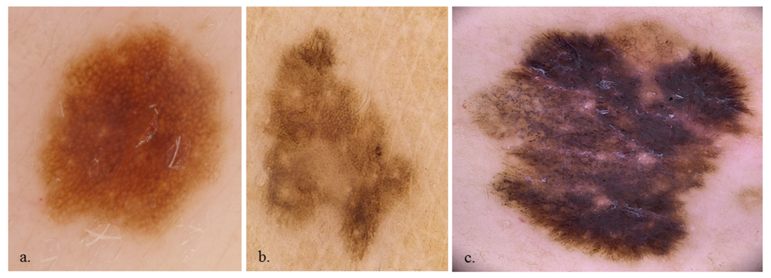
Super-High Magnification Dermoscopy in 190 Clinically Atypical Pigmented Lesions
Abstract
1. Introduction
2. Materials and Methods
2.1. Study Design
2.2. Setting
2.3. Participants
2.4. Data Sources
2.5. Variables
2.6. Statistical Analysis
3. Results
3.1. Participants and Lesion Data
3.2. ×20 Dermoscopy
3.3. ×400 Dermoscopy
3.4. Multivariate Analysis
4. Discussion
Supplementary Materials
Author Contributions
Funding
Informed Consent Statement
Data Availability Statement
Conflicts of Interest
References
- Garbe, C.; Peris, K.; Hauschild, A.; Saiag, P.; Middleton, M.; Spatz, A.; Grob, J.-J.; Malvehy, J.; Newton-Bishop, J.; Stratigos, A.; et al. Diagnosis and treatment of melanoma: European consensus-based interdisciplinary guideline. Eur. J. Cancer 2010, 46, 270–283. [Google Scholar] [CrossRef] [PubMed]
- Sacchetto, L.; Zanetti, R.; Comber, H.; Bouchardy, C.; Brewster, D.H.; Broganelli, P.; Chirlaque, M.D.; Coza, D.; Galceran, J.; Gavin, A.; et al. Trends in incidence of thick, thin and in situ melanoma in Europe. Eur. J. Cancer 2018, 92, 108–118. [Google Scholar] [CrossRef] [PubMed]
- Ferlay, J.; Soerjomataram, I.; Dikshit, R.; Eser, S.; Mathers, C.; Rebelo, M.; Parkin, D.M.; Forman, D.; Bray, F. Cancer incidence and mortality worldwide: Sources, methods and major patterns in GLOBOCAN 2012. Int. J. Cancer 2015, 136, E359–E386. [Google Scholar] [CrossRef] [PubMed]
- Iannacone, M.R.; Youlden, D.R.; Baade, P.D.; Aitken, J.F.; Green, A.C. Melanoma incidence trends and survival in adolescents and young adults in Queensland, Australia. Int. J. Cancer 2015, 136, 603–609. [Google Scholar] [CrossRef] [PubMed]
- Schadendorf, D.; van Akkooi, A.C.J.; Berking, C.; Griewank, K.G.; Gutzmer, R.; Hauschild, A.; Stang, A.; Roesch, A.; Ugurel, S. Melanoma. Lancet 2018, 392, 971–984. [Google Scholar] [CrossRef] [PubMed]
- Jerant, A.F.; Johnson, J.T.; Sheridan, C.D.; Caffrey, T.J. Early detection and treatment of skin cancer. Am. Fam. Physician 2000, 62, 357–368. [Google Scholar] [PubMed]
- Kittler, H.; Pehamberger, H.; Wolff, K.; Binder, M. Follow-up of melanocytic skin lesions with digital epiluminescence microscopy: Patterns of modifications observed in early melanoma, atypical nevi, and common nevi. J. Am. Acad. Dermatol. 2000, 43, 467–476. [Google Scholar] [CrossRef]
- Keung, E.Z.; Gershenwald, J.E. The eighth edition American Joint Committee on Cancer (AJCC) melanoma staging system: Implications for melanoma treatment and care. Expert Rev. Anticancer Ther. 2018, 18, 775–784. [Google Scholar] [CrossRef] [PubMed]
- Dinnes, J.; Deeks, J.J.; Chuchu, N.; Ferrante di Ruffano, L.; Matin, R.N.; Thomson, D.R.; Wong, K.Y.; Aldridge, R.B.; Abbott, R.; Fawzy, M.; et al. Dermoscopy, with and without visual inspection, for diagnosing melanoma in adults. Cochrane Database Syst. Rev. 2018, 12, Cd011902. [Google Scholar] [CrossRef] [PubMed]
- Vestergaard, M.E.; Macaskill, P.; Holt, P.E.; Menzies, S.W. Dermoscopy compared with naked eye examination for the diagnosis of primary melanoma: A meta-analysis of studies performed in a clinical setting. Br. J. Dermatol. 2008, 159, 669–676. [Google Scholar] [CrossRef] [PubMed]
- Cinotti, E.; Cortonesi, G.; Rubegni, P. High magnification and fluorescence advanced videodermoscopy for hypomelanotic melanoma. Skin Res. Technol. 2020, 26, 766–768. [Google Scholar] [CrossRef] [PubMed]
- Cinotti, E.; Labeille, B.; Debarbieux, S.; Carrera, C.; Lacarrubba, F.; Witkowski, A.M.; Moscarella, E.; Arzberger, E.; Kittler, H.; Bahadoran, P.; et al. Dermoscopy vs. reflectance confocal microscopy for the diagnosis of lentigo maligna. J. Eur. Acad. Dermatol. Venereol. 2018, 32, 1284–1291. [Google Scholar] [CrossRef] [PubMed]
- Cinotti, E.; Rossi, R.; Ferrara, G.; Tognetti, L.; Rubegni, P.; Perrot, J. Image Gallery: Super-high magnification dermoscopy can identify pigmented cells: Correlation with reflectance confocal microscopy. Br. J. Dermatol. 2019, 181, e1. [Google Scholar] [CrossRef] [PubMed]
- Cinotti, E.; Tognetti, L.; Campoli, M.; Liso, F.; Cicigoi, A.; Cartocci, A.; Rossi, R.; Rubegni, P.; Perrot, J.L. Super-high magnification dermoscopy can aid the differential diagnosis between melanoma and atypical naevi. Clin. Exp. Dermatol. 2021, 46, 1216–1222. [Google Scholar] [CrossRef] [PubMed]
- Cinotti, E.; Perrot, J.L.; Labeille, B.; Cambazard, F. Diagnosis of scabies by high-magnification dermoscopy: The “delta-wing jet” appearance of Sarcoptes scabiei. Ann. Dermatol. Venereol. 2013, 140, 722–723. [Google Scholar] [CrossRef] [PubMed]
- Puppin, D., Jr.; Salomon, D.; Saurat, J.-H. Amplified surface microscopy: Preliminary evaluation of a 400-fold magnification in the surface microscopy of cutaneous melanocytic lesions. J. Am. Acad. Dermatol. 1993, 28, 923–927. [Google Scholar] [CrossRef] [PubMed]
- Dusi, D.; Rossi, R.; Simonacci, M.; Ferrara, G. Image Gallery: The new age of dermoscopy: Optical super-high magnification. Br. J. Dermatol. 2018, 178, e330. [Google Scholar] [CrossRef] [PubMed]
- Cinotti, E.; Ekinde, S.; Labeille, B.; Raberin, H.; Tognetti, L.; Rubegni, P.; Perrot, J. Image Gallery: Pigmented hyphae can be identified in vivo by high and super-high magnification dermoscopy. Br. J. Dermatol. 2019, 181, e4. [Google Scholar] [CrossRef] [PubMed]
- Cinotti, E.; Bertello, M.; Donelli, C.; Rossi, R.; Tognetti, L.; Perrot, J.L.; Rubegni, P. Super-high magnification dermoscopy can detect Demodex folliculorum. J. Eur. Acad. Dermatol. Venereol. 2023, 37, e96–e97. [Google Scholar] [CrossRef] [PubMed]
- Scarfì, F.; Gori, A.; Topa, A.; Trane, L.; Dika, E.; Broganelli, P.; Massi, D.; De Giorgi, V. Image Gallery: In vivo fluorescence-advanced videodermatoscopy for the characterization of skin melanocytic pigmented lesions. Br. J. Dermatol. 2019, 180, e104. [Google Scholar] [CrossRef] [PubMed]


| Benign Lesions n = 117 | Malignant Lesions n = 73 | p-Value | |
|---|---|---|---|
| Age (years; SD) | 48.79 (19.19%) | 64.98 (16.70%) | <0.001 |
| Male | 68 (61.3%) | 31 (56.4%) | 0.846 |
| Female | 49 (38.7%) | 42 (43.6%) |
| Benign Lesions | Malignant Lesions | p-Value | |
|---|---|---|---|
| 7-point checklist | |||
| Atypical pigment network | 57 (48.7%) | 59 (80.8%) | <0.001 |
| Blue-whitish veil | 18 (15.4%) | 49 (67.1%) | <0.001 |
| Atypical vascular pattern | 1 (0.9%) | 12 (16.4%) | <0.001 |
| Irregular streaks | 11 (9.4%) | 22 (30.1%) | 0.001 |
| Regression structures | 33 (28.2%) | 49 (67.1%) | <0.001 |
| Irregular pigmentations | 40 (34.2%) | 23 (31.5%) | 0.823 |
| Irregular dots/globules | 36 (30.8%) | 18 (24.7%) | 0.457 |
| General pattern | |||
| Homogeneous | 32 (27.4%) | 43 (58.9%) | <0.001 |
| Globular | 41 (35.0%) | 7 (9.6%) | <0.001 |
| Network | 67 (57.3%) | 41 (56.2%) | 1.000 |
| Benign Lesions | Malignant Lesions | p-Value | |
|---|---|---|---|
| Cell presence | 113 (96.6%) | 72 (98.6%) | 0.695 |
| Keratinocytes | 109 (93.2%) | 64 (87.7%) | 0.304 |
| Roundish melanocytes | 35 (29.9%) | 36 (49.3%) | 0.011 |
| Dendritic melanocytes | 19 (16.2%) | 22 (30.1%) | 0.037 |
| Melanophages | 19 (16.2%) | 17 (23.3%) | 0.310 |
| Cell color | |||
| Black | 13 (11.1%) | 7 (9.6%) | 0.929 |
| Brown | 107 (91.5%) | 66 (90.4%) | 1.000 |
| Violet/blue | 45 (38.5%) | 29 (39.7%) | 0.983 |
| Cell irregularity in shape and size | 21 (17.9%) | 38 (52.1%) | <0.001 |
| Cell distribution: irregular arrangement | 19 (16.2%) | 30 (41.1%) | <0.001 |
| Roundish nests | 42 (35.9%) | 22 (30.1%) | 0.510 |
| Out-of-focus structureless areas | |||
| Bluish | 36 (30.8%) | 24 (32.9%) | 0.886 |
| Grey/brown | 23 (19.7%) | 11 (15.1%) | 0.543 |
| Vessels | 26 (22.2%) | 23 (31.5%) | 0.210 |
| Network | |||
| with edged papillae | 30 (25.6%) | 3 (4.1%) | <0.001 |
| without edged papillae | 18 (15.4%) | 19 (26.0%) | 0.107 |
| Angled nest | 9 (7.7%) | 16 (22.2%) | 0.008 |
| OR | p-Value | |
|---|---|---|
| Age | 1.05 (1.03–1.09) | <0.001 |
| Irregular melanocyte distribution | 4.69 (1.58–15.45) | 0.007 |
| Network with edged papillae | 0.16 (0.03–0.57) | 0.009 |
Disclaimer/Publisher’s Note: The statements, opinions and data contained in all publications are solely those of the individual author(s) and contributor(s) and not of MDPI and/or the editor(s). MDPI and/or the editor(s) disclaim responsibility for any injury to people or property resulting from any ideas, methods, instructions or products referred to in the content. |
© 2023 by the authors. Licensee MDPI, Basel, Switzerland. This article is an open access article distributed under the terms and conditions of the Creative Commons Attribution (CC BY) license (https://creativecommons.org/licenses/by/4.0/).
Share and Cite
Cinotti, E.; Cioppa, V.; Tognetti, L.; Perrot, J.L.; Rossi, R.; Gnone, M.; Cartocci, A.; Rubegni, P.; Cortonesi, G. Super-High Magnification Dermoscopy in 190 Clinically Atypical Pigmented Lesions. Diagnostics 2023, 13, 2238. https://doi.org/10.3390/diagnostics13132238
Cinotti E, Cioppa V, Tognetti L, Perrot JL, Rossi R, Gnone M, Cartocci A, Rubegni P, Cortonesi G. Super-High Magnification Dermoscopy in 190 Clinically Atypical Pigmented Lesions. Diagnostics. 2023; 13(13):2238. https://doi.org/10.3390/diagnostics13132238
Chicago/Turabian StyleCinotti, Elisa, Vittoria Cioppa, Linda Tognetti, Jean Luc Perrot, Renato Rossi, Matteo Gnone, Alessandra Cartocci, Pietro Rubegni, and Giulio Cortonesi. 2023. "Super-High Magnification Dermoscopy in 190 Clinically Atypical Pigmented Lesions" Diagnostics 13, no. 13: 2238. https://doi.org/10.3390/diagnostics13132238
APA StyleCinotti, E., Cioppa, V., Tognetti, L., Perrot, J. L., Rossi, R., Gnone, M., Cartocci, A., Rubegni, P., & Cortonesi, G. (2023). Super-High Magnification Dermoscopy in 190 Clinically Atypical Pigmented Lesions. Diagnostics, 13(13), 2238. https://doi.org/10.3390/diagnostics13132238


