
Dermatofibromas with Aberrant Expression of CD34 Protein: A Systematic Review and a Reappraisal of Clinicopathological Features and Histogenesis


Abstract
1. Background
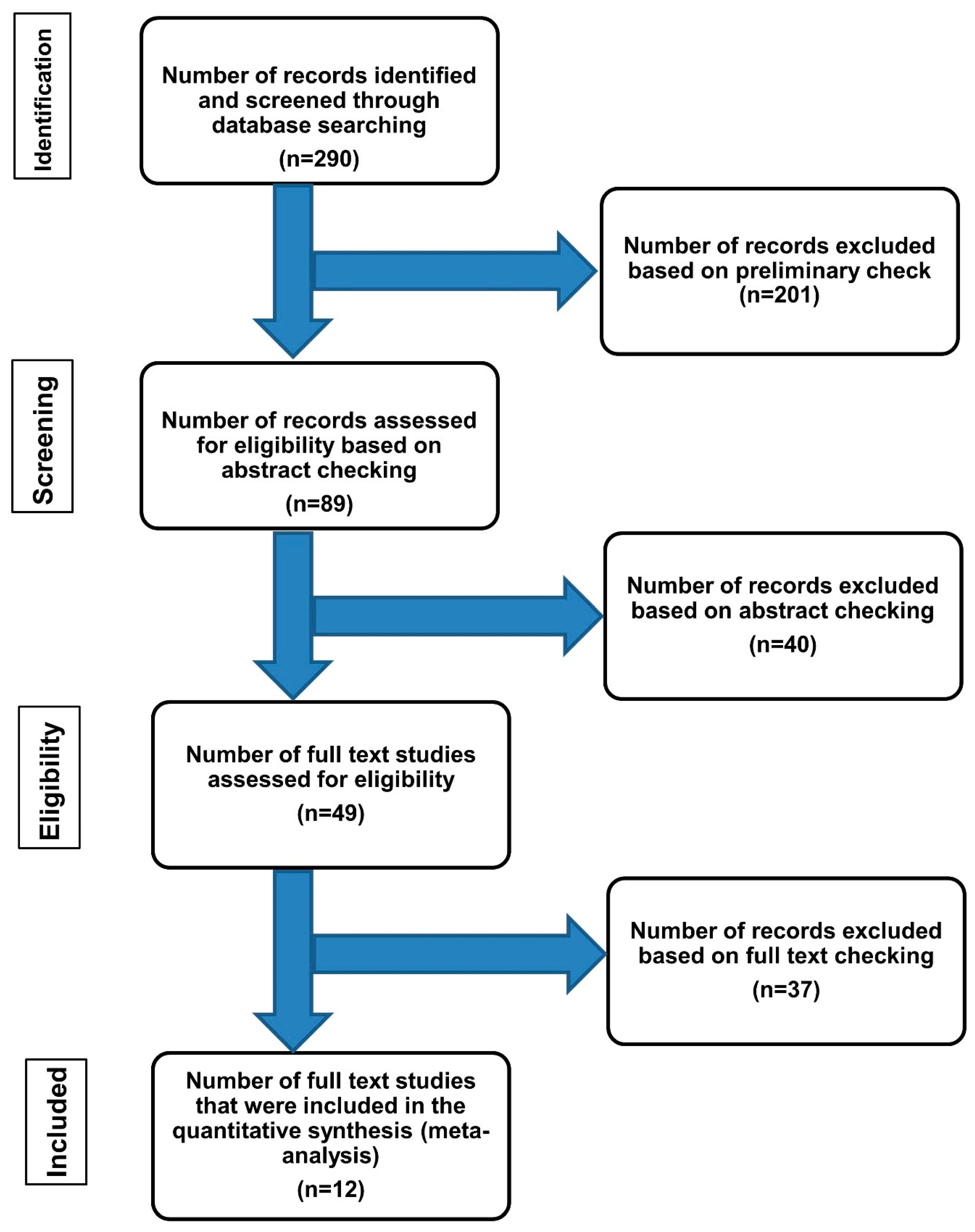
2. Methods
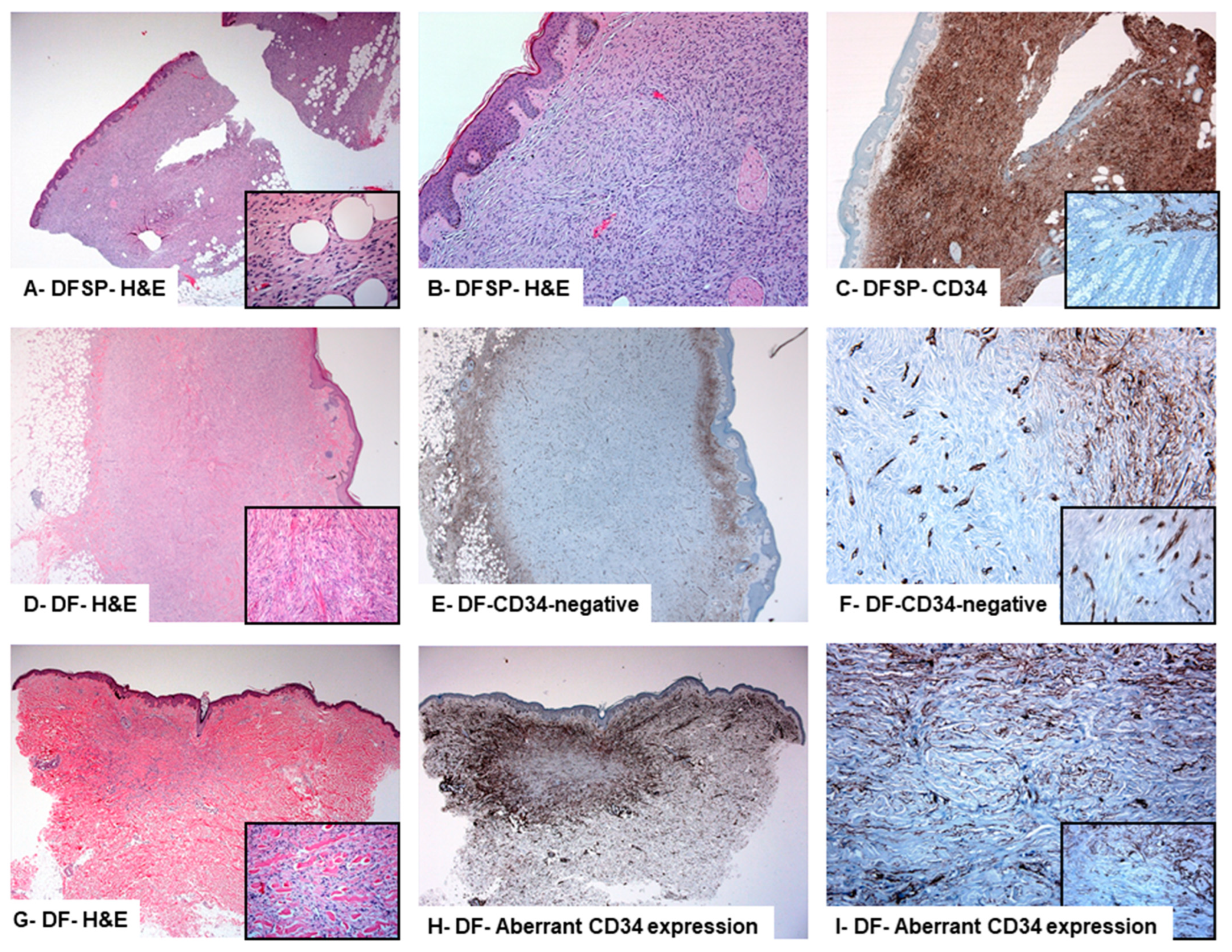
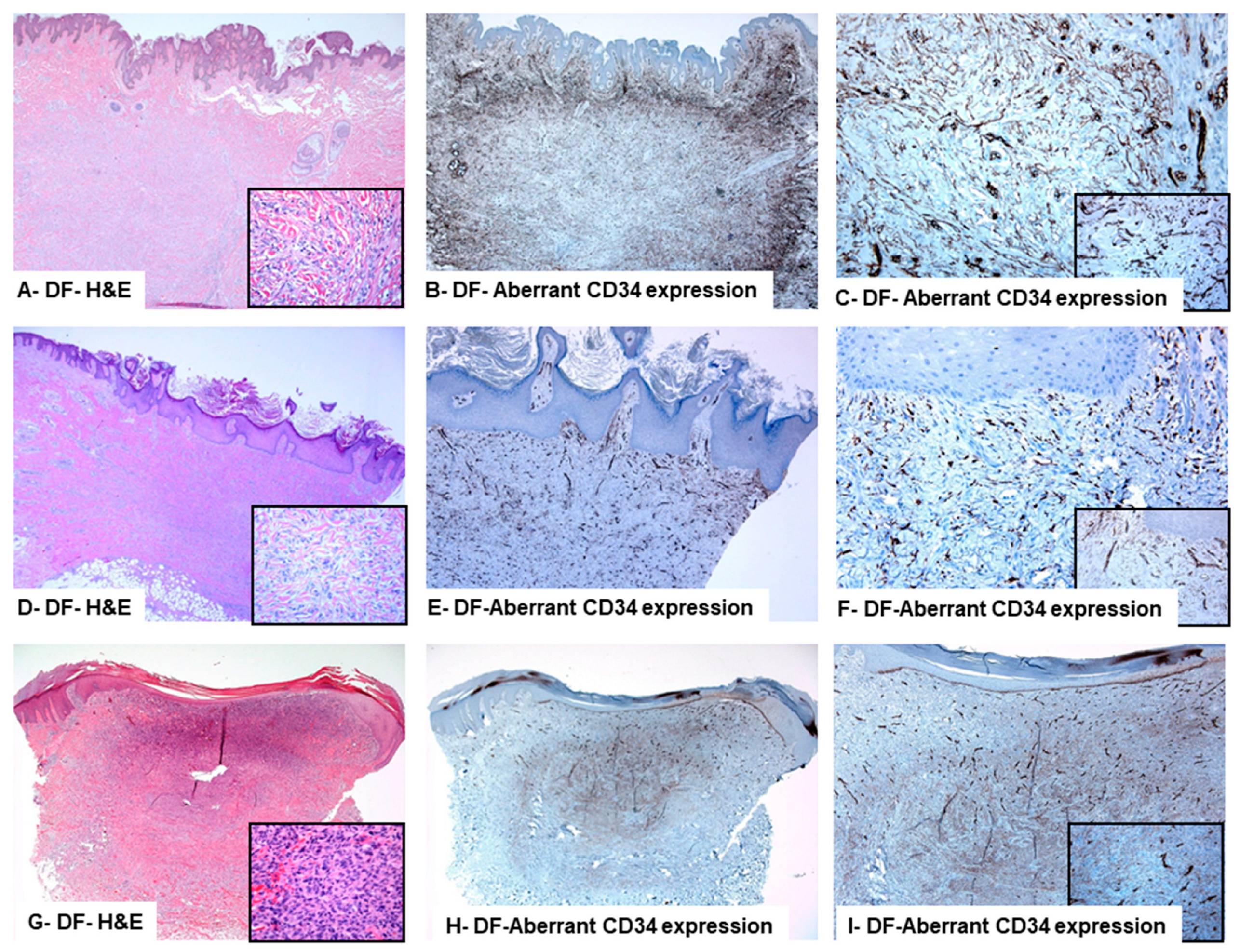
3. Results
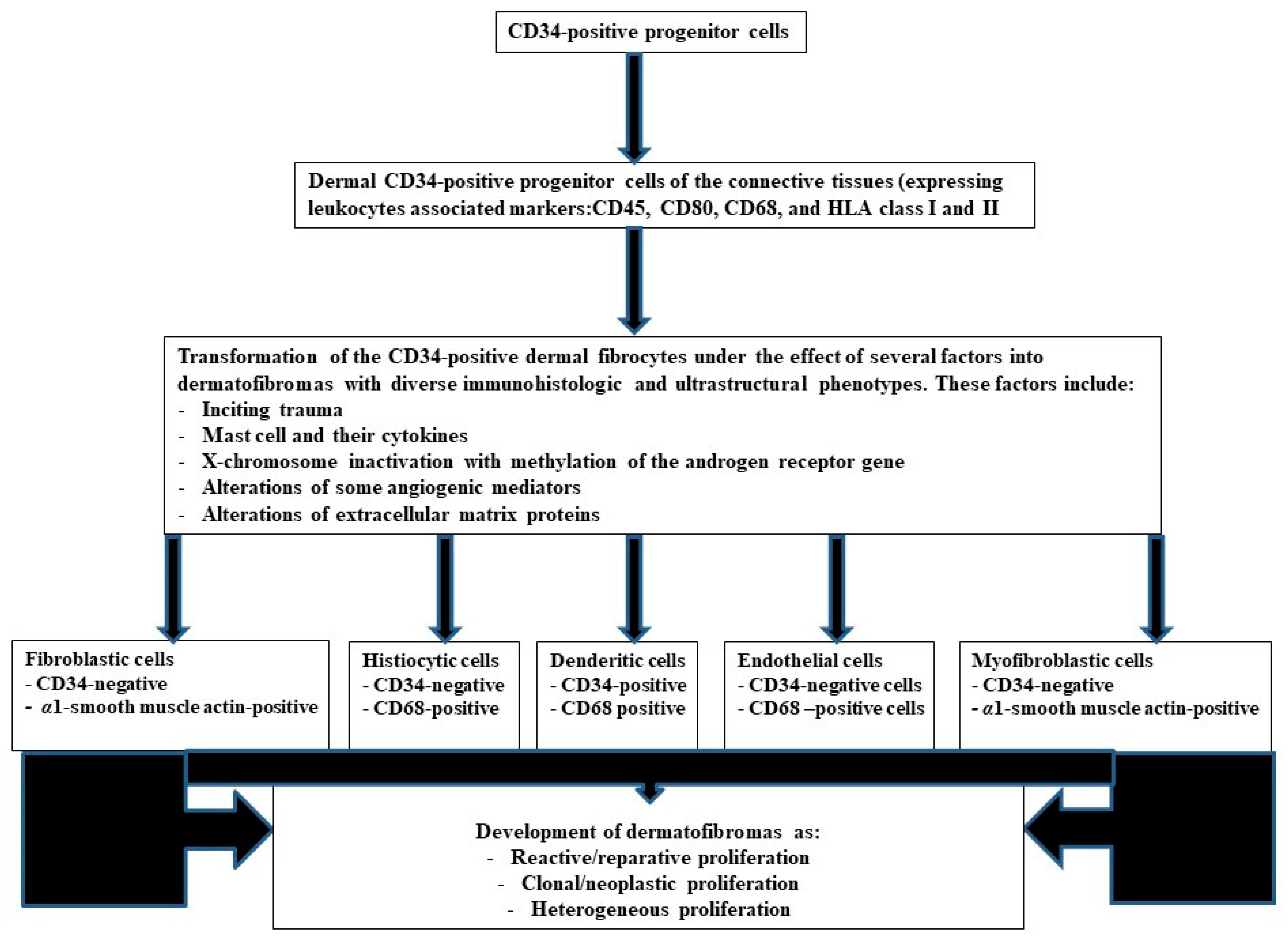
4. Discussion
5. Conclusions
6. Recommendations
Author Contributions
Funding
Institutional Review Board Statement
Informed Consent Statement
Data Availability Statement
Conflicts of Interest
Abbreviations
| DF | dermatofibroma |
| H & E | Hematoxylin and eosin |
| CD34 | Cluster of differentiation 34 |
| PRISMA | Preferred reporting items for systematic reviews and meta-analysis |
| IHC | Immunohistochemistry |
| IGFBP7 | Insulin-like growth factor-binding protein 7 |
| Matrix metalloproteinase family member | MMP-11 |
| HMGA1 and HMGA2 | High-Mobility Group Proteins |
| PDGFB gene | Platelet-derived growth factor-beta chain |
| COL1A1 gene | Collagen type 1 alpha 1 gene |
| FISH | Fluorescence in situ hybridization |
| RT-PCR | Multiplex reverse transcriptase-polymerase chain reaction |
References
- Myers, D.J.; Fillman, E.P. Dermatofibroma; StatPearls: Treasure Island, FL, USA, 2020. [Google Scholar]
- Katenkamp, D.; Stiller, D. Cellular composition of the so-called dermatofibroma (histiocytoma cutis). Virchows Arch. A Pathol. Anat. Histol. 1975, 367, 325–336. [Google Scholar] [CrossRef]
- Song, Y.; Sakamoto, F.; Ito, M. Characterization of factor XIIIa+ dendritic cells in dermatofibroma: Immunohistochemical, electron and immunoelectron microscopical observations. J. Dermatol. Sci. 2005, 39, 89–96. [Google Scholar] [CrossRef]
- Quintana, F.S.L.; Almeida, H.L., Jr.; Ruas, C.P.; Jorge, V.M. Scanning electron microscopy of dermatofibroma. An. Bras. Dermatol. 2019, 94, 358–360. [Google Scholar] [CrossRef]
- Cazzato, G.; Colagrande, A.; Cimmino, A.; Marrone, M.; Stellacci, A.; Arezzo, F.; Lettini, T.; Resta, L.; Ingravallo, G. Granular Cell Dermatofibroma: When Morphology Still Matters. Dermatopathology 2021, 8, 371–375. [Google Scholar] [CrossRef]
- Aloi, F.; Albertazzi, D.; Pippione, M. Dermatofibroma with granular cells: A report of two cases. Dermatology 1999, 199, 54–56. [Google Scholar] [CrossRef]
- LeBoit, P.E.; Barr, R.J.; Burall, S.; Metcalf, J.S.; Yen, T.S.; Wick, M.R. Primitive polypoid granular-cell tumor and other cutaneous granular-cell neoplasms of apparent nonneural origin. Am. J. Surg. Pathol. 1991, 15, 48–58. [Google Scholar] [CrossRef]
- Hussein, M.R. Evaluation of angiogenesis in normal and lichen planus skin by CD34 protein immunohistochemistry: Preliminary findings. Cell Biol. Int. 2007, 31, 1292–1295. [Google Scholar] [CrossRef]
- Gutiérrez, R.; García, M.P.; Sáez, F.; Diaz-Flores, L.; Valladares, F.; Madrid, J.F. CD34+ stromal cells/fibroblasts/fibrocytes/telocytes as a tissue reserve and a principal source of mesenchymal cells. Location, morphology, function and role in pathology. Histol. Histopathol. 2014, 29, 831–870. [Google Scholar]
- Sidney, L.E.; Branch, M.J.; Dunphy, S.E.; Dua, H.S.; Hopkinson, A. Concise review: Evidence for CD34 as a common marker for diverse progenitors. Stem. Cells 2014, 32, 1380–1389. [Google Scholar] [CrossRef]
- Monge, M.; Chauveau, D.; Cordonnier, C.; Noël, L.H.; Presne, C.; Makdassi, R.; Jauréguy, M.; Lecaque, C.; Renou, M.; Grünfeld, J.P.; et al. Localized amyloidosis of the genitourinary tract: Report of 5 new cases and review of the literature. Medicine 2011, 90, 212–222. [Google Scholar] [CrossRef]
- Zelger, B.G.; Calonje, E.; Zelger, B. Myxoid dermatofibroma. Histopathology 1999, 34, 357–364. [Google Scholar] [CrossRef] [PubMed]
- Zelger, B.W.; Zelger, B.G.; Rappersberger, K. Prominent myofibroblastic differentiation. A pitfall in the diagnosis of dermatofibroma. Am. J. Dermatopathol. 1997, 19, 138–146. [Google Scholar] [CrossRef] [PubMed]
- Zelger, B.G.; Steiner, H.; Kutzner, H.; Rutten, A.; Zelger, B. Granular cell dermatofibroma. Histopathology 1997, 31, 258–262. [Google Scholar] [CrossRef] [PubMed]
- Kiyohara, T.; Kumakiri, M.; Kobayashi, H.; Ohkawara, A.; Lao, L.M. Atrophic dermatofibroma. Elastophagocytosis by the tumor cells. J. Cutan. Pathol. 2000, 27, 312–315. [Google Scholar] [CrossRef]
- Erdag, G.; Qureshi, H.S.; Patterson, J.W.; Wick, M.R. CD34-positive dendritic cells disappear from scars but are increased in pericicatricial tissue. J. Cutan. Pathol. 2008, 35, 752–756. [Google Scholar] [CrossRef]
- Hussein, M.R.; Al-Badaiwy, Z.H.; Guirguis, M.N. Analysis of p53 and bcl-2 protein expression in the non-tumorigenic, pretumorigenic, and tumorigenic keratinocytic hyperproliferative lesions. J. Cutan. Pathol. 2004, 31, 643–651. [Google Scholar] [CrossRef]
- Hantschmann, P.; Sterzer, S.; Jeschke, U.; Friese, K. P53 expression in vulvar carcinoma, vulvar intraepithelial neoplasia, squamous cell hyperplasia and lichen sclerosus. Anticancer. Res. 2005, 25, 1739–1745. [Google Scholar]
- Hasby, E.A.; El Mashad, N.; Eltatawy, R. C-Kit, CD34 & alpha-SMA Immunohistochemical Features in Classic Kaposi Sarcoma and Kaposiform Hemangioendothelioma. J. Microsc. Ultrastruct. 2017, 5, 49–57. [Google Scholar]
- Lisovsky, M.; Hoang, M.P.; Dresser, K.A.; Kapur, P.; Bhawan, J.; Mahalingam, M. Apolipoprotein D in CD34-positive and CD34-negative cutaneous neoplasms: A useful marker in differentiating superficial acral fibromyxoma from dermatofibrosarcoma protuberans. Mod. Pathol. 2008, 21, 31–38. [Google Scholar] [CrossRef][Green Version]
- Sadullahoglu, C.; Dere, Y.; Atasever, T.R.; Oztop, M.T.; Karaaslan, O. The Role of CD34 and D2-40 in the Differentiation of Dermatofibroma and Dermatofibrosarcoma Protuberans. Turk. Patoloji Derg. 2017, 1, 223–227. [Google Scholar] [CrossRef]
- Li, J.; Yu, Y.; Yang, Y.; Wang, L.; Cao, J.; Liang, X.; Xiao, X.; Tu, Y.; Chen, H. IGFBP7, a novel immunohistochemical marker in differentiating dermatofibroma from dermatofibrosarcoma protuberans. J. Eur. Acad. Dermatol. Venereol. 2012, 26, 382–385. [Google Scholar] [CrossRef] [PubMed]
- Chen, Y.T.; Chen, W.T.; Huang, W.T.; Wu, C.C.; Chai, C.Y. Expression of MMP-2, MMP-9 and MMP-11 in dermatofibroma and dermatofibrosarcoma protuberans. Kaohsiung J. Med. Sci. 2012, 28, 545–549. [Google Scholar] [CrossRef] [PubMed]
- Xiong, Y.; Guo, H.; Zhang, S.; Zhang, B.; Li, T. Differences of the molecular phenotypes and the histogenesis between dermatofibroma and dermatofibrosarcoma protuberans. Beijing Da Xue Xue Bao Yi Xue Ban 2008, 40, 395–400. [Google Scholar] [PubMed]
- Kim, H.; Lee, J.; Kim, S.; Seo, Y.-J.; Park, J.; Kim, M.; Cinn, Y.; Cho, K.; Yoon, T. Stromelysin-3 expression in the differential diagnosis of dermatofibroma and dermatofibrosarcoma protuberans: Comparison with factor XIIIa and CD34. Br. J. Dermatol. 2007, 157, 319–324. [Google Scholar] [CrossRef]
- Sachdev, R.; Sundram, U. Expression of CD163 in dermatofibroma, cellular fibrous histiocytoma, and dermatofibrosarcoma protuberans: Comparison with CD68, CD34, and Factor XIIIa. J. Cutan. Pathol. 2006, 33, 353–360. [Google Scholar] [CrossRef]
- Li, N.; McNiff, J.; Hui, P.; Manfioletti, G.; Tallini, G. Differential expression of HMGA1 and HMGA2 in dermatofibroma and dermatofibrosarcoma protuberans: Potential diagnostic applications, and comparison with histologic findings, CD34, and factor XIIIa immunoreactivity. Am. J. Dermatopathol. 2004, 26, 267–272. [Google Scholar] [CrossRef]
- Kahn, H.J.; Fekete, E.; From, L. Tenascin differentiates dermatofibroma from dermatofibrosarcoma protuberans: Comparison with CD34 and factor XIIIa. Hum. Pathol. 2001, 32, 50–56. [Google Scholar] [CrossRef]
- Goldblum, J.R.; Tuthill, R.J. CD34 and factor-XIIIa immunoreactivity in dermatofibrosarcoma protuberans and dermatofibroma. Am. J. Dermatopathol. 1997, 19, 147–153. [Google Scholar] [CrossRef]
- Hsi, E.D.; Nickoloff, B.J. Dermatofibroma and dermatofibrosarcoma protuberans: An immunohistochemical study reveals distinctive antigenic profiles. J. Dermatol. Sci. 1996, 11, 1–9. [Google Scholar] [CrossRef]
- Zelger, B.W.; Ofner, D.; Zelger, B.G. Atrophic variants of dermatofibroma and dermatofibrosarcoma protuberans. Histopathology 1995, 26, 519–527. [Google Scholar] [CrossRef]
- Zelger, B.; Sidoroff, A.; Stanzl, U.; Fritsch, P.O.; Öfner, D.; Zelger, B.; Jasani, B.; Schmid, K.W. Deep penetrating dermatofibroma versus dermatofibrosarcoma protuberans. A clinicopathologic comparison. Am. J. Surg. Pathol. 1994, 18, 677–686. [Google Scholar] [CrossRef] [PubMed]
- Dominguez-Malagon, H.; Valdez-Carrillo Mdel, C.; Cano-Valdez, A.M. Dermatofibroma and dermatofibrosarcoma protuberans: A comparative ultrastructural study. Ultrastruct. Pathol. 2006, 30, 283–291. [Google Scholar] [CrossRef] [PubMed]
- Desmouliere, A.; Geinoz, A.; Gabbiani, F.; Gabbiani, G. Transforming growth factor-beta 1 induces alpha-smooth muscle actin expression in granulation tissue myofibroblasts and in quiescent and growing cultured fibroblasts. J. Cell Biol. 1993, 122, 103–111. [Google Scholar] [CrossRef] [PubMed]
- Cohen, P.R.; Erickson, C.P.; Calame, A. Atrophic Dermatofibroma: A Comprehensive Literature Review. Dermatol. Ther. 2019, 9, 449–468. [Google Scholar] [CrossRef] [PubMed]
- Prieto, V.G.; Reed, J.A.; Shea, C.R. Immunohistochemistry of dermatofibromas and benign fibrous histiocytomas. J. Cutan. Pathol. 1995, 22, 336–341. [Google Scholar] [CrossRef] [PubMed]
- Aiba, S.; Tagami, H. Inverse correlation between CD34 expression and proline-4-hydroxylase immunoreactivity on spindle cells noted in hypertrophic scars and keloids. J. Cutan. Pathol. 1997, 24, 65–69. [Google Scholar]
- Hui, P.; Glusac, E.J.; Sinard, J.H.; Perkins, A.S. Clonal analysis of cutaneous fibrous histiocytoma (dermatofibroma). J. Cutan. Pathol. 2002, 29, 385–389. [Google Scholar] [CrossRef]
- Mentzel, T.; Wiesner, T.; Cerroni, L.; Hantschke, M.; Kutzner, H.; Rütten, A.; Häberle, M.; Bisceglia, M.; Chibon, F.; Coindre, J.-M. Malignant dermatofibroma: Clinicopathological, immunohistochemical, and molecular analysis of seven cases. Mod. Pathol. 2013, 26, 256–267. [Google Scholar] [CrossRef]
- Taniuchi, K.; Yamada, Y.; Nonomura, A.; Takehara, K. Immunohistochemical analysis of platelet-derived growth factor and its receptors in fibrohistiocytic tumors. J. Cutan. Pathol. 1997, 24, 393–397. [Google Scholar] [CrossRef]
- Wang, W.-L.; Patel, K.U.; Coleman, N.M.; Smith-Zagone, M.J.; Ivan, D.; A Reed, J.; López-Terrada, D.; Lazar, A.J.F.; Prieto, V.G. COL1A1, PDGFB chimeric transcripts are not present in indeterminate fibrohistiocytic lesions of the skin. Am. J. Dermatopathol. 2010, 32, 149–153. [Google Scholar] [CrossRef]


| Studies | Dermatofibromas with Aberrant CD34 Protein Expression/Total Cases | Contributions of Each Study | References |
|---|---|---|---|
| 1 | 7/30 (23%) | A broad panel of immunostains is needed in DFs with CD34 positivity | [21] |
| 2 | 4/30 (13%) | A combination of CD34, FXIIIa, and Stromelysin-3 immunostains can separate DFs from DFSPs | [22] |
| 3 | 4/19 (21%) | The expressions of extracellular matrix proteins play a role in the development of DFs | [23] |
| 4 | 1/26 (3%) | A combination of CD34 and FXIIIa immune markers can separate DFs from DFSPs | [24] |
| 5 | 4/23 (17%) | A combination of CD34 and Stromelysin-3 immunostains can help to separate DFs from DFSPs | [25] |
| 6 | 1/19(5%) | The inclusion of the CD163 marker (hemoglobin scavenger receptor) can help to separate DFs from DFSPs | [26] |
| 7 | 8/22 (36%) | The use of HMGA1 and HMGA2 (members of the high mobility group protein family genes) immune markers can help to separate DFs from DFSPs | [27] |
| 8 | 5/20 (25%) | The overexpression of tenascin at the dermal-epidermal junction overlying the lesion in DFs but not in DFSPs helps separate these tumors. | [28] |
| 9 | 12/30 (40%) | A combination of CD34 and FXIIIa can separate DFs from DFSP | [29] |
| 10 | 1/13 (7%) | There is no convincing evidence indicating the derivation of DFs from cells with the vascular or hematopoietic origin | [30] |
| 11 | 2/26 (7%) | Atrophic variants of DFSP and DFs represent distinct entities that can be separated by the use of immunostains such as CD34, Factor XIIIa, and metallothionein | [31] |
| 12 | 2/20 (10%) | The deep penetrating DFs and DFSP represent distinct entities | [32] |
| Studies | Ultrastructural Findings | Number of Cases of Dermatofibromas | References |
|---|---|---|---|
| 1 | Spindle cells and dense collagen with the mesh-like appearance | 2 cases | [4] |
| 2 | Multiple capillary vessels having prominent endothelium and a perivascular ovoid or spindled cells showing intracytoplasmic lipid material and subplasmalemmal densities but lacking cell processes | 10 cases | [33] |
| 3 | Cells with histiocytic and fibroblastic features | 11 cases | [3] |
| 4 | Cells with phagocytized elastic fibers | Atrophic variant (a single case) | [15] |
| 5 | Fibrocytes amid fibrillary collagen and pools of mucin | Myxoid variant (7 cases) | [12] |
| 6 | Cells with abundant endoplasmic reticulum and Golgi complex, several intermediate filaments | Myofibroblastic variant (36 cases) | [13] |
| 7 | Cells with pools of phagolysosomes and glycogen granules | Granular variant (5 cases) | [14] |
| 8 | Most of the cells are Fibroblast-like and histiocyte-like, showing numerous rough endoplasmic reticulum, free ribosomes, bundles of filaments, macropinocytosis vesicles, and a basement membrane-like material on the outer cell surface. Some cells resembling smooth muscle | 9 cases | [2] |
| No of Case | Age | Sex | Localization | Recurrence or Distant Metastasis | IHC | ||||
|---|---|---|---|---|---|---|---|---|---|
| CD34 % of positive cells | Factor XIIIa | D2-40 | S100 | Ki67 | |||||
| 1 | 57 | Male | Upper arm | None | 35 | + | + | - | 0.0% |
| 2 | 48 | Male | Upper back | None | 35 | + | + | - | 1% |
| 3 | 57 | Female | Shoulder | None | 60 | + | + | - | 1% |
| 4 | 41 | Female | Left leg | None | 60 | + | + | - | 1% |
| 5 | 41 | Male | Left forearm | None | 60 | + | + | - | 0.0% |
| 6 | 30 | Male | Left hip | None | 75 | + | + | - | 1% |
| 7 | 45 | Male | Left scapula | None | 60 | + | + | - | 1% |
| 8 | 53 | Female | Foot right | None | 35 | + | + | - | 1% |
| 9 | 34 | Male | Shoulder | None | 60 | + | + | - | 0.0% |
| 10 | 35 | Male | Left thigh | None | 60 | + | + | - | 1% |
| 11 | 49 | Male | Left thigh | None | 60 | + | + | - | 0.0% |
| Cases | Age | Sex | Site | Recurrence or Distant Metastasis | IHC | ||||
|---|---|---|---|---|---|---|---|---|---|
| CD34 % of positive cells | Factor XIIIa | D2-40 | S100 | Ki 67 | |||||
| 1 | 52 | Female | Right deltoid | None | 0 | + | + | - | 0.0% |
| 2 | 41 | Female | Left leg | None | 0 | + | + | - | 0.0% |
| 3 | 42 | Female | Chest wall | None | 0 | + | + | - | 0.0% |
| 4 | 45 | Male | Upper back | None | 5 | + | + | - | 1% |
| 5 | 81 | Female | Upper arm | None | 0 | + | + | - | 0.0% |
| 6 | 38 | Female | Left thigh | None | 5 | + | + | - | 1% |
| 7 | 63 | Female | Right leg | None | 5 | + | + | - | 0.0% |
| 8 | 52 | Male | Left forearm | None | 0 | + | + | - | 1% |
| 9 | 58 | Female | Leg | None | 5 | + | + | - | 0.0% |
| 10 | 75 | Male | Left lower leg | None | 0 | + | + | - | 1% |
| 11 | 72 | Female | Upper back | None | 0 | + | + | - | 0.0% |
| 12 | 21 | Female | Left leg | None | 5 | + | + | - | 1% |
| 13 | 39 | Female | Left leg | None | 0 | + | + | - | 1% |
| 14 | 10 | Female | Right cheek | None | 0 | + | + | - | 0.0% |
| 15 | 24 | Female | Left-arm | None | 0 | + | + | - | 0.0% |
Disclaimer/Publisher’s Note: The statements, opinions and data contained in all publications are solely those of the individual author(s) and contributor(s) and not of MDPI and/or the editor(s). MDPI and/or the editor(s) disclaim responsibility for any injury to people or property resulting from any ideas, methods, instructions or products referred to in the content. |
© 2023 by the authors. Licensee MDPI, Basel, Switzerland. This article is an open access article distributed under the terms and conditions of the Creative Commons Attribution (CC BY) license (https://creativecommons.org/licenses/by/4.0/).
Share and Cite
Hussein, M.R.A.; Abdelwahed Hussein, T.M.R. Dermatofibromas with Aberrant Expression of CD34 Protein: A Systematic Review and a Reappraisal of Clinicopathological Features and Histogenesis. Diagnostics 2023, 13, 185. https://doi.org/10.3390/diagnostics13020185
Hussein MRA, Abdelwahed Hussein TMR. Dermatofibromas with Aberrant Expression of CD34 Protein: A Systematic Review and a Reappraisal of Clinicopathological Features and Histogenesis. Diagnostics. 2023; 13(2):185. https://doi.org/10.3390/diagnostics13020185
Chicago/Turabian StyleHussein, Mahmoud Rezk Abdelwahed, and Toka Mahmoud Rezk Abdelwahed Hussein. 2023. "Dermatofibromas with Aberrant Expression of CD34 Protein: A Systematic Review and a Reappraisal of Clinicopathological Features and Histogenesis" Diagnostics 13, no. 2: 185. https://doi.org/10.3390/diagnostics13020185
APA StyleHussein, M. R. A., & Abdelwahed Hussein, T. M. R. (2023). Dermatofibromas with Aberrant Expression of CD34 Protein: A Systematic Review and a Reappraisal of Clinicopathological Features and Histogenesis. Diagnostics, 13(2), 185. https://doi.org/10.3390/diagnostics13020185