Review

Prenatal Air Pollution Exposure and Placental DNA Methylation Changes: Implications on Fetal Development and Future Disease Susceptibility

1
Discipline of Medical Biochemistry, School of Laboratory Medicine and Medical Sciences, College of Health Sciences, University of KwaZulu-Natal, Durban 4041, South Africa
2
Discipline of Occupational and Environmental Health, School of Nursing and Public Health, College of Health Sciences, University of KwaZulu-Natal, Durban 4041, South Africa
*
Author to whom correspondence should be addressed.
Cells 2021, 10(11), 3025; https://doi.org/10.3390/cells10113025
Submission received: 4 August 2021 / Revised: 22 September 2021 / Accepted: 23 September 2021 / Published: 5 November 2021
(This article belongs to the Section Reproductive Cells and Development)

Abstract

:
The Developmental Origins of Health and Disease (DOHaD) concept postulates that in utero exposures influence fetal programming and health in later life. Throughout pregnancy, the placenta plays a central role in fetal programming; it regulates the in utero environment and acts as a gatekeeper for nutrient and waste exchange between the mother and the fetus. Maternal exposure to air pollution, including heavy metals, can reach the placenta, where they alter DNA methylation patterns, leading to changes in placental function and fetal reprogramming. This review explores the current knowledge on placental DNA methylation changes associated with prenatal air pollution (including heavy metals) exposure and highlights its effects on fetal development and disease susceptibility. Prenatal exposure to air pollution and heavy metals was associated with altered placental DNA methylation at the global and promoter regions of genes involved in biological processes such as energy metabolism, circadian rhythm, DNA repair, inflammation, cell differentiation, and organ development. The altered placental methylation of these genes was, in some studies, associated with adverse birth outcomes such as low birth weight, small for gestational age, and decreased head circumference. Moreover, few studies indicate that DNA methylation changes in the placenta were sex-specific, and infants born with altered placental DNA methylation patterns were predisposed to developing neurobehavioral abnormalities, cancer, and atopic dermatitis. These findings highlight the importance of more effective and stricter environmental and public health policies to reduce air pollution and protect human health.

1. Introduction

Air pollution is an environmental problem that threatens human health and is a major cause of mortality worldwide. In 2019, air pollution caused an estimated 6.67 million deaths, accounting for 12% of all deaths globally [1]. Pregnant women and their developing fetuses are particularly vulnerable to the adverse health effects of air pollution. In pregnant women, the respiratory adaptation to pregnancy leads to an increase in tidal volume and an increase in oxygen consumption [2,3]. Air pollution particles, due to their small size, are inhaled into the lungs, and the smallest particles infiltrate into the bloodstream reaching the placenta and fetus [4,5,6]. Such in utero exposures can affect fetal development, cause adverse birth outcomes, and increase the risk of developing certain diseases in later life, as postulated by the Developmental Origins of Health and Disease (DOHaD) concept [7,8,9,10].
Substantial evidence has associated prenatal air pollution exposure with a range of adverse health outcomes including gestational diabetes [11], preeclampsia [12,13], spontaneous abortions [14,15], preterm births [16,17], low birth weights [18], macrosomia [19], and stillbirths [20]. Furthermore, air pollution exposure during pregnancy was associated with an increased risk of developing cardiovascular diseases [21], neurodevelopmental alterations [22], respiratory problems [23,24], and cancer [25,26]. However, due to the complex composition of air pollution and the intricate processes involved in fetal development, the mechanisms by which air pollution causes these adverse health effects are not yet completely understood.
During the entire pregnancy, the placenta serves as a functional interface that connects the mother to the developing fetus [27]. It secretes hormones and regulates the in utero environment for optimum fetal growth and development. Subsequently, the placenta transfers nutrients from the mother to the fetus and regulates gas and waste exchanges [27]. In this way, the placenta plays a central role in fetal programming, and we suggest that altered placental physiology and function, possibly through epigenetic modifications such as DNA methylation, can provide a mechanism linking prenatal air pollution exposure with pregnancy complications, fetal growth abnormalities, altered newborn phenotypes, and an increased risk of developing certain diseases during the lifespan. This review explores the current knowledge on placental DNA methylation changes associated with prenatal air pollution (including heavy metals) exposure and highlights its effects on fetal development and disease susceptibility.

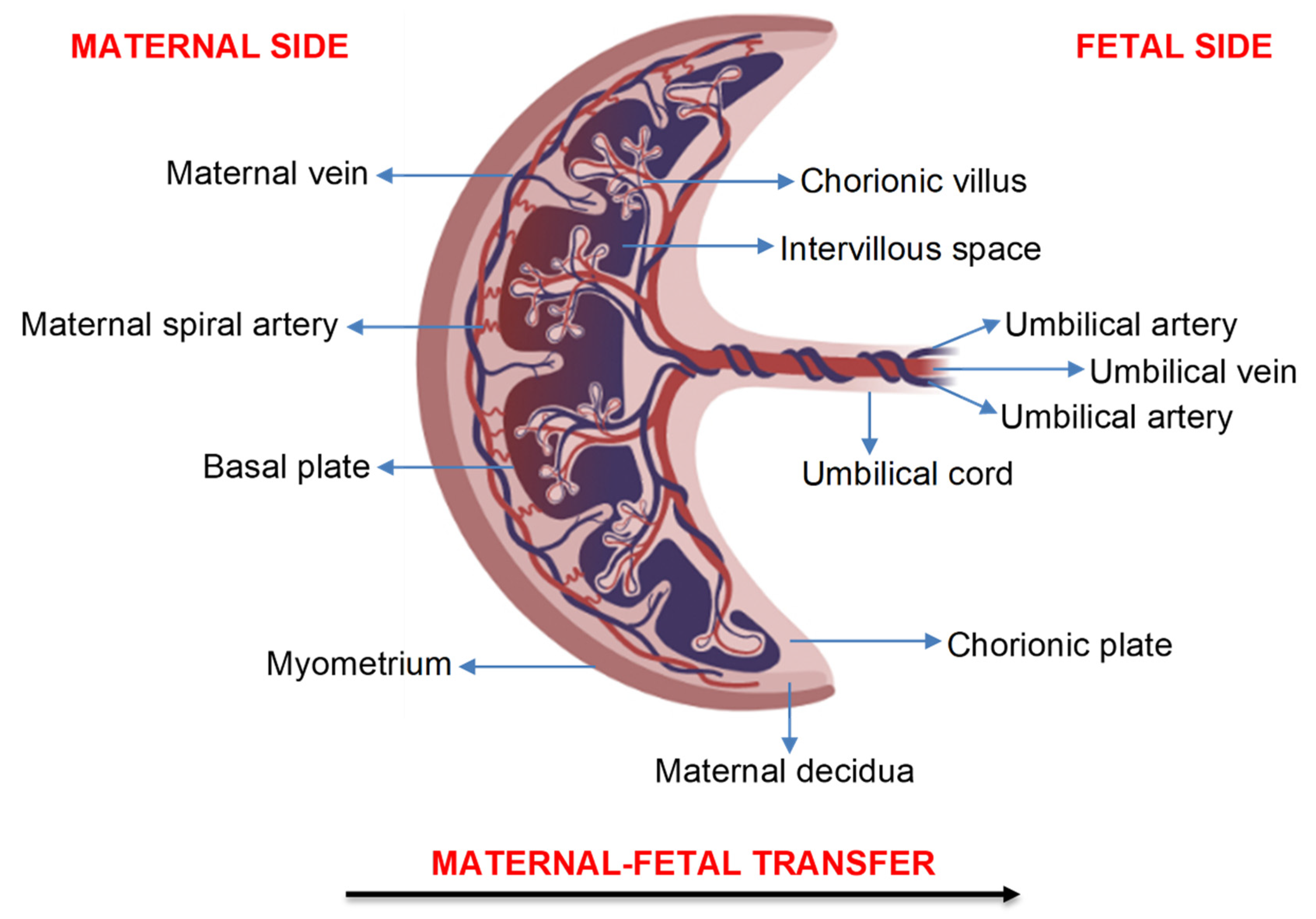
2. Transfer of Air Pollution Particles across the Human Placenta
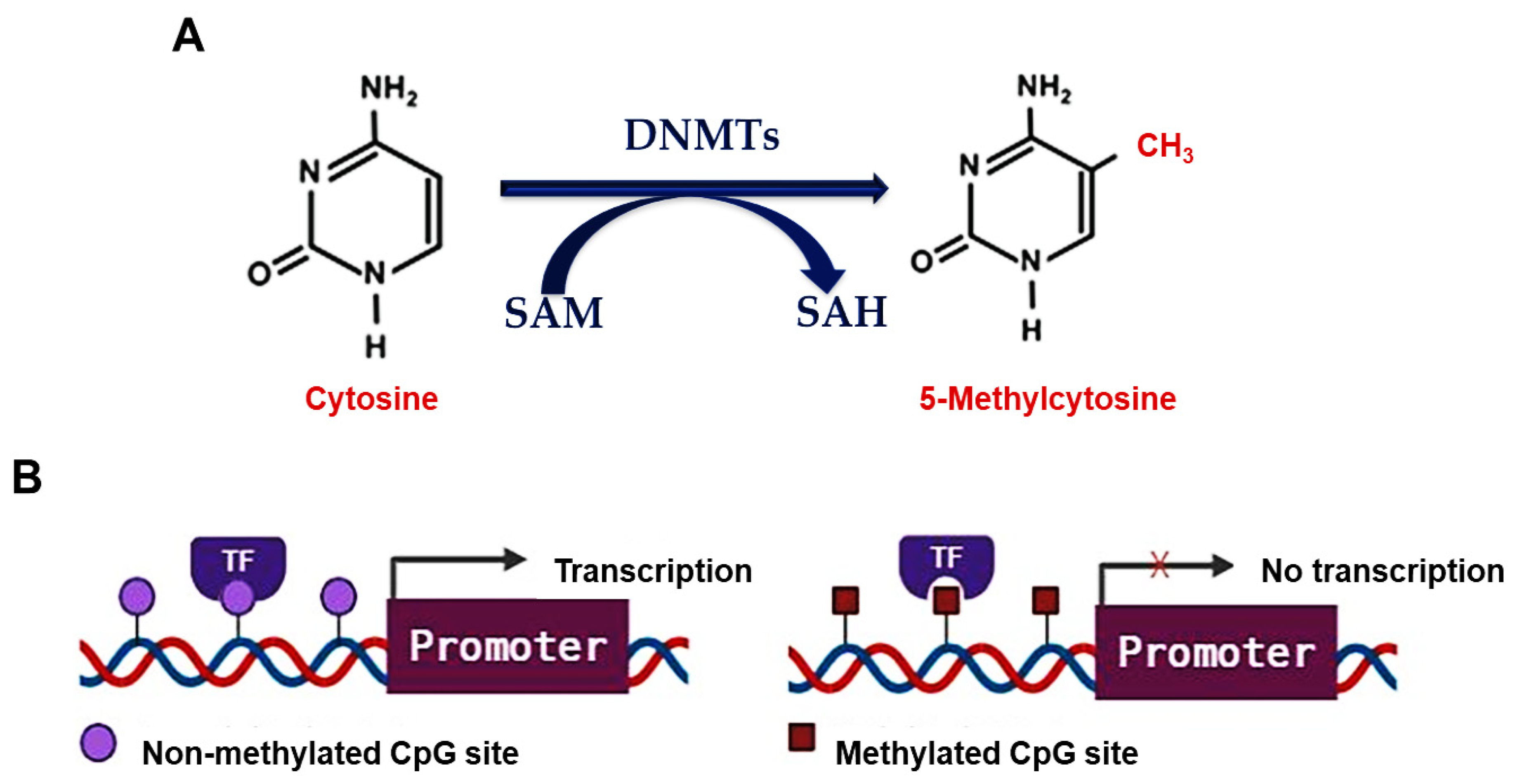
For air pollution particles to directly affect the developing fetus, they must be transported to and/or across the placenta. Figure 1 depicts the structure of the human placenta and indicates the direction of maternal-fetal transfer. Air pollution particles may be transferred to and/or across the placenta via the maternal blood supply through processes such as passive diffusion, active transport, and endocytosis [28]. In a recent review, several studies determined the ability of (ultra)fine particles and nanoparticles to cross the placenta and showed a dependency on the particle’s size, shape, dose, route of exposure, and surface composition [29]. While these studies provide evidence that particles can cross the placenta, most of them were conducted in animal models and did not focus specifically on air pollution particles or heavy metals following real-life exposure conditions.
In the ENVIRONAGE birth cohort, Bové et al. examined the presence of black carbon in placental tissue from 20 healthy, non-smoking women exposed to low (0.63–0.96 µg/m3) and high (1.70–2.42 µg/m3) levels of residential black carbon during pregnancy [4]. Black carbon particles were detected in all placentae, and the black carbon load was positively correlated with the women’s residential black carbon exposure levels. Furthermore, black carbon particles were found on both the maternal and fetal sides of the placenta, suggesting that black carbon may be transported to the developing fetus [4]. Another study by Liu et al. determined the presence of air pollution nanoparticles in placental tissue cells that were isolated from 15 healthy, non-smoking women exposed to particulate matter with an aerodynamic diameter smaller than 2.5 µm (PM2.5: 14.62–18.81 µg/m3) and 10 µm (PM10: 24.08–31.37 µg/m3) during pregnancy [5]. Carbon and metal-containing nanoparticles, usually sourced from heavily trafficked urban roads, were found in macrophage-enriched placental cells, indicating that fine metal particles are phagocytized as foreign bodies in placental tissue [5]. Similarly, Reichrtová et al. investigated the accumulation of two industry and traffic-related air pollutants, lead and nickel, in the placental tissue of 100 women residing in industrial and rural Slovak regions [6]. It was found that both lead and nickel were accumulated in the basal plate, chorionic villi, and chorionic plate of all placentae and that these metals were higher in the placenta from women residing in the industrial region compared to those in the rural region [6]. Since the chorionic villi contain blood vessels that lead directly to the developing fetus, the accumulation of lead and nickel in this region of the placenta implies that these heavy metals can be transported directly to the fetus [6].

3. DNA Methylation

Since air pollution particles were shown to translocate into and across the placenta, they may induce placental modifications. Previously, prenatal exposure to air pollution was found to alter placental weight [30], structure [31], and vascular function [32]. This may occur through alterations in placental epigenetic patterns [33,34,35,36,37,38,39,40,41,42,43,44,45,46,47,48,49,50,51,52,53].
Epigenetics is defined as the heritable changes that affect gene expression without altering the DNA nucleotide sequence. The main epigenetic mechanisms are DNA methylation, histone modifications, and microRNAs [54]. Among these, DNA methylation was the most studied epigenetic modification regarding prenatal exposure to air pollution and heavy metals in the placenta, and hence, it was the focus of this review.
DNA methylation is a biochemical process that occurs predominantly on cytosine bases that precede guanine bases (CpG sites) and involves the covalent addition of a methyl (CH3) group to the number 5 carbon of cytosine bases (Figure 2A). This reaction forms 5-methylcytosine and is mediated by DNA methyltransferases (DNMTs), namely DNMT1, DNMT3A, and DNMT3B [55]. DNMT1 maintains cellular methylation levels by recognizing hemi-methylated DNA and preserving the methylation pattern across generations, while DNMT3A and DNMT3B establish new methylation patterns in non-methylated DNA [55]. DNA methylation regulates gene expression by modifying chromatin structure and controlling the accessibility of transcription factors to gene promoter regions (Figure 2B). DNA hypermethylation is often associated with an inactive chromatin structure that prevents transcription factors (TFs) from binding to gene promoters and is involved in gene silencing. In contrast, DNA hypomethylation is associated with an active chromatin structure that enables TFs to bind to gene promoters and initiate transcription [55].
DNA methylation plays a crucial role in fetal development; it creates distinct cell lineages and regulates genome stability, cell proliferation, differentiation, genomic imprinting, and X-chromosome inactivation [54]. During pregnancy, DNA methylation is also involved in fetal epigenetic reprogramming, a process in which DNA methylation marks are erased and re-established, and it is during this time when the fetus is most susceptible to environmental insults [54]. DNA methylation changes in the placenta enable fetal metabolic adaptation in response to environmental stimuli and can affect the cellular phenotype, thus predisposing the fetus to developmental changes with both short-term and long-term health consequences [56].

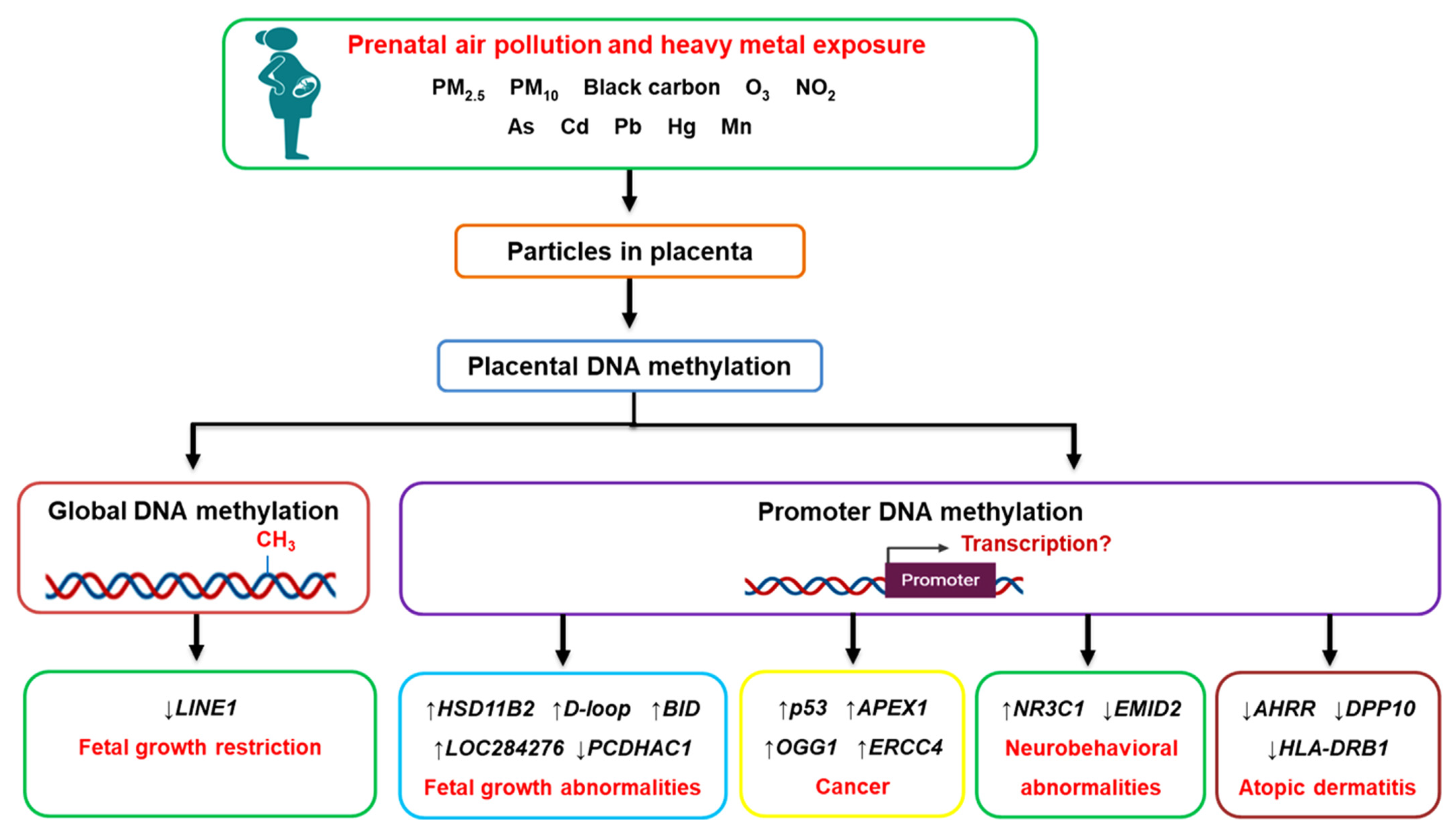
4. Prenatal Air Pollution Exposure and Placental Global DNA Methylation

Numerous studies have shown that prenatal air pollution exposure affects global DNA methylation patterns in the human placenta [33,34,35,36,37,38]. These studies are summarized in Table 1. In the ENVIRONAGE birth cohort, exposure to PM2.5 during different stages of pregnancy was associated with a lower degree of placental global DNA methylation, as measured by 5-methyl-deoxycytidine and deoxycytidine levels [33]. In another study, mothers residing near major roadways, an indicator of traffic-related air pollution, showed decreased placental LINE1 but not AluYb8 methylation, common markers of global DNA methylation [34]. Furthermore, residing near a major roadway was associated with newborns that had low birth weights; however, the change in placental LINE1 methylation did not mediate this relationship [34]. In contrast, a Chinese case-control study found that exposure to PM10 in the first trimester was associated with decreased LINE1 methylation in the placenta of fetal growth-restricted newborns [35].
A nested case-control study in Iran observed a positive correlation between global DNA methylation in the placenta (measured as 5-methyl-deoxycytidine and deoxycytidine levels) and exposure to PM2.5 and PM10 during the first trimester [36]. However, no significant correlation was found between exposures to particulate matter or placental global DNA methylation and birth outcomes such as gestational age, weight, length, and head and chest circumference [36]. The EDEN cohort showed a positive association with PM10 exposure the day before birth and placental Alu methylation; yet, no significant association was observed with LINE1 methylation [37]. Furthermore, the EARLI study found that nitrogen dioxide and ozone also induced changes in placental global DNA methylation levels [38]. Together, the above studies showed that prenatal air pollution exposure induced inconsistent DNA methylation changes in the placenta, and there was insufficient evidence linking these air pollution-induced placental global DNA methylation changes with particular birth outcomes or disease susceptibility.

5. Prenatal Air Pollution Exposure and Placental Candidate Gene Methylation

Apart from global DNA methylation, prenatal air pollution exposure was found to alter the promoter methylation of placental candidate genes that are involved in key biological processes [34,35,37,38,39,40,41,42,43,44,45]. These studies are summarized in Table 2. In the EDEN cohort, exposure to nitrogen dioxide and PM10 altered the methylation patterns of ADORA2B, PXT1, KCTD20, CAPN10, SLC44A5, ADCK5, TGM6, TUBGCP2, and KYNU in placental tissues [37]. These genes function in placental development and were previously associated with hypoxia and preeclampsia [57,58], a pathology that has been linked with air pollution exposure during pregnancy [12,13]. In a separate study, mothers living near major roadways showed differential methylation at seven CpG sites, three of which were located in protein-coding genes (PTPRN2, TMEM125, and VPS4A) [34]. Similarly, in the EARLI cohort, differentially methylated regions were found in five protein-coding genes (F11R, ZNF442, SLC25A44, STK38, and PTPRH) in the placenta of women exposed to high levels of nitrogen dioxide and ozone during pregnancy [38]. These differentially methylated regions in the placenta did not show DNA methylation changes in cord blood, and hence, they appeared to be tissue-specific. Additionally, differentially methylated regions of three genes (RNF39, CYP2E1, and PM20D1) in cord blood showed consistent nitrogen dioxide and ozone exposure-related altered DNA methylation in the placenta [38], indicating that placental DNA methylation changes may be passed onto the developing fetus. These genes regulate immune and inflammatory responses, and their altered methylation patterns might play a role in preeclampsia.
In China, exposure to PM10 during the first and second trimesters increased HSD11B2 methylation, a gene involved in fetal growth and glucocorticoid metabolism [35]. Furthermore, the increase in HSD11B2 methylation was more prominent in the placental tissues of fetal growth-restricted newborns compared to the normal-growth newborns. Previously, HSD11B2 methylation was negatively associated with placental HSD11B2 gene expression, fetal growth indices, and adverse neurobehavioral outcomes in infants [59]. The Shanghai Mother-Child Pairs Cohort found that exposure to PM2.5 during the second and third trimesters, as well as the entire pregnancy, was associated with increased methylation of BID and decreased methylation of IGF2; these genes are essential for fetal growth [42]. Furthermore, for every 1% increase in BID methylation, there was a decrease in head circumference (−1.396 mm, 95% CI: −2.582, −0.209) and biparietal diameter (−0.330 mm, 95% CI: −0.635, −0.026) in the second trimester [42]. In Korea, the COCOA study showed that high PM2.5 exposure and low cord blood vitamin D levels during the first trimester were associated with decreased placental methylation of AHRR, DPP10, and HLA-DRB1, and early-onset persistent atopic dermatitis in children [43].
In the ENVIRONAGE birth cohort, PM2.5 and black carbon exposure increased placental DNA mutation rates (determined by an increase in the DNA mutation marker, Alu) as well as increased the promoter methylation of tumor suppressor (p53) and DNA repair (APEX1, OGG1, and ERCC4) genes. This study suggested that prenatal PM2.5 and black carbon exposure reduces the DNA repair capacity of the placenta and fetus, which may increase the risk for carcinogenesis in later life [39]. In the same cohort, the analysis of placental tissue from mothers exposed to PM2.5 in the first and third trimesters showed altered methylation of the genes (CLOCK, CRY1, NPAS2, and PER1–3) involved in circadian rhythm regulation [40]. Previously, dysregulation in placental methylation of these circadian pathway genes was associated with preeclampsia [60]. Another study indicated that second-trimester PM2.5 exposure decreased promoter methylation of the Lep gene, a hormone involved in intrauterine development, embryo implantation, energy regulation, and fetal growth [41]. Interestingly, the promoter methylation of Lep was higher in the placental tissue of male neonates compared to those of female neonates, suggesting that altered DNA methylation in the placenta may be sex-specific [41].
Prenatal air pollution exposure was also associated with altered placental mitochondrial DNA methylation [44,45]. In the ENVIRONAGE cohort, placental mitochondrial DNA methylation was analyzed in the D-loop control region and MT-RNR1 region. First trimester PM2.5 exposure was associated with increased mitochondrial DNA methylation at both the D-loop and MT-RNR1 regions [44]. An increase in D-loop (non-significant) and MT-RNR1 (significant) methylation was also observed in placental tissue following exposure to PM2.5 for the entire pregnancy [44]. These findings were confirmed in a smaller sample population of the ENVIRONAGE cohort, where PM2.5 and black carbon exposure, throughout pregnancy, non-significantly increased D-loop and LDLR methylation [45]. In both studies, mitochondrial DNA methylation was negatively associated with mitochondrial DNA content (a measure of damaged mitochondria and mitophagy) [44,45]. PM2.5 and black carbon exposure for the entire pregnancy also decreased placental promoter methylation of PINK1, a gene involved in mitochondrial quality control and mitophagy [45]. Moreover, a 0.42% increase in D-loop methylation was associated with decreased newborn birth weight (−106.98 g, 95% CI: −209.60 g, −4.36 g) [45].

6. Prenatal Heavy Metal Exposure and Placental DNA Methylation

Heavy metals such as arsenic, cadmium, lead, manganese, mercury, and nickel are common constituents of industry and traffic-related air pollution. Particulate matter, mainly PM2.5 and PM10, has a strong potential for adsorbing heavy metals, which then enter the human body through inhalation [61]. There is increasing evidence that heavy metals bound to particulate matter play a crucial role in the adverse health effects caused by particulate matter [62,63,64]. Therefore, we also included studies investigating placental DNA methylation changes and prenatal exposure to heavy metals [46,47,48,49,50,51,52,53]. These studies are summarized in Table 3.
Most studies on prenatal heavy metal exposure and placental DNA methylation changes were conducted within the RICHS cohort. In the RICHS cohort, maternal exposure to high levels of arsenic, cadmium, lead, mercury, and manganese were associated with increased placental NR3C1 methylation compared to the low exposure groups [46]. Placental NR3C1 plays a vital role in cognitive and neurodevelopment by regulating the development of the child’s hypothalamic-pituitary-adrenal (HPA) axis and cortisol levels. Therefore, altered placental NR3C1 methylation may provide insight into cognitive and neurodevelopmental abnormalities in children in later life [46]. In a separate study, mothers with high toenail cadmium concentrations were found to have low levels of placental PCDHAC1 methylation and were at an increased odds of giving birth to an infant that was small for gestational age or with a decreased head circumference [50]. Another study showed that high placental cadmium levels were associated with differential methylation at 17 CpG sites, and DNA methylation at 9 of these 17 CpG sites was associated with increased expression of genes involved in inflammatory signaling and cell growth (TNFAIP2, EXOC3L4, GAS7, SREBF1, ACOT7, and RORA) [51]. Furthermore, high placental expressions of TNFAIP2 and ACOT7 were associated with decreased birth weight (Tau: −0.099, p = 0.039 and Tau: −0.134, p = 0.0048, respectively). High placental expression of ACOT7 was also associated with decreased birth length (Tau: –0.106, p = 0.029) and decreased head circumference (Tau: −0.145, p = 0.0032) [51]. Moreover, within the RICHS cohort, exposure to mercury, measured in infant toenails, was associated with differential methylation at 339 loci; 10 of these differentially methylated loci resided in the CPLX1, TTC23, and EMID2 genes and were associated with a high risk for adverse neurobehavioral profiles [52]. Exposure to high levels of manganese, measured in infant toenails, was associated with differential methylation at 5 CpG loci (EMX2OS, ATAD2B, FTO/RPGRIP1L, EN1, and LOC284276). These CpG loci resided in genes involved in neurodevelopment (EMX2OS, ATAD2B, and EN1) and fetal growth (FTO/RPGRIP1L) [53]. The function of LOC284276 is currently unknown; however, for every 10% increase in placental LOC284276 methylation, there was a decrease in birth weight (−293.44 g, p = 0.018) [53].
In the New Hampshire Birth Cohort Study, placental arsenic levels were associated with differential methylation at 163 CpG sites. Of these, 13 CpG sites attained genome-wide significance and were located at the LYRM2 (11 CpG sites), CAMTA1 (1 CpG site), and CCDC57 (1 CpG site) genes [48]. A nested cohort in Bangladesh found that maternal exposure to arsenic, via contaminated drinking water, was associated with hypermethylation at several CpG sites, which were mainly located within open sea regions [47]. Moreover, prenatal arsenic exposure was associated with CpG methylation at the NR3C1 gene (unadjusted analysis) and the TRA2B, PLCE1, and CD36 genes (adjusted analysis) [47].
The omega cohort and pilot case-control placenta microarray study showed that cadmium levels were higher in the placental tissues from female neonates (5 ng/g) compared to male neonates (2 ng/g). High cadmium levels were associated with hypomethylation at three CpG sites (ARL9, SIAH3, and HS3ST4) and one genomic region (region 86974674 to 86975244 on chromosome 7; CROT and TP53TG1) in the placental tissue of female neonates [49]. In the placental tissue of male neonates, high cadmium levels were associated with hypomethylation at two CpG sites (MECOM) and two genomic regions (region 169379554 to 169380078 on chromosome 3 and region 1792758 to 1792758 on chromosome 8; MECOM and ARHGEF10) as well as hypermethylation at one CpG site (SALL1) [49]. These differentially methylated genes are involved in cell damage response (SIAH3, HS3ST4, and TP53TG1) in females and cell differentiation, angiogenesis, and organ development (MECOM and ARHGEF10) in males. These results suggest that cadmium-associated placental DNA methylation changes may induce fetal growth abnormalities in a sex-dependent manner [49].

7. Conclusions

As a natural barrier that directly connects the mother to the developing fetus, the placenta is continuously in contact with substances to which both the mother and fetus are exposed [27]. Perturbations in the maternal environment can be transferred to the fetus through altered placental functions, a concept known as fetal reprogramming. As a result, the placenta, which is genetically identical to the fetus, contains important information on the in utero fetal life and can be considered as a “mirror” of the future health and development of the newborn [40]. The studies included in this review provide evidence that air pollution particles, including heavy metals, can be transported to the placenta [4,5,6]. Moreover, the finding of air pollution particles and heavy metals on both the maternal and fetal sides of the placenta suggests a strong possibility that these particles may be transported directly to the developing fetus. Air pollution particles and heavy metals in the placenta can modify placental functions by altering the DNA methylome, which mediates the transcriptional activation and silencing of genes involved in various physiological and developmental processes [33,34,35,36,37,38,39,40,41,42,43,44,45,46,47,48,49,50,51,52,53]. The effect of air pollution exposure on DNA methylation changes during the first 1000 days of life has been systematically reviewed [65]; however, this review did not include heavy metals as a constituent of air pollution.
Prenatal exposure to air pollution and heavy metals was associated with altered placental DNA methylation at both the global and candidate gene promoter levels [33,34,35,36,37,38,39,40,41,42,43,44,45,46,47,48,49,50,51,52,53] (summarized in Figure 3); however, whether the changes in gene promoter methylation affected the expression of the gene was not established in the majority of studies. Aside from the studies on placental mitochondrial DNA methylation [44,45], no genes were investigated in more than one study. This makes it difficult to determine if the alterations in candidate gene promoter methylation are specific to a particular socio-demographic population or to prenatal air pollution exposure in a certain geographical location. Different locations have different sources, and chemical compositions of air pollution and individual components are not often encountered in isolation within natural settings.
Two studies also showed that PM2.5 and cadmium-induced DNA methylation of genes were different in the placentae from male and female neonates [41,49], suggesting that the adverse health effects of these air pollutants may be sex-specific. Furthermore, prenatal exposure to air pollution induced inconsistent placental global DNA methylation changes, which may be the result of several other factors such as maternal diet or nutritional status and folate supplementation; folate is an essential micronutrient and methyl donor required during pregnancy to prevent neural tube defects in the fetus. Interestingly, trimester-specific analyses indicated that most of the placental DNA methylation changes observed at birth followed air pollution exposures during the early stages of pregnancy [33,35,36,37,40,43,44], which is also when fetal epigenetic reprogramming occurs. Although this suggests that air pollution and heavy metal exposure during the early stages of pregnancy may, through altered placental DNA methylation, be responsible for fetal growth abnormalities, there was insufficient evidence linking prenatal air pollution and heavy metal-induced placental DNA methylation changes with specific diseases. Follow-up studies are required to determine if placental DNA methylation changes persist to the fetus and into adulthood and its possible implications on fetal development and disease susceptibility throughout the lifespan. Together, the findings depicted in this review highlight the need for more effective and stricter environmental and public health policies to reduce air pollution and protect human health.

Author Contributions

T.G., conceptualization and writing-original draft preparation; P.N., writing-review and editing; R.N.N., writing-review and editing; A.A.C., conceptualization and writing-review and editing. All authors have read and agreed to the published version of the manuscript.

Funding

This work was supported by the National Research Foundation (grant numbers: 120716 and 120820).

Conflicts of Interest

The authors declare no conflict of interest.

References

  1. Health Effects Institute. State of Global Air 2020: A Special Report on Global Exposure to Air Pollution and Its Health Impacts. Available online: https://www.stateofglobalair.org/ (accessed on 15 July 2021).
  2. Chang, J.; Streitman, D. Physiologic adaptations to pregnancy. Neurol. Clin. 2012, 30, 781–789. [Google Scholar] [CrossRef] [PubMed]
  3. Hegewald, M.J.; Crapo, R.O. Respiratory physiology in pregnancy. Clin. Chest Med. 2011, 32, 1–13. [Google Scholar] [CrossRef] [PubMed]
  4. Bové, H.; Bongaerts, E.; Slenders, E.; Bijnens, E.M.; Saenen, N.D.; Gyselaers, W.; Van Eyken, P.; Plusquin, M.; Roeffaers, M.B.J.; Ameloot, M.; et al. Ambient black carbon particles reach the fetal side of human placenta. Nat. Commun. 2019, 10, 3866. [Google Scholar] [CrossRef] [PubMed]
  5. Liu, N.M.; Miyashita, L.; Maher, B.A.; McPhail, G.; Jones, C.J.P.; Barratt, B.; Thangaratinam, S.; Karloukovski, V.; Ahmed, I.A.; Aslam, Z.; et al. Evidence for the presence of air pollution nanoparticles in placental tissue cells. Sci. Total Environ. 2021, 751, 142235. [Google Scholar] [CrossRef] [PubMed]
  6. Reichrtová, E.; Dorociak, F.; Palkovičová, L. Sites of lead and nickel accumulation in the placental tissue. Hum. Exp. Toxicol. 1998, 17, 176–181. [Google Scholar] [CrossRef]
  7. Barker, D.J. In utero programming of chronic disease. Clin Sci. 1998, 95, 115–128. [Google Scholar] [CrossRef]
  8. Gluckman, P.D.; Hanson, M.A.; Cooper, C.; Thornburg, K.L. Effect of in utero and early-life conditions on adult health and disease. N. Engl. J. Med. 2008, 359, 61–73. [Google Scholar] [CrossRef] [Green Version]
  9. Swanson, J.M.; Entringer, S.; Buss, C.; Wadhwa, P.D. Developmental origins of health and disease: Environmental exposures. Semin. Reprod. Med. 2009, 27, 391–402. [Google Scholar] [CrossRef] [Green Version]
  10. Barker, D.J. The developmental origins of adult disease. J. Am. Coll. Nutr. 2004, 23, 588S–595S. [Google Scholar] [CrossRef]
  11. Zhang, H.; Wang, Q.; He, S.; Wu, K.; Ren, M.; Dong, H.; Di, J.; Yu, Z.; Huang, C. Ambient air pollution and gestational diabetes mellitus: A review of evidence from biological mechanisms to population epidemiology. Sci. Total Environ. 2020, 719, 137349. [Google Scholar] [CrossRef] [PubMed]
  12. Wang, Q.; Zhang, H.; Liang, Q.; Knibbs, L.D.; Ren, M.; Li, C.; Bao, J.; Wang, S.; He, Y.; Zhu, L.; et al. Effects of prenatal exposure to air pollution on preeclampsia in Shenzhen, China. Environ. Pollut. 2018, 237, 18–27. [Google Scholar] [CrossRef]
  13. Pereira, G.; Haggar, F.; Shand, A.W.; Bower, C.; Cook, A.; Nassar, N. Association between pre-eclampsia and locally derived traffic-related air pollution: A retrospective cohort study. J. Epidemiol. Community Health 2013, 67, 147–152. [Google Scholar] [CrossRef] [Green Version]
  14. Gaskins, A.J.; Hart, J.E.; Chavarro, J.E.; Missmer, S.A.; Rich-Edwards, J.W.; Laden, F.; Mahalingaiah, S. Air pollution exposure and risk of spontaneous abortion in the Nurses’ Health Study II. Hum. Reprod. 2019, 34, 1809–1817. [Google Scholar] [CrossRef]
  15. Kioumourtzoglou, M.-A.; Raz, R.; Wilson, A.; Fluss, R.; Nirel, R.; Broday, D.M. Traffic-related air pollution and pregnancy loss. Epidemiology 2019, 30, 4–10. [Google Scholar] [CrossRef] [PubMed]
  16. Ji, X.; Meng, X.; Liu, C.; Chen, R.; Ge, Y.; Kan, L.; Fu, Q.; Li, W.; Tse, L.A.; Kan, H. Nitrogen dioxide air pollution and preterm birth in Shanghai, China. Environ. Res. 2019, 169, 79–85. [Google Scholar] [CrossRef] [PubMed]
  17. Liu, X.; Ye, Y.; Chen, Y.; Li, X.; Feng, B.; Cao, G.; Xiao, J.; Zeng, W.; Li, X.; Sun, J.; et al. Effects of prenatal exposure to air particulate matter on the risk of preterm birth and roles of maternal and cord blood LINE-1 methylation: A birth cohort study in Guangzhou, China. Environ. Int. 2019, 133, 105177. [Google Scholar] [CrossRef] [PubMed]
  18. Gong, X.; Lin, Y.; Zhan, F.B. Industrial air pollution and low birth weight: A case-control study in Texas, USA. Environ. Sci. Pollut. Res. Int. 2018, 25, 30375–30389. [Google Scholar] [CrossRef] [PubMed]
  19. Chen, S.; Wang, S.; Li, T.; Zhu, H.; Liang, S.; Xu, K.; Zhang, Y.; Yuan, X.; Yang, Y.; Pan, H.; et al. Effect of PM2.5 on macrosomia in China: A nationwide prospective cohort study. Pediatr. Obes. 2020, 15, e12584. [Google Scholar] [CrossRef]
  20. Zang, H.; Cheng, H.; Song, W.; Yang, M.; Han, P.; Chen, C.; Ding, R. Ambient air pollution and the risk of stillbirth: A population-based prospective birth cohort study in the coastal area of China. Environ. Sci. Pollut. Res. Int. 2019, 26, 6717–6724. [Google Scholar] [CrossRef]
  21. Breton, C.V.; Mack, W.J.; Yao, J.; Berhane, K.; Amadeus, M.; Lurmann, F.; Gilliland, F.; McConnell, R.; Hodis, H.N.; Kunzli, N.; et al. Prenatal air pollution exposure and early cardiovascular phenotypes in young adults. PLoS ONE 2016, 11, e0150825. [Google Scholar] [CrossRef]
  22. Guxens, M.; Lubczynska, M.J.; Muetzel, R.L.; Dalmau-Bueno, A.; Jaddoe, V.W.V.; Hoek, G.; van der Lugt, A.; Verhulst, F.C.; White, T.; Brunekreef, B.; et al. Air pollution exposure during fetal life, brain morphology, and cognitive function in school-age children. Biol. Psychiatry 2018, 84, 295–303. [Google Scholar] [CrossRef] [PubMed] [Green Version]
  23. Hehua, Z.; Qing, C.; Shanyan, G.; Qijun, W.; Yuhong, Z. The impact of prenatal exposure to air pollution on childhood wheezing and asthma: A systematic review. Environ. Res. 2017, 159, 519–530. [Google Scholar] [CrossRef] [PubMed]
  24. Sbihi, H.; Tamburic, L.; Koehoorn, M.; Brauer, M. Perinatal air pollution exposure and development of asthma from birth to age 10 years. Eur. Respir. J. 2016, 47, 1062–1071. [Google Scholar] [CrossRef] [Green Version]
  25. Ghosh, J.K.; Heck, J.E.; Cockburn, M.; Su, J.; Jerrett, M.; Ritz, B. Prenatal exposure to traffic-related air pollution and risk of early childhood cancers. Am. J. Epidemiol. 2013, 178, 1233–1239. [Google Scholar] [CrossRef]
  26. Lavigne, E.; Belair, M.A.; Do, M.T.; Stieb, D.M.; Hystad, P.; van Donkelaar, A.; Martin, R.V.; Crouse, D.L.; Crighton, E.; Chen, H.; et al. Maternal exposure to ambient air pollution and risk of early childhood cancers: A population-based study in Ontario, Canada. Environ. Int. 2017, 100, 139–147. [Google Scholar] [CrossRef]
  27. Burton, G.J.; Fowden, A.L. The placenta: A multifaceted, transient organ. Philos. Trans. R. Soc. Lond. B Biol. Sci. 2015, 370, 20140066. [Google Scholar] [CrossRef] [Green Version]
  28. Wick, P.; Malek, A.; Manser, P.; Meili, D.; Maeder-Althaus, X.; Diener, L.; Diener, P.-A.; Zisch, A.; Krug, H.F.; von Mandach, U. Barrier capacity of human placenta for nanosized materials. Environ. Health Perspect. 2010, 118, 432–436. [Google Scholar] [CrossRef]
  29. Bongaerts, E.; Nawrot, T.S.; Van Pee, T.; Ameloot, M.; Bove, H. Translocation of (ultra)fine particles and nanoparticles across the placenta; a systematic review on the evidence of in vitro, ex vivo, and in vivo studies. Part. Fibre Toxicol. 2020, 17, 56. [Google Scholar] [CrossRef]
  30. Ghasemi-Tehrani, H.; Fallah, S.; Mozafarian, N.; Miranzadeh, S.; Sadeghi, S.; Azidhak, A. Effect of exposure to air pollution on placental weight in Isfahan-Iran. J. Family Reprod. Health 2017, 11, 90–96. [Google Scholar] [PubMed]
  31. Soto, S.F.; Melo, J.O.; Marchesi, G.D.; Lopes, K.L.; Veras, M.M.; Oliveira, I.B.; Souza, R.M.; de Castro, I.; Furukawa, L.N.S.; Saldiva, P.H.N.; et al. Exposure to fine particulate matter in the air alters placental structure and the renin-angiotensin system. PLoS ONE 2017, 12, e0183314. [Google Scholar] [CrossRef]
  32. Yue, H.; Ji, X.; Zhang, Y.; Li, G.; Sang, N. Gestational exposure to PM2.5 impairs vascularization of the placenta. Sci. Total Environ. 2019, 665, 153–161. [Google Scholar] [CrossRef] [PubMed]
  33. Janssen, B.G.; Godderis, L.; Pieters, N.; Poels, K.; Kicinski, M.; Cuypers, A.; Fierens, F.; Penders, J.; Plusquin, M.; Gyselaers, W.; et al. Placental DNA hypomethylation in association with particulate air pollution in early life. Part. Fibre Toxicol. 2013, 10, 22. [Google Scholar] [CrossRef] [Green Version]
  34. Kingsley, S.L.; Eliot, M.N.; Whitsel, E.A.; Huang, Y.T.; Kelsey, K.T.; Marsit, C.J.; Wellenius, G.A. Maternal residential proximity to major roadways, birth weight, and placental DNA methylation. Environ. Int. 2016, 92, 43–49. [Google Scholar] [CrossRef] [Green Version]
  35. Cai, J.; Zhao, Y.; Liu, P.; Xia, B.; Zhu, Q.; Wang, X.; Song, Q.; Kan, H.; Zhang, Y. Exposure to particulate air pollution during early pregnancy is associated with placental DNA methylation. Sci. Total Environ. 2017, 607, 1103–1108. [Google Scholar] [CrossRef]
  36. Maghbooli, Z.; Hossein-Nezhad, A.; Adabi, E.; Asadollah-Pour, E.; Sadeghi, M.; Mohammad-Nabi, S.; Zakeri Rad, L.; Malek Hosseini, A.A.; Radmehr, M.; Faghihi, F.; et al. Air pollution during pregnancy and placental adaptation in the levels of global DNA methylation. PLoS ONE 2018, 13, e0199772. [Google Scholar] [CrossRef] [PubMed] [Green Version]
  37. Abraham, E.; Rousseaux, S.; Agier, L.; Giorgis-Allemand, L.; Tost, J.; Galineau, J.; Hulin, A.; Siroux, V.; Vaiman, D.; Charles, M.A.; et al. Pregnancy exposure to atmospheric pollution and meteorological conditions and placental DNA methylation. Environ. Int. 2018, 118, 334–347. [Google Scholar] [CrossRef] [Green Version]
  38. Ladd-Acosta, C.; Feinberg, J.I.; Brown, S.C.; Lurmann, F.W.; Croen, L.A.; Hertz-Picciotto, I.; Newschaffer, C.J.; Feinberg, A.P.; Fallin, M.D.; Volk, H.E. Epigenetic marks of prenatal air pollution exposure found in multiple tissues relevant for child health. Environ. Int. 2019, 126, 363–376. [Google Scholar] [CrossRef]
  39. Neven, K.Y.; Saenen, N.D.; Tarantini, L.; Janssen, B.G.; Lefebvre, W.; Vanpoucke, C.; Bollati, V.; Nawrot, T.S. Placental promoter methylation of DNA repair genes and prenatal exposure to particulate air pollution: An ENVIRONAGE cohort study. Lancet Planet. Health 2018, 2, e174–e183. [Google Scholar] [CrossRef] [Green Version]
  40. Nawrot, T.S.; Saenen, N.D.; Schenk, J.; Janssen, B.G.; Motta, V.; Tarantini, L.; Cox, B.; Lefebvre, W.; Vanpoucke, C.; Maggioni, C.; et al. Placental circadian pathway methylation and in utero exposure to fine particle air pollution. Environ. Int. 2018, 114, 231–241. [Google Scholar] [CrossRef]
  41. Saenen, N.D.; Vrijens, K.; Janssen, B.G.; Roels, H.A.; Neven, K.Y.; Vanden Berghe, W.; Gyselaers, W.; Vanpoucke, C.; Lefebvre, W.; De Boever, P. Lower placental leptin promoter methylation in association with fine particulate matter air pollution during pregnancy and placental nitrosative stress at birth in the ENVIRONAGE cohort. Environ. Health Perspect 2017, 125, 262–268. [Google Scholar] [CrossRef] [PubMed] [Green Version]
  42. Zhao, Y.; Wang, P.; Zhou, Y.; Xia, B.; Zhu, Q.; Ge, W.; Li, J.; Shi, H.; Xiao, X.; Zhang, Y. Prenatal fine particulate matter exposure, placental DNA methylation changes, and fetal growth. Environ. Int. 2021, 147, 106313. [Google Scholar] [CrossRef]
  43. Yang, S.-I.; Lee, S.-H.; Lee, S.-Y.; Kim, H.-C.; Kim, H.-B.; Kim, J.-H.; Lim, H.; Park, M.J.; Cho, H.-J.; Yoon, J. Prenatal PM2.5 exposure and vitamin D–associated early persistent atopic dermatitis via placental methylation. Ann. Allergy Asthma Immunol. 2020, 125, 665–673. [Google Scholar] [CrossRef]
  44. Janssen, B.G.; Byun, H.M.; Gyselaers, W.; Lefebvre, W.; Baccarelli, A.A.; Nawrot, T.S. Placental mitochondrial methylation and exposure to airborne particulate matter in the early life environment: An ENVIRONAGE birth cohort study. Epigenetics 2015, 10, 536–544. [Google Scholar] [CrossRef] [Green Version]
  45. Vos, S.; Nawrot, T.S.; Martens, D.S.; Byun, H.M.; Janssen, B.G. Mitochondrial DNA methylation in placental tissue: A proof of concept study by means of prenatal environmental stressors. Epigenetics 2021, 16, 121–131. [Google Scholar] [CrossRef]
  46. Appleton, A.A.; Jackson, B.P.; Karagas, M.; Marsit, C.J. Prenatal exposure to neurotoxic metals is associated with increased placental glucocorticoid receptor DNA methylation. Epigenetics 2017, 12, 607–615. [Google Scholar] [CrossRef] [PubMed] [Green Version]
  47. Cardenas, A.; Houseman, E.A.; Baccarelli, A.A.; Quamruzzaman, Q.; Rahman, M.; Mostofa, G.; Wright, R.O.; Christiani, D.C.; Kile, M.L. In utero arsenic exposure and epigenome-wide associations in placenta, umbilical artery, and human umbilical vein endothelial cells. Epigenetics 2015, 10, 1054–1063. [Google Scholar] [CrossRef] [PubMed] [Green Version]
  48. Green, B.B.; Karagas, M.R.; Punshon, T.; Jackson, B.P.; Robbins, D.J.; Houseman, E.A.; Marsit, C.J. Epigenome-wide assessment of DNA methylation in the placenta and arsenic exposure in the New Hampshire Birth Cohort Study (USA). Environ. Health Perspect. 2016, 124, 1253–1260. [Google Scholar] [CrossRef] [PubMed] [Green Version]
  49. Mohanty, A.F.; Farin, F.M.; Bammler, T.K.; MacDonald, J.W.; Afsharinejad, Z.; Burbacher, T.M.; Siscovick, D.S.; Williams, M.A.; Enquobahrie, D.A. Infant sex-specific placental cadmium and DNA methylation associations. Environ. Res. 2015, 138, 74–81. [Google Scholar] [CrossRef] [Green Version]
  50. Everson, T.M.; Armstrong, D.A.; Jackson, B.P.; Green, B.B.; Karagas, M.R.; Marsit, C.J. Maternal cadmium, placental PCDHAC1, and fetal development. Reprod. Toxicol. 2016, 65, 263–271. [Google Scholar] [CrossRef] [Green Version]
  51. Everson, T.M.; Punshon, T.; Jackson, B.P.; Hao, K.; Lambertini, L.; Chen, J.; Karagas, M.R.; Marsit, C.J. Cadmium-associated differential methylation throughout the placental genome: Epigenome-wide association study of two US birth cohorts. Environ. Health Perspect. 2018, 126, 017010. [Google Scholar] [CrossRef] [Green Version]
  52. Maccani, J.Z.; Koestler, D.C.; Lester, B.; Houseman, E.A.; Armstrong, D.A.; Kelsey, K.T.; Marsit, C.J. Placental DNA methylation related to both infant toenail mercury and adverse neurobehavioral outcomes. Environ. Health Perspect. 2015, 123, 723–729. [Google Scholar] [CrossRef] [PubMed] [Green Version]
  53. Maccani, J.Z.; Koestler, D.C.; Houseman, E.A.; Armstrong, D.A.; Marsit, C.J.; Kelsey, K.T. DNA methylation changes in the placenta are associated with fetal manganese exposure. Reprod. Toxicol. 2015, 57, 43–49. [Google Scholar] [CrossRef] [PubMed] [Green Version]
  54. Handy, D.E.; Castro, R.; Loscalzo, J. Epigenetic modifications: Basic mechanisms and role in cardiovascular disease. Circulation 2011, 123, 2145–2156. [Google Scholar] [CrossRef] [PubMed] [Green Version]
  55. Moore, L.D.; Le, T.; Fan, G. DNA methylation and its basic function. Neuropsychopharmacology 2013, 38, 23–38. [Google Scholar] [CrossRef] [PubMed] [Green Version]
  56. Koukoura, O.; Sifakis, S.; Spandidos, D.A. DNA methylation in the human placenta and fetal growth. Mol. Med. Rep. 2012, 5, 883–889. [Google Scholar] [CrossRef] [PubMed] [Green Version]
  57. Jia, R.-Z.; Zhang, X.; Hu, P.; Liu, X.-M.; Hua, X.-D.; Wang, X.; Ding, H.-J. Screening for differential methylation status in human placenta in preeclampsia using a CpG island plus promoter microarray. Int. J. Mol. Med. 2012, 30, 133–141. [Google Scholar] [CrossRef] [PubMed] [Green Version]
  58. Acurio, J.; Troncoso, F.; Bertoglia, P.; Salomon, C.; Aguayo, C.; Sobrevia, L.; Escudero, C. Potential role of A2B adenosine receptors on proliferation/migration of fetal endothelium derived from preeclamptic pregnancies. Biomed. Res. Int. 2014, 2014, 274507. [Google Scholar] [CrossRef] [Green Version]
  59. Marsit, C.J.; Maccani, M.A.; Padbury, J.F.; Lester, B.M. Placental 11-beta hydroxysteroid dehydrogenase methylation is associated with newborn growth and a measure of neurobehavioral outcome. PLoS ONE 2012, 7, e33794. [Google Scholar] [CrossRef] [Green Version]
  60. Van den Berg, C.B.; Chaves, I.; Herzog, E.M.; Willemsen, S.P.; van der Horst, G.T.J.; Steegers-Theunissen, R.P.M. Early- and late-onset preeclampsia and the DNA methylation of circadian clock and clock-controlled genes in placental and newborn tissues. Chronobiol. Int. 2017, 34, 921–932. [Google Scholar] [CrossRef]
  61. Li, H.; Qian, X.; Wang, Q.g. Heavy metals in atmospheric particulate matter: A comprehensive understanding is needed for monitoring and risk mitigation. Environ. Sci. Technol. 2013, 47, 13210–13211. [Google Scholar] [CrossRef]
  62. Zhang, Y.; Ji, X.; Ku, T.; Li, G.; Sang, N. Heavy metals bound to fine particulate matter from northern China induce season-dependent health risks: A study based on myocardial toxicity. Environ. Pollut. 2016, 216, 380–390. [Google Scholar] [CrossRef] [PubMed] [Green Version]
  63. Jung, K.H.; Torrone, D.; Lovinsky-Desir, S.; Perzanowski, M.; Bautista, J.; Jezioro, J.R.; Hoepner, L.; Ross, J.; Perera, F.P.; Chillrud, S.N. Short-term exposure to PM2.5 and vanadium and changes in asthma gene DNA methylation and lung function decrements among urban children. Respir. Res. 2017, 18, 63. [Google Scholar] [CrossRef] [PubMed] [Green Version]
  64. Bollati, V.; Marinelli, B.; Apostoli, P.; Bonzini, M.; Nordio, F.; Hoxha, M.; Pegoraro, V.; Motta, V.; Tarantini, L.; Cantone, L.; et al. Exposure to metal-rich particulate matter modifies the expression of candidate microRNAs in peripheral blood leukocytes. Environ. Health Perspect. 2010, 118, 763–768. [Google Scholar] [CrossRef] [PubMed] [Green Version]
  65. Isaevska, E.; Moccia, C.; Asta, F.; Cibella, F.; Gagliardi, L.; Ronfani, L.; Rusconi, F.; Stazi, M.A.; Richiardi, L. Exposure to ambient air pollution in the first 1000 days of life and alterations in the DNA methylome and telomere length in children: A systematic review. Environ. Res. 2021, 193, 110504. [Google Scholar] [CrossRef] [PubMed]
Figure 1. Structure of the human placenta. Created with BioRender.com (accessed on 19 September 2021).
Cells 10 03025 g001
Figure 2. DNA methylation and gene expression regulation. (A) Process of DNA methylation. DNMTs covalently add a CH3 group from SAM to the number 5 carbon of cytosine bases yielding 5-methylcytosine and SAH. (B) Gene expression regulation by DNA methylation. DNA hypomethylation (non-methylated CpG site) enables TF to bind to gene promoters and activate its transcription; however, DNA hypermethylation (methylated CpG site) prevents the binding of TF to gene promoters and inhibits gene transcription. Abbreviations: CH3: methyl group; CpG: cytosine bases preceding guanine bases; DNMTs: DNA methyltransferases; SAM: S-adenosylmethionine; SAH: S-adenosylhomocysteine; TF: transcription factor. Created with BioRender.com.
Cells 10 03025 g002
Figure 3. The effects of prenatal air pollution exposure on placental DNA methylation patterns and its implications on fetal development and future disease susceptibility. Maternal exposure to air pollution, including heavy metals, can reach the placenta, where they alter DNA methylation patterns at both the global and gene promoter level. The aberrant methylation of genes affects fetal growth (HSD11B2, BID, D-loop, PCDHAC1, LOC284276) and increases the risk of developing cancer (p53, APEX1, OGG1, ERCC4), neurobehavioral abnormalities (NR3C1, EMID2), and atopic dermatitis (AHRR, DPP10, HLA-DRB1) in later life. Abbreviations: AHRR: aryl hydrocarbon receptor repressor; APEX1: AP endonuclease 1; As: arsenic; BID: BH3 interacting domain death agonist; Cd: cadmium; D-loop: displacement loop control region (heavy strand); DPP10: dipeptidyl peptidase 10; EMID2: EMI domain-containing protein 2; ERCC4: excision repair 4; Hg: mercury; HLA-DRB1: HLA class II histocompatibility antigen, DRB1 beta chain; HSD11B2: 11β-hydroxysteroid dehydrogenase 2; LINE1: long interspersed nuclear element 1; Mn: manganese; NO2: nitrogen dioxide; NR3C1: nuclear receptor subfamily 3 group C member 1 glucocorticoid receptor; O3: ozone; OGG1: oxoguanine glycosylase 1; p53: tumor suppressor protein 53; Pb: lead; PCDHAC1: protocadherin alpha subfamily C1; PM2.5: particulate matter with an aerodynamic diameter smaller than 2.5 µm; PM10: particulate matter with an aerodynamic diameter smaller than 10 µm.
Cells 10 03025 g003
Table 1. Studies on prenatal air pollution exposure and placental global DNA methylation.
AuthorStudySample SizeMethodAir PollutantDuration ExposedFindings
Janssen et al. [33]ENVIRONAGE cohort, Belgium240UPLC/MS-MSPM2.5: 5 µg/m3 incrementImplantation (6–21 days after conception)↓ Global DNA methylation (−1.08%, 95% CI: −1.80, −0.36%, p = 0.004)
PM2.5: 5 µg/m3 incrementFirst trimester↓ Global DNA methylation (−2.41%, 95% CI: −3.62, −1.20%, p = 0.0001)
PM2.5: 5 µg/m3 incrementSecond trimester↓ Global DNA methylation (−1.51%, 95% CI: −2.66, −0.36%, p = 0.01)
PM2.5: 5 µg/m3 incrementEntire pregnancy↓ Global DNA methylation (−2.19%, 95% CI: −3.65, −0.73%, p = 0.004)
Kingsley et al. [34]RICHS cohort, US471Bisulfite-PCR pyrosequencingTraffic-related air pollution: Women residing ≤ 150 m from a major roadway or ≤50 m from a secondary roadEntire pregnancyResiding near a major roadway: ↓ LINE1 methylation (−0.82%, 95% CI: −1.57, −0.07%, p = 0.03); No significant association with AluYb8 methylation (p = 0.07)
Cai et al. [35]Case-control study, China181Bisulfite-PCR pyrosequencingPM10: 10 µg/m3 incrementFirst trimesterPlacenta of fetal growth restricted newborns: ↓ LINE1 methylation (−1.78%, 95% CI: −3.35, −0.22%, p < 0.05)
Placenta of normal growth newborns: No significant association with LINE1 methylation (p > 0.05)
Maghbooli et al. [36]Nested case-control study, Iran92RP-HPLCPM2.5: 20.43 ± 0.68 µg/m3 and 30.99 ± 0.86 µg/m3
PM10: 64.97 ± 2.52 µg/m3 and 74.34 ± 2.66 µg/m3
First trimesterPM2.5: ↑ Global DNA methylation (r = 0.26, p = 0.01)
PM10: ↑ Global DNA methylation (r = 0.38, p = 0.0001)
Abraham et al. [37]EDEN cohort, France668Illumina Infinium HumanMethylation450K BeadChipPM10: 10 µg/m3 incrementDay before birthAlu methylation (β = 0.08, p = 0.01); No significant association with LINE1 methylation (β = 0.09, p = 0.28)
Ladd-Acosta et al. [38]EARLI cohort, US124Illumina Infinium HumanMethylation450K BeadChipNO2 and O3Entire pregnancyO3: ↓ DNA methylation at shelf regions (p = 0.00028), ↑ DNA methylation at CpG islands (p = 0.00295) and shore regions (p = 0.00266)
NO2: ↓ DNA methylation in CpG islands (p = 0.00359) and shore regions (p = 0.04284)
↓: decrease; ↑: increase; Alu/AluYb8: arthrobacter luteus elements; CI: confidence interval; EARLI: Early Autism Risk Longitudinal Investigation; EDEN: Etude de cohorte g’en’eraliste men’ee en France sur les D’eterminants pr’e et post natals pr’ecoces du d´eveloppement psychomoteur et de la sant´e de l’Enfant; ENVIRONAGE: ENVIRonmental influence ON early AGEing; LINE1: long interspersed nuclear element 1; MS-MS: tandem mass spectrometry; NO2: nitrogen dioxide; O3: ozone; PM2.5: particulate matter with an aerodynamic diameter smaller than 2.5 µm; PM10: particulate matter with an aerodynamic diameter smaller than 10 µm; RICHS: Rhode Island Child Health Study; RP-HPLC: reversed-phase high-pressure liquid chromatography; UPLC: ultra-pressure liquid chromatography; US: United States.
Table 2. Studies on prenatal air pollution exposure and placental candidate gene methylation.
AuthorStudySample SizeTechniqueAir PollutantDuration ExposedFindings
Kingsley et al. [34]RICHS cohort, US215Illumina Infinium HumanMethylation450K BeadChipTraffic-related air pollution: Women residing ≤150 m from a major roadway or ≤50 m from a secondary roadEntire PregnancyResiding near a major roadway: Differential methylation of 7 CpG sites—4 were mapped to non-genic regions and 3 were mapped to genes. ↑ PTPRN2 methylation (+0.061%, p = 2.904 × 10⁻⁶), ↓ TMEM125 methylation (−0.012%, p = 1.077 × 10⁻³), ↓ VPS4A methylation (−0.016%, p = 3.151 × 10⁻⁵)
Cai et al. [35]Case-control study, China181Bisulfite-PCR pyrosequencingPM10: 10 µg/m3 incrementFirst trimesterPlacenta of fetal growth restricted newborns: ↑ HSD11B2 methylation (+1.03%, 95% CI: 0.07, 1.98%, p < 0.05)
Placenta of normal growth newborns: No significant association with HSD11B2 methylation (p > 0.05)
PM10: 10 µg/m3 incrementSecond trimesterPlacenta of fetal growth restricted newborns: ↑ HSD11B2 methylation (+2.23%, 95% CI: 0.69, 3.76%, p < 0.05)
Placenta of normal growth newborns: No significant association with HSD11B2 methylation (p > 0.05)
Total population: ↑ HSD11B2 methylation (+1.42%, 95% CI: 0.24, 2.57%, p < 0.05)
PM10: 10 µg/m3 incrementEntire pregnancyTotal population: ↑ HSD11B2 methylation (+1.98%, 95% CI: 0.53, 3.43%, p < 0.05)
Abraham et al. [37]EDEN cohort, France668Illumina Infinium HumanMethylation450K BeadChipNO2: 10 µg/m3 incrementFirst trimesterADORA2B methylation at 2 CpG sites (cg17580614: β = −0.037, p < 0.001; cg07563400: β = −0.042, p < 0.001)
NO2: 10 µg/m3 incrementSecond trimesterADORA2B methylation at 2 CpG sites (cg17580614: β = −0.044, p < 0.0001; cg07563400: β = −0.047, p < 0.0001), ↑ PXT1/KCTD20 methylation (cg10984505: β = 0.002, p = 0.02
NO2: 10 µg/m3 incrementThird trimesterCAPN10 methylation (cg01712700: β = −0.004, p = 0.02)
PM10: 10 µg/m3 incrementOne month before birthSLC44A5 methylation (cg12659128: β = 0.037, p = 0.03), ↑ ADCK5 methylation (cg23075260: β = 0.018, p = 0.03), ↑ TGM6 methylation (cg06967014: β = 0.007, p = 0.03), ↓ TUBGCP2 methylation (cg05142592: β = −0.008, p = 0.03)
PM10: 10 µg/m3 increment3 days before birthKYNU methylation (cg04112100: β = −0.012, p = 0.04)
Ladd-Acosta et al. [38]EARLI cohort, US124Illumina Infinium HumanMethylation450K BeadChipNO2 and O3Entire pregnancyDifferentially methylated regions in 5 genes that seemed to be specific to placental tissue: ZNF442, PTPRH, SLC25A44, F11R, and STK38
Differentially methylated regions in 3 genes found in cord blood and showed similar methylation patterns in placental tissue: RNF39, CYP2E1, and PM20D1
Neven et al. [39]ENVIRONAGE cohort, Belgium463Bisulfite-PCR pyrosequencingPM2.5: 3.84 µg/m3 incrementEntire pregnancyAPEX1 methylation (+7.34%, 95% CI: 0.52, 14.16%, p = 0.0089), ↑ OGG1 methylation (+13.06%, 95% CI: 3.88, 22.24%, p = 0.0054), ↑ ERCC4 methylation (+ 16.31%, 95% CI: 5.43, 27.18%, p = 0.0034), ↑ p53 methylation (+10.60%, 95% CI: 4.46, 16.74%, p = 0.0008), ↓ DAPK1 methylation (−12.92%, 95% CI: −22.35, −3.49%, p = 0.0073)
Black carbon: 0.36 µg/m3 incrementEntire pregnancyAPEX1 methylation (+9.16%, 95% CI: 4.06, 14.25%, p = 0.005), ↑ ERCC4 methylation (+27.56%, 95% CI: 17.58, 37.55%, p < 0.0001)
Nawrot et al. [40]ENVIRONAGE cohort, Belgium407Bisulfite-PCR pyrosequencingPM2.5: 7.9 µg/m3 incrementFirst trimesterCLOCK methylation (−0.59 Log (fold-change), 95% CI: −0.93, −0.25, p = 0.0007)
PM2.5: 8.9 µg/m3 incrementThird trimesterNPAS2 methylation (+0.16 Log (fold-change), 95% CI: 0.06, 0.27, p = 0.002), ↑ CRY1 methylation (+0.59 Log (fold-change), 95% CI: 0.22, 0.95, p = 0.002), ↑ PER2 methylation (+0.36 Log (fold-change), 95% CI: 0.16, 0.57, p = 0.0005), ↑ PER3 methylation (+0.42 Log (fold-change), 95% CI: 0.18, 0.67, p = 0.0008), ↓ PER1 methylation (−0.51 Log (fold-change), 95% CI: −0.90, −0.13, p = 0.01)
PM2.5: 9.7 µg/m3 incrementLast month of pregnancyCRY1 methylation (p = 0.01), ↑ PER2 methylation (p = 0.0003), ↑ PER3 methylation (p = 0.02)
Saenen et al. [41]ENVIRONAGE cohort, Belgium361Bisulfite-PCR pyrosequencingPM2.5: 7.5 µg/m3 incrementSecond trimesterLep methylation (−1.4%, 95% CI: −2.7, −0.19%, p = 0.02), ↑ Lep methylation in placentae from male neonates compared to placentae from female neonates (+1.33%, 95% CI: 0.40, 2.27%, p = 0.005)
Zhao et al. [42]Shanghai MCPC, China287Bisulfite-PCR pyrosequencingPM2.5: 1 µg/m3 incrementSecond trimesterIGF2 methylation (−0.135%, 95% CI: −0.236, −0.034), ↑ BID methylation (+ 0.132%, 95% CI: 0.047, 0.217), ↑ FOXN3 methylation (position 1, +0.091, 95% CI: 0.008, 0.174)
PM2.5: 1 µg/m3 incrementThird trimesterIGF2 methylation (−0.229%, 95% CI: −0.384, −0.073), ↑ BID methylation (+ 0.209%, 95% CI: 0.072, 0.346)
PM2.5: 1 µg/m3 incrementEntire pregnancyIGF2 methylation (−0.297%, 95% CI: −0.489, −0.105), ↑ BID methylation (+ 0.209%, 95% CI: 0.039, 0.380)
Yang et al. [43]COCOA study, Korea1180Illumina Infinium HumanMethylationEPIC BeadChipPM2.5First trimester (3–7 weeks of pregnancy)Placenta of children with high PM2.5 exposure, low cord blood vitamin D levels, and atopic dermatitis: ↓ AHRR methylation (cg16371648: β = −0.367, p = 0.026), ↓ DPP10 methylation
(cg19211931: β = −0.263, p = 0.013), ↓ HLA-DRB1 methylation (cg10632894: β = −0.318, p = 0.026)
Janssen et al. [44]ENVIRONAGE cohort, Belgium381Bisulfite-PCR pyrosequencingPM2.5: 7.8 µg/m3 incrementFirst trimesterD-loop methylation (+0.44%, 95% CI: 0.12, 0.75%, p < 0.05), ↑ MT-RNR1 methylation (+1.27%, 95% CI: 0.23, 2.32%, p < 0.05)
PM2.5: 3.0 µg/m3 incrementEntire pregnancyD-loop methylation (+0.21%, 95% CI: -0.003, 1.02%, p > 0.05), ↑ MT-RNR1 methylation (+0.91, 95% CI: 0.56, 4.18%, p < 0.05)
Vos et al. [45]ENVIRONAGE cohort, Belgium60Bisulfite-PCR pyrosequencingPM2.5 (5.4 µg/m3 increment) and black carbon (0.9 µg/m3 increment)Entire pregnancyD-loop methylation (+0.47%, 95% CI: 0.20, 0.73%, p = 0.61), ↑ LDLR2 methylation (+0.81%, 95% CI: −0.17, 1.78, p = 0.09), ↓ PINK1 methylation (−0.42%, 95% CI: −0.60, −0.24%, p < 0.05)
↑: increase; ↓: decrease; ADCK5: aarF domain containing kinase; ADORA2B: adenosine A2B receptor; AHRR: aryl hydrocarbon receptor repressor; APEX1: AP endonuclease 1; BID: BH3 interacting domain death agonist; CAPN10: calcium-activated neutral proteinase 10; CI: confidence interval; CLOCK: circadian locomotor output cycles kaput; COCOA: Cohort for Childhood Origin of Asthma and Allergic Diseases; CpG: cytosine bases preceding guanine bases; CRY1: cryptochrome circadian clock 1; CYP2E1: cytochrome P450 2E1; DAPK1: death-associated protein kinase 1; D-loop: displacement loop control region (heavy strand); DPP10: dipeptidyl peptidase 10; EARLI: Early Autism Risk Longitudinal Investigation; EDEN: Etude de cohorte g´en´eraliste men´ee en France sur les D´eterminants pr´e et post natals pr´ecoces du d´eveloppement psychomoteur et de la sant´e de l’Enfant; ENVIRONAGE: ENVIRonmental influence ON early AGEing; ERCC4: excision repair 4; F11R: F11 receptor; FOXN3: forkhead box N3; HLA-DRB1: HLA class II histocompatibility antigen, DRB1 beta chain; HSD11B2: 11β-hydroxysteroid dehydrogenase 2; IGF2: insulin-like growth factor 2; KCTD20: potassium channel tetramerization domain containing 20; KYNU: kynureninase; LDLR2: displacement loop control region (light strand); Lep: leptin; MCPC: Mother Child Pairs Cohort; MT-RNR1: mitochondrial region RNR1; NO2: nitrogen dioxide; NPAS2: neuronal PAS domain-containing protein 2; O3: ozone; OGG1: oxoguanine glycosylase 1; p53: tumor suppressor protein 53; PER1-3: period circadian clock 1, 2, or 3; PINK1: PTEN-induced kinase 1; PM2.5: particulate matter with an aerodynamic diameter smaller than 2.5 µm; PM10: particulate matter with an aerodynamic diameter smaller than 10 µm; PM20D1: Peptidase M20 domain containing 1; PTPRH: protein tyrosine phosphatase receptor type H; PTPRN2: protein tyrosine phosphatase receptor type N2; PXT1: peroxisomal testis specific 1; RICHS: Rhode Island Child Health Study; RNF39: ring finger protein 39; SLC25A44: solute carrier family 25 member 44; SLC44A5: solute carrier family 44 member 5; STK38: serine/threonine kinase 38; TGM6: transglutaminase 6; TMEM125: transmembrane protein 125; TUBGCP2: tubulin gamma complex associated protein 2; US: United States; VPS4A: vacuolar protein sorting 4 homolog A; ZNF442: zinc finger protein 442.
Table 3. Studies on prenatal heavy metal exposure and placental DNA methylation.
AuthorStudySample SizeMethodHeavy MetalFindings
Appleton et al. [46]RICHS cohort, US222Bisulfite-PCR pyrosequencingArsenic (0.14 µg/g; measured in toenail clippings)NR3C1 methylation (+0.71, p = 0.0002)
Cadmium (0.17 µg/g; measured in toenail clippings)NR3C1 methylation (+0.74, p < 0.001)
Lead (2.3 µg/g; measured in toenail clippings)NR3C1 methylation (+0.77, p = 0.004)
Mercury (0.17 µg/g; measured in toenail clippings)NR3C1 methylation (+1.41, p < 0.001)
Manganese (2.2 µg/g; measured in toenail clippings)NR3C1 methylation (+0.80, p = 0.02)
Cardenas et al. [47]Nested cohort, Bangladesh37Illumina Infinium HumanMethylation450 BeadChipArsenic (63.7 ± 116.5 µg/L; measured in maternal drinking water by ICP-MS)CpG methylation at 3 genes—TRA2B, PLCE1, and CD36; hypermethylation of open sea regions
Green et al. [48]NHBCS, US285Illumina Infinium HumanMethylation450K BeadChipArsenic (0.82 µg/kg; measured in placental tissue by ICP-MS)Differential methylation at 163 CpG sites (q < 0.05). Of these, 13 attained genome-wide significance and were tracked to LYRM2 (11 CpG sites), CAMTA1 (1 CpG site), and CCDC57 (1 CpG site) genes
Mohanty et al. [49]Omega cohort, Pacific Northwest
Placenta MicroArray Study (pilot case-control study)
24Illumina Infinium HumanMethylation450K BeadChipCadmium (5 ng/g in placental tissue from female neonates and 2 ng/g in placental tissue from male neonates; measured by ICP-MS)Placenta of female neonates: hypomethylation of 3 CpG sites located near ARL9 (p = 0.01), SIAH3 (p = 0.08), and HS3ST4 (p = 0.08) genes; hypomethylation of 1 genomic region on chromosome 7 (region 86974674 to 86975244, including CROT and TP53TG1 genes; p = 0.06)
Placenta of male neonates: hypomethylation of 2 CpG sites located near MECOM (p < 0.01); hypermethylation of 1 CpG site located near SALL1 (p = 0.08); hypomethylation of 2 genomic regions (region 169379554 to 169380078 on chromosome 3, including the MECOM gene (p = 0.03) and region 1792758 to 1792758 on chromosome 8, including the ARHGEF10 gene (p = 0.07))
Everson et al. [50]RICHS cohort, US94Illumina Infinium HumanMethylation450K BeadArrayCadmium (0.01 µg/g; measured in maternal toenail clippings by ICP-MS)PCDHAC1 methylation (TSS200 and TSS1500)
Everson et al. [51]NHBCS and RICHS cohort, US343 (NHBCS)
141 (RICHS cohort)
Illumina Infinium HumanMethylation450K BeadArrayCadmium (3.13 ng/g (NHBCS) and 4.37 ng/g (RICHS cohort); measured in placental tissue by ICP-MS)Differential methylation of 17 CpG sites (p < 1 × 10⁻⁵); DNA methylation at 9 of these 17 CpG sites were associated with ↑ expression of TNFAIP2, EXOC3L4, GAS7, SREBF1, ACOT7, and RORA
Maccani et al. [52]RICHS, US41Illumina Infinium HumanMethylation450 BeadArrayMercury (0.077–0.425 μg/g; measured in infant toenail clippings)Differential methylation at 339 loci; 10 loci residing in CPLX1, TTC23, and EMID2 were associated with a high risk for adverse neurobehavioral profiles (p < 0.01)
Maccani et al. [53]RICHS cohort, US61Illumina Infinium HumanMethylation450 BeadChipManganese (0.858–5.666 μg/g; measured in infant toenail clippings)Differential methylation at 5 CpG loci: EMX2OS (cg16063747; p = 3.15 × 10⁻⁸), ATAD2B (cg08192560; p = 3.48 × 10⁻⁸), FTO/RPGRIP1L (cg26692097; p = 8.69 × 10⁻⁸), EN1 (cg07419575; p = 1.26 × 10⁻⁷), and LOC284276 (cg22284422; p = 1.29 × 10⁻⁷)
↑: increase; ↓: decrease; ACOT7: acyl-CoA thioesterase 7; ARL9: ADP-ribosylation factor-like 9; ARHGEF10: rho guanine nucleotide exchange factor 10; ATAD2B: ATPase family AAA domain containing 2B; CAMTA1: calmodulin-binding transcription activator-1; CCDC57: coiled-coil domain containing 57; CD36: cluster of differentiation 36; CpG: cytosine bases preceding guanine bases; CPLX1: complexin 1; CROT: carnitine O-octanoyltransferase; EMID2: EMI domain containing protein 2; EMX2OS: EMX2 opposite strand; EN1: engrailed 1 (homeobox protein); EXOC3L4: exocyst complex component 3 like 4; FTO: fat mass and obesity-associated protein; GAS7: growth arrest specific 7; HS3ST4: heparin sulfate (glucosamine) 3-O-sulfotransferase 4; ICP-MS: inductively coupled plasma mass spectrometry; LYRM2: LYR-motif containing 2; MECOM: MDS1 and EVI1 complex locus; NHBCS: New Hampshire Birth Cohort Study; NR3C1: nuclear receptor subfamily 3 group C member 1 glucocorticoid receptor; PCDHAC1: protocadherin alpha subfamily C1; PLCE1: phospholipase C epsilon 1; RICHS: Rhode Island Child Health Study; RORA: retinoic acid receptor-related orphan receptor alpha; RPGRIP1L: retinitis pigmentosa GTPase regulator-interacting protein 1 like; SALL1: spalt-like transcription factor 1; SIAH3: siah E3 ubiquitin protein ligase family member 3; SREBF1: sterol regulatory element binding factor 1; TNFAIP2: tumor necrosis factor alpha induced protein 2; TP53TG1: TP53 target 1; TRA2B: transformer 2 beta homolog; TTC23: tetratricopeptide repeat domain 23; US: United States.
Publisher’s Note: MDPI stays neutral with regard to jurisdictional claims in published maps and institutional affiliations.

