Next Article in Journal
Trends in Efficacy Endpoints in Phase II Glioblastoma Trials: A Regulatory Science Analysis (FY2020–FY2022)
Previous Article in Journal
Oncogenic Mutations and the Tumor Microenvironment: Drivers of Non-Small Cell Lung Cancer Progression
Previous Article in Special Issue
Implications of Clonal Hematopoiesis in Hematological and Non-Hematological Disorders
 
 
cancers-logo
Article Menu

Article Menu

Article

Conventional PCR Versus Next Generation Sequencing for Diagnosis of FLT3, IDH and NPM1 Mutations in Acute Myeloid Leukemia: Results of the PETHEMA PCR-LMA Study

by
Blanca Boluda
1,*,
Rebeca Rodriguez-Veiga
1,
Claudia Sargas
2,
Rosa Ayala
3,
María J. Larráyoz
4,
María Carmen Chillón
5,
Elena Soria-Saldise
6,
Cristina Bilbao
7,
Esther Prados de la Torre
8,
Irene Navarro
1,
David Martinez-Cuadron
1,9,
Cristina Gil
10,
Teresa Bernal
11,
Juan Bergua
12,
Lorenzo Algarra
13,
Mar Tormo
14,
Pilar Martínez-Sanchez
3,
Estrella Carrillo-Cruz
6,
Josefina Serrano
8,
Juan M. Alonso-Domínguez
15,
Raimundo García
16,
Maria Luz Amigo
17,
Pilar Herrera-Puente
18,
María J. Sayas
19,
Esperanza Lavilla-Rubira
20,
María José García-Pérez
21,
Julia Morán
22,
Esther Pérez-Santaolalla
23,
Natalia Alonso-Vence
24,
Ana Oliva
25,
Juan Antonio López
26,
Manuel Barrios
27,
María García-Fortes
28,
María Teresa Olave
29,
Jorge Labrador
30,
Joaquín Martínez-López
3,
María J. Calasanz
4,
Ramón García-Sanz
5,
José A. Pérez-Simón
6,
María T. Gómez-Casares
7,
Joaquín Sánchez-Garcia
8,
Yolanda Mendizabal
1,
Eva Barragán
2,9 and
Pau Montesinos
1,9,† on behalf of the PETHEMA Group
add Show full author list remove Hide full author list
1
Hematology Department, Hospital Universitari i Politécnic-IIS La Fe, 46026 Valencia, Spain
2
Molecular Biology Unit, Hospital Universitari i Politécnic-IIS La Fe, 46026 Valencia, Spain
3
Hematology Department, Hospital Universitario 12 de Octubre, CNIO, Complutense University, 28041 Madrid, Spain
4
CIMA LAB Diagnostics, Universidad de Navarra, 31008 Pamplona, Spain
5
Hospital Universitario de Salamanca (HUS/IBSAL), CIBERONC and Center for Cancer Research-IBMCC (USAL-CSIC), 37007 Salamanca, Spain
6
Hospital Universitario Virgen del Rocío, Instituto de Biomedicina (IBIS/CSIC/CIBERONC), Universidad de Sevilla, 41013 Sevilla, Spain
7
Hospital Universitario de Gran Canaria Dr. Negrín, 35010 Las Palmas de Gran Canaria, Spain
8
IMIBIC, Hematology, Hospital Universitario Reina Sofía, UCO, 14004 Córdoba, Spain
9
CIBERONC Instituto de Salud Carlos III, 28029 Madrid, Spain
10
Hospital General Universitario de Alicante, 03010 Alicante, Spain
11
Hospital Universitario Central de Asturias, Instituto Universitario (IUOPA), Instituto de Investigación del Principado de Asturias (ISPA), 33011 Oviedo, Spain
12
Hospital Universitario San Pedro de Alcántara, 10003 Cáceres, Spain
13
Hospital Universitario General de Albacete, 02008 Albacete, Spain
14
Hematology Department, Hospital Clínico Universitario-INCLIVA, 46010 Valencia, Spain
15
Hospital Universitario Fundación Jiménez Díaz, 28040 Madrid, Spain
16
Hospital Universitari General de Castelló, 12004 Castellón, Spain
17
Hospital Universitario Morales Messeguer, 300008 Murcia, Spain
18
Hospital Universitario Ramón y Cajal, 28034 Madrid, Spain
19
Hospital Universitari Dr. Peset, 46017 Valencia, Spain
20
Complexo Hospitalario Lucus Augusti, 27003 Lugo, Spain
21
Complejo Hospitalario Torrecardenas, 04009 Almería, Spain
22
Hospital U. Puerta del Mar, 11009 Cadiz, Spain
23
Hospital Donostia, 20014 Donostia, Spain
24
Hospital Santiago de Compostela, 15706 Santiago, Spain
25
Hospital Universitario Nuestra Señora de Candelaria, 38010 Tenerife, Spain
26
Hospital General Ciudad de Jaen, 23007 Jaen, Spain
27
Hospital Carlos Haya, 29010 Málaga, Spain
28
Hospital U. Virgen de la Victoria, 29010 Málaga, Spain
29
Hospital Clínico U. Lozano Blesa, 50009 Zaragoza, Spain
30
Department of Hematology, Hospital University Burgos, 09006 Burgos, Spain
*
Author to whom correspondence should be addressed.
Collaborators of the PETHEMA Group are indicated in Supplementary Materials.
Cancers 2025, 17(5), 854; https://doi.org/10.3390/cancers17050854
Submission received: 13 January 2025 / Revised: 10 February 2025 / Accepted: 24 February 2025 / Published: 1 March 2025
(This article belongs to the Special Issue New Approaches in Leukemia)

Simple Summary

This PETHEMA PCR-LMA study aimed to evaluate whether mutations detected by NGS (VAF cut-off of ≥5%) correlate with NPM1, FLT3-ITD, FLT3-TKD, IDH1, and IDH2 mutations detected using conventional PCR (analytical sensitivity 3%) in a nationwide network of seven reference laboratories. Between 2019 and 2021, 1685 adult acute myeloid leukemia (AML) patients with at least one centralized sample (NGS or PCR) at primary diagnosis or relapse/refractory episode were included, and 1288 paired NGS/PCR samples were analyzed. Considering PCR the gold-standard, for NPM1 NGS sensitivity was 98.5% and specificity 98.9%, for FLT3-ITD 73.8% and 99.6%, for FLT3-TKD 84.5% and 99.3%, for IDH1 98.7% and 98.7%, and for IDH2 99.1% and 97.7%, respectively. Overall, median days from sample reception until report were 7 for PCR and 28 for NGS. This study shows high concordance between NPM1 and IDH results using PCR and NGS. However, sensible important discrepancies are observed for FLT3 mutations.

Abstract

Background/Objectives: This PETHEMA PCR-LMA study aimed to evaluate whether mutations detected by NGS (VAF cut-off of ≥5%) correlate with NPM1, FLT3-ITD, FLT3-TKD, IDH1, and IDH2 mutations detected using conventional PCR (analytical sensitivity 3%) in a nationwide network of seven reference laboratories. Methods: Between 2019 and 2021, 1685 adult AML patients with at least one centralized sample (NGS or PCR) at primary diagnosis or relapse/refractory episode were included. Results: During this period, 1288 paired NGS/PCR samples (1094 at diagnosis, 103 at relapse and 88 at refractoriness) were analyzed. Considering PCR the gold-standard, for NPM1 NGS sensitivity was 98.5% and specificity 98.9%, for FLT3-ITD 73.8% and 99.6%, for FLT3-TKD 84.5% and 99.3%, for IDH1 98.7% and 98.7%, and for IDH2 99.1% and 97.7%, respectively. Overall concordance rate of positive results between NGS (and PCR was 95% (262/276) for NPM1, 72% (149/206) for FLT3-ITD, 74% (49/66) for FLT3-TKD, 87% (77/89) for IDH1 and 84% (107/127) for IDH2. Overall, median days from sample reception until report were 7 for PCR and 28 for NGS. Conclusions: This study shows high concordance between NPM1 and IDH results using PCR and NGS. However, sensible important discrepancies are observed for FLT3 mutations. In our context, rapid screening for these druggable mutations should be performed by conventional PCR.

1. Introduction

In recent years, the application of the NGS (next generation sequencing) to the study of patients with acute myeloid leukemia (AML) has revealed new molecular alterations that have been incorporated into diagnosis classification and risk stratification systems [1,2,3,4]. The current AML diagnostic algorithm requires a rapid knowledge of molecular alterations to start tailored therapy, including targeted therapies, and an exhaustive molecular profile to stratify patients and refine post-remission treatment [5,6]. Nowadays, the genetic profile has been integrated into day-to-day clinical decision making, and molecular laboratories are under pressure to obtain results in a timely manner to develop a personalized management approach [7]. However, conventional PCR methods based on gene-to-gene analysis have proven to be inefficient, and clinical laboratories have had to adapt technologically, complementing PCR-based assays with NGS to address the growing number of clinically relevant variants [8,9]. Despite the critical role that molecular genetic testing plays in AML treatment, several factors (methodological, geographical, age of patient, and cost) determine differences in access to molecular diagnosis for AML patients [10]. Given the complexity of the current molecular diagnostic approach to AML diagnosis, the Spanish Programa Español para Tratamientos en Hematología) PETHEMA group implemented a nationwide network of seven reference laboratories in order to provide a rapid and standardized molecular diagnosis for adult patients with AML [11].
The PCR-LMA study aimed to consolidate the PETHEMA nationwide centralized diagnostic and monitoring platform (PLATAFO-LMA), focusing on providing timely molecular diagnoses for adult patients with AML, prioritizing potentially druggable mutations (i.e., NPM1, FLT3, and IDH). Another goal was to evaluate whether mutations detected by NGS correlate with mutations detected using conventional PCR.

2. Materials and Methods

2.1. Platform and Reference Laboratorios

The PETHEMA (Programa Español de Tratamientos en Hematología) co-operative group established a nationwide NGS diagnostic platform (PLATAFO-LMA) composed of seven reference laboratories: the Hospital Universitario La Fe (HULF, Valencia, Spain), the Hospital Universitario de Salamanca (HUS, Salamanca, Spain), the Hospital Universitario 12 de Octubre (H12O, Madrid, Spain), the Hospital Universitario Virgen del Rocío (HUVR, Sevilla, Spain), the Hospital Universitario Reina Sofía (HURS, Córdoba, Spain), the Hospital Universitario de Gran Canaria Dr. Negrín (HUDN, Las Palmas de Gran Canaria, Spain), and CIMA LAB Diagnostics (UNAV, Pamplona, Spain).
The platform strategy consisted of rapid conventional PCR screening for actionable genes (NPM1, FLT3, IDH1 and IDH2) followed by an NGS consensus myeloid panel to complete molecular characterization (ASXL1, CALR, CBL, CEBPA, CSF3R, DNMT3A, EZH2, FLT3, GATA2, IDH1, IDH2, JAK2, KIT, KRAS, MPL, NPM1, NRAS, PHF6, PTPN11, RUNX1, SETBP1, SF3B1, SRSF2, TET2, TP53, U2AF1, WT1, and ZRSF2).

2.2. Patients

From October 2019 to April 2021, unselected consecutive patients with AML at diagnosis, resistance, or relapse were registered from 71 PETHEMA institutions and were included in the PCR-LMA non-interventional study (NCT04446741). Bone marrow samples were collected at diagnosis or at relapse/refractory AML, and shipped to central PLATAFO-LMA laboratories. The Institutional Ethics Committee for Clinical Research approved this study. Written informed consent in accordance with the recommendations of the Declaration of Human Rights, the Conference of Helsinki, and institutional regulations were obtained from all patients.

2.3. Molecular Studies

2.3.1. Single Gene Testing Methods

Screening for NPM1 and FLT3-ITD mutational status were assessed using capillary electrophoresis following methods previously described by Gale et al. [12] and Thiede et al. [13]. The analytical sensitivity was 3%. Samples with positive results from fragment analysis were analyzed by Sanger sequencing to determine the NPM1 type.
For FLT3-TKD2 (Asp835/Ile836), IDH1 (Arg132), and IDH2 (Arg140/Arg172) mutation detection, a High Resolution Melting (HRM) method was designed using primers described by Yamamoto et al., 2001 [14] and Tefferi et al., 2010 [15]. The analytical sensitivity was 3%.

2.3.2. NGS Methodology

The adopted NGS strategy was previously described in [11]. ASXL1, CEBPA, FLT3, IDH1, IDH2, NPM1, RUNX1, and TP53 were considered clinically relevant and mandatory for their implication in clinical guidelines, clinical trials, and risk stratification, and they were necessarily assessed in all cases. Other genes related to AML pathogenesis were also recommended, although their study was not performed for all network participants (BRAF, CALR, CBL, CSF3R, DNMT3A, ETV6, EZH2, GATA2, JAK2, KIT, KRAS, MPL, NRAS, PTPN11, SETBP1, SF3B1, SRSF2, TET2, TP53, U2AF1, WT1, and ZRSF2). Analytical sensitivity was variant allele frequency (VAF 1%), although the co-operative group established VAF ≥ 5% for variant reporting as clinically relevant.
External Quality Control:
The diagnostic platform established a quality control assay by exchanging control samples among reference laboratories every 9–12 months. To date, four cross validation rounds have been performed to ensure homogeneous results between laboratories [16].
Furthermore, in cases where officially recognized quality-control programs exist (UK NEQAS and others) for the different molecular determinations included in the platform, laboratories are recommended to participate in them.

2.4. Study End-Points and Statistical Analyses

The study end-points were: (1) frequency of mutations detected by conventional PCR, including NPM1, FLT3-ITD, FLT3-TKD, IDH1, and IDH2, (2) frequency of mutations detected by NGS panel (mandatory genes as described), (3) sensitivity and specificity of NGS for NPM1, FLT3-ITD, FLT3-TKD, IDH1, and IDH2, with conventional PCR used as a reference test, and (4) turn-around time for the diagnostic report (conventional PCR and NGS, at initial diagnosis and at R/R status.
Sensitivity is defined as the ability of a test to detect a positive result. The specificity of a test is defined as its ability to designate a sample which does not have a mutation as negative. Considering PCR as the orthogonal method, the sensitivity of NGS was assessed as the ratio between positive results in samples with NGS (using VAF ≥ 5% and ≥1%) and positive results with PCR; specificity was calculated as the ratio between negative results in NGS and negative results with PCR. The concordance rate of positive results was calculated by dividing the number of agreement results by the total number of positive results.
Turnaround time was calculated as the time from the sample’s receipt by the central laboratory to complete reports uploaded to the web platform (which will automatically email the report to the referral site).
Frequencies, means and deviations, and medians and intervals were used to describe the frequency of each mutation determined by PCR and NGS.

3. Results

From October 2019 to April 2021, 2009 Spanish AML patients were registered in the PETHEMA AML registry (NCT02607059), with 1685 (84%) patients from 71 institutions with at least one sample (NGS or PCR) available. Overall, 1411 PCR samples and 1671 NGS samples were included in the analyses, with 1288 paired NGS/PCR samples (1094 corresponding to diagnosis of AML, 103 to relapse and 88 to refractory AML).

3.1. Patient Baseline Characteristics

Among 1512 patients with an available sample at diagnosis, the median age was 65 years (range 19–98), and 57% (n = 860) were male. The median leucocyte count at diagnosis was 8.6 × 10⁹/L (range 0.1–401), and 79% had ECOG < 2 (Table 1).

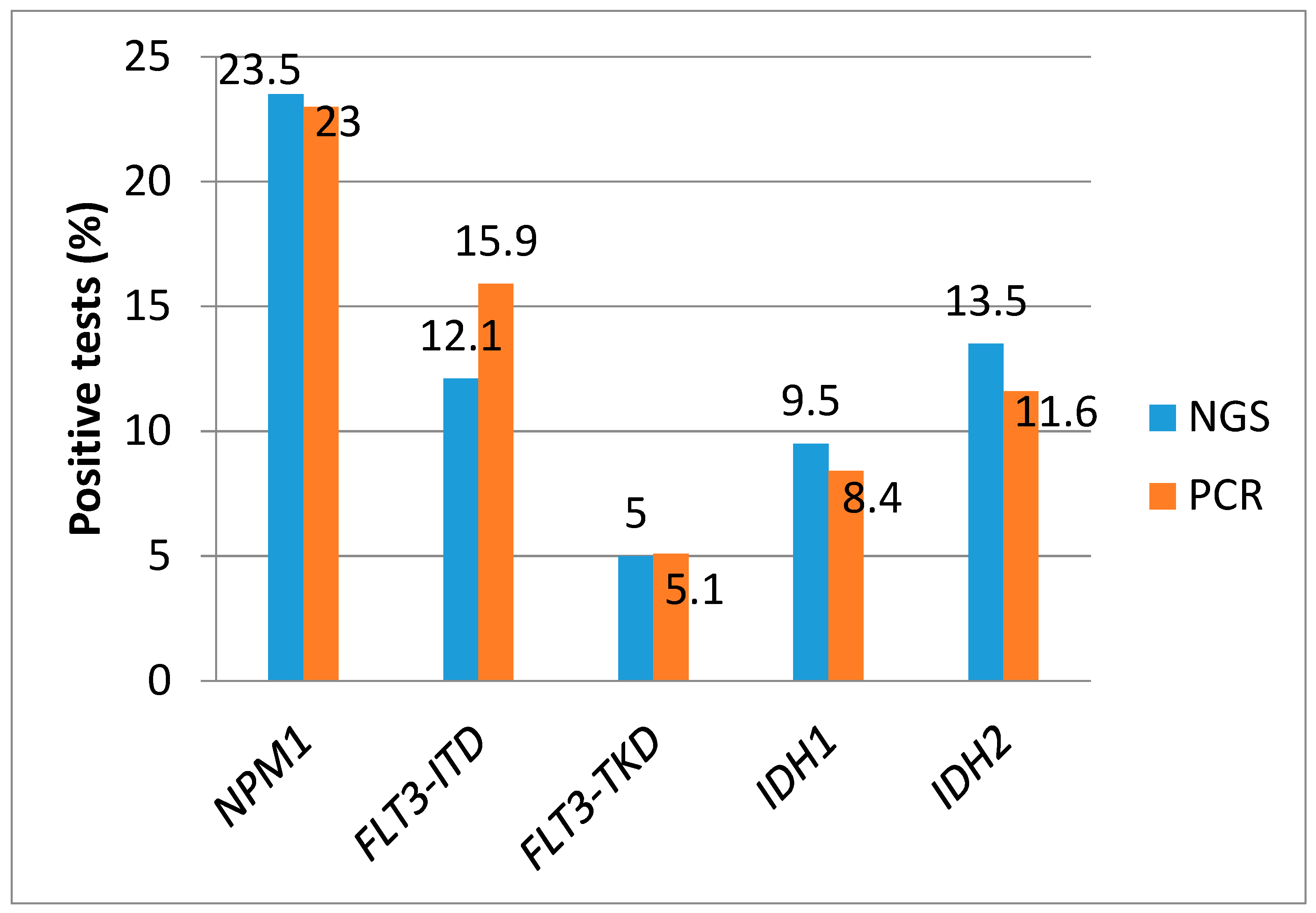
3.2. Frequency of NPM1, FLT3-ITD, FLT3-TKD, IDH1, and IDH2 Mutations by PCR

Among 1411 PCR samples analysed, 1200 corresponded to AML diagnosis, and 211 to R/R episodes.
NPM1 mutational status was determined in 1257 (89%) samples, and was not tested in 46/1200 (3.8%) samples at diagnosis vs. 46/211 (21.8%) in R/R status. Overall, 22% (N = 276) of samples were positive, 22.4% (258/1154) at diagnosis vs. 17.5% (18/103) at R/R episode (p = 0.25). The NPM1 mutation types were A (c.860_836dup863dup) (56%), B (c.863_864insCATG) (4.3%), D (c.863_864insCCTG) (3.1%), atypical (6.2%), and not available (29.5%).
FLT3-ITD mutation was tested in 1386 (98%) samples. The frequency of FLT3-ITD was 17.2% (N = 239), 17.2% (203/1181) at diagnosis, and 18.9% (36/192) at R/R episode.
FLT3-TKD mutation was tested in 1220 samples (86%). The frequency of FLT3-TKD was 5.1% (N = 62), 5% (55/1026) at diagnosis, and 3.7% (7/187) at R/R episode.
IDH1 mutation was tested in 1031 (73.1%) samples. The frequency of IDH1 mutation was 8.2% (N = 85), 7.2% (61/841) at diagnosis, and 12.7% (22/173) at R/R episode.
IDH2 mutation was tested in 1032 (73.1%) samples. The frequency of IDH2 mutation was 11.1% (N = 115), 11.6% (99/851) at diagnosis, and 8.4% (14/172) at R/R episode.

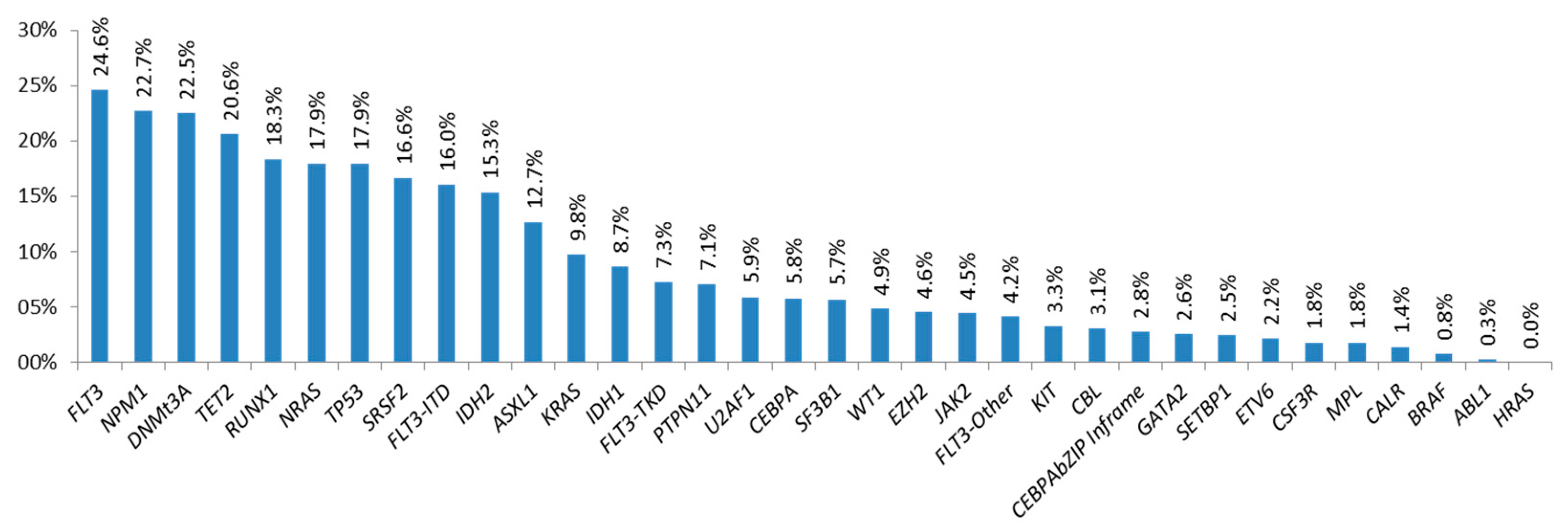
3.3. Frequency of Mutations Detected by NGS

The NGS mutational profile was available in 1671 samples, showing that almost all samples (96.5%) had at least one of the studied genes mutated, namely the most frequently mutated genes (frequency > 15%) FLT3 (both) (24.6%), NPM1 (22.7%), DNMT3A (22.5%), TET2 (20.6%), RUNX1 (18.3%), NRAS (17.9%), TP53 (17.9%), SRSF2 (16.6%), FLT3-ITD (16%), and IDH2 (15.3%) (Figure 1).

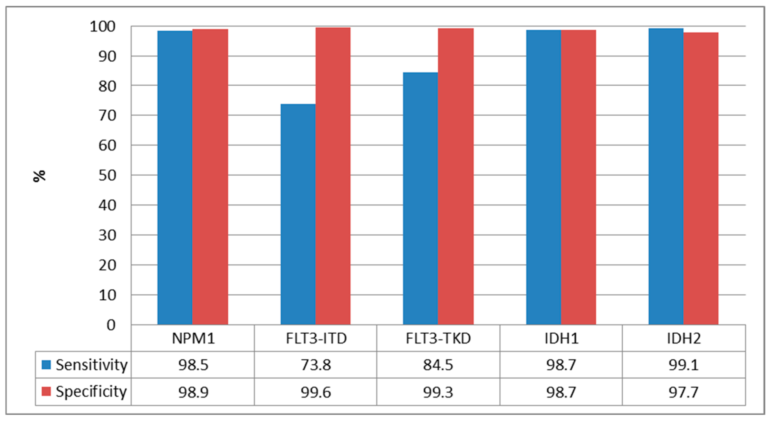
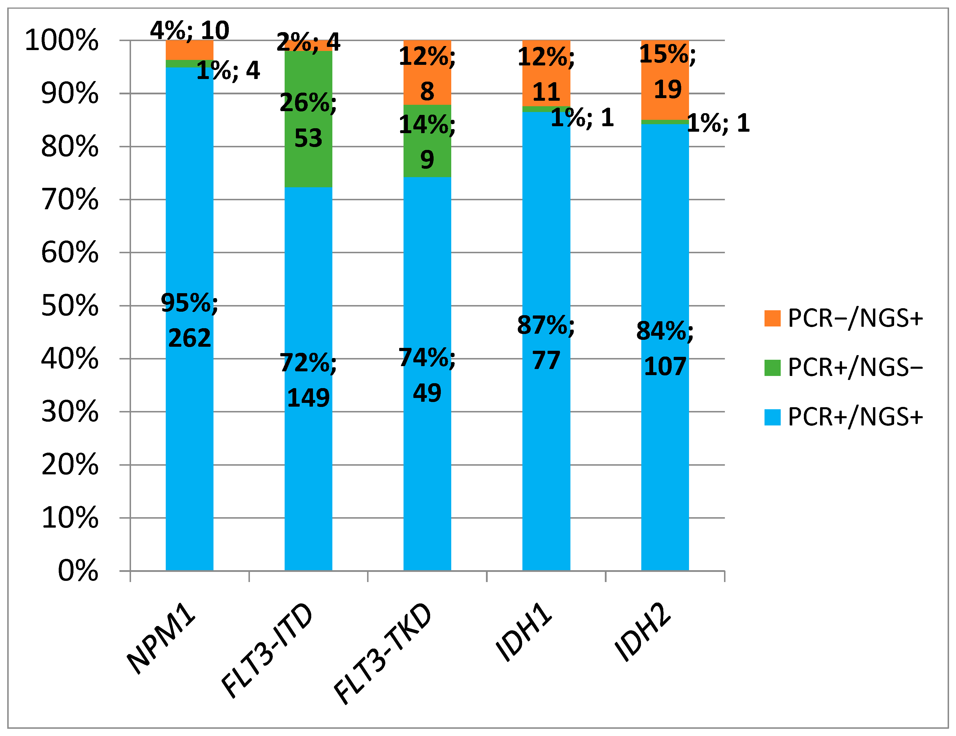
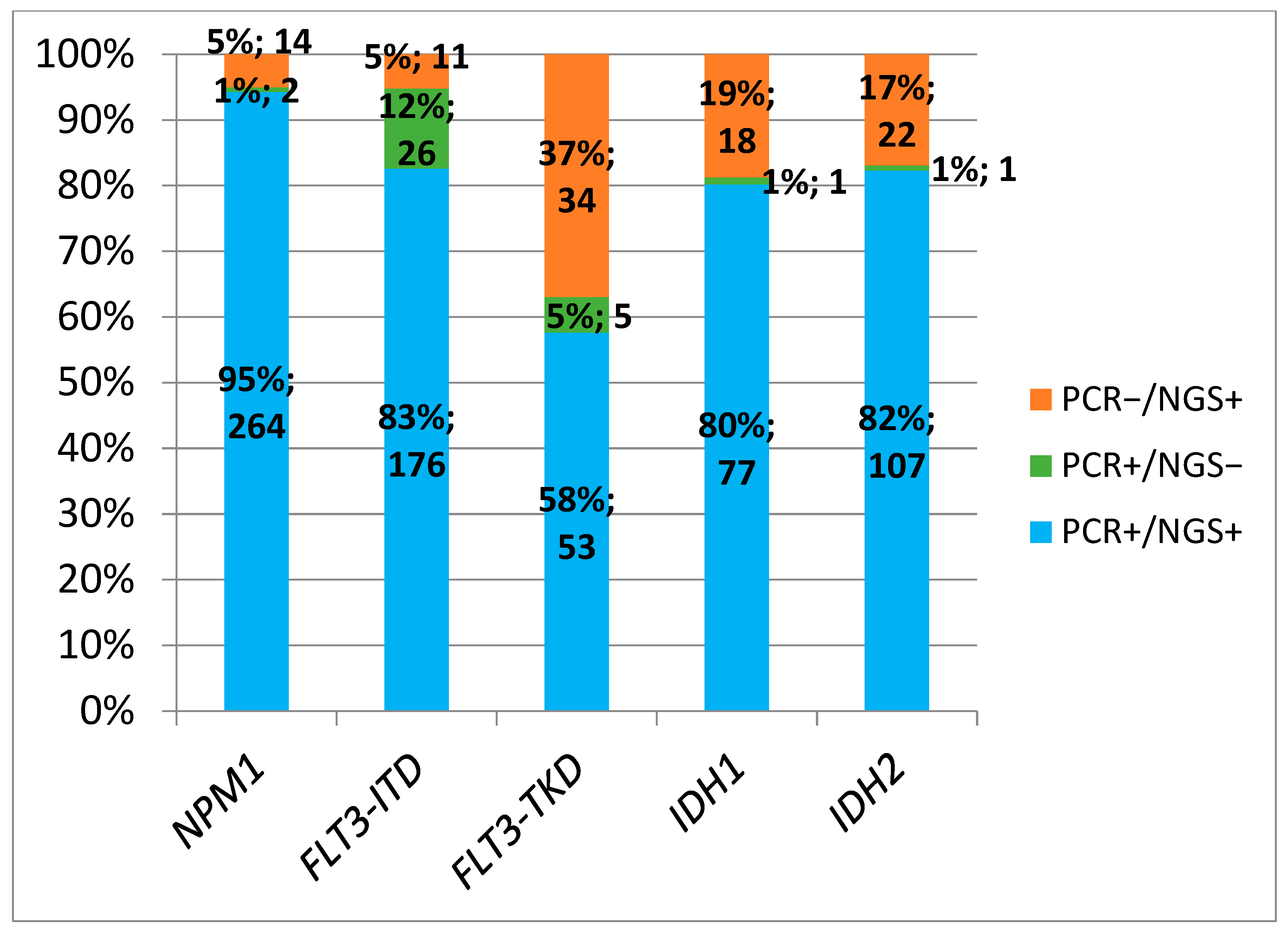
3.4. Comparison Between PCR and NGS

Among the 1288 paired NGS/PCR samples, NPM1 was available in 1285 (99.7%) for NGS and 1171 (90.8%) for PCR. Both results were available in 1156 (89.8%). Using a VAF cut-off of 5% for NGS, 23.5% were positive (272/1156) with a median VAF of 35.5% (range 7.1–74.9). The comparison of the positive results for each mutation for both techniques with VAF cut-off of ≥5% are shown in Figure 2. The NGS sensitivity was 98.5% (262/266) and specificity wad 98.9% (880/890) (Table 2 and Figure 3). Using a cut-off of ≥1% for NGS, 24% were positive for NPM1 (278/1156), with a sensitivity of 99.2% (264/266) and a specificity of 98.4% (876/890) (Table 3).
FLT3-ITD was available in 1284 (99.7%) for NGS and 1272 (98.8%) for PCR, with both available in 1267 (98.4%). With NGS VAF cut-off ≥ 5%, 12.1% were positive (153/1267) with a median VAF of 29.6 (range 5.21–97.4). By conventional PCR (cut-off ≥0.03), 15.9% were positive (202/1267), with a median ratio of 0.61 (range 0.03–11.6). We observed an NGS sensitivity of 73.8% and specificity of 99.6% (Table 2). Using a VAF cut-off ≥1%, the sensitivity increased to 87.1% (176/202) and specificity was 99% (1054/1065) (Table 3).
FLT3-TKD was available in 1283 (99.6%) for NGS and 1137 (88.3%) for PCR, with both available in 1130 (87.7%). With NGS VAF cut-off ≥5%, 5% were positive (57/1130) with median VAF 24.7 (range 5.06–51.8), resulting in sensitivity 84.5% and specificity 99.3% (Table 2). With VAF cut-off ≥1%, 7.7% were positive for FLT3-TKD (87/1130), with sensitivity of 91.4% (53/58) and specificity of 96.8% (1038/1072) (Table 3). Table 4 shows the FLT3-TKD alterations identified by NGS (cut-off ≥1%) and not detected by PCR.
IDH1 was available in 1283 (99.6%) for NGS and 929 (72.1%) for PCR, with both available in 925 (71.8%). With an NGS VAF cut-off of ≥5%, 9.5% were positive (88/925) with a median VAF of 41.8 (range 5.7–51.6), resulting in sensitivity and specificity of 98.7% (Table 2). With a VAF cut-off of ≥1%, 10.3% were positive for IDH1 (95/925), with a sensitivity of 98.7% (73/78) and a specificity of 97.9% (829/847) (Table 3).
IDH2 was available in 1285 (99.8%) for NGS and 931 (72.3%) for PCR, with both available in 931 (72.3%). With an NGS VAF cut-off of ≥5%, 13.5% were positive (126/931) with a median VAF of 42.2 (range 5.6–92.6), resulting in a sensitivity of 99.1% and a specificity of 97.7% (Table 2). With a VAF cut-off of ≥1%, 13.9% were positive for IDH2 (129/931), with a sensitivity of 99.7% (107/108) and a specificity of 97.3% (804/823) (Table 3).
The concordance rate between both techniques using NGS cut-off ≥5% was as follows: 95% for NPM1, 72% for FLT3-ITD, 74% for FLT3-TKD, 87% for IDH1, and 84% for IDH2 (Figure 4). The concordance rate between both techniques using an NGS cut-off of ≥1% were as follows: 95% for NPM1, 83% for FLT3-ITD, 58% for FLT3-TKD, 80% for IDH1, and 82% for IDH2 (Figure 5).

3.5. Comparative Between PCR and NGS at AML Diagnosis

Overall, 1094 patients were analyzed at diagnosis, NPM1 was available in 1091 (99.7%) for NGS and 1074 (98.2%) for PCR, with both results available in 1061 (97%). Using a VAF cut-off of ≥5%, 24% were positive with a median VAF of 35.8% (range 7.1–74.9).
FLT3-ITD was available in 1091 (99.7%) for NGS and 1084 (99%) for PCR, with both available in 1079 (98.5%). With an NGS VAF cut-off of ≥5%, 11.8% were positive with median VAF of 29.6% (range 5.27–97.4), and 15.8% were positive by PCR using a cut-off 0.03 ratio (median ratio 0.59; range 0.03–11.6).
FLT3-TKD was available in 1090 (99.5%) for NGS and 958 (87.6%) for PCR, with both results available in 951 (86.9%). With an NGS VAF cut-off of ≥5%, 5.2% were positive with a median VAF of 24.7% (range 5.1–51.8).
IDH1 was available in 1090 (99.5%) for NGS and 769 (70.3%) for PCR, with both available in 766 (70%). With an NGS VAF cut-off of ≥5%, 8.6% were positive with a median VAF of 41.3% (range 5.7–51.6).
IDH2 was available in 1092 (99.7%) for NGS and 771 (70.4%) for PCR, with both available in 771 (70.5%). With an NGS VAF cut-off of ≥5%, 14.3% were positive with a median VAF of 42.5% (range 6.2–92.6), NGS sensitivity and specificity for different genes are shown in Supplementary Figure S1 and Table S1 (using VAF cut-off of ≥5%) (Figure S1 and Table S1).

3.6. Comparative Between PCR and NGS at R/R AML Episode

Overall, 192 samples were tested at different R/R AML episodes. NPM1 was available in 191 (100%) for NGS and 96 (50%) for PCR, and for both in 95 (49.5%).
FLT3-ITD was available in all samples for NGS and 188 (97.9%) for PCR, and for both in 188 (97.9%). In positive cases, the median NGS VAF was 29.9% (range 5.2–92.6) and the median PCR ratio was 0.78 (range 0.03–11).
FLT3-TKD was available in all samples for NGS and 181 (93.8%) for PCR, and for both in 179 (93.2%).
IDH1 was available in all samples for NGS and 160 (82.9%) for PCR.
IDH2 was available in all samples for NGS and 160 (82.9%) for PCR. NGS sensitivity and specificity for different genes are shown in Supplementary Figure S2 and Table S2 (using cut-off VAF ≥ 5%) (Figure S2 and Table S2).

3.7. Turnaraound Time

Overall, the median days until full PCR report were 7 (range 0–84/interquartile range 3–14), and 28 (range 5–80/interquartile range 21–38) for NGS. At AML primary diagnosis, the median days until full PCR report were 5 (range 0–84/interquartile range 2–8), and 28 (range 5–80/interquartile range 20–38) for NGS. At R/R AML episodes, the median days until full PCR report were 12 (range 0–81/interquartile range 6–20), and 30 (range 11–74/interquartile range 24–41) for NGS (Supplementary Figure S3).

4. Discussion

This study is the first national large study of a wide range of patients, comparing the results of NGS and PCR performed via a uniform process for the detection of relevant genes in an AML clinical setting. We show high concordance between NPM1, IDH1, and IDH2 mutations detection using conventional PCR or NGS for AML patients. However, there were remarkable discrepancies between PCR and NGS to detect FLT3-ITD and FLT3-TKD mutations. The NGS approach showed several weaknesses to detect FLT3-ITD mutations with large duplications, whereas non canonical FLT3-TKD mutations and variants with low alleles were missed by PCR. On the other hand, the turnaround time for NPM1, IDH, and FLT3 mutations was more rapid using conventional PCR (7 days) as compared to NGS (28 days). Our national diagnostic platform allowed for rapid access to potential targeted therapies using conventional PCR, and for full classification of AML and risk stratification for post-remission therapy using NGS.
The consolidation of the national diagnostic platform has definitively facilitated the access of AML patients to precision medicine. This new strategy has meant an increase in the quality of molecular studies at the clinical level and also in the field of translational research. In fact, in our study period (2019–2021) we found that 84% of registered patients had a centralized NGS, while the centralized PCR rate was 80%. These data contrast with our recently published REALMOL study analyzing treatment patterns of the AML diagnostic [17], where up to 95% of Spanish patients had NGS and PCR at diagnosis between 2017 and 2021. These differences reflect that a percentage of patients had only local diagnostic tests, especially for conventional PCR. Another clear area of improvement is the R/R AML episode, as presumably only a small fraction of episodes were centrally tested by NGS and/or PCR.
Our study cohort of 1512 patients was representative of AML epidemiology, but with a bias towards younger patients included (median age 65 years old), in line with the REALMOL study that showed that patients aged 70 years or more had less frequent diagnostic tests [17]. Nevertheless, the frequency of detected mutations by conventional PCR at diagnosis reflect a real world AML cohort (22% NPM1, 17% FLT3-ITD, 5% FLT3-TKD, IDH1 7%, and IDH2 11%) [18]. Of note, NGS and PCR testing at diagnosis for NPM1 and FLT3-ITD were performed in parallel for 99% of patients, but IDH1/2 genes were predominantly tested by NGS (99%) and less frequently by PCR (70%). These patterns may reflect a lack of tailored induction therapies for newly diagnosed IDH1/2 AML during the study period, in contrast with NPM1 and FLT3 patients where rapid PCR screening was required to potentially add gentuzumab ozogamicine [19,20,21] or midostaurin [22], which could be to 3 + 7 chemotherapy.
We found that NPM1 and IDH2 mutations were less prevalent in R/R patients, indicating that NPM1 AML [1], and even some IDH2 types (i.e., R172) [5] could have a lower relapse rate, and that IDH2 affects older patients [16,23] that are less frequently tested at R/R episodes. Conversely, we found that FLT3-ITD and IDH1 mutations were more frequent at R/R, supporting the idea that these leukemias could be more resistant [1] (although the prognostic impact of IDH1 remains controversial) [24,25,26]. Interestingly, we showed that NGS was performed more frequently than conventional PCR at R/R episodes for NPM1 and IDH mutations. The lower rate of PCR performed at R/R suggests that the rapid diagnosis of these mutations was much less required in clinical practice, as no targeted therapies were available in our country beyond clinical trials. In fact, PCR was as frequent as NGS for the FLT3 R/R episode diagnosis, influenced by gilteritinib accessibility [27,28,29].
The comparison between NGS and PCR performance showed a high concordance rate between NPM1 and IDH results. This suggests that both techniques would be suitable for a precise molecular diagnosis [30]. NGS was able to detect between 4 and 15% (5–19% using VAF ≥1% cut-off) of additional NPM1 or IDH mutations, respectively, which could be interpreted as a technique with a better sensitivity for the assessment of these alterations [7,9]. Nevertheless, the current timelines for NGS reporting indicate that, in our context, a more rapid screening for these mutations can be systematically provided by conventional PCR. On the other hand, for FLT3-TKD, NGS missed nine positive samples using a cut-off of 5%, probably because the sensitivity of PCR is 3%. Lowering the cut-off to 1% allowed additional positive samples to be identified. For the purposes of this study we consider these as true positives, and from a clinical point of view the FLT3 inhibitor would be included as part of the treatment. This is in line with the advantage associated with NGS in detecting a wider range of mutations compared to PCR detecting only canonical mutations in I836 and D835. Another NGS advantage is the accuracy to identify subclonal variants with low allele frequency. Our study showed up to 12% and 37% of FLT3-TKD mutations found only by NGS using VAF ≥ 5% and ≥1% cut-offs, respectively. Therefore, we consider that NGS should replace PCR for FLT3-TKD and also IDH testing if the results delivery timelines are shortened for the purposes of target therapy establishment.
Regarding FLT3-ITD detection, we found a low sensitivity of NGS due to the limitation that this technique has for detecting duplications with long fragments [31,32,33]. The use of a VAF ≥ 1% cut-off can improve detection efficiency but 12% of false negative results still persisted with NGS. As a consequence, the detection of FLT3-ITD mutation by NGS could be underestimated [34]. Therefore, we believe that nowadays NGS still cannot replace PCR for FLT3-ITD screening, since missing these mutations could impact patient stratification for FLT3 inhibitors (e.g., midostaurin, gilteritinib).
In this study, we have employed two different VAF cut-offs for NGS detection in order to explore performance outcomes, which result in noticeably different results. For these mutations with a high clinical impact, we consider these results as true positives and they are thus informed and considered. We recommend adopting the 1% cut-off as the standard for these mutations and their clinical implications.
The median time from sample receipt until full report delivery was 7 days for PCR and 28 days for NGS. This timing could be more or less influenced by shipment distance from referral site to central laboratory, which is estimated in 24 h in Spain. However, courier logistical issues or sample drawing on Friday (with delivery on Monday) could lead to substantial additional delays affecting centralization applicability. It should be noted that we captured the date of the full diagnostic report, while central labs were routinely offering partial results (e.g., FLT3 positive or negative only) once this was available. Overall, our PCR and NGS timelines were in line with those recommended by the European Leukemia Net panel of experts (3–5 days for NPM1, FLT3, and IDH mutations, and before the end of the first cycle for remaining mutations by NGS) [1]. Interestingly, we show delayed timelines for molecular reporting in R/R AML, which may obey the general characteristics of R/R patients (usually less hyperleukocytosis and urgency to start therapy).
Our study has been carried out in a context in which molecular diagnosis laboratories in AML still need to combine conventional PCR with NGS. We can imagine a not-too-distant future in which, for the sake of efficiency, NGS will replace conventional PCR, at least for the vast majority of relevant mutations. The use of NGS for routine clinical care will require new NGS technologies (e.g., nanopore sequencing), more robust bioinformatic tools to improve detection of FLT3-ITD mutations, the ability to detect a wide range of alterations (including rearrangements i.e., MLLr) [35], and automated workflow to deliver a rapid turnaround time. These technological advances could pave the way to have a fully NGS-based molecular diagnosis in acceptable timelines (<5–7 days), allowing for the urgent personalized medicine treatment of AML. We could hypothesize that in a local laboratory with fewer samples it would be difficult to obtain these NGS timelines due to associated costs, while the centralization of samples would make this strategy affordable.

5. Conclusions

In conclusion, we show that a centralized platform at the national level is capable of providing molecular results by PCR and NGS according to the currently recommended timelines. While PCR remains the standard for the rapid screening of mutations in NPM1, IDH, and FLT3, NGS is capable of detecting more variants, especially in FLT3-TKD and IDH genes, which could have an impact on therapeutics. For these mutations with high clinical impact, a VAF cut-off of 1% should be adopted. Efforts should be made to report NGS results in acceptable times (<5–7 days) that may allow conventional PCR to be replaced and the daily routine in diagnostic laboratories to be optimized.

Supplementary Materials

The following supporting information can be downloaded at: https://www.mdpi.com/article/10.3390/cancers17050854/s1, Figure S1: sensitivity and specificity at diagnosis with NGS cut-off VAF ≥ 5%; Figure S2: NGS sensitivity and specificity at R/R AML (NGS cut-off VAF ≥ 5%); Figure S3: number of days elapsing from specimen receipt to diagnostic report, (A) overall, (B) at diagnosis, and (C) at R/R episode; Table S1: NGS and PCR results for each mutation at AML diagnosis, (NGS VAF cut-off ≥5%); Table S2: NGS and PCR results for each mutation at R/R AML, with NGS cut-off VAF ≥ 5%.

Author Contributions

P.M., B.B., R.R.-V. and E.B. conceived and designed the study. B.B., R.R.-V. and P.M. analyzed and interpreted the data, and wrote the paper; C.S., R.A., M.J.L., M.C.C., E.C.-C., C.B., E.P.d.l.T., E.S.-S., J.M.-L., M.J.C., R.G.-S., J.A.P.-S., M.T.G.-C., J.S.-G. and E.B. collected and analyzed the samples in the central laboratories; B.B., R.R.-V., I.N., D.M.-C., C.G., T.B., J.B., L.A., M.T., P.M.-S., J.S., J.M.A.-D., R.G., M.L.A., P.H.-P., M.J.S., E.L.-R., M.J.G.-P., J.M., E.P.-S., N.A.-V., A.O., J.A.L., M.B., M.G.-F., M.T.O., J.L. and P.M. included data of patients treated in their institutions; Y.M. performed data curation activities; B.B., C.S., R.R.-V., R.A., M.J.L., M.C.C., E.C.-C., C.B., E.P-DLT., E.S.-S., J.M.-L., M.J.C., R.G.-S., J.A.P.-S., M.T.G.-C., J.S.-G., E.B., I.N., D.M.-C., C.G., T.B., J.B., L.A., M.T., P.M.-S., J.S., J.M.A.-D., R.G., M.L.A., P.H.-P., M.J.S., E.L.-R., M.J.G.-P., J.M., E.P.-S., N.A.-V., A.O., J.A.L., M.B., M.G.-F., M.T.O., J.L., Y.M. and P.M. reviewed the manuscript, and contributed to the final draft. All authors have read and agreed to the published version of the manuscript.

Funding

This study was supported in part by Novartis and PMP22/00069, PI18/01340, PI18/01946 and PI21/01705 Instituto de Salud Carlos III grants.

Institutional Review Board Statement

The study was conducted in accordance with the Declaration of Helsinki, and approved by the Institutional Review Board of Instituto Investigación Sanitaria La Fe (protocol code PCR-LMA, approved the 5 June 2019).

Informed Consent Statement

Informed consent was obtained from all subjects involved in the study.

Data Availability Statement

The original contributions presented in this study are included in the article/Supplementary Materials. Further inquiries can be directed to the corresponding authors.

Acknowledgments

The authors would like to thank all the PETHEMA institutions and clinicians, PETHEMA data managers for data collection and management, and the central laboratory staff for sample management. A complete list of the institutions and clinicians participating in the PETHEMA epidemiologic registry of acute myeloid leukemia appears in the Supplementary Materials “Appendix”. This study was supported in part by Novartis and PMP22/00069, PI18/01340, PI18/01946 and PI21/01705 Instituto de Salud Carlos III grants.

Conflicts of Interest

The authors declare no conflicts of interest.

References

  1. Döhner, H.; Wei, A.H.; Appelbaum, F.R.; Craddock, C.; DiNardo, C.D.; Dombret, H.; Ebert, B.L.; Fenaux, P.; Godley, L.A.; Hasserjian, R.P.; et al. Diagnosis and Management of AML in Adults: 2022 Recommendations from an International Expert Panel on Behalf of the ELN. Blood 2022, 140, 1345–1377. [Google Scholar] [CrossRef] [PubMed]
  2. Arber, D.A.; Orazi, A.; Hasserjian, R.P.; Borowitz, M.J.; Calvo, K.R.; Kvasnicka, H.M.; Wang, S.A.; Bagg, A.; Barbui, T.; Branford, S.; et al. International Consensus Classification of Myeloid Neoplasms and Acute Leukemias: Integrating Morphologic, Clinical, and Genomic Data. Blood 2022, 140, 1200–1228. [Google Scholar] [CrossRef]
  3. Khoury, J.D.; Solary, E.; Abla, O.; Akkari, Y.; Alaggio, R.; Apperley, J.F.; Bejar, R.; Berti, E.; Busque, L.; Chan, J.K.C.; et al. The 5th Edition of the World Health Organization Classification of Haematolymphoid Tumours: Myeloid and Histiocytic/Dendritic Neoplasms. Leukemia 2022, 36, 1703–1719. [Google Scholar] [CrossRef]
  4. Döhner, H.; Estey, E.; Grimwade, D.; Amadori, S.; Appelbaum, F.R.; Büchner, T.; Dombret, H.; Ebert, B.L.; Fenaux, P.; Larson, R.A.; et al. Diagnosis and Management of AML in Adults: 2017 ELN Recommendations from an International Expert Panel. Blood 2017, 129, 424–447. [Google Scholar] [CrossRef]
  5. Papaemmanuil, E.; Gerstung, M.; Bullinger, L.; Gaidzik, V.I.; Paschka, P.; Roberts, N.D.; Potter, N.E.; Heuser, M.; Thol, F.; Bolli, N.; et al. Genomic Classification and Prognosis in Acute Myeloid Leukemia. N. Engl. J. Med. 2016, 374, 2209–2221. [Google Scholar] [CrossRef] [PubMed]
  6. Tazi, Y.; Arango-Ossa, J.E.; Zhou, Y.; Bernard, E.; Thomas, I.; Gilkes, A.; Freeman, S.; Pradat, Y.; Johnson, S.J.; Hills, R.; et al. Unified Classification and Risk-Stratification in Acute Myeloid Leukemia. Nat. Commun. 2022, 13, 1–16. [Google Scholar] [CrossRef] [PubMed]
  7. Bacher, U.; Shumilov, E.; Flach, J.; Porret, N.; Joncourt, R.; Wiedemann, G.; Fiedler, M.; Novak, U.; Amstutz, U.; Pabst, T. Challenges in the Introduction of next-Generation Sequencing (NGS) for Diagnostics of Myeloid Malignancies into Clinical Routine Use. Blood Cancer J. 2018, 8, 113. [Google Scholar] [CrossRef]
  8. Leisch, M.; Jansko, B.; Zaborsky, N.; Greil, R.; Pleyer, L. Next Generation Sequencing in AML-on the Way to Becoming a New Standard for Treatment Initiation And/or Modulation? Cancers 2019, 11, 252. [Google Scholar] [CrossRef]
  9. Jennings, L.J.; Arcila, M.E.; Corless, C.; Kamel-Reid, S.; Lubin, I.M.; Pfeifer, J.; Temple-Smolkin, R.L.; Voelkerding, K.V.; Nikiforova, M.N. Guidelines for Validation of Next-Generation Sequencing–Based Oncology Panels: A Joint Consensus Recommendation of the Association for Molecular Pathology and College of American Pathologists. J. Mol. Diagn. 2017, 19, 341–365. [Google Scholar] [CrossRef]
  10. Pollyea, D.A.; George, T.I.; Abedi, M.; Bejar, R.; Cogle, C.R.; Foucar, K.; Garcia-Manero, G.; Grinblatt, D.L.; Komrokji, R.S.; Maciejewski, J.P.; et al. Diagnostic and Molecular Testing Patterns in Patients with Newly Diagnosed Acute Myeloid Leukemia in the Connect® MDS/AML Disease Registry. eJHaem 2020, 1, 58–68. [Google Scholar] [CrossRef]
  11. Sargas, C.; Ayala, R.; Chillón, M.C.; Larráyoz, M.J.; Carrillo-Cruz, E.; Bilbao, C.; Yébenes-Ramírez, M.; Llop, M.; Rapado, I.; García-Sanz, R.; et al. Networking for Advanced Molecular Diagnosis in Acute Myeloid Leukemia Patients Is Possible: The PETHEMA NGS-AML Project. Haematologica 2021, 106, 3079–3089. [Google Scholar] [CrossRef]
  12. Gale, R.E.; Green, C.; Allen, C.; Mead, A.J.; Burnett, A.K.; Hills, R.K.; Linch, D.C. The Impact of FLT3 Internal Tandem Duplication Mutant Level, Number, Size, and Interaction with NPM1 Mutations in a Large Cohort of Young Adult Patients with Acute Myeloid Leukemia. Blood 2008, 111, 2776–2784. [Google Scholar] [CrossRef] [PubMed]
  13. Thiede, C.; Steudel, C.; Mohr, B.; Schaich, M.; Schäkel, U.; Platzbecker, U.; Wermke, M.; Bornhäuser, M.; Ritter, M.; Neubauer, A.; et al. Analysis of FLT3-Activating Mutations in 979 Patients with Acute Myelogenous Leukemia: Association with FAB Subtypes and Identification of Subgroups with Poor Prognosis. Blood 2002, 99, 4326–4335. [Google Scholar] [CrossRef] [PubMed]
  14. Yamamoto, Y.; Kiyoi, H.; Nakano, Y.; Suzuki, R.; Kodera, Y.; Miyawaki, S.; Asou, N.; Kuriyama, K.; Yagasaki, F.; Shimazaki, C.; et al. Activating Mutation of D835 within the Activation Loop of FLT3 in Human Hematologic Malignancies. Blood 2001, 97, 2434–2439. [Google Scholar] [CrossRef]
  15. Tefferi, A.; Lasho, T.L.; Abdel-Wahab, O.; Guglielmelli, P.; Patel, J.; Caramazza, D.; Pieri, L.; Finke, C.M.; Kilpivaara, O.; Wadleigh, M.; et al. IDH1 and IDH2 Mutation Studies in 1473 Patients with Chronic-, Fibrotic- or Blast-Phase Essential Thrombocythemia, Polycythemia Vera or Myelofibrosis. Leukemia 2010, 24, 1302–1309. [Google Scholar] [CrossRef] [PubMed]
  16. Sargas, C.; Ayala, R.; Larráyoz, M.J.; Chillón, M.C.; Carrillo-Cruz, E.; Bilbao-Sieyro, C.; Prados de la Torre, E.; Martínez-Cuadrón, D.; Rodríguez-Veiga, R.; Boluda, B.; et al. Molecular Landscape and Validation of New Genomic Classification in 2668 Adult AML Patients: Real Life Data from the PETHEMA Registry. Cancers 2023, 15, 438. [Google Scholar] [CrossRef]
  17. Labrador, J.; Martínez-Cuadrón, D.; Boluda, B.; Serrano, J.; Gil, C.; Pérez-Simón, J.A.; Bernal, T. Evolving Patterns and Clinical Outcome of Genetic Studies Performed at Diagnosis in Acute Myeloid Leukemia Patients: Real Life Data from the PETHEMA Registry. Cancer 2024, 130, 3436–3451. [Google Scholar] [CrossRef]
  18. Nagel, G.; Weber, D.; Fromm, E.; Erhardt, S.; Lübbert, M.; Fiedler, W.; Kindler, T.; Krauter, J.; Brossart, P.; Kündgen, A.; et al. Epidemiological, Genetic, and Clinical Characterization by Age of Newly Diagnosed Acute Myeloid Leukemia Based on an Academic Population-Based Registry Study (AMLSG BiO). Ann. Hematol. 2017, 96, 1993–2003. [Google Scholar] [CrossRef]
  19. Schlenk, R.F.; Paschka, P.; Krzykalla, J.; Weber, D.; Kapp-Schwoerer, S.; Gaidzik, V.I.; Leis, C.; Fiedler, W.; Kindler, T.; Schroeder, T.; et al. Gemtuzumab Ozogamicin in NPM1-Mutated Acute Myeloid Leukemia: Early Results from the Prospective Randomized AMLSG 09-09 Phase III Study. J. Clin. Oncol. 2020, 38, 623–632. [Google Scholar] [CrossRef]
  20. Petersdorf, S.H.; Kopecky, K.J.; Slovak, M.; Willman, C.; Nevill, T.; Brandwein, J.; Larson, R.A.; Erba, H.P.; Stiff, P.J.; Stuart, R.K.; et al. A Phase 3 Study of Gemtuzumab Ozogamicin during Induction and Postconsolidation Therapy in Younger Patients with Acute Myeloid Leukemia. Blood 2013, 121, 4854–4860. [Google Scholar] [CrossRef]
  21. Hills, R.K.; Castaigne, S.; Appelbaum, F.R.; Delaunay, J.; Petersdorf, S.; Othus, M.; Estey, E.H.; Dombret, H.; Chevret, S.; Ifrah, N.; et al. Addition of Gemtuzumab Ozogamicin to Induction Chemotherapy in Adult Patients with Acute Myeloid Leukaemia: A Meta-Analysis of Individual Patient Data from Randomised Controlled Trials. Lancet Oncol. 2014, 15, 986–996. [Google Scholar] [CrossRef]
  22. Stone, R.M.; Mandrekar, S.J.; Sanford, B.L.; Laumann, K.; Geyer, S.; Bloomfield, C.D.; Thiede, C.; Prior, T.W.; Döhner, K.; Marcucci, G.; et al. Midostaurin plus Chemotherapy for Acute Myeloid Leukemia with a FLT3 Mutation. N. Engl. J. Med. 2017, 377, 454–464. [Google Scholar] [CrossRef]
  23. Döhner, H.; Pratz, K.W.; DiNardo, C.D.; Wei, A.H.; Jonas, B.A.; Pullarkat, V.A.; Thirman, M.J.; Récher, C.; Schuh, A.C.; Babu, S.; et al. Genetic Risk Stratification and Outcomes among Treatment-Naive Patients with AML Treated with Venetoclax and Azacitidine. Blood 2024, 144, 2211–2222. [Google Scholar] [CrossRef] [PubMed]
  24. Megías-Vericat, J.E.; Ballesta-López, O.; Barragán, E.; Montesinos, P. IDH1-Mutated Relapsed or Refractory AML: Current Challenges and Future Prospects. Blood Lymphat. Cancer Targets Ther. 2019, 9, 19–32. [Google Scholar] [CrossRef] [PubMed]
  25. Zarnegar-Lumley, S.; Alonzo, T.A.; Gerbing, R.B.; Othus, M.; Sun, Z.; Ries, R.E.; Wang, J.; Leonti, A.; Kutny, M.A.; Ostronoff, F.; et al. Characteristics and Prognostic Impact of IDH Mutations in AML: A COG, SWOG, and ECOG Analysis. Blood Adv. 2023, 7, 5941–5953. [Google Scholar] [CrossRef]
  26. Paschka, P.; Schlenk, R.F.; Gaidzik, V.I.; Habdank, M.; Krönke, J.; Bullinger, L.; Späth, D.; Kayser, S.; Zucknick, M.; Götze, K.; et al. IDH1 and IDH2 Mutations Are Frequent Genetic Alterations in Acute Myeloid Leukemia and Confer Adverse Prognosis in Cytogenetically Normal Acute Myeloid Leukemia with NPM1 Mutation without FLT3 Internal Tandem Duplication. J. Clin. Oncol. 2010, 28, 3636–3643. [Google Scholar] [CrossRef] [PubMed]
  27. Perl, A.E.; Martinelli, G.; Cortes, J.E.; Neubauer, A.; Berman, E.; Paolini, S.; Montesinos, P.; Baer, M.R.; Larson, R.A.; Ustun, C.; et al. Gilteritinib or Chemotherapy for Relapsed or Refractory FLT3 -Mutated AML. N. Engl. J. Med. 2019, 381, 1728–1740. [Google Scholar] [CrossRef]
  28. Daver, N.; Schlenk, R.F.; Russell, N.H.; Levis, M.J. Targeting FLT3 Mutations in AML: Review of Current Knowledge and Evidence. Leukemia 2019, 33, 299–312. [Google Scholar] [CrossRef]
  29. Dumas, P.Y.; Raffoux, E.; Bérard, E.; Bertoli, S.; Hospital, M.A.; Heiblig, M.; Desbrosses, Y.; Bonmati, C.; Pautas, C.; Lambert, J.; et al. Gilteritinib Activity in Refractory or Relapsed FLT3-Mutated Acute Myeloid Leukemia Patients Previously Treated by Intensive Chemotherapy and Midostaurin: A Study from the French AML Intergroup ALFA/FILO. Leukemia 2023, 37, 91–101. [Google Scholar] [CrossRef]
  30. Haferlach, T. Advancing Leukemia Diagnostics: Role of next Generation Sequencing (Ngs) in Acute Myeloid Leukemia. Hematol. Rep. 2020, 12, 8957. [Google Scholar] [CrossRef]
  31. Mezei, Z.A.; Tornai, D.; Földesi, R.; Madar, L.; Sümegi, A.; Papp, M.; Antal-Szalmás, P. A DNA Pool of FLT3-ITD Positive DNA Samples Can Be Used Efficiently for Analytical Evaluation of NGS-Based FLT3-ITD Quantitation—Testing Several Different ITD Sequences and Rates, Simultaneously. J. Biotechnol. 2019, 303, 25–29. [Google Scholar] [CrossRef] [PubMed]
  32. Schranz, K.; Hubmann, M.; Harin, E.; Vosberg, S.; Herold, T.; Metzeler, K.H.; Rothenberg-Thurley, M.; Janke, H.; Bräundl, K.; Ksienzyk, B.; et al. Clonal Heterogeneity of FLT3-ITD Detected by High-Throughput Amplicon Sequencing Correlates with Adverse Prognosis in Acute Myeloid Leukemia. Oncotarget 2018, 9, 30128–30145. [Google Scholar] [CrossRef] [PubMed]
  33. Alvarado, Y.; Kantarjian, H.M.; Luthra, R.; Ravandi, F.; Borthakur, G.; Garcia-Manero, G.; Konopleva, M.; Estrov, Z.; Andreeff, M.; Cortes, J.E. Treatment with FLT3 Inhibitor in Patients with FLT3-Mutated Acute Myeloid Leukemia Is Associated with Development of Secondary FLT3-Tyrosine Kinase Domain Mutations. Cancer 2014, 120, 2142–2149. [Google Scholar] [CrossRef] [PubMed]
  34. Grob, T.; Sanders, M.A.; Vonk, C.M.; Kavelaars, F.G.; Rijken, M.; Hanekamp, D.W.; Gradowska, P.L.; Cloos, J.; Fløisand, Y.; Van Marwijk Kooy, M.; et al. Prognostic Value of FLT3 -Internal Tandem Duplication Residual Disease in Acute Myeloid Leukemia. J. Clin. Oncol. 2023, 41, 756–765. [Google Scholar] [CrossRef]
  35. Wang, E.S.; Issa, G.C.; Erba, H.P.; Altman, J.K.; Montesinos, P.; DeBotton, S.; Walter, R.B.; Pettit, K.; Savona, M.R.; Shah, M.V.; et al. Ziftomenib in Relapsed or Refractory Acute Myeloid Leukaemia (KOMET-001): A Multicentre, Open-Label, Multi-Cohort, Phase 1 Trial. Lancet Oncol. 2024, 25, 1310–1324. [Google Scholar] [CrossRef]
Figure 1. Frequency of mutations at diagnosis by NGS.
Cancers 17 00854 g001
Figure 2. Positive results by conventional PCR and NGS (VAF cut-off ≥5%).
Cancers 17 00854 g002
Figure 3. Sensitivity and specificity of NGS (VAF cut-off ≥5%) with respect to PCR.
Cancers 17 00854 g003
Figure 4. Concordance of positive results between PCR and NGS (VAF cut-off ≥5%).
Cancers 17 00854 g004
Figure 5. Concordance of positive results with PCR and NGS (VAF cut-off ≥1%).
Cancers 17 00854 g005
Table 1. Main demographic and clinical characteristics of the study cohort.
CharacteristicMedian (Range)Number (%)
n = 1512
Age at diagnosis65 (19–98)
<60 years511 (34)
60–70 years428 (28)
>70 years573 (38)
Male sex 860 (57)
ECOG 1 (0–4)
0–1 789 (79)
≥2 210 (21)
WBC count (×109/L)8.6 (0.1–401)
Hemoglobin (g/dL)8.9 (2–15.9)
Platelet count (×109/L)54 (3–907)
BM blast %54 (1–100)
Cytogenetic risk MRC (n = 824)
Low68 (8)
Intermediate485 (59)
High271 (33)
Abbreviations: MRC, Medical Research Council; AML, acute myeloid leukemia; BM, bone marrow; WBC, white blood cell count.
Table 2. NGS and PCR results for each mutation, with NGS VAF cut-off ≥5%.
Gene
Mutation
PCR+
N(%)
PCR−
N(%)
TOTAL
NPM1NGS+262 (98.5)10 (1.1)272
NGS−4 (1.5)880 (98.9)884
TOTAL266 (100)890 (100)1156
FLT3-ITDNGS+149 (73.8)4 (0.04)153
NGS−53 (26.2)1061 (99.6)1114
TOTAL202 (100)1065 (100)1267
FLT3-TKDNGS+49 (84.5)8 (0.07)57
NGS−9 (15.5)1064 (99.3)1073
TOTAL58 (100)1072 (100)1130
IDH1NGS+77 (98.7)11 (1.3)88
NGS−1 (1.3)836 (98.7)837
TOTAL78 (100)847 (100)925
IDH2NGS+107 (99.1)19 (2.3)126
NGS−1 (0.9)804 (97.7)805
TOTAL108 (100)823 (100)931
Abbreviations: NGS, next generation sequencing; PCR, polymerase chain reaction; FLT3, FMS-like tyrosine kinase-3.
Table 3. NGS and PCR results for each mutation using NGS VAF cut-off ≥1%.
Gene Mutation PCR+
N(%)
PCR−
N(%)
TOTAL
NPM1NGS+264 (99.2)14 (1.6)278
NGS−2 (0.8)876 (98.4)878
TOTAL266 (100)890 (100)1156
FLT3-ITDNGS+176 (87.1)11 (0.1)187
NGS−26 (12.9)1054 (99)1080
TOTAL202 (100)1065 (100)1267
FLT3-TKDNGS+53 (91.4)34 (3.2)87
NGS−5 (8.6)1038 (96.8)1043
TOTAL58 (100)1072 (100)1130
IDH1NGS+77 (98.7)18 (2.1)95
NGS−1 (1.3)829 (97.9)830
TOTAL78 (100)847 (100)925
IDH2NGS+107 (99.1)22 (2.7)129
NGS−1 (0.9)801 (97.3)802
TOTAL108 (100)823 (100)931
Abbreviations: NGS, next generation sequencing; PCR, polymerase chain reaction; FLT3, FMS-like tyrosine kinase-3.
Table 4. FLT3-TKD alterations identified by NGS (cut-off ≥1%) and not detected in PCR analysis.
Protein Change DetectedNumber of PatientsVAF (%)
Median (Range)
Asp835Tyr143.2 (1–44.29)
Asp835His62.1 (1.23–46.6)
Asp835Glu51.9 (1.13–4.68)
Ile836del53 (1.91–12.53)
Asp835Val221.43 (1.4–41.46)
Asp835Ile13.11
Asp835del14.3
Abbreviations: VAF, variant allele frequency.
Disclaimer/Publisher’s Note: The statements, opinions and data contained in all publications are solely those of the individual author(s) and contributor(s) and not of MDPI and/or the editor(s). MDPI and/or the editor(s) disclaim responsibility for any injury to people or property resulting from any ideas, methods, instructions or products referred to in the content.

