Physiological and Pathophysiological Effects of C-Type Natriuretic Peptide on the Heart
Abstract
Simple Summary
Abstract
1. Introduction
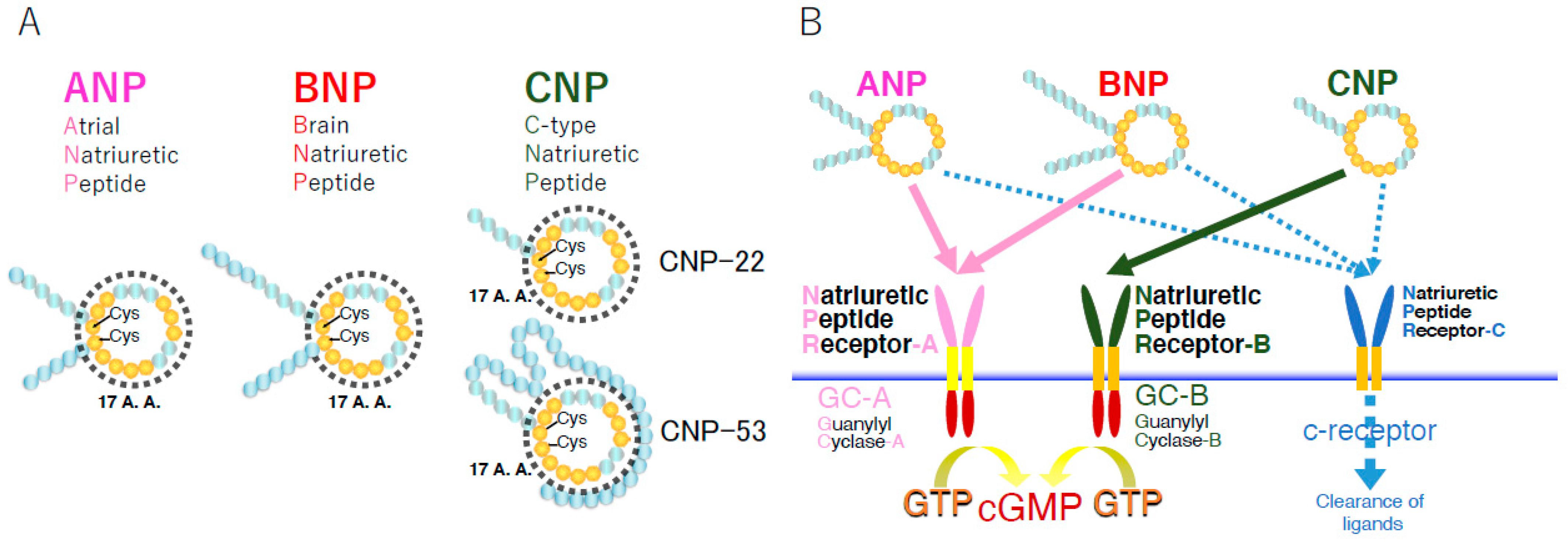
2. General Features of CNP
2.1. Generation of CNP
2.2. Receptors for CNP and Their Downstream Signaling
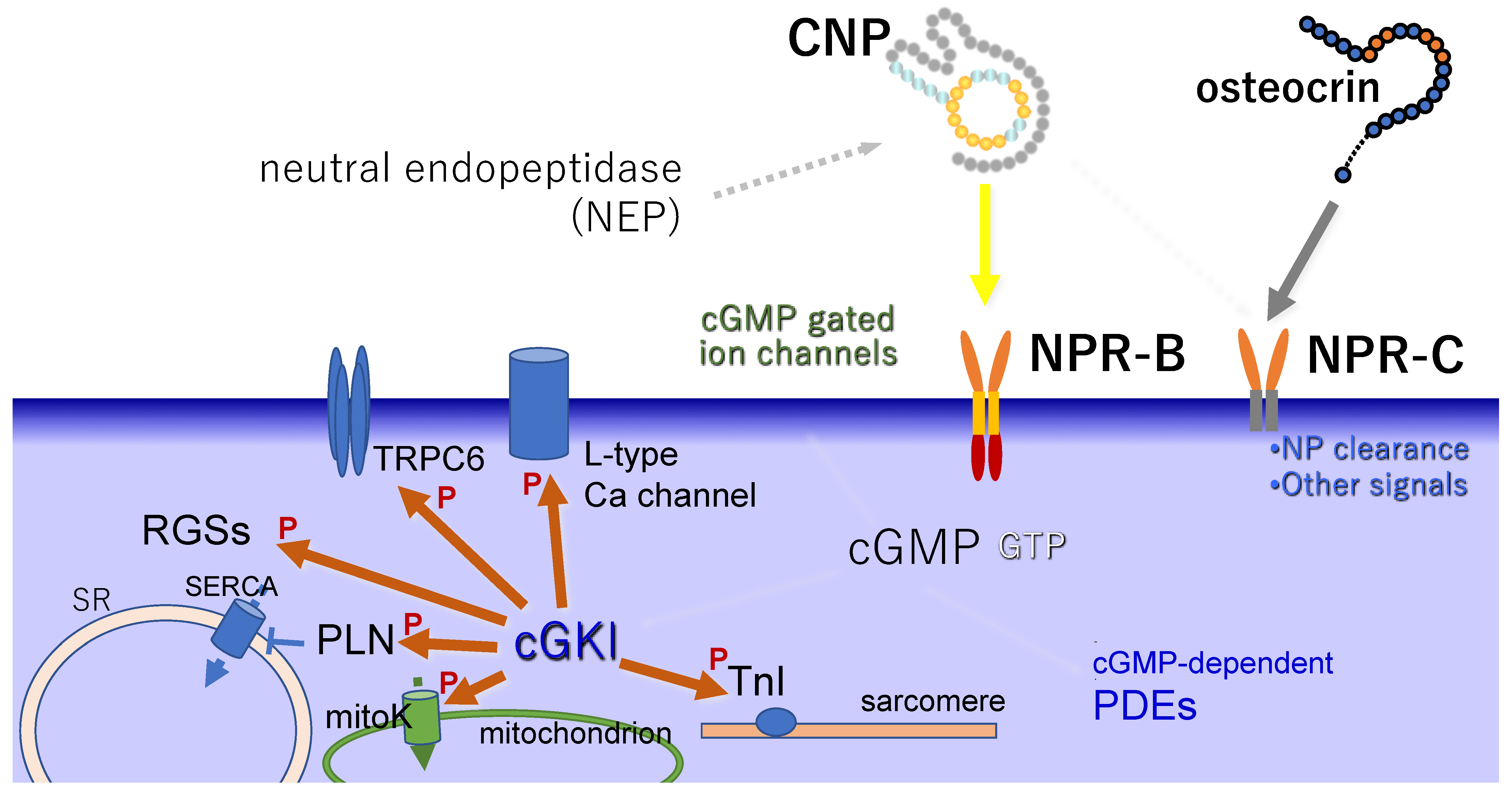
2.3. Degradation of CNP
2.4. Distribution of CNP
3. Physiological Roles of CNP in the Heart
3.1. Distribution of CNP and NPR-B in the Heart
3.2. Downstream Signaling of CNP in the Cardiac Cells
3.3. Physiological Effects of CNP on the Heart
4. Effects of CNP on Heart Failure
5. Roles of CNP on Cardiac Hypertrophy
6. Roles of CNP on MI
7. Effects of CNP on Heart Rate and Electrical Conduction in the Sinoatrial Node (SAN)
8. Conclusions and Further Discussion
Funding
Institutional Review Board Statement
Informed Consent Statement
Data Availability Statement
Conflicts of Interest
References
- Sudoh, T.; Minamino, N.; Kangawa, K.; Matsuo, H. C-type natriuretic peptide (CNP): A new member of natriuretic peptide family identified in porcine brain. Biochem. Biophys. Res. Commun. 1990, 168, 863–870. [Google Scholar] [CrossRef]
- Tawaragi, Y.; Fuchimura, K.; Nakazato, H.; Tanaka, S.; Minamino, N.; Kangawa, K.; Matsuo, H. Gene and precursor structure of porcine C-type natriuretic peptide. Biochem. Biophys. Res. Commun. 1990, 172, 627–632. [Google Scholar] [CrossRef]
- Kojima, M.; Minamino, N.; Kangawa, K.; Matsuo, H. Cloning and sequence analysis of a cDNA encoding a precursor for rat C-type natriuretic peptide (CNP). FEBS Lett. 1990, 276, 209–213. [Google Scholar] [CrossRef]
- Tawaragi, Y.; Fuchimura, K.; Tanaka, S.; Minamino, N.; Kangawa, K.; Matsuo, H. Gene and precursor structures of human C-type natriuretic peptide. Biochem. Biophys. Res. Commun. 1991, 175, 645–651. [Google Scholar] [CrossRef]
- Furuya, M.; Tawaragi, Y.; Minamitake, Y.; Kitajima, Y.; Fuchimura, K.; Tanaka, S.; Minamino, N.; Kangawa, K.; Matsuo, H. Structural requirements of C-type natriuretic peptide for elevation of cyclic GMP in cultured vascular smooth muscle cells. Biochem. Biophys. Res. Commun. 1992, 183, 964–969. [Google Scholar] [CrossRef]
- Koller, K.J.; Lowe, D.G.; Bennett, G.L.; Minamino, N.; Kangawa, K.; Matsuo, H.; Goeddel, D.V. Selective activation of the B natriuretic peptide receptor by C-type natriuretic peptide (CNP). Science 1991, 252, 120–123. [Google Scholar] [CrossRef] [PubMed]
- Suga, S.; Nakao, K.; Hosoda, K.; Mukoyama, M.; Ogawa, Y.; Shirakami, G.; Arai, H.; Saito, Y.; Kambayashi, Y.; Inouye, K. Receptor selectivity of natriuretic peptide family, atrial natriuretic peptide, brain natriuretic peptide, and C-type natriuretic peptide. Endocrinology 1992, 130, 229–239. [Google Scholar] [CrossRef] [PubMed]
- Furuya, M.; Takehisa, M.; Minamitake, Y.; Kitajima, Y.; Hayashi, Y.; Ohnuma, N.; Ishihara, T.; Minamino, N.; Kangawa, K.; Matsuo, H. Novel natriuretic peptide, CNP, potently stimulates cyclic GMP production in rat cultured vascular smooth muscle cells. Biochem. Biophys. Res. Commun. 1990, 170, 201–208. [Google Scholar] [CrossRef]
- Saito, Y.; Nakao, K.; Itoh, H.; Yamada, T.; Mukoyama, M.; Arai, H.; Hosoda, K.; Shirakami, G.; Suga, S.; Minamino, N. Brain natriuretic peptide is a novel cardiac hormone. Biochem. Biophys. Res. Commun. 1989, 158, 360–368. [Google Scholar] [CrossRef]
- Mukoyama, M.; Nakao, K.; Hosoda, K.; Suga, S.; Saito, Y.; Ogawa, Y.; Shirakami, G.; Jougasaki, M.; Obata, K.; Yasue, H. Brain natriuretic peptide as a novel cardiac hormone in humans. Evidence for an exquisite dual natriuretic peptide system, atrial natriuretic peptide and brain natriuretic peptide. J. Clin. Investig. 1991, 87, 1402–1412. [Google Scholar] [CrossRef]
- Ogawa, Y.; Nakao, K.; Mukoyama, M.; Hosoda, K.; Shirakami, G.; Arai, H.; Saito, Y.; Suga, S.; Jougasaki, M.; Imura, H. Natriuretic peptides as cardiac hormones in normotensive and spontaneously hypertensive rats. The ventricle is a major site of synthesis and secretion of brain natriuretic peptide. Circ. Res. 1991, 69, 491–500. [Google Scholar] [CrossRef] [PubMed]
- Ueda, S.; Minamino, N.; Aburaya, M.; Kangawa, K.; Matsukura, S.; Matsuo, H. Distribution and characterization of immunoreactive porcine C-type natriuretic peptide. Biochem. Biophys. Res. Commun. 1991, 175, 759–767. [Google Scholar] [CrossRef]
- Konrad, E.M.; Thibault, G.; Schiffrin, E.L. Autoradiographic visualization of the natriuretic peptide receptor-B in rat tissues. Regul. Pept. 1992, 39, 177–189. [Google Scholar] [CrossRef]
- Komatsu, Y.; Nakao, K.; Suga, S.; Ogawa, Y.; Mukoyama, M.; Arai, H.; Shirakami, G.; Hosoda, K.; Nakagawa, O.; Hama, N. C-type natriuretic peptide (CNP) in rats and humans. Endocrinology 1991, 129, 1104–1106. [Google Scholar] [CrossRef] [PubMed]
- Herman, J.P.; Langub, M.C.; Watson, R.E. Localization of C-type natriuretic peptide mRNA in rat hypothalamus. Endocrinology 1993, 133, 1903–1906. [Google Scholar] [CrossRef][Green Version]
- Langub, M.C.; Dolgas, C.M.; Watson, R.E.; Herman, J.P. The C-type natriuretic peptide receptor is the predominant natriuretic peptide receptor mRNA expressed in rat hypothalamus. J. Neuroendocrinol. 1995, 7, 305–309. [Google Scholar] [CrossRef]
- Yandle, T.G.; Fisher, S.; Charles, C.; Espiner, E.A.; Richards, A.M. The ovine hypothalamus and pituitary have markedly different distribution of C-type natriuretic peptide forms. Peptides 1993, 14, 713–716. [Google Scholar] [CrossRef]
- Suga, S.; Nakao, K.; Itoh, H.; Komatsu, Y.; Ogawa, Y.; Hama, N.; Imura, H. Endothelial production of C-type natriuretic peptide and its marked augmentation by transforming growth factor-beta. Possible existence of “vascular natriuretic peptide system”. J. Clin. Investig. 1992, 90, 1145–1149. [Google Scholar] [CrossRef]
- Zhang, M.; Su, Y.Q.; Sugiura, K.; Xia, G.; Eppig, J.J. Granulosa cell ligand NPPC and its receptor NPR2 maintain meiotic arrest in mouse oocytes. Science 2010, 330, 366–369. [Google Scholar] [CrossRef]
- Tsuji, T.; Kiyosu, C.; Akiyama, K.; Kunieda, T. CNP/NPR2 signaling maintains oocyte meiotic arrest in early antral follicles and is suppressed by EGFR-mediated signaling in preovulatory follicles. Mol. Reprod. Dev. 2012, 79, 795–802. [Google Scholar] [CrossRef]
- Suda, M.; Tanaka, K.; Fukushima, M.; Natsui, K.; Yasoda, A.; Komatsu, Y.; Ogawa, Y.; Itoh, H.; Nakao, K. C-type natriuretic peptide as an autocrine/paracrine regulator of osteoblast. Evidence for possible presence of bone natriuretic peptide system. Biochem. Biophys. Res. Commun. 1996, 223, 1–6. [Google Scholar] [CrossRef] [PubMed]
- Yasoda, A.; Ogawa, Y.; Suda, M.; Tamura, N.; Mori, K.; Sakuma, Y.; Chusho, H.; Shiota, K.; Tanaka, K.; Nakao, K. Natriuretic peptide regulation of endochondral ossification. Evidence for possible roles of the C-type natriuretic peptide/guanylyl cyclase-B pathway. J. Biol. Chem. 1998, 273, 11695–11700. [Google Scholar] [CrossRef] [PubMed]
- Chusho, H.; Tamura, N.; Ogawa, Y.; Yasoda, A.; Suda, M.; Miyazawa, T.; Nakamura, K.; Nakao, K.; Kurihara, T.; Komatsu, Y.; et al. Dwarfism and early death in mice lacking C-type natriuretic peptide. Proc. Natl. Acad. Sci. USA 2001, 98, 4016–4021. [Google Scholar] [CrossRef] [PubMed]
- Tamura, N.; Doolittle, L.K.; Hammer, R.E.; Shelton, J.M.; Richardson, J.A.; Garbers, D.L. Critical roles of the guanylyl cyclase B receptor in endochondral ossification and development of female reproductive organs. Proc. Natl. Acad. Sci. USA 2004, 101, 17300–17305. [Google Scholar] [CrossRef]
- Stingo, A.J.; Clavell, A.L.; Heublein, D.M.; Wei, C.M.; Pittelkow, M.R.; Burnett, J.C. Presence of C-type natriuretic peptide in cultured human endothelial cells and plasma. Am. J. Physiol. 1992, 263, H1318–H1321. [Google Scholar] [CrossRef]
- Hama, N.; Itoh, H.; Shirakami, G.; Suga, S.; Komatsu, Y.; Yoshimasa, T.; Tanaka, I.; Mori, K.; Nakao, K. Detection of C-type natriuretic peptide in human circulation and marked increase of plasma CNP level in septic shock patients. Biochem. Biophys. Res. Commun. 1994, 198, 1177–1182. [Google Scholar] [CrossRef]
- Prickett, T.C.; Yandle, T.G.; Nicholls, M.G.; Espiner, E.A.; Richards, A.M. Identification of amino-terminal pro-C-type natriuretic peptide in human plasma. Biochem. Biophys. Res. Commun. 2001, 286, 513–517. [Google Scholar] [CrossRef]
- Suga, S.; Itoh, H.; Komatsu, Y.; Ogawa, Y.; Hama, N.; Yoshimasa, T.; Nakao, K. Cytokine-induced C-type natriuretic peptide (CNP) secretion from vascular endothelial cells--evidence for CNP as a novel autocrine/paracrine regulator from endothelial cells. Endocrinology 1993, 133, 3038–3041. [Google Scholar] [CrossRef]
- Kuwahara, K. The natriuretic peptide system in heart failure: Diagnostic and therapeutic implications. Pharmacol. Ther. 2021, 227, 107863. [Google Scholar] [CrossRef]
- Goetze, J.P.; Bruneau, B.G.; Ramos, H.R.; Ogawa, T.; de Bold, M.K.; de Bold, A.J. Cardiac natriuretic peptides. Nat. Rev. Cardiol. 2020, 17, 698–717. [Google Scholar] [CrossRef]
- Savarirayan, R.; Irving, M.; Bacino, C.A.; Bostwick, B.; Charrow, J.; Cormier-Daire, V.; Le Quan Sang, K.H.; Dickson, P.; Harmatz, P.; Phillips, J.; et al. C-Type Natriuretic Peptide Analogue Therapy in Children with Achondroplasia. N. Engl. J. Med. 2019, 381, 25–35. [Google Scholar] [CrossRef] [PubMed]
- Savarirayan, R.; Tofts, L.; Irving, M.; Wilcox, W.; Bacino, C.A.; Hoover-Fong, J.; Ullot Font, R.; Harmatz, P.; Rutsch, F.; Bober, M.B.; et al. Once-daily, subcutaneous vosoritide therapy in children with achondroplasia: A randomised, double-blind, phase 3, placebo-controlled, multicentre trial. Lancet 2020, 396, 684–692. [Google Scholar] [CrossRef]
- Lumsden, N.G.; Khambata, R.S.; Hobbs, A.J. C-type natriuretic peptide (CNP): Cardiovascular roles and potential as a therapeutic target. Curr. Pharm. Des. 2010, 16, 4080–4088. [Google Scholar] [CrossRef] [PubMed]
- Wu, C.; Wu, F.; Pan, J.; Morser, J.; Wu, Q. Furin-mediated processing of Pro-C-type natriuretic peptide. J. Biol. Chem. 2003, 278, 25847–25852. [Google Scholar] [CrossRef]
- Prickett, T.C.; Espiner, E.A. Circulating products of C-type natriuretic peptide and links with organ function in health and disease. Peptides 2020, 132, 170363. [Google Scholar] [CrossRef]
- Minamino, N.; Kangawa, K.; Matsuo, H. N-terminally extended form of C-type natriuretic peptide (CNP-53) identified in porcine brain. Biochem. Biophys. Res. Commun. 1990, 170, 973–979. [Google Scholar] [CrossRef]
- Parmar, K.M.; Larman, H.B.; Dai, G.; Zhang, Y.; Wang, E.T.; Moorthy, S.N.; Kratz, J.R.; Lin, Z.; Jain, M.K.; Gimbrone, M.A.; et al. Integration of flow-dependent endothelial phenotypes by Kruppel-like factor 2. J. Clin. Investig. 2006, 116, 49–58. [Google Scholar] [CrossRef]
- Klinger, J.R.; Siddiq, F.M.; Swift, R.A.; Jackson, C.; Pietras, L.; Warburton, R.R.; Alia, C.; Hill, N.S. C-type natriuretic peptide expression and pulmonary vasodilation in hypoxia-adapted rats. Am. J. Physiol. 1998, 275, L645–L652. [Google Scholar] [CrossRef]
- Chun, T.H.; Itoh, H.; Ogawa, Y.; Tamura, N.; Takaya, K.; Igaki, T.; Yamashita, J.; Doi, K.; Inoue, M.; Masatsugu, K.; et al. Shear stress augments expression of C-type natriuretic peptide and adrenomedullin. Hypertension 1997, 29, 1296–1302. [Google Scholar] [CrossRef]
- Surendran, K.; Simon, T.C. CNP gene expression is activated by Wnt signaling and correlates with Wnt4 expression during renal injury. Am. J. Physiol. Ren. Physiol. 2003, 284, F653–F662. [Google Scholar] [CrossRef]
- Hagiwara, H.; Sakaguchi, H.; Itakura, M.; Yoshimoto, T.; Furuya, M.; Tanaka, S.; Hirose, S. Autocrine regulation of rat chondrocyte proliferation by natriuretic peptide C and its receptor, natriuretic peptide receptor-B. J. Biol. Chem. 1994, 269, 10729–10733. [Google Scholar] [CrossRef]
- Pfeifer, A.; Aszódi, A.; Seidler, U.; Ruth, P.; Hofmann, F.; Fässler, R. Intestinal secretory defects and dwarfism in mice lacking cGMP-dependent protein kinase II. Science 1996, 274, 2082–2086. [Google Scholar] [CrossRef] [PubMed]
- Miyazawa, T.; Ogawa, Y.; Chusho, H.; Yasoda, A.; Tamura, N.; Komatsu, Y.; Pfeifer, A.; Hofmann, F.; Nakao, K. Cyclic GMP-dependent protein kinase II plays a critical role in C-type natriuretic peptide-mediated endochondral ossification. Endocrinology 2002, 143, 3604–3610. [Google Scholar] [CrossRef][Green Version]
- Chikuda, H.; Kugimiya, F.; Hoshi, K.; Ikeda, T.; Ogasawara, T.; Shimoaka, T.; Kawano, H.; Kamekura, S.; Tsuchida, A.; Yokoi, N.; et al. Cyclic GMP-dependent protein kinase II is a molecular switch from proliferation to hypertrophic differentiation of chondrocytes. Genes Dev. 2004, 18, 2418–2429. [Google Scholar] [CrossRef] [PubMed]
- Kawasaki, Y.; Kugimiya, F.; Chikuda, H.; Kamekura, S.; Ikeda, T.; Kawamura, N.; Saito, T.; Shinoda, Y.; Higashikawa, A.; Yano, F.; et al. Phosphorylation of GSK-3beta by cGMP-dependent protein kinase II promotes hypertrophic differentiation of murine chondrocytes. J. Clin. Investig. 2008, 118, 2506–2515. [Google Scholar] [CrossRef] [PubMed]
- Nussenzveig, D.R.; Lewicki, J.A.; Maack, T. Cellular mechanisms of the clearance function of type C receptors of atrial natriuretic factor. J. Biol. Chem. 1990, 265, 20952–20958. [Google Scholar] [CrossRef]
- Anand-Srivastava, M.B.; Sairam, M.R.; Cantin, M. Ring-deleted analogs of atrial natriuretic factor inhibit adenylate cyclase/cAMP system. Possible coupling of clearance atrial natriuretic factor receptors to adenylate cyclase/cAMP signal transduction system. J. Biol. Chem. 1990, 265, 8566–8572. [Google Scholar] [CrossRef]
- Murthy, K.S.; Makhlouf, G.M. Identification of the G protein-activating domain of the natriuretic peptide clearance receptor (NPR-C). J. Biol. Chem. 1999, 274, 17587–17592. [Google Scholar] [CrossRef]
- Murthy, K.S.; Teng, B.Q.; Zhou, H.; Jin, J.G.; Grider, J.R.; Makhlouf, G.M. Gi−1/Gi−2-dependent signaling by single-transmembrane natriuretic peptide clearance receptor. Am. J. Physiol. Gastrointest. Liver Physiol. 2000, 278, G974–G980. [Google Scholar] [CrossRef]
- Pagano, M.; Anand-Srivastava, M.B. Cytoplasmic domain of natriuretic peptide receptor C constitutes Gi activator sequences that inhibit adenylyl cyclase activity. J. Biol. Chem. 2001, 276, 22064–22070. [Google Scholar] [CrossRef]
- Sangaralingham, S.J.; McKie, P.M.; Ichiki, T.; Scott, C.G.; Heublein, D.M.; Chen, H.H.; Bailey, K.R.; Redfield, M.M.; Rodeheffer, R.J.; Burnett, J.C. Circulating C-type natriuretic peptide and its relationship to cardiovascular disease in the general population. Hypertension 2015, 65, 1187–1194. [Google Scholar] [CrossRef] [PubMed]
- Hunt, P.J.; Richards, A.M.; Espiner, E.A.; Nicholls, M.G.; Yandle, T.G. Bioactivity and metabolism of C-type natriuretic peptide in normal man. J. Clin. Endocrinol. Metab. 1994, 78, 1428–1435. [Google Scholar] [CrossRef] [PubMed]
- Brandt, R.R.; Heublein, D.M.; Aarhus, L.L.; Lewicki, J.A.; Burnett, J.C. Role of natriuretic peptide clearance receptor in in vivo control of C-type natriuretic peptide. Am. J. Physiol. 1995, 269, H326–H331. [Google Scholar] [CrossRef] [PubMed]
- Kanai, Y.; Yasoda, A.; Mori, K.P.; Watanabe-Takano, H.; Nagai-Okatani, C.; Yamashita, Y.; Hirota, K.; Ueda, Y.; Yamauchi, I.; Kondo, E.; et al. Circulating osteocrin stimulates bone growth by limiting C-type natriuretic peptide clearance. J. Clin. Investig. 2017, 127, 4136–4147. [Google Scholar] [CrossRef]
- Miyazaki, T.; Otani, K.; Chiba, A.; Nishimura, H.; Tokudome, T.; Takano-Watanabe, H.; Matsuo, A.; Ishikawa, H.; Shimamoto, K.; Fukui, H.; et al. A New Secretory Peptide of Natriuretic Peptide Family, Osteocrin, Suppresses the Progression of Congestive Heart Failure After Myocardial Infarction. Circ. Res. 2018, 122, 742–751. [Google Scholar] [CrossRef]
- Watanabe-Takano, H.; Ochi, H.; Chiba, A.; Matsuo, A.; Kanai, Y.; Fukuhara, S.; Ito, N.; Sako, K.; Miyazaki, T.; Tainaka, K.; et al. Mechanical load regulates bone growth via periosteal Osteocrin. Cell Rep. 2021, 36, 109380. [Google Scholar] [CrossRef]
- Kenny, A.J.; Bourne, A.; Ingram, J. Hydrolysis of human and pig brain natriuretic peptides, urodilatin, C-type natriuretic peptide and some C-receptor ligands by endopeptidase-24.11. Biochem. J. 1993, 291 Pt 1, 83–88. [Google Scholar] [CrossRef]
- Brandt, R.R.; Mattingly, M.T.; Clavell, A.L.; Barclay, P.L.; Burnett, J.C. Neutral endopeptidase regulates C-type natriuretic peptide metabolism but does not potentiate its bioactivity in vivo. Hypertension 1997, 30, 184–190. [Google Scholar] [CrossRef]
- Ohbayashi, H.; Yamaki, K.; Suzuki, R.; Kume, H.; Takagi, K. Neutral endopeptidase 3.4.24.11 inhibition potentiates the inhibitory effects of type-C natriuretic peptide on leukotriene D4-induced airway changes. Clin. Exp. Pharmacol. Physiol. 1998, 25, 986–991. [Google Scholar] [CrossRef]
- Márton, Z.; Pataricza, J.; Krassói, I.; Varró, A.; Papp, J.G. NEP inhibitors enhance C-type natriuretic peptide-induced relaxation in porcine isolated coronary artery. Vasc. Pharmacol. 2005, 43, 207–212. [Google Scholar] [CrossRef]
- Hu, P.; Xia, X.; Xuan, Q.; Huang, B.Y.; Liu, S.Y.; Zhang, D.D.; Jiang, G.M.; Xu, Y.; Qin, Y.H. Neutral endopeptidase and natriuretic peptide receptors participate in the regulation of C-type natriuretic peptide expression in renal interstitial fibrosis. J. Recept. Signal Transduct. 2017, 37, 71–83. [Google Scholar] [CrossRef] [PubMed]
- Doi, K.; Itoh, H.; Komatsu, Y.; Igaki, T.; Chun, T.H.; Takaya, K.; Yamashita, J.; Inoue, M.; Yoshimasa, T.; Nakao, K. Vascular endothelial growth factor suppresses C-type natriuretic peptide secretion. Hypertension 1996, 27, 811–815. [Google Scholar] [CrossRef] [PubMed]
- Komatsu, Y.; Itoh, H.; Suga, S.; Ogawa, Y.; Hama, N.; Kishimoto, I.; Nakagawa, O.; Igaki, T.; Doi, K.; Yoshimasa, T.; et al. Regulation of endothelial production of C-type natriuretic peptide in coculture with vascular smooth muscle cells. Role of the vascular natriuretic peptide system in vascular growth inhibition. Circ. Res. 1996, 78, 606–614. [Google Scholar] [CrossRef] [PubMed]
- Chun, T.H.; Itoh, H.; Saito, T.; Yamahara, K.; Doi, K.; Mori, Y.; Ogawa, Y.; Yamashita, J.; Tanaka, T.; Inoue, M.; et al. Oxidative stress augments secretion of endothelium-derived relaxing peptides, C-type natriuretic peptide and adrenomedullin. J. Hypertens. 2000, 18, 575–580. [Google Scholar] [CrossRef]
- Yeung, V.T.; Mak, A.S.; Chui, Y.L.; Ho, S.K.; Lai, K.N.; Nicholls, M.G.; Cockram, C.S. Identification of C-type natriuretic peptide gene transcripts in glial cells. Neuroreport 1996, 7, 1709–1712. [Google Scholar] [CrossRef]
- Middendorff, R.; Maronde, E.; Paust, H.J.; Muller, D.; Davidoff, M.; Olcese, J. Expression of C-type natriuretic peptide in the bovine pineal gland. J. Neurochem. 1996, 67, 517–524. [Google Scholar] [CrossRef]
- Middendorff, R.; Paust, H.J.; Davidoff, M.S.; Olcese, J. Synthesis of C-type natriuretic peptide (CNP) by immortalized LHRH cells. J. Neuroendocrinol. 1997, 9, 177–182. [Google Scholar] [CrossRef]
- Yamamoto, S.; Morimoto, I.; Yanagihara, N.; Kangawa, K.; Inenaga, K.; Eto, S.; Yamashita, H. C-type natriuretic peptide suppresses arginine-vasopressin secretion from dissociated magnocellular neurons in newborn rat supraoptic nucleus. Neurosci. Lett. 1997, 229, 97–100. [Google Scholar] [CrossRef]
- Decker, J.M.; Wójtowicz, A.M.; Ul Haq, R.; Braunewell, K.H.; Heinemann, U.; Behrens, C.J. C-type natriuretic peptide decreases hippocampal network oscillations in adult rats in vitro. Neuroscience 2009, 164, 1764–1775. [Google Scholar] [CrossRef]
- Decker, J.M.; Wójtowicz, A.M.; Bartsch, J.C.; Liotta, A.; Braunewell, K.H.; Heinemann, U.; Behrens, C.J. C-type natriuretic peptide modulates bidirectional plasticity in hippocampal area CA1 in vitro. Neuroscience 2010, 169, 8–22. [Google Scholar] [CrossRef]
- Yamada-Goto, N.; Katsuura, G.; Ebihara, K.; Inuzuka, M.; Ochi, Y.; Yamashita, Y.; Kusakabe, T.; Yasoda, A.; Satoh-Asahara, N.; Ariyasu, H.; et al. Intracerebroventricular administration of C-type natriuretic peptide suppresses food intake via activation of the melanocortin system in mice. Diabetes 2013, 62, 1500–1504. [Google Scholar] [CrossRef]
- Fujii, T.; Hirota, K.; Yasoda, A.; Takizawa, A.; Morozumi, N.; Nakamura, R.; Yotsumoto, T.; Kondo, E.; Yamashita, Y.; Sakane, Y.; et al. Rats deficient C-type natriuretic peptide suffer from impaired skeletal growth without early death. PLoS ONE 2018, 13, e0194812. [Google Scholar] [CrossRef] [PubMed]
- Hirota, K.; Furuya, M.; Morozumi, N.; Yoshikiyo, K.; Yotsumoto, T.; Jindo, T.; Nakamura, R.; Murakami, K.; Ueda, Y.; Hanada, T.; et al. Exogenous C-type natriuretic peptide restores normal growth and prevents early growth plate closure in its deficient rats. PLoS ONE 2018, 13, e0204172. [Google Scholar] [CrossRef] [PubMed]
- Bartels, C.F.; Bukulmez, H.; Padayatti, P.; Rhee, D.K.; van Ravenswaaij-Arts, C.; Pauli, R.M.; Mundlos, S.; Chitayat, D.; Shih, L.Y.; Al-Gazali, L.I.; et al. Mutations in the transmembrane natriuretic peptide receptor NPR-B impair skeletal growth and cause acromesomelic dysplasia, type Maroteaux. Am. J. Hum. Genet. 2004, 75, 27–34. [Google Scholar] [CrossRef]
- Miura, K.; Namba, N.; Fujiwara, M.; Ohata, Y.; Ishida, H.; Kitaoka, T.; Kubota, T.; Hirai, H.; Higuchi, C.; Tsumaki, N.; et al. An overgrowth disorder associated with excessive production of cGMP due to a gain-of-function mutation of the natriuretic peptide receptor 2 gene. PLoS ONE 2012, 7, e42180. [Google Scholar] [CrossRef]
- Hisado-Oliva, A.; Ruzafa-Martin, A.; Sentchordi, L.; Funari, M.F.A.; Bezanilla-López, C.; Alonso-Bernáldez, M.; Barraza-García, J.; Rodriguez-Zabala, M.; Lerario, A.M.; Benito-Sanz, S.; et al. Mutations in C-natriuretic peptide (NPPC): A novel cause of autosomal dominant short stature. Genet. Med. 2018, 20, 91–97. [Google Scholar] [CrossRef] [PubMed]
- Wei, C.M.; Heublein, D.M.; Perrella, M.A.; Lerman, A.; Rodeheffer, R.J.; McGregor, C.G.; Edwards, W.D.; Schaff, H.V.; Burnett, J.C. Natriuretic peptide system in human heart failure. Circulation 1993, 88, 1004–1009. [Google Scholar] [CrossRef]
- Del Ry, S.; Cabiati, M.; Vozzi, F.; Battolla, B.; Caselli, C.; Forini, F.; Segnani, C.; Prescimone, T.; Giannessi, D.; Mattii, L. Expression of C-type natriuretic peptide and its receptor NPR-B in cardiomyocytes. Peptides 2011, 32, 1713–1718. [Google Scholar] [CrossRef]
- Del Ry, S. C-type natriuretic peptide: A new cardiac mediator. Peptides 2013, 40, 93–98. [Google Scholar] [CrossRef]
- Minamino, N.; Aburaya, M.; Kojima, M.; Miyamoto, K.; Kangawa, K.; Matsuo, H. Distribution of C-type natriuretic peptide and its messenger RNA in rat central nervous system and peripheral tissue. Biochem. Biophys. Res. Commun. 1993, 197, 326–335. [Google Scholar] [CrossRef]
- Suzuki, R.; Takahashi, A.; Hazon, N.; Takei, Y. Isolation of high-molecular-weight C-type natriuretic peptide from the heart of a cartilaginous fish (European dogfish, Scyliorhinus canicula). FEBS Lett. 1991, 282, 321–325. [Google Scholar] [CrossRef]
- Schofield, J.P.; Jones, D.S.; Forrest, J.N. Identification of C-type natriuretic peptide in heart of spiny dogfish shark (Squalus acanthias). Am. J. Physiol. 1991, 261, F734–F739. [Google Scholar] [CrossRef] [PubMed]
- Suzuki, R.; Takahashi, A.; Takei, Y. Different molecular forms of C-type natriuretic peptide isolated from the brain and heart of an elasmobranch, Triakis scyllia. J. Endocrinol. 1992, 135, 317–323. [Google Scholar] [CrossRef] [PubMed]
- Vollmar, A.M.; Gerbes, A.L.; Nemer, M.; Schulz, R. Detection of C-type natriuretic peptide (CNP) transcript in the rat heart and immune organs. Endocrinology 1993, 132, 1872–1874. [Google Scholar] [CrossRef] [PubMed]
- Öztop, M.; Cinar, K.; Turk, S. Immunolocalization of natriuretic peptides and their receptors in goat (Capra hircus) heart. Biotech. Histochem. 2018, 93, 389–404. [Google Scholar] [CrossRef]
- Takahashi, T.; Allen, P.D.; Izumo, S. Expression of A-, B-, and C-type natriuretic peptide genes in failing and developing human ventricles. Correlation with expression of the Ca(2+)-ATPase gene. Circ. Res. 1992, 71, 9–17. [Google Scholar] [CrossRef]
- Kalra, P.R.; Clague, J.R.; Bolger, A.P.; Anker, S.D.; Poole-Wilson, P.A.; Struthers, A.D.; Coats, A.J. Myocardial production of C-type natriuretic peptide in chronic heart failure. Circulation 2003, 107, 571–573. [Google Scholar] [CrossRef]
- Wright, S.P.; Prickett, T.C.; Doughty, R.N.; Frampton, C.; Gamble, G.D.; Yandle, T.G.; Sharpe, N.; Richards, M. Amino-terminal pro-C-type natriuretic peptide in heart failure. Hypertension 2004, 43, 94–100. [Google Scholar] [CrossRef][Green Version]
- Del Ry, S.; Passino, C.; Maltinti, M.; Emdin, M.; Giannessi, D. C-type natriuretic peptide plasma levels increase in patients with chronic heart failure as a function of clinical severity. Eur. J. Heart Fail. 2005, 7, 1145–1148. [Google Scholar] [CrossRef]
- Del Ry, S.; Cabiati, M.; Lionetti, V.; Emdin, M.; Recchia, F.A.; Giannessi, D. Expression of C-type natriuretic peptide and of its receptor NPR-B in normal and failing heart. Peptides 2008, 29, 2208–2215. [Google Scholar] [CrossRef]
- Palmer, S.C.; Prickett, T.C.; Espiner, E.A.; Yandle, T.G.; Richards, A.M. Regional release and clearance of C-type natriuretic peptides in the human circulation and relation to cardiac function. Hypertension 2009, 54, 612–618. [Google Scholar] [CrossRef] [PubMed]
- Lin, X.; Hänze, J.; Heese, F.; Sodmann, R.; Lang, R.E. Gene expression of natriuretic peptide receptors in myocardial cells. Circ. Res. 1995, 77, 750–758. [Google Scholar] [CrossRef] [PubMed]
- Doyle, D.D.; Upshaw-Earley, J.; Bell, E.L.; Palfrey, H.C. Natriuretic peptide receptor-B in adult rat ventricle is predominantly confined to the nonmyocyte population. Am. J. Physiol. Heart Circ. Physiol. 2002, 282, H2117–H2123. [Google Scholar] [CrossRef] [PubMed]
- Dickey, D.M.; Dries, D.L.; Margulies, K.B.; Potter, L.R. Guanylyl cyclase (GC)-A and GC-B activities in ventricles and cardiomyocytes from failed and non-failed human hearts: GC-A is inactive in the failed cardiomyocyte. J. Mol. Cell. Cardiol. 2012, 52, 727–732. [Google Scholar] [CrossRef]
- Nakamura, T.; Tsujita, K. Current trends and future perspectives for heart failure treatment leveraging cGMP modifiers and the practical effector PKG. J. Cardiol. 2021, 78, 261–268. [Google Scholar] [CrossRef]
- Dorn, G.W.; Force, T. Protein kinase cascades in the regulation of cardiac hypertrophy. J. Clin. Investig. 2005, 115, 527–537. [Google Scholar] [CrossRef]
- Fiedler, B.; Lohmann, S.M.; Smolenski, A.; Linnemuller, S.; Pieske, B.; Schroder, F.; Molkentin, J.D.; Drexler, H.; Wollert, K.C. Inhibition of calcineurin-NFAT hypertrophy signaling by cGMP-dependent protein kinase type I in cardiac myocytes. Proc. Natl. Acad. Sci. USA 2002, 99, 11363–11368. [Google Scholar] [CrossRef]
- Kuwahara, K.; Wang, Y.; McAnally, J.; Richardson, J.A.; Bassel-Duby, R.; Hill, J.A.; Olson, E.N. TRPC6 fulfills a calcineurin signaling circuit during pathologic cardiac remodeling. J. Clin. Investig. 2006, 116, 3114–3126. [Google Scholar] [CrossRef]
- Moltzau, L.R.; Meier, S.; Aronsen, J.M.; Afzal, F.; Sjaastad, I.; Skomedal, T.; Osnes, J.B.; Levy, F.O.; Qvigstad, E. Differential regulation of C-type natriuretic peptide-induced cGMP and functional responses by PDE2 and PDE3 in failing myocardium. Naunyn-Schmiedeberg’s Arch. Pharmacol. 2014, 387, 407–417. [Google Scholar] [CrossRef]
- Meier, S.; Andressen, K.W.; Aronsen, J.M.; Sjaastad, I.; Hougen, K.; Skomedal, T.; Osnes, J.B.; Qvigstad, E.; Levy, F.O.; Moltzau, L.R. PDE3 inhibition by C-type natriuretic peptide-induced cGMP enhances cAMP-mediated signaling in both non-failing and failing hearts. Eur. J. Pharmacol. 2017, 812, 174–183. [Google Scholar] [CrossRef]
- Burley, D.S.; Cox, C.D.; Zhang, J.; Wann, K.T.; Baxter, G.F. Natriuretic peptides modulate ATP-sensitive K+ channels in rat ventricular cardiomyocytes. Basic Res. Cardiol. 2014, 109, 402. [Google Scholar] [CrossRef] [PubMed]
- Moyes, A.J.; Chu, S.M.; Aubdool, A.A.; Dukinfield, M.S.; Margulies, K.B.; Bedi, K.C.; Hodivala-Dilke, K.; Baliga, R.S.; Hobbs, A.J. C-type natriuretic peptide co-ordinates cardiac structure and function. Eur. Heart J. 2020, 41, 1006–1020. [Google Scholar] [CrossRef] [PubMed]
- Dickey, D.M.; Flora, D.R.; Bryan, P.M.; Xu, X.; Chen, Y.; Potter, L.R. Differential regulation of membrane guanylyl cyclases in congestive heart failure: Natriuretic peptide receptor (NPR)-B, Not NPR-A, is the predominant natriuretic peptide receptor in the failing heart. Endocrinology 2007, 148, 3518–3522. [Google Scholar] [CrossRef] [PubMed]
- Yoshizumi, M.; Houchi, H.; Tsuchiya, K.; Minakuchi, K.; Horike, K.; Kitagawa, T.; Katoh, I.; Tamaki, T. Atrial natriuretic peptide stimulates Na+-dependent Ca2+ efflux from freshly isolated adult rat cardiomyocytes. FEBS Lett. 1997, 419, 255–258. [Google Scholar] [CrossRef]
- Brusq, J.M.; Mayoux, E.; Guigui, L.; Kirilovsky, J. Effects of C-type natriuretic peptide on rat cardiac contractility. Br. J. Pharmacol. 1999, 128, 206–212. [Google Scholar] [CrossRef] [PubMed]
- Nir, A.; Zhang, D.F.; Fixler, R.; Burnett, J.C.; Eilam, Y.; Hasin, Y. C-type natriuretic peptide has a negative inotropic effect on cardiac myocytes. Eur. J. Pharmacol. 2001, 412, 195–201. [Google Scholar] [CrossRef]
- Fixler, R.; Hasin, Y.; Eilam, Y.; Zhang, D.F.; Nir, A. Opposing effects of endothelin-1 on C-type natriuretic peptide actions in rat cardiomyocytes. Eur. J. Pharmacol. 2001, 423, 95–98. [Google Scholar] [CrossRef]
- Han, B.; Fixler, R.; Beeri, R.; Wang, Y.; Bachrach, U.; Hasin, Y. The opposing effects of endothelin-1 and C-type natriuretic peptide on apoptosis of neonatal rat cardiac myocytes. Eur. J. Pharmacol. 2003, 474, 15–20. [Google Scholar] [CrossRef]
- Zhang, Q.; Moalem, J.; Tse, J.; Scholz, P.M.; Weiss, H.R. Effects of natriuretic peptides on ventricular myocyte contraction and role of cyclic GMP signaling. Eur. J. Pharmacol. 2005, 510, 209–215. [Google Scholar] [CrossRef]
- Moalem, J.; Davidov, T.; Zhang, Q.; Grover, G.J.; Weiss, H.R.; Scholz, P.M. Negative inotropic effects of C-type natriuretic peptide are attenuated in hypertrophied ventricular myocytes associated with reduced cyclic GMP production. J. Surg. Res. 2006, 135, 38–44. [Google Scholar] [CrossRef]
- Subramanian, H.; Froese, A.; Jönsson, P.; Schmidt, H.; Gorelik, J.; Nikolaev, V.O. Distinct submembrane localisation compartmentalises cardiac NPR1 and NPR2 signalling to cGMP. Nat. Commun. 2018, 9, 2446. [Google Scholar] [CrossRef] [PubMed]
- Manfra, O.; Calamera, G.; Froese, A.; Arunthavarajah, D.; Surdo, N.C.; Meier, S.; Melleby, A.O.; Aasrum, M.; Aronsen, J.M.; Nikolaev, V.O.; et al. CNP regulates cardiac contractility and increases cGMP near both SERCA and TnI—Difference from BNP visualized by targeted cGMP biosensors. Cardiovasc. Res. 2022, 118, 1506–1519. [Google Scholar] [CrossRef] [PubMed]
- Wollert, K.C.; Yurukova, S.; Kilic, A.; Begrow, F.; Fiedler, B.; Gambaryan, S.; Walter, U.; Lohmann, S.M.; Kuhn, M. Increased effects of C-type natriuretic peptide on contractility and calcium regulation in murine hearts overexpressing cyclic GMP-dependent protein kinase I. Br. J. Pharmacol. 2003, 140, 1227–1236. [Google Scholar] [CrossRef] [PubMed]
- Zhang, Q.; Scholz, P.M.; Pilzak, A.; Su, J.; Weiss, H.R. Role of phospholamban in cyclic GMP mediated signaling in cardiac myocytes. Cell Physiol. Biochem. 2007, 20, 157–166. [Google Scholar] [CrossRef]
- Horio, T.; Tokudome, T.; Maki, T.; Yoshihara, F.; Suga, S.; Nishikimi, T.; Kojima, M.; Kawano, Y.; Kangawa, K. Gene expression, secretion, and autocrine action of C-type natriuretic peptide in cultured adult rat cardiac fibroblasts. Endocrinology 2003, 144, 2279–2284. [Google Scholar] [CrossRef]
- Del Ry, S.; Maltinti, M.; Piacenti, M.; Passino, C.; Emdin, M.; Giannessi, D. Cardiac production of C-type natriuretic peptide in heart failure. J. Cardiovasc. Med. 2006, 7, 397–399. [Google Scholar] [CrossRef]
- Lok, D.J.; Klip, I.T.; Voors, A.A.; Lok, S.I.; Bruggink-André de la Porte, P.W.; Hillege, H.L.; Jaarsma, T.; van Veldhuisen, D.J.; van der Meer, P. Prognostic value of N-terminal pro C-type natriuretic peptide in heart failure patients with preserved and reduced ejection fraction. Eur. J. Heart Fail. 2014, 16, 958–966. [Google Scholar] [CrossRef]
- Qvigstad, E.; Moltzau, L.R.; Aronsen, J.M.; Nguyen, C.H.; Hougen, K.; Sjaastad, I.; Levy, F.O.; Skomedal, T.; Osnes, J.B. Natriuretic peptides increase beta1-adrenoceptor signalling in failing hearts through phosphodiesterase 3 inhibition. Cardiovasc. Res. 2010, 85, 763–772. [Google Scholar] [CrossRef]
- Moltzau, L.R.; Aronsen, J.M.; Meier, S.; Nguyen, C.H.; Hougen, K.; Ørstavik, Ø.; Sjaastad, I.; Christensen, G.; Skomedal, T.; Osnes, J.B.; et al. SERCA2 activity is involved in the CNP-mediated functional responses in failing rat myocardium. Br. J. Pharmacol. 2013, 170, 366–379. [Google Scholar] [CrossRef]
- Michel, K.; Herwig, M.; Werner, F.; Špiranec Spes, K.; Abeßer, M.; Schuh, K.; Dabral, S.; Mügge, A.; Baba, H.A.; Skryabin, B.V.; et al. C-type natriuretic peptide moderates titin-based cardiomyocyte stiffness. JCI Insight 2020, 5, e139910. [Google Scholar] [CrossRef]
- Tokudome, T.; Horio, T.; Soeki, T.; Mori, K.; Kishimoto, I.; Suga, S.; Yoshihara, F.; Kawano, Y.; Kohno, M.; Kangawa, K. Inhibitory effect of C-type natriuretic peptide (CNP) on cultured cardiac myocyte hypertrophy: Interference between CNP and endothelin-1 signaling pathways. Endocrinology 2004, 145, 2131–2140. [Google Scholar] [CrossRef] [PubMed]
- Soeki, T.; Kishimoto, I.; Okumura, H.; Tokudome, T.; Horio, T.; Mori, K.; Kangawa, K. C-type natriuretic peptide, a novel antifibrotic and antihypertrophic agent, prevents cardiac remodeling after myocardial infarction. J. Am. Coll. Cardiol. 2005, 45, 608–616. [Google Scholar] [CrossRef] [PubMed]
- Izumiya, Y.; Araki, S.; Usuku, H.; Rokutanda, T.; Hanatani, S.; Ogawa, H. Chronic C-Type Natriuretic Peptide Infusion Attenuates Angiotensin II-Induced Myocardial Superoxide Production and Cardiac Remodeling. Int. J. Vasc. Med. 2012, 2012, 246058. [Google Scholar] [CrossRef] [PubMed]
- Su, J.; Zhang, Q.; Moalem, J.; Tse, J.; Scholz, P.M.; Weiss, H.R. Functional effects of C-type natriuretic peptide and nitric oxide are attenuated in hypertrophic myocytes from pressure-overloaded mouse hearts. Am. J. Physiol. Heart Circ. Physiol. 2005, 288, H1367–H1373. [Google Scholar] [CrossRef][Green Version]
- Langenickel, T.H.; Buttgereit, J.; Pagel-Langenickel, I.; Lindner, M.; Monti, J.; Beuerlein, K.; Al-Saadi, N.; Plehm, R.; Popova, E.; Tank, J.; et al. Cardiac hypertrophy in transgenic rats expressing a dominant-negative mutant of the natriuretic peptide receptor B. Proc. Natl. Acad. Sci. USA 2006, 103, 4735–4740. [Google Scholar] [CrossRef]
- Nakao, K.; Kuwahara, K.; Nishikimi, T.; Nakagawa, Y.; Kinoshita, H.; Minami, T.; Kuwabara, Y.; Yamada, C.; Yamada, Y.; Tokudome, T.; et al. Endothelium-Derived C-Type Natriuretic Peptide Contributes to Blood Pressure Regulation by Maintaining Endothelial Integrity. Hypertension 2017, 69, 286–296. [Google Scholar] [CrossRef]
- Moyes, A.J.; Khambata, R.S.; Villar, I.; Bubb, K.J.; Baliga, R.S.; Lumsden, N.G.; Xiao, F.; Gane, P.J.; Rebstock, A.S.; Worthington, R.J.; et al. Endothelial C-type natriuretic peptide maintains vascular homeostasis. J. Clin. Investig. 2014, 124, 4039–4051. [Google Scholar] [CrossRef]
- Hobbs, A.; Foster, P.; Prescott, C.; Scotland, R.; Ahluwalia, A. Natriuretic peptide receptor-C regulates coronary blood flow and prevents myocardial ischemia/reperfusion injury: Novel cardioprotective role for endothelium-derived C-type natriuretic peptide. Circulation 2004, 110, 1231–1235. [Google Scholar] [CrossRef]
- Wang, Y.; de Waard, M.C.; Sterner-Kock, A.; Stepan, H.; Schultheiss, H.P.; Duncker, D.J.; Walther, T. Cardiomyocyte-restricted over-expression of C-type natriuretic peptide prevents cardiac hypertrophy induced by myocardial infarction in mice. Eur. J. Heart Fail. 2007, 9, 548–557. [Google Scholar] [CrossRef]
- Del Ry, S.; Cabiati, M.; Martino, A.; Cavallini, C.; Caselli, C.; Aquaro, G.D.; Battolla, B.; Prescimone, T.; Giannessi, D.; Mattii, L.; et al. High concentration of C-type natriuretic peptide promotes VEGF-dependent vasculogenesis in the remodeled region of infarcted swine heart with preserved left ventricular ejection fraction. Int. J. Cardiol. 2013, 168, 2426–2434. [Google Scholar] [CrossRef]
- Frankenreiter, S.; Bednarczyk, P.; Kniess, A.; Bork, N.I.; Straubinger, J.; Koprowski, P.; Wrzosek, A.; Mohr, E.; Logan, A.; Murphy, M.P.; et al. cGMP-Elevating Compounds and Ischemic Conditioning Provide Cardioprotection Against Ischemia and Reperfusion Injury via Cardiomyocyte-Specific BK Channels. Circulation 2017, 136, 2337–2355. [Google Scholar] [CrossRef] [PubMed]
- Lukowski, R.; Cruz Santos, M.; Kuret, A.; Ruth, P. cGMP and mitochondrial K+ channels—Compartmentalized but closely connected in cardioprotection. Br. J. Pharmacol. 2021, 179, 2344–2360. [Google Scholar] [CrossRef] [PubMed]
- Lee, J.; Than, M.; Aldous, S.; Troughton, R.; Richards, M.; Pemberton, C.J. CNP Signal Peptide in Patients with Cardiovascular Disease. Front. Cardiovasc. Med. 2015, 2, 28. [Google Scholar] [CrossRef][Green Version]
- Prickett, T.C.; Doughty, R.N.; Troughton, R.W.; Frampton, C.M.; Whalley, G.A.; Ellis, C.J.; Espiner, E.A.; Richards, A.M. C-Type Natriuretic Peptides in Coronary Disease. Clin. Chem. 2017, 63, 316–324. [Google Scholar] [CrossRef] [PubMed]
- Mark, P.D.; Frydland, M.; Helgestad, O.K.L.; Holmvang, L.; Møller, J.E.; Johansson, P.I.; Ostrowski, S.R.; Prickett, T.; Hassager, C.; Goetze, J.P. Sex-specific mortality prediction by pro-C-type natriuretic peptide measurement in a prospective cohort of patients with ST-elevation myocardial infarction. BMJ. Open 2021, 11, e048312. [Google Scholar] [CrossRef]
- Beaulieu, P.; Cardinal, R.; De Léan, A.; Lambert, C. Direct chronotropic effects of atrial and C-type natriuretic peptides in anaesthetized dogs. Br. J. Pharmacol. 1996, 118, 1790–1796. [Google Scholar] [CrossRef]
- Beaulieu, P.; Cardinal, R.; Pagé, P.; Francoeur, F.; Tremblay, J.; Lambert, C. Positive chronotropic and inotropic effects of C-type natriuretic peptide in dogs. Am. J. Physiol. 1997, 273, H1933–H1940. [Google Scholar] [CrossRef]
- Rose, R.A.; Lomax, A.E.; Kondo, C.S.; Anand-Srivastava, M.B.; Giles, W.R. Effects of C-type natriuretic peptide on ionic currents in mouse sinoatrial node: A role for the NPR-C receptor. Am. J. Physiol. Heart Circ. Physiol. 2004, 286, H1970–H1977. [Google Scholar] [CrossRef][Green Version]
- Springer, J.; Azer, J.; Hua, R.; Robbins, C.; Adamczyk, A.; McBoyle, S.; Bissell, M.B.; Rose, R.A. The natriuretic peptides BNP and CNP increase heart rate and electrical conduction by stimulating ionic currents in the sinoatrial node and atrial myocardium following activation of guanylyl cyclase-linked natriuretic peptide receptors. J. Mol. Cell. Cardiol. 2012, 52, 1122–1134. [Google Scholar] [CrossRef]
- Azer, J.; Hua, R.; Vella, K.; Rose, R.A. Natriuretic peptides regulate heart rate and sinoatrial node function by activating multiple natriuretic peptide receptors. J. Mol. Cell. Cardiol. 2012, 53, 715–724. [Google Scholar] [CrossRef]
- Dorey, T.W.; Mackasey, M.; Jansen, H.J.; McRae, M.D.; Bohne, L.J.; Liu, Y.; Belke, D.D.; Atkinson, L.; Rose, R.A. Natriuretic peptide receptor B maintains heart rate and sinoatrial node function via cyclic GMP-mediated signaling. Cardiovasc. Res. 2021, cvab245. [Google Scholar] [CrossRef] [PubMed]
- Szaroszyk, M.; Kattih, B.; Martin-Garrido, A.; Trogisch, F.A.; Dittrich, G.M.; Grund, A.; Abouissa, A.; Derlin, K.; Meier, M.; Holler, T.; et al. Skeletal muscle derived MusClin. protects the heart during pathological overload. Nat. Commun. 2022, 13, 149. [Google Scholar] [CrossRef] [PubMed]


Publisher’s Note: MDPI stays neutral with regard to jurisdictional claims in published maps and institutional affiliations. |
© 2022 by the author. Licensee MDPI, Basel, Switzerland. This article is an open access article distributed under the terms and conditions of the Creative Commons Attribution (CC BY) license (https://creativecommons.org/licenses/by/4.0/).
Share and Cite
Yasoda, A. Physiological and Pathophysiological Effects of C-Type Natriuretic Peptide on the Heart. Biology 2022, 11, 911. https://doi.org/10.3390/biology11060911
Yasoda A. Physiological and Pathophysiological Effects of C-Type Natriuretic Peptide on the Heart. Biology. 2022; 11(6):911. https://doi.org/10.3390/biology11060911
Chicago/Turabian StyleYasoda, Akihiro. 2022. "Physiological and Pathophysiological Effects of C-Type Natriuretic Peptide on the Heart" Biology 11, no. 6: 911. https://doi.org/10.3390/biology11060911
APA StyleYasoda, A. (2022). Physiological and Pathophysiological Effects of C-Type Natriuretic Peptide on the Heart. Biology, 11(6), 911. https://doi.org/10.3390/biology11060911


