
COVID-19 Diagnosis: A Review of Rapid Antigen, RT-PCR and Artificial Intelligence Methods

 ,
,  , , , ,
Abstract
:1. Introduction
2. Current and Emerging COVID-19 Diagnostic Tests
3. Real-Time Reverse Transcriptase-Polymerase Chain Reaction
4. Rapid Antigen Detection Test
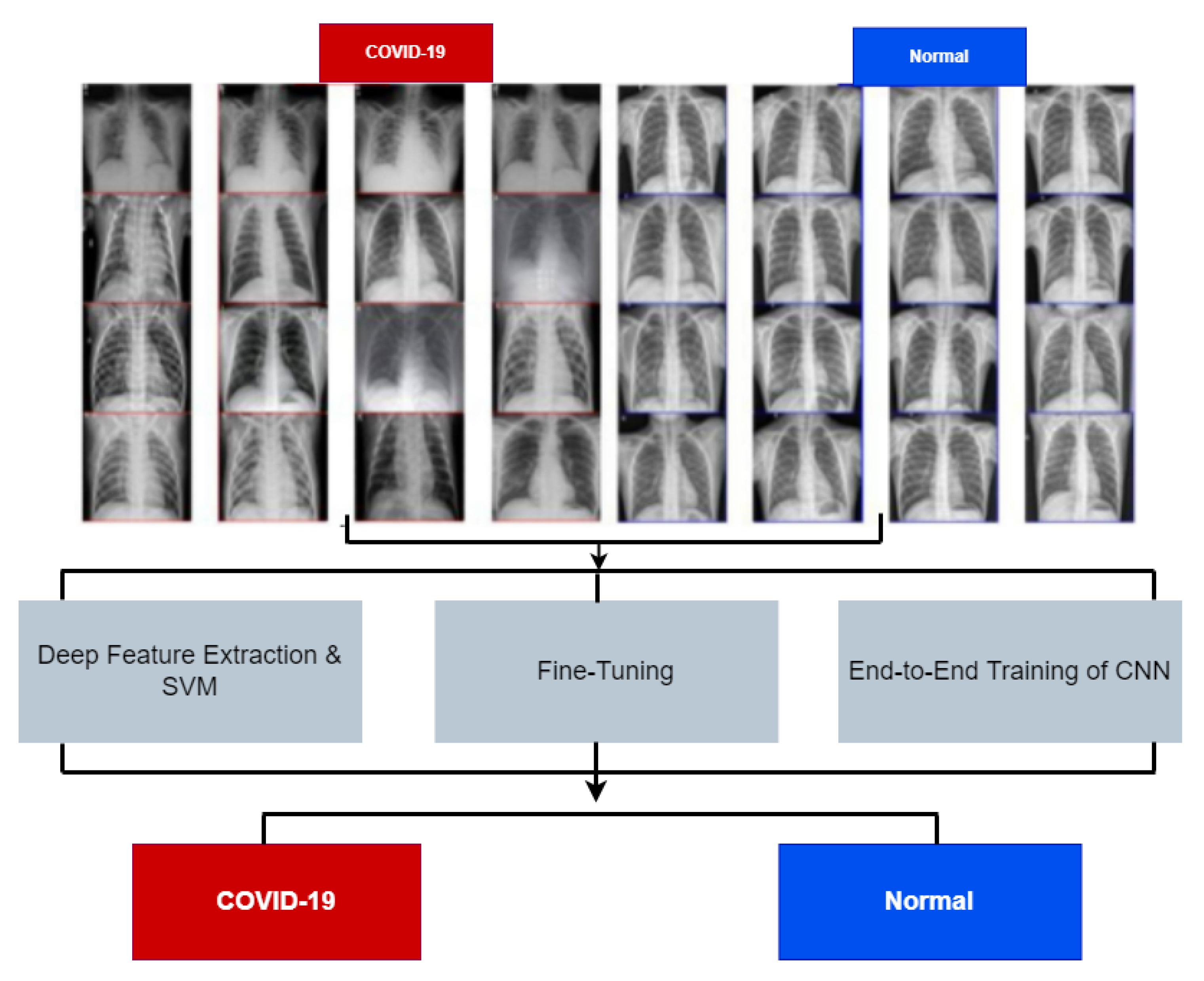
5. Artificial Intelligence and COVID-19
5.1. Machine Learning
5.2. Deep Learning
5.3. COVID-19 Datasets
6. Notable Contributions of AI in the Fight against COVID-19
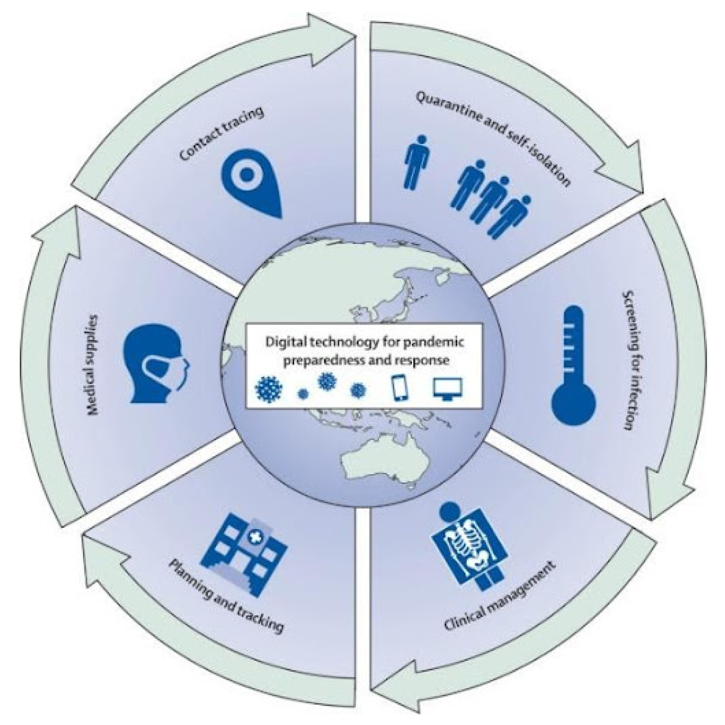
6.1. AI for COVID-19 Tracking and Dashboarding
6.2. AI for COVID-19 Diagnosis and Forecasting
6.3. AI for the Treatment of COVID-19
6.4. AI for COVID-19 Surveillance
7. Discussion and Conclusions
Author Contributions
Funding
Institutional Review Board Statement
Informed Consent Statement
Data Availability Statement
Conflicts of Interest
References
- Worldometer. COVID-19 Coronavirus Pandemic Weekly Coronavirus Cases. Available online: https://www.worldometers.info/coronavirus/ (accessed on 27 December 2021).
- Sheridan, C. Coronavirus and the race to distribute reliable diagnostics. Nat. Biotechnol. 2020, 38, 382–384. [Google Scholar] [CrossRef] [PubMed]
- Corman, V.; Bleicker, T.; Brünink, S.; Drosten, C.; Landt, O.; Koopmans, M.; Zambon, M.; Peiris, M. Diagnostic Detection of Wuhan Coronavirus 2019 by Real-Time RT-PCR; World Health Organization: Geneva, Switzerland, 2020; Volume 13. [Google Scholar]
- Xie, X.; Zhong, Z.; Zhao, W.; Zheng, C.; Wang, F.; Liu, J. Chest CT for typical coronavirus disease 2019 (COVID-19) pneumonia: Relationship to negative RT-PCR testing. Radiology 2020, 296, E41–E45. [Google Scholar] [CrossRef] [Green Version]
- WHO. Laboratory Testing for Coronavirus Disease (COVID-19) in Suspected Human Cases: Interim Guidance, 19 March 2020; Technical Report; World Health Organization: Geneva, Switzerland, 2020. [Google Scholar]
- Guan, W.J.; Ni, Z.Y.; Hu, Y.; Liang, W.H.; Ou, C.Q.; He, J.X.; Liu, L.; Shan, H.; Lei, C.L.; Hui, D.S.; et al. Clinical characteristics of coronavirus disease 2019 in China. N. Engl. J. Med. 2020, 382, 1708–1720. [Google Scholar] [CrossRef]
- Lu, R.; Zhao, X.; Li, J.; Niu, P.; Yang, B.; Wu, H.; Wang, W.; Song, H.; Huang, B.; Zhu, N.; et al. Genomic characterisation and epidemiology of 2019 novel coronavirus: Implications for virus origins and receptor binding. Lancet 2020, 395, 565–574. [Google Scholar] [CrossRef] [Green Version]
- Wu, A.; Peng, Y.; Huang, B.; Ding, X.; Wang, X.; Niu, P.; Meng, J.; Zhu, Z.; Zhang, Z.; Wang, J.; et al. Genome composition and divergence of the novel coronavirus (2019-nCoV) originating in China. Cell Host Microbe 2020, 27, 325–328. [Google Scholar] [CrossRef] [PubMed] [Green Version]
- Yang, Y.; Yang, M.; Yuan, J.; Wang, F.; Wang, Z.; Li, J.; Zhang, M.; Xing, L.; Wei, J.; Peng, L.; et al. Laboratory diagnosis and monitoring the viral shedding of SARS-CoV-2 infection. Innovation 2020, 1, 100061. [Google Scholar] [CrossRef] [PubMed]
- Wu, Z.; McGoogan, J.M. Characteristics of and important lessons from the coronavirus disease 2019 (COVID-19) outbreak in China: Summary of a report of 72 314 cases from the Chinese Center for Disease Control and Prevention. JAMA 2020, 323, 1239–1242. [Google Scholar] [CrossRef]
- Aruleba, K.; Obaido, G.; Ogbuokiri, B.; Fadaka, A.O.; Klein, A.; Adekiya, T.A.; Aruleba, R.T. Applications of computational methods in biomedical breast cancer imaging diagnostics: A review. J. Imaging 2020, 6, 105. [Google Scholar] [CrossRef]
- Mienye, I.D.; Sun, Y. Improved heart disease prediction using particle swarm optimization based stacked sparse autoencoder. Electronics 2021, 10, 2347. [Google Scholar] [CrossRef]
- Mienye, I.D.; Sun, Y. Performance analysis of cost-sensitive learning methods with application to imbalanced medical data. Inform. Med. Unlock. 2021, 25, 100690. [Google Scholar] [CrossRef]
- Vaishya, R.; Javaid, M.; Khan, I.H.; Haleem, A. Artificial Intelligence (AI) applications for COVID-19 pandemic. Diabetes Metab. Syndr. Clin. Res. Rev. 2020, 14, 337–339. [Google Scholar] [CrossRef] [PubMed]
- Kumari, P.; Singh, A.; Ngasainao, M.R.; Shakeel, I.; Kumar, S.; Lal, S.; Singhal, A.; Sohal, S.S.; Singh, I.K.; Hassan, M.I. Potential diagnostics and therapeutic approaches in COVID-19. Clin. Chim. Acta 2020, 510, 488–497. [Google Scholar] [CrossRef] [PubMed]
- Kriza, C.; Amenta, V.; Zenié, A.; Panidis, D.; Chassaigne, H.; Urbán, P.; Holzwarth, U.; Sauer, A.V.; Reina, V.; Griesinger, C.B. Artificial intelligence for imaging-based COVID-19 detection: Systematic review comparing added value of AI versus human readers. Eur. J. Radiol. 2021, 145, 110028. [Google Scholar] [CrossRef]
- Cau, R.; Faa, G.; Nardi, V.; Balestrieri, A.; Puig, J.; Suri, J.S.; SanFilippo, R.; Saba, L. Long-COVID diagnosis: From diagnostic to advanced AI-driven models. Eur. J. Radiol. 2022, 148, 110164. [Google Scholar] [CrossRef]
- Esenogho, E.; Mienye, I.D.; Swart, T.G.; Aruleba, K.; Obaido, G. A neural network ensemble with feature engineering for Improved Credit Card Fraud Detection. IEEE Access 2022, 10, 16400–16407. [Google Scholar] [CrossRef]
- Hassan, H.; Ren, Z.; Zhao, H.; Huang, S.; Li, D.; Xiang, S.; Kang, Y.; Chen, S.; Huang, B. Review and classification of AI-enabled COVID-19 CT imaging models based on computer vision tasks. Comput. Biol. Med. 2021, 141, 105123. [Google Scholar] [CrossRef]
- Verma, A.; Amin, S.B.; Naeem, M.; Saha, M. Detecting COVID-19 from chest computed tomography Scans using AI-Driven Android Application. arXiv 2021, arXiv:2111.06254. [Google Scholar] [CrossRef]
- Bernheim, A.; Mei, X.; Huang, M.; Yang, Y.; Fayad, Z.A.; Zhang, N.; Diao, K.; Lin, B.; Zhu, X.; Li, K.; et al. Chest CT findings in coronavirus disease-19 (COVID-19): Relationship to duration of infection. Radiology 2020, 295, 1–7. [Google Scholar] [CrossRef] [Green Version]
- World Health Organization (WHO). Available online: https://www.who.int/news/item/26-11-2021-classification-of-omicron-(b.1.1.529)-sars-cov-2-variant-of-concern (accessed on 21 December 2021).
- Bosch, I.; de Puig, H.; Hiley, M.; Carré-Camps, M.; Perdomo-Celis, F.; Narváez, C.F.; Salgado, D.M.; Senthoor, D.; O’Grady, M.; Phillips, E.; et al. Rapid antigen tests for dengue virus serotypes and Zika virus in patient serum. Sci. Transl. Med. 2017, 9, eaan1589. [Google Scholar] [CrossRef] [Green Version]
- Rowe, T.; Abernathy, R.A.; Hu-Primmer, J.; Thompson, W.W.; Lu, X.; Lim, W.; Fukuda, K.; Cox, N.J.; Katz, J.M. Detection of antibody to avian influenza A (H5N1) virus in human serum by using a combination of serologic assays. J. Clin. Microbiol. 1999, 37, 937–943. [Google Scholar] [CrossRef] [Green Version]
- Thaxton, C.S.; Elghanian, R.; Thomas, A.D.; Stoeva, S.I.; Lee, J.S.; Smith, N.D.; Schaeffer, A.J.; Klocker, H.; Horninger, W.; Bartsch, G.; et al. Nanoparticle-based bio-barcode assay redefines “undetectable” PSA and biochemical recurrence after radical prostatectomy. Proc. Natl. Acad. Sci. USA 2009, 106, 18437–18442. [Google Scholar] [CrossRef] [PubMed] [Green Version]
- Kim, J.; Biondi, M.J.; Feld, J.J.; Chan, W.C. Clinical validation of quantum dot barcode diagnostic technology. ACS Nano 2016, 10, 4742–4753. [Google Scholar] [CrossRef] [PubMed]
- Nilsson, H.O.; Aleljung, P.; Nilsson, I.; Tyszkiewicz, T.; Wadström, T. Immunomagnetic bead enrichment and PCR for detection of Helicobacter pylori in human stools. J. Microbiol. Methods 1996, 27, 73–79. [Google Scholar] [CrossRef]
- Imai, M.; Ninomiya, A.; Minekawa, H.; Notomi, T.; Ishizaki, T.; Van Tu, P.; Tien, N.T.K.; Tashiro, M.; Odagiri, T. Rapid diagnosis of H5N1 avian influenza virus infection by newly developed influenza H5 hemagglutinin gene-specific loop-mediated isothermal amplification method. J. Virol. Methods 2007, 141, 173–180. [Google Scholar] [CrossRef] [PubMed]
- Laksanasopin, T.; Guo, T.W.; Nayak, S.; Sridhara, A.A.; Xie, S.; Olowookere, O.O.; Cadinu, P.; Meng, F.; Chee, N.H.; Kim, J.; et al. A smartphone dongle for diagnosis of infectious diseases at the point of care. Sci. Transl. Med. 2015, 7, 273re1. [Google Scholar] [CrossRef] [Green Version]
- Shirato, K.; Nishimura, H.; Saijo, M.; Okamoto, M.; Noda, M.; Tashiro, M.; Taguchi, F. Diagnosis of human respiratory syncytial virus infection using reverse transcription loop-mediated isothermal amplification. J. Virol. Methods 2007, 139, 78–84. [Google Scholar] [CrossRef]
- Wang, X.; Xiong, E.; Tian, T.; Cheng, M.; Lin, W.; Wang, H.; Zhang, G.; Sun, J.; Zhou, X. Clustered regularly interspaced short palindromic repeats/Cas9-mediated lateral flow nucleic acid assay. ACS Nano 2020, 14, 2497–2508. [Google Scholar] [CrossRef]
- Kellner, M.J.; Koob, J.G.; Gootenberg, J.S.; Abudayyeh, O.O.; Zhang, F. SHERLOCK: Nucleic acid detection with CRISPR nucleases. Nat. Protoc. 2019, 14, 2986–3012. [Google Scholar] [CrossRef]
- Tahamtan, A.; Ardebili, A. Real-time RT-PCR in COVID-19 detection: Issues affecting the results. Expert Rev. Mol. Diagn. 2020, 20, 453–454. [Google Scholar] [CrossRef] [Green Version]
- Udugama, B.; Kadhiresan, P.; Kozlowski, H.N.; Malekjahani, A.; Osborne, M.; Li, V.Y.; Chen, H.; Mubareka, S.; Gubbay, J.B.; Chan, W.C. Diagnosing COVID-19: The disease and tools for detection. ACS Nano 2020, 14, 3822–3835. [Google Scholar] [CrossRef] [Green Version]
- Brihn, A.; Chang, J.; OYong, K.; Balter, S.; Terashita, D.; Rubin, Z.; Yeganeh, N. Diagnostic performance of an antigen test with RT-PCR for the detection of SARS-CoV-2 in a hospital setting—Los Angeles county, California, June–August 2020. Morb. Mortal. Wkly. Rep. 2021, 70, 702. [Google Scholar] [CrossRef] [PubMed]
- Reich, N.; Lowe, C.F.; Puddicombe, D.; Matic, N.; Greiner, J.; Simons, J.; Leung, V.; Chu, T.; Naik, H.; Myles, N.; et al. Diagnostic accuracy of RT-PCR for detection of SARS-CoV-2 compared to a “composite reference standard” in hospitalized patients. medRxiv 2021. [Google Scholar] [CrossRef]
- Ferté, T.; Ramel, V.; Cazanave, C.; Lafon, M.E.; Bébéar, C.; Malvy, D.; Georges-Walryck, A.; Dehail, P. Accuracy of COVID-19 rapid antigenic tests compared to RT-PCR in a student population: The StudyCov study. J. Clin. Virol. 2021, 141, 104878. [Google Scholar] [CrossRef] [PubMed]
- Khatami, F.; Saatchi, M.; Zadeh, S.S.T.; Aghamir, Z.S.; Shabestari, A.N.; Reis, L.O.; Aghamir, S.M.K. A meta-analysis of accuracy and sensitivity of chest CT and RT-PCR in COVID-19 diagnosis. Sci. Rep. 2020, 10, 22402. [Google Scholar] [CrossRef]
- Jakobsen, K.K.; Jensen, J.S.; Todsen, T.; Kirkby, N.; Lippert, F.; Vangsted, A.M.; Klokker, M.; von Buchwald, C. Accuracy of anterior nasal swab rapid antigen tests compared with RT-PCR for massive SARS-CoV-2 screening in low prevalence population. Apmis 2022, 130, 95–100. [Google Scholar] [CrossRef]
- Sethuraman, N.; Jeremiah, S.S.; Ryo, A. Interpreting diagnostic tests for SARS-CoV-2. JAMA 2020, 323, 2249–2251. [Google Scholar] [CrossRef]
- Fazio, E.; Abousiam, M.; Caselli, A.; Accorona, R.; Nebiaj, A.; Ermoli, I.; Erckert, B.; Calabrese, L.; Gazzini, L. Proper procedures for performing nasopharyngeal and oropharyngeal swabs for COVID-19. ATS Sch. 2020, 1, 495–497. [Google Scholar] [CrossRef]
- Yüce, M.; Filiztekin, E.; Özkaya, K.G. COVID-19 diagnosis—A review of current methods. Biosens. Bioelectron. 2021, 172, 112752. [Google Scholar] [CrossRef]
- Point-of-Care-Testing. Available online: https://www.cdc.gov/csels/dls/point-of-care-testing-risk-assessment-basics.html (accessed on 21 December 2021).
- Li, C.; Zhao, C.; Bao, J.; Tang, B.; Wang, Y.; Gu, B. Laboratory diagnosis of coronavirus disease-2019 (COVID-19). Clin. Chim. Acta Int. J. Clin. Chem. 2020, 510, 35. [Google Scholar] [CrossRef]
- Gremmels, H.; Winkel, B.M.; Schuurman, R.; Rosingh, A.; Rigter, N.A.; Rodriguez, O.; Ubijaan, J.; Wensing, A.M.; Bonten, M.J.; Hofstra, L.M. Real-life validation of the Panbio™ COVID-19 antigen rapid test (Abbott) in community-dwelling subjects with symptoms of potential SARS-CoV-2 infection. EClinicalMedicine 2021, 31, 100677. [Google Scholar] [CrossRef]
- Torres, I.; Poujois, S.; Albert, E.; Colomina, J.; Navarro, D. Evaluation of a rapid antigen test (Panbio™ COVID-19 Ag rapid test device) for SARS-CoV-2 detection in asymptomatic close contacts of COVID-19 patients. Clin. Microbiol. Infect. 2021, 27, 636-e1. [Google Scholar] [CrossRef] [PubMed]
- Mak, G.C.; Lau, S.S.; Wong, K.K.; Chow, N.L.; Lau, C.; Lam, E.T.; Chan, R.C.; Tsang, D.N. Evaluation of rapid antigen detection kit from the WHO Emergency Use List for detecting SARS-CoV-2. J. Clin. Virol. 2021, 134, 104712. [Google Scholar] [CrossRef] [PubMed]
- Merino, P.; Guinea, J.; Muñoz-Gallego, I.; González-Donapetry, P.; Galán, J.C.; Antona, N.; Cilla, G.; Hernáez-Crespo, S.; Díaz-de Tuesta, J.L.; Gual-de Torrella, A.; et al. Multicenter evaluation of the Panbio™ COVID-19 rapid antigen-detection test for the diagnosis of SARS-CoV-2 infection. Clin. Microbiol. Infect. 2021, 27, 758–761. [Google Scholar] [CrossRef]
- Ade-Ibijola, A.; Aruleba, K. Automatic attendance capturing using histogram of oriented gradients on facial images. In Proceedings of the 2018 IST-Africa Week Conference (IST-Africa), Gaborone, Botswana, 19–21 October 2018; IEEE: Piscataway, NJ, USA, 2018; p. 1. [Google Scholar]
- Zhuang, B.; Shen, C.; Tan, M.; Liu, L.; Reid, I. Structured binary neural networks for accurate image classification and semantic segmentation. In Proceedings of the IEEE/CVF Conference on Computer Vision and Pattern Recognition, Long Beach, CA, USA, 15–20 June 2019; pp. 413–422. [Google Scholar]
- Mienye, I.D.; Sun, Y.; Wang, Z. Improved sparse autoencoder based artificial neural network approach for prediction of heart disease. Inform. Med. Unlock. 2020, 18, 100307. [Google Scholar] [CrossRef]
- Weiss, P.; Murdoch, D.R. Clinical course and mortality risk of severe COVID-19. Lancet 2020, 395, 1014–1015. [Google Scholar] [CrossRef]
- Phua, J.; Weng, L.; Ling, L.; Egi, M.; Lim, C.M.; Divatia, J.V.; Shrestha, B.R.; Arabi, Y.M.; Ng, J.; Gomersall, C.D.; et al. Intensive care management of coronavirus disease 2019 (COVID-19): Challenges and recommendations. Lancet Respir. Med. 2020, 8, 506–517. [Google Scholar] [CrossRef]
- Mushtaq, J.; Pennella, R.; Lavalle, S.; Colarieti, A.; Steidler, S.; Martinenghi, C.M.; Palumbo, D.; Esposito, A.; Rovere-Querini, P.; Tresoldi, M.; et al. Initial chest radiographs and artificial intelligence (AI) predict clinical outcomes in COVID-19 patients: Analysis of 697 Italian patients. Eur. Radiol. 2021, 31, 1770–1779. [Google Scholar] [CrossRef]
- Bachtiger, P.; Peters, N.S.; Walsh, S.L. Machine learning for COVID-19—Asking the right questions. Lancet Digit. Health 2020, 2, e391–e392. [Google Scholar] [CrossRef]
- Rinderknecht, M.D.; Klopfenstein, Y. Predicting critical state after COVID-19 diagnosis: Model development using a large US electronic health record dataset. NPJ Digit. Med. 2021, 4, 1–14. [Google Scholar] [CrossRef]
- Roberts, M.; Driggs, D.; Thorpe, M.; Gilbey, J.; Yeung, M.; Ursprung, S.; Aviles-Rivero, A.I.; Etmann, C.; McCague, C.; Beer, L.; et al. Machine learning for COVID-19 detection and prognostication using chest radiographs and CT scans: A systematic methodological review. arXiv 2020, arXiv:2008.06388. [Google Scholar]
- Magar, R.; Yadav, P.; Farimani, A.B. Potential neutralizing antibodies discovered for novel corona virus using machine learning. Sci. Rep. 2021, 11, 1–11. [Google Scholar]
- Hazarika, B.B.; Gupta, D. Modelling and forecasting of COVID-19 spread using wavelet-coupled random vector functional link networks. Appl. Soft Comput. 2020, 96, 106626. [Google Scholar] [CrossRef] [PubMed]
- Elaziz, M.A.; Hosny, K.M.; Salah, A.; Darwish, M.M.; Lu, S.; Sahlol, A.T. New machine learning method for image-based diagnosis of COVID-19. PLoS ONE 2020, 15, e0235187. [Google Scholar] [CrossRef]
- Ghosh, A.; Nundy, S.; Mallick, T.K. How India is dealing with COVID-19 pandemic. Sens. Int. 2020, 1, 100021. [Google Scholar] [CrossRef]
- Sujath, R.; Chatterjee, J.M.; Hassanien, A.E. A machine learning forecasting model for COVID-19 pandemic in India. Stoch. Environ. Res. Risk Assess. 2020, 34, 959–972. [Google Scholar] [CrossRef] [PubMed]
- Kushwaha, S.; Bahl, S.; Bagha, A.K.; Parmar, K.S.; Javaid, M.; Haleem, A.; Singh, R.P. Significant applications of machine learning for COVID-19 pandemic. J. Ind. Integr. Manag. 2020, 5, 453–479. [Google Scholar] [CrossRef]
- Pinter, G.; Felde, I.; Mosavi, A.; Ghamisi, P.; Gloaguen, R. COVID-19 pandemic prediction for Hungary; a hybrid machine learning approach. Mathematics 2020, 8, 890. [Google Scholar] [CrossRef]
- Li, L.; Qin, L.; Xu, Z.; Yin, Y.; Wang, X.; Kong, B.; Bai, J.; Lu, Y.; Fang, Z.; Song, Q.; et al. Artificial intelligence distinguishes COVID-19 from community acquired pneumonia on chest CT. Radiology 2020, 2, E65–E71. [Google Scholar] [CrossRef]
- Khan, R.S.; Rehman, I.U. Spectroscopy as a tool for detection and monitoring of Coronavirus (COVID-19). Expert Rev. Mol. Diagn. 2020, 20, 647–649. [Google Scholar] [CrossRef]
- LeCun, Y.; Bengio, Y.; Hinton, G. Deep learning. Nature 2015, 521, 436–444. [Google Scholar] [CrossRef]
- Rusk, N. Deep learning. Nature Methods 2016, 13, 35. [Google Scholar] [CrossRef]
- Yu, K.; Jia, L.; Chen, Y.; Xu, W. Deep learning: Yesterday, today, and tomorrow. J. Comput. Res. Dev. 2013, 50, 1799. [Google Scholar]
- Zeroual, A.; Harrou, F.; Dairi, A.; Sun, Y. Deep learning methods for forecasting COVID-19 time-Series data: A Comparative study. Chaos Solitons Fractals 2020, 140, 110121. [Google Scholar] [CrossRef] [PubMed]
- Chimmula, V.K.R.; Zhang, L. Time series forecasting of COVID-19 transmission in Canada using LSTM networks. Chaos Solitons Fractals 2020, 135, 109864. [Google Scholar] [CrossRef]
- Asraf, A.; Islam, M.Z.; Haque, M.R.; Islam, M.M. Deep learning applications to combat novel coronavirus (COVID-19) pandemic. SN Comput. Sci. 2020, 1, 1–7. [Google Scholar] [CrossRef]
- Shan, K.; Ouyang, T.; Wang, X.; Yang, H.; Zhou, B.; Wu, Z.; Shang, M. Temporal prediction of algal parameters in Three Gorges Reservoir based on highly time-resolved monitoring and long short-term memory network. J. Hydrol. 2021, 605, 127304. [Google Scholar] [CrossRef]
- Hochreiter, S.; Schmidhuber, J. Long short-term memory. Neural Comput. 1997, 9, 1735–1780. [Google Scholar] [CrossRef]
- Alzubaidi, L.; Zhang, J.; Humaidi, A.J.; Al-Dujaili, A.; Duan, Y.; Al-Shamma, O.; Santamaría, J.; Fadhel, M.A.; Al-Amidie, M.; Farhan, L. Review of deep learning: Concepts, CNN architectures, challenges, applications, future directions. J. Big Data 2021, 8, 1–74. [Google Scholar] [CrossRef]
- Nasir, J.A.; Khan, O.S.; Varlamis, I. Fake news detection: A hybrid CNN-RNN based deep learning approach. Int. J. Inf. Manag. Data Insights 2021, 1, 100007. [Google Scholar] [CrossRef]
- Desai, M.; Shah, M. An anatomization on breast cancer detection and diagnosis employing multi-layer perceptron neural network (MLP) and Convolutional neural network (CNN). Clin. eHealth 2021, 4, 1–11. [Google Scholar] [CrossRef]
- Ismael, A.M.; Şengür, A. Deep learning approaches for COVID-19 detection based on chest X-ray images. Expert Syst. Appl. 2021, 164, 114054. [Google Scholar] [CrossRef] [PubMed]
- Kedia, P.; Anjum Katarya, R. CoVNet-19: A Deep Learning model for the detection and analysis of COVID-19 patients. Appl. Soft Comput. 2021, 104, 107184. [Google Scholar] [CrossRef] [PubMed]
- Madhavan, M.V.; Khamparia, A.; Gupta, D.; Pande, S.; Tiwari, P.; Hossain, M.S. Res-CovNet: An internet of medical health things driven COVID-19 framework using transfer learning. Neural Comput. Appl. 2021, 1–14. [Google Scholar] [CrossRef] [PubMed]
- Kavuran, G.; İn, E.; Geçkil, A.A.; Şahin, M.; Berber, N.K. MTU-COVNet: A hybrid methodology for diagnosing the COVID-19 pneumonia with optimized features from multinet. Clin. Imaging 2022, 81, 1–8. [Google Scholar] [CrossRef]
- Mgboh, U.; Ogbuokiri, B.; Obaido, G.; Aruleba, K. Visual Data Mining: A Comparative Analysis of Selected Datasets. In International Conference on Intelligent Systems Design and Applications; Springer: Berlin/Heidelberg, Germany, 2020; pp. 377–391. [Google Scholar]
- Mohamadou, Y.; Halidou, A.; Kapen, P.T. A review of mathematical modeling, artificial intelligence and datasets used in the study, prediction and management of COVID-19. Appl. Intell. 2020, 50, 3913–3925. [Google Scholar] [CrossRef]
- He, X.; Yang, X.; Zhang, S.; Zhao, J.; Zhang, Y.; Xing, E.; Xie, P. Sample-efficient deep learning for COVID-19 diagnosis based on CT scans. medRxiv 2020. [Google Scholar] [CrossRef]
- Angelov, P.; Almeida Soares, E. SARS-CoV-2 CT-scan dataset: A large dataset of real patients CT scans for SARS-CoV-2 identification. medRxiv 2020. [Google Scholar] [CrossRef]
- Jaiswal, A.; Gianchandani, N.; Singh, D.; Kumar, V.; Kaur, M. Classification of the COVID-19 infected patients using DenseNet201 based deep transfer learning. J. Biomol. Struct. Dyn. 2020, 39, 5682–5689. [Google Scholar] [CrossRef]
- Harmon, S.A.; Sanford, T.H.; Xu, S.; Turkbey, E.B.; Roth, H.; Xu, Z.; Yang, D.; Myronenko, A.; Anderson, V.; Amalou, A.; et al. Artificial intelligence for the detection of COVID-19 pneumonia on chest CT using multinational datasets. Nat. Commun. 2020, 11, 1–7. [Google Scholar] [CrossRef]
- Silva, P.; Luz, E.; Silva, G.; Moreira, G.; Silva, R.; Lucio, D.; Menotti, D. COVID-19 detection in CT images with deep learning: A voting-based scheme and cross-datasets analysis. Informatics Med. Unlocked 2020, 20, 100427. [Google Scholar] [CrossRef]
- Wang, L.; Lin, Z.Q.; Wong, A. Covid-net: A tailored deep convolutional neural network design for detection of covid-19 cases from chest X-ray images. Sci. Rep. 2020, 10, 19549. [Google Scholar] [CrossRef] [PubMed]
- Huang, L.; Han, R.; Ai, T.; Yu, P.; Kang, H.; Tao, Q.; Xia, L. Serial quantitative chest CT assessment of COVID-19: A deep learning approach. Radiol. Cardiothorac. Imaging 2020, 2, e200075. [Google Scholar] [CrossRef] [PubMed] [Green Version]
- Wang, S.; Kang, B.; Ma, J.; Zeng, X.; Xiao, M.; Guo, J.; Cai, M.; Yang, J.; Li, Y.; Meng, X.; et al. A deep learning algorithm using CT images to screen for Corona Virus Disease (COVID-19). Eur. Radiol. 2021, 31, 6096–6104. [Google Scholar] [CrossRef] [PubMed]
- Yang, X.; He, X.; Zhao, J.; Zhang, Y.; Zhang, S.; Xie, P. COVID-CT-dataset: A CT scan dataset about COVID-19. arXiv 2020, arXiv:2003.13865. [Google Scholar]
- Afshar, P.; Heidarian, S.; Enshaei, N.; Naderkhani, F.; Rafiee, M.J.; Oikonomou, A.; Fard, F.B.; Samimi, K.; Plataniotis, K.N.; Mohammadi, A. COVID-CT-MD, COVID-19 computed tomography scan dataset applicable in machine learning and deep learning. Sci. Data 2021, 8, 121. [Google Scholar] [CrossRef] [PubMed]
- Hall, L.O.; Paul, R.; Goldgof, D.B.; Goldgof, G.M. Finding covid-19 from chest X-rays using deep learning on a small dataset. arXiv 2020, arXiv:2004.02060. [Google Scholar]
- Choudrie, J.; Banerjee, S.; Kotecha, K.; Walambe, R.; Karende, H.; Ameta, J. Machine learning techniques and older adults processing of online information and misinformation: A COVID-19 study. Comput. Hum. Behav. 2021, 119, 106716. [Google Scholar] [CrossRef]
- Budd, J.; Miller, B.S.; Manning, E.M.; Lampos, V.; Zhuang, M.; Edelstein, M.; Rees, G.; Emery, V.C.; Stevens, M.M.; Keegan, N.; et al. Digital technologies in the public-health response to COVID-19. Nat. Med. 2020, 26, 1183–1192. [Google Scholar] [CrossRef]
- Vargo, D.; Zhu, L.; Benwell, B.; Yan, Z. Digital technology use during COVID-19 pandemic: A rapid review. Hum. Behav. Emerg. Technol. 2021, 3, 13–24. [Google Scholar] [CrossRef]
- Guidotti, E.; Ardia, D. COVID-19 data hub. J. Open Source Softw. 2020, 5, 2376. [Google Scholar] [CrossRef]
- Dong, E.; Du, H.; Gardner, L. An interactive web-based dashboard to track COVID-19 in real time. Lancet Infect. Dis. 2020, 20, 533–534. [Google Scholar] [CrossRef]
- Naudé, W. Artificial Intelligence against COVID-19: An Early Review; Institute of Labor Economics (IZA): Bonn, Germany, 2020. [Google Scholar]
- Il Yooa, K.; Kronenfelda, B.J. An evaluation of COVID-19 dashboards from cartographic and epidemiological perspectives. Cartogr. Geogr. Inf. Sci. (CaGIS) 2020, 1–8. [Google Scholar]
- Stiegler, N.; Bouchard, J.P. South Africa: Challenges and successes of the COVID-19 lockdown. In Annales Médico-Psychologiques, Revue Psychiatrique; Elsevier: Amsterdam, The Netherlands, 2020; Volume 178, pp. 695–698. [Google Scholar]
- Pietz, J.; McCoy, S.; Wilck, J.H. Chasing John Snow: Data analytics in the COVID-19 era. Eur. J. Inf. Syst. 2020, 29, 388–404. [Google Scholar] [CrossRef]
- Hassounah, M.; Raheel, H.; Alhefzi, M. Digital response during the COVID-19 pandemic in Saudi Arabia. J. Med. Internet Res. 2020, 22, e19338. [Google Scholar] [CrossRef] [PubMed]
- For Disease Control (CDC), A.C. Africa Centre for Disease Control (CDC) Dashboard. 2021. Available online: https://africacdc.org/covid-19/ (accessed on 9 December 2021).
- Muñoz, L.; Villarreal, V.; Nielsen, M.; Caballero, Y.; Sittón-Candanedo, I.; Corchado, J.M. Artificial intelligence models and techniques applied to COVID-19: A review. Electronics 2021, 10, 2901. [Google Scholar] [CrossRef]
- Patel, N.V. The Best, and the Worst, of the Coronavirus Dashboards. Available online: https://www.technologyreview.com/2020/03/06/905436/best-worst-coronavirus-dashboards/ (accessed on 9 December 2021).
- Florez, H.; Singh, S. Online dashboard and data analysis approach for assessing COVID-19 case and death data. F1000Research 2020, 9, 1–13. [Google Scholar] [CrossRef] [PubMed]
- Zohner, Y.E.; Morris, J.S. COVID-TRACK: World and USA SARS-COV-2 testing and COVID-19 tracking. BioData Min. 2021, 14, 1–15. [Google Scholar] [CrossRef]
- Ye, Y.; Hou, S.; Fan, Y.; Qian, Y.; Zhang, Y.; Sun, S.; Peng, Q.; Laparo, K. α-Satellite: An AI-driven System and Benchmark Datasets for Hierarchical Community-level Risk Assessment to Help Combat COVID-19. arXiv 2020, arXiv:2003.12232. [Google Scholar]
- Dsfsi.github.io. COVID-19 ZA South Africa Dashboard. 2021. Available online: https://sacoronavirus.co.za/ (accessed on 9 December 2021).
- Arabia, S. COVID 19 Dashboard: Saudi Arabia. 2021. Available online: https://covid19.moh.gov.sa/ (accessed on 9 December 2021).
- Wu, J.; Zhang, P.; Zhang, L.; Meng, W.; Li, J.; Tong, C.; Li, Y.; Cai, J.; Yang, Z.; Zhu, J.; et al. Rapid and accurate identification of COVID-19 infection through machine learning based on clinical available blood test results. MedRxiv 2020. [Google Scholar]
- Tang, Z.; Zhao, W.; Xie, X.; Zhong, Z.; Shi, F.; Liu, J.; Shen, D. Severity assessment of coronavirus disease 2019 (COVID-19) using quantitative features from chest CT images. arXiv 2020, arXiv:2003.11988. [Google Scholar]
- Yan, R.; Zhang, Y.; Li, Y.; Xia, L.; Guo, Y.; Zhou, Q. Structural basis for the recognition of SARS-CoV-2 by full-length human ACE2. Science 2020, 367, 1444–1448. [Google Scholar] [CrossRef] [PubMed] [Green Version]
- Bertsimas, D.; Lukin, G.; Mingardi, L.; Nohadani, O.; Orfanoudaki, A.; Stellato, B.; Wiberg, H.; Gonzalez-Garcia, S.; Parra-Calderon, C.L.; Robinson, K.; et al. COVID-19 mortality risk assessment: An international multi-center study. PLoS ONE 2020, 15, e0243262. [Google Scholar] [CrossRef] [PubMed]
- Sun, P.; Lu, X.; Xu, C.; Sun, W.; Pan, B. Understanding of COVID-19 based on current evidence. Journal of medical virology 2020, 92, 548–551. [Google Scholar] [CrossRef] [PubMed]
- Chintalapudi, N.; Battineni, G.; Amenta, F. COVID-19 virus outbreak forecasting of registered and recovered cases after sixty day lockdown in Italy: A data driven model approach. J. Microbiol. Immunol. Infect. 2020, 53, 396–403. [Google Scholar] [CrossRef] [PubMed]
- Gupta, R.; Pal, S.K. Trend Analysis and Forecasting of COVID-19 outbreak in India. MedRxiv 2020. [Google Scholar]
- Hu, Z.; Ge, Q.; Li, S.; Jin, L.; Xiong, M. Artificial intelligence forecasting of covid-19 in china. arXiv 2020, arXiv:2002.07112. [Google Scholar] [CrossRef]
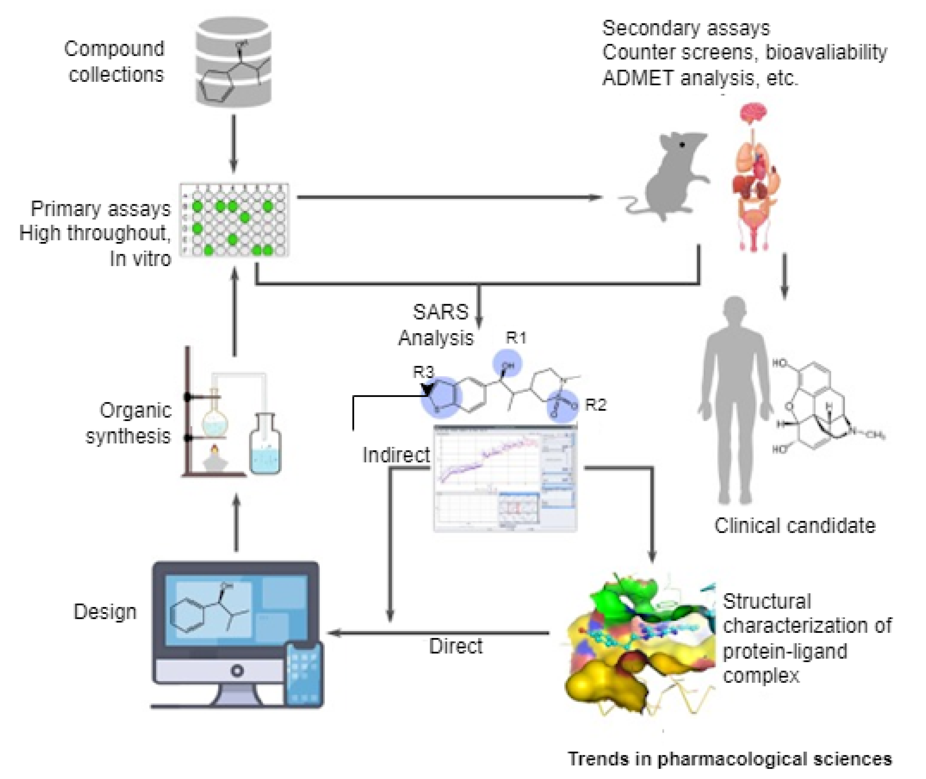
- Fleming, N. How artificial intelligence is changing drug discovery. Nature 2018, 557, S55. [Google Scholar] [CrossRef] [PubMed]
- Yang, X.; Wang, Y.; Byrne, R.; Schneider, G.; Yang, S. Concepts of artificial intelligence for computer-assisted drug discovery. Chem. Rev. 2019, 119, 10520–10594. [Google Scholar] [CrossRef] [Green Version]
- Jing, Y.; Bian, Y.; Hu, Z.; Wang, L.; Xie, X.Q.S. Deep learning for drug design: An artificial intelligence paradigm for drug discovery in the big data era. AAPS J. 2018, 20, 1–10. [Google Scholar]
- Álvarez-Machancoses, Ó.; Fernández-Martínez, J.L. Using artificial intelligence methods to speed up drug discovery. Expert Opin. Drug Discov. 2019, 14, 769–777. [Google Scholar] [CrossRef]
- Chan, H.S.; Shan, H.; Dahoun, T.; Vogel, H.; Yuan, S. Advancing drug discovery via artificial intelligence. Trends Pharmacol. Sci. 2019, 40, 592–604. [Google Scholar] [CrossRef] [PubMed]
- Zhavoronkov, A. Artificial intelligence for drug discovery, biomarker development, and Generation of Novel Chemistry. Mol. Pharm. 2018, 15, 4311–4313. [Google Scholar] [CrossRef] [PubMed] [Green Version]
- Keshavarzi Arshadi, A.; Webb, J.; Salem, M.; Cruz, E.; Calad-Thomson, S.; Ghadirian, N.; Collins, J.; Diez-Cecilia, E.; Kelly, B.; Goodarzi, H.; et al. Artificial intelligence for COVID-19 drug discovery and vaccine development. Front. Artif. Intell. 2020, 3, 65. [Google Scholar] [CrossRef] [PubMed]
- Mohanty, S.; Rashid, M.H.A.; Mridul, M.; Mohanty, C.; Swayamsiddha, S. Application of Artificial Intelligence in COVID-19 drug repurposing. Diabetes Metab. Syndr. Clin. Res. Rev. 2020, 14, 1027–1031. [Google Scholar] [CrossRef]
- Kim, H.; Han, G.; Song, J.H. A Review for Artificial Intelligence Proving to Fight Against COVID-19 Pandemic And Prefatory Health Policy. J. Med. Biomed. Appl. Sci. 2020, 8, 494–506. [Google Scholar] [CrossRef]
- Mohanty, C.; Vinod, C.; Acharya, S.; Mahapatra, N. COVID-19 drug repositioning: Present status and prospects. In Modeling, Control and Drug Development for COVID-19 Outbreak Prevention; Springer: Berlin/Heidelberg, Germany, 2022; pp. 645–671. [Google Scholar]
- Delijewski, M.; Haneczok, J. AI drug discovery screening for COVID-19 reveals zafirlukast as a repurposing candidate. Med. Drug Discov. 2021, 9, 100077. [Google Scholar] [CrossRef]
- Pham, T.H.; Qiu, Y.; Zeng, J.; Xie, L.; Zhang, P. A deep learning framework for high-throughput mechanism-driven phenotype compound screening and its application to COVID-19 drug repurposing. Nat. Mach. Intell. 2021, 3, 247–257. [Google Scholar] [CrossRef]
- Kabra, R.; Singh, S. Evolutionary artificial intelligence based peptide discoveries for effective Covid-19 therapeutics. Biochim. Biophys. Acta (BBA) Mol. Basis Dis. 2021, 1867, 165978. [Google Scholar] [CrossRef]
- Jin, W.; Stokes, J.M.; Eastman, R.T.; Itkin, Z.; Zakharov, A.V.; Collins, J.J.; Jaakkola, T.S.; Barzilay, R. Deep learning identifies synergistic drug combinations for treating COVID-19. Proc. Natl. Acad. Sci. USA 2021, 118, e2105070118. [Google Scholar] [CrossRef]
- Mbunge, E. Integrating emerging technologies into COVID-19 contact tracing: Opportunities, challenges and pitfalls. Diabetes Metab. Syndr. Clin. Res. Rev. 2020, 14, 1631–1636. [Google Scholar] [CrossRef]
- Munzert, S.; Selb, P.; Gohdes, A.; Stoetzer, L.F.; Lowe, W. Tracking and promoting the usage of a COVID-19 contact tracing app. Nat. Hum. Behav. 2021, 5, 247–255. [Google Scholar] [CrossRef] [PubMed]
- Mbunge, E.; Fashoto, S.G.; Akinnuwesi, B.; Metfula, A.; Simelane, S.; Ndumiso, N. Ethics for integrating emerging technologies to contain COVID-19 in Zimbabwe. Hum. Behav. Emerg. Technol. 2021, 3, 1–15. [Google Scholar] [CrossRef] [PubMed]
- Arora, G.; Joshi, J.; Mandal, R.S.; Shrivastava, N.; Virmani, R.; Sethi, T. Artificial Intelligence in Surveillance, Diagnosis, Drug Discovery and Vaccine Development against COVID-19. Pathogens 2021, 10, 1048. [Google Scholar] [CrossRef] [PubMed]
- Whitelaw, S.; Mamas, M.A.; Topol, E.; Van Spall, H.G. Applications of digital technology in COVID-19 pandemic planning and response. Lancet Digit. Health 2020, 2, E435–E440. [Google Scholar] [CrossRef]
- Wu, J.T.; Leung, K.; Leung, G.M. Nowcasting and forecasting the potential domestic and international spread of the 2019-nCoV outbreak originating in Wuhan, China: A modelling study. Lancet 2020, 395, 689–697. [Google Scholar] [CrossRef] [Green Version]
- Chen, B.; Marvin, S.; While, A. Containing COVID-19 in China: AI and the robotic restructuring of future cities. Dialogues Hum. Geogr. 2020, 10, 238–241. [Google Scholar] [CrossRef]
- Lin, L.; Hou, Z. Combat COVID-19 with artificial intelligence and big data. J. Travel Med. 2020, 27, taaa080. [Google Scholar] [CrossRef]
- Do Carmo Barriga, A.; Martins, A.F.; Simões, M.J.; Faustino, D. The COVID-19 pandemic: Yet another catalyst for governmental mass surveillance? Soc. Sci. Humanit. Open 2020, 2, 100096. [Google Scholar]
- Calvo, R.A.; Deterding, S.; Ryan, R.M. Health Surveillance during COVID-19 Pandemic. BMJ 2020, 369, m1373. [Google Scholar] [CrossRef] [Green Version]


| Detection System | Technology | Biomarker | Principle |
|---|---|---|---|
| Rapid antigen test | Lateral flow | Protein | Detection of Colorimetric through the use of paper with gold-coated antibodies [23] |
| ELISA | ELISA | Protein | The induction of virus colour change in enzymatic reaction in the presence of target antigen [24] |
| Biobarcode assay | DNA-mediated immunoassay | Protein | Involve the conjugation of gold nanoparticles with DNA through the help of protein signal detection [25] |
| Quantum dots barcode | Barcode | Nucleic acid | Capture of viral DNA and RNA through quantum beads [26] |
| Magnetic bead | Magnetic | Nucleic acid | Detection of PCR through the help of magnetically isolated bacteria [27] |
| LAMP | LAMP | Nucleic acid | Isothermal DNA synthesis through the signal of turbidity detection [28] |
| Smartphone dongle | ELISA | Protein | ELISA by microfluidic set up [29] |
| RT-LAMP | LAMP | Nucleic acid | RNA target generation through reverse transcriptase LAMP reaction [30] |
| CRISPR | RPA | Nucleic acid | Lateral flow nucleic assay by the help of PCR and CRISPR/Ca9 [31] |
| CRISPR | RT-RPA | Nucleic acid | SHERLOCK, RPA detection by multiplexed fluorescence spectroscopy [32] |
| References | Dataset | Size | Image Modality | Techniques | Evaluation Result (%) |
|---|---|---|---|---|---|
| [85] | SARS-CoV-2 | 2482 scans (1252–positive, 1230–negative) | CT | xDNN | F1 = 97.31 |
| [87] | LIDC | CT | Deep Learning | Acc = 90.8, Sen = 84, Spe = 93 | |
| [88] | SARS-CoV-2 | 2482 | CT | EfficientNet | Acc = 87.6, F1 = 86.19, AUC = 90.5 |
| [89] | COVIDx | 13,975–13,870 positive patient | CXR | DCNN | Sen = 91.0 |
| [94] | OSR, Istituto Ortopedico Galeazzi (IOG) | 1925 | CXR | Logistic regression, Naïve bayes, KNN, Random forest, SVM | AUC = 87, Spe = 94 |
| [65] | COVIDx | X-ray | CNN—Capsule network | Acc = 95.7, Sen = 90, Spe = 95.8, AUC = 0.97 |
| References | Name | Country | Purpose | Coverage | Medium |
|---|---|---|---|---|---|
| [99] | John Hopkins CSSE | United States | Tracking and Prediction | Worldwide | Web |
| [98] | COVID-19 Data Hub | Canada | Tracking | Worldwide | Web |
| [107] | COVID-19 Tracker | United States | Tracking | Worldwide | Web |
| [108] | COVID-19 Dashboard | Cyprus | Tracking | Worldwide | Web |
| [109] | COVID-Track | United States | Tracking | Worldwide | Web |
| [105] | Africa CDC COVID-19 | All member states | Tracking | Africa | Web |
| [106] | COVID-19 Open data | Panama | Tracking and Prediction | Panama | Web |
| [110] | -Satellite | United States | Risk assessment | United States | Web |
| [111] | COVID-19 ZA South Africa | South-Africa | Tracking | South-Africa | Web |
| [112] | Saudi MoH COVID-19 Dashboard | Saudi Arabia | Tracking | Saudi Arabia | Web |
| References | Model | Scope | Evaluation Results | Datasets |
|---|---|---|---|---|
| [113] | Random Forest | Diagnosis | Accuracy = 96.9 | Private, Blood samples |
| [89] | CNN | Diagnosis | Accuracy = 93.3% | Private, Chest X-ray images |
| [115] | XGBoost | Mortality risk prediction | Survival Accuracy = 100%, Mortality Risk = 81% | Private, Blood samples |
| [116] | XGBoost | Mortality risk prediction | AUC = 90% (Out of sample) AUC = 0.92 (Seville) | Private |
| [117] | Support Vector Machine | Prediction | Accuracy = 77.5% AUC = 78.4% | Private, Chest X-ray images |
| [71] | LSTM-RNN | Forecasting | Accuracy = 93.4% | Public dataset: John Hopkins and Canadian Health Authority |
| [119] | ARIMA | Forecasting | Accuracy = 90% | Public dataset: John Hopkins |
| [120] | Stacked Auto-Encoder | Forecasting | Unknown | WHO |
| [114] | Random Forest | Diagnosis | Accuracy = 87.5, AUC = 91% | Private, Chest X-ray images |
| [118] | ARIMA | Forecasting | Accuracy = 93.75% | Public dataset: Italian Ministry of Health |
Publisher’s Note: MDPI stays neutral with regard to jurisdictional claims in published maps and institutional affiliations. |
© 2022 by the authors. Licensee MDPI, Basel, Switzerland. This article is an open access article distributed under the terms and conditions of the Creative Commons Attribution (CC BY) license (https://creativecommons.org/licenses/by/4.0/).
Share and Cite
Aruleba, R.T.; Adekiya, T.A.; Ayawei, N.; Obaido, G.; Aruleba, K.; Mienye, I.D.; Aruleba, I.; Ogbuokiri, B. COVID-19 Diagnosis: A Review of Rapid Antigen, RT-PCR and Artificial Intelligence Methods. Bioengineering 2022, 9, 153. https://doi.org/10.3390/bioengineering9040153
Aruleba RT, Adekiya TA, Ayawei N, Obaido G, Aruleba K, Mienye ID, Aruleba I, Ogbuokiri B. COVID-19 Diagnosis: A Review of Rapid Antigen, RT-PCR and Artificial Intelligence Methods. Bioengineering. 2022; 9(4):153. https://doi.org/10.3390/bioengineering9040153
Chicago/Turabian StyleAruleba, Raphael Taiwo, Tayo Alex Adekiya, Nimibofa Ayawei, George Obaido, Kehinde Aruleba, Ibomoiye Domor Mienye, Idowu Aruleba, and Blessing Ogbuokiri. 2022. "COVID-19 Diagnosis: A Review of Rapid Antigen, RT-PCR and Artificial Intelligence Methods" Bioengineering 9, no. 4: 153. https://doi.org/10.3390/bioengineering9040153
APA StyleAruleba, R. T., Adekiya, T. A., Ayawei, N., Obaido, G., Aruleba, K., Mienye, I. D., Aruleba, I., & Ogbuokiri, B. (2022). COVID-19 Diagnosis: A Review of Rapid Antigen, RT-PCR and Artificial Intelligence Methods. Bioengineering, 9(4), 153. https://doi.org/10.3390/bioengineering9040153