Article

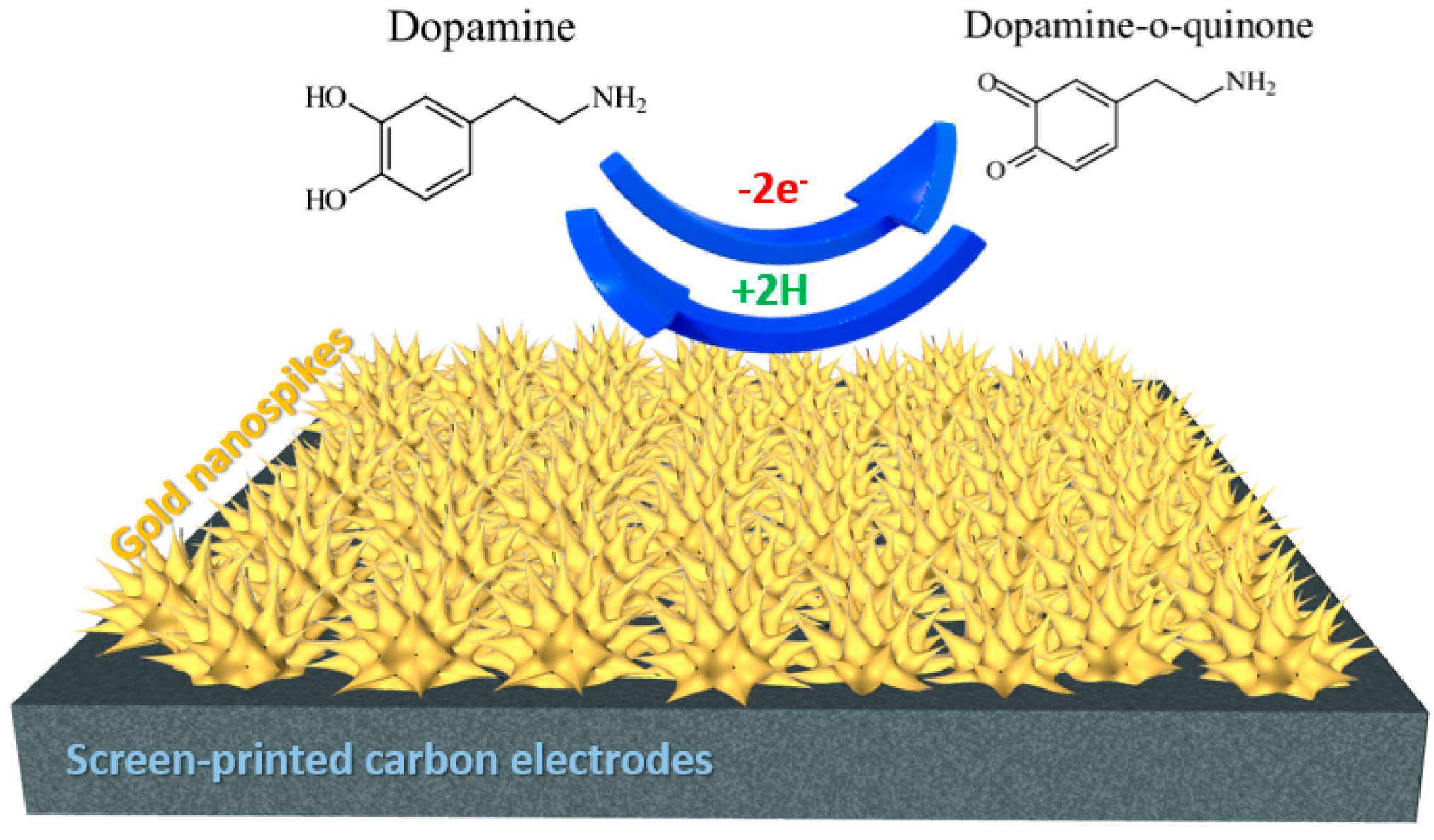
Gold Nanospikes Formation on Screen-Printed Carbon Electrode through Electrodeposition Method for Non-Enzymatic Electrochemical Sensor

by
Isa Anshori
1,2,*,
Raih Rona Althof
1,3,
Lavita Nuraviana Rizalputri
1,3,
Eduardus Ariasena
1,
Murni Handayani
4,*,
Arfat Pradana
2,
Mohammad Rizki Akbar
5,
Mas Rizky Anggun Adipurna Syamsunarno
6,
Agnes Purwidyantri
7,
Briliant Adhi Prabowo
8,*,
Muhammad Sjahrul Annas
9,
Hasim Munawar
10 and
Brian Yuliarto
2,11
1
Lab-on-Chip Group, Biomedical Engineering Department, Bandung Institute of Technology, Bandung 40132, Indonesia
2
Research Center for Nanoscience and Nanotechnology (RCNN), Bandung Institute of Technology, Bandung 40132, Indonesia
3
Department of Nanotechnology, Graduate School, Bandung Institute of Technology, Bandung 40132, Indonesia
4
National Research and Innovation Agency (BRIN), Tangerang Selatan 15314, Indonesia
5
Department of Cardiology and Vascular Medicine, Faculty of Medicine, Universitas Padjadjaran and Dr. Hasan Sadikin General Hospital, Bandung 40161, Indonesia
6
Department of Biomedical Sciences, Faculty of Medicine, Universitas Padjadjaran, Bandung 45363, Indonesia
7
School of Chemistry and Chemical Engineering, Queen’s University Belfast, Belfast BT7 1NN, UK
8
School of Mathematics and Physics, Queen’s University Belfast, Belfast BT7 1NN, UK
9
Mechanical Engineering Department, Trisakti University, Jakarta 11440, Indonesia
10
PT Mitra Asiatek Biosensor, Jakarta 12520, Indonesia
11
Advanced Functional Materials Laboratory, Engineering Physics Department, Bandung Institute of Technology, Bandung 40132, Indonesia
*
Authors to whom correspondence should be addressed.
Metals 2022, 12(12), 2116; https://doi.org/10.3390/met12122116
Submission received: 4 November 2022 / Revised: 5 December 2022 / Accepted: 7 December 2022 / Published: 9 December 2022
(This article belongs to the Section Metallic Functional Materials)

Abstract

:
In this study, we reported the construction of Gold Nanospike (AuNS) structures on the surface of screen-printed carbon electrode (SPCE) used for non-enzymatic electrochemical detection. This modification was prepared with a one-step electrodeposition method by controlling the electrodeposition parameters, such as applied potential and deposition time, via Constant Potential Amperometry (CPA). Those parameters and precursor solution concentration were varied to investigate the optimum electrodeposition configuration. The results confirmed that AuNS were homogenously deposited and well-dispersed on the working electrode surface of SPCE. The AuNS-modified SPCE was implemented as a non-enzymatic sensor toward dopamine and could enhance the electrocatalytic ability compared with the bare SPCE. Further examination shows that the sensing performance of the AuNS-modified SPCE produced an increase in electrochemical surface area (ECSA) at 17.25 times higher than the bare electrode, a sensitivity of 0.056 µA mM−1 cm−2 with a wide linear range of 0.2–50 µM and a detection limit of 0.33 µM. In addition, AuNS-modified SPCE can selectively detect dopamine among other interfering analytes such as ascorbic acid, urea, and uric acid, which commonly coexist in the body fluid. This work demonstrated that AuNS-modified SPCE is a prospective sensing platform for non-enzymatic dopamine detection.

1. Introduction

Electrochemical-based detection provides relatively low-cost instrumentation, performs rapid response acquisition, and allows real-time and on-site monitoring that could be used as point-of-care (POC) testing [1]. However, overlap potential oxidation windows toward molecules in body fluids, such as dopamine, ascorbic acid, and uric acid on non-modified electrode surfaces, become the issues in developing non-enzymatic-based electrochemical sensors [2]. The problems can be overcome by applying noble metal nanomaterials as electrocatalysts. Noble metal nanoparticles, such as Pd [3,4,5], Ag [6,7], Pt [8,9,10], and Au [11,12], have been applied in the development of electrochemical-based sensing to enhance the catalytic reaction. Au nanoparticles are extensively studied as potential modification materials for electrochemical sensing for their biological compatibility, fast electron transfer due to excellent electrical conductivity, and high chemical stability [13]. Moreover, research shows that the size effect of Au nanostructure can enhance the conductivity and electron transfer ability since it has a high surface-to-volume ratio and high surface energy [14,15]. These properties of Au nanostructure would be expected to meet the requirement of on-enzymatic sensing.
Electrode modification by Au nanostructure is generally prepared by the drop-casting method, in which the synthesized Au nanostructure is directly dropped on top of the working electrode. The Au nanostructured synthesized process can be conducted using either chemical (seed-mediated approaches), physical (the use of radiation), biological (the use of microalgae or fungi), or hybrid method [16]. Another strategy for electrode modification using Au nanostructured is electrodeposition using specific precursors [17]. Compared with the drop-casting method, the electrodeposition method has the excellencies of ease in modification with a rapid and straightforward process without any particular condition. Furthermore, the homogeneity of deposition results can significantly enhance and minimize the final result’s potential impurities due to the usage of many materials needed [18]. This method can also produce varied morphology by controlling electrodeposition parameters, including working potential, deposition time, and addition of growth direction agent.
Nanostructures of noble metals, such as gold or silver, have been introduced for several sensing applications [19,20,21]. Nevertheless, the gold nanostructure construction still faces challenges such as low yield, uniformity issues, and laboriousness. One of the well-known methods for gold coating in the sensing electrode is the electrodeposition technique [22]. Ren et al. introduced the Au nanostructuring electrodeposition to detect Arsenic contamination [22]. In addition, an electrodeposition technique for the construction of an Au hexagonal structure was proposed by Liu et al. for surface-enhanced Raman scattering (SERS). It was reported that the electrodeposition technique for SERS substrate fabrication was also reported by Yao et al. for forming gold nanoparticles modified graphdiyne/carbon cloth. It was reported to detect the rhodamine 6 G (R6G) down to subfemtomolar concentration with an enhancement factor of more than 2 × 10¹² [23]. While for electrochemical biosensing, Madhu et al. and Sun et al. recently reported the electrodeposition methods to produce highly unique gold nanostructures [24,25].
It is known that Au nanostructures with different morphology would provide different performances in electrochemical sensors [26]. Particular morphology, such as pyramidal, rod, spherical, nanodendrite, and nanocoral, has been successfully produced using electrodeposition [27,28]. Au nanospike (AuNS), one potential candidate for electrochemical application, has attracted much attention since the properties of the dominant crystal orientation of (111) produce a high electrocatalytic ability [29]. In addition, numerous active facets (311) on AuNS indicate a surface comprising enormous atomic steps and a kink to increase active sites for the electrochemical experiment [28]. However, up to this point, the electrochemical-based detection of dopamine using the application of AuNS has not been reported.
Monitoring dopamine levels in body fluid is crucial, taking note that the abnormality of the dopamine level is strongly correlated with neurology disorders that affect physiological disorders, such as Parkinson’s, schizophrenia, attention deficit hyperactivity disorder (ADHD), and Alzheimer’s disease [30,31]. In addition, research findings recently have highlighted the importance of dopamine level effect on posttraumatic stress disorder (PTSD) [32]. The need for dopamine sensors becomes essential for early detection and for those who need to monitor regularly. The main challenges of dopamine detection are the demand for high sensitivity and selectivity due to low dopamine concentration in body fluid and the presence of many substances which coexist in the sample as interferences [33]. Most studies on developed dopamine sensors rely on electrochemical-based detection noting that dopamine is a redox-active molecule that can be easily oxidized without using an enzyme, bioligand, or any other probes [34,35], thus opening the chances for the non-enzymatic electrochemical sensor.
In this work, we developed the simple and clean electrodeposition of AuNS on the electrode to apply non-enzymatic detection toward dopamine. We have conducted several optimizations of electrodeposition parameters to obtain optimal configuration results. The homogenous and precise structure of AuNS provides a high enhancement to electrocatalytic performance. Screen-printed carbon electrode (SPCE) was chosen as the type of electrochemical electrode with consideration of the distinction, such as the potential for portable testing, simplicity, ease to use, and cost-effective fabrication so that befitting for mass production [36]. Furthermore, interference substances such as ascorbic acid, uric acid, and urea were examined and effectively distinguished toward the dopamine range of detection. With those outcomes, this paper shows excellent sensing performance in developing sensitive, selective, portable, cost-effective, and mass-producible electrochemical-based dopamine detection.

2. Materials and Methods

The Sulfuric Acid (H2SO4) 95–97%, gold (III) chloride trihydrate (HAuCl4·3H2O), Phosphate Buffered Saline (PBS) (pH 7.4 at 25 °C), Potassium Chloride (KCl) solution, Hydrochloric (HCl) Acid Fuming 35%, Dopamine Hydrochloride (C8H12ClNO2), Ascorbic Acid (C6H8O6), Urea (CH4N2O), and Uric Acid (C5H4N4O3) ≥ 99% powder were obtained from Merck. All the chemicals mentioned earlier were analytical grade. Analyte (Dopamine, AA, UA, and urea) solutions were prepared using 0.1 M HCl. All aqueous solutions were prepared using sterilized distilled water. The Hyper-Value Screen-Printed Carbon Electrode (SPCE) was purchased from Zimmer Peacock, Ltd. (Coventry, UK). The electrode was made of carbon, silver/silver chloride (Ag/AgCl), and silver for the working electrode, reference electrode, and counter electrode, respectively.
All the electrochemical procedures, including electrodeposition and electrochemical measurements, were carried out using a super flexible USB-powered mini electrochemical workstation Ana Pot Potensiostat from Zimmer Peacock, Ltd. The Hitachi SU3500 Scanning Electron Microscope (SEM) was used for the morphology characterization of Au nanostructures deposited on the SPCE working electrode. In addition, material characterization was also performed using JIB-4610F Field Emission Scanning Electron Microscope (FE-SEM) and X-Ray Diffractometer (XRD) Smartlab Rigaku for more detailed observation.
Before the experiment, the electrode activation procedure was carried out to the SPCE by doing repeated potential cycling in a 0.5 M H2SO4 solution at a potential range of 0.9 to 1.5 V vs. Ag/AgCl. Then, SPCE is ready for electrodeposition procedure or electrochemical measurement. The deposition solution consists of a particular concentration of HAuCl4 as a precursor in 0.1 M KCl [37]. The electrodeposition was performed using a constant potential amperometry (CPA) method, where immersed SPCE in a precursor solution and applied certain electrodeposition conditions, such as deposition potential and deposition time. The variation of deposition parameters was conducted to obtain optimum performance. The resulting SPCE was left to dry at room temperature for about 30 minutes and was thoroughly rinsed using PBS solution to remove the deposition materials, which are not entirely deposited. Au-modified SPCE is then ready to be examined for morphology characterization and electrochemical surface area (ECSA) calculation by electrochemical analysis (redox peak current on 0.5 M H2SO4). All experiments were carried out at room temperature.
In this work, we investigated the catalytic performance of the Au-modified SPCE toward the oxidation of dopamine. The SPCE fabricated under the optimal deposition configuration was used to identify the improvement of electrocatalytic ability, sensitivity, limits of detection, and selectivity. The non-enzymatic dopamine detection was performed using cyclic voltammetry (CV), differential potential voltammetry (DPV), and constant potential amperometry (CPA). In addition, uric acid (UA), ascorbic acid (AA), and urea were added as interference materials for the selectivity test.

3. Results and Discussion

3.1. Characterization of the AuNS Morphology

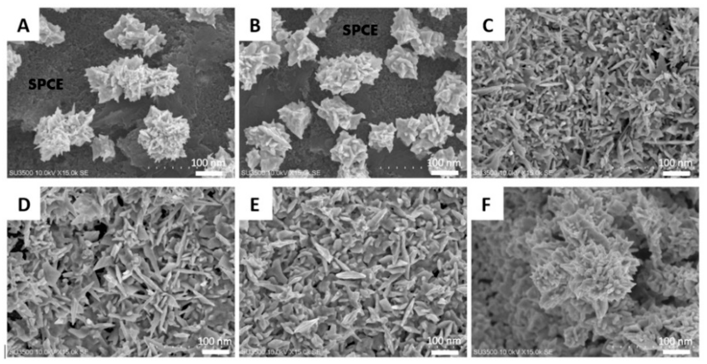
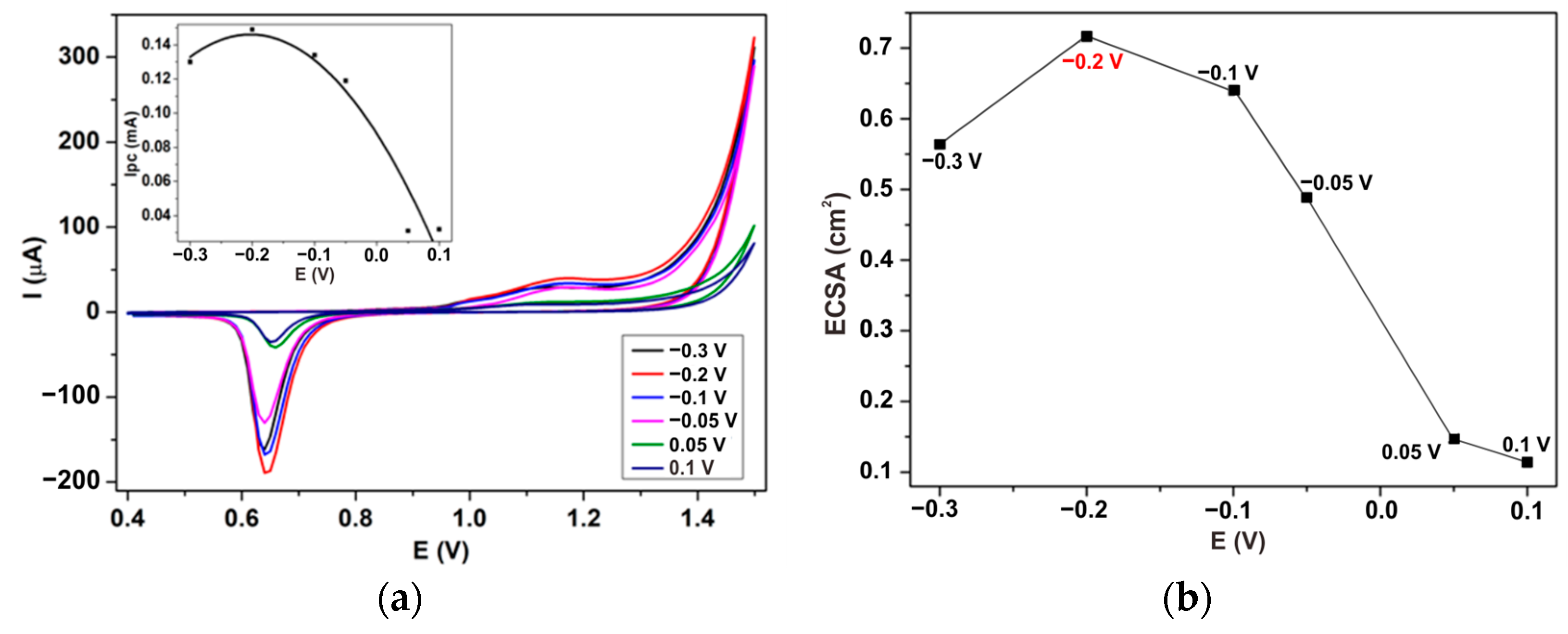
3.1.1. The Effect of the Electrodeposition Potential

Applied potential in electrodeposition is crucial in controlling deposited materials’ morphology [26]. Electrodepositions with the potential of −0.3 to +0.1 V vs. Ag/AgCl were conducted for the 1200 s in a 10 mM HAuCl4 and 0.1 M KCl solution to investigate the effect of Au morphology on the SPCE surface. The Au morphologies are depicted in Figure 1. As shown in Figure 1A, the potential of +0.1 V vs. Ag/AgCl produces dispersed Au flower-like in micrometer-sized spike-like nanostructures. The dark part in the image refers to the carbon working electrode surface. Under an applied potential of +0.05 V vs. Ag/AgCl (Figure 1B), the flower-like Au structures remain unchanged but dispersed denser. When the applied potential decreased to negative, the flower-like structures disappeared and formed uniform Au nanospike (AuNS) structures all over the carbon surface (Figure 1C). The more negative the applied potential, the AuNS is slightly thicker, as shown in Figure 1D,E. In addition, the surface coverage increases until almost entirely covered by AuNS at an applied potential of −0.1 V vs. Ag/AgCl. Further decreased to −0.3 V vs. Ag/AgCl (Figure 1F), the Au crystallinity is an overgrowth, which produces several non-uniform structures over the gold nano spikes formation.
At a positive potential, the current recorded during deposition is very small compared to the current at the negative applied working potentials, making the nucleation rate slower. This effect causes only a tiny number of nucleus crystals to be formed, which is continued in the growth process into larger particles and is less frequent [38]. Meanwhile, the more negative the potential, the greater the reduction current produced. As a result, the reduced nanomaterial becomes denser due to the increased rate of the growth process for forming Au nanostructures [39]. These results indicate that the applied potential significantly affects the deposited Au morphology in the electrodeposition method.
The electrochemical properties of the Au nanostructure-modified SPCE were investigated using a 0.5 M H2SO4 solution, as shown in Figure 2A. The anodic response starting from 1.0 V vs. Ag/AgCl and the cathodic peak at around 0.65 V vs. Ag/AgCl was associated with forming Au oxide on the carbon surface and reducing the oxide from the forward scan, respectively. For comparison, the typical response of Au nanostructure deposited on Glassy Carbon Electrode is at 1.1 V vs. Ag/AgCl for oxidation and 0.9 V vs. Ag/AgCl for the reduction peak [29]. Different types of electrodes affect the peak potential of the reaction but still produce a similar oxidation/reduction profile. In addition, the integral area of the reduction peak represents the charge required to reduce the Au oxide layer, thereby can be used to calculate the ECSA of the modified electrode surface. ECSA for each electrodeposition potential can be estimated, as shown in Figure 2B. Note that the charge density required to reduce monolayer oxygen is 0.390 mC/cm2 [40]. In line with the SEM characterization, Au electrodeposition produced at −0.2 V vs. Ag/AgCl exhibits the highest ECSA (0.716 cm2). The oxidation of dopamine characterized the catalytic performances of the modified SPCE. Figure 3 shows the Differential Potential Voltammograms of the 1 mM dopamine in 0.1 M HCl. HCl solution was used as the electrolyte as dopamine has the best stability in acidic aqueous solution compared with neutral or basic solution [41]. The dopamine oxidation peak appears approximately at 0.45 V vs. Ag/AgCl attributed to the reaction as written in Scheme 1.
The trend of the peak current from each different electrodeposition potential toward dopamine is slightly different from the ECSA trend. However, the highest catalytic performance is obtained by modified SPCE prepared at −0.2 V vs. Ag/AgCl. Therefore, it concludes that the optimum potential electrodeposition is −0.2 V vs. Ag/AgCl, which will be used in further experiments.

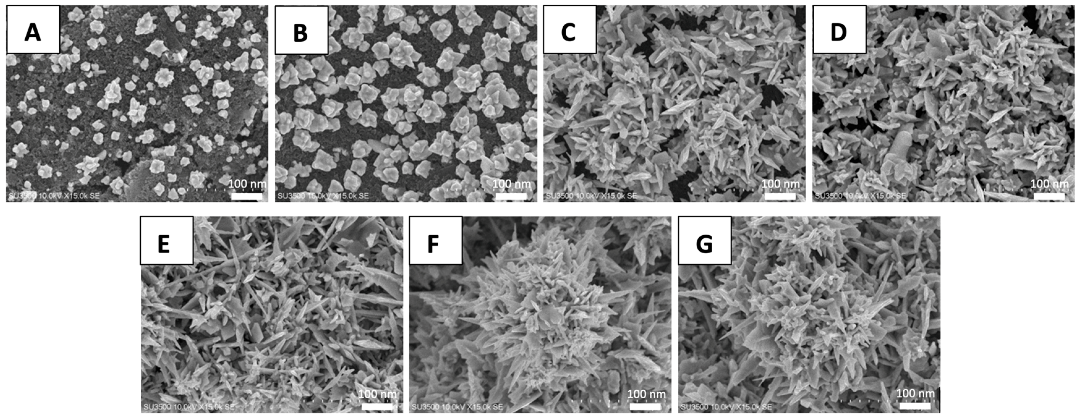
3.1.2. The Optimization of Deposition Time and Precursor Concentration on the AuNS-Modified SPCE

The optimum deposition time was determined by electrodeposition at −2.0 V vs. Ag/AgCl in a 10 mM HAuCl4 and 0.1 M KCl solution by 60, 300, 600, 900, 1200, 1500, and 1800 s. The resulting morphologies are shown in Figure 4. At 60 s, the initial gold formation on the SPCE surface from the precursor solution has begun with a well-dispersed nanometer-sized tetrahedral shape (Figure 4A). The tetrahedral gold nanostructures remained and grew bigger when extended until 300 s, shown in Figure 4B. However, after 600 s, the gold nanostructures change and become spiky surfaces (Figure 4C). When the deposition time increased, the gold nanospike deposited was denser (Figure 4D). The carbon surface was well-covered at 1200 s (Figure 4E). When the deposition time is 1500 and 1800 s, it can be found that the spike-like deposited gold was over-growth, which indicates the excess gold deposition at the SPCE surface (Figure 4F,G). Therefore, it can be concluded that the gold electrodeposition is time-dependent and requires 1200 s to cover the entire working electrode surface. Compared with other research, Shu’s [37] study requires 5400 s to cover the GCE working electrode with Au nanoparticles via the electrodeposition method. Therefore, SPCE in this study delivers a more efficient way to produce modified electrodes with gold nanostructures by electrodeposition.
As in the previous experiment, we analyzed the electrochemical properties of modified SPCE by different deposition times using a 0.5 M H2SO4 solution. Figure 5A shows the cyclic voltammogram and the correlation curve of the reduction peak current towards deposition time (inset). Along with the increasing deposition time, the surface area of active sites on gold-modified SPCE is continuously enhanced. After 1200 s, the surface area enhancement is not significant anymore and tends to be saturated in 1800 s. This result corresponds with the SEM images, which indicate that deposition around 1200 s produces maximum surface coverage due to the limited working electrode surface area. The later deposition exhibits stacking deposition of a gold nanostructure, which is not significantly advantageous to electrochemical property enhancement. In conclusion, 1200 s is considered the optimum deposition time in this study.
The other parameter that affects the electrodeposition product is the concentration of the precursor. The HAuCl4 concentration of 2, 4, 6, 8, 10, and 12 mM was used in the 1.5 mL of deposition solution with 0.1 M KCl under an applied potential of −0.2 V vs. Ag/AgCl for 1200 s. The Au-modified SPCE with different HAuCl4 concentrations was characterized using 0.5 M H2SO4, as shown in Figure 5. It is found that the ECSA of the modified SPCE is significantly influenced by the variation of HAuCl4 concentration, especially in between the interval 4–10 mM. When the concentration was increased to 12 mM, the peak current obtained was not improved compared with the 10 mM. It indicates that at 1200 s, the 10 mM of HAuCl4 in 1.5 mL deposition solution is sufficient to cover the active area of the carbon electrode. In conclusion, from the morphology and electrochemical performance above, the optimal configuration to conduct Au nanostructure electrodeposition onto the SPCE is using 10 mM HAuCl4 for 1200 s at an applied potential of −0.2 V vs. Ag/AgCl.

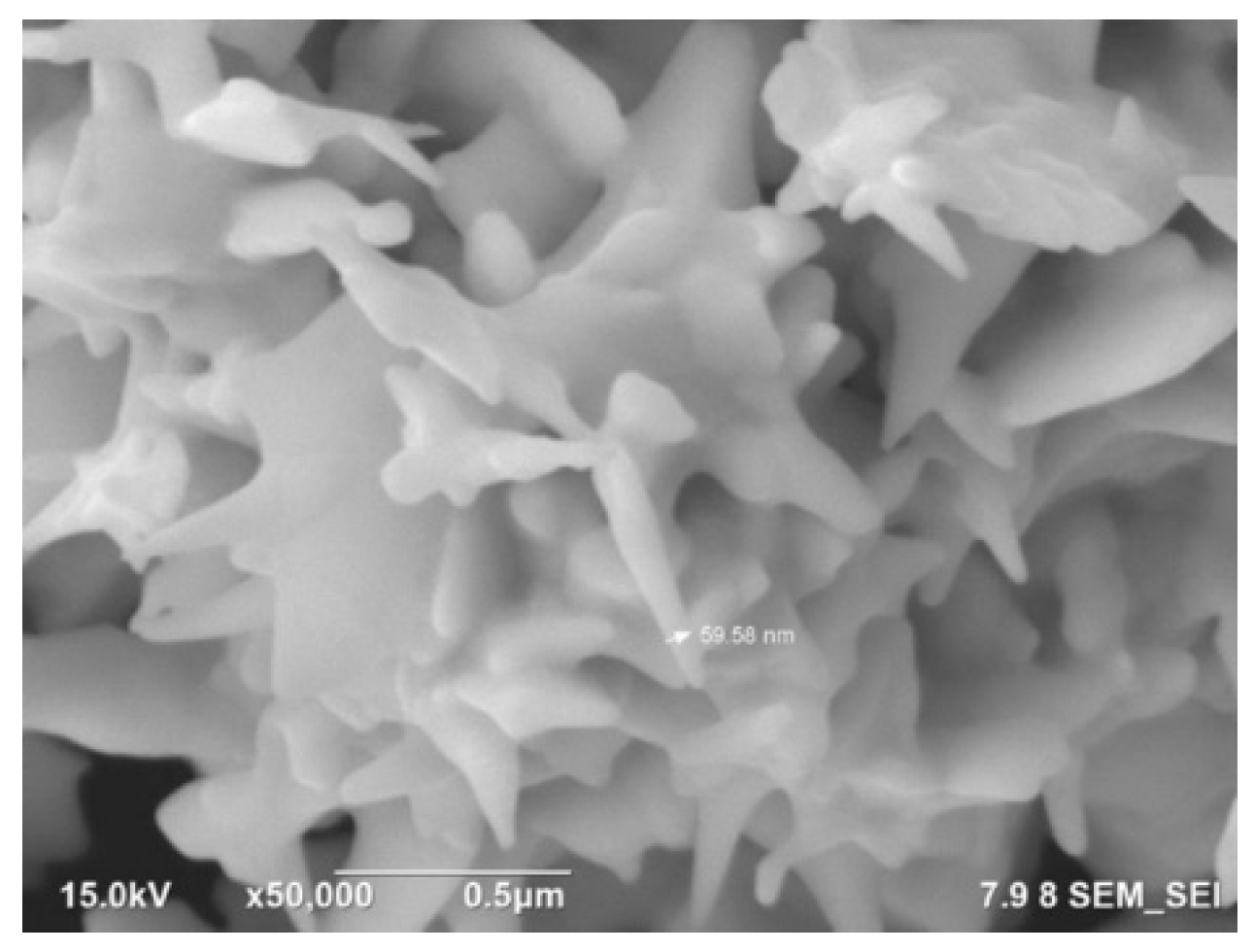
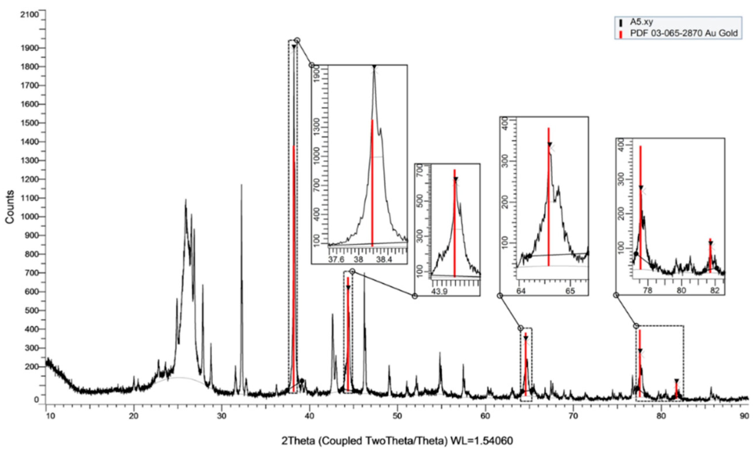
3.1.3. FE-SEM and XRD Characterization on the AuNS-Modified SPCE

After the optimum electrodeposition parameters for forming AuNS-modified SPCE were obtained, the electrodes were characterized using FE-SEM, as shown in Figure 6. From the image, a more explicit depiction of the nanospike morphology, which tends to be a random shape, with the diameter of the sharpest spike being around 59.58 nm. By being modified into nano spikes, the surface area of the working electrode was significantly increased up to 0.716 cm2. For more information, SPCE has a working electrode with a geometric area of 0.0415 cm2. Thus, the electrode surface area increases 17.25 times higher with the modification of AuNS. The enhancement is essential to make AuNS-modified SPCEs have excellent electrocatalytic abilities.
The following characterization was an XRD measurement to determine the crystallinity of AuNS. Due to equipment limitations, the scanned area is large enough that the peak results included in Figure 7 are attributed to the modified carbon working electrode, silver/silver chloride reference electrode, a silver counter electrode, and SPCE substrate. It can be observed that the AuNS material has 5 peaks of Au, and their intensity is compared with the peaks in the database (red line). The intensity of the sample in the (111) field is higher than the intensity of the standard database, while the other 4 fields have a lower intensity than the standard. The result shows that the dominant AuNS growth produces facets in the (111) direction with lower growth in other facets.

3.2. Electrocatalytic Activity of the AuNS-Modified SPCE towards Dopamine Detection

3.2.1. Electrochemical Measurement towards Dopamine

The cyclic voltammetry (CV) measurement was conducted to compare the performance of bare SPCE and the optimized AuNS-modified SPCE towards dopamine oxidation and reduction reaction using the 1 mM dopamine in 0.1 M HCl solution, as shown in Figure 8A. The current response of AuNS-modified SPCE is significantly improved and exhibits the shifting of working potential to negative potential. The signal enhancement is attributed to the high active site area and electrocatalytic ability of the AuNS towards dopamine. Anyway, the potential shifting indicates the improvement of electrocatalytic activity, so it becomes easier to oxidize dopamine [29]. Furthermore, the peak potential shifting slightly allows the electrode to distinguish the working potential of the dopamine oxidation among the coexisting interference considering that it is usually oxidized in the adjacent potential [2]. In addition, the peak-to-peak separation (ΔE) becomes narrower, showing an increase in the electron transfer rate due to an enhancement in the electroactive surface area of the modified electrode [11]. The reversibility of dopamine oxidation and reduction can be found using this measurement by observing the peak current of forward and backward scanning. As a result, the AuNS-modified SPCE performed better reversibility than the bare SPCE. The DPV measurement using bare SPCE and AuNS-modified SPCE is also conducted (Figure 8B) to support the analysis, which shows the enhanced and shifted peak corresponding with the CV result.

3.2.2. Effect of the Potential Scan Rate Variation

The CV measurement of 1 mM Dopamine using AuNS-modified SPCE was conducted at a potential scan rate of 25–200 mV/s. It turns out that the peak current rises proportionally to the potential scan rate (Figure 9A) with a slightly shifted positively for the oxidation peak, while the reduction peak is shifted to the negative potential. It shows that the electrochemical activity of dopamine follows Nernstian behavior and indicates that the faster scan rate will facilitate fewer occurred redox reactions [42]. Figure 9B points out the linear relation between the square root of the potential scan rate and the peak current with the linear equation, as seen in Equations (1) and (2) for anodic and cathodic peaks, respectively. The linearity indicates that the diffusion-controlled reaction dominates dopamine’s electrochemical detection [43].
J   ( μ A / cm 2 ) = 55.98 υ 29.22
J   ( μ A / cm 2 ) = 45.54 υ 7.12

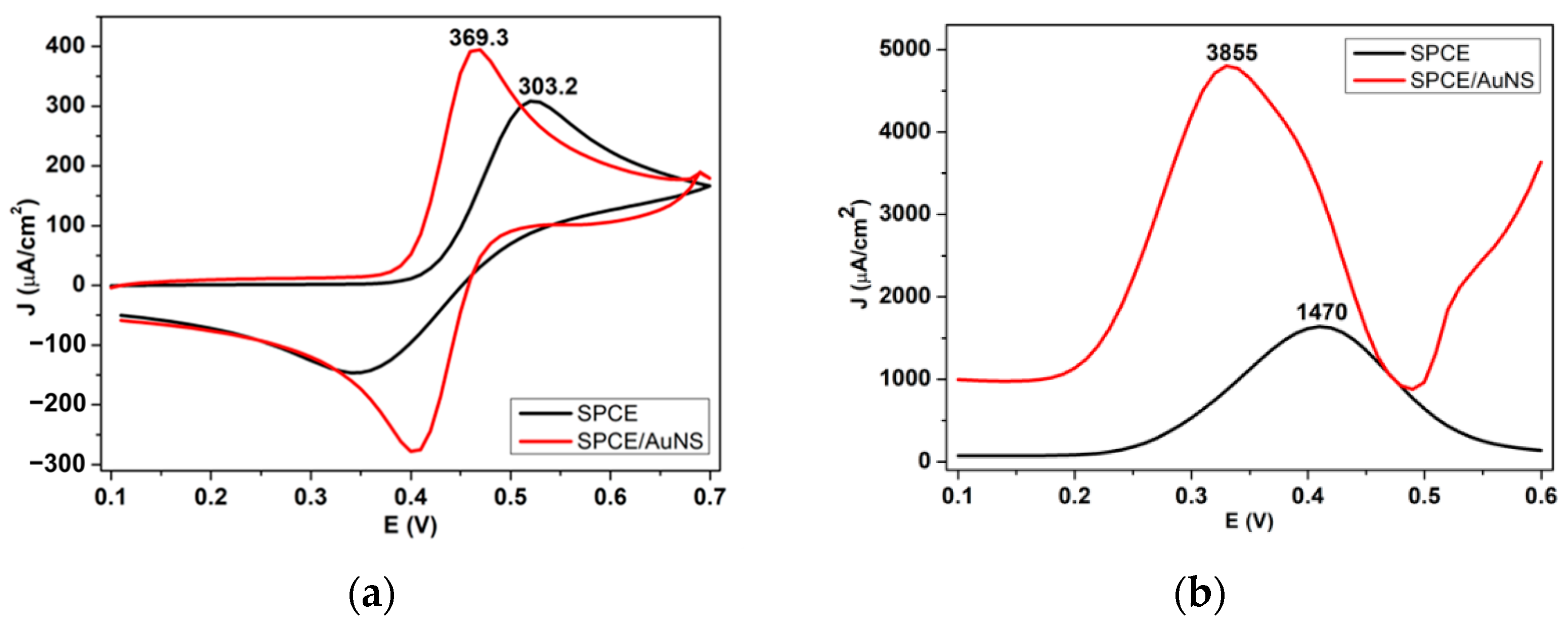
3.2.3. Sensitivity and Linearity of the AuNS-Modified SPCE for Dopamine Concentration

The amperometry measurement of the variation of dopamine concentration was investigated using the detection potential obtained by the previous CV measurement, which is +0.45 V vs. Ag/AgCl (Figure 10A). Dopamine solution with different concentrations ranging from 0.2 µM to 50 µM was examined sequentially for every 60 s. It is expected to produce a linear response between the concentration and the steady current signals. Figure 10B shows a calibration curve with a wide linear range from 0.2 µM to 50 µM, having a correlation coefficient of 0.994 and a calculated selectivity of 0.056 µA µM−1 cm−2. Noting that the dopamine level in urine is 0.3–3 µM [6], the developed electrode in this work meets the requirement to be used as a dopamine sensor. The limit of detection (LOD) and the limit of quantity (LOQ) are estimated using Equations (3) and (4), noting that σ refers to the standard deviation of y-intercepts of the calibration curve. In contrast, S refers to the curved slope. It is calculated as 0.33 µM and 1.1 µM for LOD and LOQ, respectively. The comparison of the electrode performance modified by gold nanostructures has been listed in Table 1. Currently, none of the literature presents gold nano spikes for the non-enzymatic detection of dopamine using SPCE. As written in the table, it can be concluded that using SPCE instead of conventional electrodes such as GCE and ITO also demonstrates good performance of its sensing ability against dopamine. In addition, compositing AuNS with other promising materials as sensor modification materials can be considered for further development, considering that these additions can potentially enhance sensor detection capabilities.
LOD = 3.3   σ S
LOQ = 10   σ S

3.2.4. Selectivity of the AuNS-Modified SPCE Dopamine Sensor

Dopamine detection using the electrochemical method is challenging due to the coexisting substances with usually adjacent oxidation potential, which is likely to interfere with the dopamine signals. The organic substances are ascorbic acid (AA), uric acid (UA), and urea. DPV measurement was performed for each substance and the all-mixed substance to demonstrate the selectivity of the AuNS-modified SPCE toward dopamine in an acid environment (0.1 M HCl). The AA, UA, and urea concentration used in this experiment are 1 mM, 1 mM, and 10 mM, noting that the average levels in the urine are 0.1–2.6 mM, 0.14–0.47, and 2.5–6.5 mM, respectively [51,52,53]. Figure 10c shows that the interference substances are not detected in the detection range of dopamine. AA and UA generate oxidation currents in DPV measurements at the potential of 0.3 V vs. Ag/AgCl and 0.6 V vs. Ag/AgCl, while urea cannot be oxidized using AuNP-modified SPCE. The investigation of the mixed solution of all substances produces a similar peak current compared with the dopamine-only solution. It concludes that AuNS-modified SPCE could clearly suppress the interference effect and selectively detect dopamine in an acid environment (pH 1).

4. Conclusions

In this work, AuNS were successfully developed on the working electrode surface of an SPCE using a one-step electrodeposition method via Constant Potential Amperometry. Previous experiments imply that applied potential and electrodeposition duration could significantly control the deposited morphology. Electrodeposition of gold nanostructure using the applied potential of −0.2 V vs. Ag/AgCl for the 1200 s in 10 mM precursor is the optimum configuration to produce well-dispersed AuNS on the carbon working electrode surface. According to the enhancement of the surface area and the nanospike morphology on the AuNS-modified SPCE, dopamine oxidation sensing capability is significantly improved. The AuNS-modified SPCE could perform a wide linear range from 0.2 µM to 50 µM with a sensitivity of 0.056 µA mM−1 cm−2 and a limit of detection of 0.33 µM for dopamine detection. In addition, in terms of selectivity, the AuNS-modified SPCE could suppress the interference effect caused by Uric Acid, Ascorbic Acid, and Urea. These results infer that AuNS-modified SPCE has the potential to be used as a non-enzymatic dopamine detection sensor.

Author Contributions

Conceptualization, I.A., L.N.R. and E.A.; Data curation, R.R.A.; Formal analysis, I.A., R.R.A., L.N.R., E.A., A.P. (Arfat Pradana), A.P. (Agnes Purwidyantri) and B.A.P.; Investigation, R.R.A., L.N.R. and E.A.; Methodology, R.R.A., L.N.R. and E.A.; Validation: I.A., A.P. (Agnes Purwidyantri) and B.A.P.; Project administration, M.R.A., M.R.A.A.S., M.S.A. and H.M.; Resources, I.A., M.H., M.R.A., M.R.A.A.S., M.S.A. and B.Y.; Software, H.M.; Supervision, I.A., M.H. and B.Y.; Writing—original draft, R.R.A. and L.N.R.; Writing—review & editing, I.A., E.A., A.P. (Arfat Pradana), A.P. (Agnes Purwidyantri) and B.A.P. All authors have read and agreed to the published version of the manuscript.

Funding

This research was funded by Indonesia Endowment Fund for Education (LPDP), Ministry of Finance of the Republic of Indonesia, under the RISPRO Invitasi program with grant number PRJ-81/LPDP/2019.

Institutional Review Board Statement

Not applicable.

Informed Consent Statement

Not applicable.

Data Availability Statement

Not applicable.

Acknowledgments

The authors thank the financial grants for this research from the Indonesia Endowment Fund for Education (LPDP), Ministry of Finance of the Republic of Indonesia under the RISPRO Invitasi program with grant number PRJ-81/LPDP/2019. The authors acknowledge the facilities, scientific and technical support from Advanced Characterization Laboratories Serpong, National Research and Innovation Agency through E-Layanan Sains, Badan Riset dan Inovasi Nasional. Brilliant Adhi Prabowo thanks The World Academy of Sciences (TWAS) young affiliate networks.

Conflicts of Interest

The authors declare no conflict of interest.

References

  1. Lakard, S.; Pavel, I.A.; Lakard, B. Electrochemical Biosensing of Dopamine Neurotransmitter: A Review. Biosensors 2021, 11, 179. [Google Scholar] [CrossRef] [PubMed]
  2. Plowman, B.J.; Mahajan, M.; O’Mullane, A.P.; Bhargava, S.K. Electrochemical Detection of Dopamine and Cytochrome c at a Nanostructured Gold Electrode. Electrochim. Acta 2010, 55, 8953–8959. [Google Scholar] [CrossRef]
  3. Huang, J.; Liu, Y.; Hou, H.; You, T. Simultaneous Electrochemical Determination of Dopamine, Uric Acid and Ascorbic Acid Using Palladium Nanoparticle-Loaded Carbon Nanofibers Modified Electrode. Biosens. Bioelectron. 2008, 24, 632–637. [Google Scholar] [CrossRef] [PubMed]
  4. Choe, J.E.; Ahmed, M.S.; Jeon, S. Trouble Free Dopamine Sensing by Palladium Nanoparticles Fabricated Poly(3,4-Ethylenedioxythiophene) Functionalized Graphene. J. Electrochem. Soc. 2016, 163, B113–B118. [Google Scholar] [CrossRef]
  5. Wei, Y.; Liu, Y.; Xu, Z.; Wang, S.; Chen, B.; Zhang, D.; Fang, Y. Simultaneous Detection of Ascorbic Acid, Dopamine, and Uric Acid Using a Novel Electrochemical Sensor Based on Palladium Nanoparticles/Reduced Graphene Oxide Nanocomposite. Int. J. Anal. Chem. 2020, 2020, 8812443. [Google Scholar] [CrossRef]
  6. Anshori, I.; Nuraviana Rizalputri, L.; Rona Althof, R.; Sean Surjadi, S.; Harimurti, S.; Gumilar, G.; Yuliarto, B.; Handayani, M. Functionalized Multi-Walled Carbon Nanotube/Silver Nanoparticle (f-MWCNT/AgNP) Nanocomposites as Non-Enzymatic Electrochemical Biosensors for Dopamine Detection. Nanocomposites 2021, 7, 97–108. [Google Scholar] [CrossRef]
  7. Adhikari, A.; De, S.; Rana, D.; Nath, J.; Ghosh, D.; Dutta, K.; Chakraborty, S.; Chattopadhyay, S.; Chakraborty, M.; Chattopadhyay, D. Selective Sensing of Dopamine by Sodium Cholate Tailored Polypyrrole-Silver Nanocomposite. Synth. Met. 2020, 260, 116296. [Google Scholar] [CrossRef]
  8. Yang, Z.; Liu, X.; Zheng, X.; Zheng, J. Synthesis of Au@Pt Nanoflowers Supported on Graphene Oxide for Enhanced Electrochemical Sensing of Dopamine. J. Electroanal. Chem. 2018, 817, 48–54. [Google Scholar] [CrossRef] [Green Version]
  9. Zhao, D.; Yu, G.; Tian, K.; Xu, C. A Highly Sensitive and Stable Electrochemical Sensor for Simultaneous Detection towards Ascorbic Acid, Dopamine, and Uric Acid Based on the Hierarchical Nanoporous PtTi Alloy. Biosens. Bioelectron. 2016, 82, 119–126. [Google Scholar] [CrossRef]
  10. Chen, D.; Tian, C.; Li, X.; Li, Z.; Han, Z.; Zhai, C.; Quan, Y.; Cui, R.; Zhang, G. Electrochemical Determination of Dopamine Using a Glassy Carbon Electrode Modified with a Nanocomposite Consisting of Nanoporous Platinum-Yttrium and Graphene. Microchimica. Acta 2018, 185, 13–15. [Google Scholar] [CrossRef]
  11. Elewi, A.S.; Al-Shammaree, S.A.W.; AL Sammarraie, A.K.M.A. Hydrogen Peroxide Biosensor Based on Hemoglobin-Modified Gold Nanoparticles–Screen Printed Carbon Electrode. Sens. Biosensing Res. 2020, 28, 100340. [Google Scholar] [CrossRef]
  12. Ramanaviciene, A.; German, N.; Kausaite-minkstimiene, A. Glucose Biosensor Based on Dendritic Gold Nanostructures Electrodeposited on Graphite Electrode by Different Electrochemical Methods. Chemosensors 2021, 9, 188. [Google Scholar] [CrossRef]
  13. Xiao, T.; Huang, J.; Wang, D.; Meng, T.; Yang, X. Au and Au-Based Nanomaterials: Synthesis and Recent Progress in Electrochemical Sensor Applications. Talanta 2020, 206, 120210. [Google Scholar] [CrossRef] [PubMed]
  14. Alonso-Lomillo, M.A.; Domínguez-Renedo, O.; Saldaña-Botín, A.; Arcos-Martínez, M.J. Determination of Ascorbic Acid in Serum Samples by Screen-Printed Carbon Electrodes Modified with Gold Nanoparticles. Talanta 2017, 174, 733–737. [Google Scholar] [CrossRef]
  15. Pingarrón, J.M.; Yáñez-Sedeño, P.; González-Cortés, A. Gold Nanoparticle-Based Electrochemical Biosensors. Electrochim. Acta 2008, 53, 5848–5866. [Google Scholar] [CrossRef]
  16. Liu, X.Y.; Wang, J.Q.; Ashby, C.R.; Zeng, L.; Fan, Y.F.; Chen, Z.S. Gold Nanoparticles: Synthesis, Physiochemical Properties and Therapeutic Applications in Cancer. Drug Discov. Today 2021, 26, 1284–1292. [Google Scholar] [CrossRef]
  17. Malvano, F.; Albanese, D.; Crescitelli, A.; Pilloton, R.; Esposito, E. Impedimetric Label-Free Immunosensor on Disposable Modified Screen-Printed Electrodes for Ochratoxin A. Biosensors 2016, 6, 33. [Google Scholar] [CrossRef] [Green Version]
  18. Graule, T.; Michler, J. Electrodeposition of Gold Thin Films with Controlled Morphologies and Their Applications in Electrocatalysis and SERS. Nanotechnology 2012, 23, 255705. [Google Scholar] [CrossRef]
  19. Purwidyantri, A.; Karina, M.; Hsu, C.-H.; Srikandace, Y.; Prabowo, B.A.; Lai, C.-S. Facile Bacterial Cellulose Nanofibrillation for the Development of Plasmonic Paper Sensor. ACS Biomater. Sci. Eng. 2020, 6, 3122–3131. [Google Scholar] [CrossRef]
  20. Purwidyantri, A.; Tian, Y.; Muhammad, G.; Saputra, A.; Prabowo, B.A. Gold Nanoframe Array Electrode for Straightforward Detection of Hydrogen Peroxide. Chemosensors 2021, 9, 37. [Google Scholar] [CrossRef]
  21. Hsu, C.H.; Gupta, A.K.; Purwidyantri, A.; Prabowo, B.A.; Chen, C.H.; Chuang, C.C.; Tian, Y.C.; Lu, Y.J.; Lai, C.S. Sensing Alzheimer’s Disease Utilizing Au Electrode by Controlling Nanorestructuring. Chemosensors 2022, 10, 94. [Google Scholar] [CrossRef]
  22. Ren, B.; Jones, L.A.; Chen, M.; Oppedisano, D.K.; Qiu, D.; Ippolito, S.J.; Bhargava, S.K. The Effect of Electrodeposition Parameters and Morphology on the Performance of Au Nanostructures for the Detection of As (III). J. Electrochem. Soc. 2017, 164, H1121. [Google Scholar] [CrossRef] [Green Version]
  23. Yao, S.; Lv, Y.; Wang, Q.; Yang, J.; Li, H.; Gao, N.; Zhong, F.; Fu, J.; Tang, J.; Wang, T.; et al. Facile Preparation of Highly Sensitive SERS Substrates Based on Gold Nanoparticles Modified Graphdiyne/Carbon Cloth. Appl. Surf. Sci. 2023, 609, 155098. [Google Scholar] [CrossRef]
  24. Madhu, S.; Han, J.H.; Jeong, C.W.; Choi, J. Sensitive Electrochemical Sensing Platform Based on Au Nanoflower-Integrated Carbon Fiber for Detecting Interleukin-6 in Human Serum. Anal. Chim. Acta 2023, 1238, 340644. [Google Scholar] [CrossRef]
  25. Sun, C.; Shen, Y.; Zhang, Y.; Du, Y.; Peng, Y.; Xie, Q. Sandwich Photoelectrochemical Biosensing of Concanavalin A Based on CdS/AuNPs/NiO Z-Scheme Heterojunction and Lectin-Sugar Binding. Talanta 2023, 253, 123882. [Google Scholar] [CrossRef] [PubMed]
  26. Mohan Mundotiya, B.; Ullah, W. Morphology Controlled Synthesis of the Nanostructured Gold by Electrodeposition Techniques. In Novel Metal Electrodeposition and the Recent Application; IntechOpen: London, UK, 2018; pp. 1–17. [Google Scholar] [CrossRef] [Green Version]
  27. Tian, Y.; Liu, H.; Zhao, G.; Tatsuma, T. Shape-Controlled Electrodeposition of Gold Nanostructures. J. Phys. Chem. B 2006, 110, 23478–23481. [Google Scholar] [CrossRef]
  28. Stine, K.J. Biosensor Applications of Electrodeposited Nanostructures. Appl. Sci. 2019, 9, 797. [Google Scholar] [CrossRef] [Green Version]
  29. Plowman, B.; Ippolito, S.J.; Bansal, V.; Sabri, Y.M.; O’Mullane, A.P.; Bhargava, S.K. Gold Nanospikes Formed through a Simple Electrochemical Route with High Electrocatalytic and Surface Enhanced Raman Scattering Activity. Chem. Commun. 2009, 33, 5039–5041. [Google Scholar] [CrossRef] [Green Version]
  30. Weinstein, J.J.; Chohan, M.O.; Slifstein, M.; Kegeles, L.S.; Moore, H.; Abi-dargham, A. Review Pathway-Speci Fi c Dopamine Abnormalities in Schizophrenia. Biol. Psychiatry 2017, 81, 31–42. [Google Scholar] [CrossRef]
  31. Prieto, G.A. Abnormalities of Dopamine D 3 Receptor Signaling in the Diseased Brain. J. Cent. Nerv. Syst. Dis. 2017, 9, 1179573517726335. [Google Scholar] [CrossRef] [Green Version]
  32. Lin, C.; Chen, T.; Cheng, P.; Liu, Y. Early Life Social Experience affects Adulthood Fear Extinction deficit and Associated Dopamine profile Abnormalities in a Rat Model of PTSD. Prog. Neuropsychopharmacol. Biol. Psychiatry 2020, 101, 109914. [Google Scholar] [CrossRef] [PubMed]
  33. Cernat, A.; Geanina, S.; Tertis, M.; Cristea, C.; Simon, I. An Overview of the Detection of Serotonin and Dopamine with Graphene-Based Sensors. Bioelectrochemistry 2020, 136, 107620. [Google Scholar] [CrossRef] [PubMed]
  34. Liu, X.; Liu, J. Biosensors and Sensors for Dopamine Detection. View 2021, 2, 20200102. [Google Scholar] [CrossRef]
  35. Cho, Y.W.; Park, J.H.; Lee, K.H.; Lee, T.; Luo, Z.; Kim, T.H. Recent Advances in Nanomaterial-Modified Electrical Platforms for the Detection of Dopamine in Living Cells. Nano Converg. 2020, 7. [Google Scholar] [CrossRef] [PubMed]
  36. Tangkuaram, T.; Ponchio, C.; Kangkasomboon, T.; Katikawong, P.; Veerasai, W. Design and Development of a Highly Stable Hydrogen Peroxide Biosensor on Screen Printed Carbon Electrode Based on Horseradish Peroxidase Bound with Gold Nanoparticles in the Matrix of Chitosan. Biosens. Bioelectron. 2007, 22, 2071–2078. [Google Scholar] [CrossRef]
  37. Shu, H.; Cao, L.; Chang, G.; He, H.; Zhang, Y.; He, Y. Electrochimica Acta Direct Electrodeposition of Gold Nanostructures onto Glassy Carbon Electrodes for Non-Enzymatic Detection of Glucose. Electrochim. Acta 2014, 132, 524–532. [Google Scholar] [CrossRef]
  38. Zhang, K.; Wei, J.; Zhu, H.; Ma, F.; Wang, S. Electrodeposition of Gold Nanoparticle Arrays on ITO Glass as Electrode with High Electrocatalytic Activity. Mater. Res. Bull. 2013, 48, 1338–1341. [Google Scholar] [CrossRef]
  39. Gowthaman, N.S.K.; Arul, P.; Lim, H.N.; John, S.A. Negative Potential-Induced Growth of Surfactant-Free CuO Nanostructures on an Al−C Substrate: A Dual In-Line Sensor for Biomarkers of Diabetes and Oxidative Stress. ACS Sustainable Chem. Eng. 2020, 8, 2640–2651. [Google Scholar] [CrossRef]
  40. Zakaria, N.D.; Omar, M.H.; Ahmad Kamal, N.N.; Abdul Razak, K.; Sönmez, T.; Balakrishnan, V.; Hamzah, H.H. Effect of Supporting Background Electrolytes on the Nanostructure Morphologies and Electrochemical Behaviors of Electrodeposited Gold Nanoparticles on Glassy Carbon Electrode Surfaces. ACS Omega. 2021, 6, 24419–24431. [Google Scholar] [CrossRef]
  41. Shen, Y. Determination of the Stability of Dopamine in Aqueous Solutions by High Performance Liquid Chromatography. J. Liq. Chromatogr. Relat. Technol. 2006, 17, 1557–1565. [Google Scholar] [CrossRef]
  42. Aoki, K.J.; Chen, J.; Liu, Y.; Jia, B. Peak Potential Shift of Fast Cyclic Voltammograms Owing to Capacitance of Redox Reactions. J. Electroanal. Chem. 2020, 856, 113609. [Google Scholar] [CrossRef]
  43. Speiser, B. Linear Sweep and Cyclic Voltammetry. In Comprehensive Chemical Kinetics; Elsevier: Amsterdam, The Netherlands, 2007; Volume 26, pp. 145–202. [Google Scholar] [CrossRef]
  44. Zheng, Y.; Huang, Z.; Zhao, C.; Weng, S.; Zheng, W.; Lin, X. A Gold Electrode with a Flower-like Gold Nanostructure for Simultaneous Determination of Dopamine and Ascorbic Acid. Microchimica Acta 2013, 180, 537–544. [Google Scholar] [CrossRef]
  45. Hsu, M.S.; Chen, Y.L.; Lee, C.Y.; Chiu, H.T. Gold Nanostructures on Flexible Substrates as Electrochemical Dopamine Sensors. ACS Appl. Mater Interfaces 2012, 4, 5570–5575. [Google Scholar] [CrossRef] [PubMed]
  46. Ramirez, S.; Silva, N.; Oyarzun, M.P.; Pavez, J.; Silva, J.F. Gold Nanostructures on Self-Assembled Monolayers Activity for Epinephrine, Noradrenaline and Dopamine. J. Electroanal. Chem. 2017, 799, 349–357. [Google Scholar] [CrossRef]
  47. Kumar, K.K.; Devendiran, M.; Kalaivani, R.A.; Narayanan, S.S. Enhanced Electrochemical Sensing of Dopamine in the Presence of AA and UA Using a Curcumin Functionalized Gold Nanoparticle Modified Electrode. New J. Chem. 2019, 43, 19003–19013. [Google Scholar] [CrossRef]
  48. Kim, D.S.; Kang, E.S.; Baek, S.; Choo, S.S.; Chung, Y.H.; Lee, D.; Min, J.; Kim, T.H. Electrochemical Detection of Dopamine Using Periodic Cylindrical Gold Nanoelectrode Arrays. Sci. Rep. 2018, 8, 14049. [Google Scholar] [CrossRef] [PubMed] [Green Version]
  49. Wang, C.; Du, J.; Wang, H.; Zou, C.; Jiang, F.; Yang, P.; Du, Y. A Facile Electrochemical Sensor Based on Reduced Graphene Oxide and Au Nanoplates Modified Glassy Carbon Electrode for Simultaneous Detection of Ascorbic Acid, Dopamine and Uric Acid. Sens. Actuators B Chem. 2014, 204, 302–309. [Google Scholar] [CrossRef]
  50. Immanuel, S.; Aparna, T.K.; Sivasubramanian, R. A Facile Preparation of Au—SiO2 Nanocomposite for Simultaneous Electrochemical Detection of Dopamine and Uric Acid. Surf. Interfaces 2019, 14, 82–91. [Google Scholar] [CrossRef]
  51. Venkata, S.J.A.; Narayanasamy, A.; Srinivasan, V.; Iyer, G.; Sivaramakrishnan, R.; Subramanian, M.; Mahadevan, R. Tear Ascorbic Acid Levels and the Total Antioxidant Status in Contact Lens Wearers: A Pilot Study. Indian J. Ophthalmol. 2009, 57, 289–292. [Google Scholar] [CrossRef]
  52. Zhao, J.; Huang, Y. Salivary Uric Acid as a Noninvasive Biomarker for Monitoring the Efficacy of Urate-Lowering Therapy in a Patient with Chronic Gouty Arthropathy. Clinica Chimica Acta 2015, 450, 115–120. [Google Scholar] [CrossRef]
  53. Roy, D.; Singh, P.; Halder, S.; Chanda, N.; Mandal, S. 3-D Printed Electrode Integrated Sensing Chip and a PoC Device for Enzyme Free Electrochemical Detection of Blood Urea. Bioelectrochemistry 2021, 142, 107893. [Google Scholar] [CrossRef] [PubMed]
Figure 1. SEM images of electrodeposited Au nanostructure at potentials deposition of (A) +0.1 V, (B) +0.05 V, (C) −0.05 V, (D) −0.1 V, (E) −0.2 V, and (F) −0.3 V (vs Ag/AgCl). The coverage of the AuNS over the SPCE can be achieved using negative potentials (CF).
Metals 12 02116 g001
Scheme 1. The gold nano spikes on screen-printed carbon electrodes for dopamine detection incorporated with oxidation-reduction reaction.
Metals 12 02116 sch001
Figure 2. (a) Cyclic voltammograms of Au nanostructured-modified SPCE in a 0.5 M H2SO4 solution. Inset shows the correlation curve between the reduction peak current with electrodeposition applied potential; (b) ECSA value for each SPCE with different electrodeposition applied potential.
Metals 12 02116 g002
Figure 3. DPV voltammograms of Au nanostructured-modified SPCE in a 1 mM dopamine in 0.1 M HCl Solution. The inset shows the correlation curve between the oxidation peak current with electrodeposition.
Metals 12 02116 g003
Figure 4. SEM images of electrodeposited Au nanostructure at various deposition times: (A) 60 s, (B) 300 s, (C) 600 s, (D) 900 s, (E) 1200 s, (F) 1500 s, and (G) 1800 s.
Metals 12 02116 g004
Figure 5. Cyclic voltammogram of AuNS-modified SPCE on the variation of (a) deposition time and (b) precursor concentration measured in 0.5 M H2SO4. The inset shows the correlation curve of the reduction peak current towards (a) deposition time and (b) precursor concentration.
Metals 12 02116 g005
Figure 6. SPCE/AuNS working electrode surface prepared with optimal configuration (applied potential of −0.2 V vs. Ag/AgCl for 1200 s using a precursor of 10 mM) characterized with FE-SEM with the magnification of 50,000×.
Metals 12 02116 g006
Figure 7. SPCE/AuNS working electrode surface prepared with optimal configuration (applied potential of −0.2 V vs. Ag/AgCl for 1200 s using a precursor of 10 mM) characterized with XRD.
Metals 12 02116 g007
Figure 8. (a) CV and (b) DPV results of the AuNS-modified SPCE of 1 mM dopamine in 0.1 M HCl.
Metals 12 02116 g008
Figure 9. (a) CV results of the scan rate variation of 1 mM dopamine in 0.1 M HCl of AuNS-modified SPCE and (b) the linearity curve between the square root of the potential scan rate and the anodic-cathodic peak current.
Metals 12 02116 g009
Figure 10. (A) CPA results for dopamine concentration ranging from 0.2 µM to 50 µM in 0.1 HCl solution at a constant potential of 0.45 V. (B) the calibration curve of AuNS-modified SPCE towards dopamine. (C) DPV measurements for selectivity test of 1 mM dopamine, 1 mM AA, 1 mM UA, 10 mM urea, and the mix of the mentioned solution.
Metals 12 02116 g010
Table 1. Sensing performance comparison with published works regarding Au-based dopamine electrochemical sensing.
MaterialLinear Range (µM)Limit of Detection (µM)Detection TechniqueRef.
Gold electrode/GNF1.0–1500.2DPV[44]
PET/Au thin film/GNC0.2–5000.06CPA[45]
Au/SAMs/AuNRs10–607.76SWV[46]
PIG/CR-GNP0.4–560.042CPA[47]
ITO/CAuNE1–1005.83CV[48]
GCE/Au/rGO6.8–411.4DPV[49]
GCE/Au-SiO210–1001.98DPV[50]
SPCE/AuNS0.2–500.33CPAThis work
GNF = gold nanoflower; GNC = gold nanocoral; AuNRs = Au nanorods; SAMs = self-assembled monolayer; CR-GNP = curcumin functionalized gold nanoparticle; PIG = paraffin wax impregnated graphite; CAuNE = cylindrical gold nanoelectrode; rGO = reduced graphene oxide; GCE = glassy carbon electrode; ITO = indium tin oxide.
Publisher’s Note: MDPI stays neutral with regard to jurisdictional claims in published maps and institutional affiliations.

