Article

Influence of Arch-Support Orthoses with Heel Lift Manipulation on Joint Moments and Forefoot Mechanics in Running

by
Jun-Xiang Pan
1,
Wing-Kai Lam
2,3,4,*,
Peter Lung-Wai Sze
5,
Mohammad Farhan Tan
4 and
Aaron Kam-Lun Leung
5
1
Civil Aviation Security College & Physical Education Department, Civil Aviation Flight University of China, Sichuan 618307, China
2
Department of Kinesiology, Shenyang Sport University, Shenyang 110102, China
3
Guangdong Provincial Engineering Technology Research Center for Sports Assistive Devices, Guangzhou Sport University, Guangzhou 510000, China
4
Li Ning Sports Science Research Center, Li Ning (China) Sports Goods Company, Beijing 101111, China
5
Department of Biomedical Engineering, The Hong Kong Polytechnic University, Hong Kong 999077, China
*
Author to whom correspondence should be addressed.
Appl. Sci. 2021, 11(4), 1613; https://doi.org/10.3390/app11041613
Submission received: 22 December 2020 / Revised: 2 February 2021 / Accepted: 7 February 2021 / Published: 10 February 2021
(This article belongs to the Special Issue Applied Biomechanics: Sport Performance and Injury Prevention II)

Abstract

:
While foot orthosis is suggested to improve rearfoot motion in running, little information is known about forefoot biomechanics. The objective of this study was to examine the effects of arch-support orthoses with various heel lift manipulation on the loading rate, spatiotemporal, and forefoot joint mechanics using a skin marker set model. Fifteen male habitual rearfoot strikers ran at their selected speeds on an instrumented treadmill in four foot orthoses conditions: flat-control, D2 (2 mm heel lift, arch-support), D6 (6 mm heel lift, arch-support), and D10 (10 mm heel lift, arch-support). A repeated measures ANOVA was performed to examine any significant difference in each of the tested variables, with α = 0.05. Wearing D10 led to smaller maximum loading rate than D2 (p < 0.001) and control (p = 0.002). For sagittal plane, D10 had larger rearfoot touchdown dorsiflexion than D2 (p = 0.027) and control (p = 0.007) and larger in D6 than control (p = 0.025). For frontal plane, wearing D10 demonstrated larger rearfoot frontal RoM than D2 (p = 0.018) and peak forefoot eversion than D6 (p = 0.047) and control (p = 0.048). Furthermore, the forefoot frontal range of motion was lowest when wearing D6. For joint moment, wearing control orthosis exhibited larger peak rearfoot eversion moment than D6 (p = 0.035), but smaller peak knee extension moment than D2 (p = 0.025) and D10 (p = 0.010). These findings indicate that the use of arch-support orthoses would alter the running mechanics that are related to injury potential. Lower heel lift orthoses led to alternations to most of the biomechanical variables than higher heel lift orthoses. Further longitudinal study seems necessary to optimize arch-support orthoses design in running.

1. Introduction

There has been a large increase in the participation of running events within the past decades. However, it has been reported that 37–79% of the runners in a given year experienced running-related injuries at a lower extremity [1,2]. Improper running mechanics and footwear contribute to the risk of developing running-related injuries [2,3,4]. Thus, effective interventions are required to reduce the risk potential in running. Medial longitudinal arch is the largest and most important arch of the foot to provide rigidity of the mid and forefoot structures for effective push-off. Foot orthoses, which refer to shoe inserts that contour plantar foot surface, are frequently used in footwear in attempts to re-distribute the plantar loading, maintain stability, and minimize the localized plantar pressure, hence reducing injuries [5,6] and improving comfort for better running performance [7]. The use of arch-support orthoses with arch-support aims to increase the foot-insole contact area and pressure at medial longitudinal arch of a foot, enhancing somatosensory inputs over the plantar foot [8] to provide foot support and shock absorption during contact on the ground [9].
Biomechanically, it was shown that the use of arch-support orthoses increased maximum ankle inversion angle during the landing of the basketball lay-up and the shuttle run, suggesting that this may contribute to cumulative fatigue of the proximal portion of the fifth metatarsal bone [10]. This type of injury could theoretically predispose an individual to stress fracture, acute fracture, or combined stress/acute fracture [10]. Furthermore, the use of arch-support orthoses increased vertical impact force and loading rate in running [11,12]. The higher ground reaction force (GRF) magnitudes were identified as key biomechanical indicators in running-related injury risk among recreational athletes. Collectively, arch-support orthoses can significantly decrease strain in the plantar aponeurosis and reduce the rearfoot pronation, but the orthoses may lead to a poorer perception of forefoot cushioning, heel cushioning, and overall comfort [11,13]. Furthermore, the inconsistent findings across studies could be related to the effectiveness of arch-support, such as the stack height and heel to toe drop. For the same medial arch-support height, the decreased heel thickness could biomechanically lead to higher pressure at the medial arch region. Though extensive research has been done on the use of foot orthoses with medial arch support, little attention has been paid to the combined effects of arch-support orthoses with heel lifts on running biomechanics.
A heel lift is an insert which placed over the heel region of shoes to elevate the foot into a more plantarflexed position during walking and running [14,15]. It is usually prescribed to manage lower limb pain and injuries such as plantar heel pain [16], posterior leg compartment muscles strains [17], and inflammation at the Achilles tendon [18,19,20,21] and calcaneus [22]. It can also be used to improve between-leg symmetry for patients suffering from leg length discrepancy [23] and with limited joint range of motion of the affected side [24]. However, heel lifts are rather individualized and specific to symptomatic participants. Furthermore, some heel lift heights can be greater than 15 mm, which limits the usage to a broader healthy population [14]. Akin to the heel lift concept applied in recreational sports, shoe heel-toe drop is suggested to alter weight transfer by tilting the foot longitudinally toward the mid and forefoot regions of a shoe, and thus affecting running kinematics and loading [14,15,16,17,18,19,20,21,22,23,24]. In short, lower shoe heel-toe drop would result in smaller foot strike angles [25,26], which is expected to reduce the impact peak and loading rate [27], thereby reducing the risk potential of running-related injuries [14,25,26,28]. To date, most referenced studies applied experimental heel lift conditions with only heel inserts or flat/built in insoles, the interaction of arch-support and heel lift has yet to be tested and warrants further investigation.
Considering that it is plausible to induce running injuries in occasional runners when running in shoes with higher heel lift or heel-toe drop [14,29,30,31,32,33], it is believed to lower loading rate and thereby reduce the risk of running-related injuries [28,34,35]. Moreover, arch-support orthoses may provide support on the midfoot structure and thereby affect forefoot mechanics during push-off [36], but this was not investigated in previous biomechanical studies on the effectiveness of arch-support orthoses. The forefoot is commonly injured and can be an insidious comorbidity in lateral ankle sprains and chronic ankle instability [37]. Both ankle sprains and chronic ankle instability are common musculoskeletal injuries that are a result of inversion injury during cutting and landing in various sports [37]. Forefoot impairment contributes to functional limitation and disability of individuals who experience lateral ankle sprain and chronic ankle instability [37]. Most arch-support orthoses are built with different contours of arch support (medial-lateral and rearfoot-midfoot offsets) to control flatfooted deformation [37]. The orthoses are suggested to influence midfoot kinematics possibly by minimizing arch motion during locomotion [38]. Most previous studies predominantly applied a single-segment foot model to evaluate primarily on rearfoot movements [39]. Furthermore, using shoe markers, instead of skin markers, for foot motion measurement do not reflect the actual foot motion inside the shoe [40]. It seems warranted to examine the combined effects of arch-support orthoses with varying heel lifts on forefoot joint kinematics and joint moment variables.
Hence, the purpose of this study was to examine whether arch-support orthoses combined with various heel lifts (2-mm, 6-mm, and 10-mm) would influence GRF loading, spatiotemporal, sagittal, and frontal joint kinematics and joint moments using the skin marker set model. It is hypothesized that running in shoes with arch-support orthoses and lower heel lift can lead to smaller loading rate, stride frequency, larger stride length and contact time, and altered forefoot and rearfoot joint mechanics. Such information would aid coaches, physicians, and runners in understanding the effectiveness of foot orthoses with different heel lifts during running. The study results may also contribute to improving running shoes and training regimes to help prevent running-related injuries.

2. Materials and Methods

2.1. Participants

A priori power analysis indicated that a minimum number of 15 participants were required to obtain α = 0.05, effect size = 0.80 large, and power = 0.80, based on the impact loading, temporal-distance parameters from the previous studies [14,29]. To ensure a sufficient sample size, nineteen male recreational runners (mean age: 26.4 ± 6.5 yrs; mean mass: 72.0 ± 7.1 kg; mean height: 177.0 ± 4.8 cm) were recruited from local running clubs. All runners were self-reported habitual rearfoot strikes. Their usual shoe sizes were the same as our test shoe model. All participants ran more than 10 km per week (average mileage: 35.2 ± 25.1 h) in the past six months. The ink footprint of each participant was obtained, and all of the footprints were categorized as normal arch (0.21 to 0.26, [41]). Participant was excluded if he had musculoskeletal injuries within the past six months, severe musculoskeletal disorder or foot deformities. The experimental procedure was approved by the university ethics committee (HSEAR20180915002), based on the rules of the Declaration of Helsinki. Written consent was obtained from each participant prior to the commencement of the study.

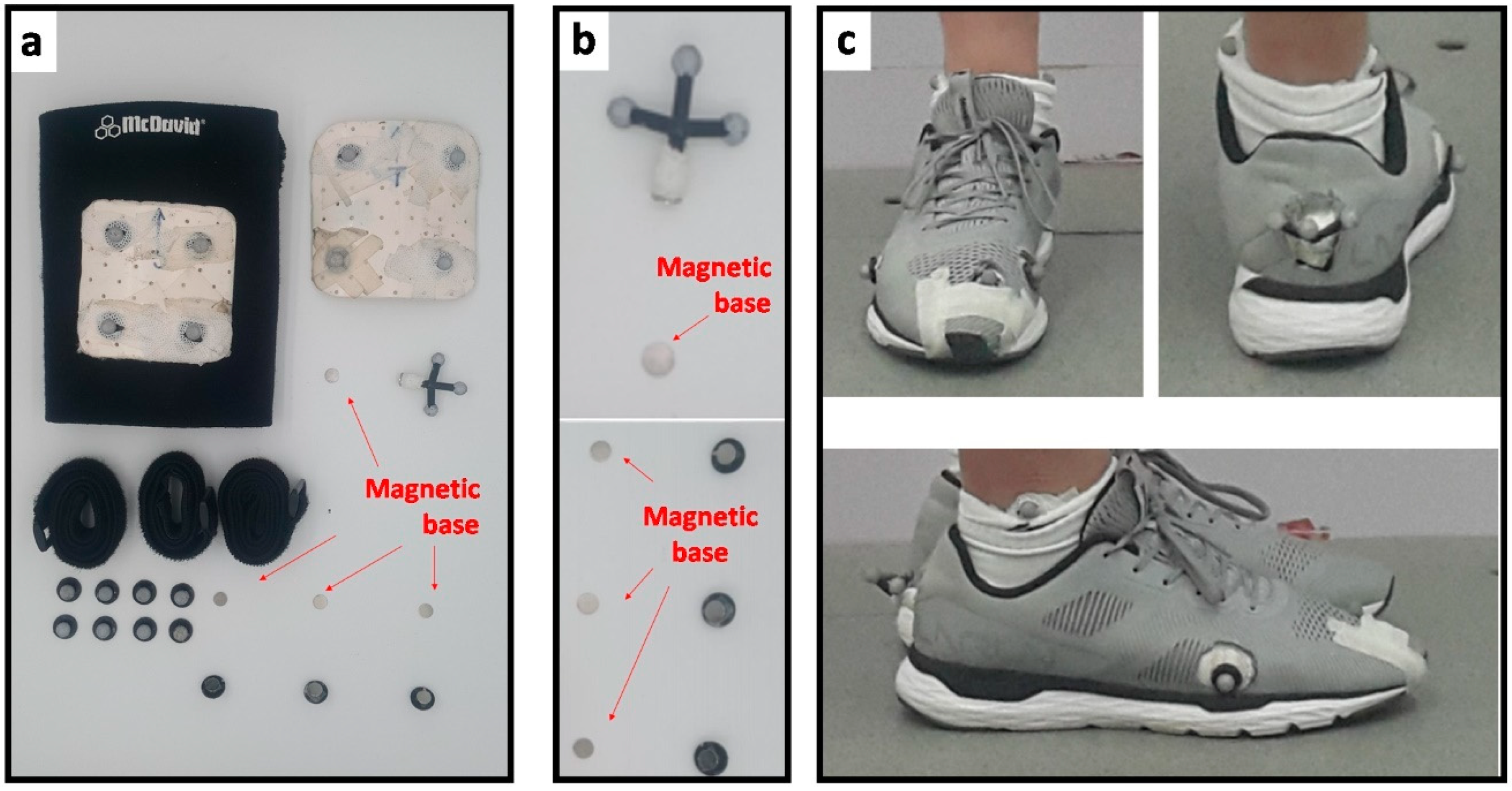
2.2. Experimental Conditions and Procedure

Comparisons were made for four orthosis conditions (Figure 1): prefabricated arch-support orthoses [2-mm (D2), 6-mm (D6), and 10-mm (D10) heel lift] versus control condition (2-mm flat control). The three arch-support orthoses (D2, D6 and D10) had the same arch-support of 25 mm, which was made of Polyurethane (PU) as specified with the off-the-shelf orthosis (Arch-support series-Universal II, Dr. Kong Footwear Ltd., Hong Kong), while the control orthosis did not have medial arch-support. The D2 orthosis had the same heel lift as the original prefabricated orthosis, while the D6 and D10 conditions were modified by adding the same PU material at the heel regions, respectively. The information on stack height and heel lift is provided in Figure 1. All arch-support orthoses were fit into the identical neutral running shoe (ARBN023, Li Ning, Beijing, China). Ten holes with a diameter of about 15 mm were cut in the shoe to allow direct placement of the reflective markers on the skin to assess forefoot and rearfoot kinematics.
After anthropometrical measurements were taken, a total of 28 reflective markers (diameter 14 mm) were placed on the skin according to the modified Oxford Foot Model [42]: four pelvis markers (left and right anterior and posterior superior iliac spines), medial and lateral femoral condyles, medial and lateral malleolus, 1st and 5th metatarsal heads, and triads markers on middle thigh, middle shank, posterior heel, and forefoot. The Oxford Foot Model is a multi-segment foot model used to calculate forefoot and rearfoot kinematics and it has been shown to have reliable gait measurements in various populations [42,43,44]. The foot markers were attached to the skin via a magnetic base that attached to the skin using strong adhesive tape [36] (Figure 2). This allowed markers to be attached to and detached from the foot through holes in the shoe, and ensured that the marker placement kept consistent among all test conditions since the magnetic bases stayed attached to the foot during the entire data collection session. The markers on the medial and lateral epicondyles were used during the static trial and then removed before commencing with the movement trials. The markers were placed by the same experimenter to ensure consistency across the participants.
The participants were instructed to run at their selected speeds (2.65 ± 0.45 m/s) in four tested orthoses (D2, D6, D10, and control) on an instrumented treadmill with force plate embedded (Bertec Corp, Columbus, Ohio, sampling at 1000 Hz). At first, the preferred speed of each participant was determined while running on the treadmill by gradually increased the running speed [45]. Briefly, while gradually increasing the treadmill speed, the participants were instructed to identify their preferred speed for their regular running. A synchronized 10-camera motion capturing system (Vicon MX, Vicon Motion System Ltd., Oxford, England, sampling at 200 Hz) was used to record kinematic data for the study. Running test for each condition lasted 1 min to achieve stable gait before data collection [29,46]. The last 10 footfalls from each condition were extracted for subsequent analysis. The testing order was randomly assigned across participants using an online program (www.random.org, accessed on 1 December 2018). To avoid any fatigue, they were required to rest unshod (i.e., barefoot) for 5 min between orthosis conditions [29,45].

2.3. Data Processing

The marker trajectory data were filtered with a Butterworth fourth order filter at a cut-off frequency of 12 Hz [47]. The data were then exported to Visual 3D program (C-motion, Germantown, MD, USA) to define segments from body markers and joint kinematics for subsequent analyses. Spatiotemporal variables, such as contact time, stride length, and stride frequency were computed. Contact time was the duration between initial contact and toe-off for each step. Stride length was calculated as the product of the treadmill speed and time between successive initial contacts of the right foot [29]. Stride frequency was calculated by reciprocal of averaged stride time of the ten strides. Vertical instantaneous loading rate was calculated as the maximum slope of the vertical impact peak [28]. Joint angle and moment in sagittal and coronal planes including initial, peak, and total range of motion (RoM) of the rearfoot, forefoot and knee joint were selected for this study. An inverse-dynamic model which comprised of the thigh, shank, rearfoot, and forefoot segments were built in Visual 3D program (C-Motion Inc., Germantown, MD, USA) to determine peak sagittal and coronal rearfoot and knee joint moments. In brief, the metatarsophalangeal joint was modeled as a single hinge joint rotating about an axis perpendicular to the sagittal plane (i.e., heel-to-toe direction). The segmental masses of segments were taken from a universal model from Visual3D program. The segmental mass of forefoot and rearfoot was then partitioned in the same ratios as their respective volumes, modeling each as simple geometric solids with uniform densities [48]. The positive values denoted as rearfoot dorsiflexion/inversion, forefoot dorsiflexion/inversion, and knee extension/adduction. All GRF and joint moments were normalized to body weight (BW).

2.4. Data Analysis

All statistical analyses were computed using the SPSS package (SPSS v22.0 Inc., Chicago, IL, USA). All outcome measures showed normal distributions (Shapiro Wilk’s normality test) and homogeneity of variances (Levene’s test). A repeated measures ANOVA was performed to examine if there was any significant difference between orthotic conditions (D2, D6, D8, and control). Bonferroni Post-hoc comparisons were applied when any significant effect of orthosis was determined. The data were screened for sphericity using Mauchly’s test. If sphericity was violated, significance of main effects was corrected through Greenhouse Geisser’s test. Alpha level was set at 0.05. The effect sizes for partial eta-squared (ηp2) and Cohen’s d (d) were calculated and interpreted as small (0.01 ≤ ηp2 < 0.06; 0.2 ≤ d < 0.5), medium (0.06 ≤ ηp2 < 0.14; 0.5 ≤ d <0.8) and large (ηp2 ≥ 0.14; d ≥ 0.5) [49].

3. Results

3.1. Spatiotemporal and GRF Variables

For GRF variable (Table 1), wearing D10 orthosis led to smaller maximum loading rate than D2 (p < 0.001, d = 0.50) and control orthoses (p = 0.002, d = 0.46).

3.2. Sagittal Joint Kinematics Variables

Rearfoot touchdown dorsiflexion was significantly larger in D10 than D2 (p = 0.027, d = 0.70) and control (p = 0.007, d = 0.97) orthoses and larger in D6 orthosis than the control condition (Table 2, p = 0.025, d = 0.80).

3.3. Frontal Joint Kinematics Variables

For frontal joint variables (Table 3), wearing D6 orthosis had significant smaller forefoot frontal RoM than D2 (p = 0.013, d = 0.73) and D10 orthoses (p = 0.018, d = 0.80). Wearing D10 orthosis demonstrated larger rearfoot frontal RoM than D2 (p = 0.018, d = 0.37) and larger peak forefoot eversion than D6 (p = 0.047, d = 0.89) and control orthoses (p = 0.048, d = 0.91).

3.4. Joint Moment Variables

Joint moment variables (Table 4), wearing control orthosis exhibited larger peak rearfoot pronation moment than D6 orthosis (p = 0.035, d = 0.39), but smaller peak knee extension moment than D2 (p = 0.025, d = 0.31) and D10 (p = 0.010, d = 0.28).

4. Discussion

This study examined the changes in loading rate and spatiotemporal, sagittal, and frontal joint kinematics and moment variables across arch-support orthosis conditions (2-mm, 6-mm, and 10-mm heel lift). In our study, wearing higher heel lift (e.g., D10) orthoses was shown to be associated with smaller loading rate, but larger rearfoot touchdown angle when compared to wearing lower heel lift orthoses (e.g., D2 and control). This is in line with previous studies, which suggested that wearing shoes with higher heel lift would lead to a smaller loading rate and larger foot strike angle [14,25,29,32,47,50]. The decrease in loading rate would be resulted from lower peak pressure and contact area at rearfoot region in higher heel lift condition [51]. Previous studies have reported a decrease in loading rate when increasing the foot strike angle would be associated with the reduction of the risk potential of running-related injuries [28]. Our results found that orthoses with higher heel lift were associated with smaller maximum loading rate, which is not in line with another study which found higher loading rate in higher heel lift condition instead [47]. The contradicting findings for higher maximum loading rate could be due to the interaction of medial arch-support and fatigue. It is reported that fatigue alters running biomechanics and neuromuscular output in as little as 15 min of running [52]. Another study reported that the loading rate significantly increased following fatigue-inducing exercise of ankle dorsiflexors [53]. Since foot orthoses may reduce fatigue and overuse injuries through minimizing the localized plantar loading and altering the “fit” of the contour of the foot contact surface [5,6,12]. Future research should examine the efficiency of arch-support orthoses combined with varying heel lifts in reducing loading rate and altering sagittal joint kinematics when running during training and match.
A higher heel lift is believed to increase the available range of motion of the rearfoot into dorsiflexion by placing the ankle in a more plantarflexed position [14]. Acute adaptations that may indicate a shift towards a more mid-foot running pattern have been reported to alter lower limb biomechanical characteristics included temporal-distances, plantar pressures, joint kinematics and loading, as well as muscle function [14,15]. However, our study results indicated that some of the frontal forefoot and rearfoot angles (1.1 to 2.4 degree) were regarded as clinically insignificant (<2 degree), although they displayed statistically differences. Such small absolute amount of ankle angle changes was often found across various studies done on heel lift [19], heel curvature [54], and arch-support manipulation [55]. The results from this study could be clinically applicable for individuals with heel pain, Achilles tendon pain, and limited dorsiflexion RoM, who may benefit by wearing footwear/orthoses with a larger heel lift [16,17,20,22,24].
Additionally, we found an inverted U pattern for forefoot frontal RoM, with medium heel lift orthosis (D6) significantly differ from the D2 and D10. This partly supports the contention that an increase in the magnitude of heel lift does not necessarily result in a proportional change in joint kinematics [19]. Similarly, Mo et al. [29] investigated the effect of shoes with various heel lifts relative to forefoot (0 mm, 4 mm, 8 mm, and 12 mm) on running biomechanics and found no systematic changes across heel lifts. While no significant difference was found between the large (12 mm) and small (0 mm) heel lift conditions, the authors found that the medium heel lift condition (8 mm) was significantly higher foot strike angle than small heel lift (0 mm) and significantly longer stride length than the large heel lift (12 mm) in self-preferred speed running. When heel lift increases, the windlass effect is activated and produced a relative shortening of longitudinal arches that is suggested to alter the plantar loading and joint positioning transmitted from the rearfoot to the forefoot [16]. These changes of joint kinematics lead to different relative points of force application and shift the GRF vector, resulting in the similar pattern with rearfoot and knee moments in our study. Our results, along with previous findings, suggest that optimal range of heel lift seems to exist to optimize running mechanics and performances.
Wearing arch-support orthoses lead to smaller peak rearfoot eversion moment but larger peak knee extension moment when compared to wearing flat orthoses. This is in line with previous studies [56,57], which suggested that wearing foot orthosis reduced peak rearfoot eversion moment but increased peak knee extension moment. Foot orthosis with arch-support could sustain the medial longitudinal arch and control excessive pronation [56]. The significant reduction in the peak ankle eversion moment facilitated by the orthosis could alleviate further pronation and progression [56,58], which could be difficult to obtain by surgical intervention alone [59]. On the other hand, the larger extension moment when wearing arch-support orthoses could be resulted from restricted plantarflexion of ankle [57]. MacLean and his colleagues [60] stated it is possible that increasing the knee extension moment could be deleterious to patellofemoral dynamics. However, in the studies of Peng et al. study [56] and Fatona et al. study [57], only participants with flatfoot and post-stroke hemiplegia were recruited. Hence, further studies may consider conducting studies on the use of arch-support orthoses by injury-free and healthy participants before a viable conclusion can be made.
When interpreting our results, it is important to consider several limitations in our study. Firstly, only male habitual rearfoot strikers were recruited in this study. The findings may not be generalizable to habitual midfoot/forefoot strikers and female population as biomechanical data differ across various foot strikes and between gender. Females are expected to have larger Q-angle and lower heel pad stiffness that may predispose distinct biomechanics pattern and loading characteristics and thus different running injuries [14,17,22,61]. Secondly, we did not measure comfort perception variables. Comfort perception has been received considerable interest by coaches and sports scientists as increase in perceived comfort was related to better running economy [7] and impact attenuation and performance during running [62]. Thirdly, we compared a large number of biomechanical variables to provide a comprehensive analysis of the orthotic effect in running. There is an increased chance of type I error when interpreting our study findings. Fourthly, while there are well over 40 different ways of foot type classification and there is no international standard on which method is the most appropriate [63], our study screened the participants using only single foot type classification method and instructed the participants to run with their preferred running speed. It should pay particular attention when comparing the orthotic effects across the studies which had different foot type classification methods and running speeds. Finally, the study was conducted without using incorporated arch-support on different medial-lateral wedge orthoses. The addition of custom arch-support to standard wedge orthoses may improve foot and knee symptoms in people with knee osteoarthritis and concomitant pronated feet [64]. Future studies may consider conducting studies using arch-support wedge insoles as it would be great potential to aid daily uses in training and competition. Fourthly, the study was also conducted using shoes with holes to get better accuracy on foot movement. This may affect shoe integrity and consequently running movement. Hence, future studies may consider conducting studies using shoes with holes as this might affect running biomechanics.

5. Conclusions

Wearing arch-support orthoses with various heel lifts leads to different movement mechanics and loading on ankle and knee joints compared to flat-control orthoses. Wearing D10 orthosis can lead to smaller impact loading, larger peak rearfoot dorsiflexion, peak forefoot eversion, and peak knee extension moment than flat-control orthosis. Wearing D6 orthosis was effective to reduce rearfoot eversion moment than control and smaller forefoot RoM than D2 and D10 orthoses, implying a higher rearfoot stability and control in frontal plane. These findings could be insightful for training and insole development in running.

Author Contributions

Conceptualization, W.-K.L., P.L.-W.S. and A.K.-L.L.; Data curation, J.-X.P., P.L.-W.S. and M.F.T.; Formal analysis, J.-X.P., P.L.-W.S. and M.F.T.; Funding acquisition, J.-X.P. and A.K.-L.L.; Investigation, P.L.-W.S. and M.F.T.; Methodology, W.-K.L., P.L.-W.S. and A.K.-L.L.; Supervision, W.-K.L. and A.K.-L.L.; Writing—original draft, J.-X.P., W.-K.L.; Writing—review & editing, P.L.-W.S., M.F.T. and A.K.-L.L. All authors have read and agreed to the published version of the manuscript.

Funding

General Program of Civil Aviation Flight University of China (grant number: J2020-023)

Institutional Review Board Statement

This study was conducted according to the guidelines of the Declaration of Helsinki and approved by the Ethics Committee of the Hong Kong Polytechnic University (HSEAR20180915002).

Informed Consent Statement

Informed consent was obtained from all subjects involved in the study.

Data Availability Statement

The data presented in this study are available on request from the corresponding author. The data are not publicly available due to the originality of the data.

Conflicts of Interest

W.-K.L. is an employee of Li Ning Sports Goods Company Limited which supplied the standard compression garments and footwear in the experiments. Other authors declared no potential conflict of interest.

References

  1. Filo, K.; Funk, D.C.; O’Brien, D. Examining motivation for charity sport event participation: A comparison of recreational-based and charity-based motives. J. Leis. Res. 2011, 43, 491–518. [Google Scholar] [CrossRef]
  2. van Gent, R.N.; Siem, D.; van Middeloop, M.; van Os, A.G.; Bierma-Zeinstra, S.M.A.; Koes, B.W. Incidence and determinants of lower extremity running injuries in long distance runners: A systematic review. Br. J. Sports Med. 2007, 41, 469–480. [Google Scholar] [CrossRef] [PubMed] [Green Version]
  3. Fredericson, M.; Misra, A.K. Epidemiology and aetiology of marathon-running injuries. Sports Med. 2007, 37, 437–439. [Google Scholar] [CrossRef]
  4. Lopes, A.D.; Hespanhol Junior, L.C.; Yeung, S.S.; Costa, L.O.P. What are the main running-related musculoskeletal injuries? Sports Med. 2012, 42, 891–905. [Google Scholar] [CrossRef]
  5. Lucas-Cuevas, A.G.; Perez-Soriano, P.; Llana-Belloch, S.; Macian-Romero, C.; Sanchez-Zuriaga, D. Effect of custom-made and prefabricated insoles on plantar loading parameters during running with and without fatigue. J. Sports Sci. 2014, 32, 1712–1721. [Google Scholar] [CrossRef] [PubMed]
  6. O’Leary, K.; Vorpahl, K.A.; Heiderscheit, B. Effect of Cushioned Insoles on Impact Forces During Running. J. Am. Podiatr. Med. Assoc. 2008, 98, 36–41. [Google Scholar] [CrossRef] [PubMed] [Green Version]
  7. Luo, G.; Stergiou, P.; Worobets, J.; Nigg, B.; Stefanyshyn, D. Improved footwear comfort reduces oxygen consumption during running. Footwear Sci. 2009, 1, 25–29. [Google Scholar] [CrossRef]
  8. Davidson, D.M. Prefabricated insoles and modifications in sports medicine. In Athletic Footwear and Orthoses in Sports Medicine; Werd, M.B., Knight, E.L., Eds.; Springer: New York, NY, USA, 2020; pp. 89–94. [Google Scholar]
  9. Zhang, X.; Li, B.; Hu, K.; Wan, Q.; Ding, Y.; Vanwanseele, B. Adding an arch support to a heel lift improves stability and comfort during gait. Gait Posture 2017, 58, 94–97. [Google Scholar] [CrossRef]
  10. Yu, B.; Preston, J.J.; Queen, R.M.; Byram, I.R.; Hardaker, W.M.; Gross, M.T.; Davis, J.M.; Taft, T.N.; Garrett, W.E. Effects of wearing foot orthosis with medial arch support on the fifth metatarsal loading and ankle inversion angle in selected basketball tasks. J. Orthop. Sports Phys. Ther. 2007, 37, 186–191. [Google Scholar] [CrossRef]
  11. Ng, J.W.; Chong, L.J.; Pan, J.W.; Lam, W.K.; Ho, M.; Kong, P.W. Effects of foot orthosis on ground reaction forces and perception during short sprints in flat footed athletes. Res. Sports Med. 2021, 29, 43–55. [Google Scholar] [CrossRef]
  12. Mundermann, A.; Nigg, B.M.; Humble, R.N.; Stefanyshyn, D.J. Foot orthotics affect lower extremity kinematics and kinetics during running. Clin. Biomech. 2003, 18, 254–262. [Google Scholar] [CrossRef]
  13. Kogler, G.F.; Solomonidis, S.E.; Paul, J.P. Biomechanics of longitudinal arch support mechanisms in foot orthoses and their effect on plantar aponeurosis strain. Clin. Biomech. 1996, 11, 243–252. [Google Scholar] [CrossRef]
  14. Rabusin, C.L.; Menz, H.B.; McClelland, J.A.; Tan, J.M.; Whittaker, G.A.; Evans, A.M.; Munteanu, S.E. Effects of heel lifts on lower limb biomechanics and muscle function: A systematic review. Gait Posture 2019, 69, 224–234. [Google Scholar] [CrossRef] [PubMed]
  15. Reinschmidt, C.; Nigg, B.M. Influence of heel height on ankle joint moments in running. Med. Sci. Sports Exerc. 1995, 27, 410–416. [Google Scholar] [CrossRef]
  16. Kogler, G.; Veer, F.; Verhulst, S.; Solomonidis, S.E.; Paul, J.P. The effect of heel elevation on strain within the plantar aponeurosis: In vitro study. Foot Ankle Int. 2001, 11, 433–439. [Google Scholar] [CrossRef]
  17. Lipton, J.; Flowers-Johnson, J.; Bunnell, M.; Carter, L. The use of heel lifts and custom orthotics in reducing self-reported chronic musculoskeletal pain scores. AAO J. 2009, 19, 15–21. [Google Scholar]
  18. Dixon, S.J.; Kerwin, D.G. The influence of heel lift manipulation on Achilles tendon loading in running. J. Appl. Biomech. 1998, 14, 374–389. [Google Scholar] [CrossRef]
  19. Dixon, S.J.; Kerwin, D.G. The influence of heel lift manipulation on sagittal plane kinematics in running. J. Appl. Biomech. 1999, 15, 139–151. [Google Scholar] [CrossRef]
  20. Grisogono, V. Physiotherapy treatment for Achilles tendon injuries. Physiotherapy 1989, 75, 562–572. [Google Scholar] [CrossRef]
  21. MacLellan, G.; Vyvyan, B. Management of pain beneath the heel and Achilles tendonitis with visco-elastic heel inserts. Br. Joint J. 1981, 63, 394–399. [Google Scholar]
  22. James, A.; Williams, C.; Haines, T. Effectiveness of interventions in reducing pain and maintaining physical activity in children and adolescents with calcaneal apophysitis (sever’s disease): A systematic review. J. Foot Ankle Res. 2013, 6, 16. [Google Scholar] [CrossRef] [Green Version]
  23. Liu, X.; Fabry, G.; Molenaers, G.; Lammens, J.; Moens, P. Kinematic and kinetic asymmetry in patients with leg-length discrepancy. J. Pediatr. Orthop. 1998, 18, 187–189. [Google Scholar] [CrossRef] [PubMed]
  24. Johnanson, M.; Cooksey, A.; Hillier, C.; Kobbeman, H.; Stambaugh, A. Heel lifts and the stance phase of gait in subjects with limited ankle dorsiflexion. J. Athl. Train. 2006, 41, 159–165. [Google Scholar]
  25. Besson, T.; Morio, C.; Millet, G.Y.; Rossi, J. Influence of shoe drop on running kinematics and kinetics in female runners. Eur. J. Sport Sci. 2019, 19, 1320–1327. [Google Scholar] [CrossRef] [PubMed]
  26. Horvais, N.; Samozino, P. Effect of midsole geometry on foot-strike pattern and running kinematics. Footwear Sci. 2013, 5, 81–89. [Google Scholar] [CrossRef]
  27. Lieberman, D.E.; Venkadesan, M.; Werbel, W.A.; Daoud, A.I.; D’andrea, S.; Davis, I.S.; Mang’eni, R.O.; Pitsiladis, Y. Foot strike patterns and collision forces in habitually barefoot versus shod runners. Nature 2010, 463, 531–536. [Google Scholar] [CrossRef] [PubMed]
  28. Milner, C.E.; Ferber, R.; Pollard, C.D.; Hamill, J.O.; Davis, I.S. Biomechanical factors associated with tibial stress fracture in female runners. Med. Sci. Sports Exerc. 2006, 38, 323–328. [Google Scholar] [CrossRef] [PubMed] [Green Version]
  29. Mo, S.; Lam, W.K.; Ching, E.C.K.; Chan, Z.Y.S.; Zhang, J.H.; Cheung, R.T.H. Effects of heel-toe drop on running biomechanics and perceived comfort of rearfoot strikers in standard cushioned running shoes. Footwear Sci. 2020, 12, 91–99. [Google Scholar] [CrossRef]
  30. Hoitz, F.; Mohr, M.; Asmussen, M.; Lam, W.K.; Nigg, S.; Nigg, B. The effects of systematically altered footwear features on biomechanics, injury, performance and preference in runners of different skill level: A systematic review. Footwear Sci. 2020, 12, 193–215. [Google Scholar] [CrossRef]
  31. Honert, E.C.; Mohr, M.; Lam, W.K.; Nigg, S. Shoe feature recommendations for different running levels: A Delphi study. PLoS ONE 2020, 15, e0236047. [Google Scholar] [CrossRef]
  32. Richert, F.C.; Stein, T.; Ringhof, S.; Stetter, B.J. The effect of heel-to-toe drop of standard running shoes on lower limb biomechanics. Footwear Sci. 2019, 11, 161–170. [Google Scholar] [CrossRef]
  33. Malisoux, L.; Chambon, N.; Urhausen, A.; Theisen, D. Influence of the heel-to-toe drop of standard cushioned running shoes on injury risk in leisure-time runners: A randomized controlled trial with 6-month follow-up. Am. J. Sports Med. 2016, 44, 2933–2940. [Google Scholar] [CrossRef]
  34. Davis, I.S.; Bowser, B.J.; Hamill, J. Vertical impact loading in runners with a history of patellofemoral pain syndrome. Med. Sci. Sports Exerc. 2010, 42, 682. [Google Scholar] [CrossRef]
  35. Pohl, M.B.; Hamill, J.; Davis, I.S. Biomechanical and anatomic factors associated with a history of plantar fasciitis in female runners. Clin. J. Sport Med. 2009, 19, 372–376. [Google Scholar] [CrossRef]
  36. Kosonen, J.; Kulmala, J.P.; Müller, E.; Avela, J. Effects of medially posted insoles on foot and lower limb mechanics across walking and running in overpronating men. J. Biomech. 2017, 54, 58–63. [Google Scholar] [CrossRef] [PubMed] [Green Version]
  37. Fraser, J.J.; Feger, M.A.; Hertel, J. Forefoot involvement in lateral ankle sprains and chronic ankle instability. Part 2: Clinical Considerations. Int. J. Sports Phys. Ther. 2016, 11, 1191–1203. [Google Scholar] [PubMed]
  38. Ferber, R.; Hettinga, B.A. A comparison of different over-the-counter foot orthotic devices on multi-segment foot biomechanics. Prosthet. Orthot. Int. 2016, 40, 675–681. [Google Scholar] [CrossRef] [PubMed]
  39. Nester, C.J. Lessons from dynamic cadaver and invasive bone pin studies: Do we know how the foot really moves during gait? J. Foot Ankle Res. 2009, 2, 1–7. [Google Scholar] [CrossRef] [Green Version]
  40. Sinclair, J.; Taylor, P.J.; Hebron, J.; Chockalingam, N. Differences in multi-segment foot kinematics measured using skin and shoe mounted markers. Foot Ankle Online J. 2014, 7, 7. [Google Scholar]
  41. Ojukwu, C.P.; Anyanwu, E.G.; Nwafor, G.G. Correlation between foot arch index and the intensity of foot, knee, and lower back pain among pregnant women in a south-eastern nigerian community. Med. Princ. Pr. 2017, 26, 480–484. [Google Scholar] [CrossRef] [PubMed]
  42. Levinger, P.; Murley, G.S.; Barton, C.J.; Cotchett, M.P.; McSweeney, S.R.; Menz, H.B. A comparison of foot kinematics in people with normal- and flat-arched feet using the Oxford Foot Model. Gait Posture 2010, 32, 519–523. [Google Scholar] [CrossRef] [PubMed]
  43. Stebbins, J.; Harrington, M.; Thompson, N.; Zavatsky, A.; Theologis, T. Repeatability of a model for measuring multi-segment foot kinematics in children. Gait Posture 2006, 23, 401–410. [Google Scholar] [CrossRef]
  44. Wright, C.J.; Arnold, B.L.; Coffey, T.G.; Pidcoe, P.E. Repeatability of the modified Oxford foot model during gait in healthy adults. Gait Posture 2011, 33, 108–112. [Google Scholar] [CrossRef]
  45. Wang, Y.; Lam, W.K.; Wong, C.K.; Park, L.Y.; Tan, M.F.; Leung, A.K.L. Effectiveness and reliability of foot orthoses on impact loading and lower limb kinematics when running at preferred and nonpreferred speeds. J. Appl. Biomech. 2020, 1, 1–8. [Google Scholar] [CrossRef]
  46. Park, S.K.; Jeon, H.M.; Lam, W.K.; Stefanyshyn, D.; Ryu, J. The effets of downhill slope on kinematics and kinetics of the lower extremity joints during running. Gait Posture 2018, 68, 181–186. [Google Scholar] [CrossRef]
  47. Chambon, N.; Delattre, N.; Gueguen, N.; Berton, E.; Rao, G. Shoe drop has opposite influence on running pattern when running overground or on a treadmill. Eur. J. Appl. Physiol. 2015, 115, 911–918. [Google Scholar] [CrossRef] [Green Version]
  48. Lam, W.K.; Lee, W.C.C.; Lee, W.M.; Ma, C.Z.H.; Kong, P.W. Segmental forefoot plate in basketball footwear: Does it influence performance and foot joint kinematics and kinetics. J. Appl. Biomech. 2018, 34, 31–38. [Google Scholar] [CrossRef] [PubMed] [Green Version]
  49. Cohen, J. Statistical power Analysis for the Behavioural Sciences; Lawrence Erlbaum Associate, Inc.: Mahwah, NJ, USA, 1998. [Google Scholar]
  50. Besson, T.; Morio, C.; Rossi, J. Effects of shoe drop on running mechanics in women. Comput. Methods Biomech. Biomed. Eng. 2017, 20, 19–20. [Google Scholar] [CrossRef] [PubMed]
  51. Zhang, X.; Li, B. Influence of in-shoe heel lifts on plantar pressure and center of pressure in the medial-lateral direction during walking. Gait Poture 2014, 39, 1012–1016. [Google Scholar] [CrossRef] [PubMed]
  52. Derrick, T.R.; Dereu, D.; McLean, S.P. Impacts and kinematic adjustments during an exhaustive run. Med. Sci. Sports Exerc. 2002, 34, 998–1002. [Google Scholar] [CrossRef]
  53. Christina, K.; White, S.; Gilchrist, L. Effect of localized muscle fatigue on vertical ground reaction forces and ankle joint motion during running. Hum. Mov. Sci. 2001, 20, 257–276. [Google Scholar] [CrossRef]
  54. Liu, Z.-L.; Lam, W.-K.; Zhang, X.; Vanwanseele, B.; Liu, H. Influence of heel design on lower extremity biomechanics and comfort perception in overground running. J. Sports Sci. 2021, 39, 232–238. [Google Scholar] [CrossRef]
  55. Lam, W.-K.; Pak, L.-Y.; Wong, C.K.-K.; Tan, M.F.; Park, S.-K.; Ryu, J.; Leung, A.K.-L. Effects of arch-support orthoses on ground reaction forces and lower extremity kinematics related to running at various inclinations. J. Sports Sci. 2020, 38, 1629–1634. [Google Scholar] [CrossRef] [PubMed]
  56. Peng, Y.; Wong, D.W.C.; Wang, Y.; Chen, T.L.W.; Tan, Q.; Chen, Z.; Jin, Z.; Zhang, M. Immediate effects of medially posted insoles on lower limb joint contact forces in adult acquired flatfoot: A pilot study. Int. J. Environ. Res. Public Health 2020, 17, 2226. [Google Scholar] [CrossRef] [PubMed] [Green Version]
  57. Fatone, S.; Gard, S.A.; Malas, B.S. Effect of ankle-foot orthosis alignment and foot-plate length on the gait of adults with poststroke hemiplegia. Arch. Phys. Med. Rehabil. 2009, 90, 810–818. [Google Scholar] [CrossRef]
  58. Desmyttere, G.; Hajizadeh, M.; Bleau, J.; Begon, M. Effect of foot orthosis design on lower limb joint kinematics and kinetics during walking in flexible pes planovalgus: A systematic review and meta-analysis. Clin. Biomech. 2018, 59, 117–129. [Google Scholar] [CrossRef] [PubMed]
  59. Wong, D.W.-C.; Wang, Y.; Chen, T.L.-W.; Leung, A.K.-L.; Zhang, M. Biomechanical consequences of subtalar joint arthroereisis in treating posterior tibial tendon dysfunction: A theoretical analysis using finite element analysis. Comput. Methods Biomech. Biomed. Eng. 2017, 20, 1525–1532. [Google Scholar] [CrossRef]
  60. MacLean, C.; Davis, I.S.; Hamill, J. Influence of a custom foot orthotic intervention on lower extremity dynamics in healthy runners. Clin. Biomech. 2016, 21, 623–630. [Google Scholar] [CrossRef]
  61. Ugbolue, U.C.; Yates, E.L.; Wearing, S.C.; Gu, Y.; Lam, W.K.; Valentin, S.; Baker, J.S.; Dutheil, F.; Sculthorpe, N.F. Sex differences in heel pad stiffness during in vivo loading and unloading. J. Anat. 2020, 237, 520–528. [Google Scholar] [CrossRef]
  62. Dessery, Y.; Belzile, E.; Turmel, S.; Corbeil, P. Effects of foot orthoses with medial arch support and lateral wedge on knee adduction moment in patients with medial knee osteoarthritis. Prosthet. Orthot. Int. 2017, 41, 356–363. [Google Scholar] [CrossRef] [PubMed]
  63. Tong, J.W.K.; Kong, P.W. Association between foot type and lower extremity injuries: A systematic review with meta-analysis. J. Orthop. Sports Phys. Ther. 2013, 43, 700–714. [Google Scholar] [CrossRef] [PubMed] [Green Version]
  64. Hunt, M.A.; Takacs, J.; Krowchuk, N.M.; Hatfield, G.L.; Hinman, R.S.; Chang, R. Lateral wedges with and without custom arch support for people with medial knee osteoarthritis and pronated feet: An exploratory randomized crossover study. J. Foot Ankle Res. 2017, 10, 20. [Google Scholar] [CrossRef] [PubMed] [Green Version]
Figure 1. The four experimental orthoses.
Applsci 11 01613 g001
Figure 2. Marker placements: (a) marker set, (b) magnetic base, and (c) marker placement on foot.
Applsci 11 01613 g002
Table 1. Mean (SD) values and statistical results of spatiotemporal and loading rate variables by tested conditions.
Orthosis ConditionANOVA Results
Flat
(Control)
D2D6D10F-Valuep-ValuePartial Eta Squared (ηp2)
Max loading rate (BW/s)871.4 (321.0) *881.7 (307.1) *801.7 (275.4)737.4 (266.4)11.49<0.0010.45
Contact time (ms)262.0 (31.5)263.2 (33.3)262.0 (30.5)263.6 (34.3)0.190.9010.01
Stride length (m)1.33 (0.12)1.38 (0.11)1.32 (0.11)1.36 (0.11)2.410.0800.15
Stride frequency (Hz)2.08 (0.45)1.95 (0.26)2.10 (0.42)1.99 (0.32)2.370.0840.15
Note: Max = maximum; BW = bodyweight; * sig different from D10 (p < 0.05).
Table 2. Mean (SD) values and statistical results of sagittal joint kinematics variables by tested conditions.
Orthosis ConditionANOVA Results
Flat
(Control)
D2D6D10F-Valuep-ValuePartial Eta Squared (ηp2)
Total RoM (o)
Forefoot-sagittal17.6 (4.8)19.7 (6.7)18.0 (6.2)20.7 (9.7)1.600.2240.10
Rearfoot-sagittal29.5 (5.6)31.5 (6.4)31.5 (5.5)33.2 (8.9)2.060.1520.13
Knee-sagittal25.3 (3.2)25.7 (3.0)25.3 (2.8)25.9 (2.8)0.820.4930.06
Angle at touchdown (o)
Forefoot dorsiflexion12.9 (5.4)14.3 (5.8)10.7 (6.1)10.5 (8.9)2.670.0900.16
Rearfoot dorsiflexion2.2 (5.6) *^3.3 (8.1) *7.3 (7.1)9.5 (9.5)8.96<0.0010.39
Knee flexion−18.3 (4.2)−18.4 (4.3)−18.6 (4.2)−17.6 (4.7)1.440.0930.09
Peak angle during stance (o)
Peak forefoot dorsiflexion17.4 (5.2)18.0 (5.9)17.7 (5.5)16.5 (7.9)0.390.6710.03
Peak forefoot plantarflexion−0.2 (3.7)−1.7 (4.7)−0.3 (4.9)−4.2 (7.4)2.810.0510.17
Peak rearfoot doriflexion21.3 (6.5) *23.6 (7.3) *23.7 (7.4) *27.9 (8.8)5.290.0030.27
Peak rearfoot plantarflexion−8.6 (6.8)−7.7 (7.7)−7.8 (6.0)−5.3 (8.1)1.690.1830.11
Peak knee flexion−42.9 (3.6)−43.0 (3.7)−43.1 (3.6)−42.6 (4.1)0.340.7970.02
Min knee flexion−17.5 (4.0)−17.2 (4.5)−17.8 (4.1)−16.7 (4.2)1.360.2670.09
Joint angles at push-off (o)
Forefoot dorsiflexion9.5 (5.8)10.3 (5.1)11.4 (5.7)8.8 (9.1)1.250.3020.08
Rearfoot plantarflexion−6.6 (8.5)−5.7 (8.1)−6.1 (8.0)−3.0 (8.9)1.820.1590.12
Knee flexion−23.1 (4.9)−22.3 (5.9)−23.3 (6.3)−22.4 (5.1)0.930.4350.06
Min = minimum; * sig different (p < 0.05) from D10; ^ sig different (p < 0.05) from D6.
Table 3. Mean (SD) values and statistical results of frontal joint kinematics variables by tested conditions.
Orthosis ConditionANOVA Results
Flat
(Control)
D2D6D10F-Valuep-ValuePartial Eta Squared (ηp2)
Total RoM (o)
Forefoot-frontal4.4 (1.3)5.3 (2.1) ^4.1 (1.2)5.7 (2.8) ^3.500.0240.20
Rearfoot-frontal13.2 (3.5)12.8 (3.1) *13.2 (2.2)13.9 (2.9)2.870.0480.17
Angle at touchdown (o)
Forefoot inversion1.2 (2.5)−0.03 (4.6)0.4 (2.9)−0.8 (2.2)1.530.2200.10
Rearfoot eversion1.3 (4.2)0.6 (2.6)0.5 (3.8)0.01 (4.1)0.610.6130.04
Peak angle during stance (o)
Peak forefoot eversion−1.5 (1.7) *−3.4 (4.7)−1.2 (2.5) *−3.6 (2.9)3.350.0280.19
Peak forefoot inversion2.8 (2.0)1.8 (4.5)2.9 (2.0)2.1 (2.6)0.650.5900.04
Peak rearfoot eversion−7.8 (5.0)−7.9 (4.1)−8.4 (4.7)−9.6 (3.9)1.740.1730.11
Peak rearfoot inversion5.4 (4.1)4.9 (2.6)4.8 (3.6)4.3 (3.2)0.500.6830.04
Joint angles at push-off (o)
Forefoot eversion0.4 (2.1)−1.3 (4.2)1.1 (2.1)−1.1 (3.1)2.690.0580.16
Rearfoot inversion4.9 (4.2)4.7 (2.9)4.4 (3.5)3.6 (3.5)0.770.5200.05
* sig different from D10 (p < 0.05); ^ sig different from D6.
Table 4. Mean (SD) values and statistical results of joint moment variables by tested conditions.
Orthosis ConditionANOVA Results
Flat
(Control)
D2D6D10F-Valuep-ValuePartial Eta Squared (ηp2)
Peak moment (Nm/BW)
Rearfoot plantarflexion−2.75 (0.50)−2.67 (0.52)−2.69 (0.57)−2.78 (0.54)1.560.2150.10
Rearfoot eversion0.18 (0.25)0.11 (0.23)0.08 (0.26) #0.11 (0.22)3.030.0400.18
Knee extension2.07 (0.42)2.20 (0.42) #2.19 (0.46)2.19 (0.44) #3.240.0310.19
Knee adduction−1.05 (0.37)−1.04 (0.40)−1.00 (0.31)−1.05 (0.34)0.410.7480.03
BW = body weight; # sig different from Control (p < 0.05).
Publisher’s Note: MDPI stays neutral with regard to jurisdictional claims in published maps and institutional affiliations.

