Article

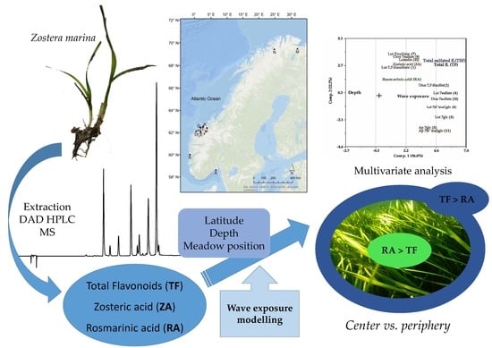
Variation in Phenolic Chemistry in Zostera marina Seagrass along Environmental Gradients

by
Cecilie Sævdal Dybsland
1,
Trine Bekkby
2,
Kjersti Hasle Enerstvedt
1,
Olav M. Kvalheim
1,
Eli Rinde
2 and
Monica Jordheim
1,*
1
Department of Chemistry, University of Bergen, Allégt. 41, N-5007 Bergen, Norway
2
Section for Marine Biology, Norwegian Institute for Water Research (NIVA), Gaustadalléen 21, N-0349 Oslo, Norway
*
Author to whom correspondence should be addressed.
Plants 2021, 10(2), 334; https://doi.org/10.3390/plants10020334
Submission received: 16 December 2020 / Revised: 26 January 2021 / Accepted: 3 February 2021 / Published: 9 February 2021
(This article belongs to the Special Issue Systematics and Ecology of Algae and Marine Plants)

Abstract

:
Chemical ecology has been suggested as a less time-consuming and more cost-efficient monitoring tool of seagrass ecosystems than traditional methods. Phenolic chemistry in Zostera marina samples was analyzed against latitude, sea depth, sample position within a seagrass meadow (periphery or center) and wave exposure. Multivariate data analysis showed that rosmarinic acid correlated moderately positively with depth, while the flavonoids had an overall strong negative correlation with increasing depth—possibly reflecting lack of stress-induced conditions with increasing depth, rather than a different response to light conditions. At a molecular level, the flavonoids were separated into two groups; one group is well described by the variables of depth and wave exposure, and the other group that was not well described by these variables—the latter may reflect biosynthetic dependencies or other unrevealed factors. A higher flavonoid/rosmarinic acid ratio was seen in the periphery of a seagrass meadow, while the contrary ratio was seen in the center. This may reflect higher plant stress in the periphery of a meadow, and the flavonoid/rosmarinic acid ratio may provide a possible molecular index of seagrass ecosystem health. Further studies are needed before the full potential of using variation in phenolic chemistry as a seagrass ecosystem monitoring tool is established.

Graphical Abstract

1. Introduction

Seagrasses are rooted vascular flowering plants (marine angiosperms) originating from land, often forming meadows in relatively shallow areas along the coast. Despite only covering less than 0.2% of the global sea surface area [1,2], they play an important role in the coastal ecosystem, as they often are the only habitat building species on shallow soft sediment. Seagrasses consist of about 60 different species with more than one thousand associated species of flora and fauna utilizing the seagrass habitat [3]. Seagrasses are in decline globally, and the rate of loss of area has increased from less than 1% per year before 1940 to 7% per year since 1990, resulting in a total loss of 29% of the known areal extent since the first seagrass recording in 1879 [4,5]. Seagrasses are threatened by many factors, such as physical modification, nutrient overload, sedimentation, the introduction of non-native species, overexploitation and climate change. The two major causes of the observed seagrass loss are, however, related to coastal development and degraded water quality [4]. Monitoring seagrass health is important to be able to detect and mitigate any severe impact of these direct and indirect human impacts on these valuable ecosystems and seagrass health indices may serve as indicators of coastal health in general. Seagrass monitoring has traditionally been based on long-term observations of changes in seagrass parameters, such as species composition, percent cover, biomass and plant morphometry that are identifiable in the field ([6,7] and refs therein). To date, molecular methods as tools for assessing plant health have been little examined. Seagrass phenolics have a high potential in this respect, as they are reckoned to play an important role in plant protection and appear to be affected by several biotic and abiotic pressures [8,9,10,11,12,13,14,15,16,17,18,19].
Phenolic chemistry has been tested as a putative descriptor of seagrass health or ecological status [7,17,20,21,22]. Migliore et al. examined Posidonia oceanica and related the spatial heterogeneity of environmental pressures to variations in the total phenolic content, which was further linked to differences in metabolic/physiological pathways as an adaptation to stress [17]. The total phenolic content was also found to be inversely related to the density of seagrass plants within meadows and linked to less stressful conditions and a better health state in meadows with high plant density than within meadows with low plant density [17,20]. Despite some research on seagrass phenolics as possible biochemical markers in seagrass monitoring, examinations at a differentiated molecular level are relatively little explored. Studies of the phenolic content of seagrasses at a molecular level often focus on simple phenolic acids [12,15,16,21,23] and not the more structurally advanced and diverse group of flavonoids. Böttner et al. (2020) recently reported that individual flavonoid glucosides have distinct cellular and subcellular locations and promote duckweed (Spirodela polyrhiza, aquatic plant) fitness under different abiotic stresses [24]. In general, the ecological roles of flavonoids are far from understood, partially due to their structural diversity, different accumulation patterns and varying abundance, all of which affect their biological activity [24,25,26].
Among the seagrasses, Zostera marina L. (eelgrass) is the most widely distributed species in the northern hemisphere, reflecting a broad tolerance of temperature, salinity and photoperiod conditions [27]. In Norway, the species is commonly found as large meadows with high plant density in the southern parts of Norway. The species is also found further north all the way to the northermost region (70° N), but then often as smaller meadows or more scattered occurrences. The existence of sulfated flavonoids in Zostera spp. is well-documented [9,28,29,30], and the appearance of these compounds has been suggested to be related to high concentration of sulfate ion in seawater [30]. The physiological role of the sulfated flavonoids in plants and the factors that regulate flavonoid sulfate accumulation is unclear. However, some evidence implies physiological protection of the seagrass [30,31,32,33]. As for the phenolic acid content of Z. marina, zosteric acid (ZA), a sulfated phenolic acid only seen in Zostera species [11], has been found to prevent the settlement of marine bacteria, algae, barnacles and tubeworms [34]. Rosmarinic acid (RA), another phenolic acid, widely known for its biological activities, including antioxidant and antifouling action [35,36], has been identified as one of the major compounds in Z. marina [23,29,37].
The main aim of this study was to analyze and assess any systematic variation in phenolic acids (ZA, RA) and flavonoids in Zostera marina, at a molecular level, along environmental gradients (latitude, depth as a proxy for the reduction in light conditions, wave exposure, and meadow positioning). The secondary aim was to identify any specific biochemical markers or molecular metrics that can be used in the context of seagrass health and monitoring.

2. Results

2.1. Phenolic Profiles

The HPLC-DAD and LR-LCMS analyses of Z. marina extracts revealed the same polyphenolic content as previously reported; eleven flavones (1–7, 912) and two phenolic acids (ZA, RA) [28,29,38] (Figure 1, Table 1 and Table 2).
The sulfated flavonoids 1, 2, 4, 10 and luteolin (12) were found in all the examined samples, covering the total range of the latitudinal gradient (see Section 4.1), as did rosmarinic acid (RA) and zosteri acid (ZA). Chrysoeriol 7-sulfate (9) was also present in most samples, with Munkefjorden (M) and Rafsbotn (N) as the only exceptions. Luteolin 7-glucoside (3) was found in three localities along the latitudinal gradient; furthest south (A), north-west (G1) and north (N). Compound 3 has previously been found in samples from Hordaland (B) as well [29]. Luteolin 7-(6″-malonyl)glucoside (6) and luteolin 3′-sulfate (7) were also found along a wide range of the latitudinal gradient.
The two samples from the northernmost region (~70° N, 27° E; M, N) (Figure 2), differed greatly in the occurrence of individual flavonoids (Table 1 and Table 2).The two apigenin derivatives, 5 and 11, were only observed in one of these samples (N). Apigenin 7-glucoside (5) and apigenin 7-sulfate (8) have previously been found in small amounts in Z. marina and in Z. noltii from Hordaland, while apigenin 7-(6″-malonyl)glucoside (11) has only been found in Z. noltii previously [29]. Apigenin 7-sulfate (8) was not found in any of the samples in the current study.
An average flavonoid amount of 6.24 ± 0.21 mg luteolin Eq./g dry weight (DW) was observed for the 24 Z. marina samples (Table 2). The highest amounts were found in Stormalen (G1) (10.20 ± 0.35 mg luteolin Eq./g DW) and Larvik (A) (10.23 ± 0.77 mg luteolin Eq./g DW). The lowest amounts were found in one of the highest latitude samples, Munkefjorden (M) (2.35 ± 0.41 mg luteolin Eq./g DW). The two seagrass samples from Finnmark region (M, N) had different qualitative profiles, and the quantitative amounts were higher in Rafsbotn (N) compared to Munkefjorden (M), with one exception. The amount of diosmetin 7,3′-disulfate (2) in Munkefjorden (M) (0.11 ± 0.04 mg luteolin Eq./g DW) exceeded the corresponding amount found in Rafsbotn (N) (0.06 ± 0.01 mg luteolin Eq./g DW), accounting for 4.75% and 0.8% of total amounts, respectively. The apigenin derivatives (5, 11) found in Rafsbotn (N) accounted for 2.2% of the total flavonoid content in this sample.
A correlation between luteolin 7-glucoside (3) and luteolin 7-sulfate (4) was observed. Luteolin 7-glucoside (3) was exclusively present in samples A, G1 and N, with high amounts of luteolin 7-sulfate (4). The observation was independent of where in Norway the samples were collected and is most likely related to the enzymatic process for the substitution of the 7-position on the ring.
For the phenolic acids, the western Møre and Romsdal sample I showed the highest content of rosmarinic acid (RA) (4.53 ± 0.31 mg luteolin Eq./g DW), approximately 29.5% of the total phenolic content, with zosteric acid (ZA) making 4.4% of the total content. The other nineteen samples in the Møre and Romsdal region (C-L) had lower RA content relative to the total phenolic content; 2-17%—while the ZA content was ranging from 0.1% to 4%. In the samples A (59° N, 10° E), B (60° N, 5° E) and G2 (63° N, 6.5° E) the highest amounts of ZA were found—with G2 showing the highest relative content with 5.6% ZA of total phenolics.

2.2. Relative Amount of Sulfated Flavonoids within Regions

A comparison of the relative amount of sulfated flavonoids (1, 2, 4, 7, 9, 10) in the Z. marina leaves between regions was made based on the average values within sampled regions (Figure 3 and Section 4.1). The northernmost regions, Finnmark (M, N; ~70° N, ~27° E) and Møre and Romsdal (C-L; ~63° N, ~7° E), had the relatively highest sulfated flavonoid content compared to the two regions further south, Hordaland (B; 60° N, 5° E) and Vestfold (A; 59° N, 10° E). There were no significant differences between the average sulfated flavonoid amounts found in Finnmark (M, N) compared to Møre and Romsdal (C-L). However, between the colder west coast of Hordaland (B) and the south situated Vestfold (A), a small significant difference was observed (p < 0.05).

2.3. Phenolic Content—Seagrass Meadow Positioning

In order to examine the phenolic content in samples with different positions in meadows, center and periphery species were analyzed from selected meadows (n = 19). An average concentration of total flavonoids (TF), sulfated flavonoids (TSF), rosmarinic acid (RA) and zosteric acid (ZA) was made for the two different positions (Figure 4). The flavonoids showed an average of 17% higher concentrations in the samples collected from the periphery (n = 9) than in the samples collected from the center (n = 10). Interestingly, and in contrast to the flavonoids, the average rosmarinic acid (RA) concentration was found to be 30% lower in the periphery samples compared to the center samples. For the small amounts of zosteric acid (ZA), no difference was observed between the two meadow positions.

2.4. Phenolic Content—Variations between Wave Exposure Classes

The analyzed Z. marina leaves were sampled within three classes of wave exposure; “extremely sheltered”, “very sheltered,” and “sheltered” see Section 4.1 and Section 4.2). The average values of total flavonoids (TF), sulfated flavonoids (TSF), rosmarinic acid (RA) and zosteric acid (ZA) content of the sampled leaves within each category were calculated (Figure 5).
Samples collected from the “sheltered” areas, representing the highest wave exposure values in this dataset, showed higher amounts of flavonoids than the “very sheltered” and “extremely sheltered” categories, 20% and 12%, respectively. For rosmarinic acid (RA), the “sheltered” samples had as much as 57% higher RA content than the “very sheltered” samples. Although the analytical amounts of zosteric acid (ZA) are low, a steady increase with respect to wave exposure was observed.

2.5. Variations in Phenolic Content Explained by Depth and Wave Exposure

The PCA explained 58.8% of the total variation (PC1; 36.6% and PC2; 22.2%). The cosine of the angle between the loadings of pairs of variables represents their correlation in the PCA-plot (Figure 6). A negative sign means that the variables are negatively correlated in the model. As the variables were standardized, a position close to zero implies that this particular variable does not correlate with the variation that component 1 and 2 is reflecting (Figure 6) [39]. Figure 7 shows the quantitative contribution of individual variables to PC1 and PC2, as well as the variation in each variable not explained by the two-component PCA model.
The multivariate analysis indicates moderate negative correlations between depth and the total amount of flavonoids (TF) and sulfated flavonoids (TSF) (Figure 6). As shown by the low residual variance (Figure 7), the TF and TFS variations are well described by components 1 and 2. Interestingly, there are differences with respect to how well the PCA plot describes the variance of the individual flavonoids. The flavonoids luteolin-7,3′-disulfate (1), luteolin 3′-sulfate (7), chrysoeriol 7-sulfate (9) and luteolin (12), seem not to be well-described by depth, while the flavonoids; diosmetin 7,3′-disulfate (2), luteolin 7-glucoside (3), luteolin 7-sulfate (4), luteolin 7-(6″-malonyl)glucoside (6) and diosmetin 7-sulfate (10), are all strongly negatively correlated with depth. The two apigenin derivatives (5, 11), found in the north (M), have almost identical positions in the plot—and they are 100% correlated with each other. They (5, 11) correlate weakly negatively with the total flavonoid amounts (TF, TSF) and moderately negatively with depth.
Both TF and TSF correlate moderately positively with wave exposure. The exceptions are again the flavonoids; 1, 7, 9 and 12, showing only weak correlations to wave exposure—repeating their response pattern relative to flavonoids 23, 6 and 10—as seen for the depth correlation. The flavonoids 1, 7, 9 and 12 are also the ones among the flavonoids having their variances less described by the “multiple variable” PCA plot (Figure 7). However, PCA analysis for individual flavonoids with depth only and with wave exposure only resulted in the same trends for 1, 7, 9 and 12, both with respect to correlations coefficients (Figure 6), and the lack of good variance description for these flavonoids (Figure 7).
The phenolic acids (ZA, RA) correlate weakly to wave exposure and depth in the “multiple variable” PCA-loading plot (Figure 6). However, Figure 7 shows that the variance seen in RA and ZA is not well described by components 1 and 2. Figure 5 shows a wave exposure dependent trend also for these two phenolic acids. The PCA partial correlation was tested for the phenolic acids, including only the variables depth and wave exposure, describing 71% of the variance (Table 3). The PCA analysis revealed a strong correlation for both acids with wave exposure and a moderate positive correlation to depth for RA, and a moderate negative correlation to depth for ZA. The depth and wave exposure are not correlated.

3. Discussion

The current study supplement our earlier studies of phenolics in Norwegian Zostera marina meadows [28,29,38] by including a larger latitudinal gradient (from just 60° N to covering 58–70° N). This gave us new insight into the phenolic chemistry of Z. marina in the northern areas, which has never been studied before. In addition, and importantly, our new dataset also contained parameters as depth (as a proxy for the reduction in light conditions), wave exposure and meadow positioning—improving chemoecological examinations.
Based on the qualitative phenolic profiles, the Z. marina samples examined in this study seems to reflect only one chemotype [30]. However, the northernmost Z. marina sample (N; 70° N, 23.5° E) had the highest diversity of flavonoids in the dataset—despite the environmental limitations Z. marina probably meet in this part of Norway (Table 1 and Table 2) [41]. This location also had relatively high total flavonoid amounts (7.57 ± 0.58 mg luteolin Eq./g DW), and one of the highest productions of diosmetin 7-sulfate (10) (3.58 ± 0.35 mg luteolin Eq./g DW, 47.3%). Only comparable to the amounts found in the southernmost sample (A; 59° N, 10° E: 3.92 ± 0.42 mg luteolin Eq./g DW, 38.3%). Little is known about flavonoid plant functionality at a molecular level. Recently, Papazian et al. examined the surface chemical defense of Z. marina against microbial fouling, where they differentiate between surface extract and C18 extract [19]. They found that the most abundant phenolic detected on the surface was diosmetin 7-sulfate (10), with 10-fold higher concentrations than the C18 extract. The next most abundant phenolic in the surface extract (absent in the C18) was ZA, followed by lower concentrations of caffeic acid, ferulic acid, p-coumaric acid, and the sulfated flavonoids luteolin 7-sulfate (4) and apigenin 7-sulfate (8). Notably, RA had only trace levels in surface extracts (<0.60 ng.mL/1). Whether the higher diosmetin 7-sulfate (10) amounts seen in our study could be linked to chemical defense or not warrants further examination. The northernmost Z. marina sample (N; 70° N, 23.5° E) also contained 2.2% apigenin derivatives (5, 11)—not seen in the rest of the samples. However, apigenin 7-sulfate (8) and apigenin 7-(6″-malonyl)glucoside (11) were found in spring samples of Z. marina in our previous studies (1–4%) (Hordaland, B; 60° N, 5° E), while apigenin 7-glucoside (5) was not found [29]. The samples in the current study were collected in late summer/fall. It was suggested that decreasing temperature could cause a shift in secondary metabolite profiles, hence a change in the qualitative profile of flavonoids—possibly affecting the presence of apigenin in the northernmost samples [14,42,43]. However, the N neighboring northern locality, Munkefjorden (M; 69.7° N, 29.5° E), is lacking apigenin derivatives—in addition to the absence of high flavonoid diversity (Figure 2). The samples from M were collected at lower depth and less wave exposure than in N. The fjord is also exposed to erosion, resulting in increased amounts of particles in the water—possibly limiting the light availability and hence the biosynthesis.
Our previous study on Z. marina from two different localities in Hordaland, showed that the sample from the southernmost area contained the relatively lowest amounts of sulfated flavonoids [29]. In the same study, regional differences in amounts of sulfated flavonoids in Z. noltii were also seen. The two samples from Vestfold (59° N, 10° E) had significantly lower relative amounts of sulfated flavonoids than the populations on the colder west coast. These observations are in agreement with the observations in the present study (Figure 3, see also Section 4.1). Several studies have shown that low water temperatures result in an increase in flavonoid production [14,15,43]. It seems, though, to be an increase in the occurrence of the sulfated flavonoids related to the longitude and colder climatic or harsher conditions, possibly reflecting the indicated physiological protective role of sulfated flavonoids in the seagrass [30,31,32,33]. However, in Zostera spp. the production of the major flavonoid group—the sulfated flavonoids, depends on the total flavonoid biosynthesis. Hence, further studies are needed since this observation is not independent of the total flavonoid production in the plant.
For 19 of the 24 samples in the dataset, the position of the sample in the seagrass meadow was registered. Samples collected in the center (n = 9) had on average a significantly lower flavonoid amount (TF/TSF) compared to the samples at the periphery, possibly reflecting differences in light exposure, available nutrients or external stress. Interestingly, and in contrast to the flavonoids, the average rosmarinic acid (RA) concentration was found to be 30% lower in the periphery samples compared to the center samples. For the small amounts of zosteric acid (ZA), no difference was observed between the two meadow positions. The PCA modeling indicates that TF/TSF correlates moderately positively to wave exposure (Figure 6), while RA and ZA are strongly positively correlated (Table 3). This is evident from Figure 5. For rosmarinic acid (RA), the “sheltered” (most wave-exposed) samples had as much as 57% higher RA content compared to the “very sheltered” (more wave protected) samples. In comparison, the flavonoids showed a 20% increase in the most wave-exposed samples. Although the analytical amounts of zosteric acid (ZA) were low, a steady increase with respect to wave exposure was observed. Thus, explaining the higher concentration of RA in center samples compared to periphery samples, opposite to what is seen for the flavonoids, appears to be difficult. Do the samples in the center experience decreased nutrient accessibility, higher competition and lower wave exposure compared to the periphery samples? The center samples most likely experience lower physical stress and possibly also lower light exposure due to higher biomass density. Flavonoids are suggested to be relatively poor UV-B-absorbers compared to other phenylpropanoids, such as hydroxycinnamic acids [44,45]. However, the ratio of flavonoids to hydroxycinnamic acids strongly increases upon exposure to UV-B or strong sunlight, and, according to the authors, these observations suggest that UV-B screening is not the sole function for flavonoids. However, the main role of the flavonoids seems to be to reduce oxidative stress since the flavonoid biosynthesis is upregulated by a plethora of abiotic and biotic stresses that all lead to the generation of reactive oxygen species (ROS) [24,46,47,48]. This indicates that the stress in the periphery of a meadow is higher than in the center. Seagrass meadows are ecologically valuable habitats, and pair-wise interactions have traditionally been used to study the implications of secondary metabolites in these systems. Phenolic amounts have been reported to vary within a meadow, possibly reflecting the heterogeneity of environmental pressures [17,20]. However, systematic differences in the presence of flavonoids and phenolic acids within a seagrass meadow have not been reported before, as far as we know. Whether the flavonoid/RA ratio at the periphery of a seagrass meadow could be used as a biochemical marker or molecular tool to assess the health of the meadows needs further exploration.
The PCA (Figure 6 and Figure 7, Table 3) indicates a moderate positive correlation with depth for RA and a moderate negative correlation for ZA. For the total flavonoids, we observed an overall strong negative correlation with increasing depth. The different response to depth seen for RA and total flavonoids may be interpreted as lack of stress-induced conditions with increasing depth, rather than a different response to light conditions. At the molecular level, a differentiation was observed within the flavonoids: the variation in luteolin-7,3′-disulfate (1), luteolin 3′-sulfate (7), chrysoeriol 7-sulfate (9) and luteolin (12), was not well described by depth nor wave exposure, while the flavonoids; diosmetin 7,3′-disulfate (2), luteolin 7-glucoside (3), luteolin 7-sulfate (4), luteolin 7-(6″-malonyl)glucoside (6) and diosmetin 7-sulfate (10), were all strongly negatively correlated with depth and moderately positively correlated with wave exposure. The reasons for this are unclear. However, it may reflect their biosynthetic dependencies or independencies to each other. It may also reflect an unrevealed function of these flavonoids within the seagrass—not evaluated in this study. Further studies are needed to understand the additional roles of these secondary metabolites and their ecological interaction strategies in Z. marina.

4. Materials and Methods

4.1. Study Sites, Plant Collection and Explanatory Variables

The plant material of Z. marina was collected from fourteen different locations in Norway (Table 4, Figure 8), covering ecoregions for Skagerrak to the Barents Sea, including Vestfold, (59° N, 10° E; A), Hordaland (60° N, 5° E; B), Møre and Romsdal (~63° N, 7° E; C-L) and Finnmark (~70° N, 27° E; M, N). The seagrass plants were collected in the period from late summer to autumn in 2017 and 2018 during the mapping of the Z. marina meadow distribution as in the National Program for Mapping of Biodiversity—Coast [49] and during fieldwork in the EU funded project MERCES [50]. The specimens were collected in meadows with moderate to high plant density to avoid potential impacts of variation in Z. marina density. Furthermore, the sampling was designed to cover the widest possible range of wave exposure (covering “sheltered”, “very sheltered” and “extremely sheltered” areas) (see Section 4.2). Sampling was done using a throw rake or by snorkeling, collecting 10–20 leaves. Immediately after sampling, the leaves were carefully cleaned in saltwater for particles and epiphytic algae and fauna. Back on land (after a maximum of a couple of hours), the leaves were rinsed in fresh water and air dried (away from sunlight) before being shipped for analyses. The site of the sampling was georeferenced using a GPS with approx. 2 m accuracy, and depth was recorded for each station using a handheld depth sensor. At the office, wave exposure values (modeled as continuous values, as described in, e.g., Bekkby et al. (2008)) were assigned to all samples [51]

4.2. The Wave Exposure Model

The wave exposure model (m2/s) was developed with a spatial resolution of 25 m, based on fetch (distance to nearest shore, island or coast), averaged wind speed and wind frequency (estimated as the amount of time that the wind comes from one of 16 direction). Data on wind speed and direction were delivered by the Norwegian Meteorological Institute and averaged over a 10 year period (i.e., 1995-2004), and therefore provides an estimate of the relative differences in wave exposure between sites, not the exact wave exposure at each station at the time of sampling. The model was developed by NIVA as a part of the National Program for Mapping of Biodiversity—Coast [49] and has been applied in several research projects in Norway [51,53,54,55,56,57,58], Sweden [59], Finland [60], the Danish region of the Skagerrak coast and the Russian, Latvian, Estonian, Lithuanian and German territories of the Baltic Sea [61].

4.3. Analytical Instrumentation

Analytical HPLC: The HPLC-DAD analyses were performed using an Agilent 1260 Infinity series quaternary pump system (Agilent Technologies, Santa Clara, CA, USA) with an Agilent 1200 series diode array detector (DAD). The analysis was performed using two solvents, (A) super distilled water with 0.5% TFA and (B) acetonitrile with 0.5% TFA. The initial conditions were 90% A and 10% B, followed by a linear gradient to 50% B. Aliquots of 20 μL were injected using an Agilent 1100 series autosampler, and the flow rate was 1 mL/min. UV-vis absorption spectra were recorded online during HPLC analysis with a wavelength range of 190–600 nm in the step of 2 nm. An Agilent Hypersil 5 μm ODS, 250 × 4.6 mm, column was used for flavonoid analysis.
Low-resolution LC-electrospray mass spectrometry (LR-LCMS) (ESI+/ESI−) was performed using an Agilent 1260 Infinity series system in combination with Agilent Technologies 6420A triple quadrupole mass spectrometry detector. The following instrumental conditions were applied: ionization mode: positive/negative, capillary voltage = 3000 V, gas temperature = 300 °C, gas flow rate = 3.0 L/min, acquisition range = 150–800 m/z. The initial elution profile of HPLC consisted of two solvents, 90% A (super distilled water with 0.5% formic acid) and 10% B (acetonitrile with 0.5% formic acid), isocratic elution 0–2 min, followed by a linear gradient to 45% B 2–17 min, at a flow rate at 0.3 mL/min. An Agilent Zorbax SB-C18 1.8 μm, 50 × 2.1 mm internal diameter column was used for separation.

4.4. Quantitative Determination

Dried leaves of Z. marina were cut into small pieces, homogenized and extracted with 50% aqueous methanol. The flavonoids from the extracts were analyzed using HPLC with DAD and LR- LCMS detection. For the quantitative analysis, four replicate samples of Z. marina were weighted (approx. 200 mg) and added to 15 mL sealed glass tubes. The leaves were extracted with 50% aqueous methanol (7 mL) at room temperature for 60 min. The extract was removed to another tube, and the process was repeated twice. To determine the volume of the combined extracts, the solution was transferred to a volumetric flask. Prior to the chromatographic analysis, the extracts were filtered through a Millipore membrane filter (0.45 μm). All the samples analyzed by HPLC were injected three times, and the results averaged.
Quantitative determination of the polyphenolic content in Z. marina was carried out using a calibration curve of luteolin (97.0% purity, Sigma-Aldrich, St. Louis, MO, USA). Six solutions with different concentrations were made to cover a broad concentration interval. The content of polyphenols is expressed in milligrams (mg) luteolin equivalents per gram dry weight (DW) of Z. marina. All chemicals used were of analytical grade. Acetonitrile (≥99.8%), methanol (≥99.9%), trifluoracetic acid (TFA) and luteolin reference standard were all purchased from Sigma-Aldrich (Sigma-Aldrich, St. Louis, MO, USA). Two-sided t-test assuming unequal variance with a p-value < 0.05 was used to determine if the means of different measurements were equal or not. Standard error bars were calculated using the STDV function in excel and represent one standard deviation (n = 4).
The HPLC method was validated by considering linearity, precision, limit of detection (LOD) and limit of quantification (LOQ). The results of the validation are presented in Table 5. LOD and LOQ were calculated by the standard deviation of the y-intercepts of the calibration curve (SD) and the slope (S) by using the equation LOD = 3.3 × SD/S and LOQ = 10 × SD/S. The square of the correlation coefficient (R2 = 0.9993–0.9995) showed good linearity.

4.5. Multivariate Analysis

Principal component analysis (PCA) is one of the most common techniques for the investigation of multivariate data [39]. PCA explores correlation patterns in multivariate data by linearly combining variables as principal components (PCs), each one successively explaining the variance maximally in the data with the constraint of being orthogonal to the others [39]. When the variables are strongly correlated, the first few PCs can describe the data without significant loss of information. Thus, the first principal component (PC1) is the linear combination of the original variables that explain most of the data matrix. PC2 is orthogonal to PC1 and contains most of the remaining variance [39].
The software Sirius (version 10) was used for multivariate analysis based on the qualitative and quantitative amounts of phenolics (17, 912, RA and ZA) found in the collected Z. marina samples (Table 2, Figure 1) and the variables “depth” and “wave exposure”. The wave exposure values were log-transformed and all variables standardized to unit variance prior to PCA.

5. Concluding Remarks

The chemical ecology of seagrasses is complex, and a number of internal and external factors affect the phenolic chemistry besides the variables included in this study (latitude, depth, wave exposure and meadow position). We found that rosmarinic acid correlated moderately positively with depth, while the total flavonoids had an overall strong negative correlation with depth—possibly reflecting a lack of stress-induced conditions with increasing depth, rather than a different response to light conditions. At a molecular level, the flavonoids were separated into two groups; one group is well described by the variables depth and wave exposure, while the other group was not well described by these two variables. The latter could reflect biosynthetic dependencies within the flavonoids or responses to other unrevealed factors. We found a higher flavonoid/RA ratio in the periphery of the meadow—contrary to what was seen in the center of a meadow. This could be related to higher expected plant stress in the periphery of a meadow. Hence, the flavonoid/RA ratio within a seagrass meadow may be a simple biochemical marker or molecular index tool of seagrass health and warrants further exploration.

Author Contributions

M.J., C.S.D. and K.H.E. conceived and designed the experiments; T.B., C.S.D., E.R. and K.H.E. collected the samples; O.M.K. contributed with support and discussion concerning the multivariate analysis; T.B. and E.R. were responsible for the fieldwork of the samples outside of Hordaland, including documenting depth, wave exposure and meadow positioning; C.S.D. performed all the laboratory work; K.H.E. assisted the laboratory work. C.S.D. and M.J. analyzed the data. All authors contributed to the writing. All authors have read and agreed to the published version of the manuscript.

Funding

The wave exposure modeling and the sampling of seagrass were funded by NIVA and the Norwegian Environment Agency as part of the National Program for Mapping of Biodiversity—Coast.

Institutional Review Board Statement

Not applicable.

Informed Consent Statement

Not applicable.

Data Availability Statement

Raw data aviable at: Dataverse NO, UiB Open Research Data: Dybsland, Cecilie Sævdal; Enerstvedt, Kjersti Hasle; Jordheim, Monica, 2021, “Replication Data for: Variation in phenolic chemistry in Zostera marina seagrass along environmental gradients”, https://doi.org/10.18710/4UCFSF.

Acknowledgments

We thank Lise Tveiten, Guri Sogn Andersen and Siri Moy at NIVA for contributing to the collection of the data.

Conflicts of Interest

The authors declare no conflict of interest.

References

  1. Duarte, C.M.; Middelburg, J.J.; Caraco, N. Major Role of Marine Vegetation on the Oceanic Carbon Cycle. Biogeosciences 2005, 2, 1–8. [Google Scholar] [CrossRef] [Green Version]
  2. Fourqurean, J.W.; Duarte, C.M.; Kennedy, H.; Marbà, N.; Holmer, M.; Mateo, M.A.; Apostolaki, E.T.; Kendrick, G.A.; Krause-Jensen, D.; McGlathery, K.J.; et al. Seagrass Ecosystems as a Globally Significant Carbon Stock. Nat. Geosci. 2012, 5, 505–509. [Google Scholar] [CrossRef]
  3. Green, E.P.; Short, F.T. World Atlas of Seagrasses; The UNEP World Conservation Monitoring Centre, University of California Press: Berkeley, FL, USA, 2003; ISBN 0-520-24047-2. [Google Scholar]
  4. Waycott, M.; Duarte, C.M.; Carruthers, T.J.; Orth, R.J.; Dennison, W.C.; Olyarnik, S.; Calladine, A.; Fourqurean, J.W.; Heck, K.L.; Hughes, A.R. Accelerating Loss of Seagrasses across the Globe Threatens Coastal Ecosystems. Proc. Natl. Acad. Sci. USA 2009, 106, 12377–12381. [Google Scholar] [CrossRef] [PubMed] [Green Version]
  5. Liu, S.; Jiang, Z.; Wu, Y.; Deng, Y.; Chen, Q.; Zhao, C.; Cui, L.; Huang, X. Macroalgae Bloom Decay Decreases the Sediment Organic Carbon Sequestration Potential in Tropical Seagrass Meadows of the South China Sea. Mar. Pollut. Bull. 2019, 138, 598–603. [Google Scholar] [CrossRef] [PubMed]
  6. Duarte, C.M. Allometric Scaling of Seagrass Form and Productivity Malaspina-2010 View Project LIG Sensors View Project. Artic. Mar. Ecol. Prog. Ser. 1991, 77, 289–300. [Google Scholar] [CrossRef]
  7. Mejia, A.Y.; Rotini, A.; Lacasella, F.; Bookman, R.; Thaller, M.C.; Shem-Tov, R.; Winters, G.; Migliore, L. Assessing the Ecological Status of Seagrasses Using Morphology, Biochemical Descriptors and Microbial Community Analyses. A Study in Halophila Stipulacea (Forsk.) Aschers Meadows in the Northern Red Sea. Ecol. Indic. 2016, 60, 1150–1163. [Google Scholar] [CrossRef]
  8. Levin, D.A. Plant Phenolics: An Ecological Perspective. Am. Nat. 1971, 105, 157–181. [Google Scholar] [CrossRef]
  9. Harborne, J.B.; Williams, C.A. Occurrence of Sulphated Flavones and Caffeic Acid Esters in Members of the Fluviales. Biochem. Syst. Ecol. 1976, 4, 37–41. [Google Scholar] [CrossRef]
  10. Sieg, R.D.; Kubanek, J. Chemical Ecology of Marine Angiosperms: Opportunities at the Interface of Marine and Terrestrial Systems. J. Chem. Ecol. 2013, 39, 687–711. [Google Scholar] [CrossRef]
  11. Zidorn, C. Secondary Metabolites of Seagrasses (Alismatales and Potamogetonales; Alismatidae): Chemical Diversity, Bioactivity, and Ecological Function. Phytochemistry 2016, 124, 5–28. [Google Scholar] [CrossRef]
  12. Agostini, S.; Desjobert, J.-M.; Pergent, G. Distribution of Phenolic Compounds in the Seagrass Posidonia Oceanica. Phytochemistry 1998, 48, 611–617. [Google Scholar] [CrossRef]
  13. Jensen, P.R.; Jenkins, K.M.; Porter, D.; Fenical, W. Evidence That a New Antibiotic Flavone Glycoside Chemically Defends the Sea Grass Thalassia Testudinum against Zoosporic Fungi. Appl. Environ. Microbiol. 1998, 64, 1490–1496. [Google Scholar] [CrossRef] [PubMed] [Green Version]
  14. Vergeer, L.H.T.; Aarts, T.L.; de Groot, J.D. The “Wasting Disease” and the Effect of Abiotic Factors (Light Intensity, Temperature, Salinity) and Infection with Labyrinthula Zosterae on the Phenolic Content of Zostera Marina Shoots. Aquat. Bot. 1995, 52, 35–44. [Google Scholar] [CrossRef]
  15. Vergeer, L.H.T.; Develi, A. Phenolic Acids in Healthy and Infected Leaves of Zostera Marina and Their Growth-Limiting Properties towards Labyrinthula Zosterae. Aquat. Bot. 1997, 58, 65–72. [Google Scholar] [CrossRef]
  16. Dumay, O.; Costa, J.; Desjobert, J.-M.; Pergent, G. Variations in the Concentration of Phenolic Compounds in the Seagrass Posidonia Oceanica under Conditions of Competition. Phytochemistry 2004, 65, 3211–3220. [Google Scholar] [CrossRef] [PubMed]
  17. Migliore, L.; Rotini, A.; Randazzo, D.; Albanese, N.N.; Giallongo, A. Phenols Content and 2-D Electrophoresis Protein Pattern: A Promising Tool to Monitor Posidonia Meadows Health State. BMC Ecol. 2007, 7, 6. [Google Scholar] [CrossRef] [PubMed] [Green Version]
  18. Qi, S.H.; Zhang, S.; Qian, P.Y.; Wang, B.G. Antifeedant, Antibacterial, and Antilarval Compounds from the South China Sea Seagrass Enhalus Acoroides. Bot. Mar. 2008, 51, 441–447. [Google Scholar] [CrossRef]
  19. Papazian, S.; Parrot, D.; Burýšková, B.; Weinberger, F.; Tasdemir, D. Surface Chemical Defence of the Eelgrass Zostera Marina against Microbial Foulers. Sci. Rep. 2019, 9, 3323. [Google Scholar] [CrossRef] [Green Version]
  20. Rotini, A.; Belmonte, A.; Barrote, I.; Micheli, C.; Peirano, A.; Santos, R.O.; Silva, J.; Migliore, L. Effectiveness and Consistency of a Suite of Descriptors for Assessing the Ecological Status of Seagrass Meadows (Posidonia Oceanica L. Delile). Estuar. Coast. Shelf Sci. 2013, 130, 252–259. [Google Scholar] [CrossRef] [Green Version]
  21. Pergent, G.; Boudouresque, C.-F.; Dumay, O.; Pergent-Martini, C.; Wyllie-Echeverria, S. Competition between the Invasive Macrophyte Caulerpa Taxifolia and the Seagrass Posidonia Oceanica: Contrasting Strategies. BMC Ecol. 2008, 8, 20. [Google Scholar] [CrossRef] [Green Version]
  22. Mannino, A.M.; Micheli, C. Ecological Function of Phenolic Compounds from Mediterranean Fucoid Algae and Seagrasses: An Overview on the Genus Cystoseira Sensu Lato and Posidonia Oceanica (L.) Delile. J. Mar. Sci. Eng. 2020, 8, 19. [Google Scholar] [CrossRef] [Green Version]
  23. Ravn, H.; Pedersen, M.F.; Borum, J.; Andary, C.; Anthoni, U.; Christophersen, C.; Nielsen, P.H. Seasonal Variation and Distribution of Two Phenolic Compounds, Rosmarinic Acid and Caffeic Acid, in Leaves and Roots-Rhizomes of Eelgrass (Zostera Marina L.). Ophelia 1994. [Google Scholar] [CrossRef]
  24. Böttner, L.; Grabe, V.; Gablenz, S.; Böhme, N.; Appenroth, K.J.; Gershenzon, J.; Huber, M. Differential Localization of Flavonoid Glucosides in an Aquatic Plant Implicates Different Functions under Abiotic Stress. Plant Cell Environ. 2020, 13974. [Google Scholar] [CrossRef]
  25. Hernández, I.; Alegre, L.; Van Breusegem, F.; Munné-Bosch, S. How Relevant Are Flavonoids as Antioxidants in Plants? Trends Plant Sci. 2009, 14, 125–132. [Google Scholar] [CrossRef]
  26. Winkel-Shirley, B. It Takes a Garden. How Work on Diverse Plant Species Has Contributed to an Understanding of Flavonoid Metabolism. Plant Physiol. 2001, 127, 1399–1404. [Google Scholar] [CrossRef]
  27. Olesen, B.; Krause-Jensen, D.; Christensen, P.B. Depth-Related Changes in Reproductive Strategy of a Cold-Temperate Zostera Marina Meadow. Estuaries Coasts 2017, 40, 553–563. [Google Scholar] [CrossRef]
  28. Enerstvedt, K.H. Analysis of Polyphenolic Content in Marine and Aquatic Angiosperms from Norwegian Coastal Waters. Ph.D. Thesis, University of Bergen, Bergen, Norway, 2018. Available online: http://hdl.handle.net/1956/17676 (accessed on 24 January 2021).
  29. Enerstvedt, K.H.; Lundberg, A.; Sjøtun, I.K.; Fadnes, P.; Jordheim, M. Characterization and Seasonal Variation of Individual Flavonoids in Zostera Marina and Zostera Noltii from Norwegian Coastal Waters. Biochem. Syst. Ecol. 2017, 74, 42–50. [Google Scholar] [CrossRef]
  30. Grignon-Dubois, M.; Rezzonico, B. Phenolic Chemistry of the Seagrass Zostera Noltei Hornem. Part 1: First Evidence of Three Infraspecific Flavonoid Chemotypes in Three Distinctive Geographical Regions. Phytochemistry 2018, 146, 91–101. [Google Scholar] [CrossRef]
  31. Laabir, M.; Grignon-Dubois, M.; Masseret, E.; Rezzonico, B.; Soteras, G.; Rouquette, M.; Rieuvilleneuve, F.; Cecchi, P. Algicidal Effects of Zostera Marina L. and Zostera Noltii Hornem. Extracts on the Neuro-Toxic Bloom-Forming Dinoflagellate Alexandrium Catenella. Aquat. Bot. 2013, 111, 16–25. [Google Scholar] [CrossRef] [Green Version]
  32. Noleto-Dias, C.; Harflett, C.; Beale, M.H.; Ward, J.L. Sulfated Flavanones and Dihydroflavonols from Willow. Phytochem. Lett. 2020, 35, 88–93. [Google Scholar] [CrossRef]
  33. Zimmermann, R.C.; Alberte, R.J.; Todd, S.; Crew, P. Phenolic Acid Sulfate Esters for Prevention of Marine Biofouling. U.S. Patent 5384176, 24 January 1995. [Google Scholar]
  34. Nieto, G.; Ros, G.; Castillo, J. Antioxidant and Antimicrobial Properties of Rosemary (Rosmarinus Officinalis, L.): A Review. Medicines 2018, 5, 98. [Google Scholar] [CrossRef] [Green Version]
  35. Guan, C.; Parrot, D.; Wiese, J.; Sönnichsen, F.D.; Saha, M.; Tasdemir, D.; Weinberger, F. Identification of Rosmarinic Acid and Sulfated Flavonoids as Inhibitors of Microfouling on the Surface of Eelgrass Zostera Marina. Biofouling 2017, 33, 867–880. [Google Scholar] [CrossRef] [PubMed]
  36. Petersen, M.; Simmonds, M.S.J. Rosmarinic Acid. Phytochemistry 2003, 62, 121–125. [Google Scholar] [CrossRef]
  37. Achamlale, S.; Rezzonico, B.; Grignon-Dubois, M. Rosmarinic Acid from Beach Waste: Isolation and HPLC Quantification in Zostera Detritus from Arcachon Lagoon. Food Chem. 2009, 113, 878–883. [Google Scholar] [CrossRef]
  38. Enerstvedt, K.H.; Jordheim, M.; Andersen, Ø.M. Isolation and Identification of Flavonoids Found in Zostera Marina Collected in Norwegian Coastal Waters. Am. J. Plant Sci. 2016, 7, 1163–1172. [Google Scholar] [CrossRef] [Green Version]
  39. Bro, R.; Smilde, A.K. Principal Component Analysis. Anal. Methods 2014, 6, 2812–2831. [Google Scholar] [CrossRef] [Green Version]
  40. Liu, C.-W.; Lin, K.-H.; Kuo, Y.-M. Application of Factor Analysis in the Assessment of Groundwater Quality in a Blackfoot Disease Area in Taiwan. Sci. Total Environ. 2003, 313, 77–89. [Google Scholar] [CrossRef]
  41. Olsen, J.; Coyer, J.; Stam, W.; Moy, F.; Christie, H.; Jørgensen, N. Eelgrass Zostera Marina Populations in Northern Norwegian Fjords Are Genetically Isolated and Diverse. Mar. Ecol. Prog. Ser. 2013, 486, 121–132. [Google Scholar] [CrossRef] [Green Version]
  42. Albert, A.; Sareedenchai, V.; Heller, W.; Seidlitz, H.K.; Zidorn, C. Temperature Is the Key to Altitudinal Variation of Phenolics in Arnica Montana L. Cv. ARBO. Oecologia 2009, 160, 1–8. [Google Scholar] [CrossRef]
  43. Jaakola, L.; Hohtola, A. Effect of Latitude on Flavonoid Biosynthesis in Plants: Effect of Latitude on Flavonoid Biosynthesis. Plant Cell Environ. 2010. [Google Scholar] [CrossRef]
  44. Agati, G.; Brunetti, C.; Di Ferdinando, M.; Ferrini, F.; Pollastri, S.; Tattini, M. Functional Roles of Flavonoids in Photoprotection: New Evidence, Lessons from the Past. Plant Physiol. Biochem. 2013, 72, 35–45. [Google Scholar] [CrossRef] [PubMed]
  45. Agati, G.; Stefano, G.; Biricolti, S.; Tattini, M. Mesophyll Distribution of ‘antioxidant’ Flavonoid Glycosides in Ligustrum Vulgare Leaves under Contrasting Sunlight Irradiance. Ann. Bot. 2009, 104, 853–861. [Google Scholar] [CrossRef] [PubMed] [Green Version]
  46. Agati, G.; Biricolti, S.; Guidi, L.; Ferrini, F.; Fini, A.; Tattini, M. The Biosynthesis of Flavonoids Is Enhanced Similarly by UV Radiation and Root Zone Salinity in L. Vulgare Leaves. J. Plant Physiol. 2011, 168, 204–212. [Google Scholar] [CrossRef]
  47. Babu, T.; Akhtar, T.; Lampi, M.; Tripuranthakam, S.; Dixon, D.; Greenberg, B. Similar Stress Responses Are Elicited by Copper and Ultraviolet Radiation in the Aquatic Plant Lemna Gibba: Implication of Reactive Oxygen Species as Common Signals. Plant Cell Physiol. 2003, 44, 1320–1329. [Google Scholar] [CrossRef] [Green Version]
  48. Vogt, T.; Gülz, P.-G.; Reznik, H. UV Radiation Dependent Flavonoid Accumulation of Cistus Laurifolius L. Z. Nat. C 1991, 46, 37–42. [Google Scholar] [CrossRef]
  49. Bekkby, T.; Bodvin, T.; Bøe, R.; Moy, F.; Olsen, H.; Rinde, E. Nasjonalt Program for Kartlegging Og Overvåking Av Biologisk Mangfold—Marint. Sluttrapport for Perioden 2007–2010; NIVA Report 6105; Norwegian Institute for Water Research: Oslo, Norway, 2011; ISBN 978-82-577-5840-0. Available online: https://niva.brage.unit.no/niva-xmlui/handle/11250/215287 (accessed on 24 January 2021).
  50. Merces—Marine Ecosystem Restoration in Changing European Seas. Available online: http://www.merces-project.eu/ (accessed on 25 January 2021).
  51. Bekkby, T.; Rinde, E.; Erikstad, L.; Bakkestuen, V.; Longva, O.; Christensen, O.; Isæus, M.; Isachsen, P.E. Spatial Probability Modelling of Eelgrass (Zostera Marina) Distribution on the West Coast of Norway. ICES J. Mar. Sci. 2008, 65, 1093–1101. [Google Scholar] [CrossRef] [Green Version]
  52. Rinde, E.; Rygg, B.; Bekkby, T.; Isæus, M.; Erikstad, L.; Sloreid, S.-E.; Longva, O. Documentation of Marine Nature Type Models Included in Directorate of Nature Management’s Database Naturbase. First Generation Models for the Municipalities Mapping of Marine Biodiversity 2007; Report LNR 5321-2006; Norwegian Institute for Water Research: Oslo, Norway, 2009; ISBN 82-577-5053-0. Available online: https://niva.brage.unit.no/niva-xmlui/handle/11250/213435 (accessed on 24 January 2021).
  53. Bekkby, T.; Smit, C.; Gundersen, H.; Rinde, E.; Steen, H.; Tveiten, L.; Gitmark, J.K.; Fredriksen, S.; Albretsen, J.; Christie, H. The Abundance of Kelp Is Modified by the Combined Impact of Depth, Waves and Currents. Front. Mar. Sci. 2019, 6, 475. [Google Scholar] [CrossRef] [Green Version]
  54. Bekkby, T.; Moy, F.E.; Olsen, H.; Rinde, E.; Bodvin, T.; Bøe, R.; Steen, H.; Grefsrud, E.S.; Espeland, S.H.; Pedersen, A.; et al. The Norwegian Programme for Mapping of Marine Habitats—Providing Knowledge and Maps for ICZMP. In Global Challenges in Integrated Coastal Zone Management; John Wiley & Sons, Ltd.: Oxford, UK, 2013; pp. 19–30. [Google Scholar]
  55. Bekkby, T.; Moy, F.E. Developing Spatial Models of Sugar Kelp (Saccharina Latissima) Potential Distribution under Natural Conditions and Areas of Its Disappearance in Skagerrak. Estuar. Coast. Shelf Sci. 2011, 95, 477–483. [Google Scholar] [CrossRef]
  56. Norderhaug, K.M.; Christie, H.; Rinde, E.; Gundersen, H.; Bekkby, T. Importance of Wave and Current Exposure to Fauna Communities in Laminaria Hyperborea Kelp Forests. Mar. Ecol. Prog. Ser. 2014, 502, 295–301. [Google Scholar] [CrossRef] [Green Version]
  57. Pedersen, M.F.; Nejrup, L.B.; Fredriksen, S.; Christie, H.; Norderhaug, K.M. Effects of Wave Exposure on Population Structure, Demography, Biomass and Productivity of the Kelp Laminaria Hyperborea. Mar. Ecol. Prog. Ser. 2012, 451, 45–60. [Google Scholar] [CrossRef] [Green Version]
  58. Rinde, E.; Christie, H.; Fagerli, C.W.; Bekkby, T.; Gundersen, H.; Norderhaug, K.M.; Hjermann, D.Ø. The Influence of Physical Factors on Kelp and Sea Urchin Distribution in Previously and Still Grazed Areas in the NE Atlantic. PLoS ONE 2014, 9, e100222. [Google Scholar] [CrossRef] [PubMed] [Green Version]
  59. Eriksson, B.K.; Sandström, A.; Isæus, M.; Schreiber, H.; Karås, P. Effects of Boating Activities on Aquatic Vegetation in the Stockholm Archipelago, Baltic Sea. Estuar. Coast. Shelf Sci. 2004, 61, 339–349. [Google Scholar] [CrossRef] [Green Version]
  60. Isæus, M.; Rygg, B. Wave Exposure Calculations for the Finnish Coast; NIVA Report LNR 5075-2005; Norwegian Institute for Water Research: Oslo, Norway, 2005; ISBN 82-577-4780-7. Available online: https://niva.brage.unit.no/niva-xmlui/handle/11250/212934 (accessed on 24 January 2021).
  61. Wijkmark, N.; Isæus, M. Baltic Sea Wave Exposure Calculations for the Baltic Sea; AquaBiota Report 2010:2; AquaBiota Water Research: Stockholm, Sweden, 2010; ISBN 978-91-85975-07-5. Available online: https://www.aquabiota.se/wp-content/uploads/ABWR_Report_2010_02_BaltExp.pdf (accessed on 24 January 2021).
Figure 1. Structures of the flavonoids and phenolic acids found in the examined Z. marina samples collected. FL: 1 = luteolin 7,3′-disulfate (R1 = OH, R2 = R3 = SO3OH), 2 = diosmetin 7,3′-disulfate (R1 = OCH3, R2 = R3 = SO3OH), 3 = luteolin 7-glucoside (R1 = R2 = OH, R3 = glucoside), 4 = luteolin 7-sulfate (R1 = R2 = OH, R3 = SO3OH), 5 = apigenin 7-glucoside (R1 = OH, R2 = H, R3 = C6H12O6), 6 = luteolin 7-(6″-malonyl)glucoside (R1 = R2 = OH, R3 = 6″-malonyl-glucoside), 7 = luteolin 3′-sulfate (R1 = R3= OH, R2 = SO3OH), (8 = apigenin 7-sulfate (R1 = OH, R2 = H, R3 = SO3OH)), 9 = chrysoeriol 7-sulfate (R1 = OH, R2 = OCH3, R3 = SO3OH), 10 = diosmetin 7-sulfate (R1 = OCH3, R2 = OH, R3 = SO3OH), 11 = apigenin 7-(6″-malonyl)glucoside (R1 = OH, R2 = H, R3 = 6″-malonyl-glucoside), 12 = luteolin (R1 = R2 = R3 = OH), RA = rosmarinic acid, ZA = zosteric acid.
Plants 10 00334 g001
Figure 2. HPLC-DAD chromatograms recorded at 360 ± 20 nm for the two northernmost Zostera marina samples (~70° N, 27° E; M; Munkefjorden, N; Rafsbotn).
Plants 10 00334 g002
Figure 3. Relative sulfated flavonoid content (%) (1, 2, 4, 7, 9, 10, Figure 1) found in Zostera marina leaves collected from localities in different regions (Vestfold, Hordaland, Møre and Romsdal, Finnmark, see Section 4.1) in Norway.
Plants 10 00334 g003
Figure 4. Average of total flavonoids (TF), sulfated flavonoids (TSF), rosmarinic acid (RA) and zosteric acid (ZA) in selected Zostera marina samples (n = 19) with different positions in seagrass meadows (center samples, n = 10 and periphery samples, n = 9).
Plants 10 00334 g004
Figure 5. Average values of total flavonoids (TF), total sulfated flavonoids (TSF), rosmarinic acid (RA) and zosteric acid (ZA) content for the 24 Zostera marina samples in the dataset (A-N, see Section 4.1).
Plants 10 00334 g005
Figure 6. PCA loadings (PC1 vs. PC2), showing the correlations between variables depth, wave exposure, flavonoids (17, 912, TF and TSF) and phenolic acids (RA, ZA) in the current Zostera marina dataset (see Figure 1 for structures).
Plants 10 00334 g006
Figure 7. Variation plot for the extracted components 1 (PC1) and 2 (PC2) of the variables in the Zostera marina dataset. See Figure 1 for structures.
Plants 10 00334 g007
Figure 8. Sample localities (A–N) (Table 4) of collected Zostera marina samples along the Norwegian coast.
Plants 10 00334 g008
Table 1. Flavonoids and phenolic acids in Zostera marina samples collected along a gradient of latitude.
Location 1 Compound 2
ZA1234567RA9101112
Vestfold (A) (59° N 10° E)+++++ + +++ +
Hordaland (B) (60° N 5° E)+++ + ++++ +
Møre and Romsdal (C-L) (~63° N 7° E)+++++ +++++ +
Finnmark (M, N) (~70° N 27° E)t++++++t+ ++t
t = traces < 0.01 mg/g, 1 For sample localities see Section 4.1. 2 Structures of individual flavonoids (17, 912) and phenolic acids (ZA, RA) see Figure 1.
Table 2. Quantitative amounts (mg/g Lut. Eq.) of phenolics found in Zostera marina leaves sampled from different parts of Norway (see Section 4.1).
ZA1234567RA9101112TF
A0.36 ± 0.013.12 ± 0.100.41 ± 0.010.11 ± 0.030.74 ± 0.23 0.31 ± 0.04 0.83 ± 0.071.13 ± 0.123.92 ± 0.42 0.50 ± 0.0310.23 ± 0.77
B0.32 ± 0.063.36 ± 0.110.59 ± 0.02 0.44 ± 0.02 0.26 ± 0.020.51 ± 0.082.07 ± 0.182.02 ± 0.20 0.75 ± 0.049.49 ± 0.60
C10.05 ± 0.0012.21 ± 0.290.18 ± 0.04 0.12 ± 0.04 <0.010.73 ± 0.120.76 ± 0.090.96 ± 0.22 0.33 ± 0.054.58 ± 0.69
C20.06 ± 0.023.05 ± 0.180.53 ± 0.02 0.58 ± 0.14 0.04 ± 0.010.05 ± 0.021.73 ± 0.131.49 ±0.172.56 ± 0.4 0.88 ± 0.089.18 ± 1.02
C30.03 ± 0.0021.84 ± 0.140.19±0.01 0.10 ± 0.04 0.05 ± 0.020.81 ± 0.080.67 ± 0.130.55 ± 0.13 0.21 ± 0.043.61 ± 0.51
D10.12 ± 0.014.73 ± 0.220.18 ± 0.01 0.15 ± 0.04 0.06 ± 0.020.96 ± 0.140.49 ± 0.070.35 ± 0.05 0.06 ± 0.016.03 ± 0.39
D20.21 ± 0.015.45 ± 0.170.46 ± 0.02 0.74 ± 0.21 0.05 ± 0.020.50 ± 0.060.38 ± 0.071.01 ± 0.19 0.05 ± 0.028.14 ± 0.67
E10.05 ± 0.0032.18 ± 0.110.21 ± 0.01 0.06 ± 0.05 <0.010.12 ± 0.071.16 ± 0.210.74 ± 0.21 0.32 ± 0.044.70 ± 0.59
E20.13 ± 0.032.97 ± 0.110.38 ± 0.02 0.07 ± 0.04 <0.010.31 ± 0.080.90 ± 0.140.78 ± 0.15 0.39 ± 0.045.51 ± 0.51
F10.16 ± 0.013.25 ± 0.100.23 ± 0.003 0.40 ± 0.09 0.05 ± 0.040.06 ± 0.021.24 ± 0.171.31 ± 0.130.84 ± 0.09 0.46 ± 0.036.61 ± 0.39
F20.08 ± 0.0042.76 ± 0.160.18 ± 0.01 0.13 ± 0.05 0.03 ± 0.010.53 ± 0.061.00 ± 0.150.32 ± 0.08 0.23 ± 0.034.65 ± 0.49
G10.08 ± 0.013.51 ± 0.170.33 ± 0.010.09 ± 0.041.28 ± 0.15 0.17 ± 0.040.13 ± 0.0031.41± 0.042.48 ± 0.071.63 ± 0.16 0.59 ± 0.0510.20 ± 0.35
G20.38 ± 0.024.25 ± 0.080.18 ± 0.01 0.17 ± 0.01 0.04 ± 0.021.46 ± 0.180.14 ± 0.010.12 ± 0.02 4.90 ± 0.11
H0.14 ± 0.024.60 ± 0.040.16 ± 0.003 0.11 ± 0.01 0.07 ± 0.011.80 ± 0.050.75 ± 0.020.16 ± 0.01 0.16 ± 0.015.83 ± 0.07
I0.21 ± 0.014.71 ± 0.070.27 ± 0.003 0.36 ± 0.02 0.16 ± 0.024.53 ± 0.310.12 ± 0.0050.22 ± 0.01 5.84 ± 0.05
J10.20 ± 0.022.42 ± 0.210.23 ± 0.04 0.47 ± 0.11 0.08 ± 0.021.20 ± 0.061.36 ± 0.171.03 ± 0.17 0.47 ± 0.066.08 ± 0.74
J20.18 ± 0.013.78 ± 0.380.25 ± 0.01 0.19 ± 0.06 0.07 ± 0.020.73 ± 0.051.43 ± 0.220.40 ± 0.07 0.30 ± 0.036.42 ± 0.77
K10.05 ± 0.0022.87 ± 0.110.30 ± 0.01 0.11 ± 0.04 0.04 ± 0.020.47 ± 0.020.94 ± 0.130.25 ± 0.05 0.19 ± 0.024.69 ± 0.38
K20.06 ± 0.0053.08 ± 0.240.54 ± 0.06 0.17 ± 0.06 0.03 ± 0.020.79 ± 0.200.92 ± 0.180.47 ± 0.11 0.31 ± 0.055.52 ± 0.70
K30.01 ± 0.0024.87 ± 0.120.57 ± 0.02 0.33 ± 0.08 0.09 ± 0.030.32 ± 0.100.10 ± 0.020.87 ± 0.11 6.84 ± 0.31
K40.09 ± 0.0023.13 ± 0.190.39 ± 0.01 0.25 ± 0.06 0.08 ± 0.020.11 ± 0.021.44 ± 0.111.14 ± 0.160.62 ± 0.09 0.34 ± 0.036.05 ± 0.56
L0.15 ± 0.013.40 ± 0.190.16 ± 0.01 0.13 ± 0.04 0.07 ± 0.030.91 ± 0.010.50 ± 0.080.41 ± 0.07 0.09 ± 0.024.76 ± 0.42
M<0.010.84 ± 0.140.11 ± 0.04 <0.01 <0.01<0.01 1.40 ± 0.24 2.35 ± 0.41
N<0.011.83 ± 0.090.06 ± 0.0050.24 ± 0.021.37 ± 0.200.06 ± 0.010.31 ± 0.06<0.010.86 ± 0.14 3.58 ± 0.350.11 ± 0.01<0.017.57 ± 0.58
The quantitative amouns (mg/g Lut. Eq.) are presented as mean ± standard deviation of four replicated HPLC analysis (n = 4), see Section 4.4.
Table 3. Principal component analysis (PCA) partial correlation coefficient table for the variables; depth, wave exposure, rosmarinic acid (RA) and zosteric acid (ZA)—describing 71% of the variance.
VariablesDepthWave ExposureRosmarinic Acid (RA)Zosteric Acid (ZA)
Depth1−0.110.52−0.47
Wave exposure 10.790.93
Rosmarinic acid (RA) 10.51
Zosteric acid (ZA) 1
Correlation coefficient category: strong (>0.75), moderate (0.75–0.5), weak (<0.5) [40].
Table 4. Overview of locations, depths and wave exposure (value and classes) for the collected Zostera marina sampling (see Figure 8).
Collection SitesRegion, MunicipalityLatitudeLongitudeDepth (cm)Wave Exposure (m2/s)Wave Exposure ClassMeadow Position
AØlbergholmen southVestfold, Larvik59.0068110.131635034,131ShelteredP
BEspegrendHordaland, Bergen60.161205.1320340–1003739Extremely shelteredP
C1Inner SykkylvsfjordenMøre and Romsdal, Sykkylven62.340956.584655803334Extremely shelteredC
C262.341506.58665903357Extremely shelteredP
C362.344286.584723503333Extremely shelteredP
D1VolsundMøre and Romsdal, Herøy62.363195.666832809561Very shelteredC
D262.363875.666371606332Very shelteredP
E1Hjartevika, SykkylvsfjordenMøre and Romsdal, Sykkylven62.372406.571993004253Very shelteredC
E262.372676.572372504120Very shelteredC
F1Bøleira, InnfjordenMøre and Romsdal, Vestnes62.497677.550124006003Very shelteredn.r.
F262.497967.548642506237Very shelteredn.r.
G1StormalenMøre and Romsdal, Sandøy62.752386.4893457072,851ShelteredC
G262.750896.4884839063,739ShelteredC
HStorholmenMøre and Romsdal, Sandøy62.754396.4099154021,214ShelteredC
IMalesandenMøre and Romsdal, Sandøy62.773016.4730123033,657ShelteredC
J1FannefjordenMøre and Romsdal, Molde62.789167.710183604050Very shelteredn.r.
J262.789577.707251404050Very shelteredn.r.
K1Between Nautneset and Halsan (Gossa)Møre and Romsdal, Aukra62.811136.911114502690Extremely shelteredC
K262.811366.912246403177Extremely shelteredC
K362.811376.910392802788Extremely shelteredP
K462.811676.911937403257Extremely shelteredP
LKalsvika, BudMøre and Romsdal, Sandøy62.901806.9282332051,652Shelteredn.r.
MMunkefjorden Finnmark, South-Varanger,69.6606829.51747120–1507449Very shelteredP
NRafsbotnFinnmark, Alta70.0145423.489845025,053ShelteredP
Wave exposure values were extracted from a model developed by NIVA in the National Program for Mapping of Biodiversity—Coast. Meadow positions are recorded as either in the center (C, i.e., in the middle) or periphery (P, i.e., towards the border) of the meadow [52], n.r. = not registered.
Table 5. Luteolin calibration curves, test range, limit of detection (LOD) and limit of quantification (LOQ) for HPLC analysis of Zostera marina.
StandardCalibration Curve aTest Range bR2LOD bLOQ b
Luteolin 1Y = 27353x + 57.680.0048–0.3670.99930.0130.038
Luteolin 2Y = 29977x + 63.330.0061–0.48910.99950.0130.041
a Y = peak area, x = concentration (mM) b (mM).
Publisher’s Note: MDPI stays neutral with regard to jurisdictional claims in published maps and institutional affiliations.

