Article

Asian Population Is More Prone to Develop High-Risk Myelodysplastic Syndrome, Concordantly with Their Propensity to Exhibit High-Risk Cytogenetic Aberrations

1
Univ Brest, Inserm, EFS, UMR 1078, GGB, F-29200 Brest, France
2
Department of Hematology, The First Hospital of Jilin University, Changchun 130021, China
3
CHRU Brest, Service d’Hématologie, F-29200 Brest, France
4
CHRU Brest, Service de Génétique, Laboratoire de Génétique Chromosomique, F-29200 Brest, France
*
Author to whom correspondence should be addressed.
Cancers 2021, 13(3), 481; https://doi.org/10.3390/cancers13030481
Submission received: 14 December 2020 / Revised: 13 January 2021 / Accepted: 19 January 2021 / Published: 27 January 2021
(This article belongs to the Special Issue Recent Advances in Myelodysplastic Syndrome)

Abstract

:

Simple Summary

The world population is genetically and environmentally diverse. In particular, genetic differences related to an ethnic factor may underlie differences in cancer phenotypic expression. Therefore, we compared the epidemiology, and the clinical, biological and genetic characteristics of myelodysplastic syndrome (MDS) between Asian and Western countries. Our results show substantial differences in the incidence and age of onset between Asian and Western MDS patients. A higher proportion of Asian MDS patients fall into the high- and very-high risk prognostic MDS groups. This finding is supported by the identification of a higher proportion of high-risk cytogenetic aberrations in Asian MDS patients. However, the survival rate is similar for Western and Asian MDS patients. Our findings may impact the clinical management as well as the strategy of clinical trials targeting those genetic aberrations and mutations depending on the world area where they are run.

Abstract

This study explores the hypothesis that genetic differences related to an ethnic factor may underlie differences in phenotypic expression of myelodysplastic syndrome (MDS). First, to identify clear ethnic differences, we systematically compared the epidemiology, and the clinical, biological and genetic characteristics of MDS between Asian and Western countries over the last 20 years. Asian MDS cases show a 2- to 4-fold lower incidence and a 10-year younger age of onset compared to the Western cases. A higher proportion of Western MDS patients fall into the very low- and low-risk categories while the intermediate, high and very high-risk groups are more represented in Asian MDS patients according to the Revised International Prognostic Scoring System. Next, we investigated whether differences in prognostic risk scores could find their origin in differential cytogenetic profiles. We found that 5q deletion (del(5q)) aberrations and mutations in TET2, SF3B1, SRSF2 and IDH1/2 are more frequently reported in Western MDS patients while trisomy 8, del(20q), U2AF1 and ETV6 mutations are more frequent in Asian MDS patients. Treatment approaches differ between Western and Asian countries owing to the above discrepancies, but the overall survival rate within each prognostic group is similar for Western and Asian MDS patients. Altogether, our study highlights greater risk MDS in Asians supported by their cytogenetic profile.

Graphical Abstract

1. Introduction

Myelodysplastic syndromes (MDS) are acquired clonal stem cell disorders characterized by inefficient hematopoiesis, refractory cytopenia and appreciable risk of progression to acute myeloid leukemia [1]. MDS are very heterogeneous for their morphology, clinical features, and the survival of patients.
Recent advances on MDS have been achieved by targeted therapies. Isolated del(5q) MDS patients are more responsive to lenalidomide [2,3,4]. Moreover, clinical trials are running with drugs targeting TP53 mutation or IDH1/IDH2 mutations [5,6]. To anticipate the world-wide use of those new targeted therapies and appreciate the range of their use, we decided to explore whether ethnic differences are observed in MDS presentation and, if appropriate, whether those differences are supported by genetic differences. In order to simplify the question, we decided to focus on two parts of the world: The Western population including America and Europe, and the Asian population. Indeed, several studies, mainly Asian [7,8,9] ones, report some differences in clinical features between Western and Asian MDS patients. However, till now, no systematic reports about these variations have been published, and their implications not yet fully considered. The current paradigms for diagnosis and treatments are mainly based on Western data, which may not be entirely appropriate for Asian populations in clinical practice.

2. Results and Discussion

2.1. The Age of Onset Is Earlier and the Incidence Lower in Asia Compared to Western Population

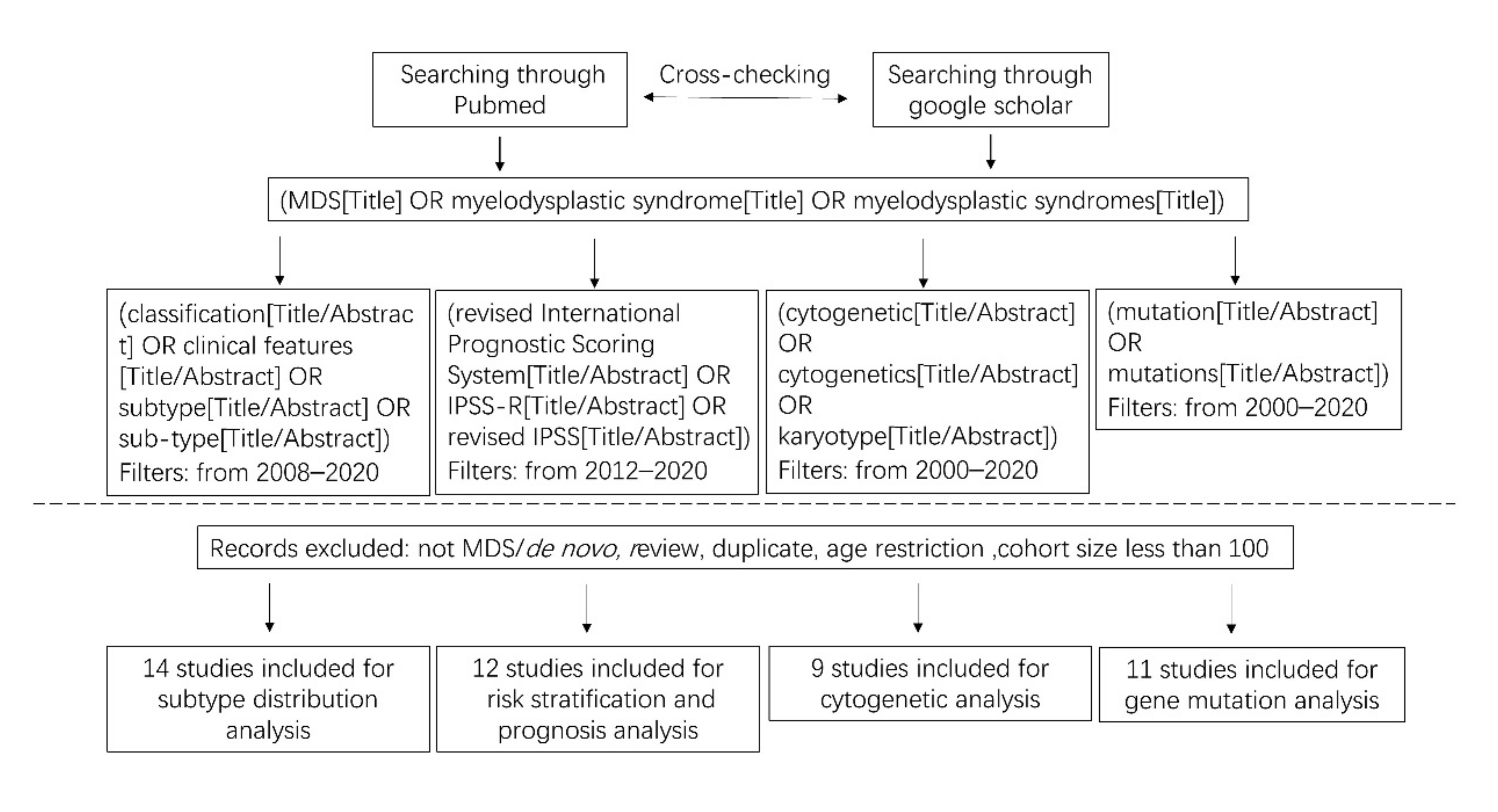
To compare the epidemiological data from Western and Asian countries for the past 20 years, we follow the flow chart presented in Figure 1. A Pubmed search was done on MESH terms and specific criteria (incidence, epidemiology, myelodysplastic syndrome, MDS, publication between 2000–2020). We also included data from the national reference registries of MDS. After exclusion of some articles, we based our study on 10 articles [10,11,12,13,14,15,16,17,18,19] (Figure 1).

2.1.1. Incidence and Age-Adjusted Incidence

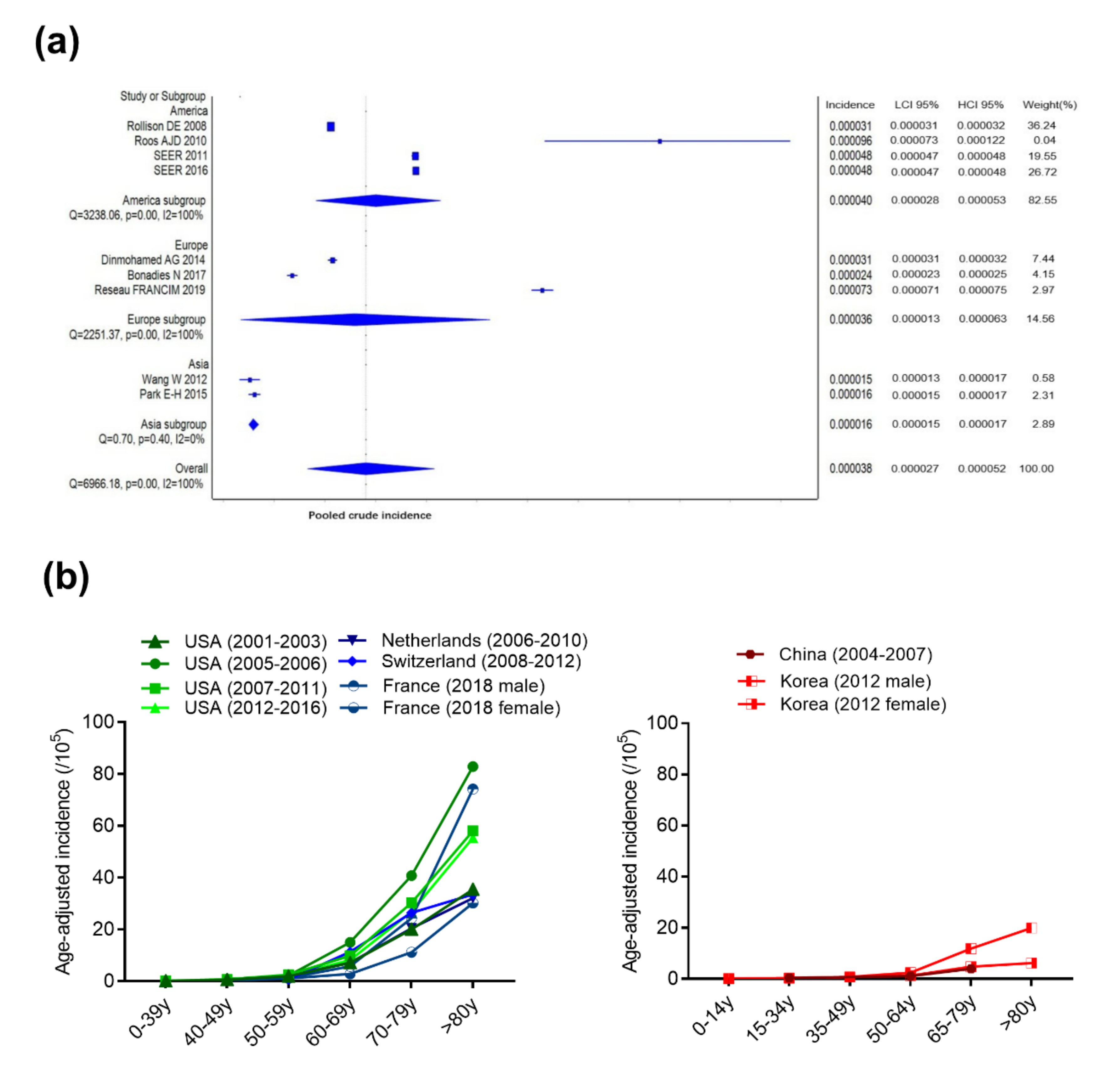
The overall incidence for the Asian subgroup is 1.6 cases per 100,000 inhabitants/year (95% confidence interval (CI): 1.5–1.7) (Figure 2a). On the contrary, the incidence rises to 3.6 cases per 100,000 inhabitants/year in the European subgroup (95% CI: 1.3–6.3) and 4.0 cases per 100,000 inhabitants/year in the American subgroup (95% CI: 2.8–5.3) (Figure 2a). The differences of incidence between Asian, European and American countries are statistically significant (p < 0.0001). More precisely, the raw data on reported age-adjusted incidence ranges from 1.13–1.51 cases per 100,000 inhabitants/year for independent studies in Asian countries to 2.26–7 cases per 100,000 inhabitants/year for independent studies in Western countries (Figure 2b, Table S1).
The diagnostic criteria have evolved over the past two decades; therefore, the incidence reported along different time periods may vary even within the same region of the world. From the same resource, the incidence reported for the United States from 2001 to 2016, the Netherlands from 2001 to 2010, and Switzerland from 2001 to 2012 shows a slight increase, which is mainly attributed to the elderly group [10,13,14,15] (Table S1, Figure 2b).
The incidence of MDS is higher in Western countries compared to Asian ones. For instance, when considering the year 2012, the incidence of MDS in Western countries is still 2- to 4-fold higher than that of Korean patients [13,15,19] (Table S1, Figure 2b).
Innate and acquired causes can explain such a discrepancy of the MDS incidence between Western and Asian populations: different genetic susceptibilities among ethnic groups, geographical and dietary reasons, occupational and environmental stresses among which toxin exposures, such as benzene, insecticide and cigarette smoke [20], virus infection [8], previous therapeutic treatments, such as chemotherapy and radiotherapy, and finally, suboptimal case ascertainment and underreporting [1]. For instance, in the United States, the incidence of MDS within the “White” people population is higher than that of Asian, Pacific Islander, American Indian, and Alaska Native people [10,13].

2.1.2. Age of Onset

MDS occurs mainly in the elderly; the median age at diagnosis is ~76 years old in Europe (Figure 2b, Table S1). The data from French register, 2018 FRANCIM, indicates the median age in France to be 78 years old for men and 80 years old for women [16] (Figure 2b, Table S1). By contrast, the median age in China and Japan is earlier than that of Western countries, ranging from 62–76 years old [17,18]. Punctually, a median age of 57 years old has been once reported in a Chinese study [21], and another Chinese report from 2013 showed that over half of the patients with MDS (61.9%) were younger than 60 year-old at the first diagnosis [22]. By contrast, about 10% of the patients were younger than 50 years of age in a German study from 2011 [23]. The causes explaining such a difference in the age of onset between Asian and Western countries are still unclear. The use of different diagnostic criteria, and genetic and environmental factors may be part of the explanation, in addition to differences in percentage of older population and life expectancy between different countries [18,24]. In particular, genetic factors may have their importance in those differences. This specific point will be investigated in Section 2.4.

2.1.3. Gender Ratio

A clear male predominance in MDS patients exists both in Western and Asian countries; the reported gender ratio is 1.24–2.125. The difference tends to be more significant in patients older than 60 years of age, based on the French and the Korean national cancer registries (Figure 2b, Table S1). In patients older than 80 years old, the incidence of males with MDS is three times higher than that of females [19]. More female patients have been once reported in a Chinese study, but a high percentage of young patients could have been responsible for this result [18]. Interestingly, the female prevalence is more significant in 5q- syndrome presenting the del(5q) as the sole abnormality, but the reason for this female representation is still unknown [25].
Altogether, MDS is less frequent in Asia compared to Western population, but the disease may be more severe as it appears earlier. To go further, we analyzed the distribution of MDS subtypes.

2.2. Asian Population is More Prone to Develop Severe MDS Subtypes

2.2.1. Subtype Distribution

We collected the data according to the 2008 World Health organization (WHO) classification and the International Classification of Diseases for Oncology (ICD-O) [10,13,14,15,18,19,21,24,26,27,28,29,30,31] (Figure 3 and Figure 4a, Tables S2 and S3). Collectively, the sizes of Western and Asian cohorts are respectively 63,394 and 9675 patients (Table S3).
The frequency of MDS with single lineage dysplasia (MDS-SLD) among all MDS subtypes is statistically higher in Asian countries compared to Western countries (18.57% vs. 9.70%, p < 0.0001).
The data show statistically lower percentages of MDS with ring sideroblasts (MDS-RS) (0.97% vs. 7.83%, p < 0.0001) and MDS with isolated del(5q) (MDS-5q-) (1.12% vs. 2.67%, p < 0.0001) in Asian countries compared to Western countries. MDS-RS and MDS with isolated del(5q) mostly occur in old patients and comprise good- and intermediate- cytogenetic risk [32]. The high proportion of these two subtypes may be a reason explaining that older patients are more frequently found in Western countries [15]. However, the 2016 WHO classification revised its criteria from the previous 2008 WHO classification for MDS with isolated del(5q). The revised classification defines MDS with isolated del(5q) even when 1 additional cytogenetic abnormality is also present besides the del(5q), excepting monosomy 7 or del(7q) [33]. Upon the revised criteria, the subtype distribution has changed. As most of the del(5q) aberrations in Asian population are accompanied by other chromosomal abnormalities [29], the percentage of MDS with isolated del(5q) in Asian countries would increase more significantly.
More MDS cases with excess of blasts (MDS-EB) are statistically present in the Asian studies compared to Western ones (41.46% vs. 15.52%, p < 0.0001).
More strikingly, higher percentages of MDS with multilineage dysplasia (MDS-MLD) are reported in Asian countries compared to Western countries (36.56% vs. 7.35%, p < 0.0001).
By contrast, MDS unclassifiable (MDS-U) are rarely reported in the Asian countries compared to European and North American studies (5.62% vs. 55.80%, p < 0.0001). However, it is noteworthy that the data from two European countries, Poland and Sweden, are similar to the Asian results (Figure 4b). The geographical location and past oriental migrations are considered plausible explanations for these similarities [34]. Besides, a study from Mayo clinic evaluated the patients who met WHO criteria for MDS at their institution. In this study, ninety patients (11%) were initially classified as MDS-U, and after pathological review, only half of the cases were confirmed to be MDS-U, while the other half were reclassified to another subtype by follow-up [35]. Making the MDS diagnosis at an early stage of the disease may also be a reason explaining that more MDS-U subtype is reported in European and North American countries compared to Asian ones.
Finally, rare literature data exists in terms of t-MDS subtype. As more patients survive primary malignancies after receiving intensive chemotherapy and/or radiation, the proportion of t-MDS is likely to increase. Still, the difference of t-MDS between Western and Asian countries needs more evidence to be asserted.

2.2.2. Cell Morphology

In the past 30 years, a noticeable discrepancy has existed in various reports due to heterogeneous diagnostic criteria. However, a study from 2005 reported that a significant concordance is achieved when using the same classification [7]. In this study, the morphological diagnosis between Japanese and German hematologists was 98.4% concordant according to FAB classification, and 83.8% concordant with the WHO classification. Interestingly, still from this study and under the same classification, Asian patients compared to the patients in Western countries present a more pronounced cytopenia, especially more severe thrombocytopenia, and a higher frequency of pancytopenia or bicytopenia [7].
Altogether, those results show that Asian MDS patients present more frequently MDS considered as severe phenotype (MDS-EB, MDS-MLD, more pronounced cytopenia, higher frequency of pancytopenia or bicytopenia). To further explore the severity of MDS, we analyzed the prognostic score.

2.3. Asian MDS Patients Fall More Frequently into the Intermediate-, High- and Very High-Risk Pronostic Groups

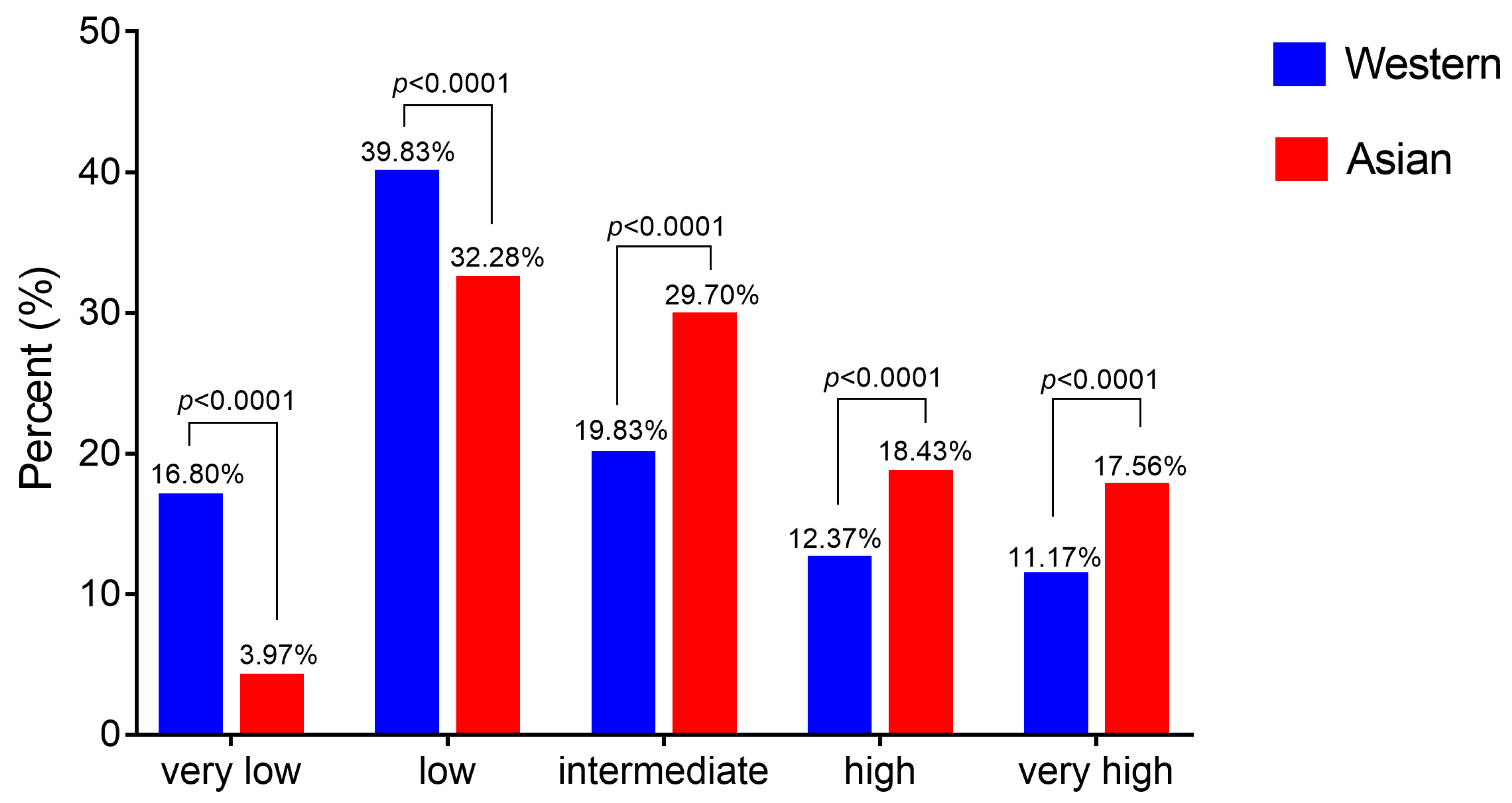
The International Prognostic Scoring System (IPSS), the WHO Prognostic Scoring System (WPSS), and the Revised-IPSS (IPSS-R) are the commonly used risk stratification systems for MDS patients. Among them, IPSS-R is well recommended for assessing prognosis because of its high evaluation effectiveness. It is based on five cytogenetic groups, in addition to the definitions of the depth of cytopenias and of bone marrow blast infiltration. For this reason, we only considered articles based on IPSS-R criteria [26,36,37,38,39]. Previous reports demonstrate that ethnic differences may exist in the prognosis of MDS patients in scoring category [28,40]. Here we collected and compared the prognosis scoring for MDS based on IPSS-R from different world areas [26,29,31,36,39,41,42,43,44,45,46,47] (Figure 3 and Figure 5, Table S4). Collectively, the sizes of Western and Asian cohorts are respectively 16,432 and 1458 patients (Table S4).
The IPSS-R classifies the risk among the following categories: very low, low, intermediate, high and very high risk. The IPSS-R is determined by combining the scores of 5 main features, including cytogenetics, bone marrow blasts, hemoglobin, platelets and absolute neutrophil count; among them, cytogenetics indicates the highest value. From the collected data (Figure 5), it appears that more patients are found in the low- and very low- risk categories in Europe and North America compared to Asian countries, and more patients are distributed in intermediate-, high- and very high- risk groups in Asia.
Altogether, the data show that MDS is rare but at higher-risk in Asian population. We investigated the genetic factors to better understand these ethnic differences.

2.4. Genetic Characteristics Are Concordant with the Prognosis Difference Observed between Asian and Western MDS Patients

2.4.1. Cytogenetics

Since the size of cohorts of recent MDS cytogenetic reports is small, we mainly concentrated our comparison on data collected a decade ago (Figure 3). Data are presented in Figure 6 and Tables S5 and S6 [8,26,29,31,32,48,49,50,51]. Differences in recurrent cytogenetic abnormalities between Western and Asian countries are consistently reported [8,21,28,29,31,48]. As a whole, the rate of detection of abnormal karyotypes is slightly higher in Asian vs. Western MDS populations (53.10% vs. 43.87%, p < 0.0001) (Figure 6a). Karyotypes with 2 abnormalities are again slightly more frequently reported in Asian compared to Western MDS population (10.96% vs. 7.73%, p = 0.0007). However, we do not observe significant differences in the rate of detection of karyotypes with 1 or more than 3 abnormalities in Asian compared to Western MDS populations (Figure 6a).
Cytogenetics associated with very good prognosis: -Y, -11 or del(11q). Chromosomal abnormalities -Y, -11 or del(11q) are associated with very good prognosis. There is no important difference in the rate of Y loss between Asian and Western populations (Figure 6b). On the contrary, the -11 or del(11q) abnormalities are significantly more observed in the Asian population compared to Western MDS patients (2.45%, vs. 1.09% p = 0.0001).
Cytogenetics associated with good prognosis: del(5q), del(12p), -20 or del(20q). Data show that the del(5q) chromosomal aberration is twice less frequently reported in the Asian MDS patients compared to the Western MDS patients (4.63% vs. 8.81%, p < 0.0001) (Figure 6c). In particular, MDS with isolated del(5q) is less frequent in Asian countries compared to Western countries (Tables S5 and S6). MDS with isolated del(5q) is considered to have a good prognosis, with a low probability of progression to secondary acute myeloid leukemia (AML) and longer life expectancy. The median survival of the Western patients with MDS with isolated del(5q) is 80 months, but decreases dramatically when additional karyotype abnormalities are present [32]. Unexpectedly, the Chinese median survival is 62 months in patients with isolated del(5q) and 78 months in patients with del(5q) plus one additional cytogenetic abnormality except monosomy 7 or del (7q) [52]. The rate of detection of del(12p) is slightly higher in Asian compared to Western MDS population (2.10% vs. 1.09%, p = 0.0035) (Figure 6c). Importantly, the del(20q) abnormality is more than twice frequently observed in the Asian patients (7.41% vs. 2.79%, p < 0.0001) (Figure 6c). MDS with isolated del(20q) show lower platelet counts, lower marrow blast counts, higher reticulocyte counts, and is associated with a favorable prognosis with a median survival of 54–71 months, and 12–15 months for del(20q) with additional abnormalities [32,53,54]. The AML progression rate is 14% for patients with isolated del(20q), 11% with one and 24% with several additional abnormalities in the Western MDS population [53]. However, in a Japanese study [55], the median survival of MDS patients with sole del(20q) is 80 months and 23 months when del(20q) is not the sole anomaly. Furthermore, del(20q) abnormality develops as a minor clone in the late stages of MDS indicating a clonal evolution toward leukemia, and poor prognosis. Accumulated genetic damage and genetic instability are thought to be the reasons [55].
Cytogenetics associated with intermediate prognosis: trisomy 8, i(17q), del(17p) or -17. The average rate of trisomy 8 in the Asian MDS population is 14.43%, whereas the average rate of trisomy 8 in the Western MDS population is down to 6.30%. This difference is significant (p < 0.001) but with a range of values in the Asian dataset ranging from 3.8% to 20% (Figure 6d). Trisomy 8 is observed in all the MDS subtypes, and isolated trisomy 8 is included in the IPSS-R intermediate cytogenetic risk group [41]. According to Western reports, the median survival of isolated +8 is 22–34.3 months, but increases to 40–44 months with 1 additional abnormality, and decreases to 23.8 months with 2 additional abnormalities, and to 5.8 months with three or more aberrations [32,56]. However, a Chinese report also indicated an association of isolated trisomy 8 with a much better prognosis, and a median survival of 44 months [29], which was higher than the Western results. A previous study disclosed that a high frequency of myeloproliferative features either at diagnosis or during evolution in MDS with isolated +8, respond poorly to hypomethylating agents (HMAs) [57]. Moreover, patients with isolated trisomy 8 present a high risk of progression to AML [26]. However immunosuppressive therapy response rate of MDS with +8 was significant [58]. The discrepancy in isolated trisomy 8 between Western and Asian countries should be verified in larger cohorts. Finally, no important differences is observed for the rate of i(17q), del(17p) or -17 between Western and Asian MDS populations (Figure 6d). They are rare and range from 0 to 3% in the different studies.
Cytogenetics associated with poor prognosis: -7, del(7q), inv(3), t(3q) or del(3q). We have also compared the distribution of -7 or del(7q) abnormalities, which correlate to a poor prognosis, and no significant differences were found between the Asian and the Western MDS populations (Figure 6e). Abnormalities on chromosome 3 including inv(3), t(3q) or del(3q) are also consistently reported in Western and Asian populations with a rate under 3%.
Cytogenetics associated with very poor prognosis. The distribution of the complex karyotype (>3 chromosomal abnormalities), which correlates to a very poor prognosis, does not show a significant difference between the Asian and the Western MDS populations (Figure 6a, enlarged in Figure 6f). The average percentage of detection of complex karyotypes is about 15% both in Asian and Western MDS populations. Additional publications showed that other abnormal karyotypes including +1/+1q, -1/del(1q), der(1;7), -9/del(9q) and del(16q) are significantly increased in Japanese compared to Caucasian populations [31], and a good prognostic impact is identified in +1/+1q, der(1;7),and del(9q) abnormalities [32,59]. The cytogenetic abnormalities play an important role on prognosis; the IPSS-R cytogenetic categories have discounted some rare karyotypes with independent prognostic values. Due to the profound cytogenetic heterogeneity of MDS, further investigation of the prognostic significances of many rare abnormalities is warranted.
Altogether, combining with cytogenetic data, we propose that the over-representation of MDS associated to del(5q) may account for the higher proportion of the low risk group in Western countries. Similarly, the higher proportion of trisomy 8 in Asian countries may explain the higher representation of patients in the intermediate risk category.
Considering age of onset, the median age of patients with trisomy 8 was younger than that of patients with del(5q) (43 vs. 59 years old). Patients with sole trisomy 8 were also younger than that with sole del(5q) (median age of 43 vs. 63 year-old) [29]. Therefore, the higher proportion of trisomy 8 observed in the Asian countries and the higher proportion of del(5q) observed in the Western countries are one of the plausible genetic factors explaining the discrepancy of almost two decades for the age of MDS onset in those countries.

2.4.2. Molecular Genetics

Gene mutations are thought to be acquired and positively selected to allow the expansion of the initiating clone to compromise normal hematopoiesis, and in due course give rise to MDS and subsequent transformation to AML [60]. Acquired mutations in genes involved in epigenetic regulation and chromatin remodeling (TET2, DNMT3A, ASXL1, IDH1/2, EZH2), pre-mRNA splicing (SF3B1, SRSF2, U2AF1), transcription (TP53, RUNX1, ETV6) and signaling transduction (KRAS/NRAS) are recurrently seen in most MDS [61,62,63,64], and some somatic mutations in certain genes can predict patient outcomes [65]. Different mutation topographies have been reported in hematological neoplasms between Asian and Western countries [66]; herein we analyzed the frequency of the most common 12 mutated genes in different countries [30,62,63,67,68,69,70,71,72,73,74] (Figure 3 and Figure 7, Table S7). Western and Asian mutations data include cohorts of respectively up to 2920 and 1790 patients (Table S7).
Distribution of the gene mutations. The gene mutation rate differs significantly among different studies, ranging from 51.5 to 89.5% in Western countries and from 55 to 91.4% in Asian countries. Genes with a mutation frequency greater than 10% are SF3B1, TET2, ASXL1, SRSF2, DNMT3A and RUNX1 in Western countries, and ASXL1, RUNX1, U2AF1 and TP53 in Asia. The genes IDH1/2, KRAS/NRAS, EZH2 and ETV6 are mutated for less than 10% in MDS population, both in Asian and Western countries (Figure 7, Table S7).
In particular, SF3B1, TET2, SRSF2 and IDH1/2 are reported mutated three-time less in Asia compared to the Western MDS population (SF3B1: 7.17% vs. 25.19%; SRSF2: 6.15% vs. 16.15%; TET2: 8.86% vs. 23.35%; IDH1/2: 3.25% vs. 7.09%; p < 0.0001 for each gene). The ASXL1 and DNMT3A genes are slightly less mutated in Asia compared to the Western MDS population (ASXL1: 15.12% vs. 17.90%, p < 0.05; DNMT3A: 8.95% vs. 11.55%, p < 0.05). On the contrary, U2AF1 and ETV6 are reported mutated twice as often among Asians (U2AF1: 13.25% vs. 7.76%, p < 0.0001; ETV6: 5.02% vs. 1.70%, p < 0.0001). Finally, the RUNX1, TP53, KRAS/NRAS and EZH2 genes present equivalent mutation rates (RUNX1: ~12%; TP53: ~10%; KRAS/NRAS: ~7%; EZH2: ~5%, in Asian and Western populations respectively).
Prognosis associated to gene mutation. No systematic studies have been conducted to compare the prognosis of these mutations between the two parts of the world. TET2 and SF3B1 mutations are independent favorable prognostic factors [75,76,77]. On the contrary, SRSF2 [78,79,80], ASXL1 [81], RUNX1 [82,83], DNMT3A [84,85], U2AF1 [86], TP53 [87,88,89,90], EZH2 [91,92], IDH1/2 [93,94,95,96], KRAS/NRAS [97,98] and ETV6 [99] mutations are all associated with poorer clinical outcomes.
Gene mutations are related to the therapeutic response. TET2 and DNMT3A mutations predict a higher response rate to hypomethylating agents in MDS patients [100,101,102,103]. RUNX1 and TP53 mutations are associated with significantly lower responses to hypomethylating treatment, and TP53 mutations show a high risk of relapse after allogeneic hematopoietic cell transplantation [81,87,88,89,90]. A Korean study described that U2AF1 mutation does not affect the response rate to first-line decitabine treatment [104]. The presence of an SF3B1 mutation adversely influences response to immunosuppressive therapy [105]. TP53 mutations prove to have a negative impact on sensitivity to lenalidomide in MDS with isolated del(5q) [106].
However, new target therapies may improve the clinical prognosis of these gene mutations. For example, spliceosome modulators may be a therapeutic window of MDS with SF3B1 mutations [107]. In this case, Western population may be more concerned by this therapy than Asian population because SF3B1 is more frequently mutated in Western MDS patients rather that in Asia. Small molecule inhibitor APR-246 seems to improve the prognosis of TP53 mutation MDS both as a single agent as well as in combination with AZA [6,108]. Ivosidenib and enasidenib may be an effective option for IDH1 and IDH2 mutations [5,109]. Less MDS patients are affected by IDH1 and IDH2 mutations in Asia than in Western countries. Pre-mRNA splicing modulators like sudemycins demonstrate a potential effect for treating hematological cancers harboring U2AF1 mutations [110].
The relationships between gene mutations and gender, age and cytogenetics are also reported. An Asian study demonstrated that SRSF2 mutation is closely associated with male sex and older age, and the prognostic impact of SRSF2 mutation might be attributed to its close association with old age [80]. TP53 mutations in MDS are strongly correlated with childhood and therapy-related MDS [87,111,112]. U2AF1 mutation is more prevalent in younger MDS patients [86], and the U2AF1 mutation is strongly associated with isolated trisomy 8 and del(20q) in Asian people, but not in Caucasian people, and is characterized by a younger age of MDS onset (median 39 year-old) [113].
MDS is typically present in older adults with the acquisition of age-related somatic mutations, whereas MDS present in children and younger adults is more frequently associated with germline genetic predisposition [114]. Considering the significant difference of onset age between Western and Asian populations, Yu et al. compared the difference of gene mutation between younger groups (16~59 years) and older groups (60~87 years) of MDS patients; they reported a trend towards higher incidence of gene mutations in the older group [115].
Altogether, cytogenetic aberrations appear to be more frequent in some ethnic groups, as well as some gene mutations. The reasons explaining those observations are not clear. The genetic differences observed between Western and Asian populations are relatively consistent with the distribution of MDS subtypes and the age of MDS onset. A higher frequency of SF3B1 mutation is concordantly associated with a higher proportion of MDS with ring sideroblasts (MDS-RS) in Western countries. TET2 is an age-associated mutation found in normal elderly individuals with acquired clonal hematopoiesis [116,117]. TET2 inactivation results in clonal hematopoiesis of indeterminate potential (CHIP) and favors transformation to myeloproliferative and myelodysplastic neoplasms (MPN, MDS) and AML [117]. The fact that MDS appears in older people in Western rather that in Asian population may explain that the rate of TET2 mutation is higher in Western MDS population. Moreover, MDS, like other cancers, is considered to emerge after successive positive selections, where gene mutations and genetic alterations are central players [60]. We propose that specific ethnic variants or combination of variants will differently favor a positive selection of the initiating clone in a permissive microenvironment with specific sets of mutations.
Importantly, Asian MDS patients show more frequently high-risk cytogenetic aberrations. Therefore, we next question the survival of patients.

2.5. Survival Rates Are Equivalent for Asian and Western MDS Patients and Strictly Correlated to the Prognostic Groups

2.5.1. Therapeutic Options

A variety of risk-adapted treatment strategies are adopted by clinicians due to the heterogeneity of MDS. The MDS International Working Group (IWG) puts forward the response criteria to treatment in 2000 and further revised it in 2006 to make the results of different clinical treatments comparable. Allogeneic hematopoietic stem cell transplantation (HSCT) is currently the only potentially curative option. Over the past two decades, azacitidine, lenalidomide, decitabine, deferasirox, epoetin alpha and luspatercept have gained market authorization from the US Food and Drug Administration (FDA) and the European Medicines Agency (EMA) for MDS. Among them, azacitidine, lenalidomide and decitabine are considered to modify the disease course. Epoetin alpha and luspatercept improve cytopenia. Deferasirox, a major oral iron chelator, reduces chronic iron overload in patients who are receiving long-term blood transfusions. The exact impact of transfusional systemic iron overload in MDS to AML transformation is still under question [118]. However, as far as we know, there is currently no systematic study comparing the deferasirox treatment response rates between Asian and Western countries. Treatment approach differences still exist in Western and Asian countries due to the discrepancy of MDS onset age, cytogenetics and prognostic groups.
The therapeutic response rate of lenalidomide is higher in patients with del(5q) (83%) compared to patients with normal karyotypes (57%) and other chromosomal abnormalities (12%). In addition, in intermediate-2 or high-risk patients with isolated del(5q), lenalidomide was proved to have a much better complete response rate than patients with one more additional cytogenetic abnormality [3]. Lenalidomide has been recommended by the National Comprehensive Cancer Network (NCCN) in the United States as the first-choice treatment for patients with del(5q) chromosomal abnormalities alone or with an additional cytogenetic abnormality, except those involving chromosome 7. Del(5q) is less frequently detected in the Asian population, and few studies have been conducted on lenalidomide treatment on MDS del(5q) in this population. Clinical trials implicating small size cohorts from China and Japan showed that the therapeutic response rate for 5q- syndrome is comparable to that in Europe and the USA [52,119]. However, the MDS patients with del(5q) in Asian countries are mostly accompanied by complex abnormalities [29].
Hypomethylating therapy using 5-azacitidine and 5-aza-2-deoxycytidine (decitabine) can be proposed to patients with MDS in higher-risk groups. Compared with supportive treatment groups, it can reduce the risk of patients progressing to AML and can improve survival. A Chinese report showed that azacitidine emerges as an effective and safe treatment strategy for Chinese patients with high-risk MDS, and therapeutic response rates are comparable to those from the Western patients in the phase 3 AZA-001 study [120]. A prospective study (DIVA) demonstrated that the effectiveness of hypomethylating agents in Korean patients is similar to those of the American multi-centre, phase II, clinical trial (ADOPT) [121]. Another study enrolling 135 Chinese cases with a median age of 54.1 years, all having de novo MDS and the majority of intermediate-1 or intermediate-2 IPSS, showed the complete response rate to be higher (66.7%) compared to that in the ADOPT study (52%) [122]. However, the complete response rate of a Japanese study was lower (32.4%) [123]. The difference in the baseline clinical characteristics of patients and number of treatment cycles may result in the disparity. An international, multi-centre, prospective clinical trial may help explain these differences.
As the only curative strategy, HSCT is recommended for younger MDS patients in higher risk groups and lower risk groups with poor prognosis genetic abnormalities. Despite its curative potential, the treatment-related mortality and the risk of relapse, age and comorbidities turn out to be a serious obstacle for elderly patients [124]. Albeit reduced-intensity conditioning transplantation offers overall and quality-adjusted survival benefit for patients with de novo MDS aged 60 to 70 years old in intermediate-2/high IPSS [125], post-HCT relapse still should be taken into account [126]. MDS patients being usually elderly with a median age of approximately 75 years at diagnosis in Western countries, compared to 60 years of age in Asian countries; transplant accessibility and the intensity of transplant conditioning are doomed to be different.

2.5.2. Survival Rate

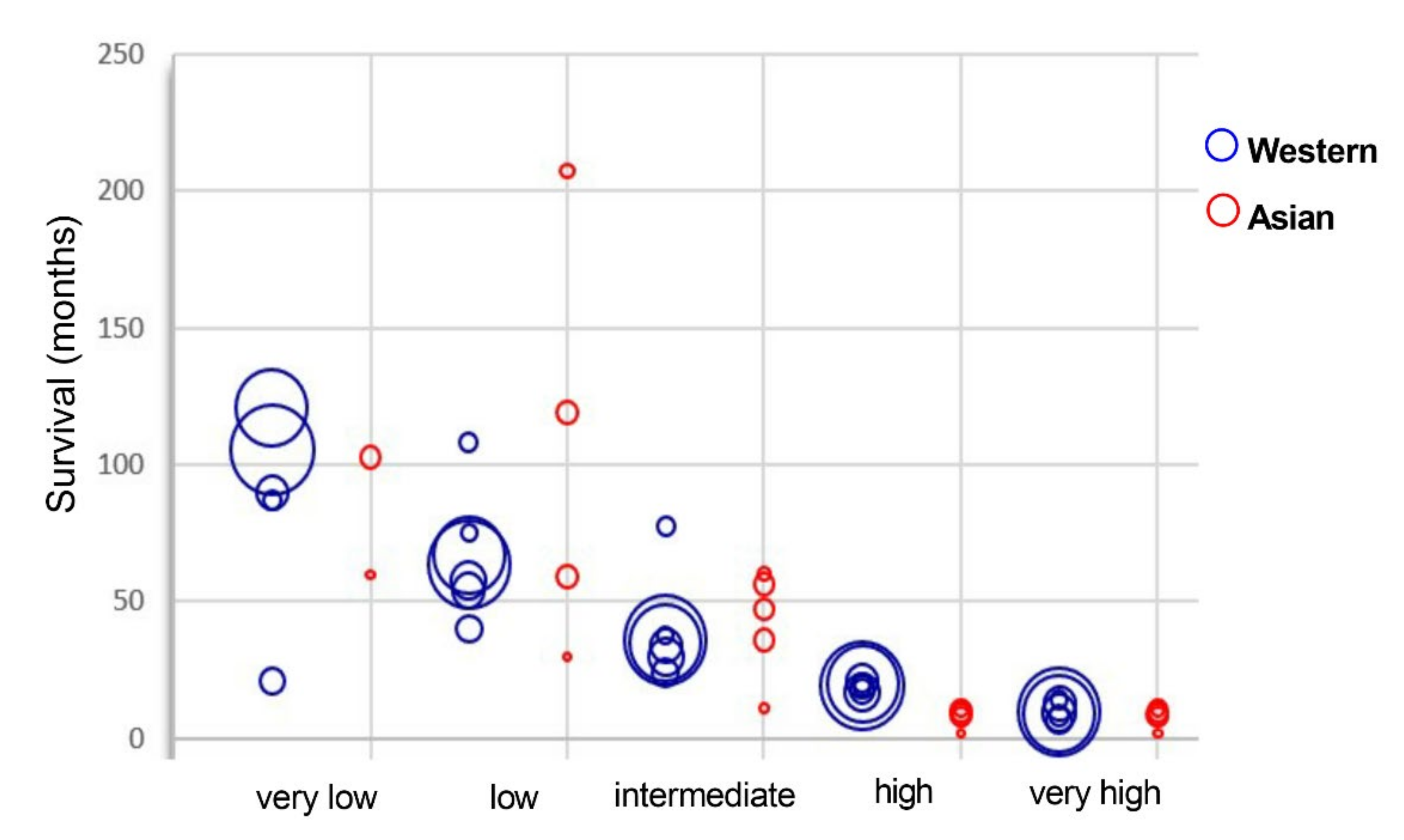
We collected and compared the survival data from different world areas [26,29,31,36,39,41,42,43,44,45,46,47] (Figure 3 and Figure 8, Table S8).
The survival rate varies significantly among the studies, and we could not draw a clear difference between Asian and Western countries (Figure 8). However, a trend appears from regional reports. The prognostic discrimination of IPSS-R for survival is shown in most risk groups, but for the very low and low risk categories, some studies show an even longer survival for the low risk group than the very low risk group. Additional significant factors for predicting survival, including performance status, serum ferritin levels, lactate dehydrogenase levels, chronic comorbidity conditions, and mutations are thought to be the causes of this unexpected result [26,44,45,46]. Among them, gene mutations such as TP53 are important factors explaining poor prognosis in patients at low risk category [26,127]. Furthermore, geriatric and male gender are also associated with reduced overall survival and more advanced leukemia [41,42,48,128].
Akira Matsuda et al. compared the overall survival (OS) of Japanese patients with refractory anemia in French-American-British (FAB) classification to that of German patients. Japanese patients show significantly more favorable prognosis for patients aged 60 years or younger. For patients older than 60 years, no favorable OS is shown [7]. In another study comparing clinical features of Japanese and Caucasian MDS patients, a striking difference is found in OS but not in time to AML transformation. The significantly improved survival in Japanese populations holds even after considering age, FAB and IPSS-R categories. The difference in OS mainly comes from lower-risk IPSS-R categories, including very low, low, and intermediate risk groups [31]. About AML transformation rate, the way of presenting this rate varies a lot from one study to another, so we could not get enough comparable data to perform a relevant comparison of the AML transformation rate between the Western and Asian populations. One study presenting the AML transformation rate between the Japanese and Caucasian, did not identify any significant difference [31].

3. Materials and Methods

Selection of studies. We performed a literature search in PubMed and Google Scholar over the past two decades, with no restriction to language of publication. MESH terms and exclusion criteria are indicated in Figure 1 and Figure 3. For incidence and epidemiology data, we also considered the national cancer registries of reference with available published results. We finally considered 10 articles (Figure 1). For classification data, and due to successive modifications of diagnosis criteria [1], the studies based on World Health Organization (WHO) 2008 classification system were included, therefore restricted to 2008–2020. Additionally, we manually checked all the articles and excluded the studies based on FAB classification (Figure 3). For prognostic studies, we collected data under the Revised International Prognostic Scoring System (IPSS-R) so restricted to 2012–2020. In total, we retrieved 14 studies for the subtype distribution, 12 for the risk stratification, 9 for cytogenetics, and 11 for gene mutations (Figure 3).
Statistical analysis.Incidence. We calculated the weighted crude incidence for each study. Pooled incidence estimates were calculated with MetaXL version 5.3 software (Epigear International) using a quality-effects model by area. The quality scores of all the studies included into the analysis were more than 8 reflecting the very high quality of each study. Heterogeneity between studies was assessed with a X2 test (Cochran Q statistic) and quantified with the I2 statistic. Differences of incidence between the Western and Asian datasets were calculated using t-tests. Classifications, cytogenetics, subtype distribution and prognosis. We performed weighted analyses. The differences of count data between the Western and Asian datasets were calculated using contingency tables and X2 tests (GraphPad Prism version 7.0). A p-value less than 0.05 is considered statistically significant.
Geographic criteria. We refer to Asian population for China, Korea, and Japan. Western countries are more representative of Caucasian population. They consist of USA, Canada, Argentina (or America) and Netherland, Switzerland, France, Italy, Germany, Greece, Austria, Sweden, Poland (or Europe), and Australia because of the high representation of inhabitants from European origin in this country.

4. Conclusions

MDS is a group of very heterogeneous syndromes. Here, we show that both differences and similarities exist in epidemiology, clinical features, genetics, and prognosis between Western and Asian countries (Graphical Abstract). In the era of precision medicine, those ethnic specificities should be taken into account for drug development.
Compared to the Western MDS population, Asian MDS patients show a 2- to 4-fold lower incidence and up to 10 years of earlier onset age. Analyzing the causes of this early onset in Asia will be of great interest and may be beneficial to understand the pathogenesis of younger Western MDS patients. The subtypes MDS-RS, MDS-del(5q) and MDS-U are more represented in Western countries whereas MDS-SLD, MDS-MLD and MDS-EB are more represented in Asian countries.
Genetic characteristics present subtle differences between Western and Asian MDS population. As for the cytogenetics abnormalities, the del(5q) is more frequently found in Western compared to Asian MDS population, whilst trisomy 8 and del(20q) are more frequently identified in Asian compared to the Western MDS population. The reasons of such cytogenetic differences remain unknown but this genetic specificity may contribute to explain the severity of MDS in Asian population. The rate of mutation is found half that of the Western for four genes, TET2, SF3B1, SRSF2 and IDH1/2 in the Asian MDS population, while, U2AF1 and ETV6 are more frequently reported in Asian MDS patients. Mutation rates are equally identified among the other major reported mutated genes of MDS, ASXL1, RUNX1, DNMT3A, TP53 (with a frequency of more than or close to 10% each), and in EZH2 and KRAS/NRAS (with a mutation frequency of about 5%).
Meanwhile, very low- and low-risk groups of IPSS-R are reported in Western MDS population whereas the intermediate, high and very high-risk groups are more represented in Asian MDS population. The above differences lead to disparities in therapeutic effects of immunomodulation, immunosuppression, hypomethylating reagents, as well as accessibility and conditioning of hematopoietic stem cell transplantation. Finally, we did not show substantial differences for the survival rate within each prognostic group for MDS patients between Western and Asian countries.
Altogether, systematic research to analyze and compare the molecular, genetic, and environmental differences among different regions of the world may open a window for a better understanding of MDS pathogenesis. This may lead to the design of personalized tailored treatments adapted to the diversity of the world populations.

Supplementary Materials

The following are available online at https://www.mdpi.com/2072-6694/13/3/481/s1, Table S1. Studies comparing the epidemiological difference between Western an Asian MDS patients. Table S2. Studies reporting the percentage of MDS subtypes according to WHO 2008 and ICD-O classification. Table S3. Raw data and statistical analysis of MDS subtypes according to different areas in the world. Table S4. Raw data and statistical analysis of MDS IPSS-R distribution according to different areas in the world. Table S5. Studies reporting cytogenetic characteristics in MDS. Table S6. Raw data and statistical analysis of recurrent cytogenetic abnormalities in MDS according to different areas in the world. The tables are separately presented according to the cytogenetic score. Table S7. Raw data and statistical analysis of recurrently mutated genes in MDS according to different areas in the world. Table S8. Raw data of MDS median overall survival in month and distribution among each IPSS-R group.

Author Contributions

Conceptualization, Y.J. and M.-B.T.; methodology, Y.J., J.-M.C. and M.-B.T.; validation, Y.J., J.-R.E., M.-A.C., B.S., J.-M.C., S.G., A.B., F.M., N.D.-G. and M.-B.T.; formal analysis, Y.J.; writing—original draft preparation, Y.J. and M.-B.T.; writing—review and editing, Y.J., J.-R.E., M.-A.C., B.S., J.-M.C., S.G., A.B., F.M., N.D.-G. and M.-B.T.; supervision, M.-B.T.; funding acquisition, M.-B.T. All authors have read and agreed to the published version of the manuscript.

Funding

This research was funded by The Ligue Régionale contre le Cancer (comité 22, 29, 35, 41, 56, 79), the Association Gaetan Saleun, the Association Connaître et Combattre les Myélodysplasies, and the Association Halte au Cancer. The APC was funded by The Ligue Régionale contre le Cancer.

Institutional Review Board Statement

Ethical review and approval were waived for this study, because ethical approval was already granted in each study on which we based our work.

Informed Consent Statement

Patient consent was waived because patient consent was already granted in each study on which we based our work.

Data Availability Statement

Publicly available datasets were analyzed in this study. This data can be found in each publication that we have referenced.

Conflicts of Interest

The authors declare no conflict of interest.

References

  1. Zeidan, A.M.; Shallis, R.M.; Wang, R.; Davidoff, A.; Ma, X. Epidemiology of Myelodysplastic Syndromes: Why Characterizing the Beast Is a Prerequisite to Taming It. Blood Rev. 2019, 34, 1–15. [Google Scholar] [CrossRef]
  2. List, A.; Ebert, B.L.; Fenaux, P. A Decade of Progress in Myelodysplastic Syndrome with Chromosome 5q Deletion. Leukemia 2018, 32, 1493–1499. [Google Scholar] [CrossRef]
  3. Adès, L.; Boehrer, S.; Prebet, T.; Beyne-Rauzy, O.; Legros, L.; Ravoet, C.; Dreyfus, F.; Stamatoullas, A.; Chaury, M.P.; Delaunay, J.; et al. Efficacy and Safety of Lenalidomide in Intermediate-2 or High-Risk Myelodysplastic Syndromes with 5q Deletion: Results of a Phase 2 Study. Blood 2009, 113, 3947–3952. [Google Scholar] [CrossRef] [Green Version]
  4. Magalhães, S.M.M.; Velloso, E.D.R.P.; Buzzini, R.; Bernardo, W.M. Part 4: Myelodysplastic Syndromes—Treatment of Low-Risk Patients with the 5q Deletion. Hematol. Transfus. Cell Ther. 2018, 40, 274–277. [Google Scholar] [CrossRef]
  5. Richard-Carpentier, G.; DeZern, A.E.; Takahashi, K.; Konopleva, M.Y.; Loghavi, S.; Masarova, L.; Alvarado, Y.; Ravandi, F.; Montalban Bravo, G.; Naqvi, K.; et al. Preliminary Results from the Phase II Study of the IDH2-Inhibitor Enasidenib in Patients with High-Risk IDH2-Mutated Myelodysplastic Syndromes (MDS). Blood 2019, 134, 678. [Google Scholar] [CrossRef]
  6. Sallman, D.A.; DeZern, A.E.; Steensma, D.P.; Sweet, K.L.; Cluzeau, T.; Sekeres, M.A.; Garcia-Manero, G.; Roboz, G.J.; McLemore, A.F.; McGraw, K.L.; et al. Phase 1b/2 Combination Study of APR-246 and Azacitidine (AZA) in Patients with TP53 Mutant Myelodysplastic Syndromes (MDS) and Acute Myeloid Leukemia (AML). Blood 2018, 132, 3091. [Google Scholar] [CrossRef]
  7. Matsuda, A.; Germing, U.; Jinnai, I.; Misumi, M.; Kuendgen, A.; Knipp, S.; Aivado, M.; Iwanaga, M.; Miyazaki, Y.; Tsushima, H.; et al. Difference in Clinical Features between Japanese and German Patients with Refractory Anemia in Myelodysplastic Syndromes. Blood 2005, 106, 2633–2640. [Google Scholar] [CrossRef]
  8. Chen, B.; Zhao, W.-L.; Jin, J.; Xue, Y.-Q.; Cheng, X.; Chen, X.-T.; Cui, J.; Chen, Z.-M.; Cao, Q.; Yang, G.; et al. Clinical and Cytogenetic Features of 508 Chinese Patients with Myelodysplastic Syndrome and Comparison with Those in Western Countries. Leukemia 2005, 19, 767–775. [Google Scholar] [CrossRef] [Green Version]
  9. Lee, J.-H.; Lee, J.-H.; Shin, Y.-R.; Lee, J.-S.; Kim, W.-K.; Chi, H.-S.; Park, C.-J.; Seo, E.-J.; Lee, K.-H. Application of Different Prognostic Scoring Systems and Comparison of the FAB and WHO Classifications in Korean Patients with Myelodysplastic Syndrome. Leukemia 2003, 17, 305–313. [Google Scholar] [CrossRef] [Green Version]
  10. Rollison, D.E.; Howlader, N.; Smith, M.T.; Strom, S.S.; Merritt, W.D.; Ries, L.A.; Edwards, B.K.; List, A.F. Epidemiology of Myelodysplastic Syndromes and Chronic Myeloproliferative Disorders in the United States, 2001-2004, Using Data from the NAACCR and SEER Programs. Blood 2008, 112, 45–52. [Google Scholar] [CrossRef]
  11. Roos, A.J.D.; Deeg, H.J.; Onstad, L.; Kopecky, K.J.; Bowles, E.J.A.; Yong, M.; Fryzek, J.; Davis, S. Incidence of Myelodysplastic Syndromes within a Nonprofit Healthcare System in Western Washington State, 2005–2006. Am. J. Hematol. 2010, 85, 765–770. [Google Scholar] [CrossRef]
  12. Sect_30_mds.Pdf. Available online: https://seer.cancer.gov/archive/csr/1975_2011/results_merged/sect_30_mds.pdf (accessed on 27 July 2020).
  13. Sect_30_mds.Pdf. Available online: https://seer.cancer.gov/csr/1975_2016/results_merged/sect_30_mds.pdf#search=mds%20incidence (accessed on 9 December 2019).
  14. Dinmohamed, A.G.; Visser, O.; van Norden, Y.; Huijgens, P.C.; Sonneveld, P.; van de Loosdrecht, A.A.; Jongen-Lavrencic, M. Trends in Incidence, Initial Treatment and Survival of Myelodysplastic Syndromes: A Population-Based Study of 5144 Patients Diagnosed in the Netherlands from 2001 to 2010. Eur. J. Cancer 2014, 50, 1004–1012. [Google Scholar] [CrossRef]
  15. Bonadies, N.; Feller, A.; Rovo, A.; Ruefer, A.; Blum, S.; Gerber, B.; Stuessi, G.; Benz, R.; Cantoni, N.; Holbro, A.; et al. Trends of Classification, Incidence, Mortality, and Survival of MDS Patients in Switzerland between 2001 and 2012. Cancer Epidemiol. 2017, 46, 85–92. [Google Scholar] [CrossRef]
  16. Source: Réseau FRANCIM. Available online: https://lesdonnees.e-cancer.fr/Informations/Sources/SOURCE-Reseau-FRANCIM (accessed on 9 December 2019).
  17. Incidence of Myelodysplastic Syndrome in Japan. J. Epidemiol. 2014, 24, 469–473. [CrossRef] [Green Version]
  18. Wang, W.; Wang, H.; Wang, X.-Q.; Lin, G.-W. First Report of Incidence of Adult Myelodysplastic Syndrome in China. Ann. Hematol. 2012, 91, 1321–1322. [Google Scholar] [CrossRef]
  19. Park, E.-H.; Lee, H.; Won, Y.-J.; Ju, H.Y.; Oh, C.-M.; Ingabire, C.; Kong, H.-J.; Park, B.-K.; Yoon, J.Y.; Eom, H.-S.; et al. Nationwide Statistical Analysis of Myeloid Malignancies in Korea: Incidence and Survival Rate from 1999 to 2012. Blood Res. 2015, 50, 204–217. [Google Scholar] [CrossRef] [Green Version]
  20. Bowen, D.T. Occupational and Environmental Etiology of MDS. Best Pract. Res. Clin. Haematol. 2013, 26, 319–326. [Google Scholar] [CrossRef]
  21. Zhang, T.T.; Sun, A.N.; Pan, J.L.; Wu, D.P.; Qiu, H.Y.; Tang, X.W.; Miao, M.; Chen, S.N. The clinical features, cytogenetic characteristics and survival analysis of 550 myelodysplastic syndromes in a single center. Zhonghua Xue Ye Xue Za Zhi 2016, 37, 864–869. [Google Scholar] [CrossRef]
  22. Li, X.; Xiao, Z.; Chang, C.; Xu, F.; Wu, L.; He, Q.; Xu, Z.; Song, L.; Zhang, Z.; Zhou, L.; et al. Distinct Clinical and Experimental Characteristics in the Patients Younger than 60 Years Old with Myelodysplastic Syndromes. PLoS ONE 2013, 8, e57392. [Google Scholar] [CrossRef]
  23. Neukirchen, J.; Schoonen, W.M.; Strupp, C.; Gattermann, N.; Aul, C.; Haas, R.; Germing, U. Incidence and Prevalence of Myelodysplastic Syndromes: Data from the Düsseldorf MDS-Registry. Leuk. Res. 2011, 35, 1591–1596. [Google Scholar] [CrossRef]
  24. Mądry, K.; Machowicz, R.; Waszczuk-Gajda, A.; Drozd-Sokołowska, J.; Hołowiecka, B.S.; Wiater, E.; Mital, A.; Obara, A.; Szmigielska-Kapłon, A.; Kołkowska-Leśniak, A.; et al. Demographic, Hematologic, and Clinical Features of Myelodysplastic Syndrome Patients: Results from the First Polish Myelodysplastic Syndrome Registry. AHA 2015, 134, 125–134. [Google Scholar] [CrossRef]
  25. Van den Berghe, H.; Michaux, L. 5q-, Twenty-Five Years Later: A Synopsis. Cancer Genet. Cytogenet. 1997, 94, 1–7. [Google Scholar] [CrossRef]
  26. Berggren, D.M.; Folkvaljon, Y.; Engvall, M.; Sundberg, J.; Lambe, M.; Antunovic, P.; Garelius, H.; Lorenz, F.; Nilsson, L.; Rasmussen, B.; et al. Prognostic Scoring Systems for Myelodysplastic Syndromes (MDS) in a Population-Based Setting: A Report from the Swedish MDS Register. Br. J. Haematol. 2018, 181, 614–627. [Google Scholar] [CrossRef] [PubMed] [Green Version]
  27. McQuilten, Z.K.; Wood, E.M.; Polizzotto, M.N.; Campbell, L.J.; Wall, M.; Curtis, D.J.; Farrugia, H.; McNeil, J.J.; Sundararajan, V. Underestimation of Myelodysplastic Syndrome Incidence by Cancer Registries: Results from a Population-Based Data Linkage Study. Cancer 2014, 120, 1686–1694. [Google Scholar] [CrossRef] [PubMed]
  28. Wang, H.; Wang, X.; Xu, X.; Lin, G. Cytogenetic Features and Prognosis Analysis in Chinese Patients with Myelodysplastic Syndrome: A Multicenter Study. Ann. Hematol. 2010, 89, 535–544. [Google Scholar] [CrossRef] [PubMed]
  29. Qu, S.; Xu, Z.; Zhang, Y.; Qin, T.; Zhang, T.; Cui, R.; Xiao, Z. Impacts of Cytogenetic Categories in the Revised International Prognostic Scoring System on the Prognosis of Primary Myelodysplastic Syndromes: Results of a Single-Center Study. Leuk. Lymphoma 2012, 53, 940–946. [Google Scholar] [CrossRef] [PubMed]
  30. Yao, C.-Y.; Hou, H.-A.; Lin, T.-Y.; Lin, C.-C.; Chou, W.-C.; Tseng, M.-H.; Chiang, Y.-C.; Liu, M.-C.; Liu, C.-W.; Kuo, Y.-Y.; et al. Distinct Mutation Profile and Prognostic Relevance in Patients with Hypoplastic Myelodysplastic Syndromes (h-MDS). Oncotarget 2016, 7, 63177–63188. [Google Scholar] [CrossRef] [PubMed] [Green Version]
  31. Miyazaki, Y.; Tuechler, H.; Sanz, G.; Schanz, J.; Garcia-Manero, G.; Solé, F.; Bennett, J.M.; Bowen, D.; Fenaux, P.; Dreyfus, F.; et al. Differing Clinical Features between Japanese and Caucasian Patients with Myelodysplastic Syndromes: Analysis from the International Working Group for Prognosis of MDS. Leuk. Res. 2018, 73, 51–57. [Google Scholar] [CrossRef] [PubMed]
  32. Haase, D.; Germing, U.; Schanz, J.; Pfeilstöcker, M.; Nösslinger, T.; Hildebrandt, B.; Kundgen, A.; Lübbert, M.; Kunzmann, R.; Giagounidis, A.A.N.; et al. New Insights into the Prognostic Impact of the Karyotype in MDS and Correlation with Subtypes: Evidence from a Core Dataset of 2124 Patients. Blood 2007, 110, 4385–4395. [Google Scholar] [CrossRef] [PubMed]
  33. Arber, D.A.; Orazi, A.; Hasserjian, R.; Thiele, J.; Borowitz, M.J.; Le Beau, M.M.; Bloomfield, C.D.; Cazzola, M.; Vardiman, J.W. The 2016 Revision to the World Health Organization Classification of Myeloid Neoplasms and Acute Leukemia. Blood 2016, 127, 2391–2405. [Google Scholar] [CrossRef] [PubMed]
  34. Gologan, R.; Georgescu, D.; Tatic, A.; Radulescu, I.; Vasilache, D. Epidemiological Data from the Registry of Patients with Myelodysplastic Syndrome in a Single Hospital Center of Romania. Leuk. Res. 2009, 33, 1556–1561. [Google Scholar] [CrossRef] [PubMed]
  35. Baidoun, F.; Chen, D.; Patnaik, M.; Gangat, N.; Begna, K.; Elliott, M.; Hogan, W.; Litzow, M.; Al-Kali, A. Clinical Outcome of Patients Diagnosed with Myelodysplastic Syndrome-Unclassifiable (MDS-U): Single Center Experience. Leuk. Lymphoma 2019, 60, 2483–2487. [Google Scholar] [CrossRef] [PubMed]
  36. Du, M.; Xu, M.; Deng, J.; Liu, L.; Guo, T.; Xia, L.; Hu, Y.; Mei, H. Evaluation of Different Scoring Systems and Gene Mutations for the Prognosis of Myelodysplastic Syndrome (MDS) in Chinese Population. J. Cancer 2020, 11, 508–519. [Google Scholar] [CrossRef] [PubMed]
  37. Chinese Society of Hematology, Chinese Medical Association [Chinese guidelines for diagnosis and treatment of myelodysplastic syndromes (2019)]. Zhonghua Xue Ye Xue Za Zhi 2019, 40, 89–97. [CrossRef]
  38. Haferlach, T. The Molecular Pathology of Myelodysplastic Syndrome. Pathobiology 2019, 86, 24–29. [Google Scholar] [CrossRef] [PubMed] [Green Version]
  39. Voso, M.T.; Fenu, S.; Latagliata, R.; Buccisano, F.; Piciocchi, A.; Aloe-Spiriti, M.A.; Breccia, M.; Criscuolo, M.; Andriani, A.; Mancini, S.; et al. Revised International Prognostic Scoring System (IPSS) Predicts Survival and Leukemic Evolution of Myelodysplastic Syndromes Significantly Better than IPSS and WHO Prognostic Scoring System: Validation by the Gruppo Romano Mielodisplasie Italian Regional Database. J. Clin. Oncol. 2013, 31, 2671–2677. [Google Scholar] [CrossRef]
  40. Kawabata, H.; Tohyama, K.; Matsuda, A.; Araseki, K.; Hata, T.; Suzuki, T.; Kayano, H.; Shimbo, K.; Zaike, Y.; Usuki, K.; et al. Validation of the Revised International Prognostic Scoring System in Patients with Myelodysplastic Syndrome in Japan: Results from a Prospective Multicenter Registry. Int. J. Hematol. 2017, 106, 375–384. [Google Scholar] [CrossRef]
  41. Greenberg, P.L.; Tuechler, H.; Schanz, J.; Sanz, G.; Garcia-Manero, G.; Solé, F.; Bennett, J.M.; Bowen, D.; Fenaux, P.; Dreyfus, F.; et al. Revised International Prognostic Scoring System for Myelodysplastic Syndromes. Blood 2012, 120, 2454–2465. [Google Scholar] [CrossRef]
  42. Della Porta, M.G.; Tuechler, H.; Malcovati, L.; Schanz, J.; Sanz, G.; Garcia-Manero, G.; Solé, F.; Bennett, J.M.; Bowen, D.; Fenaux, P.; et al. Validation of WHO Classification-Based Prognostic Scoring System (WPSS) for Myelodysplastic Syndromes and Comparison with the Revised International Prognostic Scoring System (IPSS-R). A Study of the International Working Group for Prognosis in Myelodysplasia (IWG-PM). Leukemia 2015, 29, 1502–1513. [Google Scholar] [CrossRef]
  43. Mishra, A.; Corrales-Yepez, M.; Ali, N.A.; Kharfan-Dabaja, M.; Padron, E.; Zhang, L.; Epling-Burnette, P.K.; Pinilla-Ibarz, J.; Lancet, J.E.; List, A.F.; et al. Validation of the Revised International Prognostic Scoring System in Treated Patients with Myelodysplastic Syndromes. Am. J. Hematol. 2013, 88, 566–570. [Google Scholar] [CrossRef] [Green Version]
  44. Gangat, N.; Patnaik, M.M.; Begna, K.; Kourelis, T.; Knudson, R.A.; Ketterling, R.P.; Hodnefield, J.M.; Hanson, C.A.; Pardanani, A.; Tefferi, A. Evaluation of Revised IPSS Cytogenetic Risk Stratification and Prognostic Impact of Monosomal Karyotype in 783 Patients with Primary Myelodysplastic Syndromes. Am. J. Hematol. 2013, 88, 690–693. [Google Scholar] [CrossRef] [PubMed]
  45. Kaivers, J.; Lauseker, M.; Hildebrandt, B.; Fenaux, P.; Pfeilstöcker, M.; Valent, P.; Platzbecker, U.; Latagliata, R.; Oliva, E.N.; Xicoy, B.; et al. The IPSS-R Has Prognostic Impact in Untreated Patients with MDS Del(5q). Leuk. Res. 2018, 72, 27–33. [Google Scholar] [CrossRef] [PubMed]
  46. Yang, Y.-T.; Hou, H.-A.; Liu, C.-Y.; Lin, C.-C.; Chou, W.-C.; Lee, F.-Y.; Liu, M.-C.; Liu, C.-W.; Tang, J.-L.; Yao, M.; et al. IPSS-R in 555 Taiwanese Patients with Primary MDS: Integration of Monosomal Karyotype Can Better Risk-Stratify the Patients. Am. J. Hematol. 2014, 89, E142–E149. [Google Scholar] [CrossRef] [PubMed]
  47. Hou, H.-A.; Tsai, C.-H.; Lin, C.-C.; Chou, W.-C.; Kuo, Y.-Y.; Liu, C.-Y.; Tseng, M.-H.; Peng, Y.-L.; Liu, M.-C.; Liu, C.-W.; et al. Incorporation of Mutations in Five Genes in the Revised International Prognostic Scoring System Can Improve Risk Stratification in the Patients with Myelodysplastic Syndrome. Blood Cancer J. 2018, 8. [Google Scholar] [CrossRef] [PubMed]
  48. Li, L.; Liu, X.-P.; Nie, L.; Yu, M.-H.; Zhang, Y.; Qin, T.-J.; Xiao, Z.-J. Unique Cytogenetic Features of Primary Myelodysplastic Syndromes in Chinese Patients. Leuk. Res. 2009, 33, 1194–1198. [Google Scholar] [CrossRef] [PubMed]
  49. Schanz, J.; Steidl, C.; Fonatsch, C.; Pfeilstöcker, M.; Nösslinger, T.; Tuechler, H.; Valent, P.; Hildebrandt, B.; Giagounidis, A.; Aul, C.; et al. Coalesced Multicentric Analysis of 2,351 Patients With Myelodysplastic Syndromes Indicates an Underestimation of Poor-Risk Cytogenetics of Myelodysplastic Syndromes in the International Prognostic Scoring System. J. Clin. Oncol. 2011, 29, 1963–1970. [Google Scholar] [CrossRef] [PubMed] [Green Version]
  50. Avgerinou, C.; Alamanos, Y.; Zikos, P.; Lampropoulou, P.; Melachrinou, M.; Labropoulou, V.; Tavernarakis, I.; Aktypi, A.; Kaiafas, P.; Raptis, C.; et al. The Incidence of Myelodysplastic Syndromes in Western Greece Is Increasing. Ann. Hematol. 2013, 92, 877–887. [Google Scholar] [CrossRef] [PubMed] [Green Version]
  51. Belli, C.B.; Bengió, R.; Aranguren, P.N.; Sakamoto, F.; Flores, M.G.; Watman, N.; Nucifora, E.; Prates, M.V.; Arbelbide, J.; Larripa, I. Partial and Total Monosomal Karyotypes in Myelodysplastic Syndromes: Comparative Prognostic Relevance among 421 Patients. Am. J. Hematol. 2011, 86, 540–545. [Google Scholar] [CrossRef] [PubMed]
  52. Dan, L.; Zefeng, X.; Tiejun, Q.; Chengwen, L.; Naibo, H.; Lijuan, P.; Shiqiang, Q.; Bing, L.; Zhijian, X. Analysis of clinical characteristics, treatment response rate and survival of 77 myelodysplastic syndrome patients with del (5q) syndrome. Chin. J. Hematol. 2019, 40, 895–900. [Google Scholar] [CrossRef]
  53. Braun, T.; de Botton, S.; Taksin, A.-L.; Park, S.; Beyne-Rauzy, O.; Coiteux, V.; Sapena, R.; Lazareth, A.; Leroux, G.; Guenda, K.; et al. Characteristics and Outcome of Myelodysplastic Syndromes (MDS) with Isolated 20q Deletion: A Report on 62 Cases. Leuk. Res. 2011, 35, 863–867. [Google Scholar] [CrossRef] [PubMed]
  54. Gupta, R.; Soupir, C.P.; Johari, V.; Hasserjian, R.P. Myelodysplastic Syndrome with Isolated Deletion of Chromosome 20q: An Indolent Disease with Minimal Morphological Dysplasia and Frequent Thrombocytopenic Presentation. Br. J. Haematol. 2007, 139, 265–268. [Google Scholar] [CrossRef] [PubMed]
  55. Liu, Y.-C.; Ito, Y.; Hsiao, H.-H.; Sashida, G.; Kodama, A.; Ohyashiki, J.H.; Ohyashiki, K. Risk Factor Analysis in Myelodysplastic Syndrome Patients with Del(20q): Prognosis Revisited. Cancer Genet. Cytogenet. 2006, 171, 9–16. [Google Scholar] [CrossRef] [PubMed]
  56. Saumell, S.; Florensa, L.; Luño, E.; Sanzo, C.; Cañizo, C.; Hernández, J.M.; Cervera, J.; Gallart, M.A.; Carbonell, F.; Collado, R.; et al. Prognostic Value of Trisomy 8 as a Single Anomaly and the Influence of Additional Cytogenetic Aberrations in Primary Myelodysplastic Syndromes. Br. J. Haematol. 2012, 159, 311–321. [Google Scholar] [CrossRef] [PubMed]
  57. Drevon, L.; Marceau, A.; Maarek, O.; Cuccuini, W.; Clappier, E.; Eclache, V.; Cluzeau, T.; Richez, V.; Berkaoui, I.; Dimicoli-Salazar, S.; et al. Myelodysplastic Syndrome (MDS) with Isolated Trisomy 8: A Type of MDS Frequently Associated with Myeloproliferative Features? A Report by the Groupe Francophone Des Myélodysplasies. Br. J. Haematol. 2018, 182, 843–850. [Google Scholar] [CrossRef] [PubMed] [Green Version]
  58. Sloand, E.M.; Mainwaring, L.; Fuhrer, M.; Ramkissoon, S.; Risitano, A.M.; Keyvanafar, K.; Lu, J.; Basu, A.; Barrett, A.J.; Young, N.S. Preferential Suppression of Trisomy 8 Compared with Normal Hematopoietic Cell Growth by Autologous Lymphocytes in Patients with Trisomy 8 Myelodysplastic Syndrome. Blood 2005, 106, 841–851. [Google Scholar] [CrossRef] [PubMed]
  59. Ganster, C.; Müller-Thomas, C.; Haferlach, C.; Strupp, C.; Ogata, K.; Germing, U.; Hildebrandt, B.; Mallo, M.; Lübbert, M.; Müller, C.; et al. Comprehensive Analysis of Isolated Der(1;7)(Q10;P10) in a Large International Homogenous Cohort of Patients with Myelodysplastic Syndromes. Genes Chromosomes Cancer 2019, 58, 689–697. [Google Scholar] [CrossRef]
  60. Ogawa, S. Genetics of MDS. Blood 2019, 133, 1049–1059. [Google Scholar] [CrossRef] [Green Version]
  61. Yoshida, K.; Sanada, M.; Shiraishi, Y.; Nowak, D.; Nagata, Y.; Yamamoto, R.; Sato, Y.; Sato-Otsubo, A.; Kon, A.; Nagasaki, M.; et al. Frequent Pathway Mutations of Splicing Machinery in Myelodysplasia. Nature 2011, 478, 64–69. [Google Scholar] [CrossRef]
  62. Papaemmanuil, E.; Gerstung, M.; Malcovati, L.; Tauro, S.; Gundem, G.; van Loo, P.; Yoon, C.J.; Ellis, P.; Wedge, D.C.; Pellagatti, A.; et al. Clinical and Biological Implications of Driver Mutations in Myelodysplastic Syndromes. Blood 2013, 122, 3616–3627. [Google Scholar] [CrossRef]
  63. Haferlach, T.; Nagata, Y.; Grossmann, V.; Okuno, Y.; Bacher, U.; Nagae, G.; Schnittger, S.; Sanada, M.; Kon, A.; Alpermann, T.; et al. Landscape of Genetic Lesions in 944 Patients with Myelodysplastic Syndromes. Leukemia 2014, 28, 241–247. [Google Scholar] [CrossRef] [Green Version]
  64. Makishima, H.; Visconte, V.; Sakaguchi, H.; Jankowska, A.M.; Abu Kar, S.; Jerez, A.; Przychodzen, B.; Bupathi, M.; Guinta, K.; Afable, M.G.; et al. Mutations in the Spliceosome Machinery, a Novel and Ubiquitous Pathway in Leukemogenesis. Blood 2012, 119, 3203–3210. [Google Scholar] [CrossRef] [PubMed] [Green Version]
  65. Lindsley, R.C.; Saber, W.; Mar, B.G.; Redd, R.; Wang, T.; Haagenson, M.D.; Grauman, P.V.; Hu, Z.-H.; Spellman, S.R.; Lee, S.J.; et al. Prognostic Mutations in Myelodysplastic Syndrome after Stem-Cell Transplantation. N. Engl. J. Med. 2017, 376, 536–547. [Google Scholar] [CrossRef] [PubMed] [Green Version]
  66. Zhang, M.; Yin, J.; He, Q.; Zhang, F.; Huang, H.; Wu, B.; Wang, X.; Liu, H.; Yin, H.; Zeng, Y.; et al. Chinese and Europeans with Acute Myeloid Leukemia Have Discordant Mutation Topographies. Leuk. Res. 2018, 70, 8–12. [Google Scholar] [CrossRef] [PubMed]
  67. Walter, M.J.; Shen, D.; Shao, J.; Ding, L.; White, B.S.; Kandoth, C.; Miller, C.A.; Niu, B.; McLellan, M.D.; Dees, N.D.; et al. Clonal Diversity of Recurrently Mutated Genes in Myelodysplastic Syndromes. Leukemia 2013, 27, 1275–1282. [Google Scholar] [CrossRef] [PubMed] [Green Version]
  68. Bejar, R.; Stevenson, K.; Abdel-Wahab, O.; Galili, N.; Nilsson, B.; Garcia-Manero, G.; Kantarjian, H.; Raza, A.; Levine, R.L.; Neuberg, D.; et al. Clinical Effect of Point Mutations in Myelodysplastic Syndromes. N. Engl. J. Med. 2011, 364, 2496–2506. [Google Scholar] [CrossRef] [PubMed] [Green Version]
  69. Montalban-Bravo, G.; Takahashi, K.; Patel, K.; Wang, F.; Xingzhi, S.; Nogueras, G.M.; Huang, X.; Pierola, A.A.; Jabbour, E.; Colla, S.; et al. Impact of the Number of Mutations in Survival and Response Outcomes to Hypomethylating Agents in Patients with Myelodysplastic Syndromes or Myelodysplastic/Myeloproliferative Neoplasms. Oncotarget 2018, 9, 9714–9727. [Google Scholar] [CrossRef] [Green Version]
  70. Yu, Y.; Zhang, T.; Bao, X.; Wang, Q.; Zhang, L.; Hong, Y.; Zeng, Z.; Shen, H.; Wu, D.; Pan, J.; et al. Combining Gene Variants with Clinical Characteristics Improves Outcome Prediction in Chinese Patients with Myelodysplastic Syndromes. Leuk. Lymphoma 2020, 61, 919–926. [Google Scholar] [CrossRef]
  71. Xu, Y.; Li, Y.; Xu, Q.; Chen, Y.; Lv, N.; Jing, Y.; Dou, L.; Bo, J.; Hou, G.; Guo, J.; et al. Implications of Mutational Spectrum in Myelodysplastic Syndromes Based on Targeted Next-Generation Sequencing. Oncotarget 2017, 8, 82475–82490. [Google Scholar] [CrossRef] [Green Version]
  72. Tefferi, A.; Lasho, T.L.; Patnaik, M.M.; Saeed, L.; Mudireddy, M.; Idossa, D.; Finke, C.; Ketterling, R.P.; Pardanani, A.; Gangat, N. Targeted Next-Generation Sequencing in Myelodysplastic Syndromes and Prognostic Interaction between Mutations and IPSS-R. Am. J. Hematol. 2017, 92, 1311–1317. [Google Scholar] [CrossRef] [Green Version]
  73. Della Porta, M.G.; Gallì, A.; Bacigalupo, A.; Zibellini, S.; Bernardi, M.; Rizzo, E.; Allione, B.; van Lint, M.T.; Pioltelli, P.; Marenco, P.; et al. Clinical Effects of Driver Somatic Mutations on the Outcomes of Patients With Myelodysplastic Syndromes Treated With Allogeneic Hematopoietic Stem-Cell Transplantation. J. Clin. Oncol. 2016, 34, 3627–3637. [Google Scholar] [CrossRef]
  74. Yoshizato, T.; Nannya, Y.; Atsuta, Y.; Shiozawa, Y.; Iijima-Yamashita, Y.; Yoshida, K.; Shiraishi, Y.; Suzuki, H.; Nagata, Y.; Sato, Y.; et al. Genetic Abnormalities in Myelodysplasia and Secondary Acute Myeloid Leukemia: Impact on Outcome of Stem Cell Transplantation. Blood 2017, 129, 2347–2358. [Google Scholar] [CrossRef] [PubMed]
  75. Kosmider, O.; Gelsi-Boyer, V.; Cheok, M.; Grabar, S.; Della-Valle, V.; Picard, F.; Viguié, F.; Quesnel, B.; Beyne-Rauzy, O.; Solary, E.; et al. TET2 Mutation Is an Independent Favorable Prognostic Factor in Myelodysplastic Syndromes (MDSs). Blood 2009, 114, 3285–3291. [Google Scholar] [CrossRef] [PubMed] [Green Version]
  76. Malcovati, L.; Papaemmanuil, E.; Bowen, D.T.; Boultwood, J.; Della Porta, M.G.; Pascutto, C.; Travaglino, E.; Groves, M.J.; Godfrey, A.L.; Ambaglio, I.; et al. Clinical Significance of SF3B1 Mutations in Myelodysplastic Syndromes and Myelodysplastic/Myeloproliferative Neoplasms. Blood 2011, 118, 6239–6246. [Google Scholar] [CrossRef] [PubMed] [Green Version]
  77. Cui, R.; Gale, R.P.; Xu, Z.; Qin, T.; Fang, L.; Zhang, H.; Pan, L.; Zhang, Y.; Xiao, Z. Clinical Importance of SF3B1 Mutations in Chinese with Myelodysplastic Syndromes with Ring Sideroblasts. Leuk. Res. 2012, 36, 1428–1433. [Google Scholar] [CrossRef] [PubMed]
  78. Jafari, P.A.; Ayatollahi, H.; Sadeghi, R.; Sheikhi, M.; Asghari, A. Prognostic Significance of SRSF2 Mutations in Myelodysplastic Syndromes and Chronic Myelomonocytic Leukemia: A Meta-Analysis. Hematology 2018, 23, 778–784. [Google Scholar] [CrossRef] [PubMed] [Green Version]
  79. Thol, F.; Kade, S.; Schlarmann, C.; Löffeld, P.; Morgan, M.; Krauter, J.; Wlodarski, M.W.; Kölking, B.; Wichmann, M.; Görlich, K.; et al. Frequency and Prognostic Impact of Mutations in SRSF2, U2AF1, and ZRSR2 in Patients with Myelodysplastic Syndromes. Blood 2012, 119, 3578–3584. [Google Scholar] [CrossRef] [Green Version]
  80. Wu, S.-J.; Kuo, Y.-Y.; Hou, H.-A.; Li, L.-Y.; Tseng, M.-H.; Huang, C.-F.; Lee, F.-Y.; Liu, M.-C.; Liu, C.-W.; Lin, C.-T.; et al. The Clinical Implication of SRSF2 Mutation in Patients with Myelodysplastic Syndrome and Its Stability during Disease Evolution. Blood 2012, 120, 3106–3111. [Google Scholar] [CrossRef]
  81. Wu, P.; Weng, J.; Li, M.; Lu, Z.; Deng, C.; Sun, Q.; Xu, R.; Geng, S.; Du, X. Co-Occurrence of RUNX1 and ASXL1 Mutations Underlie Poor Response and Outcome for MDS Patients Treated with HMAs. Am. J. Transl. Res. 2019, 11, 3651–3658. [Google Scholar]
  82. Chen, C.-Y.; Lin, L.-I.; Tang, J.-L.; Ko, B.-S.; Tsay, W.; Chou, W.-C.; Yao, M.; Wu, S.-J.; Tseng, M.-H.; Tien, H.-F. RUNX1 Gene Mutation in Primary Myelodysplastic Syndrome--the Mutation Can Be Detected Early at Diagnosis or Acquired during Disease Progression and Is Associated with Poor Outcome. Br. J. Haematol. 2007, 139, 405–414. [Google Scholar] [CrossRef]
  83. Dicker, F.; Haferlach, C.; Sundermann, J.; Wendland, N.; Weiss, T.; Kern, W.; Haferlach, T.; Schnittger, S. Mutation Analysis for RUNX1, MLL -PTD, FLT3 -ITD, NPM1 and NRAS in 269 Patients with MDS or Secondary AML. Leukemia 2010, 24, 1528–1532. [Google Scholar] [CrossRef] [Green Version]
  84. Lin, M.-E.; Hou, H.-A.; Tsai, C.-H.; Wu, S.-J.; Kuo, Y.-Y.; Tseng, M.-H.; Liu, M.-C.; Liu, C.-W.; Chou, W.-C.; Chen, C.-Y.; et al. Dynamics of DNMT3A Mutation and Prognostic Relevance in Patients with Primary Myelodysplastic Syndrome. Clin. Epigenetics 2018, 10, 42. [Google Scholar] [CrossRef] [PubMed] [Green Version]
  85. Lin, J.; Yao, D.; Qian, J.; Chen, Q.; Qian, W.; Li, Y.; Yang, J.; Wang, C.; Chai, H.; Qian, Z.; et al. Recurrent DNMT3A R882 Mutations in Chinese Patients with Acute Myeloid Leukemia and Myelodysplastic Syndrome. PLoS ONE 2011, 6, e26906. [Google Scholar] [CrossRef] [PubMed] [Green Version]
  86. Wu, S.-J.; Tang, J.-L.; Lin, C.-T.; Kuo, Y.-Y.; Li, L.-Y.; Tseng, M.-H.; Huang, C.-F.; Lai, Y.-J.; Lee, F.-Y.; Liu, M.-C.; et al. Clinical Implications of U2AF1 Mutation in Patients with Myelodysplastic Syndrome and Its Stability during Disease Progression. Am. J. Hematol. 2013, 88, E277–E282. [Google Scholar] [CrossRef] [PubMed]
  87. Kulasekararaj, A.G.; Smith, A.E.; Mian, S.A.; Mohamedali, A.M.; Krishnamurthy, P.; Lea, N.C.; Gäken, J.; Pennaneach, C.; Ireland, R.; Czepulkowski, B.; et al. TP53 Mutations in Myelodysplastic Syndrome Are Strongly Correlated with Aberrations of Chromosome 5, and Correlate with Adverse Prognosis. Br. J. Haematol. 2013, 160, 660–672. [Google Scholar] [CrossRef] [PubMed]
  88. Kim, Y.-J.; Jung, S.-H.; Hur, E.-H.; Choi, E.-J.; Lee, K.-H.; Yim, S.-H.; Kim, H.-J.; Kwon, Y.-R.; Jeon, Y.-W.; Lee, S.H.; et al. TP53 Mutation in Allogeneic Hematopoietic Cell Transplantation for de Novo Myelodysplastic Syndrome. Leuk. Res. 2018, 74, 97–104. [Google Scholar] [CrossRef] [PubMed]
  89. Ciurea, S.O.; Chilkulwar, A.; Saliba, R.M.; Chen, J.; Rondon, G.; Patel, K.P.; Khogeer, H.; Shah, A.R.; Randolph, B.V.; Perez, J.M.R.; et al. Prognostic Factors Influencing Survival after Allogeneic Transplantation for AML/MDS Patients with TP53 Mutations. Blood 2018, 131, 2989–2992. [Google Scholar] [CrossRef]
  90. Kim, M.; Yahng, S.-A.; Kwon, A.; Park, J.; Jeon, Y.-W.; Yoon, J.-H.; Shin, S.-H.; Lee, S.-E.; Cho, B.-S.; Eom, K.-S.; et al. Mutation in TET2 or TP53 Predicts Poor Survival in Patients with Myelodysplastic Syndrome Receiving Hypomethylating Treatment or Stem Cell Transplantation. Bone Marrow Transpl. 2015, 50, 1132–1134. [Google Scholar] [CrossRef] [Green Version]
  91. Cabrero, M.; Wei, Y.; Yang, H.; Ganan-Gomez, I.; Bohannan, Z.; Colla, S.; Marchesini, M.; Bravo, G.M.; Takahashi, K.; Bueso-Ramos, C.; et al. Down-Regulation of EZH2 Expression in Myelodysplastic Syndromes. Leuk. Res. 2016, 44, 1–7. [Google Scholar] [CrossRef]
  92. Nikoloski, G.; Langemeijer, S.M.C.; Kuiper, R.P.; Knops, R.; Massop, M.; Tönnissen, E.R.L.T.M.; van der Heijden, A.; Scheele, T.N.; Vandenberghe, P.; de Witte, T.; et al. Somatic Mutations of the Histone Methyltransferase Gene EZH2 in Myelodysplastic Syndromes. Nat. Genet. 2010, 42, 665–667. [Google Scholar] [CrossRef]
  93. Patnaik, M.M.; Hanson, C.A.; Hodnefield, J.M.; Lasho, T.L.; Finke, C.M.; Knudson, R.A.; Ketterling, R.P.; Pardanani, A.; Tefferi, A. Differential Prognostic Effect of IDH1 versus IDH2 Mutations in Myelodysplastic Syndromes: A Mayo Clinic Study of 277 Patients. Leukemia 2012, 26, 101–105. [Google Scholar] [CrossRef]
  94. Lin, C.-C.; Hou, H.-A.; Chou, W.-C.; Kuo, Y.-Y.; Liu, C.-Y.; Chen, C.-Y.; Lai, Y.-J.; Tseng, M.-H.; Huang, C.-F.; Chiang, Y.-C.; et al. IDH Mutations Are Closely Associated with Mutations of DNMT3A, ASXL1 and SRSF2 in Patients with Myelodysplastic Syndromes and Are Stable during Disease Evolution. Am. J. Hematol. 2014, 89, 137–144. [Google Scholar] [CrossRef] [PubMed]
  95. Wang, N.; Wang, F.; Shan, N.; Sui, X.; Xu, H. IDH1 Mutation Is an Independent Inferior Prognostic Indicator for Patients with Myelodysplastic Syndromes. Acta Haematol. 2017, 138, 143–151. [Google Scholar] [CrossRef] [PubMed]
  96. Jin, J.; Hu, C.; Yu, M.; Chen, F.; Ye, L.; Yin, X.; Zhuang, Z.; Tong, H. Prognostic Value of Isocitrate Dehydrogenase Mutations in Myelodysplastic Syndromes: A Retrospective Cohort Study and Meta-Analysis. PLoS ONE 2014, 9, e100206. [Google Scholar] [CrossRef] [PubMed] [Green Version]
  97. Tien, H.F.; Wang, C.H.; Chuang, S.M.; Chow, J.M.; Lee, F.Y.; Liu, M.C.; Chen, Y.C.; Shen, M.C.; Lin, D.T.; Lin, K.H. Cytogenetic Studies, Ras Mutation, and Clinical Characteristics in Primary Myelodysplastic Syndrome. A Study on 68 Chinese Patients in Taiwan. Cancer Genet. Cytogenet. 1994, 74, 40–49. [Google Scholar] [CrossRef]
  98. Rocquain, J.; Carbuccia, N.; Trouplin, V.; Raynaud, S.; Murati, A.; Nezri, M.; Tadrist, Z.; Olschwang, S.; Vey, N.; Birnbaum, D.; et al. Combined Mutations of ASXL1, CBL, FLT3, IDH1, IDH2, JAK2, KRAS, NPM1, NRAS, RUNX1, TET2 and WT1 Genes in Myelodysplastic Syndromes and Acute Myeloid Leukemias. BMC Cancer 2010, 10, 401. [Google Scholar] [CrossRef] [Green Version]
  99. Rasighaemi, P.; Ward, A.C. ETV6 and ETV7: Siblings in Hematopoiesis and Its Disruption in Disease. Crit. Rev. Oncol. Hematol. 2017, 116, 106–115. [Google Scholar] [CrossRef]
  100. Itzykson, R.; Kosmider, O.; Cluzeau, T.; Mansat-De Mas, V.; Dreyfus, F.; Beyne-Rauzy, O.; Quesnel, B.; Vey, N.; Gelsi-Boyer, V.; Raynaud, S.; et al. Impact of TET2 Mutations on Response Rate to Azacitidine in Myelodysplastic Syndromes and Low Blast Count Acute Myeloid Leukemias. Leukemia 2011, 25, 1147–1152. [Google Scholar] [CrossRef] [Green Version]
  101. Pollyea, D.A.; Raval, A.; Kusler, B.; Gotlib, J.R.; Alizadeh, A.A.; Mitchell, B.S. Impact of TET2 Mutations on MRNA Expression and Clinical Outcomes in MDS Patients Treated with DNA Methyltransferase Inhibitors. Hematol. Oncol. 2011, 29, 157–160. [Google Scholar] [CrossRef]
  102. Bejar, R.; Lord, A.; Stevenson, K.; Bar-Natan, M.; Pérez-Ladaga, A.; Zaneveld, J.; Wang, H.; Caughey, B.; Stojanov, P.; Getz, G.; et al. TET2 Mutations Predict Response to Hypomethylating Agents in Myelodysplastic Syndrome Patients. Blood 2014, 124, 2705–2712. [Google Scholar] [CrossRef]
  103. Traina, F.; Visconte, V.; Elson, P.; Tabarroki, A.; Jankowska, A.M.; Hasrouni, E.; Sugimoto, Y.; Szpurka, H.; Makishima, H.; O’Keefe, C.L.; et al. Impact of Molecular Mutations on Treatment Response to DNMT Inhibitors in Myelodysplasia and Related Neoplasms. Leukemia 2014, 28, 78–87. [Google Scholar] [CrossRef]
  104. Hong, J.Y.; Seo, J.-Y.; Kim, S.-H.; Jung, H.A.; Park, S.; Kim, K.; Jung, C.W.; Kim, J.S.; Park, J.S.; Kim, H.-J.; et al. Mutations in the Spliceosomal Machinery Genes SRSF2, U2AF1, and ZRSR2 and Response to Decitabine in Myelodysplastic Syndrome. Anticancer Res. 2015, 35, 3081–3089. [Google Scholar] [PubMed]
  105. Zhang, Q.; Haider, M.; Al Ali, N.H.; Lancet, J.E.; Epling-Burnette, P.K.; List, A.F.; Padron, E.; Komrokji, R.S. SF3B1 Mutations Negatively Predict for Response to Immunosuppressive Therapy in Myelodysplastic Syndromes. Clin. Lymphoma Myeloma Leuk. 2020. [Google Scholar] [CrossRef] [PubMed]
  106. Mossner, M.; Jann, J.-C.; Nowak, D.; Platzbecker, U.; Giagounidis, A.; Götze, K.; Letsch, A.; Haase, D.; Shirneshan, K.; Braulke, F.; et al. Prevalence, Clonal Dynamics and Clinical Impact of TP53 Mutations in Patients with Myelodysplastic Syndrome with Isolated Deletion (5q) Treated with Lenalidomide: Results from a Prospective Multicenter Study of the German MDS Study Group (GMDS). Leukemia 2016, 30, 1956–1959. [Google Scholar] [CrossRef] [PubMed]
  107. Obeng, E.A.; Chappell, R.J.; Seiler, M.; Chen, M.C.; Campagna, D.R.; Schmidt, P.J.; Schneider, R.K.; Lord, A.M.; Wang, L.; Gambe, R.G.; et al. Physiologic Expression of Sf3b1(K700E) Causes Impaired Erythropoiesis, Aberrant Splicing, and Sensitivity to Therapeutic Spliceosome Modulation. Cancer Cell 2016, 30, 404–417. [Google Scholar] [CrossRef] [Green Version]
  108. Lehmann, S.; Bykov, V.J.N.; Ali, D.; Andrén, O.; Cherif, H.; Tidefelt, U.; Uggla, B.; Yachnin, J.; Juliusson, G.; Moshfegh, A.; et al. Targeting P53 in Vivo: A First-in-Human Study With P53-Targeting Compound APR-246 in Refractory Hematologic Malignancies and Prostate Cancer. JCO 2012, 30, 3633–3639. [Google Scholar] [CrossRef]
  109. DiNardo, C.D.; Watts, J.M.; Stein, E.M.; de Botton, S.; Fathi, A.T.; Prince, G.T.; Stein, A.S.; Foran, J.M.; Stone, R.M.; Patel, P.A.; et al. Ivosidenib (AG-120) Induced Durable Remissions and Transfusion Independence in Patients with IDH1-Mutant Relapsed or Refractory Myelodysplastic Syndrome: Results from a Phase 1 Dose Escalation and Expansion Study. Blood 2018, 132, 1812. [Google Scholar] [CrossRef]
  110. Shirai, C.L.; White, B.S.; Tripathi, M.; Tapia, R.; Ley, J.N.; Ndonwi, M.; Kim, S.; Shao, J.; Carver, A.; Saez, B.; et al. Mutant U2AF1-Expressing Cells Are Sensitive to Pharmacological Modulation of the Spliceosome. Nat. Commun. 2017, 8. [Google Scholar] [CrossRef]
  111. Duarte, F.B.; Barbosa, M.C.; dos Santos, T.E.J.; Lemes, R.P.G.; Vasconcelos, J.P.; de Vasconcelos, P.R.L.; Rocha, F.D.; Zalcberg, I.; Coutinho, D.F. Bone Marrow Fibrosis at Diagnosis Is Associated with TP53 Overexpression and Adverse Prognosis in Low-Risk Myelodysplastic Syndrome. Br. J. Haematol. 2018, 181, 547–549. [Google Scholar] [CrossRef] [Green Version]
  112. Silveira, C.G.T.; Oliveira, F.M.; Valera, E.T.; Ikoma, M.R.V.; Borgonovo, T.; Cavalli, I.J.; Tone, L.G.; Rogatto, S.R. New Recurrent Deletions in the PPARγ and TP53 Genes Are Associated with Childhood Myelodysplastic Syndrome. Leuk. Res. 2009, 33, 19–27. [Google Scholar] [CrossRef]
  113. Kim, S.Y.; Kim, K.; Hwang, B.; Im, K.; Park, S.N.; Kim, J.-A.; Hwang, S.M.; Bang, D.; Lee, D.S. The High Frequency of the U2AF1 S34Y Mutation and Its Association with Isolated Trisomy 8 in Myelodysplastic Syndrome in Asians, but Not in Caucasians. Leuk. Res. 2017, 61, 96–103. [Google Scholar] [CrossRef]
  114. Kennedy, A.L.; Shimamura, A. Genetic Predisposition to MDS: Clinical Features and Clonal Evolution. Blood 2019, 133, 1071–1085. [Google Scholar] [CrossRef] [PubMed] [Green Version]
  115. Yu, J.; Li, Y.; Li, T.; Li, Y.; Xing, H.; Sun, H.; Sun, L.; Wan, D.; Liu, Y.; Xie, X.; et al. Gene Mutational Analysis by NGS and Its Clinical Significance in Patients with Myelodysplastic Syndrome and Acute Myeloid Leukemia. Exp. Hematol. Oncol. 2020, 9. [Google Scholar] [CrossRef] [PubMed]
  116. Pellagatti, A.; Boultwood, J. The Molecular Pathogenesis of the Myelodysplastic Syndromes. Eur. J. Haematol. 2015, 95, 3–15. [Google Scholar] [CrossRef] [PubMed]
  117. Ferrone, C.K.; Blydt-Hansen, M.; Rauh, M.J. Age-Associated TET2 Mutations: Common Drivers of Myeloid Dysfunction, Cancer and Cardiovascular Disease. Int. J. Mol. Sci. 2020, 21, 626. [Google Scholar] [CrossRef] [Green Version]
  118. Brissot, E.; Bernard, D.G.; Loréal, O.; Brissot, P.; Troadec, M.-B. Too Much Iron: A Masked Foe for Leukemias. Blood Rev. 2020, 39, 100617. [Google Scholar] [CrossRef]
  119. Harada, H.; Watanabe, M.; Suzuki, K.; Yanagita, S.; Suzuki, T.; Yoshida, Y.; Kimura, A.; Tsudo, M.; Matsuda, A.; Tohyama, K.; et al. Lenalidomide Is Active in Japanese Patients with Symptomatic Anemia in Low- or Intermediate-1 Risk Myelodysplastic Syndromes with a Deletion 5q Abnormality. Int. J. Hematol. 2009, 90, 353–360. [Google Scholar] [CrossRef]
  120. Du, X.; Lai, Y.-Y.; Xiao, Z.; Liu, T.; Hu, Y.; Sun, A.; Li, X.; Shen, Z.-X.; Jin, J.; Yu, L.; et al. Efficacy, Safety and Pharmacokinetics of Subcutaneous Azacitidine in Chinese Patients with Higher Risk Myelodysplastic Syndromes: Results from a Multicenter, Single-Arm, Open-Label Phase 2 Study. Asia Pac. J. Clin. Oncol. 2018, 14, 270–278. [Google Scholar] [CrossRef] [Green Version]
  121. Lee, J.-H.; Jang, J.H.; Park, J.; Park, S.; Joo, Y.-D.; Kim, Y.-K.; Kim, H.-G.; Choi, C.W.; Kim, S.-H.; Park, S.K.; et al. A Prospective Multicenter Observational Study of Decitabine Treatment in Korean Patients with Myelodysplastic Syndrome. Haematologica 2011, 96, 1441–1447. [Google Scholar] [CrossRef]
  122. Wu, D.; Du, X.; Jin, J.; Xiao, Z.; Shen, Z.; Shao, Z.; Li, X.; Huang, X.; Liu, T.; Yu, L.; et al. Decitabine for Treatment of Myelodysplastic Syndromes in Chinese Patients: An Open-Label, Phase-3b Study. Adv. Ther. 2015, 32, 1140–1159. [Google Scholar] [CrossRef]
  123. Oki, Y.; Kondo, Y.; Yamamoto, K.; Ogura, M.; Kasai, M.; Kobayashi, Y.; Watanabe, T.; Uike, N.; Ohyashiki, K.; Okamoto, S.; et al. Phase I/II Study of Decitabine in Patients with Myelodysplastic Syndrome: A Multi-Center Study in Japan. Cancer Sci. 2012, 103, 1839–1847. [Google Scholar] [CrossRef]
  124. Kröger, N. Allogeneic Stem Cell Transplantation for Elderly Patients with Myelodysplastic Syndrome. Blood 2012, 119, 5632–5639. [Google Scholar] [CrossRef] [PubMed]
  125. Koreth, J.; Pidala, J.; Perez, W.S.; Deeg, H.J.; Garcia-Manero, G.; Malcovati, L.; Cazzola, M.; Park, S.; Itzykson, R.; Ades, L.; et al. Role of Reduced-Intensity Conditioning Allogeneic Hematopoietic Stem-Cell Transplantation in Older Patients with de Novo Myelodysplastic Syndromes: An International Collaborative Decision Analysis. J. Clin. Oncol. 2013, 31, 2662–2670. [Google Scholar] [CrossRef] [PubMed]
  126. Vaughn, J.E.; Scott, B.L.; Deeg, H.J. Transplantation for Myelodysplastic Syndromes 2013. Curr. Opin. Hematol. 2013, 20, 494–500. [Google Scholar] [CrossRef] [PubMed] [Green Version]
  127. Nazha, A.; Sekeres, M.A.; Garcia-Manero, G.; Barnard, J.; Al Ali, N.H.; Roboz, G.J.; Steensma, D.P.; DeZern, A.E.; Zimmerman, C.; Jabbour, E.J.; et al. Outcomes of Patients with Myelodysplastic Syndromes Who Achieve Stable Disease after Treatment with Hypomethylating Agents. Leuk. Res. 2016, 41, 43–47. [Google Scholar] [CrossRef] [PubMed] [Green Version]
  128. Ma, X.; Does, M.; Raza, A.; Mayne, S.T. Myelodysplastic Syndromes: Incidence and Survival in the United States. Cancer 2007, 109, 1536–1542. [Google Scholar] [CrossRef] [Green Version]
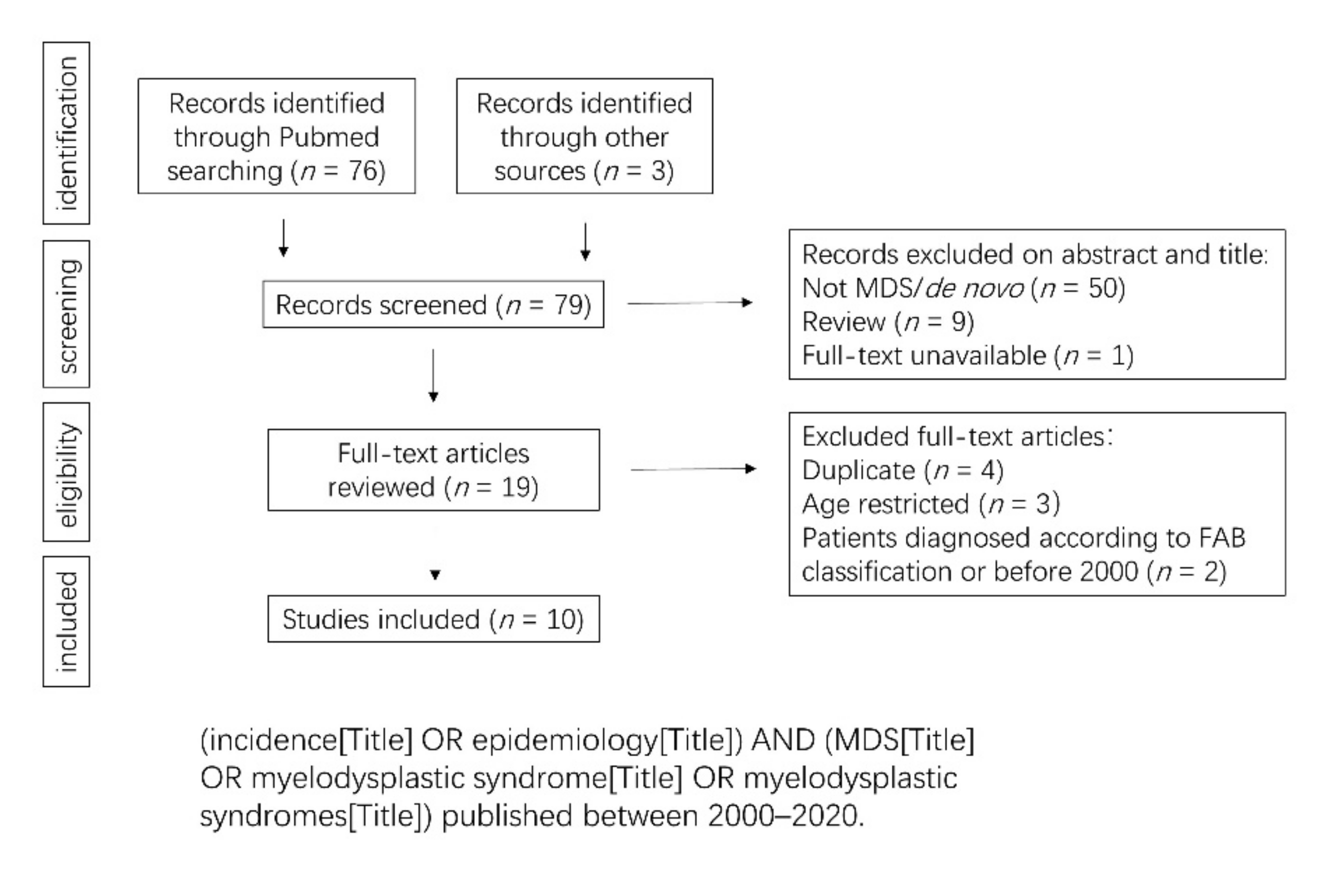
Figure 1. Flow chart for the process of identifying studies included in and excluded from the systematic review for the comparisons of MDS incidence and epidemiology data.
Cancers 13 00481 g001
Figure 2. Epidemiological characteristics of MDS in Western and Asian countries. (a) Pooled crude incidence in America, Europe, and Asia. (b) Age-adjusted incidence in Western and Asian countries. Figure 2 has been drawn based on the data and publications [10,11,12,13,14,15,16,17,18,19] described in Supplemental Table S1.
Cancers 13 00481 g002
Figure 3. Flow chart for the process of identifying studies included in and excluded from the systematic review for the comparisons of MDS subtypes, risk stratification and prognosis, cytogenetic abnormalities, and gene mutation.
Cancers 13 00481 g003
Figure 4. MDS subtypes in Western and Asian countries. (a,b), Percentage of MDS subtypes in Western and Asian countries. Figure 4 has been drawn based on the data and publications [10,13,14,15,18,19,21,24,26,27,28,29,30,31] described in Supplemental Tables S2 and S3. Importantly, the numbers indicated above each condition correspond to the weighted average (taking into account the size of the cohort of each study).
Cancers 13 00481 g004
Figure 5. Distribution depending on IPSS-R in Western and Asian countries. The graph was drawn upon publications [26,29,31,36,39,41,42,43,44,45,46,47]. Raw data and statistical analysis are presented in Supplemental Table S4.
Cancers 13 00481 g005
Figure 6. Karyotype distribution in Western and Asian MDS patients. (a) Percentage of total abnormalities, single abnormality, double abnormalities, 3 and more than 3 abnormalities. (bf) Karyotype distribution in each cytogenetic risk group depending on IPSS-R. Because of the different sources of the data, we did not list the results completely consistent with IPSS-R, -11/del(11q) instead del(11q) in very good group, -20/del(20q) instead del(20q) in good group, i(17q)/del(17p)/-17 instead i(17q) in intermediate group, -7/del(7q) instead -7 in poor group and ≥3 abnormalities instead >3 abnormalities in very poor group. Figure 6 has been drawn based on the data and publications [8,26,29,31,32,48,49,50,51] described in Supplemental Tables S4 and S5.
Cancers 13 00481 g006
Figure 7. Gene mutation topographies in Western and Asian countries. Frequency of the most common gene mutations in Western and Asian countries. The data were analyzed upon 11 publications [30,62,63,67,68,69,70,71,72,73,74]. Raw data and statistical analysis are presented in Supplemental Table S7.
Cancers 13 00481 g007
Figure 8. Survival depending on IPSS-R in Western and Asian countries. The size of the circles indicates the sample size of each study. The graph was drawn upon publications [26,29,31,36,40,42,43,44,45,46,47,48]. Raw data and statistical analyses are presented in Supplemental Table S8.
Cancers 13 00481 g008
Publisher’s Note: MDPI stays neutral with regard to jurisdictional claims in published maps and institutional affiliations.