Article

Exogenous ACC Deaminase Is Key to Improving the Performance of Pasture Legume-Rhizobial Symbioses in the Presence of a High Manganese Concentration

by
Ana Paço
1,
José Rodrigo da-Silva
1,
Denise Pereira Torres
1,
Bernard R. Glick
2 and
Clarisse Brígido
1,*
1
MED—Mediterranean Institute for Agriculture, Environment and Development, Instituto de Investigação e Formação Avançada, Universidade de Évora, Pólo da Mitra, Ap. 94, 7006-554 Évora, Portugal
2
Department of Biology, University of Waterloo, Waterloo, ON N2L 3G1, Canada
*
Author to whom correspondence should be addressed.
Plants 2020, 9(12), 1630; https://doi.org/10.3390/plants9121630
Submission received: 19 October 2020 / Revised: 20 November 2020 / Accepted: 22 November 2020 / Published: 24 November 2020
(This article belongs to the Special Issue Role of Microorganisms in Plant Growth and Phytoremediation)

Abstract

:
Manganese (Mn) toxicity is a very common soil stress around the world, which is responsible for low soil fertility. This manuscript evaluates the effect of the endophytic bacterium Pseudomonas sp. Q1 on different rhizobial-legume symbioses in the absence and presence of Mn toxicity. Three legume species, Cicer arietinum (chickpea), Trifolium subterraneum (subterranean clover), and Medicago polymorpha (burr medic) were used. To evaluate the role of 1-aminocyclopropane-1-carboxylate (ACC) deaminase produced by strain Q1 in these interactions, an ACC deaminase knockout mutant of this strain was constructed and used in those trials. The Q1 strain only promoted the symbiotic performance of Rhizobium leguminosarum bv. trifolii ATCC 14480T and Ensifer meliloti ATCC 9930T, leading to an increase of the growth of their hosts in both conditions. Notably, the acdS gene disruption of strain Q1 abolished the beneficial effect of this bacterium as well as causing this mutant strain to act deleteriously in those specific symbioses. This study suggests that the addition of non-rhizobia with functional ACC deaminase may be a strategy to improve the pasture legume–rhizobial symbioses, particularly when the use of rhizobial strains alone does not yield the expected results due to their difficulty in competing with native strains or in adapting to inhibitory soil conditions.

Graphical Abstract

1. Introduction

Acid soils represent ≈30% of the total area of the planet and 50% of the world’s arable land [1]. Soil acidity restricts agricultural production mainly due to nutrient deficiency and toxicity by metals such as manganese (Mn), iron (Fe), and aluminum (Al), with Al and Mn toxicities being the main factors limiting plant growth in acidic soils [2,3,4]. Therefore, Mn toxicity is a very common stress in arable lands around the world [5], and in particular, it is one of the most important limiting factors to plant productivity in the South of Portugal [6]. In this regard, the most common production system in Portugal, mainly in the Alentejo region in Southern Portugal, is the silvo-pastoral Montado ecosystem [7]. Animal production is an important component of the Montado system, but the natural pastures in these soils are unproductive, and the success of sown pastures, in addition to small grains, is greatly reduced due to very high soil acidity and Mn concentrations [8]. The recommended procedure for the recovery of these soils is the installation of permanent pastures and increasing soil fertility through the application of chemical phosphate fertilizers [9] or the correction of soil acidity by the addition of dolomitic limestone [10]. However, either of these approaches is expensive and not available to farmers in large areas of the world, and hence, it is important to develop less expensive solutions.
Based on the diverse expressions of Mn toxicity, it may be assumed that tolerance to Mn depends on the plant species and cultivar. Among different pasture legumes, white clover (Trifolium repens L.), alfalfa (Medicago sativa L.), and barrel medic (M. truncatula L.) are considered to be very sensitive to Mn toxicity [11], while subterranean clover (T. subterraneum L.) is considered to be relatively tolerant to soil acidity [12]. In addition, as a consequence of the negative effects of Mn on the growth and activity of the root nodules, the negative effects of Mn on plant growth are more evident on N2-fixing plants than on plants supplied with mineral N [13,14]. Thus, improving the symbiotic performance of rhizobia with legumes growing in high acidic and high Mn soils through sustainable agricultural practices is a great challenge.
Along with rhizobia, legumes can also establish associations with non-rhizobial endophytic bacteria, which also contribute to increases in plant biomass and yield [15]. Furthermore, their application in agriculture may be a promising and low-cost alternative either to increasing the availability of nutrients for the improvement of pasture productivity or to protect plants against Mn toxicity. In fact, the use of consortia, including both rhizobial and non-rhizobial endophytic bacteria, has been shown to synergistically enhance the symbiotic performance of rhizobia and consequently increase host plant growth [16,17,18], including under metal toxicity [19,20]. Moreover, studies on plant growth-promoting bacteria showed that one of the central mechanisms in plant-microbe interactions is the ability of some bacteria to synthesize the enzyme 1-aminocyclopropane-1-carboxylate (ACC) deaminase [21,22,23]. This enzyme acts to cleave the immediate ethylene precursor, ACC, into ammonia and α-ketobutyrate under free-living [24] or symbiotic conditions [25,26]. The importance of ACC deaminase for the symbiotic rhizobial-legume association has been demonstrated in several studies [27,28], particularly under stressful conditions [29,30,31,32].
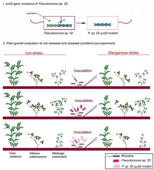
Herein, it was hypothesized that the inoculation of plants with a consortium of rhizobia and non-rhizobial bacteria where at least one bacterium in the consortium possess ACC deaminase activity could be advantageous to promote legume growth, particularly under Mn stress conditions. Therefore, in this work we investigated (i) the effects of an ACC deaminase-producing endophytic bacterium on three different rhizobium-legume symbioses (one grain and two forage legumes) under the presence and absence of a high Mn concentration, and (ii) the role of an exogenous ACC deaminase in these rhizobial–legume symbioses. For that, the Pseudomonas sp. Q1 was selected from our lab collection of endophytic bacteria isolated from legumes [18,33], and its acdS gene was deleted in order to provide evidence of the importance of the activity of an exogenous ACC deaminase in the rhizobia and legume performance under stressed and non-stressed conditions.

2. Results

2.1. Analysis of the acdS Gene Sequence from Pseudomonas sp. Q1

Data mining of the Pseudomonas sp. Q1 genome (Assembly number ASM993597v1) reveal the existence of genes related to ACC deamination. The acdS gene detected in the Pseudomonas sp. Q1 genome has a sequence of 1017 bp (Accession number MN232215) that encodes a protein of 338-amino acid residues with a predicted molecular weight of 36.9 kDa.
To confirm the annotation of the putative acdS gene from Pseudomonas sp. Q1, an alignment of its predicted amino-acid sequence with the amino-acid sequence of several functional ACC deaminase proteins was performed (Figure 1). This alignment shows that the predicted Pseudomonas sp. Q1 ACC deaminase displays a high degree of similarity to the predicted and experimentally proven ACC deaminase enzymes from a variety of organisms, including several Pseudomonas species (Figure 1). The most similar protein is the ACC deaminase of P. palleroniana (100% identity, accession WP_090372028), followed by P. fluorescens (99.7%, accession WP_060756418), P. fuscovaginae (98.5%, accession WP_019361456), P. kilonensis (97.0%, accession WP_053178893), P. thivervalensis (96.8%, accession WP_053183836), and P. migulae (96.5%, accession WP_084321532).
This alignment also showed that the Pseudomonas sp. Q1 acdS gene possesses both the pyridoxal 5′-phosphate binding sites and the catalytic residue (Figure 1) that are typical sites of the bacterial 1-aminocyclopropane-1-carboxylate deaminase (IPR005965) subfamily.

2.2. Confirmation of the ∆acdS Knockout Mutant and acdS-Complemented Strain Construction

The deletion of an internal fragment of 955 nt of the acdS gene sequence in the ∆acdS knockout mutant strain, as well as the introduction of the acdS gene plus its promoter and terminator regions (2200 nt) in the genome of the complemented strains were confirmed by PCR. Moreover, the detection of ACC deaminase activity in free-living conditions of Pseudomonas sp. Q1 but not its ∆acdS knockout mutant confirmed the presence of a functional acdS gene in this strain. The ACC deaminase activity of Pseudomonas sp. Q1 and the acdS-complemented strain was 3.78 and 3.32 µmol of α-ketobutyrate mg−1 protein·h−1, respectively. No ACC deaminase activity was detected either in the ΔacdS mutant strain or in any of the rhizobial strains used in this study. Furthermore, the acdS gene deletion in the ∆acdS knockout mutant strain did not affect its compatibility with any of the rhizobial strains in vitro. These results corroborate the fact that the ∆acdS knockout mutant phenotype is due to the loss of ACC deaminase functionality and not to other mutations that may occur during the mutant construction process.

2.3. Tolerance to a High Concentration of Mn

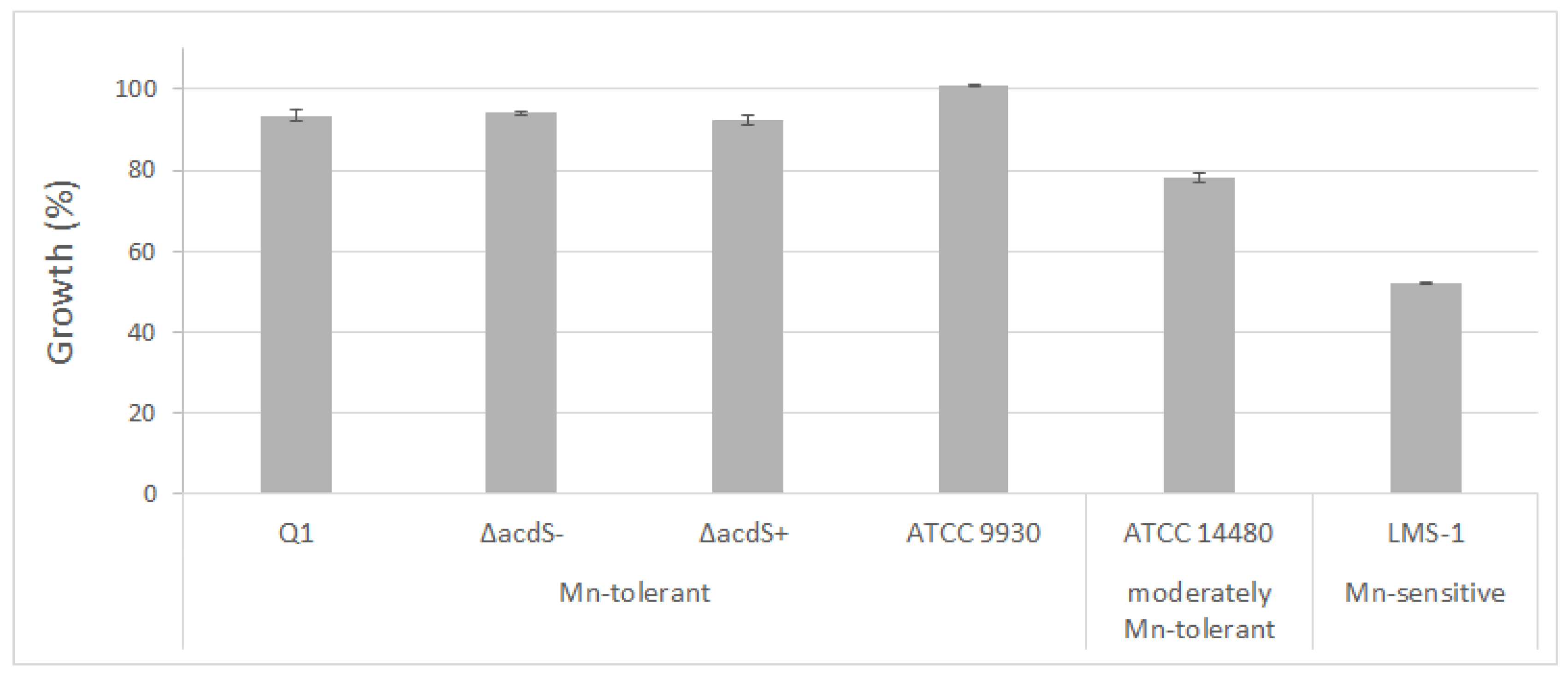
To evaluate Mn tolerance of the pseudomonads and rhizobial strains, their growth was evaluated in liquid medium under control (medium without additional Mn) and a high concentration of Mn. After 72 h, E. meliloti ATCC9930T showed similar growth in the absence and presence of Mn. Similarly, when R. leguminosarum ATCC14480T was exposed to a high level of Mn, its growth was only slightly slower than its growth under control conditions, indicating that this strain is moderately tolerant to Mn. On the other hand, the growth of M. ciceri LMS-1 was severely affected by Mn, as its growth was significantly lower in its presence compared to control conditions after 72 h (Figure 2). Altogether, E. meliloti and R. leguminosarum were considered to be Mn-tolerant, while M. ciceri was Mn-sensitive.
The phenotype of the ΔacdS knockout mutant strain was also evaluated in the absence and presence of Mn compared to the wild-type strain. No significant differences between the growth rates of the ΔacdS knockout mutant and wild-type strain under those conditions were found (Figure 2). Moreover, Pseudomonas sp. Q1 and its derivatives showed similar growth characteristics both with and without Mn, indicating that Pseudomonas sp. Q1 is tolerant to these Mn concentrations and its ACC deaminase is not involved in its tolerance response to Mn.

2.4. Benefits of the Q1 Strain and of Its ACC Deaminase Activity on the Symbiotic Rhizobial-Legume Model

From a preliminary assessment of the minimum concentration of Mn that constrains legume growth, it was determined that 30 mg L−1 of Mn is sufficient to significantly inhibit the growth of both chickpea and subterranean clover legumes (data not shown). The other two concentrations of Mn (45 and 60 mg L−1) applied to the plants greatly reduced the growth of the subterranean clover and were lethal to chickpea plants. For this reason, the concentration of 30 mg L−1 of Mn was chosen to assess the symbiotic performance of rhizobial strains under a high concentration of Mn and the effects of Pseudomonas sp. Q1 on the rhizobial-legume symbioses.

2.4.1. Chickpea-Mesorhizobium Symbiosis

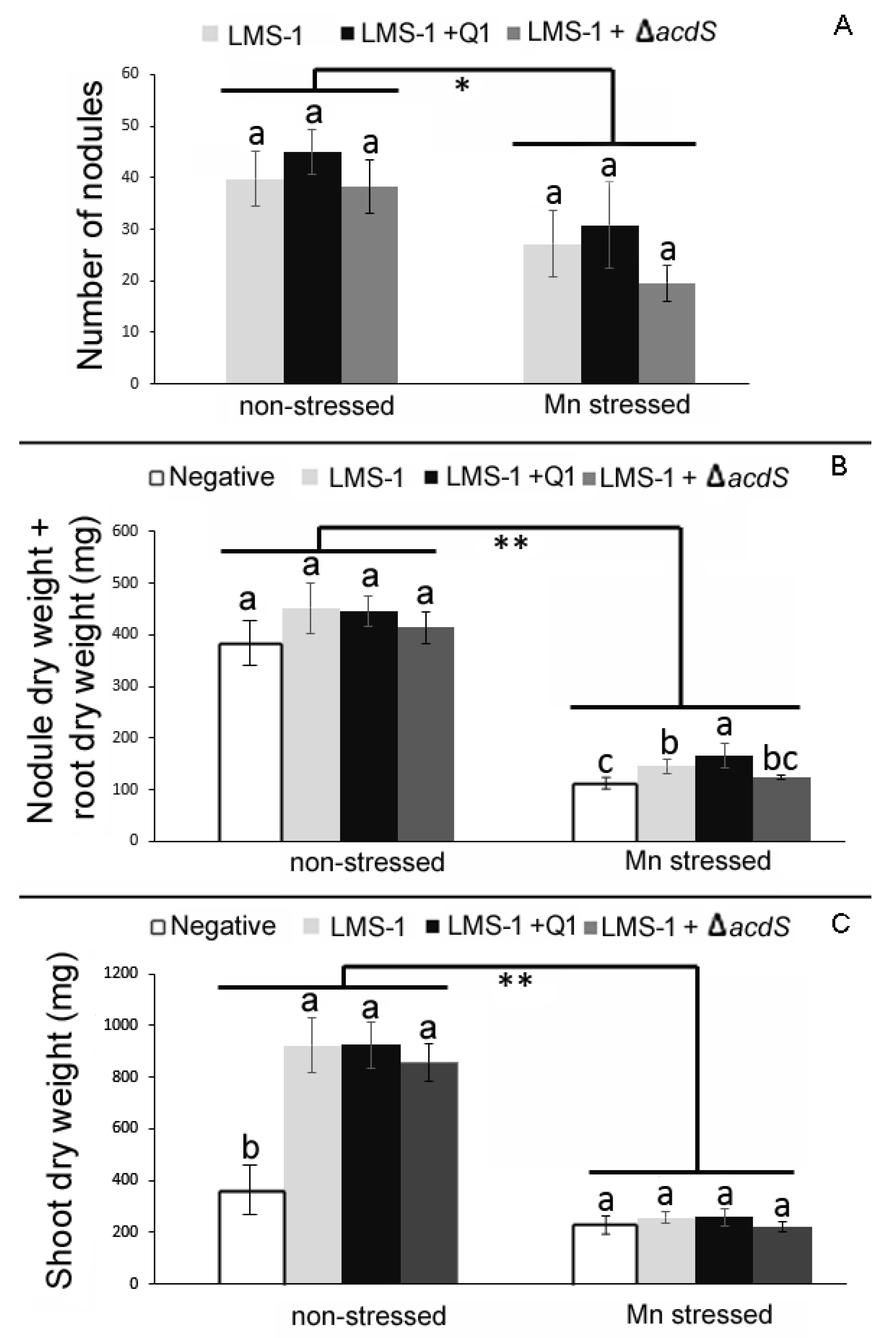
As previously observed [18], and compared to non-stressed conditions, the root and shoot dry weights of chickpea plants grown under a high concentration of Mn were significantly reduced either when single inoculated with the highly efficient strain M. ciceri LMS-1 [29,30,34] or co-inoculated with LMS-1 and Q1 strains (Figure 3). In fact, a statistically significant difference in average shoot and root dry weights by plant growth conditions was found (f(1) = 193.668, p < 0.001) for RDW and f(1) = 151.470, p < 0.001) for SDW). Furthermore, the chickpea plants co-inoculated with the LMS-1 and Q1 strains presented similar values in all parameters analyzed (number of nodules, total root and shoot dry weights) to the chickpea plants solely inoculated with LMS-1 strain in both non-stressed and Mn conditions.
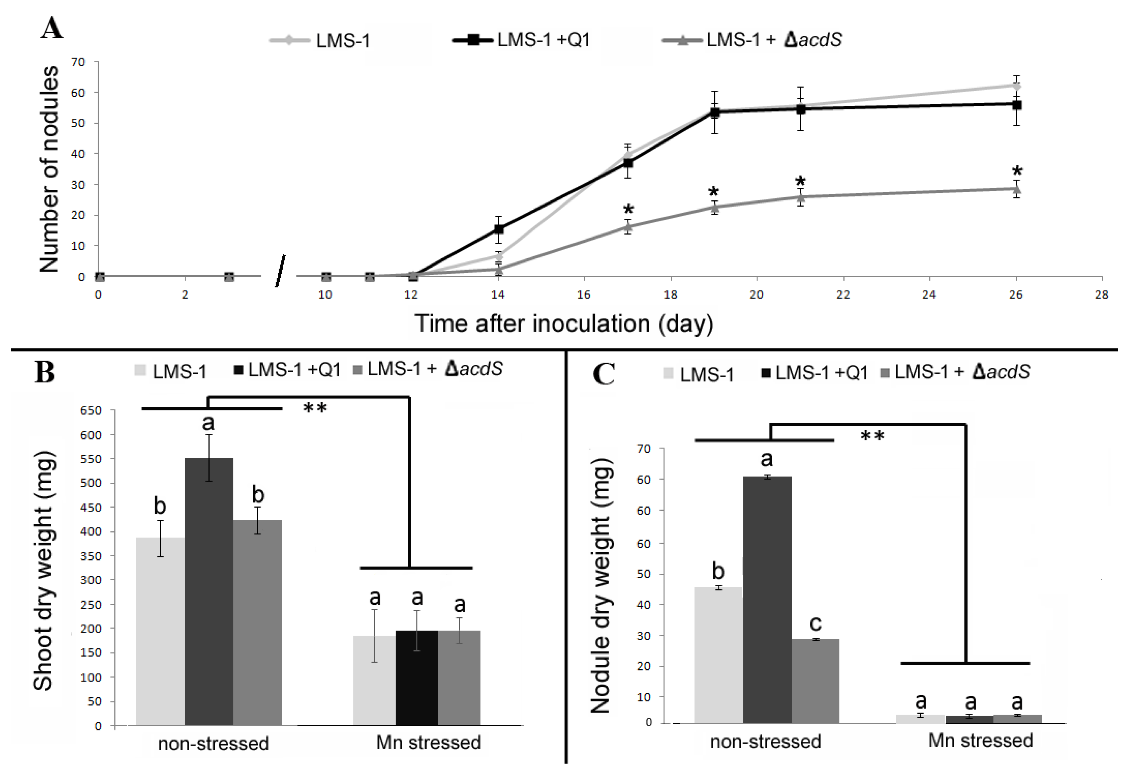
In hydroponics, the nodulation kinetics showed that the co-inoculation of chickpea plants with LMS-1 and Q1 strains did not change the nodulation abilities (number of nodules and time of appearance) of the LMS-1 strain under non-stressed conditions (Figure 4A). However, plants co-inoculated with LMS-1 and Q1 strains formed bigger nodules, which is reflected in the significant increase of the nodule dry weight of those plants compared to plants single inoculated with LMS-1. Moreover, these bigger nodules may have contributed to the observed significant increase of the shoot dry weight of those plants compared to chickpea plants single inoculated with LMS-1 strain (Figure 4B). Under Mn conditions in hydroponics, the growth of chickpea plants was also severely affected, but no significant differences between treatments were observed for any of the plant parameters analyzed at 26 days after inoculation (DAI) (Figure 4C).
The hydroponic assay also revealed that the presence of the ∆acdS mutant strain in the chickpea—M. ciceri LMS-1 symbiosis contributed to a significant reduction of the number of nodules formed by the LMS-1 strain compared to plants inoculated with LMS-1 alone (Figure 4). As a result, the nodule and shoot dry weights in those plants were significantly lower than those obtained in plants inoculated with LMS-1 alone. Nevertheless, no significant differences were observed in any of the analyzed parameters of chickpea co-inoculated with the LMS-1 and ∆acdS mutant and chickpea single inoculated with LMS-1 in the pot assay, suggesting that the differences observed in hydroponics dissipate over time.

2.4.2. Subterranean Clover-Rhizobium and Burr Medic-Ensifer Symbioses

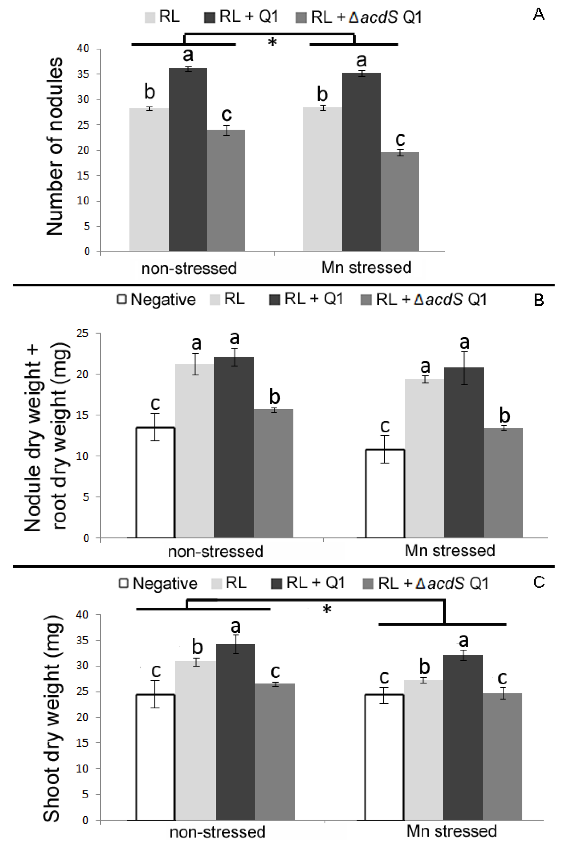
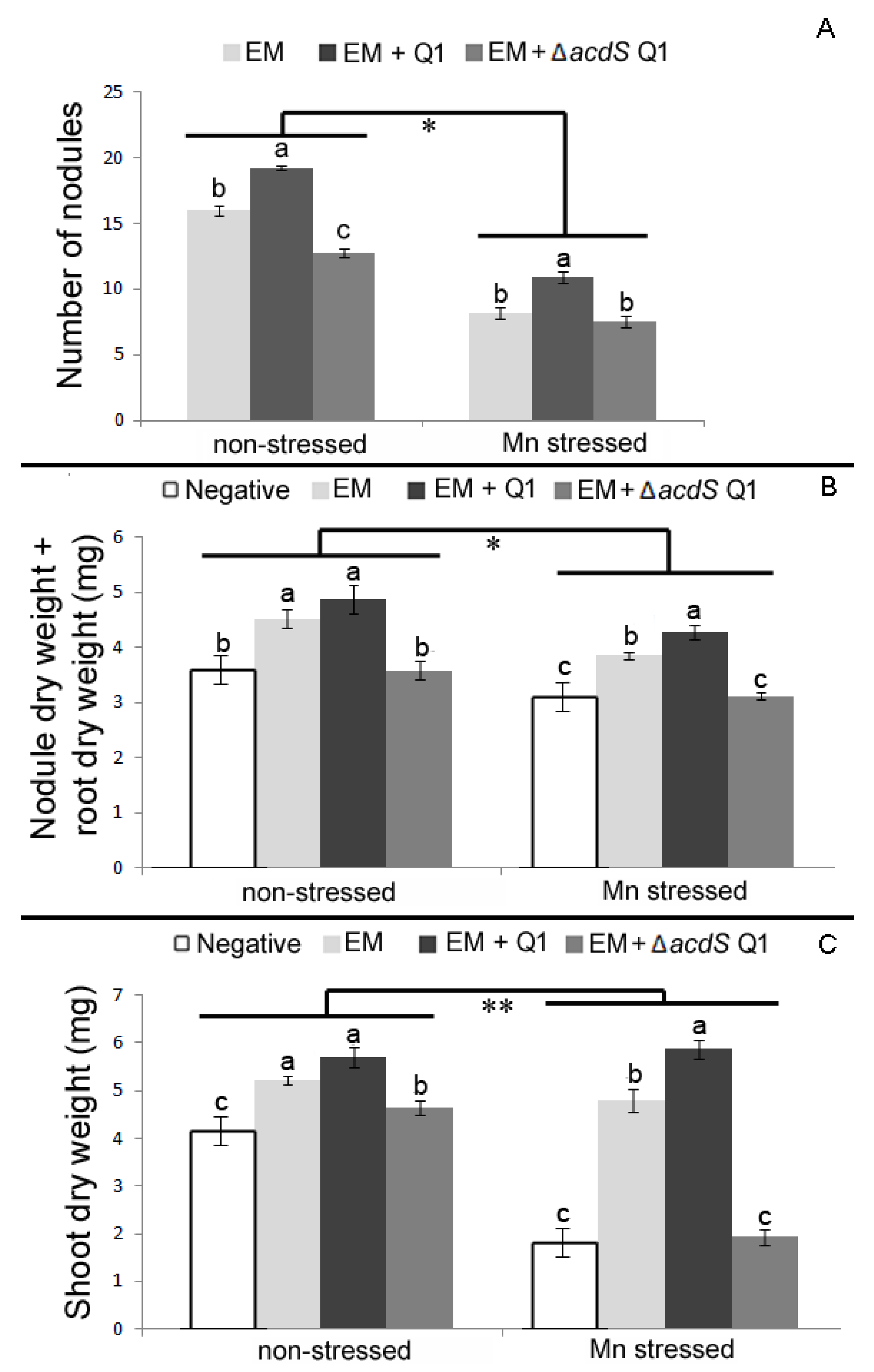
The growth of subterranean clover and burr medic species were also affected by Mn, although to a lesser extent than in chickpea plants. Compared to non-stressed conditions, the shoot dry weight of subterranean clover plants single inoculated with R. leguminosarum ATCC14480T was significantly reduced (Figure 5), while the root dry weight of burr medic plants single inoculated with E. meliloti ATCC9930T was significantly diminished under stressed conditions (Figure 6).
The nodulation ability of E. meliloti was more impaired by Mn than it was for R. leguminosarum in plants inoculated with the rhizobia alone or with Q1 strain. The nodulation ability of the symbiotic strains R. leguminosarum and E. meliloti was significantly improved when their legume hosts (subterranean clover and burr medic) were co-inoculated with the non-rhizobial endophytic bacterium Q1 strain. This happens either under non-stressed or stressed conditions (Figure 5 and Figure 6). In both cases, the number of nodules formed by the microsymbionts was significantly enhanced by the presence of strain Q1 (Figure 5A and Figure 6A). Subterranean clover plants co-inoculated with R. leguminosarum and Q1 strains presented significantly higher values for shoot dry weight compared to plants inoculated with R. leguminosarum strain alone. In contrast, no significant differences were observed between the total root dry weights of single inoculated plants with rhizobium and plants co-inoculated with rhizobium and the Q1 strain. Interestingly, the positive effects of the Q1 strain on growth of both legume hosts were even more pronounced in plants exposed to high levels of Mn. For instance, the shoot dry weight values of both subterranean clover and burr medic plants were significantly higher in plants co-inoculated with their microsymbiont and the Q1 strain compared to plants inoculated with rhizobium alone under Mn toxicity. Moreover, these results are consistent with the significant increase in the number of nodules of those plants mentioned above.
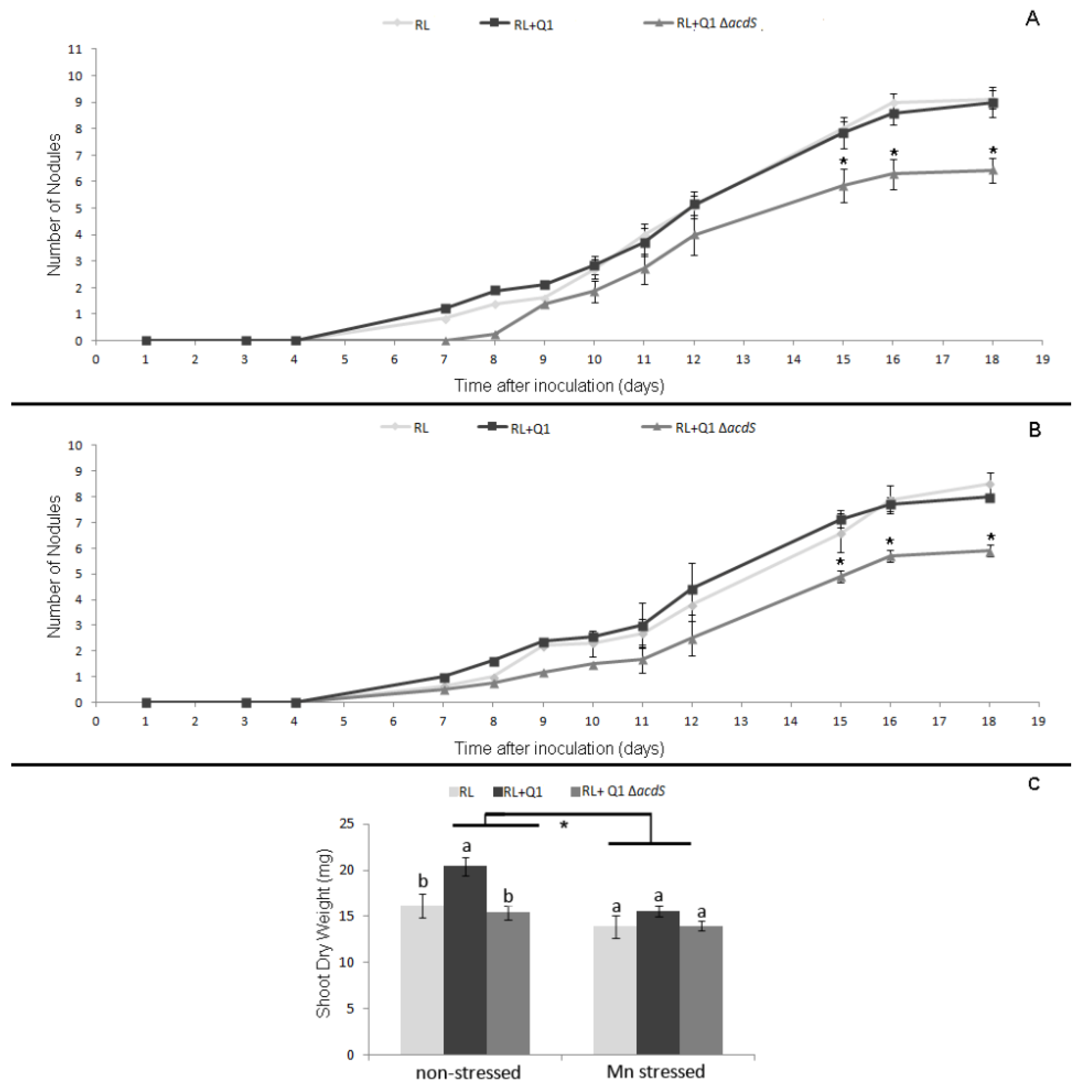
During the first 18 days in hydroponics, no significant differences were observed in the nodulation kinetics of R. leguminosarum in subterranean clover single inoculated with this strain and plants co-inoculated with rhizobium and Q1 strains either under stressed or non-stressed conditions (Figure 7A,B). Nevertheless, the shoot dry weight of double inoculated plants was significantly higher compared to plants single inoculated with the R. leguminosarum strain at the end of the experiment under non-stressed conditions. On the other hand, no significant differences were observed between these treatments under stressed conditions.
In contrast, both rhizobial–legume symbioses (subterranean clover–R. leguminosarum ATCC14480T and burr medic–E. meliloti ATCC9930T) were negatively affected by the presence of the ∆acdS mutant strain. In fact, all parameters analyzed in the forage legumes co-inoculated with the ∆acdS mutant and the respective rhizobium were significantly reduced compared to plants inoculated solely with the respective microsymbiont (Figure 5 and Figure 6). Under stressed conditions, the presence of the ∆acdS mutant strain negatively affected the nodulation ability of R. leguminosarum (Figure 5). Nevertheless, the shoot dry weight of those co-inoculated plants were similar to those in subterranean clover plants inoculated solely with R. leguminosarum, suggesting that other plant growth-promoting mechanisms besides ACC deamination from strain Q1 may have helped the growth of subterranean clover in the presence of Mn. These results were consistent with the hydroponic data conducted with subterranean clover, where is observed that the nodulation kinetics of subterranean clover by R. leguminosarum were also impaired by the presence of the ∆acdS mutant either under stressed or non-stressed conditions (Figure 7A,B). Despite these differences, no differences between the shoot dry weight of plants double inoculated with its microsymbiont and the ∆acdS mutant and the plants inoculated with rhizobium alone were observed at the end of the hydroponic assay (Figure 7C). However, the nodulation ability of E. meliloti with burr medic was not affected by the presence of the ∆acdS mutant strain under Mn stressed conditions. Although the shoot dry weight and total root dry weight of plants co-inoculated with E. meliloti and ∆acdS mutant strains were significantly reduced compared to plants inoculated with E. meliloti alone (Figure 6).

3. Discussion

Mn toxicity is one of the most important constrains to plant growth on acid soils, especially for legumes dependent on N2-fixing symbiosis. Although the supplementation of acidic soils with dolomitic limestone alleviates the problem [10], it is an expensive approach and is not universally available. The introduction of highly efficient native rhizobia or engineered rhizobia strains can be useful tools as legume inoculants, particularly under challenging soil and climate conditions [35]. However, the selection and characterization of native rhizobial strains is a slow and expensive process, and the use of genetically modified organisms (GMO) is limited or prohibited in many countries. The present study evaluated the effects of the non-rhizobial endophytic bacterium Pseudomonas sp. Q1 on the symbiotic performance of three different rhizobia strains, as well as on the growth of their hosts in the absence and presence of high levels of Mn.
As expected, all legume species were negatively affected by the presence of high levels of Mn, although expressing different levels of sensitivity. A previous study addressing the tolerance of pasture legumes to high concentrations of Mn in southern Australia revealed that in general, subterranean clover and related species are more tolerant to Mn than are Medicago species such as M. polymorpha [36]. Here, it was observed that the symbioses of these legume plants with their respective microsymbionts were differently affected by the presence of Mn. Even though E. meliloti is highly tolerant to Mn in free-living conditions, its ability to nodulate burr medic under stressed conditions was severely affected. On the other hand, no negative effect on the nodulation abilities of R. leguminosarum was observed under these Mn conditions, even though it displays lower tolerance to Mn under free-living conditions than E. meliloti. These data are consistent with the notion that the ability of legumes to grow in marginal soils is a consequence of the symbiotic associations that they are able to establish with nitrogen-fixing rhizobia [6,37,38]. Moreover, it has been reported that bacteria are capable of changing the chemical properties of rhizospheric soil and catalyzing redox reactions, altering heavy metal mobility and bioavailability, consequently reducing the toxicity of metals to plants [39,40,41,42]. In this context, the subterranean clover and burr medic symbioses with their respective microsymbionts, in the presence of Mn, may not only be a result of their nitrogen-fixation abilities but a cumulative result of several mechanisms employed by these microsymbionts; rhizobia possess a range of plant beneficial mechanisms other than nitrogen fixation [37,43].
Interestingly, the beneficial effects of the Q1 strain were only evident in the pasture legume–rhizobial symbioses while no effects were observed in chickpea plants inoculated with M. ciceri LMS-1. Despite these differences and the fact that Pseudomonas sp. Q1 was isolated from Lupinus angustifolius L., it can effectively colonize the internal root tissues of different plant species, including the legumes used herein (Unpublished results). The results presented here indicate that the presence of the endophytic non-rhizobial bacterial strain Q1 significantly increased the nodulation abilities of strains R. leguminosarum ATCC 14480T and E. meliloti ATCC 9930T in subterranean clover and burr medic, respectively, at the same time contributing to the growth promotion of these plants. The augmentation of legume nodulation through the co-inoculation of non-rhizobial bacteria along with rhizobia microsymbionts has been known for some time. For instance, the co-inoculation of a rhizobia and a Bacillus strain facilitated the nodulation of Phaseolus acutifolius by Rhizobium TAL 182, demonstrating the potential for root-associated non-rhizobial organisms to alter the dynamics of the legume–Rhizobium sp. symbiosis [44]. Similar results were reported with Pisum sativum [45], Lotus spp. [20], Lens culinaris [45,46], Vigna radiata [47], chickpea [48], and soybean [49,50] plants when co-inoculated with plant growth-promoting bacteria and with their rhizobia microsymbionts.
Under Mn stress conditions, the beneficial effects of the Q1 strain on the symbiotic performance of ATCC 14480T and ATCC 9930T were even more evident. This is unexpected, since previous studies have shown that high concentrations of Mn and other metals can negatively affect ACC deaminase activity as well as other plant growth promoting properties [51,52]. High concentrations of Mn differently affected each of these rhizobium–legume symbioses. Nevertheless, in both cases, the presence of the Q1 strain facilitated the formation of nodules by these rhizobial strains and led to an increase of the host’s growth. These results are in agreement with previous reports showing that non-rhizobial bacteria stimulated legume growth and nodulation under different abiotic stress conditions, such as salinity [18,48,53,54,55,56,57], toxicity of copper [58,59,60], Mn [18], cadmium [23,61], and zinc [60]; as well as under biotic stress, namely root rot caused by Fusarium solani [56]. On the other hand, no beneficial effects of the co-inoculation of non-rhizobial bacterium along with M. ciceri LMS-1 were observed with the chickpea plants grown either under non-stressed or stressed conditions. Nevertheless, the lack of effects in the presence of the Q1 strain on the symbiotic performance of LMS-1 or chickpea growth under Mn toxicity conditions is most probably due to the high sensitivity of this symbiotic legume–rhizobium association to the Mn levels employed.
The importance of ACC deaminase activity in plant–bacteria interactions, more specifically in the rhizobial nodulation process, has been demonstrated in several studies either by the transformation of rhizobial strains [28,29,30,31,62,63,64] or by combining rhizobia with ACC deaminase-expressing non-rhizobial bacteria [65,66,67,68,69,70,71]. Consistent with that, no beneficial effect of the ΔacdS mutant strain in the rhizobial–legumes symbioses was observed. This suggests that the ACC deaminase is a main plant growth-promoting mechanism responsible for the beneficial effects of the Q1 strain in the symbiotic subterranean clover–R. leguminosarum and burr medic–E. meliloti associations. In this regard, the presence of the ∆acdS mutant in co-inoculated plants resulted in a significant reduction of the rhizobial nodulation abilities and in the growth of the legume hosts either under non-stressed or stressed conditions. These results suggest that the importance of the ACC deaminase enzyme from Pseudomonas sp. Q1 in the rhizobium–legume symbiosis is not only on its ability to locally cleave the plant ethylene precursor ACC, which directly favors the nodulation process by the rhizobial strains, but additionally to prevent the activation of plant immune responses. Since ethylene is involved in plant immunity [72], ACC deaminase activity may block the activation of plant defenses in the roots [73]. For example, canola treated with ACC deaminase-producing Pseudomonas putida (formerly Enterobacter cloacae) UW4 down-regulated genes involved in ethylene-induced plant stress and in defense signaling pathways in comparison to canola plants inoculated with the ACC deaminase minus mutant of this bacterium [74]. Moreover, a transient induction of defense-related genes was observed in different legume species after rhizobial inoculation, and it was suggested that plant innate immunity is a crucial component in the establishment and maintenance of symbiosis [75].
Despite the fact that all rhizobial strains used in this study possess at least one copy of the acdS gene in their genomes, none of them presents the activity of this enzyme under free-living conditions. This may reflect the fact that rhizobial ACC deaminases contain a much lower level of this enzyme activity than do free-living soil bacteria [24]. Moreover, the expression of ACC deaminase in Mesorhizobium loti MAFF303099 and M. ciceri UPM-Ca7T occurs only inside of formed nodules [26,76] under the regulation of the NifA protein [77]. So far, this mechanism of ACC deaminase regulation has only been found in Mesorhizobium strains, suggesting that this regulatory mechanism is widespread in this genus [26,78]. It is possible that M. ciceri LMS-1 expresses ACC deaminase activity under symbiotic conditions, while the other two rhizobial strains used in this study do not. In this sense, it may be hypothesized that the role of ACC deaminase from strain Q1 in the tripartite associations containing R. leguminosarum ATCC 14480T or E. meliloti ATCC 9930T is important to overcome the plant defenses activated during the establishment of the rhizobium–legume symbiosis compared with the establishment of symbioses involving M. ciceri LMS-1, which could explain the less evident beneficial effects of the Q1 strain in this symbiotic association. In addition, the ACC deaminase may also affect other plant growth-promoting mechanisms that help to establish rhizobium–legume symbioses, especially those with low N2-fixing efficiency. A recent study revealed that acdS disruption in Rahnella aquatilis HX2 affected the regulation of other genes or enzymes related to plant growth-promoting features [79]. Thus, the ability of R. aquatilis HX2 to produce indole-3-acetic acid (IAA) and its antagonistic activity was diminished with the acdS disruption, while its nitrogen-fixing activity was increased. Similarly, the acdS disruption in strain Q1 may have affected the regulation of other plant growth-promoting mechanisms, altering the complementary of this strain with the rhizobial strains. Nevertheless, further studies on why the loss of ACC deaminase activity has caused deleterious effects on the pasture legume–rhizobial interactions are needed in order to better understand the precise nature of the benefits of exogenous ACC deaminase activity in the rhizobial–legume associations.

4. Materials and Methods

4.1. Bacterial Strains and Growth Conditions

The bacterial strains and plasmids used in this study are listed in Table 1. The endophytic Pseudomonas sp. Q1, which was isolated from L. angustifolius L. roots and previously characterized [18], was selected to evaluate its effects on the symbiotic rhizobium–legume associations. Three rhizobial strains, namely M. ciceri LMS-1, R. leguminosarum bv. trifolii strain ATCC 14480T, and E. meliloti strain ATCC 9930T, were used as specific microsymbionts of chickpea, subterranean clover, and burr medic, respectively. The rhizobial strains were grown in yeast extract mannitol (YEM) medium [80] or in tryptone yeast (TY) medium [81] at 28 °C. Unless otherwise indicated, strain Pseudomonas sp. Q1 and its derivatives, ΔacdS knockout mutant and complemented strains, were grown in tryptic soy broth (TSB, Liofilchem) medium or tryptic soy agar (TSA, Liofilchem) medium at 28 °C, while the Escherichia coli strains were grown in Luria–Bertani (LB) medium [82] at 37 °C. Where indicated, 100 µg·mL−1 ampicillin, 25 µg·mL−1 chloramphenicol, or 10 µg·mL−1 tetracycline were added to the medium.

4.2. DNA Methods and Construction of Pseudomonas sp. Q1 Derivatives

4.2.1. DNA Methods

The extraction of bacterial genomic DNA and plasmid DNA were conducted using the E.Z.N.A. bacterial DNA kit (Omega Bio-Tek, Norcross, GA, USA) and the Zyppy™ Plasmid Miniprep kit (Zymo Research, Freiburg, Germany), respectively, according to the manufacturer’s protocols. All PCR reactions were performed in a final volume of 50 μL containing 1× Buffer, 0.2 mM dNTPs, 1.5 mM MgCl2, 15 pmol of each forward and reverse primers and 0.02 U/µL KOD Hot Start DNA Polymerase (Novagen, WI, USA). PCR products or gel bands were purified with the DNA Clean & Concentrator or ZymocleanTM Gel DNA Recovery kits (Zymo Research, Freiburg, Germany). Unless otherwise specified, molecular techniques were performed using standard protocols [82]. The primers used in this study are listed in Table 2.

4.2.2. Construction of Pseudomonas sp. Q1 ∆acdS Mutant Strain

Based on the genome sequence of strain Q1 (SAMN09237650), the flanking regions of the acdS gene were amplified by PCR using primers designed as recommended for gene splicing by PCR-driven overlap extension [87]. The internal primers are designed to possess short complementary regions to each other (≈20 bp) that will hybridize with one another during the second PCR to serve as a chimeric template for the generation of a hybrid product [87]. PCR reactions to amplify the acdS flanking regions were performed using 10 ng of DNA and the following set of primers: acdS-Q1-Fa and acdS-Q1-Rd for upstream region and acdS-Q1-Fc and acdS-Q1-Rd primers for downstream region (Table 2). PCR reactions were performed using the followed programs: 95 °C for 2 min followed by 30 cycles of 95 °C for 20 s, 56 °C for 10 s (for upstream region) or 59 °C for 10 s (downstream region), and 70 °C for 21 s (upstream region) or 70 °C for 18 s (downstream region).
To fuse the acdS flanking regions, another PCR reaction, containing 1 µL of a mixture of upstream and downstream-amplified products (±10 ng of each) as DNA template, was performed using the following program: 95 °C for 2 min, 30 cycles of 95 °C for 20 s, 56 °C for 10 s and 70 °C for 39 s. This reaction was performed using 15 pmol of the acdS-Q1-Fa and acdS-Q1-Rd primers (Table 2) and the DNA template (a mixture of upstream and downstream-amplified products) which possesses short complementary regions allowing the fusion between the upstream and downstream fragments, generating a hybrid product during the PCR amplification.
The amplified product was purified, cloned in pNZY28-blunt vector (NZYtech, Lisbon, Portugal), and sequenced at StabVida Lda (Lisbon, Portugal). The fused-flanking region of the acdS gene was released from the pNZY28-blunt vector using the SmaI and XbaI sites and subcloned into the suicide plasmid pT18mobsacB, also linearized with the same restriction enzymes. Then, this product was transformed into Escherichia coli NZY5α competent cells (NZYtech, Lisbon, Portugal). Subsequently, these bacteria were used to mobilize the transformed pT18mobsacB into the Pseudomonas sp. Q1 strain by triparental mating, using the helper strain MT616 (pRK600) as described [29]. Double recombinants were selected as previously described [88].

4.2.3. Complementation of Pseudomonas sp. Q1 ∆acdS mutant

pGRG36-Tetr Vector Construction

A derivative of the pGRG36 vector with resistance to tetracycline was constructed and used to complement the Q1 ∆acdS mutant strain. To do this, the regulatory region of the tetA gene (tetracycline resistance gene) was amplified from the pT18mobsacB by PCR using the Tet-SdaI-Fw and Tet-SdaI-Rv primers (Table 2) and 20 ng of plasmid DNA. The amplification program was: 95 °C for 2 min followed by 30 cycles of 95 °C for 20 s, 65 °C for 10 s, and 70 °C for 53 s. The amplified product was cloned into pNZY28-blunt and then subcloned into the pGRG36 at SdaI restriction site to generate the pGRG36-Tetr. Escherichia coli DH5α transformed cells were selected on LB plates supplemented with tetracycline and glucose (0.1%) at 32 °C.

Complementation

To ensure that the ∆acdS knockout mutant phenotype is actually due to the loss of the functionality of the ACC deaminase, a complemented strain was obtained by restoring the ACC deaminase functionality in the ∆acdS knockout mutant strain. To complement the ∆acdS knockout mutant, the acdS gene and its regulatory regions were introduced into the chromosome of the mutant strain using the site-specific recombination machinery of the transposon Tn7 present in pGRG36-Tetr. The putative regulatory regions of the acdS gene were identified using the software BPROM-Prediction of bacterial promoters [89] and ARNold Finding Terminators [90]. The DNA fragment carrying the complete acdS gene, its putative promoter and terminator regions were amplified using the primers acdS-Q1-Fa and comp_acds_Q1 Rv (Table 2) and 20 ng of plasmid DNA. The amplification program was 95 °C for 2 min followed by 30 cycles of 95 °C for 20 s, 57 °C for 10 s, and 70 °C for 55 s. The amplified product was purified and then cloned in pJET1.2/blunt vector (ThermoFisher Scientific, MA, USA). The extremities of the restricted BglII-acdS fragment were prepared using Fast end Repair Kit (ThermoFisher Scientific, MA, USA) to introduce the fragment into the pGRG36-Tetr at its SmaI restriction site to generate the pGRG36-Tetr-acdS. The E.coli WM3064 transformed cells were selected on LB plates supplemented with tetracycline, 2,6-Diaminopimelic acid (DAP) (0.3 mM), and glucose (0.1%) at 32 °C, and they were used as a donor to introduce the pGRG36-Tetr -acdS into the recipient the Q1 ∆acdS mutant strain by conjugation. After conjugation, selection of the ∆acdS mutant transformed cells was done by plating the mixture on TSA plates containing tetracycline and glucose (0.1%) at 23 °C for 48 h. To induce the expression of transposition genes in pGRG36-Tetr -acdS and subsequent integration of DNA containing the acdS fragment into the chromosome of the Q1 ∆acdS mutant strain, colonies were grown at TSB supplemented with 0.1% arabinose at 23 °C on an orbital agitator. After 24 h, the bacterial cells were plated on TSA at 37 °C to cure the vector. The absence of pGRG36-Tetr -acdS inacdS cells and the integration of the acdS fragment into the chromosome of the ∆acdS mutant strain were confirmed by the inability of cells to grow in the presence of tetracycline and by PCR using the acdS-Q1-Fa and comp_acds_Q1 Rv, respectively.

4.3. ACC Deaminase Activity Assay

The ACC deaminase activity was evaluated in strain Q1 and its derivatives, as well as in the rhizobial strains ATCC 14480T and ATCC 9930T. M. ciceri LMS-1 was used as a negative control [29]. At 28 °C, Pseudomonas sp. Q1 and its derivatives were grown in TSB medium for 24 h, while the rhizobial strains were grown in TY medium for 2–3 days until they all reached stationary phase. Then, ACC deaminase activity was induced in these cells following the procedure described [29]. After induction, ACC deaminase activity was estimated based on the chemical determination of α-ketobutyrate that results from ACC cleavage by ACC deaminase [91], and the total protein contents of cells was quantified using the Bradford method [92] with bovine serum albumin as a standard. Final ACC deaminase activity was expressed in μmol α-ketobutyrate/mg protein/h.

4.4. Detection of acdS Genes by PCR

To detect the acdS gene in strains ATCC 14480T and ATCC 9930T, two sets of primers were designed based on a conserved region of the acdS gene sequences from several Rhizobium leguminosarum bv. trifolii and E. meliloti strains. PCR reactions were performed using 20 ng of total DNA and the specific set of primers for each strain (Table 2). The amplification program was 95 °C for 2 min followed by 30 cycles of 95 °C for 20 s, 55 °C for 10 s, and 70 °C for 60 s. Sequencing reactions were performed by StabVida (Lisbon, Portugal). The sequences have been deposited in the GenBank database under the accession numbers: MN255490 and MN255491.

4.5. Compatibility between the Bacterial Endophyte Q1 or Its Derivatives and the Rhizobial Strains

To determine the compatibility between the endophytic bacterium Q1 or its derivatives and each of the rhizobial strains, a bioassay combining any strain of these two groups (a Pseudomonas and a rhizobial strain) was performed on YEM and TY plates to evaluate potential antagonist effects among them. At 28 °C, Pseudomonas sp. Q1 and its derivatives were grown in TSB medium for 24 h, whereas the rhizobial strains were grown in TY medium for 2–3 days on an incubator shaker at 200 rpm. Then, each bacterial culture was standardized to an initial OD565 nm of 1.0. Each rhizobial cell suspension was spread uniformly on the entire plate and incubated for 2 h at 28 °C. Later, 10 µL of each endophyte Q1 or its derivatives culture were spotted on the previously prepared plates with rhizobial strains in triplicate. The plates were incubated at 28 °C and observed over a period of 96 h. Absence of a zone of inhibition between strains was considered as indicator of compatible strains.

4.6. Evaluation of Strains’ Tolerance to High Concentration of Mn

To evaluate Mn tolerance of the Pseudomonas sp. Q1, its derivative strains, and the rhizobial strains LMS-1, ATCC 14480T, and ATCC 9930T, the bacterial cells of each strain were grown in their respective liquid media supplemented or not with 30 mg·L−1 of Mn (MnSO4). Strain Q1 and its derivatives grew on M9 medium [24] while rhizobial strains grew on TY liquid medium. For each treatment, the bacterial culture was standardized to an initial OD565 nm of 0.1 from an overnight growth. Then, the bacterial cultures were incubated in an orbital agitator at 28 °C for 24 h (strain Q1 and its derivative strains) or 72 h (rhizobial strains). After incubation, the bacterial suspensions were read by spectrophotometry at OD565nm. At least three replicas were performed per treatment under each condition.

4.7. Effect of the Pseudomonas sp. Q1 and the ∆acdS Mutant Strains on the Rhizobia-Legume Symbioses

4.7.1. Bacterial Inoculum Preparation

The rhizobial strains, strain Pseudomonas sp. Q1, and the ∆acdS mutant strains were grown in TY medium or TSB medium, respectively, until they reached the late exponential phase at 28 °C. Then, the cells were washed twice and resuspended in sterile saline solution (0.85% w/v NaCl) or nitrogen-free nutrient solution [93] previously diluted 1:4 with sterile distilled water. The optical density of bacterial cultures at OD565 nm was recorded. For co-inoculation, the cell suspension of two bacterial isolates was mixed in a 1:1 ratio and vortexed to yield a homogenous suspension.

4.7.2. Seed Surface Sterilization and Germination

The chickpea (Cicer arietinum L. cv Elixir) seeds were surface-sterilized and pre-germinated as previously described [29]. The subterranean clover (Trifolium subterraneum L. cv Nungarin) and (Medicago polymorpha L. cv. Scimitar) seeds were surface sterilized by soaking in 70% (v/v) ethanol solution for 1 min, followed by immersion in a 0.1% (w/v) acidified mercuric chloride solution for 4 min. After disinfection, seeds were rinsed 10 times in sterile distilled water and incubated at room temperature for 2 h. Seeds were washed more three times with sterile distilled water and incubated overnight at 4 °C, with a subsequent incubation of 48 h at 28 °C.

4.7.3. Evaluation of the Minimum Concentration Levels of Mn with Significant Constraints for Legume Plants

In order to simulate the gnotobiotic assay conditions close to those found in soils of Southern Portugal in terms of Mn values [9], the minimum concentration of Mn that significantly constrains the growth of a grain (chickpea) and a forage (subterranean clover) legume was determined. A plant growth experiment was performed by testing five different Mn concentrations: no additional Mn (control), 15, 30, 45, and 60 mg·L−1 of Mn supplemented in the nutrient solution [94]. After the second week of legume growth, the nutrient solution was complemented with 140 mg·L−1 of nitrogen (KNO3) and with the respective additional Mn concentration. Five pots per treatment were used, and the nutrient solution was applied three times a week. After four weeks, plants were harvested and then dried, followed by the recorded of shoot dry weights. The minimum concentration of Mn causing significant damage to legume growth was considered as the lowest concentration of Mn in which the treatment showed a significant reduction in SDW in relation to that of plants without the addition of Mn (control).

4.7.4. Pot Experiments under Gnotobiotic Conditions

To evaluate the effect of Pseudomonas sp. Q1 and the ∆acdS mutant strains on the symbiotic performance of three different rhizobial strains, plant growth assays were conducted in a growth chamber, under non-stressed and Mn stressed conditions using gnotobiotic systems. This system allows the study of individual biotic and abiotic factors that play important roles in plant–bacteria interactions in soil under controllable and reproducible conditions.
After seed germination, the seedlings were transferred to plastic pots filled with a sterile 1:1 (v/v) vermiculite:sand mixture. Chickpea seedlings (one per pot; ≈300 cm3) were immediately inoculated as earlier described by Nascimento et al. [29] while the other legume species seedlings (four per pot; ≈200 cm3) were inoculated only two days after sowing to ensure seedlings viability, with 250 µL of a bacterial suspension with an OD565nm of 0.5 [95]. All pots were grown in a growth chamber with the light cycle, temperature, and relative humidity set up as described [34]. For non-stressed conditions, a nitrogen-free nutrient solution [94] was used to water the plants three times a week. For Mn-stressed conditions, Mn (MnSO4) was added to the nitrogen-free nutrient solution (at a final concentration of 30 mg L−1) after the second week of growth until the end of the experiment. Due to the sensibility of chickpea–Mesorhizobium symbiosis to Mn, chickpea plants were only subjected to Mn stress during the third week of the experimental assay. Three treatments for each legume-rhizobial combination were prepared as follows: (i) legume plants inoculated with a specific rhizobial strain alone; (ii) legume plants co-inoculated with a specific rhizobial strain and Pseudomonas sp. Q1; (iii) legume plants co-inoculated with a specific rhizobial strain and the Q1 ∆acdS mutant strain. Uninoculated plants were used as negative controls. Five pot replicates were used for each treatment. The chickpea plants were harvested 54 days after inoculation (DAI), while subterranean clover and burr medic were harvested at 42-DAI. For subterranean clover and burr medic, the Shoot and root dry weights were measured, and the number of nodules was recorded.

4.7.5. Hydroponic Assay

To evaluate the nodule kinetics and development, hydroponic plant-growth assays were conducted using C. arietinum and subterranean clover plants co-inoculated with either Pseudomonas sp. Q1 or the ∆acdS mutant strains, together with their specific microsymbiont. All procedures were based on the protocol described [34]. Bacterial suspensions with an OD565 nm of 0.5 and 0.4 were used for inoculating C. arietinum and subterranean clover seedlings, respectively. A nitrogen-free nutrient solution was used as a hydroponic solution [93]. When required, Mn (MnSO4) was added to the hydroponic solution at final concentration of 30 mg·L−1. Chickpea plants were grown for 26 DAI while subterranean clover plants grown for 18 DAI. The number of nodules was recorded during the experiment, while the shoot dry weights (for both plant species) and nodule dry weights (only for C. arietinum plants) were measured at the end of the experiment. Eight biological replicates were used for treatments with C. arietinum plants, while for treatments with subterranean clover plants, ten replicates were used.

4.8. Statistical Analysis

Statistical analysis was carried out using IBM SPSS Statistics for Windows, version 21 (IBM Corp., Armonk, N.Y., USA). Data obtained from plant-growth assays were checked for normality and homogeneity of variances (Kolmogorov–Smirnov test and Levene’s test, respectively). One or two-way analysis of variance (ANOVA) were used to compare the treatments or groups means. The Duncan test was used to detect significant differences between the treatments or group means (p < 0.05).

5. Conclusions

Overall, the data presented here indicate that dual inoculation of forage legumes with their respective microsymbiont and an ACC deaminase-producing non-rhizobial endophytic bacterium significantly improves the growth of these legumes even when they are growing under stressful levels of Mn. Moreover, these benefits are mainly due to the role of the ACC deaminase of the Q1 strain in favoring the establishment and maintenance of rhizobial–legume symbioses, probably by locally reducing the stress effects to the plants, or by overcoming the plant’s immunity defenses, or by regulating other complementary plant growth-promoting mechanisms that favor the tripartite interaction. Nevertheless, additional studies to identify the molecular mechanisms behind the benefits of these tripartite interactions would contribute to a better understanding of the complexity of the microbial interactions with legumes and thus facilitate optimizing the benefits of using beneficial bacteria in sustainable crop production.

Author Contributions

Conceptualization, C.B.; methodology, A.P., J.R.d.-S., D.P.T., C.B.; formal analysis, A.P. and C.B.; investigation, A.P., J.R.-d.S., D.P.T., C.B.; resources, C.B.; writing—original draft preparation, A.P. and C.B.; writing—review and editing, J.R.-d.S., D.P.T., B.R.G.; supervision, C.B.; project administration, C.B.; funding acquisition, C.B. All authors have read and agreed to the published version of the manuscript.

Funding

This work was financed by FEDER funds through the COMPETE 2020 (POCI), and FCT—Fundação para a Ciência e a Tecnologia in the framework of the project POCI-01-0145-FEDER-016810 (PTDC/AGR-PRO/2978/2014) and Project UIDB/05183/2020.

Acknowledgments

Clarisse Brígido acknowledges a Post-doctoral fellowship (SFRH/BPD/94751/2013) and a contract from the Individual Call to Scientific Employment Stimulus 2018 (CEECIND/00093/2018) from FCT. B.R. Glick was supported by the Natural Science and Engineering Research Council of Canada. The authors also thank. Kai Thormann, from the Institute for Microbiology and Molecular Biology at Justus Liebig University Giessen, for kindly providing the E. coli WM3064 strain, and Isabel Duarte Maçãs, Estação Nacional de Melhoramento de Plantas, Elvas, Portugal, for her kindness in providing the chickpea seeds.

Conflicts of Interest

The authors declare no conflict of interest. The funders had no role in the design of the study; in the collection, analyses, or interpretation of data; in the writing of the manuscript, or in the decision to publish the results.

References

  1. Sade, H.; Meriga, B.; Surapu, V.; Gadi, J.; Sunita, M.S.; Suravajhala, P.; Kavi Kishor, P.B. Toxicity and tolerance of aluminum in plants: Tailoring plants to suit to acid soils. Biometals 2016, 29, 187–210. [Google Scholar] [CrossRef]
  2. Kochian, L.V.; Piñeros, M.A.; Hoekenga, O.A. The physiology, genetics and molecular biology of plant aluminum resistance and toxicity. Plant Soil 2005, 274, 175–195. [Google Scholar] [CrossRef]
  3. Gupta, N.; Gaurav, S.; Kumar, A. Molecular basis of aluminium toxicity in plants: A review. Am. J. Plant Sci. 2013, 4, 21–37. [Google Scholar] [CrossRef] [Green Version]
  4. Bose, J.; Babourina, O.; Ma, Y.; Zhou, M.; Shabala, S.; Rengel, Z. Specificity of ion uptake and homeostasis maintenance during acid and aluminium stresses. In Aluminum Stress Adaptation in Plants. Signaling and Communication in Plants; Panda, S., Baluška, F., Eds.; Springer: Cham, Switzerland, 2015; Volume 24, pp. 229–251. [Google Scholar]
  5. von Uexküll, H.R.; Mutert, E. Global extent, development and economic impact of acid soils. Plant Soil 1995, 171, 1–15. [Google Scholar] [CrossRef]
  6. Alho, L.; Carvalho, M.; Brito, I.; Goss, M.J. The effect of arbuscular mycorrhiza fungal propagules on the growth of subterranean clover (Trifolium subterraneum L.) under Mn toxicity in ex situ experiments. Soil Use Manag. 2015, 31, 337–344. [Google Scholar] [CrossRef]
  7. Pinto-Correia, T.; Ribeiro, N.; Sá-Sousa, P. Introducing the montado, the cork and holm oak agroforestry system of Southern Portugal. Agrofor. Syst. 2011, 82, 99. [Google Scholar] [CrossRef] [Green Version]
  8. Carvalho, M.; Goss, M.J.; Teixeira, D. Manganese toxicity in Portuguese cambisols derived from granitic rocks: Causes, limitations of soil analyses and possible solutions. Rev. Ciências Agrárias 2015, 38, 518–527. [Google Scholar] [CrossRef]
  9. Serrano, J.; Shahidian, S.; Marques da Silva, J.; Moral, F.; Carvajal-Ramirez, F.; Carreira, E.; Pereira, A.; Carvalho, M.D. Evaluation of the effect of dolomitic lime application on pastures—Case study in the Montado Mediterranean ecosystem. Sustainability 2020, 12, 3758. [Google Scholar] [CrossRef]
  10. Goss, M.J.; Carvalho, M.J.G.P.R. Manganese toxicity: The significance of magnesium for the sensitivity of wheat plants. Plant Soil 1992, 139, 91–98. [Google Scholar] [CrossRef]
  11. Andrew, C.; Hegarty, M. Comparative responses to manganese excess of eight tropical and four temperate pasture legume species. Aust. J. Agric. Res. 1969, 20, 687–696. [Google Scholar] [CrossRef]
  12. Hayes, R.C.; Dear, B.S.; Orchard, B.A.; Peoples, M.B.; Eberbach, P.L. Response of subterranean clover, balansa clover, and gland clover to lime when grown in mixtures on an acid soil. Aust. J. Agric. Res. 2008, 59, 824–835. [Google Scholar] [CrossRef]
  13. Evans, J.; Scott, B.J.; Lill, W.J. Manganese tolerance in subterranean clover (Trifolium subterraneum L.) genotypes grown with ammonium nitrate or symbiotic nitrogen. Plant Soil 1987, 97, 207–215. [Google Scholar] [CrossRef]
  14. DeHaan, L.R.; Russelle, M.P.; Sheaffer, C.C.; Ehlke, N.J. Kura clover and birdsfoot trefoil response to soil pH. Commun. Soil Sci. Plant Anal. 2002, 33, 1435–1449. [Google Scholar] [CrossRef] [Green Version]
  15. Santoyo, G.; Moreno-Hagelsieb, G.; Orozco-Mosqueda, M.D.C.; Glick, B.R. Plant growth-promoting bacterial endophytes. Microbiol. Res. 2016, 183, 92–99. [Google Scholar] [CrossRef]
  16. Pandey, P.K.; Samanta, R.; Yadav, R.N.S. Inside the plant: Addressing bacterial endophytes in biotic stress alleviation. Arch. Microbiol. 2019, 201, 415–429. [Google Scholar] [CrossRef]
  17. Tariq, M.; Hameed, S.; Yasmeen, T.; Ali, A. Non-rhizobial bacteria for improved nodulation and grain yield of mung bean [Vigna radiata (L.) Wilczek]. Afr. J. Biotechnol. 2012, 11, 15012–15019. [Google Scholar] [CrossRef]
  18. Brígido, C.; Menéndez, E.; Paço, A.; Glick, B.R.; Belo, A.; Félix, M.R.; Oliveira, S.; Carvalho, M. Mediterranean native leguminous plants: A reservoir of endophytic bacteria with potential to enhance chickpea growth under stress conditions. Microorganisms 2019, 7, 392. [Google Scholar] [CrossRef] [Green Version]
  19. Ju, W.L.; Liu, L.; Fang, L.C.; Cui, Y.X.; Duan, C.J.; Wu, H. Impact of co-inoculation with plant-growth-promoting rhizobacteria and rhizobium on the biochemical responses of alfalfa-soil system in copper contaminated soil. Ecotoxicol. Environ. Saf. 2019, 167, 218–226. [Google Scholar] [CrossRef]
  20. Safronova, V.I.; Piluzza, G.; Zinovkina, N.Y.; Kimeklis, A.K.; Belimov, A.A.; Bullitta, S. Relationships between pasture legumes, rhizobacteria and nodule bacteria in heavy metal polluted mine waste of SW Sardinia. Symbiosis 2012, 58, 149–159. [Google Scholar] [CrossRef]
  21. Glick, B.R. Bacteria with ACC deaminase can promote plant growth and help to feed the world. Microbiol. Res. 2014, 169, 30–39. [Google Scholar] [CrossRef]
  22. Glick, B.R.; Todorovic, B.; Czarny, J.; Cheng, Z.; Duan, J.; McConkey, B. Promotion of plant growth by bacterial ACC deaminase. Crit. Rev. Plant Sci. 2007, 26, 227–242. [Google Scholar] [CrossRef]
  23. Belimov, A.A.; Safronova, V.I.; Sergeyeva, T.A.; Egorova, T.N.; Matveyeva, V.A.; Tsyganov, V.E.; Borisov, A.Y.; Tikhonovich, I.A.; Kluge, C.; Preisfeld, A.; et al. Characterization of plant growth promoting rhizobacteria isolated from polluted soils and containing 1-aminocyclopropane-1-carboxylate deaminase. Can. J. Microbiol. 2001, 47, 642–652. [Google Scholar] [CrossRef]
  24. Duan, J.; Muller, K.M.; Charles, T.C.; Vesely, S.; Glick, B.R. 1-aminocyclopropane-1-carboxylate (ACC) deaminase genes in rhizobia from southern Saskatchewan. Microb. Ecol. 2009, 57, 423–436. [Google Scholar] [CrossRef]
  25. Ma, W.B.; Guinel, F.C.; Glick, B.R. Rhizobium leguminosarum biovar viciae 1-aminocyclopropane-1-carboxylate deaminase promotes nodulation of pea plants. Appl. Environ. Microbiol. 2003, 69, 4396–4402. [Google Scholar] [CrossRef] [Green Version]
  26. Nascimento, F.X.; Brígido, C.; Glick, B.R.; Oliveira, S. ACC deaminase genes are conserved among Mesorhizobium species able to nodulate the same host plant. FEMS Microbiol. Lett. 2012, 336, 26–37. [Google Scholar] [CrossRef] [Green Version]
  27. Nascimento, F.X.; Brígido, C.; Glick, B.R.; Rossi, M.J. The role of rhizobial ACC deaminase in the nodulation process of leguminous plants. Int. J. Agron. 2016. [Google Scholar] [CrossRef] [Green Version]
  28. Ma, W.B.; Charles, T.C.; Glick, B.R. Expression of an exogenous 1-aminocyclopropane-1-carboxylate deaminase gene in Sinorhizobium meliloti increases its ability to nodulate alfalfa. Appl. Environ. Microbiol. 2004, 70, 5891–5897. [Google Scholar] [CrossRef] [Green Version]
  29. Nascimento, F.; Brígido, C.; Alho, L.; Glick, B.R.; Oliveira, S. Enhanced chickpea growth-promotion ability of a Mesorhizobium strain expressing an exogenous ACC deaminase gene. Plant Soil 2012, 353, 221–230. [Google Scholar] [CrossRef]
  30. Nascimento, F.X.; Brígido, C.; Glick, B.R.; Oliveira, S.; Alho, L. Mesorhizobium ciceri LMS-1 expressing an exogenous 1-aminocyclopropane-1-carboxylate (ACC) deaminase increases its nodulation abilities and chickpea plant resistance to soil constraints. Lett. Appl. Microbiol. 2012, 55, 15–21. [Google Scholar] [CrossRef]
  31. Brígido, C.; Nascimento, F.X.; Duan, J.; Glick, B.R.; Oliveira, S. Expression of an exogenous 1-aminocyclopropane-1-carboxylate deaminase gene in Mesorhizobium spp. reduces the negative effects of salt stress in chickpea. FEMS Microbiol. Lett. 2013, 349, 46–53. [Google Scholar] [CrossRef] [PubMed] [Green Version]
  32. Belimov, A.A.; Zinovkina, N.Y.; Safronova, V.I.; Litvinsky, V.A.; Nosikov, V.V.; Zavalin, A.A.; Tikhonovich, I.A. Rhizobial ACC deaminase contributes to efficient symbiosis with pea (Pisum sativum L.) under single and combined cadmium and water deficit stress. Environ. Exp. Bot. 2019, 167. [Google Scholar] [CrossRef]
  33. Brígido, C.; Singh, S.; Menéndez, E.; Tavares, M.J.; Glick, B.R.; Félix, M.d.R.; Oliveira, S.; Carvalho, M. Diversity and functionality of culturable endophytic bacterial communities in chickpea plants. Plants 2019, 8, 42. [Google Scholar] [CrossRef] [PubMed] [Green Version]
  34. Brígido, C.; Robledo, M.; Menendez, E.; Mateos, P.F.; Oliveira, S. A ClpB chaperone knockout mutant of Mesorhizobium ciceri shows a delay in the root nodulation of chickpea plants. Mol. Plant Microbe Interact. 2012, 25, 1594–1604. [Google Scholar] [CrossRef] [PubMed] [Green Version]
  35. da-Silva, J.R.; Alexandre, A.; Brígido, C.; Oliveira, S. Can stress response genes be used to improve the symbiotic performance of rhizobia? AIMS Microbiol. 2017, 3, 365–382. [Google Scholar] [CrossRef] [PubMed]
  36. De Marco, D.G.; Li, C.B.; Randall, P.J. Manganese toxicity in Trifolium balansae, T. resupinatum, T. subterraneum, Medicago murex, M. polymorpha, M. sativa, Lotus pedunculatus, and Ornithopus compressus: Relative tolerance and critical toxicity concentrations. Anim. Prod. Sci. 1995, 35, 367–374. [Google Scholar] [CrossRef]
  37. Nonnoi, F.; Chinnaswamy, A.; de la Torre, V.S.G.; de la Pena, T.C.; Lucas, M.M.; Pueyo, J.J. Metal tolerance of rhizobial strains isolated from nodules of herbaceous legumes (Medicago spp. and Trifolium spp.) growing in mercury-contaminated soils. Appl. Soil Ecol. 2012, 61, 49–59. [Google Scholar] [CrossRef] [Green Version]
  38. Brígido, C.; Glick, B.R. Phytoremediation using Rhizobia. In Phytoremediation: Management of Environmental Contaminants; Ansari, A.A., Gill, S.S., Gill, R., Lanza, G.R., Newman, L., Eds.; Springer International Publishing: Cham, Switzerland, 2015; Volume 2, pp. 95–114. [Google Scholar]
  39. Carrillo-Castañeda, G.; Juárez Muñoz, J.; Ramón Peralta-Videa, J.; Gomez, E.; Gardea-Torresdey, J.L. Plant growth-promoting bacteria promote copper and iron translocation from root to shoot in alfalfa seedlings. J. Plant Nutr. 2003, 26, 1801–1814. [Google Scholar] [CrossRef]
  40. Gupta, D.K.; Rai, U.N.; Sinha, S.; Tripathi, R.D.; Nautiyal, B.D.; Rai, P.; Inouche, M. Role of Rhizobium (CA-1) inoculation in increasing growth and metal accumulation in Cicer arietinum L. growing under my-ash stress condition. Bull. Environ. Contam. Toxicol. 2004, 73, 424–431. [Google Scholar] [CrossRef]
  41. Dimkpa, C.O.; Merten, D.; Svatoš, A.; Büchel, G.; Kothe, E. Metal-induced oxidative stress impacting plant growth in contaminated soil is alleviated by microbial siderophores. Soil Biol. Biochem. 2009, 41, 154–162. [Google Scholar] [CrossRef]
  42. Rau, N.; Mishra, V.; Sharma, M.; Das, M.K.; Ahaluwalia, K.; Sharma, R.S. Evaluation of functional diversity in rhizobacterial taxa of a wild grass (Saccharum ravennae) colonizing abandoned fly ash dumps in Delhi urban ecosystem. Soil Biol. Biochem. 2009, 41, 813–821. [Google Scholar] [CrossRef]
  43. Brígido, C.; Glick, B.R.; Oliveira, S. Survey of plant growth-promoting mechanisms in native portuguese chickpea Mesorhizobium isolates. Microb. Ecol. 2017, 73, 900–915. [Google Scholar] [CrossRef]
  44. Srinivasan, M.; Holl, F.B.; Petersen, D.J. Nodulation of Phaseolus vulgaris by Rhizobium etli is enhanced by the presence of Bacillus. Can. J. Microbiol. 1997, 43, 1–8. [Google Scholar] [CrossRef]
  45. Mishra, P.K.; Mishra, S.; Selvakumar, G.; Bisht, J.K.; Kundu, S.; Gupta, H.S. Coinoculation of Bacillus thuringeinsis-KR1 with Rhizobium leguminosarum enhances plant growth and nodulation of pea (Pisum sativum L.) and lentil (Lens culinaris L.). World J. Microbiol. Biotechnol. 2009, 25, 753–761. [Google Scholar] [CrossRef]
  46. Sepúlveda-Caamaño, M.; Gerding, M.; Vargas, M.; Moya-Elizondo, E.; Oyarzúa, P.; Campos, J. Lentil (Lens culinaris L.) growth promoting rhizobacteria and their effect on nodulation in coinoculation with rhizobia. Arch. Agron Soil Sci. 2018, 64, 244–256. [Google Scholar] [CrossRef]
  47. Pandya, M.; Rajput, M.; Rajkumar, S. Exploring plant growth promoting potential of non rhizobial root nodules endophytes of Vigna radiata. Microbiology 2015, 84, 80–89. [Google Scholar] [CrossRef]
  48. Chaudhary, D.; Sindhu, S.S. Amelioration of salt stress in chickpea (Cicer arietinum L.) by coinculation of ACC deaminase-containing rhizospheric bacteria with Mesorhizobium strains. Legume Res. 2017, 40, 80–86. [Google Scholar] [CrossRef] [Green Version]
  49. Sahur, A.; Ala, A.; Patandjengi, B.; Syam’un, E. Effect of seed inoculation with Actinomycetes and Rhizobium isolated from indigenous soybean and rhizosphere on nitrogen fixation, growth, and yield of soybean. Int. J. Agron. 2018, 2018, 4371623. [Google Scholar] [CrossRef] [Green Version]
  50. Puente, M.L.; Zawoznik, M.; de Sabando, M.L.; Perez, G.; Gualpa, J.L.; Carletti, S.M.; Cassán, F.D. Improvement of soybean grain nutritional quality under foliar inoculation with Azospirillum brasilense strain Az39. Symbiosis 2019, 77, 41–47. [Google Scholar] [CrossRef]
  51. Zhang, W.-H.; Chen, W.; He, L.-Y.; Wang, Q.; Sheng, X.-F. Characterization of Mn-resistant endophytic bacteria from Mn-hyperaccumulator Phytolacca americana and their impact on Mn accumulation of hybrid penisetum. Ecotoxicol. Environ. Saf. 2015, 120, 369–376. [Google Scholar] [CrossRef]
  52. Carlos, M.H.J.; Stefani, P.V.Y.; Janette, A.M.; Melani, M.S.S.; Gabriela, P.O. Assessing the effects of heavy metals in ACC deaminase and IAA production on plant growth-promoting bacteria. Microbiol. Res. 2016, 188, 53–61. [Google Scholar] [CrossRef]
  53. Dardanelli, M.S.; de Cordoba, F.J.F.; Espuny, M.R.; Carvajal, M.A.R.; Díaz, M.E.S.; Serrano, A.M.G.; Okon, Y.; Megías, M. Effect of Azospirillum brasilense coinoculated with Rhizobium on Phaseolus vulgaris flavonoids and Nod factor production under salt stress. Soil Biol. Biochem. 2008, 40, 2713–2721. [Google Scholar] [CrossRef]
  54. Martínez, R.; Espejo, A.; Sierra, M.; Ortiz-Bernad, I.; Correa, D.; Bedmar, E.; López-Jurado, M.; Porres, J.M. Co-inoculation of Halomonas maura and Ensifer meliloti to improve alfalfa yield in saline soils. Appl. Soil Ecol. 2015, 87, 81–86. [Google Scholar] [CrossRef]
  55. Hashem, A.; Abd_Allah, E.F.; Alqarawi, A.A.; Al-Huqail, A.A.; Wirth, S.; Egamberdieva, D. The interaction between arbuscular mycorrhizal fungi and endophytic bacteria enhances plant growth of Acacia gerrardii under salt stress. Front. Microbiol. 2016, 7, 1089. [Google Scholar] [CrossRef] [PubMed] [Green Version]
  56. Egamberdieva, D.; Wirth, S.J.; Shurigin, V.V.; Hashem, A.; Abd Allah, E.F. Endophytic bacteria improve plant growth, symbiotic performance of chickpea (Cicer arietinum L.) and induce suppression of root rot caused by Fusarium solani under salt stress. Front. Microbiol. 2017, 8, 1887. [Google Scholar] [CrossRef] [PubMed]
  57. Mayak, S.; Tirosh, T.; Glick, B.R. Plant growth-promoting bacteria confer resistance in tomato plants to salt stress. Plant Physiol. Biochem. 2004, 42, 565–572. [Google Scholar] [CrossRef] [PubMed]
  58. Fatnassi, I.C.; Chiboub, M.; Saadani, O.; Jebara, M.; Jebara, S.H. Impact of dual inoculation with Rhizobium and PGPR on growth and antioxidant status of Vicia faba L. under copper stress. Comptes Rendus Biol. 2015, 338, 241–254. [Google Scholar] [CrossRef]
  59. Fatnassi, I.C.; Chiboub, M.; Saadani, O.; Jebara, M.; Jebara, S.H. Phytostabilization of moderate copper contaminated soils using co-inoculation of Vicia faba with plant growth promoting bacteria. J. Basic Microbiol. 2015, 55, 303–311. [Google Scholar] [CrossRef]
  60. Jian, L.; Bai, X.; Zhang, H.; Song, X.; Li, Z. Promotion of growth and metal accumulation of alfalfa by coinoculation with Sinorhizobium and Agrobacterium under copper and zinc stress. PeerJ 2019, 7, e6875. [Google Scholar] [CrossRef] [Green Version]
  61. Safronova, V.I.; Stepanok, V.V.; Engqvist, G.L.; Alekseyev, Y.V.; Belimov, A.A. Root-associated bacteria containing 1-aminocyclopropane-1-carboxylate deaminase improve growth and nutrient uptake by pea genotypes cultivated in cadmium supplemented soil. Biol. Fertil. Soils. 2006, 42, 267–272. [Google Scholar] [CrossRef]
  62. Conforte, V.P.; Echeverria, M.; Sanchez, C.; Ugalde, R.A.; Menendez, A.B.; Lepek, V.C. Engineered ACC deaminase-expressing free-living cells of Mesorhizobium loti show increased nodulation efficiency and competitiveness on Lotus spp. J. Gen. Appl. Microbiol. 2010, 56, 331–338. [Google Scholar] [CrossRef] [Green Version]
  63. Kong, Z.Y.; Glick, B.R.; Duan, J.; Ding, S.L.; Tian, J.; McConkey, B.J.; Wei, G.H. Effects of 1-aminocyclopropane-1-carboxylate (ACC) deaminase-overproducing Sinorhizobium meliloti on plant growth and copper tolerance of Medicago lupulina. Plant Soil 2015, 391, 383–398. [Google Scholar] [CrossRef]
  64. Tittabutr, P.; Sripakdi, S.; Boonkerd, N.; Tanthanuch, W.; Minamisawa, K.; Teaumroong, N. Possible Role of 1-aminocyclopropane-1-carboxylate (ACC) deaminase activity of Sinorhizobium sp. BL3 on symbiosis with mung bean and determinate nodule senescence. Microbes Environ. 2015, 30, 310–320. [Google Scholar] [CrossRef] [PubMed] [Green Version]
  65. Shaharoona, B.; Arshad, M.; Zahir, Z. Effect of plant growth promoting rhizobacteria containing ACC-deaminase on maize (Zea mays L.) growth under axenic conditions and on nodulation in mung bean (Vigna radiata L.). Lett. Appl. Microbiol. 2006, 42, 155–159. [Google Scholar] [CrossRef] [PubMed]
  66. Shahzad, S.M.; Khalid, A.; Arshad, M.; Tahir, J.; Mahmood, T. Improving nodulation, growth and yield of Cicer arietinum L. through bacterial ACC-deaminase induced changes in root architecture. Eur. J. Soil Biol. 2010, 46, 342–347. [Google Scholar] [CrossRef]
  67. Zahir, Z.A.; Zafar-ul-Hye, M.; Sajjad, S.; Naveed, M. Comparative effectiveness of Pseudomonas and Serratia sp. containing ACC-deaminase for coinoculation with Rhizobium leguminosarum to improve growth, nodulation, and yield of lentil. Biol. Fertil. Soils 2011, 47, 457–465. [Google Scholar] [CrossRef]
  68. Tittabutr, P.; Piromyou, P.; Longtonglang, A.; Noisa-Ngiam, R.; Boonkerd, N.; Teaumroong, N. Alleviation of the effect of environmental stresses using co-inoculation of mungbean by Bradyrhizobium and rhizobacteria containing stress-induced ACC deaminase enzyme. Soil Sci. Plant Nutr. 2013, 59, 559–571. [Google Scholar] [CrossRef] [Green Version]
  69. Shahzad, S.M.; Khalid, A.; Arif, M.S.; Riaz, M.; Ashraf, M.; Iqbal, Z.; Yasmeen, T. Co-inoculation integrated with P-enriched compost improved nodulation and growth of chickpea (Cicer arietinum L.) under irrigated and rainfed farming systems. Biol. Fertil. Soils 2014, 50, 1–12. [Google Scholar] [CrossRef]
  70. Soussou, S.; Brunel, B.; Pervent, M.; Van Tuinen, D.; Cleyet-Marel, J.-C.; Baudoin, E. Rhizobacterial Pseudomonas spp. strains harbouring acdS gene could enhance metallicolous legume nodulation in Zn/Pb/Cd mine tailings. Water Air Soil Pollut. 2017, 228, 142. [Google Scholar] [CrossRef]
  71. Cedeño-García, G.A.; Gerding, M.; Moraga, G.; Inostroza, L.; Fischer, S.; Sepúlveda-Caamaño, M.; Oyarzúa, P. Plant growth promoting rhizobacteria with ACC deaminase activity isolated from Mediterranean dryland areas in Chile: Effects on early nodulation in alfalfa. Chil. J. Agric. Res. 2018, 78, 360–369. [Google Scholar] [CrossRef] [Green Version]
  72. Guan, R.; Su, J.; Meng, X.; Li, S.; Liu, Y.; Xu, J.; Zhang, S. Multilayered regulation of ethylene induction plays a positive role in Arabidopsis resistance against Pseudomonas syringae. Plant Physiol. 2015, 169, 299. [Google Scholar] [CrossRef] [Green Version]
  73. Khatabi, B.; Schäfer, P. Ethylene in mutualistic symbioses. Plant Signal. Behav. 2012, 7, 1634–1638. [Google Scholar] [CrossRef] [PubMed] [Green Version]
  74. Hontzeas, N.; Saleh, S.S.; Glick, B.R. Changes in gene expression in canola roots induced by ACC-deaminase-containing plant-growth-promoting bacteria. Mol. Plant Microbe Interact. 2004, 17, 865–871. [Google Scholar] [CrossRef] [PubMed]
  75. Gourion, B.; Berrabah, F.; Ratet, P.; Stacey, G. Rhizobium–legume symbioses: The crucial role of plant immunity. Trends Plant Sci. 2015, 20, 186–194. [Google Scholar] [CrossRef] [Green Version]
  76. Uchiumi, T.; Ohwada, T.; Itakura, M.; Mitsui, H.; Nukui, N.; Dawadi, P.; Kaneko, T.; Tabata, S.; Yokoyama, T.; Tejima, K.; et al. Expression islands clustered on the symbiosis island of the Mesorhizobium loti genome. J. Bacteriol. 2004, 186, 2439–2448. [Google Scholar] [CrossRef] [Green Version]
  77. Nukui, N.; Minamisawa, K.; Ayabe, S.-I.; Aoki, T. Expression of the 1-aminocyclopropane-1-carboxylic acid deaminase gene requires symbiotic nitrogen-fixing regulator gene nifA2 in Mesorhizobium loti MAFF303099. Appl. Environ. Microbiol. 2006, 72, 4964–4969. [Google Scholar] [CrossRef] [PubMed] [Green Version]
  78. Nascimento, F.X.; Rossi, M.J.; Soares, C.; McConkey, B.J.; Glick, B.R. New insights into 1-aminocyclopropane-1-carboxylate (acc) deaminase phylogeny, evolution and ecological significance. PLoS ONE 2014, 9, e99168. [Google Scholar] [CrossRef] [PubMed] [Green Version]
  79. Peng, J.; Wu, D.; Liang, Y.; Li, L.; Guo, Y. Disruption of acdS gene reduces plant growth promotion activity and maize saline stress resistance by Rahnella aquatilis HX2. J. Basic Microbiol. 2019, 59, 402–411. [Google Scholar] [CrossRef]
  80. Vincent, J.M. A Manual for the Practical Study of Root-Nodule Bacteria; Blackwell Scientific Publications: Oxford, UK, 1970; Volume 15, p. 164. [Google Scholar]
  81. Beringer, J.E. R factor transfer in Rhizobium leguminosarum. J. Gen. Microbiol. 1974, 84, 188–198. [Google Scholar] [CrossRef] [Green Version]
  82. Sambrook, J.; Russell, D.W. Molecular Cloning: A Laboratory Manual, 3rd ed.; Cold Spring Harbor Laboratory: New York, NY, USA, 2001. [Google Scholar]
  83. Wei, H.L.; Chakravarthy, S.; Worley, J.N.; Collmer, A. Consequences of flagellin export through the type III secretion system of Pseudomonas syringae reveal a major difference in the innate immune systems of mammals and the model plant Nicotiana benthamiana. Cell. Microbiol. 2013, 15, 601–618. [Google Scholar] [CrossRef]
  84. Finan, T.M.; Kunkel, B.; De Vos, G.F.; Signer, E.R. Second symbiotic megaplasmid in Rhizobium meliloti carrying exopolysaccharide and thiamine synthesis genes. J. Bacteriol. 1986, 167, 66–72. [Google Scholar] [CrossRef] [Green Version]
  85. McKenzie, G.J.; Craig, N.L. Fast, easy and efficient: Site-specific insertion of transgenes into Enterobacterial chromosomes using Tn7 without need for selection of the insertion event. BMC Microbiol. 2006, 6, 39. [Google Scholar] [CrossRef] [Green Version]
  86. Dehio, C.; Meyer, M. Maintenance of broad-host-range incompatibility group P and group Q plasmids and transposition of Tn5 in Bartonella henselae following conjugal plasmid transfer from Escherichia coli. J. Bacteriol. 1997, 179, 538–540. [Google Scholar] [CrossRef] [Green Version]
  87. Heckman, K.L.; Pease, L.R. Gene splicing and mutagenesis by PCR-driven overlap extension. Nat. Protoc. 2007, 2, 924. [Google Scholar] [CrossRef]
  88. Schafer, A.; Tauch, A.; Jager, W.; Kalinowski, J.; Thierbach, G.; Puhler, A. Small mobilizable multipurpose cloning vectors derived from the Escherichia coli plasmids pK18 and pK19—Selection of defined deletions in the chromosome of Corynebacterium glutamicum. Gene 1994, 145, 69–73. [Google Scholar] [CrossRef]
  89. Solovyev, V.; Salamov, A. Automatic Annotation of Microbial Genomes and Metagenomic Sequences. In Metagenomics and Its Applications in Agriculture, Biomedicine and Environmental Studies; Li, R.W., Ed.; Nova Science Publishers: Harpak, NY, USA, 2011. [Google Scholar]
  90. Naville, M.; Ghuillot-Gaudeffroy, A.; Marchais, A.; Gautheret, D. ARNold: A web tool for the prediction of Rho-independent transcription terminators. RNA Biol. 2011, 8, 11–13. [Google Scholar] [CrossRef] [Green Version]
  91. Penrose, D.M.; Glick, B.R. Methods for isolating and characterizing ACC deaminase-containing plant growth-promoting rhizobacteria. Physiol. Plant 2003, 118, 10–15. [Google Scholar] [CrossRef] [Green Version]
  92. Bradford, M.M. A rapid and sensitive method for the quantitation of microgram quantities of protein utilizing the principle of protein-dye binding. Anal. Biochem. 1976, 72, 248–254. [Google Scholar] [CrossRef]
  93. Harper, J.E.; Gibson, A.H. Differential nodulation tolerance to nitrate among legume species. Crop Sci. 1984, 24, 797–801. [Google Scholar] [CrossRef]
  94. Broughton, W.J.; Dilworth, M.J. Control of leghaemoglobin synthesis in snake beans. Biochem. J. 1971, 125, 1075–1080. [Google Scholar] [CrossRef] [Green Version]
  95. Menéndez, E.; Robledo, M.; Jiménez-Zurdo, J.I.; Velázquez, E.; Rivas, R.; Murray, J.D.; Mateos, P.F. Legumes display common and host-specific responses to the rhizobial cellulase CelC2 during primary symbiotic infection. Sci. Rep. 2019, 9, 13907. [Google Scholar] [CrossRef] [Green Version]
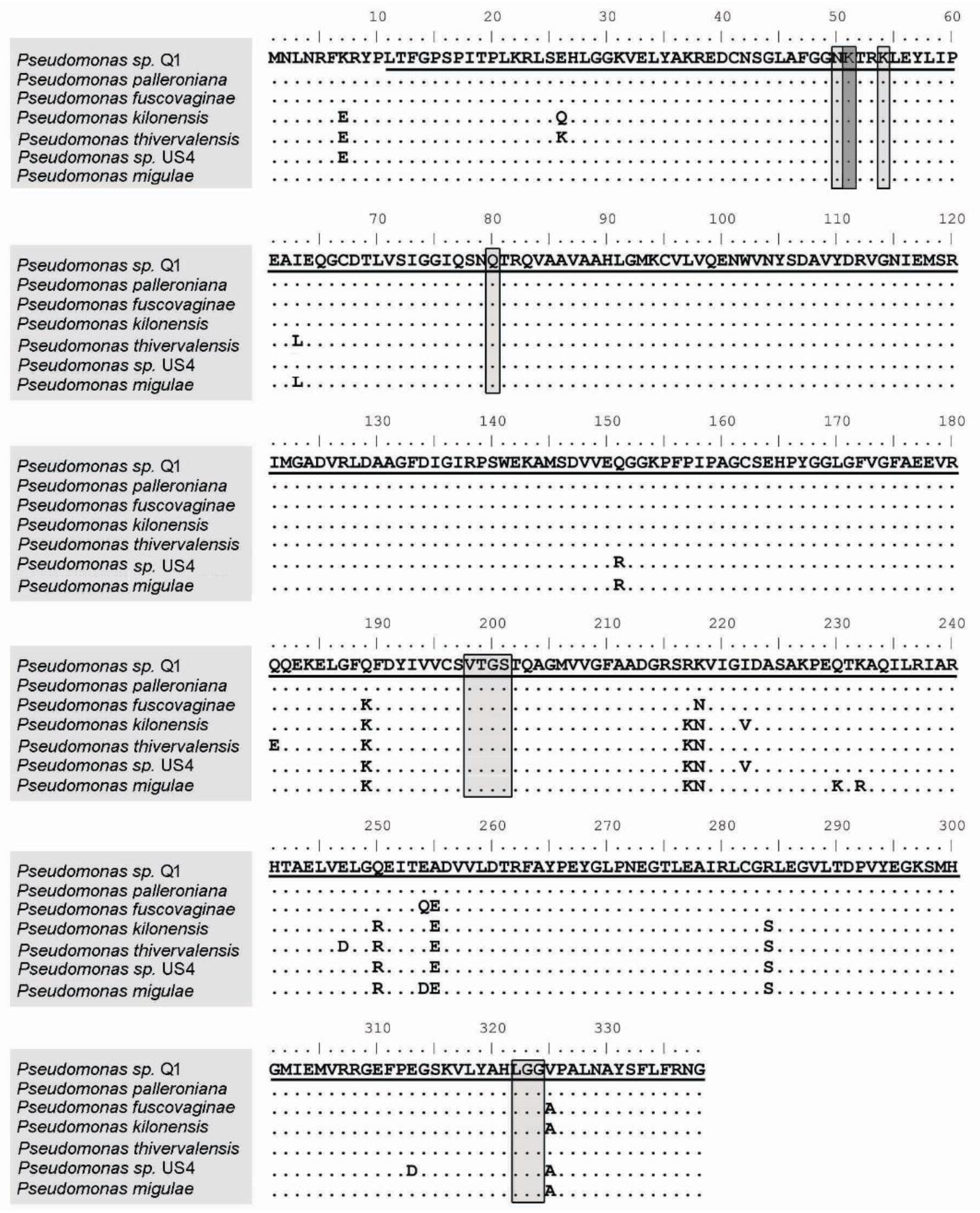
Figure 1. Alignment of AcdS protein amino-acid sequences from bacteria belonging to genus Pseudomonas. The conserved residues between all sequences are indicated by dots. The pyridoxal 5′-phosphate binding sites are indicated in light gray, and the catalytic residue is indicated in dark gray. The underlined amino acids correspond to the deletion fragment in Pseudomonas sp. Q1. Pseudomonas palleroniana accession WP_090372028, Pseudomonas fuscovaginae accession WP_019361456, Pseudomonas kilonensis WP_053178893, Pseudomonas thivervalensis accession WP_053183836, Pseudomonas sp. US4 accession WP_015096487.1 and Pseudomonas migulae accession WP_084321532.
Plants 09 01630 g001
Figure 2. Growth of Pseudomonas sp. Q1 and its derivatives strains (∆acdS mutant and ∆acdS+) and rhizobial strains (M. ciceri LMS-1, R. leguminosarum ATCC 14480T, E. meliloti ATCC 9930T) under Mn conditions (2 mM MnSO4). Percentages were calculated considering the control condition (without addition of MnSO4) as 100% growth. Bars indicate standard deviation.
Plants 09 01630 g002
Figure 3. Effects of single inoculation (M. ciceri LMS-1 (LMS-1)) and co-inoculation (M. ciceri LMS-1 and Pseudomonas sp. Q1 (LMS-1 + Q1) or M. ciceri LMS-1 and Pseudomonas sp. Q1 ∆acdS mutant (LMS-1 + ΔacdS)) on chickpea plants grown in a vermiculite pot assay for 54 days. (A) Number of nodules of plants. (B) Nodule plus root dry weight of plants. (C) Shoot dry weight of plants. Each point represents the mean and standard error values of five plants per treatment. Different letters (a–c) correspond to statistically significant differences (p < 0.05) within each group (non-stressed and Mn stressed). * denotes statistically significant differences between the treatments means in the presence and absence of high Mn concentration (* p < 0.01; ** p < 0.001).
Plants 09 01630 g003
Figure 4. Nodulation kinetics by a hydroponic assay of chickpea plants single inoculated (M. ciceri LMS-1 (LMS-1)) and co-inoculated (M. ciceri LMS-1 and Pseudomonas sp. Q1 (LMS-1 + Q1) or M. ciceri LMS-1 and Pseudomonas sp. Q1 ∆acdS mutant (LMS-1 + ΔacdS)). (A) Nodulation kinetics of chickpea plants during 26 days at non-stressed conditions. Each point represents the mean and standard error values of eight plants per treatment. The asterisks correspond to significant differences between the LMS-1 + Q1 ∆acdS treatment and the other two treatments. (B) Shoot dry weight at the end of the hydroponic assay (C) Nodule dry weight at the end of the hydroponic assay. Different letters (a–c) correspond to statistically significant differences (p < 0.05) within each group (non-stressed and Mn stressed). * denotes statistically significant differences between the treatments means in the presence and absence of high Mn concentration (** p < 0.001).
Plants 09 01630 g004
Figure 5. Effects of single inoculation (R. leguminosarum bv trifolii ATCC14480T (RL)), and co-inoculation (R. leguminosarum bv trifolii ATCC14480T and Pseudomonas sp. Q1 (RL + Q1) or R. leguminosarum bv trifolii ATCC14480T and Pseudomonas sp. Q1 ΔacdS (RL + ΔacdS Q1)) on subterranean clover plants grown in a vermiculite pot assay for 42 days. (A) Number of nodules of plants. (B) Nodule plus root dry weight of plants. (C) Shoot dry weight of plants. Each point represents the mean and standard error values of five plants per treatment. Different letters (a–c) correspond to statistically significant differences (p < 0.05) within each group (non-stressed and Mn stressed). * denotes statistically significant differences between the treatments means in the presence and absence of high Mn concentration (* p < 0.01).
Plants 09 01630 g005
Figure 6. Effects of single inoculation (E. meliloti ATCC9930T (EM)) and co-inoculation (E. meliloti ATCC9930T and Pseudomonas sp. Q1 (EM + Q1) or E. meliloti ATCC9930T and Pseudomonas sp. Q1 ΔacdS (EM + ΔacdS Q1) on burr medic plants grown in a vermiculite pot assay for 42 days. (A) Number of nodules of plants. (B) Nodule plus root dry weight of plants. (C) Shoot dry weight of plants. Each point represents the mean and standard error values of five plants per treatment. Different letters (a–c) correspond to statistically significant differences (p < 0.05) within each group (non-stressed and Mn stressed). * denotes statistically significant differences between the treatments means in the presence and absence of high Mn concentration (* p < 0.01; ** p < 0.001).
Plants 09 01630 g006
Figure 7. Nodulation kinetics by a hydroponic assay of subterranean clover plants inoculated with R. leguminosarum bv trifolii ATCC14480T (RL), and co-inoculated with R. leguminosarum bv trifolii ATCC14480T and Pseudomonas sp. Q1 (RL + Q1) or R. leguminosarum bv trifolii ATCC14480T and Pseudomonas sp. Q1 ΔacdS (RL + Q1 ΔacdS). (A) Nodulation kinetics of subterranean clover during 18 days under non-stressed conditions. (B) Nodulation kinetics of plants during 18 days under Mn conditions. Each point represents the mean and standard error values of ten plants per treatment. The asterisks correspond to significant differences between the RL + ∆acdS mutant treatment and the other two treatments. (C) Shoot dry weight of subterranean clover at the end of the hydroponics experiments. Different letters (a–b) correspond to statistically significant differences (p < 0.05) within each group (non-stressed and Mn stressed). * denotes statistically significant differences between the treatments means in the presence and absence of high Mn concentration (p < 0.01).
Plants 09 01630 g007
Table 1. Bacterial strains and plasmids used in this study.
Plasmids or BacteriaDescriptionReference or Source
Plasmids
pNZY28-bluntAmpr, blunt endsNZYtech
pJET1.2/bluntAmpr, blunt endsThermoFisher Scientific
pT18mobsacBplasmid suicide; Kmr, sacBs[83]
pRK600Helper plasmid pRK2013, npt::Tn9, Cmr[84]
pGRG36Ampr, arabinose-inducible promoter, RP4 conjugal transfer origin, pSC101 temperature-sensitive origin, presenting tnsABCD genes for Tn7 transposition[85]
pGRG36-TetrpGRG36 derivative, TetrThis work
pNZY28-∆acdSregions flanking acdS in pNZY28 This work
pJET1.2-acdSacdS gene and its promoter and terminator regionsThis work
pT18mobsacB-∆acdSregions flanking acdS in pT18mobsacBThis work
pGRG36-Tetr-acdSacdS gene and its promoter region of Pseudomonas sp. Q1 strain in pGRG36‑Tetr This work
Pseudomonas sp.
Q1Pseudomonas sp. Q1, acdS+[18]
Q1 ∆acdS Derivative of strain Q1, acdS-This work
Q1 ∆acdS+ complemented Derivative of strain Q1 ∆acdS, acdS+ with a copy of acdS gene and its promoter and terminator regionThis work
Rhizobia
LMS-1M. ciceri, acdS+, nif+, nod+, Nodulates chickpea (Cicer arietinum)[26,34]
ATCC 14480TR. leguminosarum bv. trifolii, Nodulates red clover (Trifolium praetense) and white clover (Trifolium repens) ATCC® Microbiome Standards
ATCC 9930TE. meliloti, Nodulates (Medicago sativa) and sweet clover (Melilotus alba)ATCC® Microbiome Standards
Escherichia coli
DH5αHost for cloning[82]
WM3064Host for conjugation, presence of RP4 (tra) in the chromosome[86]
MT616Strain containing helper plasmid pRK600 [84]
Note: Ampicillin (Ampr), kanamycin (Kmr), tetracycline (Tetr) and Chloramphenicol (Cmr) resistance. Sucrose sensitivity (SacBS). Presence (acdS+) or absence (acdS-) of acdS gene. Presence of nitrogen -fixation (nif)+ and nodulation (nod+) genes.
Table 2. Primers used in this study.
Primer NamesPrimer Sequence (5′→3′)Application
Tet-SdaI-Fw
Tet-SdaI-Rv
CCTGCAGGGGACAAGGGAAAACGCAAG
CCTGCAGGCCGTCAGCGTTTTGTAATGG
tetA gene and its promoter and terminator regions (2624 bp)
acdS-Q1-Fa
acdS-Q1-Rb
TCAAGAGGTTGACGGGTTCT
CTGGACGCGAGCGTGAAGGGCGTGATGGGAGA
acdS upstream region (948bp)
acdS-Q1-Fc
acdS-Q1-Rd
TCTCCCATCACGCCCTTCACGCTCGCGTCCAG
AACATCGCAAAGACGTAGGG
acdS downstream region (898bp)
acdS-Q1-Fa
comp_acds_Q1 Rv
TCAAGAGGTTGACGGGTTCT
AATGCCCTCGATTGCCGGA
acdS gene and its promoter and terminator regions (1242 bp)
RL_acdS_all_Fw
RL_acdS_all_Rv
AACGCTATCCGCTCACCTT
ATCCCCTGCATCGACTTTC
acdS gene fragment (886 bp) from R. leguminosarum bv trifolii ATCC 14480T
EM_acdS_all_Fw
EM_acdS_all_Rv
CAGCCGTCCCTGTAGTAATAGC
GAAAAGTTCGAACGCTACCC
acdS gene fragment (1006 bp) from Ensifer meliloti ATCC 9930T
Publisher’s Note: MDPI stays neutral with regard to jurisdictional claims in published maps and institutional affiliations.