Review

Immune Response to Herpes Simplex Virus Infection and Vaccine Development

1
Department of Microbiology, Faculty of Biological Sciences, University of Nigeria, Nsukka, Enugu 410001, Nigeria
2
Department of Biological Sciences, College of Liberal Arts & Sciences, Wayne State University, Detroit, MI 48202, USA
3
Department of Biotechnology, Graduate School of Engineering, Osaka University, Suita City, Osaka 565-0871, Japan
*
Author to whom correspondence should be addressed.
Vaccines 2020, 8(2), 302; https://doi.org/10.3390/vaccines8020302
Submission received: 5 May 2020 / Revised: 29 May 2020 / Accepted: 8 June 2020 / Published: 12 June 2020
(This article belongs to the Special Issue Vaccine Development for Herpes Simplex Viruses)

Abstract

:
Herpes simplex virus (HSV) infections are among the most common viral infections and usually last for a lifetime. The virus can potentially be controlled with vaccines since humans are the only known host. However, despite the development and trial of many vaccines, this has not yet been possible. This is normally attributed to the high latency potential of the virus. Numerous immune cells, particularly the natural killer cells and interferon gamma and pathways that are used by the body to fight HSV infections have been identified. On the other hand, the virus has developed different mechanisms, including using different microRNAs to inhibit apoptosis and autophagy to avoid clearance and aid latency induction. Both traditional and new methods of vaccine development, including the use of live attenuated vaccines, replication incompetent vaccines, subunit vaccines and recombinant DNA vaccines are now being employed to develop an effective vaccine against the virus. We conclude that this review has contributed to a better understanding of the interplay between the immune system and the virus, which is necessary for the development of an effective vaccine against HSV.

1. Herpes Simplex Virus and the Immune System

1.1. Introduction

Herpes simplex virus belongs to the Herpesviridae family, a group of spherical viruses measuring 120–200 nm. There are two types of Herpes simplex viruses, Herpes simplex virus type 1 and type 2 (HSV-1 and HSV-2). These viruses cause lifelong infections that are among the most common viral infections worldwide [1,2,3]. As part of the global effort to control the infections caused by these viruses, many vaccines have been developed [4,5,6,7,8]; however, to date, none has been licensed for use in humans. Following the successful development and use of vaccine against varicella zoster virus, a closely related virus in the same viral family, there has been a recent upsurge of interest in developing vaccine against HSV. It is believed that one of the major problems with vaccine development against HSV is the complex interactions that exist between the immune response and the virus. The immune system which consists of an innate and adaptive component has cytosolic sensors which sense the DNA of this virus and consequently stimulate inflammatory response against it. Intriguingly, HSV possesses a repository of arsenals that ensures its successful replication in the human host. The interplay between these arsenals and the immune system determines an outcome. In this review, we explore how the knowledge of the immune response can and has been used in the development of a functional vaccine against HSV.

1.2. Overview of the Immune System

The human body is equipped with an immune system that acts as guard against invading pathogens, which are ubiquitous in the environment. Broadly, the human immune system is of two types, the innate and the adaptive immunity. The innate immunity consists of both the structural component, and the proteins which recognize molecular patterns not present in human cell. They constitute the first line of defence against pathogens. The adaptive immunity is a more specific defence against specific pathogens mediated by the B-cells and the T- cells. One attribute of this immunity is that its effect is long lasting.

1.2.1. The Innate Immune System

The innate immune system is the first point of defence in eukaryotic organisms; it is usually fast and non-specific. It is broadly divided into two, namely, the structural component (anatomical barrier) and chemical component. The structural component involves the skin and the mucus membrane. The skin provides an outer impermeable cover against invasion by pathogens [9]. The skin also secretes chemicals (sweat) and fatty acids which are toxic to invading pathogens and exhibit antimicrobial property. The desiccation and desquamation nature of the skin are also known to prevent bacterial colonization [10]. The mucus membrane is less impermeable compared to the skin. Infection via the mucus membrane involves colonization and ability to overcome the defence of the membrane. The different mucus membranes protect the body against infections using different mechanisms, the upward flapping movement of the cilia in the respiratory tract, the mobility and low pH of the stomach in the gastrointestinal tract and the constant flushing of the urinary tract.
The chemicals components include, the lysozyme, defensins, interleukin, interferon, and complement proteins. Lysozyme is a 1, 4-β-N-acetylmuramidase enzyme present in body secretions such as tears and saliva and mainly acts on bacterial cells [11]. The complement proteins act in a cascade-dependent manner to eliminate pathogens [9]. They can also help in the phagocytic process by opsonizing the pathogen, which facilitates easy uptake by the phagocytes. Interferons are antiviral proteins made by viral-infected cells alongside the lymphocytes. They help establish antiviral state in neighbouring cells thus limiting the dissemination of the viral agent. The neutrophils remove pathogens via production of reactive oxygen species that are toxic to them. They have also been implicated in tumour necrotic factor and interleukin-12 cytokines [12]. Macrophages aid in phagocytising process. Macrophages, alongside dendritic cells, link the innate and the adaptive immunity by processing and presenting antigens to the adaptive immune cells. Eosinophils contain granules that have toxic enzymes and molecules very active against helminths and parasites. Furthermore, pathogen pattern receptors such as the toll-like receptors (TLR) and nucleotide-like oligomerization domain like (NOD-like) receptors play vital roles in innate immunity. They act as sensors of pathogen-associated molecular patterns and alert the cell of an invading danger.
Together, all these work in consortium to prevent the human cells from infection caused by pathogens by removing them and helping to recruit the adaptive immune response.

1.2.2. The Adaptive Immune System

Although the innate immune system is first to attack an invading pathogen, it can be overpowered or tricked by the invading pathogen. At this stage, adaptive immunity is called up to assist. Two distinct features of the adaptive immune system are specificity and memory. Specificity here connotes that it act only against the pathogens that elicited its response, and it keeps a memory of the pathogen for future reference. The first infection takes time for response to be mounted, while in repeated infections, the immune response is faster. The B-lymphocyte and the T-lymphocyte are the principal players of the adaptive immunity. The B-cells produce antibodies and mediates the humoral immune response while the T-cells oversee the cellular immune response.

1.3. The Interplay of Herpes Simplex Infection and the Immune System

The host–pathogen interaction is dynamic that has the potential to result in a diseased condition. Factors such as the virulent factors of the pathogen (encoded proteins) could help it in navigating the immune resistance of the host. Toll-like receptors 2 and 3 have been reported to play a role in sensing HSV-1 and the activation of interferon type 1 and inflammatory cytokines [13]. HSV-1 tegument kinase US3 has been reported to inhibit TLR-3 expression and dampen interferon type 1 synthesis [14]. Furthermore, myD88 (a critical adapter molecule of the TLR pathway) levels in the cells are drastically reduced by the infected cell polypeptide 0 (ICP0) of HSV-1 [15]. The ICP0 protein carries out this reduction via its proteosomal and E3 ligase activities. ICP0 is a promiscuous transactivator of HSV-1 immediate–early, early and late genes [16]. ICP0 is an E3 ubiquitin ligase comprising 775 amino acids with an N-terminal RING structure [17]. ICP0 promotes viral replication and reactivation from latency by inhibiting interferon synthesis [16]. This function is dependent upon the degradation of ND10 constituent proteins (PML and SP100) using its ubiquitin ligase activity [15,17,18]. The role of ICP0 protein in HSV-1 infection has been extensively reviewed [19]. The ICP0 protein mutant HSV-1 has been reported to have a low growth rate. Undegraded PML protein stimulates interferon production [17], and this can serve as a platform for an attenuated HSV-1 vaccine.
Additionally, HSV has been reported to inhibit tumour necrotic factor (TNF) alpha NF-kB activation of genes involved in inflammatory response [20]. TNF-α is a cytokine vital in innate immunity and, upon synthesis, induces the expression of genes involved in the inflammatory response. TNF-α binds its receptor TNF-R1 recruiting the adapter protein TNF receptor death domain (TRADD), which then recruits TNFR-associated factor 2 (TRAF2) and receptor-interacting protein 1 (RIP1) to the complex. TRAF 2 modulation of K63-based poly-ubiquitination of RIP1 brings about TGF-β-activated kinase-1 (TAK1), turning on the kinase activity of IκB kinase (IKK) which phosphorylates and degrades IκBα, subsequently leading to the activation of the nuclear factor kappa B (NF-κB), a transcriptional factor [21]. However, HSV-1 γ134.5 protein (late gene encoded) represses NF-κB activation in CD8+ dendritic cells [22]. Furthermore, a tegument protein, VHS, has also been reported to inhibit the viral replication independent NF-κB activation [23]. A HSV-1 protein, UL42, that enhances the processivity of the DNA polymerase, inhibits the TNF-α dependent NF-κB activation [20]. It was found out that UL42 binds the p65 and p50 subunits of NF-κB and inhibits their translocation into the nucleus. This will repress the transcription of genes involved in inflammatory reactions. These can be illustrated diagrammatically, as shown in Figure 1.
Other cytokines such as type I interferon can be activated by the RLR cytosolic signalling pathway. This is produced in response to the IRF3 activation and NF-κB in the RLR pathway. US11 protein prevents RIG-1 and IPS-1 interaction (dimerization of IPS-1 and MDA5) [24]. Similarly, the TLR2/TIR/MyD88/Mal signalling pathway stimulates the NK-Kb transcription factor, which facilitates the synthesis of pro-inflammatory cytokines such as interleukins 6, 8, and 12 [25]. Signalling through TLR2/TIR/MyD88 activates IRF3 and IRF7, which promote the production of interferon-alpha and beta [26].
Furthermore, CD8+ TRM, a subtype of memory lymphocyte which resides in non-lymphoid tissues [27], has been reported to trigger adaptive immune response during HSV-1 infection [28] through IFN-Y and granzyme B effectors. CD4+ cells have been reported to inhibit HSV-1 infection and clear HSV-1 from genital infection sites following primary infection. Naïve CD4+ can differentiate into Th1, Th2, Th17, and induced regulatory T (iTreg) upon interaction with the major histocompatibility complex (MHC) (an antigen complex). The subpopulation that naïve CD4+ T cell differentiates into depends on the cytokine within the native CD4+ environment [29]. CD4+ CD25+ has reportedly been involved in HSV-1 response [30], and the deletion of CD4+ cells increased susceptibility to HSV-1 infection in mice [31]. Yu and colleagues reported that regulatory T (Treg) level positively correlates with viral infectivity and is a requirement in establishing latency [32]. The role of adaptive immune cells in HSV-1 infection has been extensively reviewed in [33].

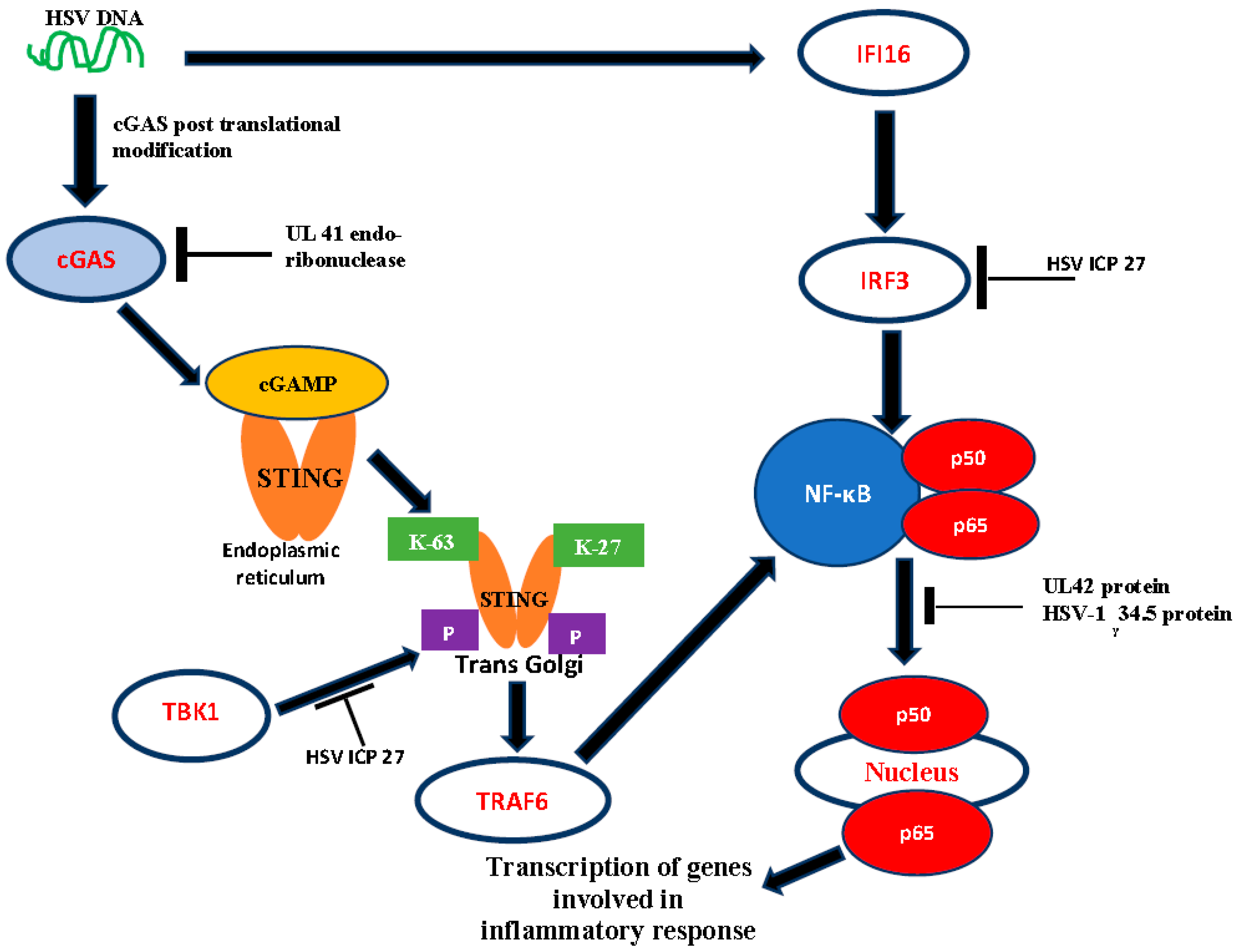
1.4. DNA Sensors as Activators of Host Antiviral Response

Pathogen-associated molecular patterns (PAMPs) are a variety of molecules found on pathogens which are usually recognized by the pathogen recognition receptors and subsequent activation of interferon and chemokines, independent of the TLR and retinoic acid-inducible gene 1 (RIG-1) [13]. Some of the DNA cytoplasmic sensors that have been reported to mediate HSV-DNA recognition include-cyclic GMP-AMP (GAMP) synthase (cGAS)—this is a cytosolic DNA sensor of viral DNA with capacity to recruit the stimulator of interferon genes (STING) adapter protein. The cGAS interacts with DNA via its N-domain, and this interaction is modulated by post-translational modifications such as sumoylation, acetylation, phosphorylation and glutamylation [34,35,36,37]. This interaction causes the synthesis of the chemical messenger cGAMP which binds the endoplasmic reticulum-anchored STING, causing its oligomerization [38]. The oligomerized STING moves to the trans-Golgi, where K27 and K63-linked poly-ubiquitin moieties are added, catalysed by E3 ubiquitin ligase [39]. The K-27 and K-63 call upon the TBK1, which is a kinase that will phosphorylate STING. STING can also recruit TRAF6 to activate the transcription factor NF-κB for expression of genes involved in inflammatory response [40]. Following NF-κB activation, STING gets degraded in the lysosome terminating the DNA sensing. STING activates the interferon regulatory factor (IRF3) and NF-κB signalling pathway to exert its effects [40]. The same authors reported that STING also activates the Jun N-terminal protein kinase/stress-activated protein kinase (JUN/SAPK) pathway.
According to Zhang and colleagues [41] DDX41 (DEAD-box helicase 41), a member of the DEAD-like helicases superfamily (DEXDc) senses DNA in myeloid dendritic cells, which stimulates type I interferon production. It has been reported that the knockdown of the DDX41 gene inhibited mitrogen-activated protein kinase TBK1 and NF-κB transcription factor by DNA, and it depends on STING to sense pathogenic DNA. HSV-1 capsid ubiquitination and Vp5 degradation occur in a proteasome-dependent manner [42]. This liberates the DNA into the cytosol where they get sensed by cytosolic DNA sensors, subsequently inducing the innate immune response. A PYRIN protein interferon gamma inducible protein 16 (IFI16), which functions as a cytosolic DNA sensor that directly binds DNA, has been reported. The study found that the knockdown of the IFI16 gene in mouse models inhibited the activation of transcription factors IRF3 and NF-κB on infection by HSV-1 [43]. This showed that IFI16 is essential for the activation of IRF3 and NF-κB. IFI16 interacts directly with interferon-β, inducing DNA to recruit IRF3 and NF-κB for the transcription of genes involved in inflammatory response. In a recent publication, HSV-1 was shown to also activate the inflammatory response via the nucleotide-binding domain and leucine-rich repeat-containing receptor 3 (NLRP3) [44].

2. HSV Immune Evasion Mechanisms

It is critical to consider the mechanisms by which HSV evades the immune system during the process of designing and administration of different vaccine compounds and formats [45]. In fact, the major impediment in the search of a potent cure or vaccine for HSV infection is the myriad of mechanisms through which the virus evades the immune system. Thus, continuous elucidation of these mechanisms is critical to achieve potent prophylactic or therapeutic intervention for HSV.

2.1. Modulation of Autophagy

Autophagy has housekeeping roles in regulating normal physiological cellular processes. Degradation of misfolded proteins, cellular differentiation, and defence against pathogens are several of the functions of autophagy [46]. Autophagy can promote innate and adaptive immunity. One study showed that HSV-1 induced autophagy in both immature and mature dendritic cells which are crucial for the induction of antiviral immune responses [47]. The virus selectively targets lamin A/C, B1, and B2 for degradation, which then facilitates the egress of newly formed infectious particles. The same authors also found out that the overexpressing of two proteins KIF1B and KIF2A, which are both members of the kinesin-3 family [48], attenuated this rate of egress via reduced lamin degradation. It has been shown that HSV-1 not only inhibits autophagy in neuronal cells, but also in non-neuronal cells. Interestingly, in the non-neuronal cells, inhibition of autophagy was achieved by the phosphorylation of two autophagy regulators: ULK1 and Beclin1, which are dependent on the HSV-1 protein Us3 ser/thr kinase. The authors also showed that depleting both ULK1 and Beclin1 rescued the replication of the Us3-deficient virus strain [49]. This is significant, as Us3 also plays a role in viral manipulation of cellular apoptosis [50]. Another novel autophagy inhibition mechanism employed by HSV is the downregulation of the mitophagy [51], adaptor optineurin (OPTIN) and the autophagy adaptor protein sequestosome (p62/SQSTM1) [52]. This downregulation is mediated through the E3 ubiquitin ligase activity of the intensely studied HSV-1 immediate early protein ICP0 [53]. It has also been demonstrated that exogenous p62/SQSTMI decreased the HSV viral yield [52], thus confirming a potent antiviral activity, but the exact mechanisms by which the proteins mount antiviral responses still needs to be elucidated. HSV-1 also inhibits autophagy through the protein Us11 [54]. Recently, Liu et al. [55] showed in their study that Us11 disrupts the tripartite motif protein 23 (TRIM23)–TANK-binding kinase 1 (TBK1) complex, thus suppressing the efficiency of the autophagy-mediated cellular response to HSV infection. Their results suggest that Us11 competes for binding to the ARF domain with TBK1, thus, inhibiting the efficient binding of TBK1. In an earlier article by the same group, It was reported that Us11 can also target the TBK1 by binding to heat shock protein 90 (hsp90), thereby preventing the formation of the TBK1-hsp90 complex, which is also required for the efficient functioning of the autophagy response mediated by TBK1 [56]. Us11 also binds to protein kinase R [PKR] and together with ICP34.5, which binds to Beclin1 modulate autophagy via the eukaryotic translation initiation factor 2-α kinase (EIF2AK2/PKR) pathway [57]. The dissimilarity between autophagic stimulation of HSV-1 and HSV-2 has been investigated [58]. While HSV-1 inhibits total autophagic activity, HSV-2 maintains basal autophagic activity. Disruption of basal autophagic activity leads to neurodegeneration, which suggests the link between HSV-1 and neurodegeneration. Basal autophagy suggests that autophagy contributes to continuous infection by limiting inflammation, IFN-1 production and NF-kβ regulation. Subsequently, the persistent latency of HSV-1 in the neuron is maintained by the stimulation of autophagy [58,59].

2.2. Interplay of HSV-1 and Host PML Protein

Promyelocytic leukaemia nuclear bodies (PML-NBs), also referred to as nuclear domains 10 (ND10), or PML oncogenic domains (PODs), are small (0.1–1.0 µm) dynamic nuclear structures made of several constant (PML, SP100, Daxx) or transiently associated proteins, depending on the cell function and/or exposed stress [60,61,62]. They respond to varieties of stimuli including apoptosis, senescence viral infections and interferon response [63]. PML-NBs are suggested to be the site of nuclear activities, nuclear protein depots and hotspots for posttranslational modifications [64]. They restrict HSV-1 gene expression and replication by forming structures called viral DNA-containing PML-NBs (vDCP NBs), which is an intrinsic antiviral response strategy of preventing lytic infection [65]. Latency could be attributed to HSV-1 chromatinization mediated by the H3.3 chaperone complex (DAXX/ATRX) [65] and HIRA/UBN1/CABIN1 [66]. It was suggested that the α- thalassemia mental retardation X-linked protein (ATRX) is dispensable in the initial loading of heterochromatin, and is only required for stability maintenance of the viral heterochromatin [67]. Histone cell cycle regulator (HIRA) is suggested to play an independent role in the regulation of intrinsic and innate immune regulation of HSV-1 [68]. Therefore, understanding the underlying mechanisms of HSV-1 chromatinization-induced latency is important. The PML-NBs is organized by protein–protein interaction between SUMOylated proteins and SUMO-interacting motifs (SIMs) [61]. PML is the key protein responsible for the assembly and maintenance of PML-NBs. PML upon SUMOylation recruits other PML-NB-associated proteins to the nuclear structure [62,69,70]. PML and SP100 is activated upon induction by interferon (IFN), leading to an increase in PML-NBs [60]. PML, together with SP100, the death domain-associated protein 6 (DAXX), and ATRX, contributes to the repression of viral gene expression in a cooperative manner [64]. However, this mechanism to repress HSV-1 gene expression is counteracted by ICP0 through its E3 ubiquitin ligase activity. Using a combination of SUMO-dependent and independent targeting strategies, HSV-1 (ICP0) ubiquitinates and degrades PML and SP100, resulting in the disruption of PML-NBs and the subsequent release of viral genes [61].
It is suggested that the biological activity of ICP0 is connected to the host cell SUMOylation events, as mutations of some SIM-like sequences (SLSs) in the c-terminal quarter of ICP0 on HSV-1 reduced ICP0-mediated degradation of PML [71]. The association between ICP0 and PML-NBs is said to be a series of process involving adhesion, fusion and retention [72]. Moreover, ICP0 utilizes different motifs of PML-NBs fusion and SUMO interaction [72], suggesting different molecular mechanisms of PML- ICP0 interaction and degradation. More so, among the PML isoforms, different methods of interactions and degradation were observed to be utilized by ICP0 in Hep-2 and U2OS cells [73]. Hembram and colleagues identified a bona fide SIM in ICP0 which is essential to target SUMOylated PML. It was reported that the phosphorylation of this ICP0 motif by host kinase Chk2 increases the potency of ICP0 to act as a SUMO targeted ubiquitin ligase [STUbl]. Their findings indicate that three post-translational modifications, namely, ubiquitination, SUMOylation and phosphorylation, were involved in an unprecedented crosstalk in the ICP0′s degradation of PML [74]. In another study, Fada and colleagues reported that a PML II mutant lacking both lysine SUMOylation and SIM rendered it unrecognizable by ICP0, preventing ICP0-mediated degradation while still maintaining PML II localization to ND10 [75]. Moreover, it was observed that SP100 degradation was delayed in PML-/-infected cells and that the accumulation of ICP0 was reduced at low but not high multiplication of infection [76]. On the contrary, higher wild-type virus yields were observed in SP100-/-infected cells than in the parental Hep-2 cells at low multiplication of infection [60]. This suggests that HSV-1 may have hijacked the PML stress response function for successful infection. The function of SP100 may be different and could have an important role in suppressing HSV-1 infection. A recent review by Full and Ensser also addressed this interplay between HSV-1 and host PML proteins [77].

2.3. Modulation of Apoptosis

Apoptosis is a crucial cellular defence mechanism that ensures the elimination of pathogen-infected cells and has been reviewed by He and Han [78], but there are recent interesting mechanisms of apoptosis modulation by HSV not captured in the aforementioned review that will be outlined in this section. One of the recent findings was on a study that focused on the Herpes simplex encephalitis and the impact of the blood–brain barrier in modulating the pathological development of the disease [79]. It was reported that HSV-1 infection led to the activation of apoptosis with accompanying golgi fragmentation and downregulation of occludin and claudin 5. These cellular responses were mediated by the protein GM130 which was downregulated due to HSV-1 infection [79]. Overexpression of the GM130 attenuated apoptosis, and in cells infected with HSV-1, the protein levels of both occludin and claudin were partially restored. Despite the potentiation of apoptosis reported above, it is established that inhibition of apoptosis is critical for the reactivation of latency by HSV [80], and this mechanism is mediated by the action of the latency-associated transcript (LAT), which is the only HSV transcript expressed significantly during latency. Another recent study showed that CD80 can compensate for the functions of the LAT, such as the establishment of latency, reactivation and immune exhaustion in cells infected with the LAT-null virus [81]. This is significant because this is the first time that such overlapping functionalities between LAT and CD80 have been reported. Although CD80 rescues the functions of LAT, it has distinct functions as CD80 exacerbated eye disease in mice compared to wild-type HSV. Furthermore, CD80 potentiated the expression of the anti-apoptotic Bcl-2 gene thus modulating apoptosis in the infected cells [81]. Another HSV-1 protein that has implications in regulating cellular apoptosis—ICP22 has been shown to function via a mechanism like that used by the cellular J-protein/HSP40 family chaperone [82]. Other HSV proteins that have been implicated in evasion of cellular immune response by interference of apoptosis include Us3 and Us5 [83]. Both proteins interact with critical proteins of the apoptotic pathway. Us3, which is a serine/threonine kinase, inhibits Bad-induced apoptosis, while Us5 is a glycosylated J protein that inhibits Fas-mediated apoptosis [50].

2.4. Intracellular Cell-to-Cell Propagation

It has been shown that HSV can make use of extracellular vesicles, not just in mediating intercellular cell to cell propagation, but also in evading the immune system [84]. The manipulation of the MHC class II processing pathway through alteration of the endosomal sorting and trafficking of HLA-DR by HSV-1 has been consistently shown to be an immune evasion mechanism. HSV-1 also exploits the anterograde transport mechanism [84] in cells to move viral particles from neuron cell bodies to axon tips during reactivation of infection, and this is implicated in the spread of viral particles in epithelial tissues and general dissemination of the virus to other hosts. It has been shown that kinesin-1 proteins KIF5A, -5B, -5C play crucial roles in this anterograde transport mechanism. Targeting the transport of reactivating HSV has crucial therapeutic significance [48].

2.5. Inactivation in Expression of Signaling Pathways

It has been suggested that HSV-induced Dok phosphorylation and Dok-2 degradation could be a strategy of immune evasion to inactivate T-cells, which might play a role in HSV pathogenesis [85]. Ectopic expression of VP22 was said to decrease cGAS/STING-mediated IFN-β promoter activation and expression of IFN-β [86]. It has been suggested through further studies that VP22 interacts with cGAS and inhibit its enzymatic activity. The γ134.5 protein of HSV-1 was said to inactivate STING and, more so, disrupts its translocation from endoplasmic reticulum to golgi apparatus, an important process necessary to prime cellular immunity [87]. This leads to downregulation of interferon regulatory factor 3 (IRF3) and IFN responses. Another HSV-1 protein, virion host shutoff endonuclease (UL41), was reported to decrease cGAS/STING-mediated IFN-β promoter activation and expression [88]. This protein was reported to degrade cGAS via its RNase activity. It has equally been observed that HSV-1 neuronal infection triggers activation of Src tyrosine kinase, phosphorylation of dynamin 2 GTPase and perturbation of golgi apparatus (GA) integrity [89]. A scattered and fragmented distribution of the GA through the cytoplasm with swollen cisternae and disorganized stacks was observed in HSV-1-infected neurons in contrast with the uniform perinuclear distribution pattern observed in control cells [89]. The evasion of HSV-1-specific CD8+ T cells which accumulates in infection sites is enhanced by HSV-1 UL13 kinase through reducing the expression of the CD8+ T cell attractant chemokine CXCL9 in the CNS of infected mice, leading to increased HSV-1 mortality due to encephalitis [90]. VP24 protein was reported to dampen interferon stimulatory DNA (ISD)-triggered IFN-β production and inhibit IFN-β promoter activation induced by cyclic cGAS and STING [91]. The ectopic expression of VP24 selectively blocked interferon regulatory factor 3 (IRF3) and downregulated ISD-induced phosphorylation and dimerization of IRF3 during HSV-1 infection in a VP24 stable knockdown human foreskin fibroblast cell line [91]. Here, VP24 disrupts the interaction between TANK- binding kinase 1 (TBK1) and IRF3, impairing IRF3 activation. It has been reported that HSV-1 downregulates CD1d cell surface expression and suppresses the function of NKT cells through its viral protein kinase US3 [92]. US3 phosphorylates KIF3A at serine 687, leading to downregulation of CD1d expression. The interferon-induced protein with tetratricopeptide repeat 3 (IFIT3) is an antiviral host intrinsic factor that restricts replication of DNA and RNA viruses. UL41 (HSV-1 tegument protein) was reported to inhibit the antiviral activity of IFIT3 [93]. UL41 diminishes the accumulation of IFIT3 mRNA to abrogate its antiviral activity. A study reported CD1d expression downregulation and subsequent suppression of NKT cells function, using its viral serine/threonine protein kinase US3 as another strategy of the virus to evade immune response [94]. Peroxisomes are thought to be important signalling platforms of antiviral innate immunity, as signalling from peroxisomal MAVS (MAVS-Pex) triggers a rapid production of IFN-independent and -stimulated genes (ISGs) against invading pathogens. Another study also revealed that HSV-1, through its tegument protein VP16, blocks MAVS-Pex-mediated early ISG expression to dampen the immediate early antiviral innate immunity signalling from peroxisomes [95].

2.6. Role of miRNA in HSV Immune Evasion

MicroRNA (miRNA) is a short (20–24 nucleotides) non-coding RNA that is involved in post-transcriptional regulation of gene expression in multicellular organisms. MicroRNA 146a (miR146a) inhibits the expression of STAT1. However, Nuclear Dbf2-related kinase 1 (NDR1), which promotes virus-induced production of type 1 IFN, proinflammatory cytokines and ISGs, enhances STAT1 translation by binding to the intergenic region of miR146a. This leads to inhibition of the expression of miR146a, subsequently liberating STAT1 from miR146a-mediated translational inhibition [96]. More so, STAT1 binds to the promoter of miR146a, decreasing its expression. A study investigated the role of LAT encoded miRNAs in resistance to apoptosis and establishment of latent infection [97]. Five miRNAs (miR-H3, miR-H4-3p, miR-H4-5p, miR-H24, and miR-H19) encoded by latency associated transcript RLI sequence are implicated due to the overexpression of miR-H3, miR-H4-5p and miR-H19 in PC12 cells [97]. Six upregulated miRNAs (miR-592, miR-1245b-5p, miR-150, miR-342-5p, miR-1245b-3p and miR-124) were reported to downregulate TLR pathway-associated genes following HSV-2 infection [98]. Host-encoded miR-649 was reported to promote HSV-1 replication through regulation of the mucosa-associated lymphoid tissue lymphoma translocation gene 1 (MALT1)-mediated antiviral signalling pathway [99]. Micro-RNA-155 (miR-155) was reported to contribute to the pathogenesis of stomal keratitis [100]. HSV-1-encoded miRH8 was reported to target PIGT of the glycosylphosphatidylinositol (GPI), resulting in reduced expression of the antiviral protein tetherin and GPI-anchored activation of NK cell ligands, as well as a subsequent decrease in viral recognition and elimination by NK cells [101], which led to enhanced viral spread. miR-23a binds to the three prime untranslated region (3′ UTR) of interferon regulatory factor 1 (IRF1) to downregulate its expression and, thus, facilitates HSV-1 spread [102]. miR-221 negatively regulates IFNβ production at the time of virus production and miR-221 is induced by ELF4 by binding to the miR-221 promoter [103]. miRNA-H4-5p binds to 3′ UTR of CDKN2A and CDKL2, which reduces their expression and subsequently leads to reduced apoptosis and cell cycle progression [104].
Moreover, miR-H6 targeting of ICP4 inhibits HSV-1 productive infection and a decrease in production of IL-6 in human cornea epithelial (HCE) cells [105]. A group of researchers reported using hsa-miR-7704, expressed on macrophage, to inhibit HSV-1 in infected HeLa cells [105]. miRNA401 delivered to cells through exosomes were demonstrated to reduce viral yields via targeting ICP4 [106], an essential viral regulatory protein in a dose-dependent manner. miR-101, an ICP4-induced expression, was reported to downregulate RNA-binding protein G-rich sequence factor 1 (GRSF1) expression, inhibiting the replication of HSV-1 [107], as binding of GRSF1 to HSV-1 p40 mRNA, enhances its expression and viral proliferation [107].

3. HSV Vaccination and Immunotherapies

3.1. HSV Vaccination

Vaccination against HSV virus has been a far-fetched success, as there are currently no approved vaccines against the virus due to the reason that those produced have not successfully passed the clinical trials for safe use in humans. The recent interest in HSV vaccine development could be attributed to the recorded success in the vaccine against varicella zoster virus (VZV) and because both viruses (HSV and VZV) are alpha-herpes viruses, sharing similar pathogenesis pattern. Both viruses infect the skin and nerves, develop latent infections in the trigeminal and dorsal root ganglia, and have a tendency to reactivate. Therefore, there seems to be intensified efforts towards HSV vaccine development. Different kinds of vaccines have been developed for the treatment and/or prevention of HSV (Table 1), however none has been licensed for human use.

3.2. Potential Vaccine Candidates

There are several recent reviews that addressed the advancement of HSV vaccine design and discussed numerous emerging HSV candidate vaccines [144,145,146]. In this section we provide an update to this discussion by highlighting the new published findings in the search for novel prophylactic and therapeutic HSV vaccines not captured in prior reviews.
Different approaches being employed in candidate vaccine development include subunit vaccines, replication incompetent viruses and live attenuated vaccines [45]. It has been reported that HSV-1 mutant strains carrying modified Us3 and Us5 when used to infect mice triggered an asymptomatic immune response when challenged with the wild-type virus [145]. Interestingly, this immune response led to significantly less inflammatory cell aggregation in nervous tissues compared to the wild-type HSV-1 which induced an intense inflammatory reaction [145].
An exciting new study reports the development of a novel DNA vaccine against HSV-2 [146]. Their DNA vaccine encoded the following viral proteins: gB, gD, ICP0, ICP4 and UL39. It was reported that upon inoculation of the test animals with the vaccine, significant T cell response was generated. CD4+ T cells and antigen-specific CD8+ were stimulated to produce high amounts of IFN-γ, both in locally infected tissues and lymphoid organs. The authors also report the generation of memory T cells which are being studied to evaluate whether they can mount an anti-HSV-2 immune response even after primary infection [147]. Recently, a group of researchers tested their DNA vaccine candidate—a codon-modified polynucleotide vaccine COR-1 in HSV-2 positive patients, and reported a reduction in viral shedding after administration [115]. The authors had earlier shown that COR-1 induces a balanced adaptive humoral and cell-mediated immune response in mice, and protected mice challenged with a lethal dose of HSV-2 [142], and was also shown to elicit minimal adverse effects when tried in healthy volunteers [116]. This positive outcome of COR-1 in HSV-2 positive patients lends credence to the potential of COR-1 as a HSV therapeutic vaccine. Another group in their study explored the possibility of maternal immunization with the single-cycle HSV candidate vaccine deleted in glycoprotein-D [148,149,150], which induces antibody-dependent cell-mediated cytotoxicity (ADCC) to confer protection to neonates. It has been reported that the vaccine significantly protected new-born mice from neonatal HSV, and showed that the neonatal protection was a consequence of the new-borns acquiring antibodies that mediate ADCC from the mother transplacentally and through breastfeeding [151].
In a clinical trial funded by Genocea, a group of researchers showed that a candidate therapeutic vaccine, GEN-003, which is a purified protein subunit vaccine made by deleting a large fragment of ICP4 in addition to a transmembrane deletion mutant of gD [152], generated promising outcomes. It was reported that the candidate vaccine stimulated both humoral and cellular immune responses while exhibiting minimal adverse effects in the HSV-2 seropositive persons with genital herpes [4]. Another candidate vaccine—an intranasal vaccine comprising of HSV-2 surface glycoproteins gD2 and gB3, has also been investigated. The vaccine was formulated in a nanoemulsion adjuvant. Using HSV-2 genital herpes in a guinea pig model, the authors reported positive outcomes for their candidate vaccine. There was a higher level of neutralizing antibodies compared to the single-surface glycoprotein candidate vaccine when both were injected into the guinea pig model. Furthermore, there was a significant reduction in the ability of the challenge of HSV-2 establishing latent infection in the dorsal root ganglia of the vaccinated guinea pigs [153].
Another promising candidate vaccine was also reported by the same group of researchers. In this study using a guinea pig HSV genital model, it was demonstrated that the candidate vaccine, which contained highly purified, inactivated HSV-2 particles (with and without additional recombinant glycoprotein D), provided protection against different HSV-2 homologous and heterologous strains. Importantly, it was reported that upon challenge, the candidate vaccine conferred protection against virus retention in the ganglia and spinal cords of most animals. The results also suggest that there was no added advantage of the glycoprotein D on the efficacy of the candidate vaccine [154].

3.3. Possible Immunotherapies

Understanding the purpose for which immunotherapies are developed for HSV infection is a key step to successful infection prevention and control. On the premise that prophylactic and immunotherapeutic vaccines have different goals and initiate different immune responses (carefully discussed by Truong et al. [144]), it was suggested that therapeutic vaccines have a lesser immunological task compared to the prophylactic vaccine. On the contrary, another group of researchers [8] argued that therapeutic vaccines are often faced with multiple immune evasion mechanisms by successfully establishing HSV infection to persist in host cells. The two groups have different views on the level of immunological tasks of prophylactic and immunotherapeutic vaccines and on the research efforts that should be focused on. Based on this contrasting opinion and on the mechanism of establishment of infection by HSV carefully discussed in this article, it is important to focus on prevention and treatment based on the presented pattern of infection of HSV over the years. More so, preventing the acquisition and colonization of the dorsal root ganglia could prevent the event of virus latency and subsequent reactivation. It is necessary to consider that HSV-1 and HSV-2 show different patterns of infection and immune invasion, as well as that the establishment of latency and viral shedding are exploited in different ways by these viruses at different sites. Therefore, it is of paramount importance to understand the type of HSV infection and the site of action, the type of effector immune response, and to identify the immunogenic pathogen proteins, the mechanism of immune evasion and the signalling pathways involved, the needed DCs and/or T cell to be targeted to stimulate response, how to target and activate the DCs, how and where the designed adjuvants will work and the possibility of off-targets which may lead to cytotoxicity. Furthermore, designing a vaccine that is both therapeutic and protective is of paramount importance for virus control and prevention. Prophylactic vaccines for HSV are needed to effectively stimulate primary immune responses at the site of pathogen entry, involving the naïve T cells of which the DCs are the important cell type for stimulating naïve T and B cells [144], thereby protecting uninfected hosts. On the other hand, immunotherapeutic vaccines are also important to reduce herpes shedding and alleviate herpetic disease in symptomatic patients with recurrent outbreaks [8]. Considering this and the viral survival mechanisms for the establishment of infection as already discussed, it is pertinent to design immunotherapies to solve both treatment and prevention problems.

4. Conclusions

Virus invasion is normally followed by activation of both the innate and adaptive immune systems, which through the production of NK cells, recognize the glycoprotein present on HSV. Through this and in the presence of TLR and other important cells, IFN-γ as well as numerous other important immune cells (as outlined in different parts of this review) are produced. On the other hand, HSV uses different mechanisms, including inhibition of induction of autophagy and apoptosis to avoid the immune system and maintain itself in latency. Designing both therapeutic and protective vaccine is of paramount importance for virus control and prevention. Prophylactic vaccines for HSV are needed to effectively stimulate primary immune responses at the site of pathogen entry. Among the naïve T cells, the DCs are the important cell type for stimulating naïve T and B cells [144], and this stimulation subsequently leads to the protection of uninfected hosts. On the other hand, immunotherapeutic vaccines are also important for the reduction in herpes shedding and alleviating herpetic disease in symptomatic patients with recurrent outbreaks [8]. The existing challenges related to the deployment of a HSV vaccine in humans include the expensive costs of carrying out trials to test the efficacy of HSV vaccine candidates, and this is especially critical, as most of the candidates that exhibited potency in animal models did not confer same protection in humans [155]. Another challenge is the uncertainty of the humoral and cell-mediated responses induced by HSV vaccine candidates to confer lasting protection. Furthermore, the development of vaccine candidates that can confer protection against both HSV-1 and HSV-2, for example in the case of genital herpes, remains a challenge. Different promising vaccines are already undergoing testing or have completed testing, and it is hoped that this review has contributed to the understanding of the interplay between the immune response and the virus evasion mechanism, which is necessary for the development of an efficient vaccine against HSV.

Author Contributions

Conceptualization, A.C.I., C.J.O., and C.M.O.; literature and data curation, C.M.O., C.J.O., E.E.R., and S.O.M.; writing, all authors; review and editing, A.C.I.; supervision, A.C.I. All authors have read and agreed to the published version of the manuscript.

Funding

This research received no external funding.

Conflicts of Interest

The authors declare no conflict of interest.

References

  1. Bradley, H.; Markowitz, L.E.; Gibson, T.; McQuillan, G.M. Seroprevalence of herpes simplex virus types 1 and 2-United States, 1999–2010. J. Infect. Dis. 2014, 209, 325–333. [Google Scholar] [CrossRef]
  2. Okonko, I.O.; Cookey, T.I. Seropositivity and determinants of immunoglobulin-G (IgG) antibodies against herpes simplex virus (HSV) types -1 and -2 in pregnant women in Port Harcourt, Nigeria. Afr. Health Sci. 2015, 15, 737–747. [Google Scholar] [CrossRef] [PubMed] [Green Version]
  3. Reward, E.E.; Muo, S.O.; Orabueze, I.N.A.; Ike, A.C. Seroprevalence of herpes simplex virus types 1 and 2 in Nigeria: A systematic review and meta-analyses. Pathog. Glob. Heath 2019, 113, 2229–2237. [Google Scholar] [CrossRef] [PubMed]
  4. Bernstein, D.I.; Flechtner, J.B.; McNeil, L.K.; Heineman, T.; Oliphant, T.; Tasker, S.; Wald, A.; Hetherington, S.; Genocea Study Group. Therapeutic HSV-2 vaccine decreases recurrent virus shedding and recurrent genital herpes disease. Vaccine 2019, 37, 3443–3450. [Google Scholar] [CrossRef] [PubMed]
  5. Awasthi, S.; Hook, L.M.; Shaw, C.E.; Friedman, H.M. A trivalent subunit antigen glycoprotein vaccine as immunotherapy for genital herpes in the guinea pig genital infection model. Hum. Vaccines Immunother. 2017, 13, 2785–2793. [Google Scholar] [CrossRef] [PubMed]
  6. Khodai, T.; Chappell, D.; Christy, C.; Cockle, P.; Eyles, J.; Hammond, D.; Gore, K.; McChuskie, M.J.; Evans, D.M.; Lang, S.; et al. Single and combination herpes simplex virus type 2 glycoprotein vaccines adjuvanted with CpG oligodeoxynucleotides or monophosphoryl lipid A exhibit differential immunity that is not correlated to protection in animal models. Clin. Vaccines Immunol. 2011, 18, 1702–1709. [Google Scholar] [CrossRef] [Green Version]
  7. Mundle, S.T.; Hernandez, H.; Hamberger, J.; Catalan, J.; Zhou, C.; Stegalkina, S.; Tiffany, A.; Kleanthous, H.; Delagrave, S.; Anderson, S.F. High-purity preparation of HSV-2 vaccine candidate ACAM529 is immunogenic and efficacious in vivo. PLoS ONE 2013, 8, e57224. [Google Scholar] [CrossRef] [Green Version]
  8. Srivastava, R.; Roy, S.; Coulon, P.G.; Vahed, H.; Prakash, S.; Dhanushkodi, N.; Kim, G.J.; Fouladi, M.A.; Campo, J.; Teng, A.A.; et al. Therapeutic mucosal vaccination of herpes simplex virus 2-infected Guinea pigs with ribonucleotide reductase 2 (RR2) protein boosts antiviral neutralizing antibodies and local tissue-resident CD4+ and CD8+ TRM cells associated with protection against recurrent genital herpes. J. Virol. 2019, 93, e02309-18. [Google Scholar]
  9. Felsburg, P.J. Overview of the immune system and immunodeficiency diseases. Vet. Clin. N. Am. Small Anim. Pract. 1994, 24, 629–653. [Google Scholar] [CrossRef]
  10. Matsui, T.; Amagai, M. Dissecting the formation, structure and barrier function of the stratum corneum. Int. Immunol. 2015, 27, 269–280. [Google Scholar] [CrossRef] [Green Version]
  11. Oliver, W.T.; Wells, J.E. Lysozyme as an alternative to growth promoting antibiotics in swine production. J. Anim. Sci. Biotechnol. 2015, 6, 35. [Google Scholar] [CrossRef] [PubMed] [Green Version]
  12. Chaplin, D.D. Overview of the immune response. J. Allergy Clin. Immunol. 2010, 125 (Suppl. 2), S3–S23. [Google Scholar] [CrossRef] [PubMed]
  13. Su, C.; Zhan, G.; Zheng, C. Evasion of host antiviral innate immunity by HSV-1, an update recruitment of the downstream adaptor TBK1. Virol. J. 2016, 13, 38. [Google Scholar] [CrossRef] [PubMed] [Green Version]
  14. Peri, P.; Mattila, R.K.; Kantola, H.; Broberg, E.; Karttunen, H.S.; Waris, M.; Vuorinen, T.; Hukkanen, V. Herpes simplex virus type 1 Us3 gene deletion influences toll-like receptor responses in cultured monocytic cells. Virol. J. 2008, 5, 140. [Google Scholar] [CrossRef] [Green Version]
  15. van Lint, A.L.; Murawski, M.R.; Goodbody, R.E.; Severa, M.; Fitzgerald, K.A.; Finberg, R.W.; Knipe, D.M.; Kurt-Jones, E.A. Herpes simplex virus immediate-early ICP0 protein inhibits Toll-like receptor 2-dependent inflammatory responses and NF-kappaB signaling. J. Virol. 2010, 84, 10802–10811. [Google Scholar] [CrossRef] [Green Version]
  16. Smith, M.C.; Boutell, C.; Davido, D.J. HSV-1 ICP0: Paving the way for viral replication. Future Virol. 2012, 6, 421–429. [Google Scholar] [CrossRef] [Green Version]
  17. Gu, H.; Roizman, B. The Two Functions of Herpes Simplex Virus 1 ICP0, Inhibition of Silencing by the CoREST/REST/HDAC Complex and Degradationof PML, Are Executed in Tandem. J. Virol. 2009, 83, 181–187. [Google Scholar] [CrossRef] [Green Version]
  18. Merkl, P.E.; Orzalli, M.H.; Knipe, D.M. Mechanisms of host IFI16, PML, and Daxxprotein restriction of herpes simplex virus 1replication. J. Virol. 2018, 92, e00057-18. [Google Scholar] [CrossRef] [Green Version]
  19. Gu, H. What role does cytoplasmic ICP0 play in HSV-1 infection? Future Virol. 2018, 13, 6. [Google Scholar] [CrossRef]
  20. Zhang, J.; Wang, S.; Wang, K.; Zheng, C. Herpes simplex virus 1 DNA polymerase processivity factor UL42 inhibits TNF-a-induced NF-jB activation by interacting with p65/RelA and p50/NF-jB1. Med. Microbiol. Immunol. 2013, 202, 313–325. [Google Scholar] [CrossRef]
  21. Chen, Z.J. Ubiquitin signaling in the NF-kappaB pathway. Nat. Cell Biol. 2005, 7, 758–765. [Google Scholar] [CrossRef] [PubMed] [Green Version]
  22. Jin, H.; Ma, Y.; Yan, Z.; Prabhakar, B.S.; He, B. Activation of NF-kappaB in CD8+ dendritic cells ex vivo by the gamma134.5 null mutant correlates with immunity against herpes simplex virus 1. J. Virol. 2012, 86, 1059–1068. [Google Scholar] [CrossRef] [PubMed] [Green Version]
  23. Cotter, C.R.; Kim, W.K.; Nguyen, M.L.; Yount, J.S.; Lopez, C.B.; Blaho, J.A.; Moran, T.M. The virion host shutoff protein of herpes simplex virus 1 blocks the replication-independent activation of NF-kappaB in dendritic cells in the absence of type I interferon signaling. J. Virol. 2011, 85, 12662–12672. [Google Scholar] [CrossRef] [PubMed] [Green Version]
  24. Xing, J.; Wang, S.; Lin, R.; Mossman, K.L.; Zheng, C. Herpes simplex virus 1 tegument protein US11 downmodulates the RLR signaling pathway via direct interaction with RIG-I and MDA-5. J. Virol. 2012, 86, 3528–3540. [Google Scholar] [CrossRef] [PubMed] [Green Version]
  25. Horng, T.; Barton, G.M.; Flavell, R.A.; Medzhitov, R. The adaptor molecule TIRAP provides signalling specificity for Toll-like receptors. Nature 2002, 420, 329–333. [Google Scholar] [CrossRef]
  26. Finberg, R.W.; Knipe, D.M.; Kurt-Jones, E.A. Herpes simplex virus and Toll-like receptors. Viral Immunol. 2005, 18, 457–465. [Google Scholar] [CrossRef]
  27. Schenkel, J.M.; Masopust, D. Tissue-resident memory T cells. Immunity 2014, 41, 886–897. [Google Scholar] [CrossRef] [Green Version]
  28. Schenkel, J.M.; Fraser, K.A.; Beura, L.K.; Kristen, E.P.; Vaiva, V.; Masopust, D. T cell memory. Resident memory CD8 T cells trigger protective innate and adaptive immune responses. Science 2014, 346, 98–101. [Google Scholar] [CrossRef] [Green Version]
  29. Zheng, S.G. Regulatory T cells vs Th17: Differentiation of Th17 versus Treg, are the mutually exclusive? Am. J. Clin. Exp. Immunol. 2013, 3, 94–106. [Google Scholar]
  30. Suvas, S.; Kumaraguru, U.; Pack, C.D.; Lee, S.; Rouse, B.T. CD4+CD25+ T cells regulate virus-specific primary and memory CD8+ T cell responses. J. Exp. Med. 2003, 198, 889–901. [Google Scholar] [CrossRef]
  31. Manickan, E.; Rouse, R.J.; Yu, Z.; Wire, W.S.; Rouse, B.T. Genetic immunization against herpes simplex virus. Protection is mediated by CD4+ T lymphocytes. J. Immunol. 1995, 155, 259–265. [Google Scholar] [PubMed]
  32. Yu, W.; Geng, S.; Suo, Y.; Wei, X.; Cai, Q.; Wu, B.; Zhou, X.; Shi, Y.; Wang, B. Critical role of regulatory T Cells in the latency and stress-induced reactivation of HSV-1. Cell Rep. 2018, 25, 2379–2389. [Google Scholar] [CrossRef] [PubMed] [Green Version]
  33. Zhang, J.; Liu, H.; Wei, B. Immune response of T cells during herpes simplex virus type 1 (HSV-1) infection. J. Zhejiang Univ. Sci. B Biomed. Biotech. 2017, 18, 277–288. [Google Scholar] [CrossRef] [PubMed]
  34. Hu, M.M.; Yang, Q.; Xie, X.Q.; Liao, C.Y.; Lin, H.; Liu, T.T.; Yin, L.; Shu, H.B. Sumoylation promotes the stability of the DNA sensor cGAS and the adaptor STING to regulate the kinetics of response to DNA virus. Immunity 2016, 45, 555–569. [Google Scholar] [CrossRef] [PubMed] [Green Version]
  35. Xia, P.; Ye, B.; Wang, S.; Zhu, X.; Du, Y.; Xiong, Z.; Tian, Y.; Fan, Z. Glutamylation of the DNA sensor cGAS regulates its binding and synthase activity in antiviral immunity. Nat. Immunol. 2016, 17, 369–378. [Google Scholar] [CrossRef] [PubMed]
  36. Tao, J.; Zhang, X.W.; Jin, J.; Du, X.X.; Lian, T.; Yang, J.; Zhou, X.; Jiang, Z.; Su, X.D. Nonspecific DNA binding of cGAS N terminus promotes cGAS activation. J. Immunol. 2017, 198, 3627–3636. [Google Scholar] [CrossRef] [Green Version]
  37. Du, M.; Chen, Z.J. DNA-induced liquid phase condensation of cGAS activates innate immune signaling. Science 2018, 361, 704–709. [Google Scholar] [CrossRef] [Green Version]
  38. Zhang, C.; Shang, G.; Gui, X.; Zhang, X.; Bai, X.C.; Chen, Z.J. Structural basis of STING binding with and phosphorylation by TBK1. Nature 2019, 567, 394–398. [Google Scholar] [CrossRef]
  39. Ishikawa, H.; Barber, G.N. The STING pathway and regulation of innate immune signaling in response to DNA pathogens. Cell. Mol. Life Sci. 2011, 68, 1157–1165. [Google Scholar] [CrossRef] [Green Version]
  40. Abe, T.; Barber, G.N. Cytosolic-DNA-Mediated, STING-dependent proinflammatory gene induction necessitates canonical NF-B activation through TBK1. J. Virol. 2014, 88, 5328–5341. [Google Scholar] [CrossRef] [Green Version]
  41. Zhang, Z.; Yuan, B.; Bao, M.; Lu, N.; Kim, T.; Liu, Y.J. The helicase DDX41 senses intracellular DNA mediated by the adaptor STING in dendritic cells. Nat. Immunol. 2011, 12, 959–965. [Google Scholar] [CrossRef] [Green Version]
  42. Horan, K.A.; Hansen, K.; Jakobsen, M.R.; Holm, C.K.; Søby, S.; Unterholzner, L.; Thompson, M.; West, J.A.; Iversen, M.B.; Rasmussen, S.B.; et al. Proteasomal degradation of herpes simplex virus capsids in macrophages releases DNA to the cytosol for recognition by DNA sensors. J. Immunol. 2013, 190, 2311–2319. [Google Scholar] [CrossRef] [PubMed]
  43. Unterholzner, L.; Keating, S.E.; Baran, M.; Horan, K.A.; Jensen, S.B.; Sharma, S.; Siros, C.M.; Jin, I.; Latz, E.; Xiao, T.S.; et al. IFI16 is an innate immune sensor for intracellular DNA. Nat. Immunol. 2010, 11, 997–1004. [Google Scholar] [CrossRef] [PubMed] [Green Version]
  44. Karaba, A.H.; Figueroa, A.; Massaccesi, G.; Botto, S.; DeFilippis, V.R.; Cox, A.L. Herpes simplex virus type 1 inflammasome activation in proinflammatory human macrophages is dependent on NLRP3, ASC, and Caspase-1. PLoS ONE 2020, 15, e0229570. [Google Scholar] [CrossRef] [PubMed] [Green Version]
  45. Koelle, D.M.; Corey, L. Recent progress in herpes simplex virus immunobiology and vaccine research. Clin. Microbiol. Rev. 2003, 16, 96–113. [Google Scholar] [CrossRef] [Green Version]
  46. Ravanan, P.; Srikumar, F.I.; Talwar, P. Autophagy: The spotlight for cellular stress responses. Life Sci. 2017, 188, 53–67. [Google Scholar] [CrossRef]
  47. Turan, A.; Grosche, L.; Krawczyk, A.; Muhl-Zurbes, P.; Drassner, C.; Duthorn, A.; Kummer, M.; Hasenberg, M.; Voortamann, S.; Jastrow, H.; et al. Autophagic degradation of lamins facilitate the nuclear egress of herpes simplex virus type 1. J. Cell Biol. 2019, 218, 508–523. [Google Scholar] [CrossRef]
  48. DuRaine, G.; Wisner, T.W.; Howard, P.; Johnson, C.D. Kinesin-1 proteins KIF5A, -5B, and -5C promote anterograde transport of herpes simplex virus enveloped virions in proteins. J. Virol. 2018, 92, e01269-18. [Google Scholar] [CrossRef] [Green Version]
  49. Rubio, R.M.; Mohr, I. Inhibition of ULK1 and Beclin1 by an α-herpesvirus Akt-like Ser/Thr kinase limits autophagy to stimulate virus replication. Proc. Natl. Acad. Sci. USA 2019, 116, 26941–26950. [Google Scholar] [CrossRef]
  50. Xu, X.; He, Y.; Fan, S.; Feng, M.; Jiang, G.; Wang, L.; Zhang, L.; Liao, Y.; Qihan, L. Reducing viral inhibition of host cellular apoptosis strengthens the immunogenicity and protective efficacy of an attenuated HSV-1 strain. Virol. Sin. 2019, 34, 673–687. [Google Scholar] [CrossRef]
  51. Zhang, L.; Qin, Y.; Chen, M. Viral strategies for triggering and manipulating mitophagy. Autophagy 2018, 14, 1665–1673. [Google Scholar] [CrossRef] [PubMed] [Green Version]
  52. Waisner, H.; Kalamvoki, M. The ICP0 protein of herpes simplex virus 1 (HSV-1) downregulates major autophagy adaptor proteins sequestosome 1 and optineurin during the early stages of HSV-1 infection. J. Virol. 2019, 93, e01258-19. [Google Scholar] [CrossRef]
  53. Gu, H. Infected cell protein O functional domains and their coordination in herpes simplex virus replication. World J. Virol. 2016, 5, 1–13. [Google Scholar] [CrossRef]
  54. Lussignol, M.; Queval, C.; Bernet-Camard, M.; Cotte-Laffitte, J.; Beau, I.; Codogno, P.; Esclatine, A. The herpes simplex virus 1 Us11 protein inhibits autophagy through its interaction with the protein kinase PKR. J. Virol. 2013, 87, 859–871. [Google Scholar] [CrossRef] [PubMed] [Green Version]
  55. Liu, X.; Matrenec, R.; Gack, M.U.; He, B. Disassembly of the TRIM23-TBK1 complex by the Us11 protein of herpes simplex virus 1 impairs autophagy. J. Virol. 2019, 93, e00497-19. [Google Scholar] [CrossRef] [Green Version]
  56. Liu, X.; Main, D.; Ma, Y.; He, B. Herpes simplex virus 1 inhibits TANK-Binding kinase 1 through formation of the Us11-Hsp90 complex. J. Virol. 2018, 92, e00402-18. [Google Scholar] [CrossRef] [Green Version]
  57. O’Connell, D.; Liang, C. Autophagy interaction with herpes simplex virus type -1 infection. Autophagy 2016, 12, 451–459. [Google Scholar] [CrossRef]
  58. Lussignol, M.; Esclatine, A. Herpes virus and autophagy: “All right, everybody be cool, this is a robbery!”. Viruses 2017, 9, 372. [Google Scholar] [CrossRef] [PubMed] [Green Version]
  59. Yakoub, A.M.; Shukla, D. Herpes simplex virus-1 fine-tunes host’s autophagic response to infection: A comprehensive analysis in productive infection models. PLoS ONE 2015, 10, e0124646. [Google Scholar] [CrossRef]
  60. Xu, P.; Roizman, B. The SP100 component of ND10 enhances accumulation of PML and suppresses replication and the assembly of HSV replication compartments. Proc. Natl. Acad. Sci. USA 2017, 114, E3823–E3829. [Google Scholar] [CrossRef] [Green Version]
  61. Rodriguez, M.C.; Dybas, J.M.; Hughes, J.; Weitzman, M.D.; Boutell, C. The HSV-1 ubiquitin ligase ICP0: Modifying the cellular proteome to promote infection. Virus Res. 2020, 285, 198015. [Google Scholar] [CrossRef] [PubMed]
  62. Stamminger, T.; Tavalai, N. Interplay between herpesvirus infection and host defense by PML Nuclear Bodies. Viruses 2009, 1, 1240–1264. [Google Scholar]
  63. Wang, S.; Long, J.; Zheng, C. The potential link between PML NBs and ICP0 in regulating lytic and latent infection of HSV-1. Protein Cell 2012, 3, 372–382. [Google Scholar] [CrossRef] [PubMed] [Green Version]
  64. Stamminger, T.; Scherer, M. Emerging role of PML Nuclear Bodies in innate immune signaling. J. Virol. 2016, 90, 5850–5854. [Google Scholar]
  65. Cohen, C.; Corpet, A.; Roubille, S.; Maroui, M.A.; Poccardi, N.; Rousseau, A.; Kleijwegt, C.; Binda, O.; Texier, P.; Sawtell, N.; et al. Promyelocytic leukemia (PML) nuclear bodies (NBs) induce latent/quiescent HSV-1 genomes chromatinization through a PML NB/Histone H3.3/H3.3 chaperone axis. PLoS Pathog. 2018, 14, e1007313. [Google Scholar] [CrossRef] [Green Version]
  66. Rai, T.S.; Glass, M.; Cole, J.J.; Rather, M.I.; Marsden, M.; Neilson, M.; Brock, C.; Humphreys, I.R.; Everett, R.D.; Adams, P.D. Histone chaperone HIRA deposits histone H3.3 onto foreign viral DNA and contributes to anti-viral intrinsic immunity. Nucleic Acids Res. 2017, 45, 11673–11683. [Google Scholar] [CrossRef] [Green Version]
  67. Cabral, J.M.; Oh, H.S.; Knipe, D.M. ATRX promotes maintenance of herpes simplex virus heterochromatin during chromatin stress. eLife 2018, 7, e40228. [Google Scholar] [CrossRef]
  68. McFarlane, S.; Orr, A.; Roberts, A.P.E.; Conn, K.L.; Llier, V.; Loney, C.; Filipe, A.S.; Smollett, K.; Gu, Q.; Robertson, N.; et al. The histone chaperon HIRA promotes the induction of host innate immune defenses in response to HSV-1 infection. PLoS Pathog. 2019, 15, e1007667. [Google Scholar] [CrossRef] [Green Version]
  69. Sahin, U.; Lallemand-Breitenbach, V.; de The, H. PML nuclear bodies: Regulation, function and therapeutic perspectives. J. Pathol. 2014, 234, 289–291. [Google Scholar] [CrossRef]
  70. Cuchet-Lourenco, D.; Boutell, C.; Lukashchuk, V.; Grant, K.; Sykes, A.; Murray, J.; Orr, A.; Everett, R.D. SUMO pathway dependent recruitment of cellular repressors to herpes simplex virus type 1 genomes. PLoS Pathog. 2011, 7, e1002123. [Google Scholar] [CrossRef] [Green Version]
  71. Everett, R.D.; Boutell, C.; Pheasant, K.; Cuchet-Lourenco, D.; Orr, A. Sequences related to SUMO interaction motifs in herpes simplex virus 1 protein ICP0 act cooperatively to stimulate virus infection. J. Virol. 2013, 88, 2763–2774. [Google Scholar] [CrossRef] [Green Version]
  72. Zheng, Y.; Gu, H. Identification of three redundant segments responsible for herpes simplex virus ICP0 to fuse with ND10 nuclear bodies. J. Virol. 2015, 89, 4214–4226. [Google Scholar] [CrossRef] [PubMed] [Green Version]
  73. Zheng, Y.; Sumrat, S.K.; Gu, H. A tale of two PMLs: Elements regulating a differential substrate recognition by the ICP0 E3 ubiquitin ligase of herpes simplex virus 1. J. Virol. 2016, 90, 10875–10885. [Google Scholar] [CrossRef] [PubMed] [Green Version]
  74. Hembram, D.S.S.; Negi, H.; Biswas, P.; Tripathi, V.; Bhushan, L.; Shet, D.; Kumar, V.; Das, R. The viral SUMO-targeted ubiquitin ligase ICP0 Is phosphorylated and activated by host kinase Chk2. J. Mol. Biol. 2020, 432, 1952–1977. [Google Scholar] [CrossRef] [PubMed]
  75. Fada, J.B.; Kaadi, E.; Samrat, S.; Zheng, Y.; Gu, H. Regulations of SUMO-SIM Interaction on the ICP0-mediated degradation of PML Isoform II and its associated proteins in HSV-1 infection. J. Virol. 2020. [Google Scholar] [CrossRef]
  76. Xu, P.; Mallon, S.; Roizman, B. PML plays both inimical and beneficial roles in HSV-1 replication. Proc. Natl. Acad. Sci. USA 2016, 113, E3022–E3028. [Google Scholar] [CrossRef] [Green Version]
  77. Full, F.; Ensser, A. Early nuclear events after herpesviral infection. J. Clin. Med. 2019, 8, 1408. [Google Scholar] [CrossRef] [Green Version]
  78. He, S.; Han, J. Manipulation of host cell death pathways by herpes simplex virus. Curr. Top. Microbiol. Immunol. 2020. [Google Scholar] [CrossRef]
  79. He, Q.; Liu, H.; Haung, C.; Wang, R.; Luo, M.; Lu, W. Herpes simplex virus 1-induced blood-brain barrier damage involves apoptosis associated with GM130-mediated golgi stress. Front. Mol. Neurosci. 2020, 13, 2. [Google Scholar] [CrossRef] [Green Version]
  80. Li, H.; Gao, Q.; Shao, Y.; Sun, B.; Wang, F.; Qiao, Y.; Wang, N.; Liu, S. Gallid herpesvirus 1 initiates apoptosis is uninfected cells through paracrine repression of p53. J. Virol. 2018, 92, e00529-18. [Google Scholar] [CrossRef] [Green Version]
  81. Jaggi, U.; Matundan, H.H.; Tormanen, K.; Wang, S.; Yu, J.; Mott, K.R.; Ghiasi, H. Expression of murine CD80 by herpes simplex virus 1 in place of latency-associated transcript (LAT) can compensate for latency reactivation and anti-apoptotic functions of LAT. J. Virol. 2020, 94, e01798-19. [Google Scholar] [CrossRef] [PubMed]
  82. Adlakha, M.; Livingston, C.M.; Bezsonva, I.; Weller, S.K. The herpes simplex virus 1 immediate early protein ICP22 is a functional mimic of a cellular J protein. J. Virol. 2020, 94, e01564-19. [Google Scholar] [CrossRef] [PubMed]
  83. Aubert, M.; Krantz, E.M.; Jerome, K.R. Herpes simplex virus genes, Us3, Us5, and Us12 differentially regulate cytotoxic T lymphocyte -induced cytotoxicity. Viral Immunol. 2006, 19, 391–408. [Google Scholar] [CrossRef] [PubMed]
  84. Smith, G. Herpesvirus transport to the nervous system and back again. Annu. Rev. Microbiol. 2012, 66, 153–176. [Google Scholar] [CrossRef] [PubMed] [Green Version]
  85. Lahmidi, S.; Strunk, U.; Smiley, J.R.; Pearson, A.; Duplay, P. Herpes simplex virus 1 infection of T cells causes VP11/12-dependent phosphorylation and degradation of the cellular protein Dok-2. Virology 2017, 511, 66–73. [Google Scholar] [CrossRef]
  86. Huang, J.; You, H.; Su, C.; Li, Y.; Chen, S.; Zheng, C. Herpes simplex virus 1 tegument protein VP22 abrogates cGAS/STING-mediated antiviral innateimmunity. J. Virol. 2018, 92, e00841-18. [Google Scholar] [CrossRef] [Green Version]
  87. Pan, S.; Liu, X.; Ma, Y.; Cao, Y.; He, B. Herpes simplex virus 1 γ134.5 protein inhibits STING activation that restricts viral replication. J. Virol. 2018, 9, e01015-18. [Google Scholar] [CrossRef] [Green Version]
  88. Su, C.; Zheng, C. Herpes simplex virus 1 abrogates the cGAS/STING-mediated cytosolic DNA-sensing pathway via its virion host shutoff protein, UL41. J. Virol. 2017, 91, e02414-16. [Google Scholar] [CrossRef] [Green Version]
  89. Martin, C.; Leyton, L.; Hott, M.; Arancibia, Y.; Spichiger, C.; McNiven, M.A.; Court, F.A.; Concha, M.I.; Burgos, P.V.; Otth, C. Herpes simplex virus type 1 neuronal infection perturbs golgi apparatus integrity through activation of Src tyrosine kinase and Dyn-2 GTPase. Front. Cell. Infect. Microbiol. 2017, 7, 1–14. [Google Scholar] [CrossRef] [Green Version]
  90. Koyanagi, N.; Imai, T.; Shindo, K.; Sato, A.; Fujii, W.; Ichinohe, T.; Takemura, N.; Kakuta, S.; Uematsu, S.; Kiyono, H. Herpes simplex virus 1 evasion of CD8+ T cell accumulation contributes to viral encephalitis. J. Clin. Investig. 2017, 127, 3784–3795. [Google Scholar] [CrossRef] [Green Version]
  91. Zhang, D.; Su, C.; Zheng, C. Herpes simplex virus 1 serine protease VP24 blocks the DNA-sensing signal pathway by abrogating activation of interferon regulatory factor 3. J. Virol. 2016, 90, 5824–5829. [Google Scholar] [CrossRef] [PubMed] [Green Version]
  92. Xiong, R.; Rao, P.; Kim, S.; Li, M.; Wen, X.; Yuan, W. Herpes simplex virus 1 US3 phosphorylates cellular KIF3A to downregulate CD1d expression. J. Virol. 2015, 89, 6646–6655. [Google Scholar] [CrossRef] [PubMed] [Green Version]
  93. Jiang, Z.; Su, C.; Zheng, C. Herpes simplex virus 1 tegument protein UL41 counteracts IFIT3 antiviral innate immunity. J. Virol. 2016, 90, 11056–11061. [Google Scholar] [CrossRef] [PubMed] [Green Version]
  94. Rao, P.; Wen, X.; Lo, J.H.; Kim, S.; Li, X.; Chen, S.; Feng, X.; Akbari, O.; Yuan, W. Herpes simplex virus 1 specifically targets human CD1d antigen presentation to enhance its pathogenicity. J. Virol. 2018, 92, e01490-18. [Google Scholar] [CrossRef] [PubMed] [Green Version]
  95. Zheng, C.; Su, C. Herpes simplex virus 1 infection dampens the immediate early antiviral innate immunity signaling from peroxisomes by tegument protein VP16. Virol. J. 2017, 14, 35. [Google Scholar] [CrossRef] [Green Version]
  96. Liu, Z.; Qin, Q.; Wu, C.; Li, H.; Shou, J.; Yang, Y.; Gu, M.; Ma, C.; Lin, W.; Zou, Y.; et al. Downregulated NDRI protein kinase inhibits innate immune response by initiating an miRNA146a-STAT1 feedback loop. Nat. Commun. 2018, 9, 1–16. [Google Scholar]
  97. Liu, Y.; Yang, H.L.; Zhong, F.F.; Fan, J.Y. Anti-apoptotic function of herpes simplex virus-2 latency-associated transcript RL1 sequence and screening of its encoded microRNAs. Clin. Exp. Dermatol. 2016, 41, 782–791. [Google Scholar] [CrossRef] [PubMed]
  98. Kuang, L.; Deng, Y.; Liu, X.; Zou, Z.; Mi, L. Differential expression of mRNA and miRNA in guinea pigs following infection with HSV2v. Exp. Ther. Med. 2017, 14, 2577–2583. [Google Scholar] [CrossRef] [Green Version]
  99. Zhang, Y.; Dai, J.; Tang, T.; Zhou, L.; Zhou, M. MicroRNA-649 promotes HSV-1 replication by directly targeting MALT1. J. Med. Virol. 2017, 89, 1069–1079. [Google Scholar] [CrossRef]
  100. Bhela, S.; Mulik, S.; Gimenez, F.; Reddy, P.B.J.; Richardson, R.L.; Varanasi, S.K.; Jaggi, U.; Xu, J.; Lu, P.Y.; Rouse, B.T. Role of miRNA-155 in the pathogenesis of herpetic stromal keratitis. Am. J. Pathol. 2015, 185, 1073–1084. [Google Scholar] [CrossRef] [Green Version]
  101. Enk, J.; Levi, A.; Weisblum, Y.; Yamin, R.; Charpak-Amikam, Y.; Wolf, D.G.; Mandelboim, O. HSV1 microRNA modulation of GPI anchoring and downstream immune evasion. Cell Rep. 2016, 17, 949–956. [Google Scholar] [CrossRef] [PubMed] [Green Version]
  102. Ru, J.; Sun, H.; Fan, H.; Wang, C.; Li, Y.; Liu, M.; Tang, H. MiR-23α facilitates the replication of HSV-1 through the suppression of the interferon regulatory factor 1. PLoS ONE 2014, 9, e114021. [Google Scholar] [CrossRef] [PubMed] [Green Version]
  103. Du, H.; Cui, S.; Li, Y.; Yang, G.; Wang, P.; Fikrig, E.; You, F. MiR-221 negatively regulates innate antiviral response. PLoS ONE 2018, 13, e0200385. [Google Scholar] [CrossRef] [Green Version]
  104. Zhao, Y.; Yang, J.; Liu, Y.; Fan, J.; Yang, H. HSV-2-encoded miRNA-H4 regulates cell cycle progression and Act-D-induced apoptosis in HeLa cells by targeting CDKL2 and CDKN2A. Virol. Sin. 2019, 34, 278–286. [Google Scholar] [CrossRef] [PubMed]
  105. Shabani, M.; Esfahani, B.N.; Ehdaei, B.S.; Moghim, S.; Mirzaei, A.; Sharifi, M.; Mouhebat, L. Inhibition of herpes simplex virus type 1 replication by novel has-miR-7704 in vitro. Res. Pharm. Sci. 2019, 14, 167–174. [Google Scholar]
  106. Wang, L.; Chen, X.; Zhou, X.; Roizman, B.; Zhou, G.G. miRNAs targeting ICP4 and delivered to susceptible cells in exosomes block HSV-1 replication in a dose-dependent manner. Mol. Ther. 2018, 26, 1032–1039. [Google Scholar] [CrossRef] [Green Version]
  107. Wang, X.; Diao, C.; Yang, X.; Yang, Z.; Liu, M.; Li, X.; Tang, H. ICP4-induced miR-101 attenuates HSV- replication. Sci. Rep. 2016, 6, 23205. [Google Scholar] [CrossRef] [Green Version]
  108. Bernstein, D.I.; Wald, A.; Warren, T.; Fife, K.; Tyring, S.; Lee, P.; Wagoner, N.V.; Margaret, A.; Flechtner, J.B.; Tasker, S.; et al. Therapeutic vaccine for genital herpes simplex virus-2 infection: Findings from a randomized trial. J. Infect. Dis. 2017, 215, 856–864. [Google Scholar] [CrossRef] [Green Version]
  109. van Wagoner, N.; Fife, K.; Leone, P.A.; Bernstein, D.I.; Warren, T.; Panther, L.; Novak, R.M.; Beigi, R.; Kriesel, J.; Tyring, S.; et al. Effect of different doses of GEN-003, a therapeutic vaccine for genital herpes simplex virus-2, on viral shedding and lesions: Results of a randomized placebo-controlled trial. J. Infect. Dis. 2018, 218, 1890–1899. [Google Scholar] [CrossRef]
  110. Bernard, M.C.; Barban, V.; Pradezynski, F.; deMontfort, A.; Ryall, R.; Caillet, C.; Londono-Hayes, P. Immunogenicity, protective efficacy, and non-replicative status of HSV-2 vaccine candidate HSV529 in mice and guinea pigs. PLoS ONE 2015, 10, e0121518. [Google Scholar] [CrossRef]
  111. Dropulic, L.K.; Oestreich, M.C.; Pietz, H.L.; Laing, K.J.; Hunsberger, S.; Lumbard, K.; Garabedian, D.; Turk, S.P.; Chen, A.; Hornung, R.L.; et al. A randomized, double-blinded, placebo-controlled, phase I study of a replication-defective herpes simplex virus (HSV) type 2 vaccine, HSV529, in adults with or without HSV infection. J. Infect. Dis. 2019, 220, 990–1000. [Google Scholar] [CrossRef]
  112. Delagrave, S.; Hernandez, H.; Zhou, C.; Hamberger, J.F.; Mundle, S.T.; Catalan, J.; Baloglu, S.; Anderson, S.F.; DiNapoli, J.M.; Londono-Hayes, P.; et al. Immunogenicity and efficacy of intramuscular replication-defective and subunit vaccines against herpes simplex virus type 2 in the mouse genital model. PLoS ONE 2012, 7, e46714. [Google Scholar] [CrossRef] [Green Version]
  113. Diaz, F.M.; Knipe, D.M. Protection from genital herpes disease, seroconversion, and latent infection in a non-lethal murine genital infection model by immunization with an HSV-2 replication-defective mutant virus. Virology 2016, 488, 61–67. [Google Scholar] [CrossRef] [Green Version]
  114. Diaz, F.; Gregory, S.; Nakashima, H.; Viapiano, M.S.; Knipe, D.M. Intramuscular delivery of replication-defective herpes simplex virus gives antigen expression in muscle syncitia and improved protection against pathogenic HSV-2 strains. Virology 2018, 513, 129–135. [Google Scholar] [CrossRef]
  115. Chandra, J.; Woo, W.P.; Dutton, J.L.; Xu, Y.; Li, B.; Kinrade, S.; Druce, J.; Finlayson, N.; Griffin, P.; Laing, K.J.; et al. Immune response to a HSV-2 polynucleotide immunotherapy COR-1 in HSV-2 positive subjects: A randomized double blinded phase I/IIa trial. PLoS ONE 2019, 14, e0226320. [Google Scholar] [CrossRef]
  116. Dutton, J.L.; Woo, W.P.; Chandra, J.; Xu, Y.; Li, B.; Finlayson, N.; Griffin, P.; Frazer, I.H. An escalating dose study to assess the safety, tolerability and immunogenicity of a herpes simplex virus DNA vaccine COR-1. Hum. Vaccines Immunother. 2016, 12, 3079–3088. [Google Scholar] [CrossRef] [Green Version]
  117. Awasthi, S.; Hook, L.M.; Shaw, C.E.; Pahar, B.; Stagray, J.A.; Liu, D.; Veazey, R.S.; Friedman, H.M. An HSV-2 trivalent vaccine is immunogenic in Rhesus Macaques and highly efficacious in guinea pigs. PLoS Pathog. 2017, 13, e1006141. [Google Scholar] [CrossRef] [Green Version]
  118. Hook, L.M.; Awasthi, S.; Dubin, J.; Flechtner, J.; Long, D.; Friedman, H.M. A trivalent gC2/gD2/gE2 vaccine for herpes simplex virus generates antibody responses that block immune evasion domains on gC2 better than natural infection. Vaccine 2019, 37, 664–669. [Google Scholar] [CrossRef]
  119. Bourne, N.; Bravo, F.J.; Francotte, M.; Bernstein, D.I.; Myers, M.G.; Slaoui, M.; Stanberry, L.R. Herpes simplex virus (HSV) type 2 glycoprotein D subunit vaccine and protection against genital HSV-1 or HSV-2 disease in guinea pigs. J. Infect. Dis. 2003, 187, 542–549. [Google Scholar] [CrossRef] [Green Version]
  120. Leroux-Roels, G.; Clement, F.; Vande-Papeliere, P.; Fourneau, M.; Heineman, T.C.; Dublin, G. Immunogenicity and safety of different formulations of an adjuvanted glycoprotein D genital herpes vaccine in healthy adults: A double-blind randomized trial. Hum. Vaccines Immunother. 2013, 9, 1254–1262. [Google Scholar] [CrossRef] [Green Version]
  121. Hook, L.M.; Cairns, T.M.; Awasthi, S.; Brooks, B.D.; Ditto, N.T.; Eisenberg, R.J.; Cohen, G.H.; Friedman, H.M. Vaccine-induced antibodies to herpes simplex virus glycoprotein D epitopes involved in virus entry and cell-to-cell spread correlate with protection against genital disease in guinea pigs. PLoS Pathog. 2018, 14, e1007095. [Google Scholar] [CrossRef]
  122. Group, H.S.V.S.; Abu-Elyazeed, R.R.; Heineman, T.; Dublin, G.; Fourneau, M.; Leroux-Roels, I.; Leroux-Roels, G.; Richardus, J.H.; Ostergaard, L.; Diez-Domingo, J.; et al. Safety and immunogenicity of a glycoprotein D genital herpes vaccine in healthy girls 10–17 years of age: Results from a randomized, controlled, double-blind trial. Vaccine 2013, 31, 6136–6143. [Google Scholar]
  123. Awasthi, S.; Berthe, R.B.; Friedman, M. Better neutralization of herpes simplex virus type 1 (HSV-1) than HSV-2 by antibody from recipients of GlaxoSmithKline HSV-2 glycoprotein D2 subunit vaccine. J. Infect. Dis. 2014, 210, 571–575. [Google Scholar] [CrossRef]
  124. Davido, D.J.; Tu, E.M.; Wang, H.; Korom, M.; Casals, A.G.; Reddy, P.J.; Mostafa, H.H.; Combs, B.; Haenchen, S.D.; Morrison, L.A. Attenuated herpes simplex virus 1 (HSV-1) expressing a mutant form of ICP6 stimulates a strong immune response that protects mice against HSV-1 induced corneal disease. J. Virol. 2018, 92, e01036-18. [Google Scholar] [CrossRef] [Green Version]
  125. Mostafa, H.H.; Thompson, T.W.; Konen, A.J.; Haenchen, S.D.; Hillard, J.G.; MacDonald, S.J.; Morrison, L.A.; Davido, D.J. Herpes simplex virus 1 mutant with point mutations in UL39 is impaired for acute viral replication in mice, establishment of latency, and explant-induced reactivation. J. Virol. 2018, 92, e01654-17. [Google Scholar] [CrossRef] [Green Version]
  126. Akhrameyeva, N.V.; Zhang, P.; Sugiyama, N.; Behar, S.M.; Yao, F. Development of a glycoprotein D-expressing dominant-negative and replication-defective herpes simplex virus 2 (HSV-2) recombinant viral vaccine against HSV-2 infection in mice. J. Virol. 2011, 85, 5036–5047. [Google Scholar] [CrossRef] [Green Version]
  127. Zhang, P.; Xie, L.; Balliet, J.W.; Casimiro, D.R.; Yao, F. A herpes simplex virus 2 (HSV-2) glycoprotein D-expressing non-replicating dominant-negative HSV-2 virus vaccine is superior to a gD2 subunit vaccine against HSV-2 genital infection in guinea pigs. PLoS ONE 2014, 9, e101373. [Google Scholar]
  128. Mo, A.; Musselli, C.; Chen, H.; Pappas, J.; Leclair, K.; Liu, A.; Chicz, R.M.; Truneh, A.; Monks, S.; Levey, D.L.; et al. A heat shock protein based polyvalent vaccine targeting HSV-2: CD4(+) and CD8(+) cellular immunity and protective efficacy. Vaccine 2011, 29, 8530–8541. [Google Scholar] [CrossRef]
  129. Wald, A.; Koelle, D.M.; Fife, K.; Warren, T.; Leclair, K.; Chicz, R.M.; Monks, S.; Levey, D.L.; Musselli, C.; Srivastava, P.K. Safety and immunogenicity of long HSV-2 peptides complexed with rhHsc70 in HSV-2 seropositive persons. Vaccine 2011, 39, 8520–8529. [Google Scholar] [CrossRef]
  130. Odegard, J.M.; Flynn, P.A.; Campbell, D.J.; Robbins, S.H.; Dong, L.; Wang, K.; Ter Meulen, J.; Cohen, J.I.; Koelle, D.M. A novel HSV-2 subunit vaccine induces GLA-dependent CD4 and CD8 T cell responses and protective immunity in mice and guinea pigs. Vaccine 2016, 34, 101–109. [Google Scholar] [CrossRef]
  131. Stanfield, B.A.; Stahl, J.; Chouljenko, V.N.; Subramanian, R.; Charles, A.-S.; Saied, A.A.; Walker, J.D.; Kousoulas, K.G. A single intramuscular vaccination of mice with the HSV-1 VC2 virus with mutations in the glycoprotein k and the membrane protein UL20 confers full protection against lethal intravaginal challenge with virulent HSV-1 and HSV-2 strains. PLoS ONE 2014, 9, e109890. [Google Scholar] [CrossRef] [PubMed] [Green Version]
  132. Stanfield, B.A.; Pahar, B.; Chouljenko, V.N.; Veazey, R.; Kousoulas, K.G. Vaccination of rhesus macaques with the live-attenuated HSV-1 vaccine VC2 stimulates the proliferation of mucosal T cells and germinal center responses resulting in sustained production of highly neutralizing antibodies. Vaccine 2017, 35, 536–543. [Google Scholar] [CrossRef]
  133. Bernstein, D.I.; Pullum, D.A.; Cardin, R.D.; Bravo, F.J.; Dixon, D.A.; Kousoulas, K.G. The HSV-1 live attenuated VC2 vaccine provides protection against HSV-2 genital infection in the guinea pig model of genital herpes. Vaccine 2019, 37, 61–68. [Google Scholar] [CrossRef]
  134. Shlapobersky, M.; Marshak, J.O.; Dong, L.; Huang, M.L.; Wei, Q.; Chu, A.; Rolland, A.; Sullivan, S.; Koelle, D.M. Vaxfectin-adjuvinated plasmid DNA vaccine improves protection and immunogenicity in a murine model of genital herpes infection. J. Gen. Virol. 2012, 93, 1305–1315. [Google Scholar] [CrossRef] [PubMed]
  135. Vaselenak, R.L.; Shlapobersky, M.; Pyles, R.B.; Wei, Q.; Sullivan, S.M.; Bourne, N. A vaxfectin (®)-adjuvanted HSV-2 plasmid DNA vaccine is effective for prophylactic and therapeutic use in the guinea pig model of genital herpes. Vaccine 2012, 30, 7046–7051. [Google Scholar] [CrossRef] [PubMed] [Green Version]
  136. Geltz, J.J.; Gershburg, E.; Halford, W.P. Herpes simplex virus 2 (HSV-2) infected cells proteins are among the most dominant antigens of a live-attenuated HSV-2 vaccine. PLoS ONE 2015, 10, e0116091. [Google Scholar] [CrossRef] [PubMed] [Green Version]
  137. Halford, W.P.; Geltz, J.; Messer, R.J.; Hasenkrug, K.J. Antibodies are required for complete vaccine-induced protection against herpes simplex virus-2. PLoS ONE 2015, 10, e0145228. [Google Scholar] [CrossRef]
  138. Govander, S.; Harandi, A.M.; Lindqvist, M.; Bergstrom, T.; Liljeqvist, J.A. Glycoprotein G of herpes simplex virus 2 as a novel vaccine antigen for immunity to genital and neurological diseases. J. Virol. 2012, 86, 7544–7553. [Google Scholar] [CrossRef] [Green Version]
  139. Onnheim, K.; Ekblad, M.; Gorander, S.; Bergstrom, T.; Liljeqvist, J.A. Vaccination with the secreted glycoprotein G of herpes simplex virus 2 induces protective immunity after genital infection. Viruses 2016, 8, 110. [Google Scholar] [CrossRef] [Green Version]
  140. Awasthi, S.; Zumbrun, E.E.; Si, H.; Wang, F.; Shaw, C.E.; Cai, M.; Lubinski, J.M.; Barrett, S.M.; Balliet, J.W.; Flynn, J.A. Live attenuated herpes simplex virus 2 glycoprotein E deletion mutant as a vaccine candidate defective in neuronal spread. J. Virol. 2012, 86, 4586–4598. [Google Scholar] [CrossRef] [Green Version]
  141. Cortesi, R.; Ravani, L.; Rinaldi, F.; Marconi, P.; Drechsler, M.; Manservigi, M.; Argnani, R.; Menegatti, E.; Esposito, E.; Manservigi, R. Intranasal immunization in mice with non-ionic surfactants vesicles containing HSV immunogens: A preliminary study as possible vaccine against genital herpes. Int. J. Pharm. 2013, 440, 229–237. [Google Scholar] [CrossRef] [PubMed]
  142. Dutton, J.L.; Li, B.; Woo, W.P.; Marshak, J.O.; Xu, Y.; Huang, M.L.; Dong, L.; Fraser, I.H.; Koelle, D.M. A novel DNA vaccine technology conveying protection against a lethal herpes simplex viral challenge in mice. PLoS ONE 2013, 8, e76407. [Google Scholar] [CrossRef] [PubMed]
  143. Koshizuka, T.; Ishioka, K.; Kobayashi, T.; Ikuta, K.; Suzutani, T. Protection from lethal herpes simplex virus type 1 infection by vaccination with a UL41-deficient recombinant strain. Fukushima J. Med. Sci. 2016, 62, 36–42. [Google Scholar] [CrossRef] [PubMed] [Green Version]
  144. Truong, N.R.; Smith, J.B.; Sandgren, K.J.; Cunningham, A.L. Mechanisms of immune control of mucosal HSV infection: A guide to rational vaccine design. Front. Immunol. 2019, 10, 373. [Google Scholar] [CrossRef] [PubMed] [Green Version]
  145. Xu, X.; Zhang, Y.; Qihan, L. Characteristics of herpes simplex virus infection and pathogenesis suggest a strategy for vaccine development. Rev. Med. Virol. 2019, 29, e2054. [Google Scholar] [CrossRef] [Green Version]
  146. Sandgren, K.J.; Truong, N.R.; Smith, J.B.; Bertram, K.; Cunningam, A.L. Vaccines for herpes simplex: Recent progress driven by viral and adjuvant immunology. Methods Mol. Biol. 2020, 2060, 31–56. [Google Scholar]
  147. Kim, C.H.; Oh, S.D.; Park, H.J.; Kim, H.; Seo, Y.B.; Yoo, J.H.; Jang, S.H.; Shin, J.; Kim, W.C.; Kwon, S.M.; et al. Multivalent DNA vaccine protects against genital herpes by T-cell immune induction in vaginal mucosa. Antivir. Res. 2020, 177, 104755. [Google Scholar] [CrossRef]
  148. Petro, C.D.; Weinrick, B.; Khajoueinejad, N.; Burn, C.; Sellers, R.; Jacobs, W.R., Jr.; Herold, B.C. HSV-2 ∆gD elicits FcγR-effector antibodies that protect against clinical isolates. JCI Insight 2016, 1, e88529. [Google Scholar] [CrossRef] [Green Version]
  149. Ratamal-Diaz, A.; Weiss, K.A.; Tognarelli, E.I.; Freire, M.; Bueno, S.M.; Herold, B.C.; Jacobs, W.R., Jr.; Gonzalez, P.A. US6 gene deletion in herpes simplex virus type 2 enhances dendritic cell function and T cell activation. Front. Immunol. 2017, 8, 1523. [Google Scholar] [CrossRef]
  150. Burn, C.; Ramsey, N.; Garforth, S.J.; Almo, S.; Jacobs, W.R., Jr.; Herold, B.C. A herpes simplex virus (HSV)-2 single-cycle candidate vaccine deleted in glycoprotein D protects male mice from lethal skin challenge with clinical isolates of HSV-1 and HSV-2. J. Infect. Dis. 2018, 217, 754–758. [Google Scholar]
  151. Kao, C.M.; Goymer, J.; Loh, L.N.; Mahant, A.; Aschner, B.C.; Herold, B.C. Murine model of maternal immunization demonstrates protective role for antibodies that mediate antibody-dependent cellular cytotoxicity in protecting neonates from herpes simplex virus type 1 and type 2. J. Infect. Dis. 2020, 221, 729–738. [Google Scholar] [CrossRef]
  152. Skoberne, M.; Cardin, R.; Lee, A.; Kazimirova, A.; Zielinski, V.; Garvie, D.; Lundberg, A.; Larson, S.; Bravo, F.J.; Bernstein, D.I.; et al. An adjuvanted herpes simplex virus 2 subunit vaccine elicits a T cell response in mice and is an effective therapeutic vaccine in guinea pigs. J. Virol. 2013, 87, 3930–3940. [Google Scholar] [CrossRef] [Green Version]
  153. Bernstein, D.I.; Cardin, R.D.; Bravo, F.J.; Hamouda, T.; Pullum, D.A.; Cohen, G.; Bitko, V.; Fattom, A. Intranasal nanoemulsion-adjuvanted HSV-2 subunit vaccine is effective as a prophylactic and therapeutic vaccine using the guinea pig model of genital herpes. Vaccine 2019, 37, 6470–6477. [Google Scholar] [CrossRef]
  154. Bernstein, D.I.; Morello, C.S.; Cardin, R.D.; Bravo, F.J.; Kraynyak, K.A.; Spector, D.H. A vaccine containing highly purified virus particles in adjuvant provides high level protection against genital infection and disease in guinea pigs challenged intravaginally with homologous and heterogenous strains of herpes simplex virus type 2. Vaccine 2020, 38, 79–89. [Google Scholar] [CrossRef]
  155. Egan, K.; Hook, L.M.; Latourette, P.; Desmond, A.; Awasthi, S.; Friedman, H.M. Vaccines to prevent genital herpes. Transl. Res. 2020. [Google Scholar] [CrossRef]
Figure 1. Cytosolic DNA sensing and pathway activation. HSV UL41 endoribonuclease breaks down. cGAS mRNA inhibits it by reaching a threshold required to activate cGAMP production. HSV ICP 27 protein inhibits TBK1 kinase by phosphorylating oligomerized STING, meaning that TRAF6 will not be recruited, subsequently inhibiting NF-κB activation. Additionally, HSV ICP 27 inhibits IRF3 in an alternative pathway that activates NF-κB. UL42 and HSV-1 γ34.5 protein inhibits the translocation of p50 and p65 subunits of NF-κB by inhibiting the transcription of genes involved in inflammatory response. UL41: Virion host shutoff (Vhs); cGAMP: Cyclic guanosine monophosphate (GMP), Adenosine monophosphate (AMP); cGAS mRNA: Cyclic GAMP synthase messenger ribonucleic acid; ICP: Infected cell protein; TBK1: TANK-binding kinase 1; STING: Stimulator of interferon genes; NF-kB: Nuclear factor kappa B; IRF: Interferon regulatory factor 3.
Vaccines 08 00302 g001
Table 1. Types of vaccines that have been developed against herpes simplex virus.
S/NName of VaccineType of VaccineAntigensAdjuvantsMode of ActionPhase of TrialCompany/Institute
1GEN-003 [4,108,109]TherapeuticgD2, ICP4Matrix-M2Stimulates both humoral and cellular immune responsePhase IIGenocea Biosciences
2HSV529(ACAM 529)/d15-29 [7,110,111,112,113,114]ProphylacticReplication-deficient derived from dl5-29Not ApplicableStimulates production of neutralizing antibody and mild CD4+ T-cellsPhase ISanofi
3COR-1 [115,116]TherapeuticHSV-2 DNAVaxfectinCell-mediated immune responsePhase I/IIaVGXI Inc. (Texas, USA) under license from Admedus
4Trivalent Vaccine [5,117,118]TherapeuticgC2, gD2, gE2CpG and alumBlocks virus entry by gD2 and immune evasion by gC2 and gE2. Induces plasma- and mucosa-neutralizing antibodies, stimulates CD4 T cell responseClinical phaseHarvey M. Friedman Penn Institute of Immunology, University Pennsylvania
5gD2 subunit vaccine [6,119,120,121,122,123]ProphylacticgD2AS04, MPL and alum,Produces neutralizing antibodies to gD2Phase IIIGlaxosmithkline
6KOS-NA [124,125]ProphylacticMutation in UL39 encoding ICP6 (Live attenuated)Not applicableAnti-apoptosis effect as a result of diminished ICP6 protein levelsPre-clinicalDavid J. David (University of Kansas, USA) and Lynda Annemarison (St. Louis University, USA)
7HSV-2 CJ2-gD2 [126,127]ProphylacticReplication defective, expressing gD2Not applicableElicits neutralizing antibodyPre-clinicalDepartment of Surgery and the Department of Medicine Brigham Hospital and Women Hospital and Harvard Medical School, Boston
8HerpV [128,129]Therapeutic32 HSV-2 peptidesQS-21Elicits CD4+ and CD8+ T cell responsesCompleted Phase IIAgenus
9G103 [130]ProphylacticgD, UL19, UL25GLAElicits antigen-specific binding and neutralizing antibody responses, including CD4 and CD8 effector and memory T cellsPre-clinicalImmune-Design (Sanofi)
10VC2 [131,132,133]ProphylacticMutations in gK and UL20 (life attenuated)Not ApplicableInduces humoral and cellular immunityPre-clinicalLousiana State University
11Vaxfectin®- gD2/UL46/UL47 [134,135]ProphylacticgD, VP11/12, VP13/14VaxfectinInduces neutralizing antibody and stimulates CD8+ T cellsPhase IIVical
12RR2 [8]TherapeuticRR2 proteinCPG and alumBoosts high neutralizing antibodies, enhance number of functioning IFN-γPre-clinical-
13HSV-2 0∆NLS [136,137]ProphylacticLive HSV-2 ICP0- (Live attenuated)Not applicableStimulates the humoral and cellular immune responsePhase IRational Vaccines Inc. (RVs)
14sgG-2 [138,139]Prophylactic vaccine candidategDCpG and alumStimulates IgG antibody responsePre-clinical-
15gE2 [140]ProphylacticgELive attenuated gE deletion mutantCPG and alumStimulates neutralizing antibody-
16gB1s-NISV [141]TherapeuticgBCpGGenerates gB-specific IgG antibody and lymphoproliferative responsesPre-clinical-
17Codon optimized polynucleotide vaccine [142]TherapeuticgDPlasmid encodedInduces both B and T cell responsesPhase IIAdmedus
18VR∆41 [143]ProphylacticLive attenuatedNot ApplicableSpreads to the CNS from the site of inoculation, evoke potent immune reaction within the CNS without the induction of lethal encephalitisPre-clinicalFukushima Medical University School of Medicine, Japan
Keys: GEN-003: HSV protein subunit vaccine consisting of 2 recombinant T cell antigens: ICP4 and gD; ICP4: Infected cell protein 4; ACAM 529: HSV-2 replication-defective vaccine with UL5 and UL29 deleted; CD: Cluster of differentiation; COR: Codon-modified and optimized plasmid; gD2: Glycoprotein D 2; MPL: Monophosphoryl lipid A; KOS-NA: Mutant HSV-1 containing novel mutations in the UL39 gene; CJ2-gD2: A novel non-replicating dominant-negative HSV-2 recombinant viral vaccine; QS-21: Active fraction of the bark of Chilean tree, Quillaja saponaria; G103: HSV-2 vaccine that consists of 3 recombinantly expressed HSV-2 proteins (gD, UL19 and UL25 gene products; GLA: Glucopyranosyl lipid adjuvant; VC-2: HSV-1 live attenuated vaccine; CPG: short single stranded DNA molecules that contain cytosins triphosphate and guanine triphosphate with a phosphodiester link between them; HSV-2 0ΔNLS: HSV-2 ICP0 negative mutant; SgG: HSV-2 cleaved to a secreted amino-terminal portion; gE2: Glycoprotein E 2; gB1s-NISV: Intranasal non-ionic surfactant vesicles containing recombinant HSV-1 glycoprotein B; VRΔ41: UL41-deleted recombinant HSV-1 strain; CNS: Central nervous system.

