Article

Alternative Bioremediation Agents against Haloacids, Haloacetates and Chlorpyrifos Using Novel Halogen-Degrading Bacterial Isolates from the Hypersaline Lake Tuz

by
Habeebat Adekilekun Oyewusi
1,2,3,*,
Roswanira Abdul Wahab
2,4,
Yilmaz Kaya
5,6,
Mohamed Faraj Edbeib
7 and
Fahrul Huyop
1,2,5,*
1
Department of Biosciences, Faculty of Science, Universiti Teknologi Malaysia, UTM Johor Bahru 81310, Johor, Malaysia
2
Enzyme Technology and Green Synthesis Group, Faculty of Science, Universiti Teknologi Malaysia, UTM Johor Bahru 81310, Johor, Malaysia
3
Department of Biochemistry, School of Science and Computer Studies, Federal Polytechnic Ado Ekiti, Ado Ekiti PMB 5351, Ekiti State, Nigeria
4
Department of Chemistry, Faculty of Science, Universiti Teknologi Malaysia, UTM Johor Bahru 81310, Johor, Malaysia
5
Department of Agricultural Biotechnology, Faculty of Agriculture, Ondokuz Mayis University, Samsun 55139, Turkey
6
Department of Biology, Faculty of Science, Kyrgyz-Turkish Manas University, Biskek 720038, Kyrgyzstan
7
Department of Medical Laboratories, Faculty of Medical Technology, Baniwalid University, Bani Walid, Libya
*
Authors to whom correspondence should be addressed.
Catalysts 2020, 10(6), 651; https://doi.org/10.3390/catal10060651
Submission received: 16 May 2020 / Revised: 8 June 2020 / Accepted: 8 June 2020 / Published: 10 June 2020

Abstract

:
The indiscriminate use of chemical pesticides alongside the expansion of large-scale industries globally can critically jeopardize marine ecology and the well-being of mankind. This is because the agricultural runoffs and industrial effluents eventually enter waterways before flowing into highly saline environments i.e., oceans. Herein, the study assessed two novel bacterial isolates, Bacillus subtilis strain H1 and Bacillus thuringiensis strain H2 from the hypersaline Lake Tuz in Turkey to degrade recalcitrant haloalkanoic acids, haloacetates and chlorpyrifos, and consequently, identify their optimal pollutant concentrations, pH and temperature alongside salt-tolerance thresholds. Bacillus strains H1 and H2 optimally degraded 2,2-dichloropropionic acid (2,2-DCP) under similar incubation conditions (pH 8.0, 30 °C), except the latter preferred a higher concentration of pollutants as well as salinity at 30 mM and 35%, respectively, while strain H1 grew well on 20 mM at <30%. While both isolates could degrade all substrates used, the dehalogenase gene from strain H1 could not be amplified. Capacity of the H2 bacterial isolate to degrade 2,2-DCP was affirmed by the detection of the 795 bp putative halotolerant dehalogenase gene after a successful polymerase chain reaction (PCR) amplification. Hence, the findings envisage the potential of both isolates as bio-degraders of recalcitrant halogenated compounds and those of the same chemical family as chlorpyrifos, in saline environments.

Graphical Abstract

1. Introduction

The indiscriminate use of chemical pesticides and herbicides alongside fumigants by the agricultural community for pest and weed management, has raised serious concerns over their deleterious effects on the ecology and human well-being. There is some truth in such reports considering the 1.8% increase in pesticide application per hectare, for every 1% increase of crop output per hectare [1,2]. More than 500 different kinds of pesticide preparation are used in agricultural fields, of which most are highly toxic, recalcitrant, non-biodegradable, and persist in the fields [3]. The issues are exacerbated by the global expansion of large-scale industries that further adds to the naturally existing halogenated substances. Also, effluents from pharmaceutical, petrochemical and agrochemical industries that produce pesticides, herbicides and polyhydric compounds have high contents of organic, inorganic and nitrogenous compounds alongside dissolved solids [4], namely 2,2-dichloropropionic acid (2,2-DCP), 2,3-dichloropropionic acid (2,3-DCP), d,l-2-chloropropionic acid (d,l-2CP), 3-chloropropionic acid (3CP)], haloacetates [monochloroacetate (MCA), dichloroacetate (DCA), trichloroacetate (TCA)] and organophosphates [5]. Such toxic contaminants can seriously jeopardize ecology and human health if disposed without any proper treatment [4,6]. Agricultural runoffs and industrial effluents eventually enter waterways, for instance, rivers, before making their way into oceans [7,8]. Over time, these hazardous chemicals are bioaccumulated in aquatic marine animals and then ingested by birds and mammals further up the food chain [2,9,10]. This is consistent with a report by the World Health Organization (WHO) describing environmental pollution alone, being the cause of 1.7 million child fatalities [10].
While bioremediation efforts to degrade toxic halogenated substances in situ, have been reported [4], the same task becomes more challenging when it comes to marine and hypersaline environments. Biological treatment on high salinity industrial wastewater is difficult due to the low removal efficiency on organic matter, as well as the high cost of a combined treatment system when it is built in populated areas [4]. Moreover, salinity of waters in marine and hypersaline environments is diverse, ranging between 3.5% to concentrations near saturation (35%) [11]. Hypersaline ecosystems alongside hypersaline habitats comprise athalassohaline or thalasohaline, wherein the former are formed from salt sediments due to evaporative processes of inland water bodies (not marine). By contrast, natural thalassohaline habitats result from seawater evaporation due to inland segregation of seawater bodies, thus NaCl and sulphate ions predominate in such water bodies, whereas artificial thalassohaline environments are solar salterns from which seawater is evaporated to obtain commercial NaCl. In fact, Lake Tuz in Central Turkey is a natural thalassohaline brine lake with a salinity of 32.4% [12].
The distinctiveness of hypersaline habitats, for instance in Lake Tuz, results in greater diversity of microbes (halophiles) inhabiting such environments, requiring their unique adaptation into salt-loving microbiomes. This group of microbes can endure physical stress of salinity, grow, and carry out optimal metabolic functions [13], hence making them ideal candidates for treating wastewater produced by coastal aquaculture industries [14]. This is because the high salt content in effluents in such industries interfere with the efficiency of wastewater treatment plants, rendering conventional biodegradation methods ineffective [4,6]. In light of this, bioprospecting for efficient salt-tolerant microbes in marine and hypersaline environments that can degrade halogenated contaminants in situ may prove useful in alleviating these issues. One of the predominant bacterial taxa isolated from such habitats include aerobic, Gram-positive members of the Bacillaceae in particular Bacillus [15].
In view of technological challenges to remediate halogen-polluted highly saline environments, this study attempted to isolate halogen-degrading halophilic bacteria from the hypersaline Lake Tuz, to be employed as bio-degraders of halogenated compounds. Moreover, biologically assisted degradation of halogens is deemed as a safe, economical, and efficient process, while being sustainable. This study successfully isolated two different kinds of bacteria, namely Bacillus subtilis strain H1, and Bacillus thuringiensis strain H2, for which their biochemical characterization and potentials to degrade halogenated compounds were assessed for the following parameters: substrate specificity, pH, temperature, and salt-tolerance test. It is important to indicate here that the two bacterial isolates have yet to be tested for their potential as bioremediation agents to degrade halogenated compounds and chlorpyrifos under highly saline conditions. Hence, the salinity tolerance test was undertaken with the anticipation that each bacterial isolate will demonstrate a certain degree of halotolerance while effectively degrading a sample pollutant, i.e., 2,2-dichloropropionic acid (2,2-DCP).

2. Results

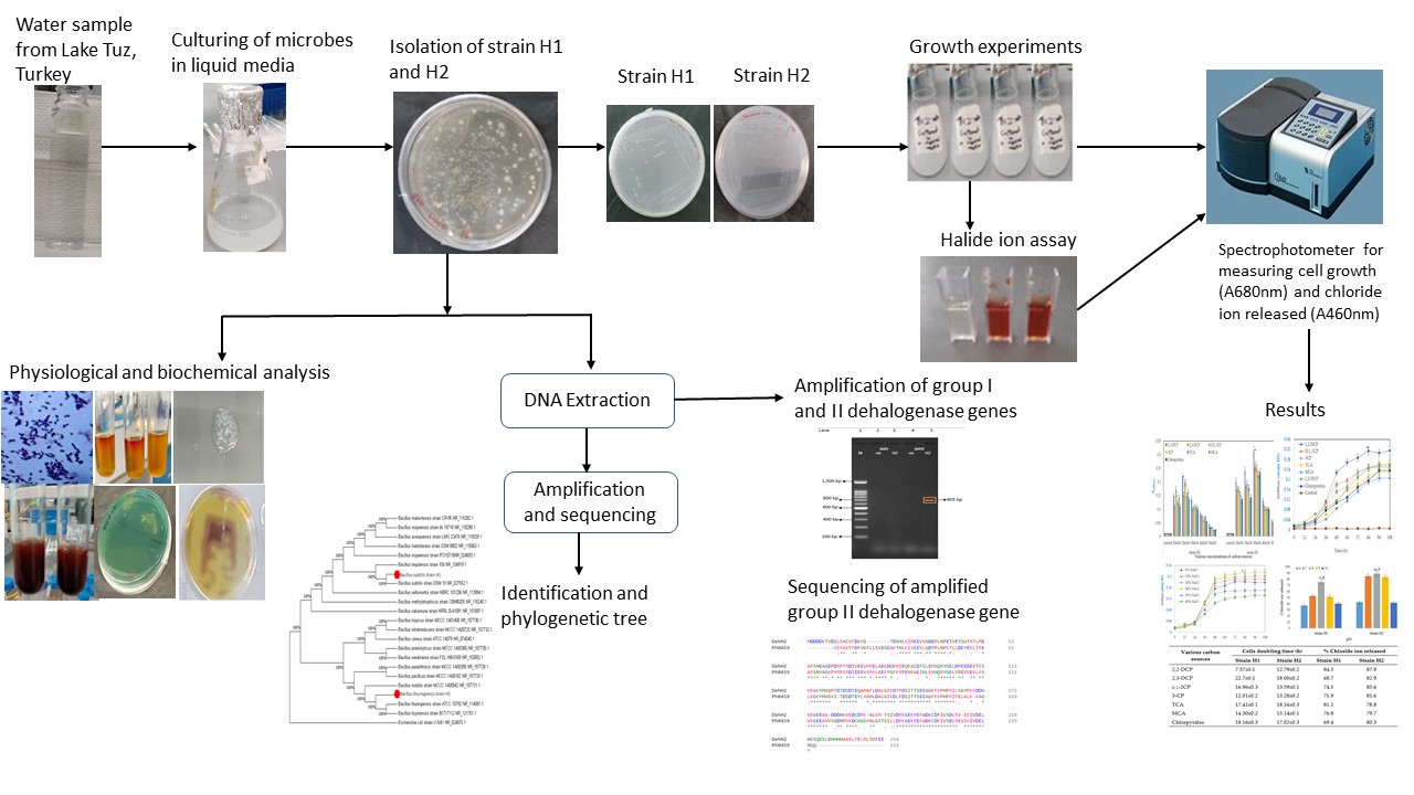
2.1. Enrichment and Isolation of the Bacterial Strains

Two bacteria were successfully isolated from the Tuz Lake sample and observably grew quite well on the haloalkanoic acids, haloacetates and chlorpyrifos selective minimal media. The bacteria were consequently designated as strains H1 and H2. After 5 days of incubation, the bacteria grew on 10–50 mM concentrations of the minimal media, beyond which growth was no longer observed.

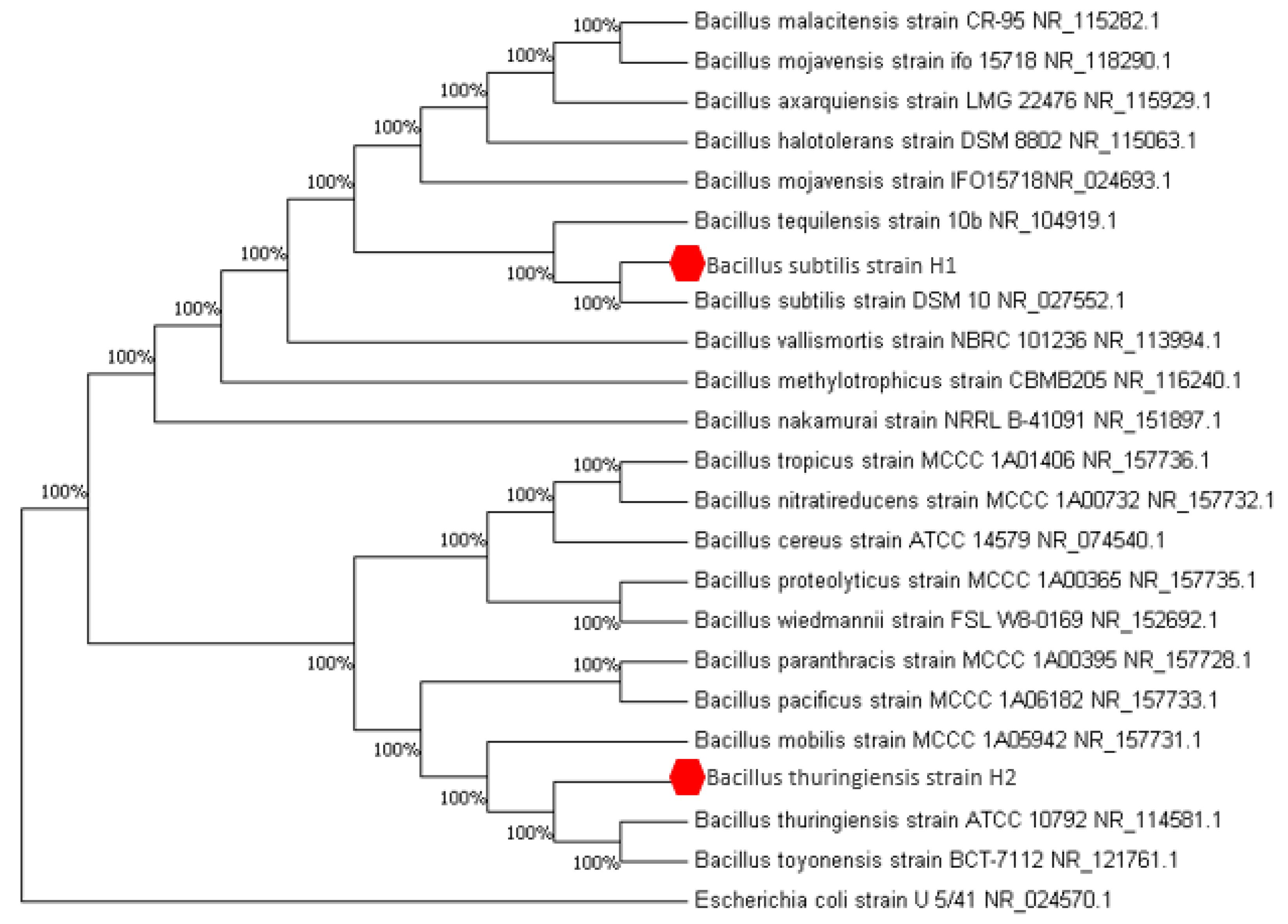
2.2. Bacteria Identification

In this study, the two bacterial strains were firstly identified by physiological and biochemical tests (Table 1). The two strains were found to be Gram-positive, with the cream-coloured colonies characteristically circular, raised/convex elevated with an entire smooth margin, and the bacteria were motile. The strains can utilize a narrow range of organic substrates, including glucose, lactose, maltose, and sucrose, except for strain H2 that is unable to utilize lactose and casein. The strains were tested positive for catalase, oxidase, amylase, and urease activities, and were negative for indole, gelatine liquefaction and H2S production. Gene sequence analysis indicated that the 16S rRNA gene of strains H1 and H2 exhibited 100% identity to the Bacillus subtilis strain DSM 10 (GenBank accession no. NR_027552.1) and B. thuringiensis strain ATCC 10792 (GenBank accession no. NR_114581.1), respectively. However, it is important to note that the two bacterial strains were never tested as bioremediation agents for in situ treatments of different halogenated compounds and chlorpyrifos, under highly saline conditions. The 16S rRNA gene sequence of both isolated strains were submitted to the Gene Sequence Database at the National Centre for Biotechnology Information (NCBI) under the accession number MK949128 and MK949122 for the bacterial strains H1 and H2, respectively. Figure 1 depicts the subsequent constructed phylogenetic tree showing strains H1 and H2 being closely related to B. subtilis and B. thuringiensis, respectively (Figure 1).

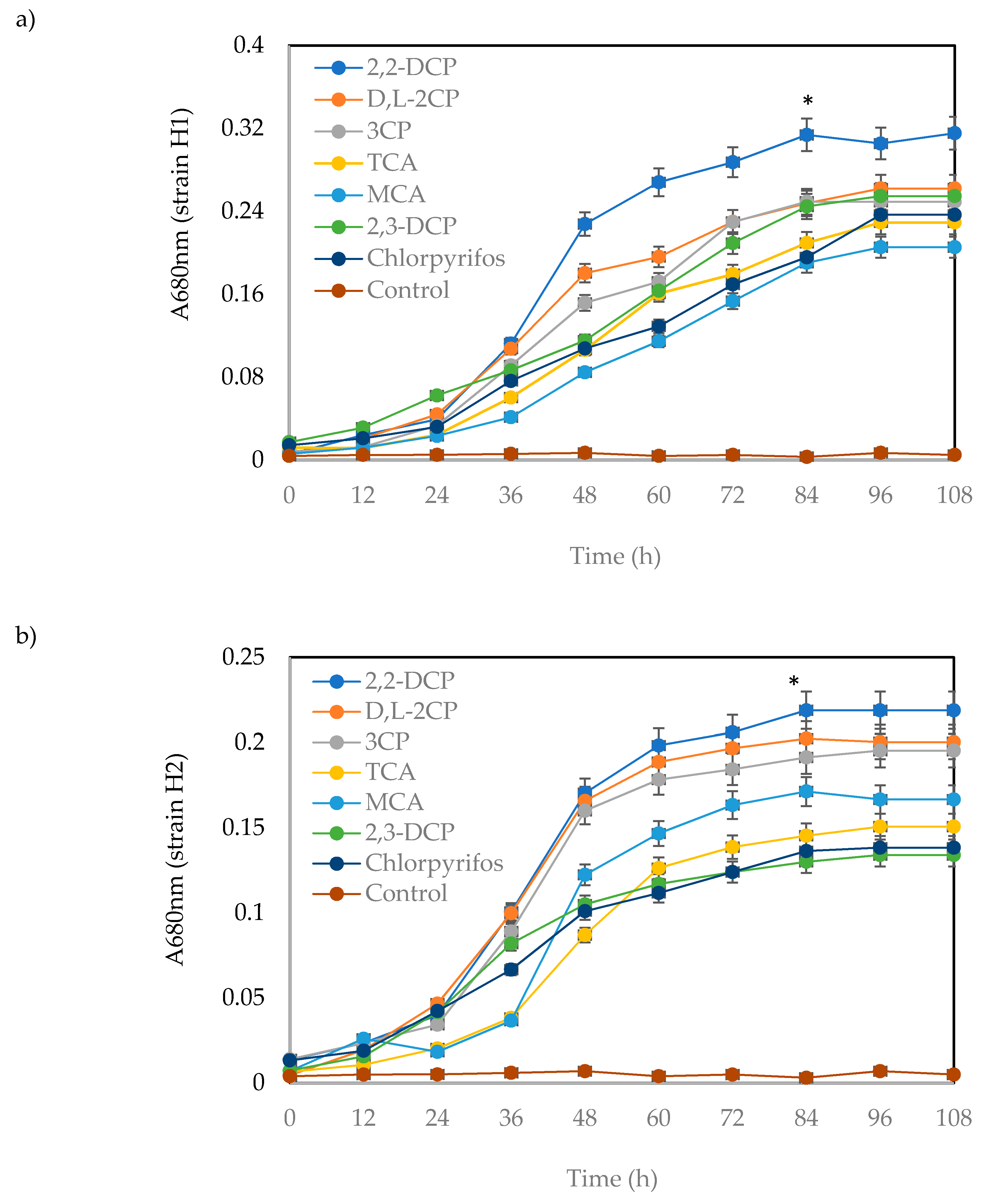
2.3. Bacterial Growth and Biodegradation Potential of the Isolated Bacteria

The study tested the bioremediation potential of the H1 and H2 bacterial isolates, whereby the rates of growth and substrate degradation by the two bacterial strains were evaluated in minimal media of various concentrations of halogenated compounds/substrates. Haloalkanoic acids (2,2-DCP, 2,3-DCP, d,l-2CP, 3-CP), haloacetates (MCA and TCA) and a commonly used pesticide, chlorpyrifos were used. So far, Bacillus species are generally non-pathogenic to humans, and have been used as a biopesticide and for promoting plant growth [16]. Nonetheless, their use in pollutant degradation remains limited.

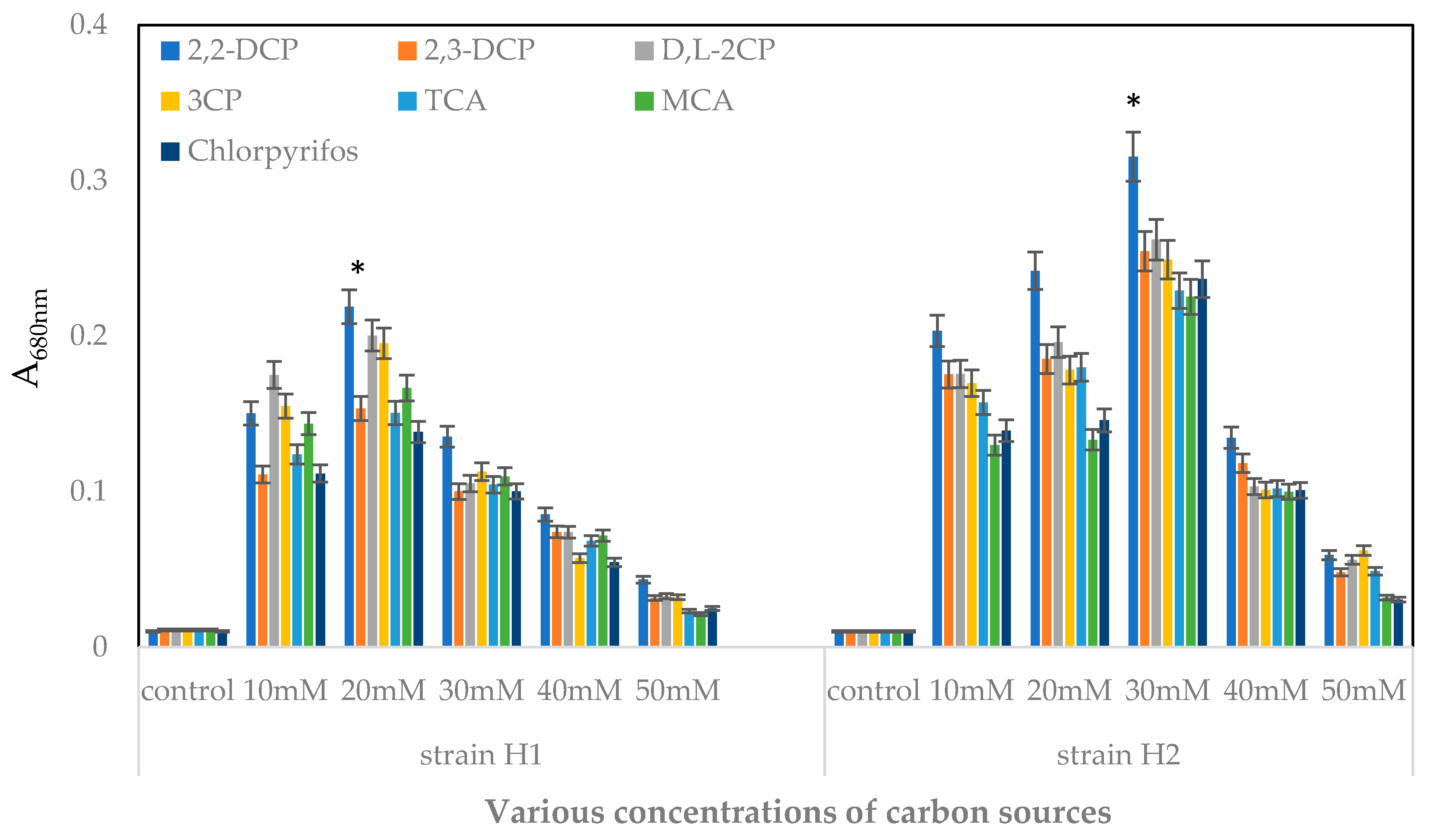
2.3.1. Effects of Various Concentrations of Substrates on Bacterial Growth

The initial concentration of the tested pollutant can substantially impact the rate of degradation by biological treatment. Effect of the initial concentration of each halogenated organic compound on the growth profile of strains H1 and H2 was observed for concentrations between 10–50 mM, growth temperature 30 °C, and the results are depicted in Figure 2. As can be seen, there was a significant (p > 0.05) increase in bacterial growth in test groups, alongside a steady bacterial growth in various substrates (haloalkanoic acids, haloacetate and chlorpyrifos). Minimal media of concentrations 10–20 mM showed a monotoic increase in growth of strain H1 at A680nm that corresponded to 0.13–0.23. Further increasing the substrates’ concentrations led to a precipitous drop in growth, plausibly from increasing toxicities of the substrates. Likewise, strain H2 grew optimally at 30 mM concentrations of the substrates, registering A680nm of between 0.25–0.32 before a general growth decline was observed. Notably, 20 mM was the optimal substrate concentration for strain H1 that grew on 2,2-DCP (0.218 ± 0.2) (p < 0.05), followed by DL-2CP (0.200 ± 0.1) (p < 0.05), 3-CP (0.195 ± 0.2) (p < 0.05), MCA (0.167 ± 0.1) (p < 0.05), 2,3-DCP (0.153 ± 0.1) (p < 0.05), TCA (0.151 ± 0.2) (p < 0.05) and chlorpyrifos (0.138 ± 0.1) (p < 0.05) (Figure 2). The data correlated with the maximum chloride ion released at 84.5, 74.5, 71.9, 76.8, 68.7, 81.1 and 69.4 %, respectively (Table 2). Whereas, growth of bacterial strain H2 was the highest in 2,2-DCP at 30 mM (0.315 ± 0.1)(p < 0.05), DL-2CP (0.262 ± 0.2) (p < 0.05), 2,3-DCP (0.255 ± 0.1) (p < 0.05), 3-CP (0.249 ± 0.1) (p < 0.05), chlorpyrifos (0.237 ± 0.1) (p < 0.05), TCA (0.229 ± 0.2) (p < 0.05), and lastly, MCA (0.225 ± 0.2) (p < 0.05) (Figure 2), corresponding to maximum release of chloride ions amounting to 87.9%, 85.6%, 82.9%, 81.6%, 80.3%, 78.8% and 79.7%, respectively (Table 2). Thus, the subsequent bacterial cultivation experiments employed the established optimum substrate concentrations.
Figure 3a,b illustrate the growth curves of bacterial strains H1 and H2, while Table 2 summarizes the corresponding cell doubling times and percentages of maximum chloride ion released by strains H1 and H2 for growths at 20 mM and 30 mM. It was apparent that among the tested substrates, growth in 2,2-DCP was the best, as depicted in Figure 3a,b. The corresponding calculated cells’ doubling times were 7.57 ± 0.1 and 12.79 ± 0.2 (Table 2).

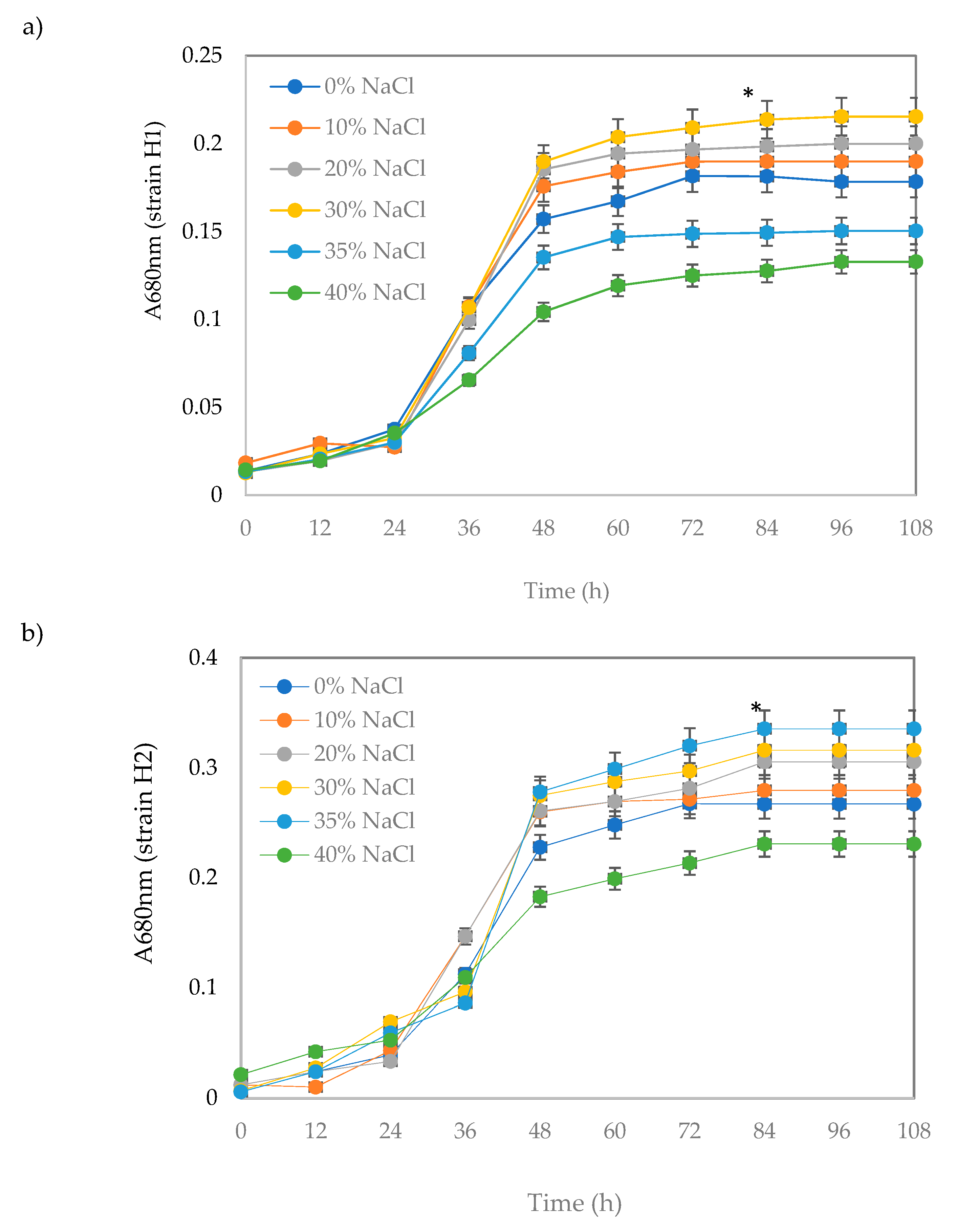
2.3.2. Bacterial Growth at Different NaCl Concentrations

Since 2,2-DCP was the preferred substrate of both the H1 and H2 bacterial strains, the compound was used in this investigation. Growths of H1 and H2 were measured on 20 mM and 30 mM of 2,2-DCP, respectively, in the presence of various concentrations of NaCl (Figure 4). Strain H1 could tolerate different concentrations of NaCl for up to 30% (w/v). Bacterial strain H2, on the other hand, was more accomplished to thriving in saline conditions with the maximum growth recorded in minimal media supplemented with 35 % (w/v) NaCl, whereas the poorest growth occurred in 40 % (w/v) of NaCl, similar to H1.

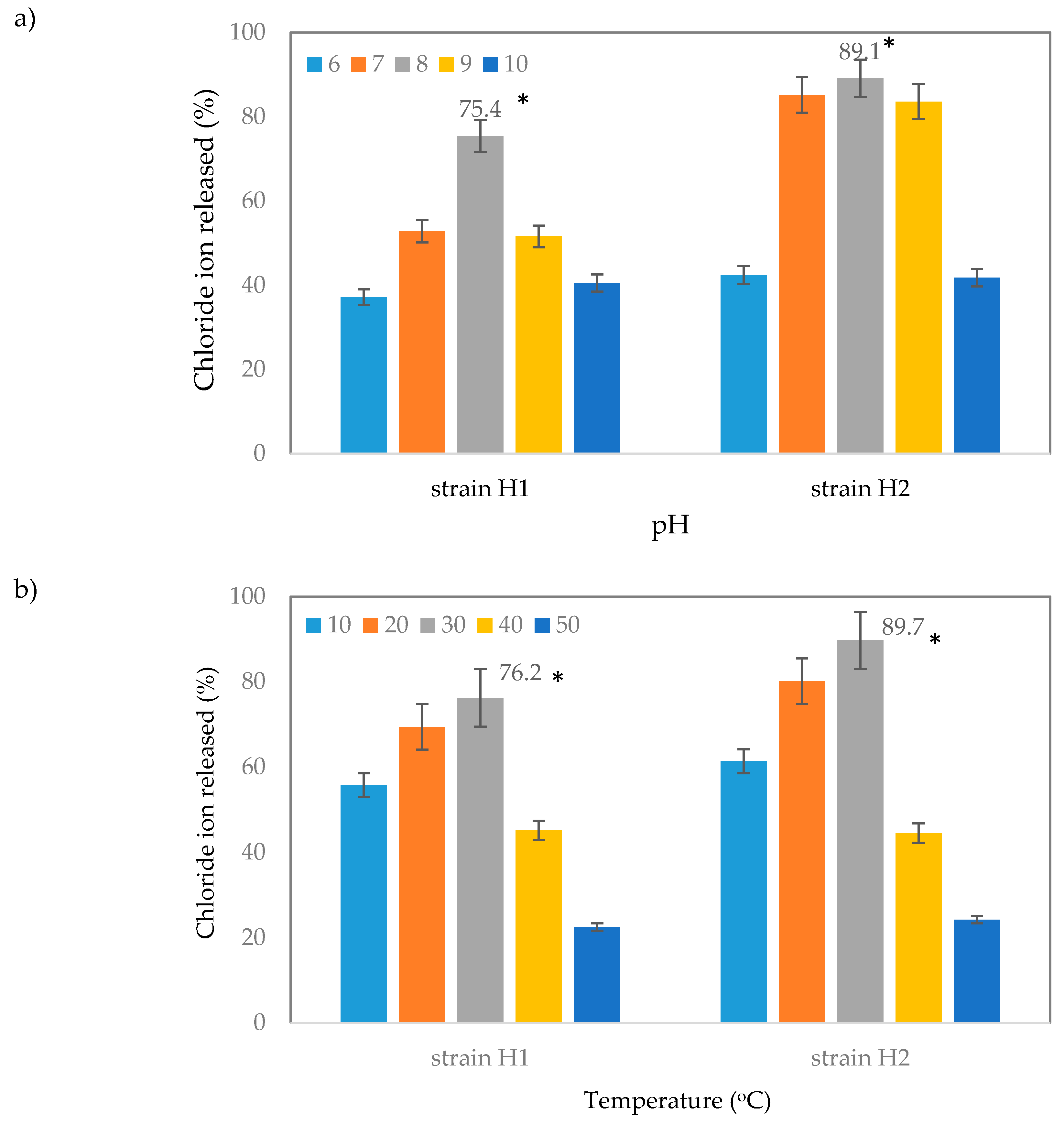
2.3.3. Effect of pH and Temperature on Dehalogenation of 2,2-Dichloropropionic Acid (2,2-DCP) by Strain H1 and H2

Changes in pH of the surrounding can substantially interfere with many bacterial activities, given that marine pH falls between pH 7.5–8.5. Having said that, the effect of pH on strains H1 and H2 was investigated for pH 6.0–10.0. Figure 5a illustrates the data for the effects of pH on the % of chloride ion released of 2,2-DCP by H1 and H2 strains. Dehalogenation of 2,2-DCP by strain H1 was significantly higher at pH 8.0, yielding a 75.4 ± 0.1 % (p < 0.05) of released chloride ions. Interestingly, strain H2 demonstrated a wider range of pH tolerance (7.0–9.0), with the percentage of released chloride ions being significantly higher at 89.1 ± 0.1 % (p < 0.05) at pH 8.0 (Figure 5a). Since strain H2 was grown in 30 mM 2,2DCP, therefore, the study observed a higher release of chloride ions compared to strain H1.
The effects of temperature (10–50 °C) on the release of chloride ions by strains H1 and H2 growing on 2,2-DCP are presented in Figure 5b. The results revealed that growth of both strains was highly significant at 30 °C and the liberated chloride ions were as much as 76.2 ± 0.01% (p < 0.05) for strain H1 and 89.7 ± 0.01 (p < 0.05) for strain H2 (Figure 5b). Incubation at higher temperatures of 40 and 50 °C, drastically reduced growth of both bacteria and produced an approximately 45 ± 0.01% (p < 0.05) and 24 ± 0.01% (p < 0.05) of the chloride ions, respectively. Consistently, the findings demonstrated that both strains H1 and H2 produced better dehalogenation of 2,2-DCP under the same incubation temperature (30 °C).

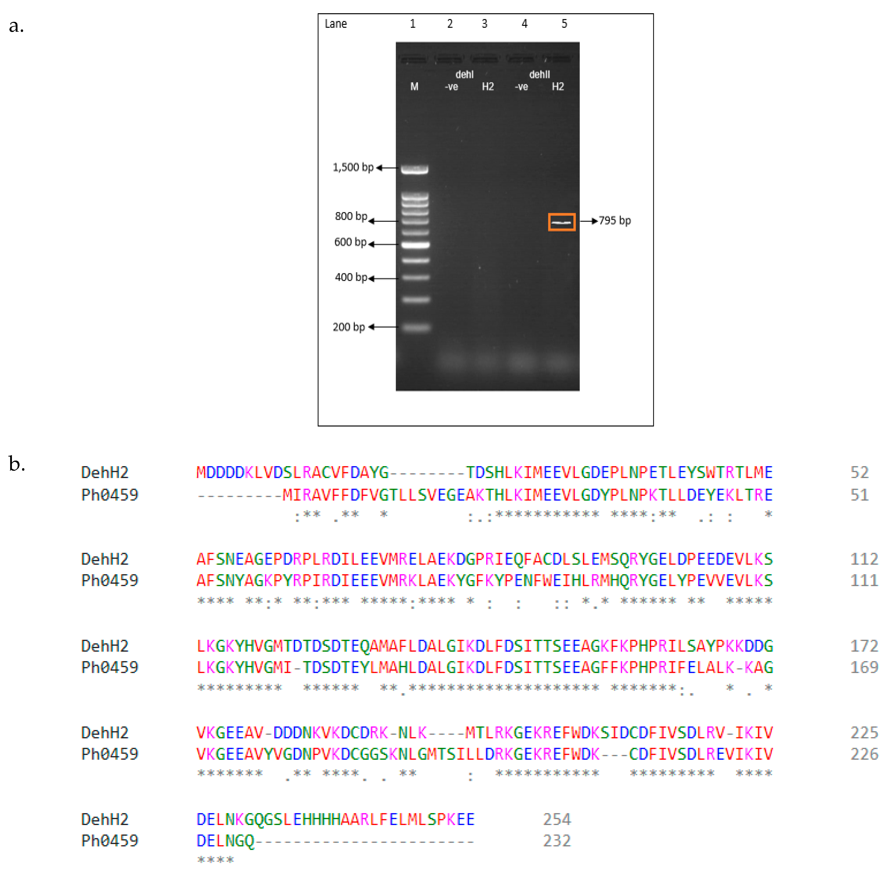
2.4. Polymerase Chain Reaction (PCR) Amplification of Putative Dehalogenase Gene

To confirm the presence of a dehalogenase gene in the H1 and H2 bacteria, we amplified a fragment of the genes by polymerase chain reaction (PCR). The amplification of dehalogenase genes from strain H1 using group I or II primers failed. Whereas, amplification using genomic DNA from strain H2 as a template, a PCR fragment of the size 795 bp was successfully generated for group II (dehII) but was not observed for group 1 (dehI) and the negative control that used water in replace of the DNA template (Figure 6a). A putative dehalogenase gene from B. thuringiensis strain H2 was successfully sequenced and analyzed which revealed a 762 bp DNA sequence that constituted of 254 amino acid residues. To check the similarity of current amino acids, Basic Local Alignment Search Tool-protein (BLASTp) (blast.ncbi.nlm.nih.gov/) and pairwise alignment was carried out (Figure 6b). Findings showed the group II sequence has 63.71% sequence identity and was similar to the PH0459 haloacid dehalogenase secreted by hyperthermophilic Pyrococcus horikoshii OT3 (PDB ID 1X42) [17].

3. Discussion

Bioremediation is an effective method for treating polluted environment which necessitates the isolation of efficacious halo-organic compounds-biodegraders to clean up the environment. In this study, two strains of bacteria were successfully isolated by enrichment cultivation in minimal medium of various concentrations of chlorinated organic substrates (haloalkanoic acids, haloacetates and chlorpyrifos). Notably, while the online BLASTn search on the 16S rRNA gene sequence H1 (GenBank: MK949128) and H2 (GenBank: MK949122) revealed 100% sequence identity to Bacillus subtilis and Bacillus thuringiensis, respectively, studies on the efficacy of the bacteria at degrading the aforementioned pollutants remains to be seen. Hence, to identify the optimal conditions to degrade the sample pollutants, this study grew the H1 and H2 bacterial isolates under various carbon sources (pollutants), temperature, pH values and in media of varying salt concentrations. As far as the biodegradation of environmental pollutants is concerned, both strains H1 and H2 are reportedly capable of degrading hydrocarbon compounds.
The nature and type of carbon sources (pollutants) is a pertinent factor for determining bacterial growth. The initial concentration of substrate can affect the degrading ability of the microbes, as an excess can stifle bacterial growth. Having said that, the effect of initial pollutant concentrations ranging from 10–50 mM were assessed for strains H1 and H2 (Figure 2). It is worth mentioning here that some bacteria can grow on halogenated compounds i.e., α-haloalkanioc acids, but fewer can do so on β-haloalkanioc acids [18,19,20]. Micrococcus denitrificans [21], Pseudomonas sp. B6P [22], Rhodococcus sp. HJ1 [23], and Bacillus sp. CGMCC no. 4169 [24], Bacillus sp. H4 [25], Burkholderia cepacia W1 [26], Pseudomonas halophila HX1 [8] are examples of microbials that can degrade either α-haloalkanioc acids or β-haloalkanioc acids. Additionally, Bagherbaigi, Gicana [27] described the Arthrobacter sp. S1 that can degrade both α-haloalkanoic acids and β-haloalkanoic acids but not haloacetates (MCA and TCA). Additionally, to the best of our knowledge this is the first report on Bacillus sp. isolated from a hypersaline environment adapted in metabolizing haloalkanoic acids (both αHA and βHA), haloacetates (MCA and TCA) and chlorpyrifos as the sole carbon source (Table 2).
Until now, there have been relatively few microbes capable of degrading chlorpyrifos and its metabolite chlorinated-pyridinol (3,5,6-trichloropyridiol) [28,29]. However, chlorpyrifos hydrolytic dehalogenase-producing bacteria that can effectively render chlorpyrifos and its residues harmLess in soil and water environments, remains to be seen. In fact, strains of the Bacillus genus have been reported as rather competent bioremediation and biopesticides agents [30,31,32]. This is consistent with our current findings which showed that strains H1 and H2 as quite accomplished bioremediation agents.
Pertinently, the isolation of pollutants degraders showing salt tolerance is crucial, when considering their practical applicability to degrade highly saline effluents or for cleaning up marine environments contaminated with toxic halogenated compounds. For this part of the study, we intended identify the best salinity for the H1 and H2 to degrade 2,2-DCP that represents halogenated compounds, by optimizing the concentration of NaCl in the growth media. Figure 4 shows the growth curves for bacterial strains H1 and H2 at various salt NaCl concentrations (0–40% w/v). Both bacteria strains H1 and H2 showed good tolerance towards increasing NaCl concentrations, reaching as high as 30% and 35%, and correspondingly exhibited cell doubling times of 9.62 and 8.08 h. The halotolerant characteristic demonstrated here have been reported for a similar bacteria species, namely the B. megaterium CTBmeg1 [32], that tolerates up to 20% NaCl (w/v) with a cell doubling time of 26.41 h. Another bacterium is from the Pseudomonas genus, viz. P. halophila HX [8] and P. halophila DSM 3050 [33], both of which can endure up to 25% NaCl with a cell doubling time of 25.4 h. Despite the ability of strains H1 and H2 to grow in highly salt conditions, both species were not highly reliant on the high salt content for survival. This was apparent when colonies of both strains could grow on media devoid of NaCl (0% NaCl) (Figure 4). This was contrary to the Alomonas montanilacus sp. previously isolated from a hypersaline lake showed an optimal growth between 4–5% (w/v) of NaCl but was inhibited in the absence of NaCl [34]. The same was observed for Natrialba swarupiae sp. which grew optimally on a medium supplemented with 25% NaCl [35]. The ability of strains H1 and H2 to survive the highly saline environment was expected as extreme salinity tends of Lake Tuz to increase the rate of water lost, instigating the bacterial cells to shrivel and thus lead to the irreversible loss of cellular structure and function. Such an environment, in response can affect the metabolism of many microbes, hence justifying their lower substrate/pollutant degrading performance. In the case of halophiles, the present of salt for growth becomes mandatory, unlike the halotolerant ones. While the latter do not require NaCl for growth, they tolerate saline conditions very well. Consequently, the same feature was observed for the H1 and H2 bacterial isolates, thereby categorizing them as halotolerants. The unique adaptation by the two bacteria could be explained by the heterogeneity and frequent fluctuations of salinity in space and time in the Lake Tuz which predominantly supports the survival of such euryhaline microbes. The increase in salinity and oligotrophic conditions in the lake makes it impossible for non-halophiles to survive. This is because such a habitat can considerably alter the folding of their enzymes and interrupt the active conformation. Because strain H1 and H2 can grow in relatively wide salinity range (0–35% NaCl), this indicated their potential as bioremediation agents for cleaning up saline environments, especially seawater polluted with halogenated hydrocarbons.
Another crucial parameter that affects the biodegradation potential of a microbe is pH, as it is directly associated with the inter-membrane transport and bacterial growth [36]. This is particularly true when treating highly saline wastewater or industrial effluents using biological methods. As a matter of fact, the outcome of this study agreed well with previous studies for bacterial isolates from Lake Tuz (optimum pH range of pH 6.0–9.0). Equally, most bacteria isolated from hypersaline environments show optimal pHs ranging from 7.0–9.0 [37,38,39,40]. This may be explained by the amazing changes at Lake Tuz due to seasonal evaporations, in which the lake naturally and repeatedly turns alkaline and, of course highly saline, too. This causes an enormous rise in pH, resulting from the increase in NaCl content [41,42]. Such changes further add to the disparity of osmotic pressure on aquatic life and the surroundings. The ability of strains H1 and H2 to efficiently degrade the various pollutants under the assessed conditions is a testament to their evolutionary adaptation towards their highly dynamic surroundings, from which they were isolated.
Similarly, the surrounding temperature also plays a crucial role in governing rates of degradation of pollutants by biological treatments. Temperature changes can influence microbial growth rates, gas solubility, microbes’ metabolism, as well as affect the physical and chemical states of contaminants [43]. Comparatively, Halobacillus sp. strain EG1HP4QL, bacterial strain O-CH1, Haloterrigena jeotgali sp. and Natrialba swarupiae sp. isolated from hypersaline environments have optimal growths between 37–40 °C [35,38,39,44]. The result seen here agreed with reports on other hypersaline bacterial isolates showing optimum growths at 30 °C [37,45]. It was apparent that the higher incubation temperatures which exceeded 30 °C may have contributed to greater denaturation of vital structural and enzyme proteins, and adversely altered membrane structures and, subsequently growth [46]. Likewise, the mesophilic characteristic of strains H1 and H2 is comparable to P. halophila HX1 isolated from the same lake, except the latter is better adept to surviving in a wide temperature range of Lake Tuz [8], as the mean annual temperature fluctuates between 12–37 °C [47]. It was clearly shown that the optimal incubation temperature for strains H1 and H2 to effectively degrade the sample pollutants must be found, since a temperature too high can denature crucial enzymes in H1 and H2, and interfere with other metabolic activities related to growth. Conversely, a too low incubation temperature results in a sluggish enzyme action, as well as slower growth [48].
PCR amplification was used to detect dehalogenase gene. Group I dehalogenase genes are specific towards both D and L form of the substrate while group II dehalogenase genes are only active on the L form of the substrate. These indicated that the current isolate has a L-2-haloacid dehalogenase gene to produce an enzyme that is stereo-specific towards the L-isomer of the substrate. This was possibly due to the L-2-haloacid dehalogenase being more common than group I dehalogenases. More importantly, the inability of using group I primers to amplify a dehalogenase gene from strain H2 was that: (i) the primers might not be universal for some bacteria and (ii) only a single dehalogenase gene may be present that can act on α-HA and β-HA and same reasons may apply for no amplification using group I and II primers to amplify dehalogenase from strain H1. Therefore, further analysis is needed to re-confirm this observation.

4. Materials and Methods

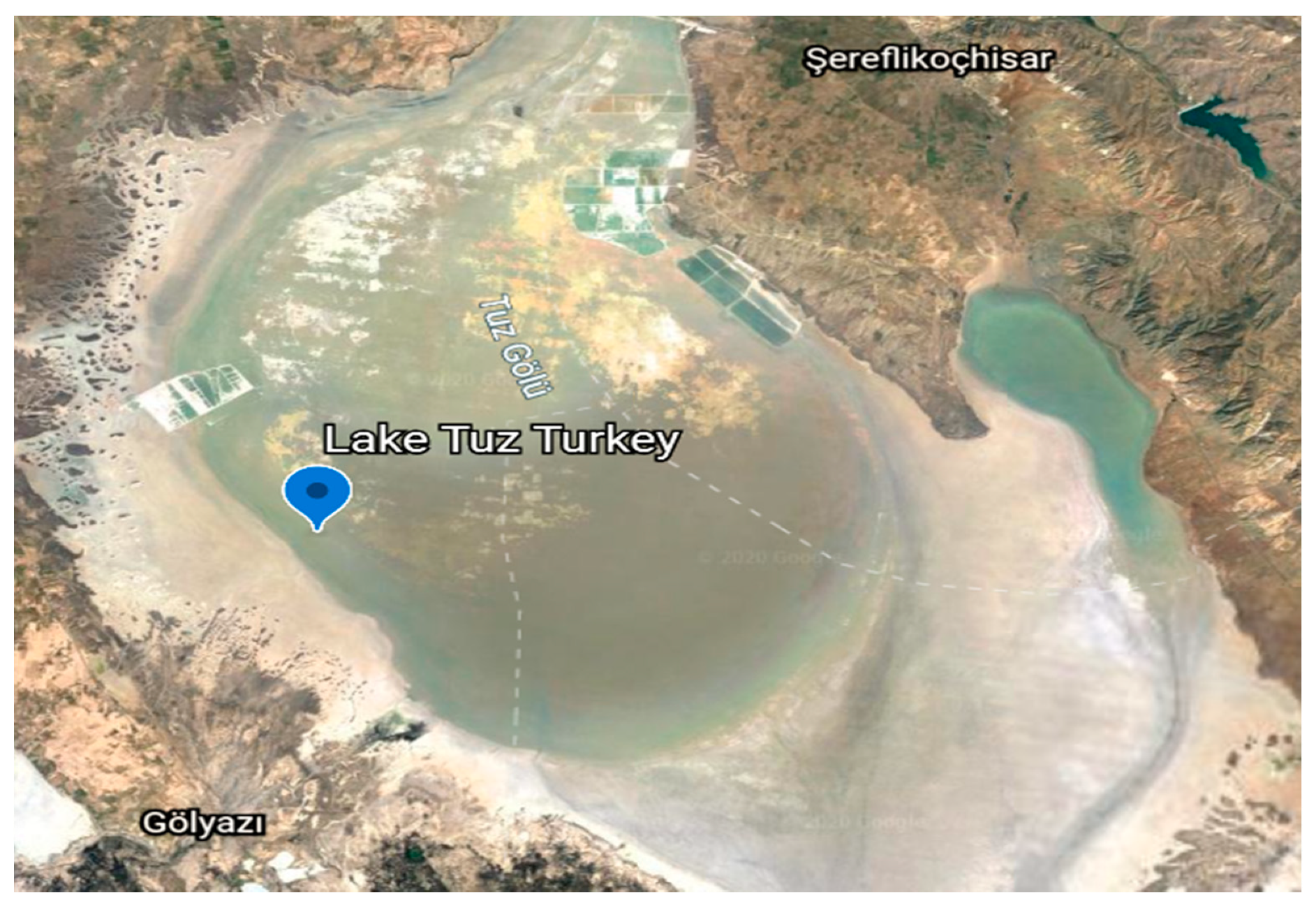
4.1. Sample Collection

The water samples were collected from the main zone of Tuz Gölü, Turkey (latitude: 38°43′41″ N; longitude: 33°14′56″ E) at 1665 km2 (643 sq mi) surface area during peak summer period (August 2018). Lake Tuz is one of the largest hypersaline lakes in the world [49] (Figure 7). The samples were collected using a sterile plastic container, immediately transferred to the laboratory and chilled at 4 °C until further analysis.

4.2. Chemicals and Media Preparation

Components and preparation of minimal medium followed a protocol described by Hareland, Crawford [50] (Table 3). Halogenated compounds, haloalkanoic acids [2,2-dichloropropionic acid (2,2-DCP), 2,3-dichloropropionic acid (2,3-DCP), d,l-2-chloropropionic acid (d,l-2-CP)], 3-chloropropionic acid (3-CP) and haloacetic acids [monochloroacetate (MCA) and trichloroacetate (TCA)] and hydrogen peroxide used in this study were all of analytical grade and purchased from Sigma-Aldrich ( St. Louis, USA). The commercial pesticide, chlorpyrifos was purchased from Majujaya Agriculture and Agrikimia Trading (Johor Bahru, Malaysia). Chlorpyrifos was evaporated and lyophilized into a powder form. All halogenated compounds were filter sterilized through a 0.2 µm nylon membrane disc (Hybond N; Amersham, Little Chalfont, UK) before use, and sterile distilled water was used as the solvent for media preparation.

4.3. Enrichment and Isolation of Dehalogenase-Producing Bacteria

Stock solution was prepared as 10× concentration of basal salts and 10× concentration of trace metals solution (Table 3). Minimal/enrichment media for growing bacteria contained 10 mL of 10× basal salts and 10 mL of 10× trace metal per 100 mL of distilled water and were autoclaved (121 °C for 15 min 15 psi). Carbon sources (2,2-DCP, 2,3-DCP, d,l-2CP, 3CP, MCA, TCA and chlorpyrifos) were separately filter sterilized and was added aseptically to the media to achieve the desired final concentration of between 10–50 mM. Bacteriological agar (1.5% w/v) was added prior to sterilization for the preparation of the solid medium. To each growth medium, 5 mL of the water sample from Lake Tuz was added followed by incubation for 5 days at 30 °C with agitation at 200 rpm. Then, 100 μL aliquot of each growth medium was transferred to the solid minimal medium and incubated for a further 5 days at 30 °C. Repeated streak plate method (5 times) was used to obtain the pure colonies of each bacterial isolates. Each isolate was transferred to solid minimal media containing various concentrations of halogenated compounds (10–50 mM) as the sole carbon sources.

4.4. Identification of Dehalogenase-Producing Bacteria

4.4.1. Morphological and Biochemical Characterization

Physiological and biochemical properties of the isolated bacterial strains were systematically assessed for Gram staining, motility, pigmentation, oxidase, amylase, utilization of glucose, sucrose, lactose and maltose, starch hydrolysis, triple sugar iron test, hydrogen sulphide production, gas production from glucose, urease, methyl red, Voges–Proskauer, indole, gelatin liquification and simon salt of citric tests, using Bergey’s Manual for Determinative Bacteriology [51]. The isolated bacteria were tested for catalase activity using a solution of 3% (w/v) hydrogen peroxide.

4.4.2. Molecular Identification

DNA Extraction, PCR Amplification of the 16S rRNA and Gene Sequencing of Bacteria Strains

Genomic DNA of the two bacterial isolates were extracted using the Wizard Genomic DNA Purification Kit (Promega kit) and the concentration of the corresponding genetic material was measured by the Nano-Drop 1000 (Thermo Scientific). Subsequently, the extracted DNA was subjected to PCR amplification using universal primers Fd1(5′-AGA GTT TGA TCC TGG CTC AG-3′) and Rv1 (5′-ACG GTC ACC TTG TTA CGA CTT-3′) [52]. Each of 16S rRNA amplification reaction contains a 25 μL total volume of 12 μL PCR master mix (Fermentas Inc. USA), 1 μL template DNA, 1 μL for ward primer (Fd1), 1 μL reverse primer (rP1), and 10 μL nuclease free water. The 16S rRNA gene amplification was run for 30 cycles whereby each cycle was set as an initial denaturation phase of 94 °C for 5 min, followed by denaturation 94 °C for 1 min, annealing 55 °C for 1 min and final extension 72 °C for 10 min. The PCR product was electrophoresed on an agarose gel (0.8%). For the sequencing reaction, QIAquick PCR purification kit was used to purify PCR product (Apical Laboratory, Selangor). The BLAST search program found on the NCBI website (http://www.ncbi.nlm.nih.gov/) was used to compare the obtained sequence with others sequences in the public database.

Phylogenetic Analysis of 16S rRNA Gene

Next, the BLASTn analysis tool was used to align and compare 16S rRNA sequences of the bacterial isolates with sequences found in the GenBank database (NCBI). The identified sequences were then selected from the BLASTn and added directly into MEGA7 software. Analysis of phylogeny and alignment of multiple sequences used the CLUSTAL‒W and Phylogeny Construction in MEGA7 software [53]. The 16S rDNA sequences of the bacterial isolates were deposited in GenBank under accession numbers MK949128 and MK949122.

4.5. Bacteria Growth and Halide Ion Assay

Dehalogenation of the various halogenated substrates by the two bacterial isolates was assessed aerobically. The change in turbidity of the growth media due to growth was read on an ultraviolet (UV)–visible spectrophotometer (T60 visible, PG instruments) at A680nm over designated time intervals, and the cell doubling time of each isolate was calculated from their corresponding growth curves. The halide ion assay that measures the liberation of chloride ion and the concentration of chloride ion released was established by converting the absorbance value to μmol Cl/L. This conversion was based on the standard curve constructed using sodium chloride as a typical measurement of soluble chloride [54]. The dehalogenation rate of each halogenated substrate was calculated by comparing the amount of chloride ion released in μmol Cl/l of test culture medium containing the bacterial suspension, versus the control without bacterial inoculation, under the same incubation conditions.

4.5.1. Effects of Various Concentrations of Carbon Source on the Bacterial Growth

A loopful of pure culture of each strain was transferred into a freshly prepared nutrient broth and was incubated overnight at 30 °C with agitation at 200 rpm. Then, 1 mL of the bacterial suspension was inoculated into 250 mL Erlenmeyer conical flask containing 50 mL of minimal medium of each carbon source (10–50 mM), while control experiments without strain H1 and H2, were prepared under similar growth conditions. Degradative potential was measured (for three independent samples) by the colorimetric method by determining the release of chloride ions during dehalogenation [54]. One unit of dehalogenase activity was defined as the amount of enzyme that catalyzed the hydrolysis of 1 µmol 2,2-DCP per min under assay condition. A 1 mL of the sample was added into a test tube containing 100 μL of 0.25M ammonium ferric sulphate prepared in 9 M nitric acid. The solution was vortexed followed by the addition of 100 μL of mercuric thiocyanate-saturated ethanol and the mixture was vortexed again. The mixture was left to stand at room temperature for 10 min to allow color development and the liberated chloride ions were observed at A460nm.

4.5.2. Effect of Salinity on Bacterial Growth

The influence of salt tolerance on growth of selected strains was checked by inoculating each isolate into selective minimal media having final concentrations of 2,2-DCP at 20 mM and 30 mM for bacterial strains H1 and H2, respectively, with different concentrations of NaCl (0–40%, w/v). Calculations of the bacterial growth curve and cell doubling time of each strain was taken from triplicates and were monitored over 12 h intervals at A680nm. Minimal media was prepared in a 250 mL sterile Erlenmeyer flask contained based salts (10 mL), trace metals (10 mL) and distilled water made up to the volume of 100 mL. The minimal salt media was sterilized by autoclave at 121 °C for 15 min at 15 psi. Sterilized 2,2-DCP and NaCl were added to the autoclaved media to the desired concentrations (0–40% w/v). The media was adjusted to the desired pH (pH 8) using the DELTA 320 pH meter and incubated at 30 °C with shaking at 200 rpm for 5 days. The experiments were set up in triplicate. It is important to note that bacteria that grow optimally in media containing 3–15% and >15% NaCl are deemed as moderately halophilic and extremely halophilic, respectively, while colonies that grow on media with or without NaCl are halotolerant [55].

4.5.3. Effect of pH and Temperature on the Bacterial Growth and Substrate Degradation

To investigate the effect of different pH and temperature on the degradation of each substrate by both bacterial strains, pH of the minimal medium (50 mL) containing suitable final concentrations of 2,2-DCP for strains H1 (20 mM) and H2 (30 mM) were adjusted (pH 6.0–10) using appropriate amounts of 1 M HCl or NaOH. A 1 mL aliquot of the cell culture suspension was transferred into a 250 mL sterile Erlenmeyer flask containing basal salts (10mL), trace metals (10 mL) and distilled water made up to the volume of 100mL and sterilized 2,2-DCP (carbon source) was added to autoclaved solution. The media was adjusted to the desired pH (pH 6, 7, 8, 9, and 10) using the DELTA 320 pH meter and was incubated at 30 °C with agitation at 200 rpm for 5 days and the experiment was setup was done in triplicate. A control experiment without strains H1 and H2 was incubated in parallel, to evaluate the ability of each isolate to degrade the substrate at pH 8.0. Meanwhile, the incubation temperatures were monitored at 10, 20, 30, 40 and 50 °C, respectively. The optimized growth conditions were determined by measuring the growth of bacteria and the amount of chloride ion released. The degradation rate of each substrate was estimated after 5 days of incubation.

4.6. Amplification of Putative Dehalogenase Gene using Group I and Group II Primers

The PCR primers were designed according to Hill, Marchesi [56] and the primers were synthesised by Apical Scientific (Malaysia). Amplification of the dehalogenase gene sequence was done using group I deh gene primers (dehI-f 5′- ACGCTGCGTGTGCCGTGGGT-3′ and dehI-r 5′- CGCAATCACATGGAAGTCACT-3′) and group II deh gene primers (dehII-f 5′ TGGCGCCACATCCAACTTGACTA-3′ and dehII-r 5′-TCATATCCTTTAGACGATGACACTAA-3′). PCR amplification was carried out in a thermal cycler (Eppendorf Master Nexus Gradient; Eppendorf) using the following PCR conditions: an initial denaturation for 10 min at 94 °C, followed by 36 cycles of denaturation (94 °C, 45 s), primer annealing (55 °C, 2 min), and extension (75 °C, 45 s), with a final extension for 5 min at 75 °C. The reaction mixture was electrophoresed on a 0.8 % agarose gel and purified using QIAGEN PCR purification kit prior sequencing using standard BigDye® Terminator v3.1 cycle sequencing kit chemistry at Apical Scientific (Malaysia). A control, leaving out one of the primers, was set up and was amplified using the same PCR conditions. For the negative control, similar PCR reaction was set up except the target/template DNA was replaced with water.

4.7. Statistical Analysis

Statistical analysis was on the obtained data was done on IBM SPSS version 20.0 software, in which significant differences among groups was based on the level of significance level of 0.05 (P < 0.05), if any. The data were shown to be normally-distributed, the parametric test of analysis of variance (ANOVA) repeated measurement was employed.

5. Conclusions

Two novel halogenated hydrocarbon-degrading bacteria were effectively isolated from the hypersaline Lake Tuz in Turkey and were identified as Bacillus subtilis strain H1 and B. thuringiensis strain H2. The phenotypic and phylogenetic characterizations suggested that B. subtilis strain H1 and B. thuringiensis strain H2 demonstrated promising potential for the degradation of halogenated organic compounds and were adapted to living under high salinity and pH conditions. Strain H2 constitutes one of the few haloacetate- and haloalkanoic-degrading B. thuringiensis strains, and to the best of our knowledge is the first of this species to be isolated from hypersaline environment of Lake Tuz in Turkey. Remarkably, the findings in this study indicates the possibility of strains H1 and H2 to be employed cleaning up halogen-polluted marine/hypersaline environments. However, complementary studies using purified dehalogenases from the two bacterial isolates are required, to better understand their halogen degradative ability in such unique hypersaline environments.

Author Contributions

H.A.O., F.H. conceived and designed research. H.A.O. prepared the manuscript. Writing—review and editing, R.A.W., Y.K., M.F.E. All authors have read and agreed to the published version of the manuscript.

Funding

This research was funded by the Scientific Technological Research Council of Turkey (TUBITAK), Program 2221-Fellowship for visiting Scientists and Scientists on Sabbatical Leave with Grant number 1059B11800242. FH and HAO thanks the Fundamental Research Grant Scheme (FRGS)-Ministry of Higher Education Malaysia R.J130000.7854.5F189 and RUG-UTMHR Q.J130000.2414.08G59 for financial assistance and the Ministry of Education through Tertiary Education Trust Fund (TETFund) Nigeria for studentship.

Conflicts of Interest

The authors declare no conflict of interest.

References

  1. Nion, Y.A.; Toyota, K. Recent trends in control methods for bacterial wilt diseases caused by Ralstonia solanacearum. Microbes Environ. 2015, 30. [Google Scholar] [CrossRef] [Green Version]
  2. Khalili, E.; Javed, M.A.; Huyop, F.; Wahab, R.A. Efficacy and cost study of green fungicide formulated from crude beta-glucosidase. Int. J. Environ. Sci. Technol. 2019, 16, 4503–4518. [Google Scholar] [CrossRef]
  3. Douglas, S.H.; Dixon, B.; Griffin, D. Assessing the abilities of intrinsic and specific vulnerability models to indicate groundwater vulnerability to groups of similar pesticides: A comparative study. Phys. Geogr. 2018, 39, 487–505. [Google Scholar] [CrossRef]
  4. Lu, J.; Yan, X.; Ma, Y.F.; Tian, C.X.; Ding, J.C. Impact of salinity on treatment of saline wastewater by sequencing batch biofilm reactor process. J. Central South Uni. 2014, 21, 1989–1994. [Google Scholar] [CrossRef]
  5. Lefebvre, O.; Moletta, R. Treatment of organic pollution in industrial saline wastewater: A literature review. Water Res. 2006, 40, 671–3682. [Google Scholar] [CrossRef] [PubMed]
  6. Aloui, F.; Khoufi, S.; Loukil, S.; Sayadi, S. Performances of an activated sludge process for the treatment of fish processing saline wastewater. Desalination 2009, 246, 389–396. [Google Scholar] [CrossRef]
  7. Nemati, M.; Abdulghader, M.F.; Gicana, R.G.; Lamis, R.J.S.; Ibrahim, N.; Hamid, A.A.A.; Huyop, F. Identification of putative cof-like hydrolase associated with dehalogenase in enterobacter cloacae mn1 isolated from the contaminated sea-side area of the Philippines. Malays. J. Microbiol. 2013, 9, 253–259. [Google Scholar] [CrossRef] [Green Version]
  8. Edbeib, M.F.; Wahab, R.A.; Kaya, Y.; Huyop, F. In silico characterization of a novel dehalogenase (DehHX) from the halophile Pseudomonas halophila HX isolated from Tuz Gölü lake, Turkey: Insights into a hypersaline-adapted dehalogenase. Ann. Microbiol. 2017, 1–12. [Google Scholar] [CrossRef]
  9. Edbeib, M.F.; Wahab, R.A.; Huyop, F. Characterization of an α-haloalkanoic acid–degrading Pseudomonas aeruginosa MX1 isolated from contaminated seawater. Bioremediat. J. 2016, 20, 89–97. [Google Scholar] [CrossRef]
  10. Abidin, M.H.Z.; Halim, K.B.A.; Huyop, F.; Hamid, T.H.T.A.; Wahab, R.A.; Hamid, A.A.A. The mechanistic role of active site residues in non-stereo haloacid dehalogenase E (DehE). J. Mol. Graph. Model. 2019, 90, 219–225. [Google Scholar] [CrossRef] [PubMed]
  11. Grattieri, M.; Minteer, S.D. Microbial fuel cells in saline and hypersaline environments: Advancements, challenges and future perspectives. Bioelectrochemistry 2018, 120, 127–137. [Google Scholar] [CrossRef] [PubMed]
  12. Edbeib, M.F.; Wahab, R.A.; Huyop, F. Halophiles: Biology, adaptation, and their role in decontamination of hypersaline environments. World J. Microbiol. Biotechnol. 2016, 32, 1–23. [Google Scholar] [CrossRef] [PubMed]
  13. Le Borgne, S.; Paniagua, D.; Vazquez-Duhalt, R. Biodegradation of organic pollutants by halophilic bacteria and archaea. J. Mol. Microbiol. Biotechnol. 2008, 15, 74–92. [Google Scholar] [CrossRef] [PubMed]
  14. Vymazal, J.; Březinová, T. Long term treatment performance of constructed wetlands for wastewater treatment in mountain areas: Four case studies from the Czech Republic. Ecol. Eng. 2014, 71, 578–583. [Google Scholar] [CrossRef]
  15. Erin, A.G.; Fenical, W.; Jensen, P.R. Phylogenetic Diversity of Gram-Positive Bacteria Cultured from Marine Sediments. Appl. Environ. Microbiol. 2007, 73, 3272–3282. [Google Scholar]
  16. Hashem, A.; Tabassum, B.; Abd_Allah, E.F. Bacillus subtilis: A plant-growth promoting rhizobacterium that also impacts biotic stress. Saudi J. Biol. Sci. 2019, 26, 1291–1297. [Google Scholar] [CrossRef] [PubMed]
  17. Arai, R.; Kukimoto-Niino, M.; Kuroishi, C.; Bessho, Y.; Shirouzu, M.; Yokoyama, S. Crystal structure of the probable haloacid dehalogenase PH0459 from Pyrococcus horikoshii OT3. Protein Sci. 2006, 15, 373–377. [Google Scholar] [CrossRef] [PubMed] [Green Version]
  18. Thasif, S.; Hamdan, S.; Huyop, F. Degradation of d,l-2-chloropropionic acid by bacterial dehalogenases that shows stereospecificity and its partial enzymatic characteristics. Biotechnology 2009, 8, 264–269. [Google Scholar]
  19. Zulkifly, A.H.; Roslan, D.D.; Hamid, A.A.A.; Hamdan, S.; Huyop, F. Biodegradation of low concentration of monochloroacetic acid-degrading Bacillus sp. TW1 isolated from Terengganu water treatment and distribution plant. J. Appl. Sci. 2010, 10, 2940–2944. [Google Scholar] [CrossRef]
  20. Muslem, W.H.; Edbeib, M.F.; Aksoy, H.M.; Kaya, Y.; Hamid, A.A.A.; Hood, M.H.M.; Wahab, R.A.; Huyop, F. Biodegradation of 3-chloropropionic acid (3-CP) by Bacillus cereus WH2 and its in silico enzyme-substrate docking analysis. J. Biomol. Struct. Dyn. 2019, 1–10. [Google Scholar] [CrossRef] [PubMed]
  21. Bollag, J.M.; Alexander, M. Bacterial dekalogenation of chlorinated aliphatic acids. Soil. Biol. Biochem. 1971, 3, 91–96. [Google Scholar] [CrossRef]
  22. Mesri, S.; Wahab, R.A.; Huyop, F. Degradation of 3-chloropropionic acid (3CP) by Pseudomonas sp. B6p isolated from a rice paddy field. Ann. Microbiol. 2009, 59, 447–451. [Google Scholar] [CrossRef]
  23. Jing, N.H.; Wahab, R.A.; Taha, A.M.; Rashid, N.A.A.; Huyop, F. A further characterization of 3-chloropropionic acid dehalogenase from Rhodococcus sp. HJ1. Res. J. Microbiol. 2008, 3, 482–488. [Google Scholar]
  24. Lin, C.; Yang, L.; Xu, G.; Wu, J. Biodegradation and metabolic pathway of β-chlorinated aliphatic acid in Bacillus sp. CGMCC no. 4196. Appl. Microbiol. Biotechnol. 2011, 90, 689–696. [Google Scholar] [CrossRef] [PubMed]
  25. Sufian, N.H.M.; Naim, M.A.; Hamid, T.H.T.A.; Huyop, F.; Hamid, A.A.A. Isolation and identification of 3-chloropropionic acid degrading bacterium from marine sponge. J. Teknologi. 2015, 77. [Google Scholar] [CrossRef] [Green Version]
  26. Muslem, W.H.; Huyop, F.; Zakaria, I.I.; Wahab, R.A. Isolation and characterization of a biodegrading 3-chloropropionic acid Burkholderia cepacia WH1 isolated from abandoned agricultural land. Asia Pac. J. Mol. Biol. Biotechnol. 2015, 23, 268–279. [Google Scholar]
  27. Bagherbaigi, S.; Gicana, R.G.; Lamis, R.J.; Nemati, M.; Huyop, F. Characterisation of Arthrobacter sp. S1 that can degrade α and β-haloalkanoic acids isolated from contaminated soil. Ann. Microbiol. 2013, 63, 1363–1369. [Google Scholar] [CrossRef]
  28. Jabeen, H.; Iqbal, S.; Anwar, S. Biodegradation of chlorpyrifos and 3, 5, 6-trichloro-2-pyridinol by a novel rhizobial strain Mesorhizobium sp. HN3. Water Environ. J. 2015, 29, 151–160. [Google Scholar] [CrossRef]
  29. Abraham, J.; Silambarasan, S. Biodegradation of chlorpyrifos and its hydrolysis product 3,5,6-trichloro-2-pyridinol using a novel bacterium Ochrobactrum sp. JAS2: A proposal of its metabolic pathway. Pestic. Biochem. Physiol. 2016, 126, 13–21. [Google Scholar] [CrossRef] [PubMed]
  30. Mnif, I.; Ghribi, D. Potential of bacterial derived biopesticides in pest management. Crop Prot. 2015, 77, 52–64. [Google Scholar] [CrossRef]
  31. Fira, D.; Dimkić, I.; Berić, T.; Lozo, J.; Stanković, S. Biological control of plant pathogens by bacillus species. J. Biotechnol. 2018, 285, 44–55. [Google Scholar] [CrossRef] [PubMed]
  32. Akcay, K.; Kaya, Y. Isolation, characterization and molecular identification of a halotolerant Bacillus megaterium CTBmeg1 able to grow on halogenated compounds. Biotechnol. Biotechnol. Equip. 2019, 33, 945–953. [Google Scholar] [CrossRef] [Green Version]
  33. Sorokin, D.Y.; Tourova, T.P.; Galinski, E.A.; Belloch, C.; Tindall, B.J. Extremely halophilic denitrifying bacteria from hypersaline inland lakes, Halovibrio denitrificans sp. nov. and Halospina denitrificans gen. nov., sp. nov., and evidence that the genus name Halovibrio Fendrich 1989 with the type species Halovibrio variabilis should be associated with DSM 3050. Int. J. Syst. Evol. Microbiol. 2006, 56, 379–388. [Google Scholar] [PubMed]
  34. Lu, H.; Xing, P.; Zhai, L.; Li, H.; Wu, Q. Halomonas montanilacus sp. nov., isolated from hypersaline Lake Pengyanco on the Tibetan Plateau. Int. J. Syst. Evol. Microbiol. 2020, 70, 2859–2866. [Google Scholar] [CrossRef] [PubMed]
  35. Kajale, S.; Deshpande, N.; Pali, S.; Shouche, Y.; Sharma, A. Natrialba swarupiae sp. nov., a halophilic archaeon isolated from a hypersaline lake in India. Int. J. Syst Evol. Microbiol. 2020, 70, 1876–1881. [Google Scholar] [CrossRef] [PubMed]
  36. Chen, X.; Liang, P.; Zhang, X.; Huang, X. Bioelectrochemical systems-driven directional ion transport enables low-energy water desalination, pollutant removal, and resource recovery. Bioresour. Technol. 2016, 215, 274–284. [Google Scholar] [CrossRef] [PubMed]
  37. Castro, D.J.; Gomez-Altuve, A.; Reina, J.C.; Rodríguez, M.; Sampedro, I.; Llamas, I.; Martínez-Checa, F. Roseovarius bejariae sp. nov., a moderately halophilic bacterium isolated from a hypersaline steep-sided riverbed. Int. J. Syst Evol. Microbiol. 2020, ijsem004154. [Google Scholar] [CrossRef]
  38. Ibrahim, I.M.; Konnova, S.A.; Sigida, E.N.; Lyubun, E.V.; Muratova, A.Y.; Fedonenko, Y.P.; Elbanna, K. Bioremediation potential of a halophilic Halobacillus sp. strain, EG1HP4QL: Exopolysaccharide production, crude oil degradation, and heavy metal tolerance. Extremophiles 2020, 24, 157–166. [Google Scholar] [CrossRef] [PubMed]
  39. Ben Abdallah, M.; Karray, F.; Sayadi, S. Production of Polyhydroxyalkanoates by Two Halophilic Archaeal Isolates from Chott El Jerid Using Inexpensive Carbon Sources. Biomolecules 2020, 10, 109. [Google Scholar] [CrossRef] [PubMed] [Green Version]
  40. Zeki Camur, M.; Mutlu, H. Major-ion geochemistry and mineralogy of the salt lake (Tuz Gölü) basin, Turkey. Chem. Geol. 1996, 127, 313–329. [Google Scholar] [CrossRef]
  41. Litchfield, C.D.; Gillevet, P.M. Microbial diversity and complexity in hypersaline environments: A preliminary assessment. J. Ind. Microbiol. Biotechnol. 2002, 28, 48–55. [Google Scholar] [CrossRef] [PubMed]
  42. Oren, A. Microbial life at high salt concentrations: Phylogenetic and metabolic diversity. Saline Syst. 2008, 4, 1–13. [Google Scholar] [CrossRef] [PubMed] [Green Version]
  43. Garg, S.K.; Tripathi, M. Microbial strategies for discoloration and detoxification of azo dyes from textile effluents. Res. J. Microbiol. 2017, 12, 1–19. [Google Scholar] [CrossRef] [Green Version]
  44. Samimi, M.; Shahriari Moghadam, M. Phenol biodegradation by bacterial strain O-CH1 isolated from seashore. Glob. J. Environ. Sci. Manag. 2020, 6, 109–118. [Google Scholar]
  45. Brandt, K.K.; Ingvorsen, K. Desulfobacter halotolerans sp. nov. a halotolerant acetate-oxidizing sulfate-reducing bacterium isolated from sediments of Great Salt Lake, Utah. Syst. Appl. Microbiol. 1997, 20, 366–373. [Google Scholar] [CrossRef]
  46. Garcia, N.F.L.; Santos, F.R.D.S.; Gonçalves, F.A.; Paz, M.F.D.; Fonseca, G.G.; Leite, R.S.R. Production of β-glucosidase on solid-state fermentation by Lichtheimia ramosa in agroindustrial residues: Characterization and catalytic properties of the enzymatic extract. Electron. J. Biotechnol. 2015, 18, 314–319. [Google Scholar] [CrossRef] [Green Version]
  47. Altin, T.B.; Barak, B.; Altın, B.N. Change in precipitation and temperature amounts over three decades in central anatolia, turkey. Atmos. Clim. Sci. 2012, 2, 107. [Google Scholar] [CrossRef] [Green Version]
  48. Ezeilo, U.R.; Wahab, R.A.; Tin, L.C.; Zakaria, I.I.; Huyop, F.; Mahat, N.A. Fungal-assisted valorization of raw oil palm leaves for production of cellulase and xylanase in solid state fermentation media. Waste Biomass. Valorization 2020, 11, 3133–3149. [Google Scholar] [CrossRef]
  49. Odongo, V.O.; Hamm, N.A.; Milton, E.J. Spatio-temporal assessment of Tuz Gölü, Turkey as a potential radiometric vicarious calibration site. Remote Sens. 2014, 6, 2494–2513. [Google Scholar] [CrossRef] [Green Version]
  50. Hareland, W.A.; Crawford, R.L.; Chapman, P.J.; Dagley, S.T. Metabolic function and properties of 4-hydroxyphenylacetic acid 1-hydroxylase from Pseudomonas acidovorans. J. Bacteriol. 1975, 121, 272–285. [Google Scholar] [CrossRef] [PubMed] [Green Version]
  51. Bergey, D.; Holt, J.; Krieg, P. Bergey’s Manual of Determinative Bacteriology; Williams and Wilkins: Baltimore, MD, USA, 1994. [Google Scholar]
  52. Fulton, C.K.; Cooper, R.A. Catabolism of sulfamate by Mycobacterium sp. CF1. Environ. Microbiol. 2005, 7, 378–381. [Google Scholar] [CrossRef] [PubMed]
  53. Kumar, S.; Stecher, G.; Tamura, K. Mega7: Molecular evolutionary genetics analysis version 7.0 for bigger datasets. Mol. Biol. Evol. 2016, 33, 1870–1874. [Google Scholar] [CrossRef] [PubMed] [Green Version]
  54. Bergmann, J.; Sanik, J.J. Determination of trace amounts of chlorine in naphtha. Anal. Chem. 1957, 29, 241–243. [Google Scholar] [CrossRef]
  55. Joghee, N.N.; Jayaraman, G. Biochemical changes induced by salt stress in halotolerant bacterial isolates are media dependent as well as species specific. Prep. Biochem. Biotechnol. 2016, 46, 8–14. [Google Scholar] [CrossRef] [PubMed]
  56. Hill, K.E.; Marchesi, J.R.; Weightman, A.J. Investigation of two evolutionarily unrelated halocarboxylic acid dehalogenase gene families. J. Bacteriol. 1999, 181, 2535–2547. [Google Scholar] [CrossRef] [PubMed] [Green Version]
Figure 1. Phylogenetic tree established by neighbour-joining method based on the 16S rRNA gene sequence of B. subtilis strain H1 and B. thuringiensis strain H2. Note: The accession number of each sequence was obtained from the National Centre for Biotechnology Information (NCBI) and microorganisms’ names are followed by the accession numbers.
Catalysts 10 00651 g001
Figure 2. The maximum growth profiles in liquid minimal media measured at A680nm for strains H1 and H2 at various concentrations of 2,2-dichloropropionic acid (2,2-DCP), 2,3-DCP, d,l-2-chloropropionic acid (d,l-2CP), 3-chloropropionic acid (3CP), trichloroacetate (TCA), monochloroacetate (MCA) and chlorpyrifos (10 mM, 20 mM, 30 mM, 40 mM and 50 mM). The control cultures without strain H1 and H2, were incubated under the same growth conditions. Bars indicate the errors of three independent samples. Factorial analysis of variance (ANOVA) with the Turkey Kramer post hoc test with the significant level of 0.05 (* p < 0.05.) was used to determine significant differences among groups.
Catalysts 10 00651 g002
Figure 3. Growth profiles of strains H1 (a) and H2 (b) in 20 mM and 30 mM of substrates (2,2-DCP, 2,3-DCP, d,l-2CP, 3CP, TCA, MCA and chlorpyrifos), respectively. Growth was measured at A680. The control cultures without strains H1 and H2 were incubated under the same growth conditions. Bars indicate the errors of three independent samples. Factorial ANOVA with the Turkey Kramer post hoc test with the significant level of 0.05 (*p < 0.05.) was used to determine significant differences among groups.
Catalysts 10 00651 g003
Figure 4. The effect of different concentrations of NaCl (0%, 10%, 20%, 30%, 35%, and 40%) on growth measured at A680nm of strains (a) H1 and (b) H2, in a minimal medium having a final concentration of 20 mM and 30 mM of 2,2-DCP, respectively. Bars indicate the errors of three independent samples. Factorial ANOVA with the Turkey Kramer post hoc test with the significant level of 0.05 (* p < 0.05.) was used to determine significant differences among groups.
Catalysts 10 00651 g004
Figure 5. The effect of (a) pHs and (b) temperatures on chloride ion released by strain H1 and H2 in minimal medium with a final concentration of 20 mM and 30 mM of 2,2-DCP, respectively. Bars indicate the errors of three independent samples. Factorial ANOVA with the Turkey Kramer post hoc test with the significant level of 0.05 (* p < 0.05.) was used to determine significant differences among groups.
Catalysts 10 00651 g005
Figure 6. (a) Agarose gel electrophoresis showing polymerase chain reaction (PCR)-amplified gene fragment of the putative dehalogenase gene. Lane 1: 1 Kb DNA Ladder; Lane 2: -ve control for group 1 (dehI); Lane 3: DNA from strain H2 as a template showing no amplification using group I (dehI) primer; Lane 4 -ve control for group II (dehII); Lane 5: DNA from strain H2 as a template using group II (dehII) primer showing an approximately 795bp fragment. (b) Pairwise of amino acid sequence alignment of dehalogenase sequence from Bacillus thuringiensis strain H2 (designated as DehH2) and Ph0459 haloacid dehalogenase secreted by hyperthermophilic Pyrococcus horikoshii OT3 (PDB ID 1X42) [17].
Catalysts 10 00651 g006
Figure 7. Map showing Lake Tuz in Turkey, blue marker indicates where water sample was collected.
Catalysts 10 00651 g007
Table 1. Physiological and biochemical characteristics of bacterial strains H1 and H2.
FeaturesStrain H1Strain H2
SizeMedium Tiny
ShapeCircular Circular
PigmentationLight creamCream
Texture Smooth/slimeSmooth/damp
Elevation Convex Raised
Margin Entire Entire
Cell morphology Rod-shapeCocci
Biochemical Characteristics
Motility++
Gram staining++
Catalase++
Amylase++
Oxidase++
Methyl red++
VP++
Indole
Urease++
Gelatine liquefaction
Simon citrate+
Nitrate++
Casein+
H2S production
Glucose ++
Lactose+
Maltose++
Sucrose ++
Note: + Positive; − Negative
Table 2. Calculated cells doubling time and percentage of maximum chloride ion released for strains H1 and H2 grown in minimal medium with a final concentration of 20 mM and 30 mM of various carbon sources at 30 °C, pH 8.0 on an orbital shaker (200 rpm).
Various Carbon SourcesCells Doubling Time (h)% Chloride Ion Released
Strain H1Strain H2Strain H1Strain H2
2,2-DCP7.57 ± 0.112.79 ± 0.284.587.9
2,3-DCP22.7 ± 0.118.00 ± 0.268.782.9
D, L-2CP16.96 ± 0.313.59 ± 0.174.585.6
3-CP12.01 ± 0.213.28 ± 0.271.981.6
TCA17.41 ± 0.118.16 ± 0.381.178.8
MCA14.30 ± 0.215.14 ± 0.176.8 79.7
Chlorpyrifos18.16 ± 0.317.02 ± 0.369.480.3
Note: 2,2-Dichloropropionic acid (2,2-DCP), 2,3-Dichloropropionic acid (2,3-DCP), d,l-2-Chloropropionic acid (d,l-2CP), 3-Chloropropionic acid (3-CP), Monochloroacetate (MCA), Trichloroacetate (TCA).
Table 3. Components and preparation of stock solutions of the minimal media [50].
Basal Salts (10× Concentration)Trace Metals (10× Concentration)
ComponentsAmount (g/L)ComponentsAmount (g/L)
K2HPO4.3H2O
NaH2PO4.2H2O
(NH4)2SO4
42.5
10.0
25.0
Nitriloacetic acid
MgSO4
FeSO4.7H2O
MnSO4.4H2O
ZnSO4.H2O
CoCl2
1.0
2.0
0.12
0.03
0.03
0.01
Preparation of minimal/enrichment media in 100 mL volume
Liquid minimal mediumSolid minimal medium
Medium componentsAmount (mL)Flask A (mL)Flask B (mL)
Distilled waterTo final volume of 100 mL To final volume of 50 mL50
Basal salts (10×) 1010__
Trace metals (10×)1010__
1M Carbon source(s)Varies (1 or above)Varies (1 or above)__
Agar No1____1.5 g
Total1005050
Note: After autoclave, the mixture were allowed to cool down (down to 60 °C) before carbon source was added. Preparation of solid media: both components of flasks A and B were mixed prior to pouring into a petri dish.

