Article

Extraction of Carnosic Acid and Carnosol from Sage (Salvia officinalis L.) Leaves by Supercritical Fluid Extraction and Their Antioxidant and Antibacterial Activity

1
Department of Biology, Josip Juraj Strossmayer University of Osijek, Cara Hadrijana 8/A, 31000 Osijek, Croatia
2
Faculty of Food Technology Osijek, Josip Juraj Strossmayer University of Osijek, Franje Kuhača 20, 31000 Osijek, Croatia
*
Author to whom correspondence should be addressed.
Plants 2019, 8(1), 16; https://doi.org/10.3390/plants8010016
Submission received: 1 December 2018 / Revised: 28 December 2018 / Accepted: 7 January 2019 / Published: 9 January 2019

Abstract

:
Sage (Salvia officinalis L.) is a good source of antioxidant compounds, carnosic acid and carnosol being the prominent ones. Both are soluble in CO2, and our goal was to investigate the application of supercritical CO2 extraction to obtain sage extracts rich in these compounds. The effect of pressure, temperature, and CO2 flow rate on the carnosic acid and carnosol yield was studied. These variables were optimized by response surface methodology (RSM). The pressure significantly affected carnosol extraction, while the extraction of carnosic acid was affected by the pressure, temperature, and CO2 flow rate. Carnosic acid content varied from 0.29–120.0 µg mg−1, and carnosol content from 0.46–65.5 µg mg−1. The optimal conditions according to RSM were a pressure of 29.5 MPa, a temperature of 49.1 °C, and a CO2 flow rate of 3 kg h−1, and the sage extract yield was calculated to be 6.54%, carnosic acid content 105 µg mg−1, and carnosol content 56.3 µg mg−1. The antioxidant activities of the sage extracts were evaluated by the scavenging activities of 2,2-diphenyl-1-picrylhydrazyl (DPPH). Sage extract obtained at 30 MPa and 40 °C with 2 kg h−1 CO2 flow rate with a carnosic acid content of 72 µg mg−1 and carnosol content of 55 µg mg−1 exhibited the highest antioxidant activity (80.0 ± 0.68%) amongst the investigated supercritical fluid extracts at 25 µg mL−1 concentration. The antimicrobial properties of extracts were tested on four bacterial strains: Escherichia coli, Pseudomonas aeruginosa, Bacillus subtilis, and Staphylococcus aureus. The extract with a carnosic acid content of 116 µg mg−1 and a carnosol content of 60.6 µg mg−1 was found to be the most potent agent against B. subtilis.

1. Introduction

Sage (Salvia officinalis L.), a member of the Lamiaceae family, is an aromatic medicinal plant often used in culinary preparations and in folk medicine for various health conditions, such as fever and sweating, rheumatism, bronchitis, and mental and nervous disorders [1,2]. Numerous studies have shown a positive effect of various sage extracts on human health (e.g., tea, essential oils, ethanolic extracts, etc.). The complex composition of sage extracts, considering bioactive compounds such as terpenes (monoterpenes, diterpenes, triterpenes) and phenolic compounds, is the reason for their biological activities and health effects [3,4,5,6].
The most prevalent phenolic components in sage extracts are phenolic acids (caffeic, vanillic, ferulic, and rosmarinic acids) and flavonoids (lutein, apigenin, and quercetin) [7,8], while the most abundant components with antioxidant activity are primary diterpenes such as carnosic acid, carnosol, and methyl carnosate [9,10], followed by flavonoids and other phenols [7].
Carnosic acid is a phenolic diterpene belonging to the class of the secondary plant metabolites called terpenoids, isoprenoids, or terpenes [11]. Carnosol (picrosalvin) is an ortho-diphenolic diterpene and an oxidative derivative of carnosic acid [12], formed in the presence of oxygen, after plant harvest and during the leaf drying process [13,14]. If exposure to air occurs during the extraction process, phenolic diterpenes with a lactone structure are also formed, such as rosmanol, epirosmanol, and 7-methyl-epirosmanol [15].
Carnosic acid is mostly present in the aerial parts of the plant, and the content of carnosic acid and carnosol in sage leaves increases with senescence [16]. Dried rosemary or sage leaves can contain between 0.1% and 7% carnosic acid, depending on the species, variety, plant growth conditions, sample treatment, and type of extract preparation [17]. In different studies [18,19], sage leaves contained 2.12 mg g−1 DW and 1.34 mg g−1 DW carnosic acid and carnosol. Carnosic acid was proven to be very efficient in the prevention of fish oil oxidation and its antioxidant activity was shown to be stronger than that of vitamin E [20], while Guitard et al. [21] also found that carnosic acid is very efficient in the preservation of omega-3 oils. Beside their antioxidant activity, the antibacterial activity of carnosic acid and carnosol against both gram-positive and gram-negative bacteria was also confirmed [22,23,24]. Furthermore, Horiuchi et al. [25] found that a crude extract of sage reduced the minimum inhibitory concentrations (MICs) of aminoglycosides in vancomycin-resistant enterococci. Carnosic acid and carnosol, as important antioxidants in plants, have been suggested to account for over 90% of the antioxidant properties of rosemary extract [26]. These are the main reasons to investigate these two compounds in sage extracts.
So far, various extraction techniques have been employed to obtain sage extracts rich in bioactive components, hydrodistillation [27,28,29,30,31], solid–liquid extraction involving ultrasonic and microwave application [8,28,32,33,34], and supercritical fluid extraction (SFE) [35,36,37,38].
SFE has attracted attention in recent years in the process of separation as a highly desirable “green“ solvent, since it is a non-toxic, non-flammable, odorless, tasteless, inexpensive, readily available in large quantities, and environmentally friendly solvent [39]. Since a large number of parameters can influence the SFE process, it is important to perform screening for the detection of the most influential ones on the investigated responses. For this purpose, response surface methodology (RSM) is appropriate, because of the possibility of finding optimal levels of factor in the SFE process [40]. RSM, which was originally described by Box and Wilson [41], proved to be very useful in modeling and analyzing problems with the influence of several variables on the response.
In our latest paper [42], we investigated the SFE of sage leaves for selected components, such as oxygenated monoterpenes, α-humulene, viridiflorol, and manool. In this work, on the other hand, we focused on the carnosic acid and carnosol content of sage SFE extracts in order to obtain sage extracts enriched in these phenolic diterpenes.
A literature search revealed that there is no data available on SFE parameter optimization in order to achieve the optimal content of carnosic acid and carnosol in sage extracts, as it is presented in our research. Both Babović et al. [43] and Glisic et al. [44] have performed SFE of sage leaves, and there are also some other researches on the SFE of sage [36,37,38]; however, those were mainly focused on yield, relative percentage of volatile compounds, or the determination of the extract’s composition at selected extraction conditions.
Considering all the above, the objectives of this study were focused on investigating the influence of various SFE parameters (pressure, temperature, and CO2 flow rate) on: (1) the content of carnosic acid and carnosol in sage extracts analyzed by HPLC; (2) defining the optimal extraction conditions by RSM for the desired antioxidant components (carnosic acid and carnosol); and (3) defining the optimal extraction conditions for antioxidant and antibacterial activity.

2. Materials and Methods

2.1. Chemicals

Commercial CO2 used for SFE was 99.97% (w/w) pure (Messer, Osijek, Croatia). 2,2-Diphenyl-1-picrylhydrazyl (DPPH), ascorbic acid (AA), gallic acid, carnosic acid, and carnosol were purchased from the Sigma Chemical Co. (St. Louis, MO, USA). Other solvents were obtained from J.T. Baker (Radnor, PA, USA).

2.2. Plant Material

Sage leaves (S. officinalis L.) were obtained in spring 2016 from herbal pharmacy Vextra d.o.o. (Mostar, Bosnia and Herzegovina). In the sage leaves, moisture content (12.42% with an S.D. of 0.06) and the particle size of the ground leaves was determined as described previously [42]. Each measurement was performed in triplicate.

2.3. Supercritical Fluid Extraction and Experimental Design

An SFE system, explained in detail in [45], was used for extraction experiments. An extraction procedure of the sage leaves was also explained in detail previously [42]. Briefly, 50.0 g of the ground sage leaves was extracted in each experiment. Box-Behnken design (BBD) was chosen to create different extraction experiments (Table 1 and Table 2) and Design-Expert® commercial software (ver. 9, Stat-Ease Inc., Minneapolis, MN, USA) was used for data analysis.

2.4. Chemical Characterization of the Extracts

HPLC analysis of carnosol and carnosic acid was performed on a Varian ProStar system (Varian Analytical Instruments, Palo Alto, CA, USA), with a Varian ProStar 230 Solvent Delivery Module, a ProStar 500 Column Valve Module, and a ProStar 330 Photodiode Array detector. Chromatographic separation was performed on a COSMOSIL 5C18-MA-II (Nacalai Tesque, Inc., Kyoto, Japan) 250 mm-long column with an internal diameter of 4.6 mm.
Separation of analyzed compounds was performed by isocratic elution for 40 min, where acetonitrile was used as phase A and 0.1% H3PO4 (in millipore water) as phase B, with a 60:40 ratio of A:B. The flow rate was 1.0 mL min−1, the injection volume was 20 μL, the UV detection wavelength was 230 nm, and the analysis was performed at room temperature.
Standard stock solutions for carnosic acid and carnosol were prepared in a solvent and calibration was obtained at eight concentrations (concentration range 10.0, 20.0, 30.0, 50.0, 75.0, 100.0, 150.0, and 200.0 mg L−1). The linearity of the calibration curve was confirmed by R2 = 0.9997 for carnosic acid and R2 = 0.9997 for carnosol. For carnosic acid, the limit of detection (LOD) was 0.082 mg L−1, the limit of quantification (LOQ) was 0.273 mg L−1, and the compound retention time was 20.3 min. For carnosol, the LOD was 0.103 mg L−1, the LOQ was 0.344 mg L−1, and the compound retention time was 13.4 min.

2.5. Determination of Total Phenolics Content

The total phenolics contents of SFE sage leaf extracts were determined by a spectrophotometric method with Folin–Ciocalteu reagent, calibrated against gallic acid [46]. The results were calculated according to the calibration curves for gallic acid, derived from triplicate analyses and expressed as milligrams of gallic acid equivalents (GAE) per gram of dry mater.

2.6. 2,2-Diphenyl-1-picrylhydrazyl (DPPH) Radical Scavenging Activity

Total antioxidant activities of SFE sage leaf extracts were determined using the DPPH radical scavenging assay described earlier [47]. The plant extracts were dissolved in methanol (25 μg mL−1) and mixed with 0.2 mM DPPH radical solution. Ascorbic acid (AA) was used as a reference compound. All measurements were done in triplicate. The absorbance was measured at 517 nm, and DPPH scavenging activity was determined using Equation (1):
DPPH activity = (Ab + As) − Am)/Ab × 100
where Ab is the absorbance of 0.1 mM DPPH radical solution at λ = 517 nm, As is the absorbance of 0.1 mM extraction solution at λ = 517 nm, and Am is the absorbance of 0.1 mM solution mixture of tested extracts and DPPH radical at 517 nm.

2.7. Antibacterial Susceptibility Testing

2.7.1. Microorganisms and Growth Conditions

Two gram-positive Bacillus subtilis and Staphylococcus aureus, and two gram-negative Escherichia coli and Pseudomonas aeruginosa, were investigated. The four bacteria were isolates from various clinical specimens obtained from the Microbiology Service of the Public Health Institute of Osijek-Baranja County, Croatia. B. subtilis and E. coli were selected as two popular laboratory model organisms representing gram-positive and gram-negative bacteria, respectively. S. aureus and P. aeruginosa were selected as human pathogens representing gram-positive and gram-negative bacteria, respectively. Working cultures were prepared from subcultures and grown overnight in Muller Hinton Broth (MHB) (Fluka, BioChemica, Germany) under optimal conditions for each microorganism. The antibacterial agent gentamicin (BioChemica, Germany) was dissolved in distilled water.

2.7.2. Minimum Inhibitory Concentration (MIC), Growth Inhibition and 50% Growth Reduction (IC50)

MIC and 50% growth reduction (IC50) values were determined by a modified broth microdilution method [48] as described in our previous work [49]. The MIC and IC50 were defined as the lowest concentration of the extract which completely inhibited the growth of a particular microorganism, and the concentration which inhibited 50% of growth, respectively. Assays were performed with sterile TPP 96-well plates (TPP Techno Plastic Products AG Trasadingen, Switzerland) in a final volume of 200 μL. A total of 100 μL of midlogarithmic-phase bacterial cultures (5 × 10⁵ CFU mL−1) in Mueller Hinton Broth were added to 100 μL of serially diluted extracts (250 to 0.122 μg mL−1). Wells containing bacterial inoculum without extracts (growth control) and wells containing only broth and ethanol (background control) were included in each plate. Controls were set up with ethanol in amounts corresponding to the highest quantity present in the test solution where appropriate. The experiments were replicated three times on different occasions with triplicate samples analyzed per replicate, and the antibacterial standard gentamycin was co-assayed under the same conditions. The microplates were incubated at 37 °C for 24 h, and the bacterial cell growth was assessed by measuring the optical density of cultures at 600 nm at zero (OD1) and 24 h (OD2) with a Tecan Spark Multimode Microplate Reader (Tecan Trading AG, Switzerland). The MIC was defined as the lowest concentrations of compound at which there was no visual turbidity due to microbial growth. Growth inhibition was estimated by the following formula:
Growth Inhibition (%) = ((ODcontrol − ODcorr)/ODcontrol) × 100
where ODcontrol is growth control at 24 h and ODcorr is OD2 − OD1.

2.8. Statistical Data Processing

The normality of the distribution of numeric variables was tested by the Shapiro–Wilk test. Since data do not follow the normal distribution, the comparison of sage extracts with the concentration of carnosic acid and carnosol and antibacterial activity was performed using the nonparametric Spearman coefficient of correlation. Data obtained from this study were processed in the STATISTICA 12.0 statistical program (Statsoft, Inc., Tulsa, OK, USA). All tests were performed at a level of significance of α = 0.05.

3. Results and Discussion

Process parameters for SFE were determined by BBD and are tabulated in Table 1. These parameters are used for the evaluation of extraction possibilities of carnosic acid and carnosol from sage leaves. In our previous work [42], where we performed SFE of sage leaves, we targeted the extraction of different volatile compounds, applying a range of process parameters, pressures of 10–30 MPa, temperatures of 40–60 °C, and CO2 flow rates of between 1–3 kg h−1 for 90 min, and the particle size of the plant material was 0.478 ± 0.36 mm. The same conditions were applied in this research as well, however we focused on the content of carnosic acid and carnosol in the obtained extracts, as prominent antioxidants in sage.
Process parameters of extraction experiments are tabulated in Table 2. The extraction process was optimized using RSM in order to achieve the highest amount of targeted compounds. The content of carnosic acid in sage extracts varied between 0.290–120.0 µg mg−1 of extract, depending on the applied extraction parameters. The lowest yield of carnosic acid was obtained at a pressure of 10 MPa and temperature of 50 °C, while the highest yield was obtained at 20 MPa and 40 °C (Table 2). The content of carnosol varied depending on the parameters used in the range of 0.460–65.5 µg mg−1, with the lowest yield obtained at 10 MPa and 50 °C and the highest yield at 20 MPa and 50 °C.
As is evident from Figure 1 (response surface plots for carnosic acid) and Table 3 (analysis of variance, ANOVA), pressure, temperature, and CO2 flow rate statistically significantly influenced the content of carnosic acid (p = 0.0063; p = 0.0282, p = 0.0198) in the obtained extracts. The content of carnosic acid increased with the pressure and CO2 flow rate, while the increase of temperature decreased the content of carnosic acid. Interactions between extraction parameters (p > 0.05) did not show a significant influence on the extract carnosic acid composition.
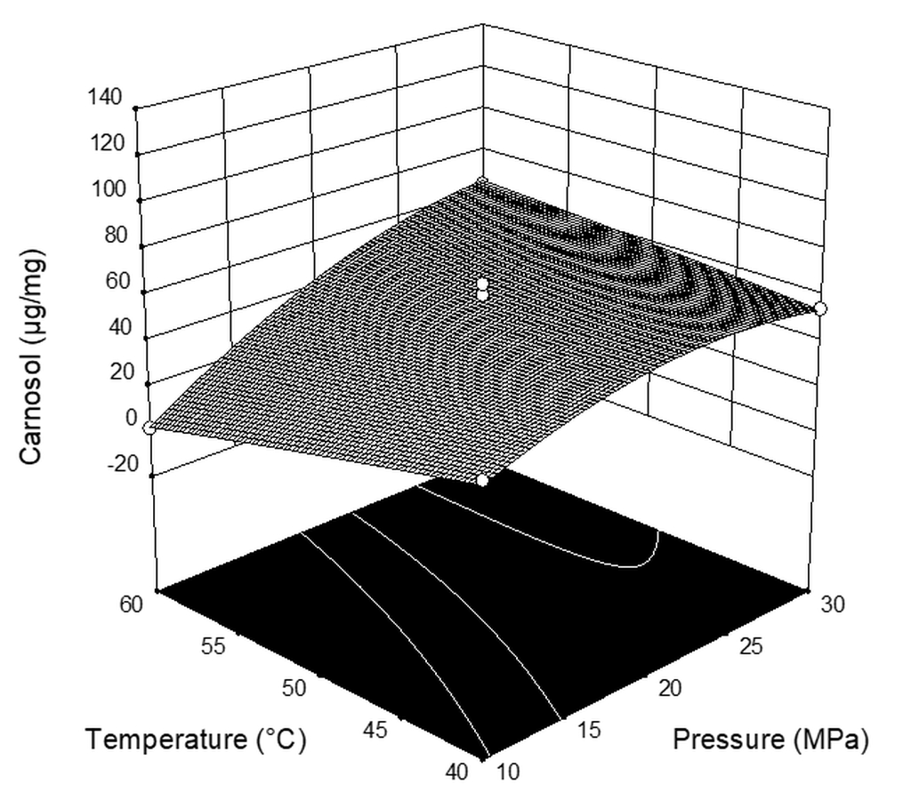
Figure 2 (response surface plots for carnosol) and Table 3 (ANOVA data) demonstrate that the content of carnosol was significantly affected by the pressure (p = 0.0014), i.e., the content of carnosol increased with the increase in pressure. Unlike the effect on carnosic acid, neither temperature (p = 0.1360), CO2 flow rate (p = 0.1258), nor interaction between parameters showed significant statistical influence on the content of carnosol.
According to previous research, the amounts of antioxidant components in plant extracts are determined by the type of extraction and solvent used [50]. It is well known that carnosic acid degrades rapidly in methanol [15,51], and that in extracts with petroleum ether a small amount of carnosic acid is extracted [50]. In methanolic leaf extract of S. officinalis, the amounts of carnosic acid and carnosol were quantified as 14.6 mg g−1 DW and 0.4 mg g−1 DW, respectively [16]. The concentrations of carnosol and carnosic acid in 100 mL of aqueous infusion of sage were 0.66 ± 0.19 mg and 1.31 ± 0.33 mg [52], respectively, while in acetone they were 1.66 ± 0.21 mg g−1 and 12.40 ± 0.43 mg g−1, respectively [53]. In methanolic extracts of 12 samples of S. officinalis L. from Northern Italy, the content of carnosic acid was 0.2–7.1 g kg−1 of extract, while the content of carnosol was 1.1–9.0 g kg−1 of extract. In methanolic extracts of sage from Tunisia, the content of carnosic acid was 746–3110 µg g−1 of dry plant material weight, and depended on geographical location [54]. Additionally, supercritical fluid extraction has been used in plant material extraction considering that it can be performed at low temperatures in a short time, which effectively prevents the oxidation of carnosic acid during extraction and can provide clean extracts without residual of solvent [38,55,56].
As already stated, the data describing the optimal conditions for the extraction of carnosic acid and carnosol from sage using SFE are not available in the literature. However, Caldera et al. [57] investigated the SFE of carnosic acid and carnosol from rosemary. They concluded that the interaction between extraction temperature and time exhibited the most significant influence on the content of carnosic acid, while the extraction temperature and extraction time exhibited the most significant influence on the content of carnosol. Our results differ from those published, which is understandable since we were investigating a different plant material considering different extraction parameters; they can be explained if we consider the solubility of carnosic acid in CO2. The solubility of carnosic acid in SFE with ethanol as a co-solvent was investigated by Cháfer et al. [58]. The solubility of carnosic acid increases with the pressure and the amount of ethanol, while, in the range of pressures that they have explored, the solubility of carnosic acid is higher at lower temperatures, which is consistent with our results. These data on the solubility of the certain components during the extraction process using different parameters are good evidence that each medicinal plant behaves differently during extraction and that each active component is extracted differently depending on the raw material from which it is extracted.
Therefore, the optimization of the extraction process is necessary, and it can be achieved using BBD or some other design. Based on the BBD, the estimated coefficients of second-order response models for carnosol and carnosic acid in S. officinalis extracts are given in Table 4. The R2 was 0.861 and 0.901 for carnosic acid and carnosol, respectively, which indicates that the empirical model shows a good fit with empirical data (R2 are close to 1). According to the ANOVA results (Table 4), the models for both investigated responses (the content of carnosic acid and carnosol in sage extracts) were statistically significant (p ≤ 0.05), and the error analysis that showed a non-significant lack of fit (p = 0.0874–0.8893). Therefore, the influence of the SFE parameters we applied on carnosol and carnosic acid content can be described by a second-order polynomial model.
According to RSM, the following optimization conditions were proposed for calculations: the maximum extraction yield as well as maximum content of carnosic acid and carnosol in obtained extracts. The extraction yields mentioned above were taken from previous work [42]. By applying the desirability function method [59], the optimum extraction conditions were obtained at a pressure of 29.5 MPa, a temperature of 49.2 °C, and a CO2 flow rate of 3 kg h−1. Under these optimal conditions, the yield of sage extract was calculated to be 6.54%, the carnosic acid content to be 105 µg mg−1, and the carnosol content to be 56.3 µg mg−1, which is in very close agreement with obtained experimental data (run 6, Table 2). The desirability for this optimization was 0.874.
The quantitative evaluation of total phenolics in supercritical fluid sage leaf extracts as estimated by the method of Folin–Ciocalteu revealed that S. officinalis exhibited high and variable contents ranging from 1.02 to 9.15 mg of GAE g−1 of DM (Table 5). The highest total phenolic content (TPC) (9.15 ± 0.09 mg of GAE g−1 of DM) was recorded in sage extract obtained at 30 MPa and 40 °C with 2 kg h−1 CO2 flow rate with a carnosic acid content of 71.94 µg mg−1 and a carnosol content of 54.75 µg mg−1, whereas the lowest content (1.02 ± 0.02 mg of GAE g−1 of DM) was found in the case of sage extract obtained at 30 MPa and 60 °C with 2 kg h−1 CO2 flow rate with a carnosic acid content of 18.9 µg mg−1 and a carnosol content of 64.2 µg mg−1. The antioxidant activities of the supercritical fluid sage leaf extracts at a concentration of 25 µg mL−1 were evaluated by the scavenging activities of DPPH, as shown in Table 5. Sage extract obtained at 30 MPa and 40 °C with 2 kg h−1 CO2 flow rate with a carnosic acid content of 71.9 µg mg−1 and a carnosol content of 54.8 µg mg−1 also exhibited the highest antioxidant activity (80.0 ± 0.68%) amongst the investigated supercritical extracts, whereas the lowest antioxidant activity (26.9 ± 0.91%) was found in the case of sage extract obtained at 30 MPa and 60 °C with 2 kg h−1 CO2 flow rate with a carnosic acid content of 18.9 µg mg−1 and a carnosol content of 64.2 µg mg−1. Non-parametric Spearman correlation analysis of all data points demonstrated significant (p < 0.050) strong positive correlation between the carnosic acid content and TPC results (R = 0.7909), and significant (p < 0.050) moderate positive correlation between the carnosic acid content and radical scavenging activity (R = 0.5636).
Supercritical fluid extracts from sage leaves were tested for in vitro antibacterial activity against E. coli, P. aeruginosa, B. subtilis, and S. aureus. MIC and IC50 results are shown in Table 6.
As shown in Table 6, all tested extracts showed good antibacterial activities against E. coli, P. aeruginosa, B. subtilis, and S. aureus. Extracts were more active against gram-positive bacteria than gram-negative bacteria. The main reason for the differences in bacterial susceptibility could be the outer membrane surrounding the cell wall in gram-negative bacteria, which restricts the diffusion of compounds through its lipopolysaccharide covering, as previously reported [60]. The best antibacterial activity was seen against B. subtilis and the lowest activity against E. coli. As reported, carnosic acid, carnosol, rosmanol, and ferruginol are also responsible for the biological activity of sage (Salvia sp.) along with the phenolic rosmarinic and salvianolic acids [61]. The results in Table 6 show supercritical fluid sage extracts to be very effective. Obviously, MIC values are in accordance with IC50 however are not as accurate. Among them, as shown in Table 6, the extract with a carnosic acid content of 116 µg mg−1 and a carnosol content of 60.6 µg mg−1 showed the lowest IC50 10.82 ± 0.02 µg mL−1 against B. subtilis, and the extract with a carnosic acid content of 66.2 µg mg−1 and a carnosol content of 61.02 µg mg−1 showed the highest IC50 40.4 ± 0.19 µg mL−1 against E. coli. As shown in Table 7, all the extracts revealed excellent growth inhibition of all tested bacteria at 62.5 µg mL−1 extract concentration, while at the extract concentration of 15.6 µg mL−1 growth inhibition varied from 14.5–76.8%. Among them, the extract with a carnosic acid content of 116 µg mg−1 and a carnosol content of 60.6 µg mg−1 (run 6, Table 2) showed the best antibacterial activity, especially against B. subtilis, with inhibition rates of 98.33 ± 1.19% at a concentration of 62.5 µg mL−1 and 76.79 ± 0.88% at a concentration of 15.6 µg mL−1. According to the results obtained in the present work, the carnosic acid/carnosol ratio of the sage extracts seems to affect the antibacterial activity of the extracts.
As shown in Table 6 and Table 7, a higher content of carnosic acid in relation to carnosol showed better antibacterial activities of the supercritical fluid sage extracts. We can not claim that the higher content of carnosic acid improves antibacterial activity, since SFE extracts contained carnosic acid and carnosol, as well as many other components such as oxygenated monoterpenes, α-humulenes, viridiflorol, and manool. Our findings are in agreement with those of different authors, such as Klancnik et al. [62] and Bubonja-Sonje et al. [63], who found that the biological activities of rosemary extracts are directly related to the presence of carnosic acid as the major phenolic component, but contrast with the findings of Jordán et al. [64], who found that a higher concentration of carnosol in relation to carnosic acid with same rosmarinic acid content improves the antibacterial activities of methanolic rosemary extracts against Listeria monocytogenes and Staphylococcus aureus strains.

4. Conclusions

In this work, optimization of the extraction of carnosol and carnosic acid from sage leaves using SFE was performed. Only the pressure significantly affected the extraction of carnosol, while in the case of carnosic acid, all investigated parameters—pressure, temperature, and CO2 flow rate—showed a significant effect. The results revealed that the optimal conditions for the maximum extraction of carnosic acid and carnosol were at a pressure of 29.45 MPa, a temperature of 49.19 °C, and a CO2 flow rate of 3 kg h−1. It was observed that by changing the applied pressure and temperature it is possible to obtain an extract with completely different contents of the desired components. The highest total phenolic content (TPC) (9.15 ± 0.09 mg of GAE g−1 of DM) and highest antioxidant activity (79.98 ± 0.68%) at 25 µg mL−1 concentration amongst the investigated supercritical fluid sage extracts was recorded in extract obtained at 30 MPa and 40 °C with 2 kg h−1 CO2 flow rate with a carnosic acid content of 71.94 µg mg−1 and a carnosol content of 54.75 µg mg−1. In this study, the best antibacterial efficiency was confirmed for supercritical fluid sage extract formulations with higher carnosic acid content against all the tested strains, especially gram-positive B. subtilis. Gram-negative tested strains were less susceptible, which could be related to the lower permeability of their surface for phenolic compounds.

Author Contributions

Conceptualization, V.P. and S.J.; Methodology, V.P. and M.J.; Software, S.J., Validation, M.M., S.J., and V.P.; Formal analysis, V.P.; Investigation, M.M., M.J., V.P., and S.J.; Resources, M.M.; Data curation, M.M., V.P., and S.J.; Writing—original draft preparation, V.P.; Writing—review and editing, V.P. and M.M.; Visualization, M.M.; Supervision, V.P.; Project administration, S.J.; Funding acquisition, V.P.

Funding

This research received no external funding.

Acknowledgments

This work was supported by the Osijek-Baranja County under the project “Possibilities of processing medicinal herbs using modern technological processes”.

Conflicts of Interest

The authors declare no conflict of interest.

References

  1. Kamatou, G.P.P.; Viljoen, A.M.; Gono-Bwalya, A.B.; van Zyl, R.L.; van Vuuren, S.F.; Lourens, A.C.U.; Başer, K.H.C.; Demirci, B.; Lindsey, K.L.; van Staden, J.; et al. The in vitro pharmacological activities and a chemical investigation of three South African Salvia species. J. Ethnopharmacol. 2005, 102, 382–390. [Google Scholar] [CrossRef] [PubMed]
  2. Martins, N.; Barros, L.; Santos-Buelga, C.; Henriques, M.; Silva, S.; Ferreira, I.C.F.R. Evaluation of bioactive properties and phenolic compounds in different extracts prepared from Salvia officinalis L. Food Chem. 2015, 170, 378–385. [Google Scholar] [CrossRef] [PubMed]
  3. Kontogianni, V.G.; Tomic, G.; Nikolic, I.; Nerantzaki, A.A.; Sayyad, N.; Stosic-Grujicic, S.; Stojanovic, I.; Gerothanassis, I.P.; Tzakos, A.G. Phytochemical profile of Rosmarinus officinalis and Salvia officinalis extracts and correlation to their antioxidant and anti-proliferative activity. Food Chem. 2013, 136, 120–129. [Google Scholar] [CrossRef] [PubMed]
  4. Bauer, J.; Kuehnl, S.; Rollinger, J.M.; Scherer, O.; Northoff, H.; Stuppner, H.; Werz, O.; Koeberle, A. Carnosol and carnosic acids from Salvia officinalis inhibit microsomal prostaglandin E2 synthase-1. J. Pharmacol. Exp. Ther. 2012, 342, 169–176. [Google Scholar] [CrossRef] [PubMed]
  5. Vuković-Gacić, B.; Nikcević, S.; Berić-Bjedov, T.; Knezević-Vukcević, J.; Simić, D. Antimutagenic effect of essential oil of sage (Salvia officinalis L.) and its monoterpenes against UV-induced mutations in Escherichia coli and Saccharomyces cerevisiae. Food Chem. Toxicol. 2006, 44, 1730–1738. [Google Scholar] [CrossRef] [PubMed]
  6. Pedro, D.F.N.; Ramos, A.A.; Lima, C.F.; Baltazar, F.; Pereira-Wilson, C. Colon Cancer Chemoprevention by Sage Tea Drinking: Decreased DNA Damage and Cell Proliferation. Phytother. Res. 2016, 30, 298–305. [Google Scholar] [CrossRef] [PubMed]
  7. Lu, Y.; Yeap Foo, L. Polyphenolics of Salvia—A review. Phytochemistry 2002, 59, 117–140. [Google Scholar] [CrossRef]
  8. Roby, M.H.H.; Sarhan, M.A.; Selim, K.A.-H.; Khalel, K.I. Evaluation of antioxidant activity, total phenols and phenolic compounds in thyme (Thymus vulgaris L.), sage (Salvia officinalis L.), and marjoram (Origanum majorana L.) extracts. Ind. Crops Prod. 2013, 43, 827–831. [Google Scholar] [CrossRef]
  9. Cuvelier, M.-E.; Richard, H.; Berset, C. Antioxidative activity and phenolic composition of pilot-plant and commercial extracts of sage and rosemary. J. Am. Oil Chem. Soc. 1996, 73, 645–652. [Google Scholar] [CrossRef]
  10. Frankel, E.N.; Huang, S.-W.; Prior, E.; Aeschbach, R. Evaluation of Antioxidant Activity of Rosemary Extracts, Carnosol and Carnosic Acid in Bulk Vegetable Oils and Fish Oil and Their Emulsions. J. Sci. Food Agric. 1996, 72, 201–208. [Google Scholar] [CrossRef]
  11. Hill, R.A.; Connolly, J.D. Triterpenoids. Nat. Prod. Rep. 2017, 34, 90–122. [Google Scholar] [CrossRef] [PubMed] [Green Version]
  12. Munné-Bosch, S.; Schwarz, K.; Alegre, L. Response of abietane diterpenes to stress in Rosmarinus officinalis L.: New insights into the function of diterpenes in plants. Free Radic. Res. 1999, 31, S107–S112. [Google Scholar] [CrossRef] [PubMed]
  13. Gajhede, M.; Anthoni, U.; Per Nielsen, H.; Pedersen, E.J.; Christophersen, C. Carnosol. Crystal structure, absolute configuration, and spectroscopic properties of a diterpene. J. Crystallogr. Spectrosc. Res. 1990, 20, 165–171. [Google Scholar] [CrossRef]
  14. Wenkert, E.; Fuchs, A.; McChesney, J.D. Chemical Artifacts from the Family Labiatae. J. Org. Chem. 1965, 30, 2931–2934. [Google Scholar] [CrossRef]
  15. Schwarz, K.; Ternes, W. Antioxidative constituents of Rosmarinus officinalis and Salvia officinalis. II. Isolation of carnosic acid and formation of other phenolic diterpenes. Z. Lebensm. Unters. Forsch. 1992, 195, 99–103. [Google Scholar] [CrossRef] [PubMed]
  16. Abreu, M.E.; Müller, M.; Alegre, L.; Munné-Bosch, S. Phenolic diterpene and α-tocopherol contents in leaf extracts of 60 Salvia species. J. Sci. Food Agric. 2008, 88, 2648–2653. [Google Scholar] [CrossRef]
  17. Tounekti, T.; Munné-Bosch, S. Enhanced Phenolic Diterpenes Antioxidant Levels Through Non-transgenic Approaches. Crit. Rev. Plant Sci. 2012, 31, 505–519. [Google Scholar] [CrossRef]
  18. Tounekti, T.; Munné-Bosch, S.; Vadel, A.M.; Chtara, C.; Khemira, H. Influence of ionic interactions on essential oil and phenolic diterpene composition of Dalmatian sage (Salvia officinalis L.). Plant Physiol. Biochem. 2010, 48, 813–821. [Google Scholar] [CrossRef]
  19. Tounekti, T.; Vadel, A.M.; Ennajeh, M.; Khemira, H.; Munné-Bosch, S. Ionic interactions and salinity affect monoterpene and phenolic diterpene composition in rosemary (Rosmarinus officinalis). J. Plant Nutr. Soil Sci. 2011, 174, 504–514. [Google Scholar] [CrossRef]
  20. Wang, H.; Liu, F.; Yang, L.; Zu, Y.; Wang, H.; Qu, S.; Zhang, Y. Oxidative stability of fish oil supplemented with carnosic acid compared with synthetic antioxidants during long-term storage. Food Chem. 2011, 128, 93–99. [Google Scholar] [CrossRef]
  21. Guitard, R.; Paul, J.-F.; Nardello-Rataj, V.; Aubry, J.-M. Myricetin, rosmarinic and carnosic acids as superior natural antioxidant alternatives to α-tocopherol for the preservation of omega-3 oils. Food Chem. 2016, 213, 284–295. [Google Scholar] [CrossRef] [PubMed]
  22. Cushnie, T.P.T.; Lamb, A.J. Antimicrobial activity of flavonoids. Int. J. Antimicrob. Agents 2005, 26, 343–356. [Google Scholar] [CrossRef] [PubMed]
  23. Del Campo, J.; Amiot, M.J.; Nguyen-The, C. Antimicrobial effect of rosemary extracts. J. Food Prot. 2000, 63, 1359–1368. [Google Scholar] [CrossRef] [PubMed]
  24. Moreno, S.; Scheyer, T.; Romano, C.S.; Vojnov, A.A. Antioxidant and antimicrobial activities of rosemary extracts linked to their polyphenol composition. Free Radic. Res. 2006, 40, 223–231. [Google Scholar] [CrossRef] [PubMed]
  25. Horiuchi, K.; Shiota, S.; Kuroda, T.; Hatano, T.; Yoshida, T.; Tsuchiya, T. Potentiation of antimicrobial activity of aminoglycosides by carnosol from Salvia officinalis. Biol. Pharm. Bull. 2007, 30, 287–290. [Google Scholar] [CrossRef] [PubMed]
  26. Aruoma, O.I.; Halliwell, B.; Aeschbach, R.; Löligers, J. Antioxidant and pro-oxidant properties of active rosemary constituents: Carnosol and carnosic acid. Xenobiotica 1992, 22, 257–268. [Google Scholar] [CrossRef] [PubMed]
  27. Dapkevicius, A.; Venskutonis, R.; van Beek, T.A.; Linssen, J.P.H. Antioxidant activity of extracts obtained by different isolation procedures from some aromatic herbs grown in Lithuania. J. Sci. Food Agric. 1998, 77, 140–146. [Google Scholar] [CrossRef]
  28. Dent, M.; Dragović, V.; Penić, M. The Effect of Extraction Solvents, Temperature and Time on the Composition and Mass Fraction of Polyphenols in Dalmatian Wild Sage (Salvia officinalis L.) Extracts. Food Technol. Biotechnol. 2013, 51, 84–91. [Google Scholar] [CrossRef]
  29. Miguel, G.; Cruz, C.; Faleiro, M.L.; Simões, M.T.F.; Figueiredo, A.C.; Barroso, J.G.; Pedro, L.G. Salvia officinalis L. essential oils: Effect of hydrodistillation time on the chemical composition, antioxidant and antimicrobial activities. Nat. Prod. Res. 2011, 25, 526–541. [Google Scholar] [CrossRef]
  30. Ollanketo, M.; Peltoketo, A.; Hartonen, K.; Hiltunen, R.; Riekkola, M.-L. Extraction of sage (Salvia officinalis L.) by pressurized hot water and conventional methods: Antioxidant activity of the extracts. Eur. Food Res. Technol. 2002, 215, 158–163. [Google Scholar] [CrossRef]
  31. Raal, A.; Orav, A.; Arak, E. Composition of the essential oil of Salvia officinalis L. from various European countries. Nat. Prod. Res. 2007, 21, 406–411. [Google Scholar] [CrossRef] [PubMed]
  32. Durling, N.E.; Catchpole, O.J.; Grey, J.B.; Webby, R.F.; Mitchell, K.A.; Foo, L.Y.; Perry, N.B. Extraction of phenolics and essential oil from dried sage (Salvia officinalis) using ethanol–water mixtures. Food Chem. 2007, 101, 1417–1424. [Google Scholar] [CrossRef]
  33. Sališová, M.; Toma, Š.; Mason, T.J. Comparison of conventional and ultrasonically assisted extractions of pharmaceutically active compounds from Salvia officinalis. Ultrason. Sonochem. 1997, 4, 131–134. [Google Scholar] [CrossRef]
  34. Velicković, D.T.; Milenović, D.M.; Ristić, M.S.; Veljković, V.B. Kinetics of ultrasonic extraction of extractive substances from garden (Salvia officinalis L.) and glutinous (Salvia glutinosa L.) sage. Ultrason. Sonochem. 2006, 13, 150–156. [Google Scholar] [CrossRef]
  35. Dauksas, E.; Venskutonis, P.R.; Povilaityte, V.; Sivik, B. Rapid screening of antioxidant activity of sage (Salvia officinalis L.) extracts obtained by supercritical carbon dioxide at different extraction conditions. Nahrung 2001, 45, 338–341. [Google Scholar] [CrossRef]
  36. Fornari, T.; Ruiz-Rodriguez, A.; Vicente, G.; Vázquez, E.; García-Risco, M.R.; Reglero, G. Kinetic study of the supercritical CO2 extraction of different plants from Lamiaceae family. J. Supercrit. Fluids 2012, 64, 1–8. [Google Scholar] [CrossRef]
  37. Menaker, A.; Kravets, M.; Koel, M.; Orav, A. Identification and characterization of supercritical fluid extracts from herbs. C. R. Chim. 2004, 7, 629–633. [Google Scholar] [CrossRef]
  38. Reverchon, E.; Taddeo, R.; Porta, G.D. Extraction of sage oil by supercritical C02: Influence of some process parameters. J. Supercrit. Fluids 1995, 8, 302–309. [Google Scholar] [CrossRef]
  39. Jokić, S.; Vidović, S.; Aladić, K. Supercritical Fluid Extraction of Edible Oils. In Supercritical Fluids: Fundamentals, Properties and Application; Osborn, J., Ed.; Nova Publishers: New York, NY, USA, 2014; pp. 205–228. ISBN 978-1-63321-946-5. [Google Scholar]
  40. Sharif, K.M.; Rahman, M.M.; Azmir, J.; Mohamed, A.; Jahurul, M.H.A.; Sahena, F.; Zaidul, I.S.M. Experimental design of supercritical fluid extraction—A review. J. Food Eng. 2014, 124, 105–116. [Google Scholar] [CrossRef]
  41. Box, G.E.P.; Wilson, K.B. On the Experimental Attainment of Optimum Conditions. J. R. Stat. Soc. Ser. B (Methodol.) 1951, 13, 1–45. [Google Scholar] [CrossRef]
  42. Jokić, S.; Molnar, M.; Jakovljević, M.; Aladić, K.; Jerković, I. Optimization of supercritical CO2 extraction of Salvia officinalis L. leaves targeted on Oxygenated monoterpenes, α-humulene, viridiflorol and manool. J. Supercrit. Fluids 2018, 133, 253–262. [Google Scholar] [CrossRef]
  43. Babovic, N.; Djilas, S.; Jadranin, M.; Vajs, V.; Ivanovic, J.; Petrovic, S.; Zizovic, I. Supercritical carbon dioxide extraction of antioxidant fractions from selected Lamiaceae herbs and their antioxidant capacity. Innov. Food Sci. Emerg. Technol. 2010, 11, 98–107. [Google Scholar] [CrossRef]
  44. Glisic, S.; Ivanovic, J.; Ristic, M.; Skala, D. Extraction of sage (Salvia officinalis L.) by supercritical CO2: Kinetic data, chemical composition and selectivity of diterpenes. J. Supercrit. Fluids 2010, 52, 62–70. [Google Scholar] [CrossRef]
  45. Jokić, S.; Horvat, G.; Aladić, K. Design of SFE System Using a Holistic Approach: Problems and Challenges. In Supercritical Fluid Extraction: Technology, Applications and Limitations; Jason, L., Ed.; Nova Publishers: New York, NY, USA, 2015; pp. 95–122. ISBN 978-1-63463-353-6. [Google Scholar]
  46. Singleton, V.L.; Rossi, J.A. Colorimetry of Total Phenolics with Phosphomolybdic-Phosphotungstic Acid Reagents. Am. J. Enol. Viticult. 1965, 16, 144–158. [Google Scholar]
  47. Shih, M.-H.; Su, Y.-S.; Wu, C.-L. Syntheses of Aromatic Substituted Hydrazino-thiazole Derivatives to Clarify Structural Characterization and Antioxidant Activity between 3-Arylsydnonyl and Aryl Substituted Hydrazino-thiazoles. Chem. Pharm. Bull. 2007, 55, 1126–1135. [Google Scholar] [CrossRef] [PubMed] [Green Version]
  48. Gu, W.; Wang, S. Synthesis and antimicrobial activities of novel 1H-dibenzo[a,c]carbazoles from dehydroabietic acid. Eur. J. Medicinal Chem. 2010, 45, 4692–4696. [Google Scholar] [CrossRef] [PubMed]
  49. Molnar, M.; Pavić, V.; Šarkanj, B.; Čačić, M.; Vuković, D.; Klenkar, J. Mono-and bis-dipicolinic acid heterocyclic derivatives–thiosemicarbazides, triazoles, oxadiazoles and thiazolidinones as antifungal and antioxidant agents. Heterocycl. Commun. 2017, 23, 35–42. [Google Scholar] [CrossRef]
  50. Wellwood, C.R.L.; Cole, R.A. Relevance of carnosic acid concentrations to the selection of rosemary, Rosmarinus officinalis (L.), accessions for optimization of antioxidant yield. J. Agric. Food Chem. 2004, 52, 6101–6107. [Google Scholar] [CrossRef]
  51. Masuda, T.; Inaba, Y.; Maekawa, T.; Takeda, Y.; Tamura, H.; Yamaguchi, H. Recovery mechanism of the antioxidant activity from carnosic acid quinone, an oxidized sage and rosemary antioxidant. J. Agric. Food Chem. 2002, 50, 5863–5869. [Google Scholar] [CrossRef]
  52. Matsingou, T.C.; Petrakis, N.; Kapsokefalou, M.; Salifoglou, A. Antioxidant Activity of Organic Extracts from Aqueous Infusions of Sage. J. Agric. Food Chem. 2003, 51, 6696–6701. [Google Scholar] [CrossRef]
  53. Okamura, N.; Fujimoto, Y.; Kuwabara, S.; Yagi, A. High-performance liquid chromatographic determination of carnosic acid and carnosol in Rosmarinus officinalis and Salvia officinalis. J. Chromatogr. A 1994, 679, 381–386. [Google Scholar] [CrossRef]
  54. Ben Farhat, M.; Jordán, M.J.; Chaouech-Hamada, R.; Landoulsi, A.; Sotomayor, J.A. Variations in Essential Oil, Phenolic Compounds, and Antioxidant Activity of Tunisian Cultivated Salvia officinalis L. J. Agric. Food Chem. 2009, 57, 10349–10356. [Google Scholar] [CrossRef] [PubMed]
  55. Tena, M.T.; Valcárcel, M.; Hidalgo, P.J.; Ubera, J.L. Supercritical Fluid Extraction of Natural Antioxidants from Rosemary:  Comparison with Liquid Solvent Sonication. Anal. Chem. 1997, 69, 521–526. [Google Scholar] [CrossRef] [PubMed]
  56. Mukhopadhyay, M. Natural Extracts Using Supercritical Carbon Dioxide; CRC Press: Boca Raton, FL, USA, 2000; ISBN 978-1-4200-4169-9. [Google Scholar]
  57. Caldera, G.; Figueroa, Y.; Vargas, M.; Santos, D.; Marquina-Chidsey, G. Optimization of Supercritical Fluid Extraction of Antioxidant Compounds from Venezuelan Rosemary Leaves. Int. J. Food Eng. 2012, 8. [Google Scholar] [CrossRef]
  58. Chafer, A.; Fornari, T.; Berna, A.; Ibáñez, E.; Reglero, G. Solubility of solid carnosic acid in supercritical CO2 with ethanol as a co-solvent. J. Supercrit. Fluids 2005, 34, 323–329. [Google Scholar] [CrossRef]
  59. Cojocaru, C.; Khayet, M.; Zakrzewska-Trznadel, G.; Jaworska, A. Modeling and multi-response optimization of pervaporation of organic aqueous solutions using desirability function approach. J. Hazard. Mater. 2009, 167, 52–63. [Google Scholar] [CrossRef] [PubMed]
  60. Vaara, M. Agents that increase the permeability of the outer membrane. Microbiol. Rev. 1992, 56, 395–411. [Google Scholar]
  61. Matkowski, A. Plant in vitro culture for the production of antioxidants—A review. Biotechnol. Adv. 2008, 26, 548–560. [Google Scholar] [CrossRef]
  62. Klancnik, A.; Guzej, B.; Kolar, M.H.; Abramovic, H.; Mozina, S.S. In vitro antimicrobial and antioxidant activity of commercial rosemary extract formulations. J. Food Prot. 2009, 72, 1744–1752. [Google Scholar] [CrossRef]
  63. Bubonja-Sonje, M.; Giacometti, J.; Abram, M. Antioxidant and antilisterial activity of olive oil, cocoa and rosemary extract polyphenols. Food Chem. 2011, 127, 1821–1827. [Google Scholar] [CrossRef]
  64. Jordán, M.J.; Lax, V.; Rota, M.C.; Lorán, S.; Sotomayor, J.A. Relevance of carnosic acid, carnosol, and rosmarinic acid concentrations in the in vitro antioxidant and antimicrobial activities of Rosmarinus officinalis (L.) methanolic extracts. J. Agric. Food Chem. 2012, 60, 9603–9608. [Google Scholar] [CrossRef] [PubMed]
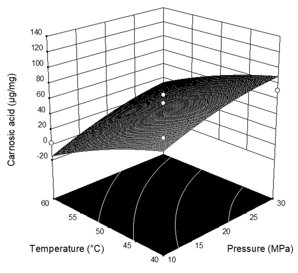
Figure 1. Three-dimensional plots for the obtained content of carnosic acid as a function of the extraction pressure and temperature.
Plants 08 00016 g001
Figure 2. Three-dimensional plots for the obtained content of carnosol as a function of the extraction pressure and temperature.
Plants 08 00016 g002
Table 1. Coded and real levels of independent variables for the designed experiment.
Independent VariableSymbolLevel
Low (−1)Middle (0)High (+1)
Pressure (MPa)X1102030
Temperature (°C)X2405060
CO2 flow rate (kg h−1)X3123
Table 2. Experimental matrix and values of observed response.
RunPressure (MPa)Temperature (°C)CO2 Flow Rate
(kg h−1)
Extraction Yield * (%)Carnosic Acid
(µg mg−1extract)
Carnosol
(µg mg−1extract)
1104020.65967.9440.71
2204013.38548.0461.70
3204034.026120.0551.08
4105031.14425.4939.77
5205023.76847.5665.51
6305037.361116.2560.63
7305015.23861.7861.47
8206035.47738.5358.00
9105010.2420.290.46
10106020.3652.931.80
11205023.30539.7837.28
12306024.31618.9364.19
13205022.55255.6146.72
14304023.80371.9454.75
15206014.8911.6435.19
16205024.52866.2061.02
* Data are from Jokić et al. (2018).
Table 3. Regression coefficient of polynomial function of all response surfaces.
TermCoefficientsStandard ErrorF-Valuep-Value *
Carnosic acid
Intercept52.9910.58
X121.537.488.280.0282
X2−30.747.4816.870.0063
X323.577.489.920.0198
X12−6.4810.580.380.5628
X22−5.3710.580.260.6298
X32−5.1510.580.240.6439
X1X23.0010.580.0800.7865
X1X37.3210.580.480.5153
X2X3−8.7810.580.690.4385
R2 = 0.8611
Carnosol
Intercept52.635.04
X119.793.5630.850.0014
X2−6.133.562.960.1360
X36.333.563.160.1258
X12−11.595.045.290.0534
X22−0.685.040.0180.0934
X32−0.465.040.030.1481
X1X212.095.045.750.0611
X1X3−10.045.043.970.8973
X2X3−0.465.042.750.9300
R2 = 0.9013
* p < 0.01 highly significant; 0.01 ≤ p < 0.05 significant; p ≥ 0.05 not significant.
Table 4. Analysis of variance (ANOVA) of the modeled responses.
SourceSum of SquaresDegrees of FreedomMean SquareF-Valuep-Value *
Carnosic acid
The recovery
Model16663.2391851.474.130.0491
Residual2688.706448.12
Lack of fit2305.193768.406.010.0874
Pure error383.513127.84
Total19351.9415
Carnosol
The recovery
Model5560.329617.816.090.0198
Residual609.146101.52
Lack of fit102.25334.080.200.8893
Pure error506.893168.96
Total6169.4615
* p < 0.01 highly significant; 0.01 ≤ p < 0.05 significant; p ≥ 0.05 not significant.
Table 5. Total phenolics content (TPC) in supercritical fluid sage leaf extracts expressed as mg of GAE g−1 of DM and 2,2-diphenyl-1-picrylhydrazyl (DPPH) radical scavenging activity expressed as % DPPH radical scavenging activity at 25 µg mL−1 concentration.
RunTotal Phenolics Content (TPC) (mgGAE gDM−1)DPPH Radical Scavenging Activity (%)
12.79 ± 0.0657.71 ± 0.33
23.08 ± 0.0538.99 ± 0.32
3NDND
42.26 ± 0.0146.87 ± 3.60
53.97 ± 0.1353.53 ± 3.37
64.32 ± 0.0347.05 ± 1.77
7NDND
8NDND
9NDND
10NDND
112.72 ± 0.0756.72 ± 0.65
121.02 ± 0.0226.91 ± 0.91
133.17 ± 0.0148.01 ± 0.86
149.15 ± 0.0979.98 ± 0.68
152.65 ± 0.0742.62 ± 1.69
162.96 ± 0.0150.54 ± 1.98
ND: not determined. Data expressed as mean ± S.D.
Table 6. Comparison of minimum inhibitory concentrations (MIC) and 50% Growth Reduction (IC50) of supercritical fluid sage leaf extracts against Escherichia coli, Pseudomonas aeruginosa, Bacillus subtilis, and Staphylococcus aureus (µg mL−1).
RunMIC (µg mL−1)IC50 (µg mL−1)
E. coliP. aeruginosaB. subtilisS. aureusE. coliP. aeruginosaB. subtilisS. aureus
131.2531.2515.62531.2529.53 ± 0.1628.54 ± 0.0512.45 ± 0.0817.17 ± 0.09
231.2531.2515.62531.2527.89 ± 0.0726.28 ± 1.7612.69 ± 0.0723.90 ± 0.10
3NDNDNDNDNDNDNDND
431.2531.2515.62515.62527.03 ± 0.5525.75 ± 0.5213.97 ± 0.0714.53 ± 0.04
531.2531.2515.62531.2531.18 ± 0.0516.33 ±0.2612.77 ± 0.0622.75 ± 0.13
631.2531.2515.62515.62531.08 ± 0.0817.82 ± 0.3910.82 ± 0.0214.61 ± 0.08
7NDNDNDNDNDNDNDND
8NDNDNDNDNDNDNDND
9NDNDNDNDNDNDNDND
10NDNDNDNDNDNDNDND
1131.2531.2515.62515.62528.43 ± 0.3622.23 ± 0.6112.70 ± 0.0814.10 ± 0.10
1262.5031.2515.62515.62539.68 ± 0.2122.36 ± 0.7914.02 ± 0.1614.12 ± 0.18
1362.5031.2515.62515.62537.80 ± 0.8323.78 ± 0.4413.18 ± 0.0812.46 ± 0.10
1431.2531.2515.62515.62524.85 ± 0.0823.57 ± 1.2112.51 ± 0.1115.04 ± 0.13
1531.2531.2515.62515.62520.29 ± 0.6227.98 ± 1.2413.01 ± 0.1613.75 ± 0.04
1662.5031.2515.62515.62540.44 ± 0.1923.61 ± 0.5313.43 ± 0.0417.91 ± 0.67
G0.9760.9761.9533.9060.58 ± 0.100.91 ± 0.071.83 ± 0.013.06 ± 0.06
ND: not determined. G-gentamicin data expressed as mean ± S.D.
Table 7. Bacterial growth inhibition in the presence of supercritical fluid sage leaf extracts against Escherichia coli, Pseudomonas aeruginosa, Bacillus subtilis, and Staphylococcus aureus.
RunGrowth Inhibition (%)
E. coliP. aeruginosaB. subtilisS. aureus
62.5
µg mL−1
15.625
µg mL−1
62.5
µg mL−1
15.625
µg mL−1
62.5
µg mL−1
15.625
µg mL−1
62.5
µg mL−1
15.625
µg mL−1
198.34 ± 0.3516.73 ± 0.5196.62 ± 1.8431.93 ± 3.2298.84 ± 0.2972.19 ± 0.2298.32 ± 0.0646.33 ± 0.15
293.04 ± 0.1920.10 ± 0.5991.42 ± 1.5635.39 ± 0.5798.26 ± 0.4471.80 ± 0.8397.56 ± 0.2221.97 ± 1.17
3NDNDNDNDNDNDNDND
497.17 ± 0.2814.45 ± 1.1897.36 ± 0.3038.30 ± 0.7999.48 ± 01061.62 ± 0.4398.31 ± 0.1257.57 ± 0.36
595.82 ± 0.2823.91 ± 0.3493.30 ± 0.6549.41 ± 0.2297.39 ± 0.4268.05 ± 0.7496.57 ± 0.8732.24 ± 0.16
699.05 ± 1.1939.22 ± 0.0695.91 ± 0.9649.63 ± 0.0898.33 ± 0.3876.79 ± 0.8897.80 ± 0.5755.47 ± 0.77
7NDNDNDNDNDNDNDND
8NDNDNDNDNDNDNDND
9NDNDNDNDNDNDNDND
10NDNDNDNDNDNDNDND
1195.75 ± 1.6228.04 ± 1.1895.07 ± 1.2944.10 ± 1.0298.88 ± 0.2272.31 ± 0.3997.74 ± 0.1956.69 ± 0.59
1294.96 ± 0.3520.21 ± 3.0097.78 ± 1.3731.93 ± 3.2299.00 ± 0.2260.99 ± 1.3197.98 ± 0.0960.77 ± 1.56
1397.41 ± 0.8226.80 ± 3.6298.07 ± 0.4141.50 ± 3.8898.72 ± 0.2267.30 ± 0.1097.37 ± 0.6674.93 ± 0.30
1497.03 ± 0.2437.76 ± 0.2599.15 ± 0.1539.71 ± 2.9599.28 ± 0.1871.42 ± 0.6497.55 ± 0.1852.30 ± 0.60
1597.47 ± 1.1040.72 ± 1.3097.45 ± 0.7042.32 ± 2.0499.41 ± 0.4569.85 ± 0.6598.24 ± 0.2961.11 ± 0.29
1696.77 ± 0.4128.47 ± 3.1897.26 ± 2.1140.38 ± 1.1998.81 ± 0.1165.04 ± 0.5897.00 ± 0.8646.29 ± 1.28
G98.18 ± 0.7695.00 ± 0.1799.83 ± 0.0296.83 ± 0.0299.86 ± 0.1494.83 ± 0.7198.58 ± 0.5697.00 ± 0.67
ND: not determined. G-gentamicin data expressed as mean ± S.D.