1. Introduction
Cannabis sativa has occupied a significant place in traditional medical systems across multiple cultures for thousands of years, and its phytochemical diversity has sustained intensive modern pharmacological investigation [
1]. Cannabidiol (CBD) is the most abundant non-psychoactive phytocannabinoid in
C. sativa, does not act as a classical orthosteric agonist at cannabinoid CB1 or CB2 receptors the way its psychoactive congener THC does; at CB1, it behaves as a non-competitive negative allosteric modulator, reducing THC and endocannabinoid signaling at a site mapped to the CB1 N-terminus [
1], with a second, structurally distinct allosteric site recently identified [
2]. This multiplicity of binding sites at a single, well-studied receptor is a caution directly relevant to the docking-only analysis performed in this study. At CB2, CBD similarly acts as a weak partial agonist at the orthosteric site while occupying a distinct allosteric pocket [
3]. Beyond this dual cannabinoid-receptor activity, CBD engages additional targets including TRPV1, serotonin 5-HT1A receptors, PPARgamma, and GPR55, a receptor promiscuity that, combined with a well-established tolerability profile, positions CBD as a pharmacologically distinctive candidate for multi-target therapeutic investigation.
The clinical application of CBD has expanded substantially since the FDA approval of Epidiolex (purified oral CBD solution) for Lennox–Gastaut and Dravet syndromes in 2018, alongside growing preclinical evidence of anti-inflammatory, antioxidant, and antineoplastic activity across cancer models. Breast, colorectal, and lung cancer are the three most commonly diagnosed cancers worldwide, together accounting for close to a third of all new cancer cases globally [
4]. Yet, they arise from substantially different driver pathways: estrogen-receptor signaling in breast cancer, Wnt/PPARgamma/COX-2-linked adenoma-to-carcinoma progression in colorectal cancer [
5,
6,
7], and EGFR/KRAS-driven epithelial transformation in lung cancer [
8,
9] (detailed further in
Section 4.2). These three cancers were selected specifically because they instantiate mechanistically distinct paradigms of oncogenic driver biology rather than three arbitrary examples of a single disease process; a CBD target-prediction pipeline that recovers cancer-type-appropriate hub proteins across all three paradigms is a more stringent test of the approach than testing it against a single cancer type or an undifferentiated pan-cancer gene set. The present study, therefore, analyzes CBD’s target engagement against gene sets curated separately for each cancer type, so that its predicted target profile can be interpreted in the context of the specific molecular biology of each tumor rather than a molecularly heterogeneous “cancer” category.
Network pharmacology provides the conceptual and computational framework best suited to characterize this type of multi-target pharmacological activity. By modeling drug action as perturbation of a protein–protein interaction (PPI) network, this approach enables a systems-level understanding of how a compound’s predicted target set maps onto disease pathways and biological processes. The Maximal Clique Centrality (MCC) algorithm, implemented in the CytoHubba plugin for Cytoscape, identifies proteins with the greatest topological centrality within the disease PPI network by evaluating their participation in maximally dense clique subnetworks. When combined with structure-based molecular docking, including re-docking of each target’s experimentally co-crystallized reference ligand as an internal benchmark, network pharmacology analysis yields not only a list of predicted targets but also a structural basis for atomic-level engagement and a calibrated point of comparison for interpreting CBD’s docking scores.
A small but growing number of independent studies have applied network pharmacology and docking specifically to CBD across individual cancer types, including a recent analysis of CBD in colorectal cancer [
10] and a 2025/2026 inverse-docking fingerprint study that benchmarks phytocannabinoid target engagement against experimentally validated ChEMBL bioactivity data [
11]. The present study extends this emerging literature by constructing a single, pooled CBD-disease PPI network built from breast, colorectal, and lung cancer gene sets, subsequently cross-referencing the resulting MCC hub list against independent, cancer-type-specific disease-association searches, and by pairing each docking result with an authentic co-crystallized reference ligand for direct potency calibration.
The objectives of this study are: to construct a pooled, cancer-associated CBD-disease PPI network spanning breast, colorectal, and lung cancer; to identify, via MCC-based hub analysis, the proteins of greatest topological centrality within this network and to relate them explicitly to the defining biology of each cancer type to characterize CBD’s docking affinity toward these hubs using GBVI/WSA dG scoring, benchmarked directly against each target’s own co-crystallized reference ligand; and to generate a cancer-type-specific, mechanistically grounded, and appropriately hedged framework for experimental validation.
This study integrates MCC-based network topology with structure-based molecular docking benchmarked against experimentally resolved co-crystallized ligands. By combining network centrality, structural binding assessment, and cancer-specific biological interpretation, this approach provides a more rigorous framework for prioritizing CBD targets for future experimental validation.
2. Materials and Methods
2.1. Target Identification for CBD and Cancer-Specific Disease Gene Sets
CBD-associated protein targets were retrieved from three complementary computational databases, SwissTargetPrediction (Swiss Institute of Bioinformatics, Lausanne, Switzerland), SEA (Similarity Ensemble Approach) (University of California, San Francisco, CA, USA), and SuperPRED (Charité–Universitätsmedizin Berlin, Berlin, Germany), using CBD’s canonical SMILES format (PubChem CID: 644019). Predictions from the three platforms were combined into a unified set of 215 unique CBD-associated targets.
Breast, colorectal, and lung cancer disease-gene sets were retrieved from three curated disease-association resources, GeneCards (Weizmann Institute of Science, Rehovot, Israel), OMIM (Johns Hopkins University, Baltimore, MD, USA), and DisGeNET (MedBioinformatics Solutions SL, Barcelona, Spain), queried with the combined set of breast, colorectal, and lung neoplasm terms. The three resources were combined into a unified set of 27,420 unique genes. This union is intentionally broad rather than a stringent disease-relevance filter: GeneCards alone accounts for the vast majority of these genes under permissive disease-term matching, and the resulting set approaches whole-genome coverage. As discussed in
Section 4.6, the meaningful narrowing in this pipeline is performed by the subsequent STRING network (STRING Consortium, Lausanne, Switzerland) construction and CytoHubba MCC (Institute of Bioinformatics, National Chiao Tung University, Hsinchu, Taiwan) ranking, not by this gene-set intersection step.
Intersecting the 215-target CBD prediction union with the 27,420-gene cancer disease-association union yielded 144 overlapping CBD-disease targets, standardized to official HGNC gene symbols (European Bioinformatics Institute, Hinxton, UK); one pair of these entries (PTGS2 and its common protein-name alias COX2) referred to the same gene and was merged into a single node before network construction, giving the final network size of 143 unique targets used throughout this study. Of these 143 targets, source-by-source confirmation was as follows: SwissTargetPrediction 99, SEA 2, SuperPRED 55, GeneCards 143, OMIM 16, and DisGeNET 140.
To determine which of these 143 network targets are attributable to breast, colorectal, or lung cancer specifically, rather than to the pooled three-cancer search used to build the network, DisGeNET was queried a second time, separately for each of the three cancer types, returning 1548 unique genes across 19 matched breast cancer disease terms, 1389 unique genes across 11 matched colorectal cancer terms, and 1211 unique genes across 14 matched lung cancer terms (union of 3199 genes; 269 common to all three cancer types). Cross-referencing these three cancer-type-specific gene sets directly against the 143 network targets shows exactly which hub and non-hub targets are individually confirmed as breast-, colorectal-, or lung-associated by name-resolved disease-term matching, rather than relying on a pooled search or general biological arguments alone.
2.2. PPI Network Construction and Topological Analysis
The 143 CBD-disease targets were submitted to the STRING database (version 12.0;
https://string-db.org, accessed on 15 June 2026) for PPI network construction, retaining interactions with a combined confidence score of at least 0.700 [
12]. The resulting network was exported and imported into Cytoscape v3.10.1 for visualization and topological analysis [
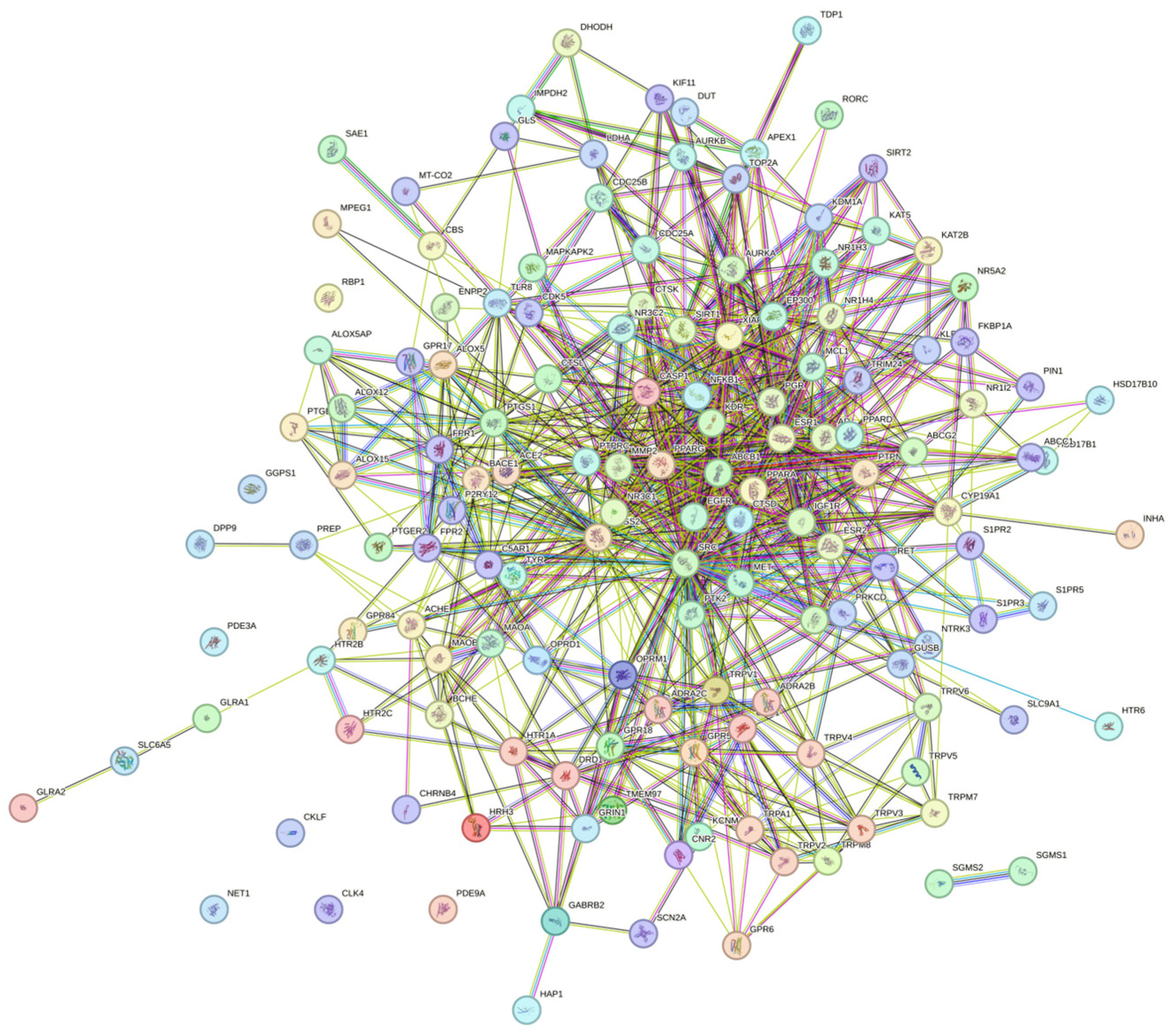
13]. The final network comprised 143 nodes and 802 edges, with an average node degree of 11.2 and an average local clustering coefficient of 0.472. Under a random network model applied to the same number of proteins, only 305 edges would be expected; the observed 802 edges represent a 2.63-fold enrichment over this expectation, and the PPI enrichment
p-value was below 1.0 × 10
−16. Network parameters were computed using the NetworkAnalyzer plugin, and hub protein identification was performed with the CytoHubba plugin using the Maximal Clique Centrality (MCC) algorithm [
14], which preferentially rewards participation in tightly interconnected functional modules rather than raw connectivity and outperforms Degree, MNC, and DMNC for recovery of disease-causal proteins in CytoHubba benchmark testing. The top 10 MCC-ranked proteins were carried forward to molecular docking.
2.3. Functional Enrichment Analysis
Functional enrichment of the 143-target network was performed directly in STRING using its whole-genome statistical background, with false discovery rate correction. This analysis returned 812 significantly enriched Gene Ontology Biological Process terms, 131 Molecular Function terms, and 69 Cellular Component terms; 55 significantly enriched KEGG pathways; 94 Reactome pathways; 134 WikiPathways entries; 27 DISEASES disease-gene associations; 85 TISSUES expression terms; 51 COMPARTMENTS subcellular localization terms; and 47 significantly enriched UniProt keywords (all FDR < 0.05). Whole-network enrichment, rather than restriction to only the ten docking hubs, was used so that the established pharmacological target classes CBD is already known to engage, GPCRs, ion channels, and nuclear hormone receptors, would be visible within the analysis, regardless of whether they were ultimately selected as MCC hubs. Given the large number of terms returned across these ten enrichment categories, only the top-ranked terms per category are discussed in the text (
Section 3.3).
2.4. Ligand Preparation
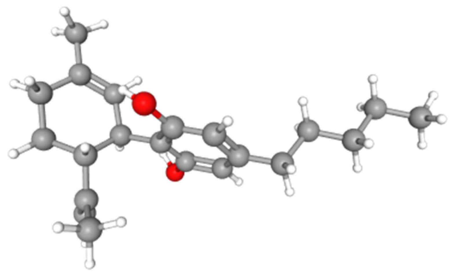
The three-dimensional structure of cannabidiol (CBD; IUPAC: 2-[(1R,6R)-6-isopropenyl-3-methylcyclohex-2-en-1-yl]-5-pentylbenzene-1,3-diol; molecular formula: C
21H
30O
2; MW: 314.46 g/mol; PubChem CID: 644019) was retrieved from PubChem in SDF format [
15]. The structure was imported into MOE (version 2022.02; Chemical Computing Group Inc., Montreal, QC, Canada) and subjected to geometry optimization to a root-mean-square gradient of 0.01 kcal/mol per angstrom (
Figure 1).
2.5. Protein Structure Retrieval and Preparation
Three-dimensional crystal structures of the ten hub proteins were retrieved from the RCSB Protein Data Bank [
16]; the original crystallographic determination is cited directly for each structure where a specific primary source paper was identified and confirmed, 1PXX/PTGS2 [
17], 1M17/EGFR [
18], the ERα/ERβ subtype-selectivity structural work relevant to 1XP1/ESR1 and 1L2J/ESR2 [
19,
20], 3K8S/PPARG (via its co-crystallized ligand T2384) [
21], 3I81/IGF1R [
22], 2SRC/SRC [
23], and 4KXQ/SIRT1 [
24]. Primary source citations for the remaining two PDB entries (8TQD/NFKB1, 8H78/MMP2) were not located as direct co-crystallographic reports at the time of writing; instead, recent primary pharmacological studies independently establishing the ligandability of NFKB1 [
25] and the structure-based design of a selective MMP2 inhibitor [
26] are cited in
Section 3.13 and
Section 3.14 as the most relevant available primary literature for those two targets. All structures were prepared using the MOE Protein Prepare Wizard workflow: removal of crystallographic waters outside the primary binding site; addition of hydrogens at pH 7.4 using Protonate3D [
27]; assignment of formal charges and bond orders; repair of missing side-chain atoms; and constrained energy minimization of hydrogen positions with heavy-atom coordinates fixed. The docking-box center coordinates and dimensions used for each target are documented in
Supplementary Table S1.
2.6. Active Site Identification
All protein binding sites were correlated with experimentally validated co-crystallized native ligands. Each of the ten targets examined here possesses an experimentally confirmed ligand-occupied active site or ligand-binding domain pocket, containing a co-crystallized small-molecule reference ligand. For the nine structures containing noncovalent reference ligands, these ligands provided target-matched references for interpreting CBD docking at the corresponding pockets. The covalently bound NFKB1 reference ligand JMR was treated separately, as described in
Section 2.7.
2.7. Molecular Docking Protocol and Co-Crystallized-Ligand Benchmarking
Docking was performed using Triangle Matcher placement, London dG initial scoring, induced-fit refinement of residues within 4.5 Å of the docked pose, and GBVI/WSA dG rescoring [
28,
29]. Among 300 poses, the five top-ranked poses per target were retained, and the pose with the most negative GBVI/WSA dG S score was selected as the representative binding mode. Each docking run was repeated in triplicate using independently regenerated CBD conformers. For the nine structures containing noncovalent reference ligands, each authentic co-crystallized ligand was extracted and redocked into its corresponding receptor using the standard docking protocol described above. This provided geometric validation through comparison of the redocked and experimentally deposited poses and a target-matched reference for interpreting CBD’s docking score at the same pocket. The covalently bound NFKB1 reference ligand JMR was treated separately using the MOE covalent docking procedure described below. S score classification thresholds: below −10 kcal/mol = strong binding; −10 to −5 kcal/mol = moderate binding; above −5 kcal/mol = low/weak binding. Redocking accuracy was quantified by calculating the root-mean-square deviation (RMSD) between each redocked co-crystallized ligand pose and its original deposited coordinates using PyMOL (version 2.5.4). Because the co-crystallized ligand JMR in the NFKB1 structure (PDB ID: 8TQD) is covalently bound, its experimental binding pose could not be appropriately reproduced using the standard noncovalent docking protocol applied to the other reference ligands. JMR was therefore redocked separately using MOE’s covalent docking protocol. Induced-fit refinement was performed for 300 generated poses, and the pose with the most favorable docking score was retained for geometric validation against the crystallographic pose. CBD, which was treated as a noncovalent ligand, was docked to NFKB1 using the standard protocol applied to all other targets. Consequently, the JMR redocking result was used primarily for pose-reproduction assessment, and its docking score was not considered directly comparable to the noncovalent CBD score.
2.8. AutoDock Vina Cross-Validation Docking
To provide an independent cross-validation of the MOE docking results, CBD was docked against all ten targets using AutoDock Vina [
30] (version 1.1.2), while the nine noncovalent co-crystallized reference ligands were redocked against their corresponding targets. Docking parameters were set to an exhaustiveness of 50 and 15 output binding poses per run; the pose with the most negative predicted binding affinity was retained as the representative result for each run. Search grids were centered on the co-crystallized ligand position in each retrieved structure; box dimensions were determined by visual inspection of each binding pocket in UCSF Chimera (version 1.2) [
31] to ensure full enclosure of the co-crystallized ligand and immediately adjacent residues. AutoDock Vina scores were interpreted comparatively across the target panel rather than assigned to rigid affinity categories. For the nine structures containing noncovalent reference ligands, each reference ligand was redocked using the same Vina protocol to provide an independently generated target-matched benchmark at the corresponding binding pocket. AutoDock Vina does not natively support covalent docking; therefore, no Vina score was reported for the covalently bound NFKB1 reference ligand JMR. Instead, JMR was redocked in MOE using the covalent docking protocol described in
Section 2.7. For all MOE and AutoDock Vina docking analyses, the search grids were centered on the corresponding co-crystallized ligands and sized to fully accommodate CBD and the relevant binding pocket. The final grid centers and dimensions used in all analyses are provided in
Supplementary Table S1.
The two docking engines use unrelated search algorithms and scoring functions: MOE’s Triangle Matcher placement with GBVI/WSA dG rescoring versus Vina’s iterated local search with an empirical scoring function; consistent results across both are therefore treated as a more robust indication of genuine structural complementarity than either method alone.
2.9. Interaction Analysis
Protein–ligand interaction profiles for the best-ranked docked poses were characterized using the MOE Ligand Interactions tool (hydrogen bonds, hydrophobic contacts, electrostatic interactions, and aromatic pi-stacking), and key contact residues were cross-referenced with the published structural and mutational literature to assess biological plausibility.
3. Results
3.1. Target Identification and Cancer-Associated PPI Network Topology
Target retrieval across SwissTargetPrediction, SEA, and SuperPRED (215 unique CBD targets in union) intersected with breast-, colorectal-, and lung-cancer-associated genes from GeneCards, OMIM, and DisGeNET (27,420 unique genes in union;
Section 2.1) yielded 143 unique CBD-disease network nodes after merging one gene-symbol alias pair. Submission to STRING produced a network of 143 nodes and 802 edges (average node degree 11.2; average local clustering coefficient 0.472), against 305 edges expected under a random model (2.63-fold enrichment; PPI enrichment
p < 1.0 × 10
−16), confirming a functionally coherent rather than random interactome (
Figure 2). This cancer-associated network explicitly contains a substantial complement of CBD’s already well-characterized pharmacological targets, including TRPV1, TRPV2–TRPV6, TRPA1, TRPM7/TRPM8, GPR55, GPR6, GPR18, GPR84, CNR1, CNR2, and HTR1A, confirming that the target-prediction and network-construction pipeline recovers established CBD pharmacology. These GPCR, ion-channel, and cannabinoid-receptor nodes did not, however, emerge as MCC hubs in the pooled cancer-associated network (
Section 3.2); their role in cancer-relevant signaling is discussed in
Section 4.4.
3.2. Hub Protein Identification by MCC Analysis and Direct Cancer-Type Attribution
Cross-referencing the 143 network targets directly against three independent, cancer-type-specific DisGeNET searches (
Section 2.1) shows that 51 of the 143 targets (36%) are individually confirmed as disease-associated with at least one of the three named cancer types by name-resolved query matching, rather than only through the broader pooled three-cancer search used to build the network; the remaining 92 targets entered the network via GeneCards and/or OMIM, or via DisGeNET disease terms not specific to a single named cancer type. Of the 51 directly cancer-type-attributed targets, 9 are confirmed in all three cancer types (ABCB1, EGFR, EP300, ESR1, KDR, MCL1, PTGS2, SRC, TYR), 29 are confirmed specifically in breast cancer searches, 28 in colorectal cancer searches, and 24 in lung cancer searches (
Table 1). Critically, this direct, name-resolved attribution independently corroborates the biological rationale given for the ten MCC hub proteins in
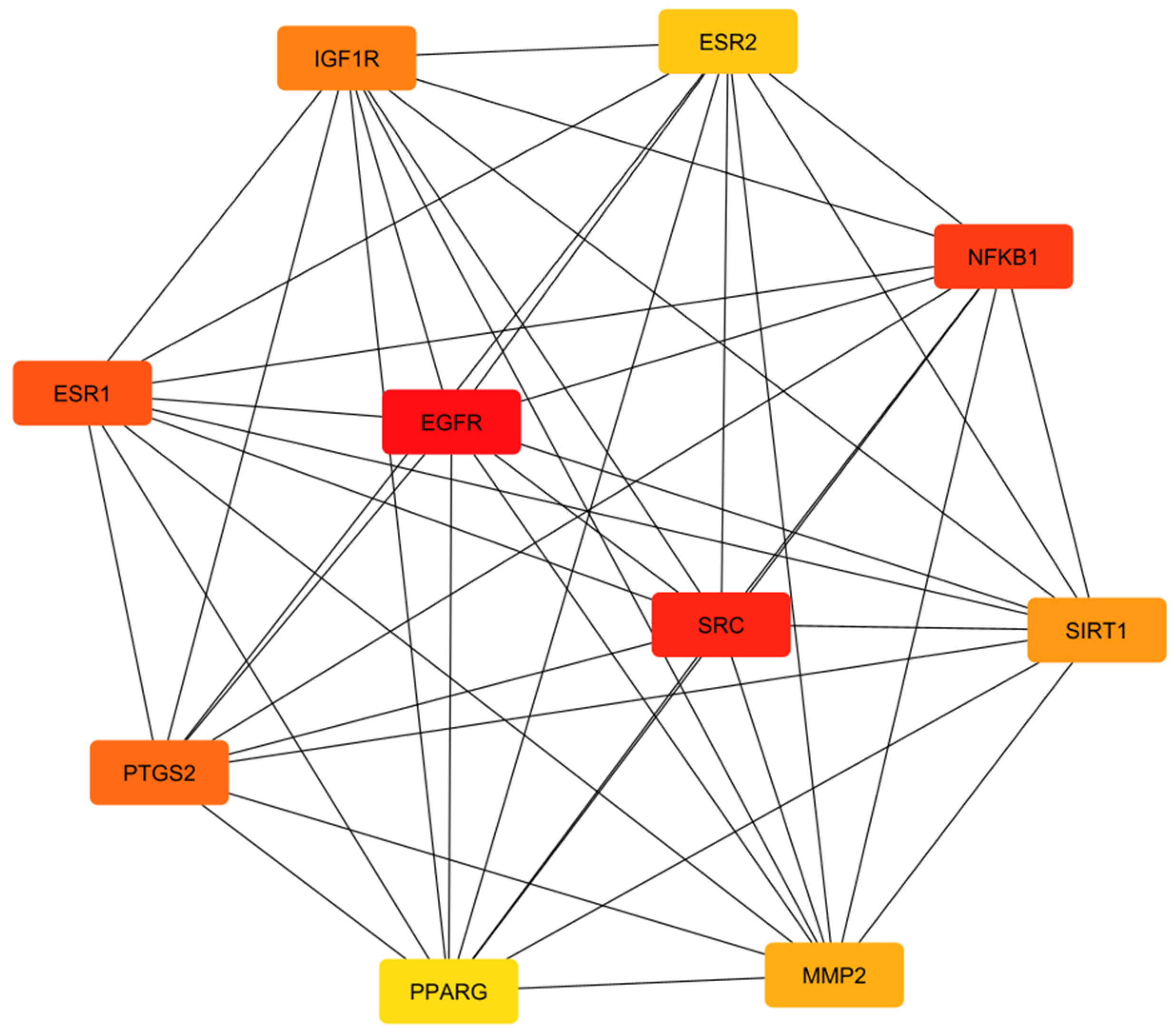
Section 3.3. All ten hubs are individually confirmed by at least one cancer-type-specific DisGeNET search, and four (SRC, PTGS2, ESR1, EGFR) are confirmed in all three cancer types, providing target-level (rather than only pathway-level) evidence for the cancer-type relevance claims made throughout this study. The interaction network among the ten MCC-ranked hub proteins is shown in
Figure 3.
CytoHubba MCC analysis of the 143-node network identified ten hub proteins (
Table 2), spanning nuclear hormone receptor, growth-factor receptor, inflammatory transcription factor, and matrix-remodeling protein classes. The hub list is directly and specifically anchored to the three named cancer types: ESR1 and ESR2 (estrogen receptor alpha and beta) are the central druggable axis of breast cancer endocrine signaling; PPARG and PTGS2/COX-2 are established colorectal cancer differentiation and chemoprevention targets; and EGFR is the principal oncogenic driver of non-small-cell lung cancer. SRC, MMP2, NFKB1, IGF1R, and SIRT1 recur across all three cancer types as convergent invasion, growth-signaling, and inflammatory nodes.
All ten hubs are independently confirmed as cancer-type-associated by the direct, name-resolved DisGeNET cross-referencing in
Table 1: four (SRC, PTGS2, ESR1, EGFR) are confirmed across all three cancer types specifically, while the remaining six are each confirmed in one or two of the three cancer types individually (SIRT1: breast only; NFKB1: colorectal only; PPARG, MMP2, and ESR2: breast and colorectal; IGF1R: breast and lung).
3.3. Functional Enrichment Confirms Breast-, Colorectal-, and Lung-Relevant Signalling
STRING functional enrichment of the full 143-target network (
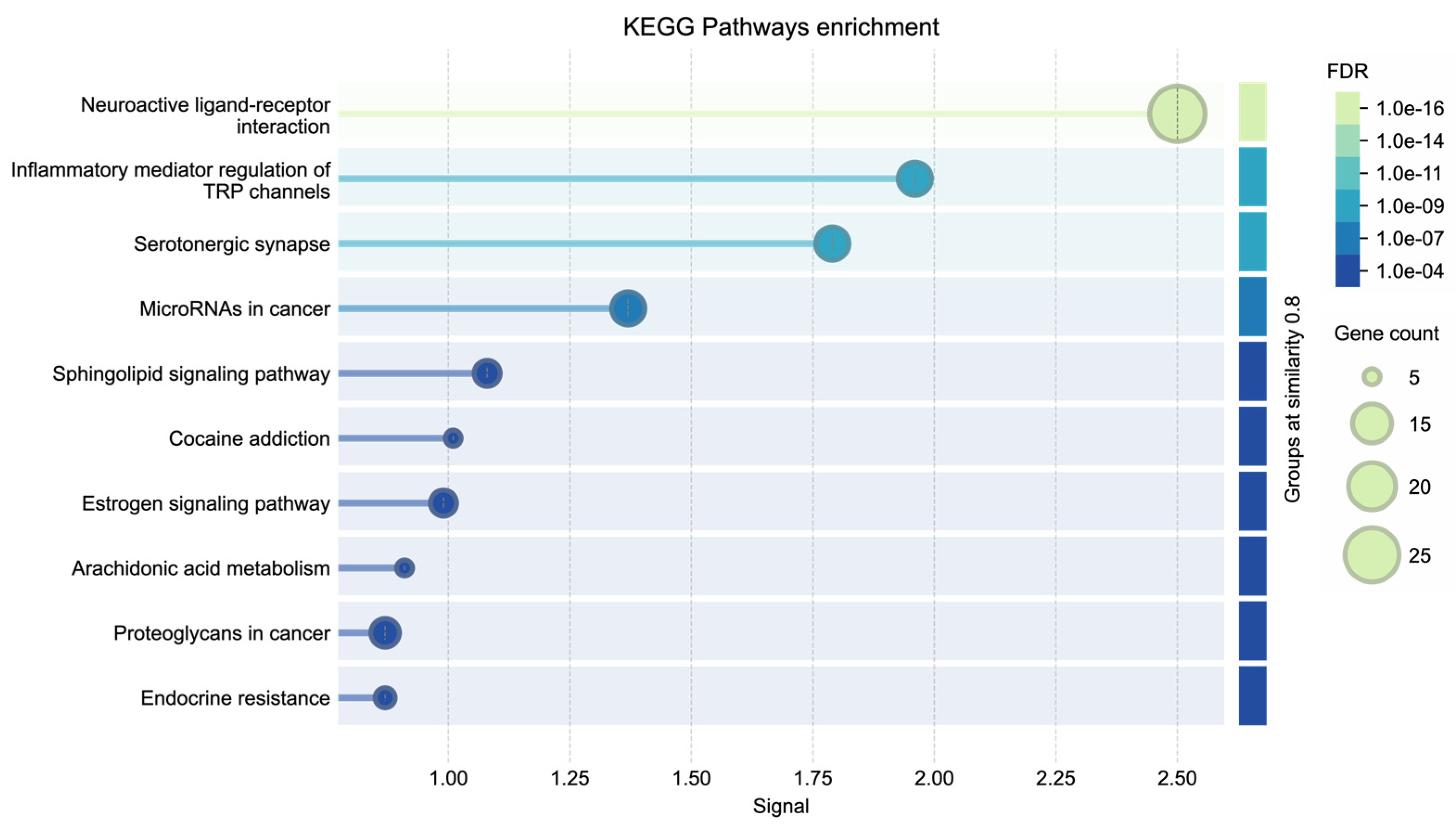
Section 2.3) returned pathway and process annotations directly consistent with the three target cancers. The top ten enriched KEGG pathways are summarized in
Figure 4, with pathway significance represented by signal strength, gene count, and false discovery rate (FDR). Among the top KEGG pathways by enrichment strength were neuroactive ligand–receptor interaction (26/329 genes; signal 2.50; FDR 1.80 × 10
−16), inflammatory mediator regulation of TRP channels (12/92; signal 1.96; FDR 2.03 × 10
−9), serotonergic synapse (12/108; signal 1.79; FDR 7.47 × 10
−9), microRNAs in cancer (12/159; signal 1.37; FDR 3.42 × 10
−7), and sphingolipid signaling (9/116; signal 1.08; FDR 2.01 × 10
−5). As shown in
Figure 4, the exported enrichment plot additionally ranks cocaine addiction, estrogen signaling pathway, arachidonic acid metabolism, proteoglycans in cancer, and endocrine resistance among the ten most enriched KEGG terms by signal strength. Two of these five, estrogen signaling pathway and endocrine resistance, map directly onto the ESR1/ESR2/IGF1R breast-cancer axis identified as MCC hubs in
Section 3.2, and a third, arachidonic acid metabolism, maps directly onto the PTGS2-driven colorectal chemoprevention axis, giving independent, network-level statistical support to the cancer-type-specific hub assignments made in
Table 2. Reactome enrichment additionally returned the Nuclear Receptor transcription pathway (14/52; FDR 3.42 × 10
−14) and Class A/1 Rhodopsin-like receptors (23/326; FDR 5.30 × 10
−13), and DISEASES-category enrichment returned adenocarcinoma (DOID:299; 9/119 genes; FDR 4.3 × 10
−4), consistent with the adenocarcinoma histology common to breast, colorectal, and the majority of lung cancers.
3.4. Molecular Docking: S Score Overview and Classification, and Co-Crystallized-Ligand Benchmarking
Eight of the ten CBD-target complexes fell within the MOE moderate-binding classification, while NFKB1 and MMP2 fell within the MOE low/weak-binding range. PTGS2/COX-2 produced the most favorable CBD docking score under both MOE and AutoDock Vina (−8.775 and −9.731 kcal/mol, respectively), whereas NFKB1 and MMP2 produced the least favorable CBD scores within the target panel. Independent cross-validation with AutoDock Vina yielded binding trends broadly comparable to those from MOE. However, minor differences in rank order and classification were observed, as expected given the distinct search algorithms and scoring functions used by the two platforms. This broad agreement provides methodological corroboration beyond what either docking engine alone would support. Redocking of each target’s own co-crystallized reference ligand (
Table 3) reproduced the experimentally observed binding pose in every case, with per-structure RMSD values calculated in PyMOL of 0.884 Å (PTGS2/COX-2), 0.724 Å (ESR1), 0.723 Å (ESR2), 0.590 Å (EGFR), 0.689 Å (PPARG), 0.699 Å (IGF1R), 0.557 Å (SRC), 0.456 Å (SIRT1), 0.746 Å (NFKB1), and 0.756 Å (MMP2), all well within the 2.0 Å threshold conventionally used to indicate successful pose reproduction. Under both MOE and Vina, the co-crystallized ligand scored more negatively than CBD at six of the nine targets with a valid comparison under both engines (ESR1, PPARG, IGF1R, SRC, SIRT1, MMP2); at three of these nine targets (PTGS2, ESR2, EGFR), CBD’s predicted score was instead marginally more negative than the co-crystallized ligand under both engines. NFKB1’s co-crystallized reference ligand (JMR) is a covalent inhibitor and was docked in MOE using a covalent docking protocol rather than the standard protocol used elsewhere (
Section 3.13); JMR produced a more negative MOE docking score than CBD (−6.986 versus −4.916 kcal/mol). However, because JMR and CBD were evaluated using covalent and noncovalent docking protocols, respectively, these scores were not treated as a direct quantitative comparison of binding potency. No corresponding Vina comparison is available for NFKB1, since AutoDock Vina does not natively support covalent docking. CBD’s predicted affinity is therefore generally, though not uniformly, weaker than that of each pocket’s own high-affinity co-crystallized ligand, with the exceptions concentrated at targets where the co-crystallized ligand’s own affinity was itself comparatively modest rather than strong (
Table 3). Within the AutoDock Vina results, CBD produced less favorable scores than six of the nine noncovalent co-crystallized reference ligands for which a valid Vina comparison was available. At the same time, PTGS2, ESR2, and EGFR showed marginally more favorable CBD scores. NFKB1’s co-crystallized ligand has no Vina score for the reasons noted above. Given the ±1–2 kcal/mol empirical error typical of both scoring functions, the three-way clustering of ESR1/ESR2/EGFR/PPARG MOE scores (−7.79 to −8.34 kcal/mol) is treated here as indicating comparable, not strictly rank-ordered, moderate-range affinity, and NFKB1 and MMP2 are explicitly reported as producing the least favorable CBD docking scores within the target panel rather than as validated targets of comparable pharmacological relevance to the higher-scoring hubs.
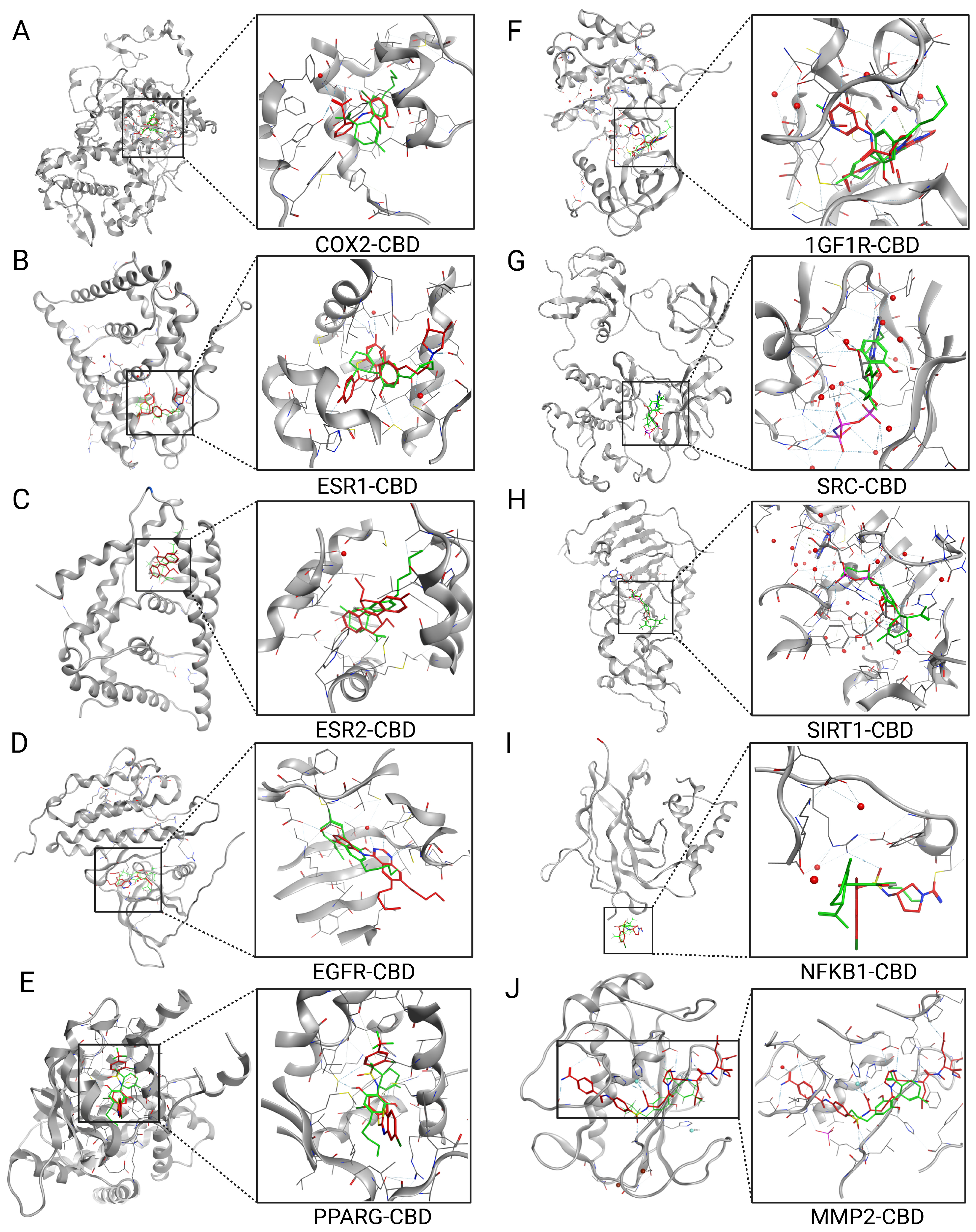
Representative binding poses of CBD within the active sites of the ten hub proteins are shown in
Figure 5, illustrating the predicted orientation of CBD in each target binding pocket.
3.5. CBD Binding to PTGS2/COX-2 (MOE S Score: −8.775 kcal/mol; Vina Score: −9.731 kcal/mol)
PTGS2 exhibited the strongest predicted binding affinity in the panel. As shown in
Figure 5A, CBD is predicted to occupy the arachidonic acid substrate channel in a geometry comparable to that of the co-crystallized diclofenac-class reference ligand (DIF), with its phenolic hydroxyl groups positioned to engage the polar residues that normally anchor the arachidonic acid carboxylate, and its hydrophobic pentyl chain extending into the apolar distal channel.
This interpretation is anchored to the validated determinants of ligand recognition at this site: Tyr385 and Ser530, rather than the canonical Arg120 salt bridge, govern diclofenac-class binding in this channel [
17], and CBD’s predicted pose is best assessed against this same Tyr385/Ser530 region rather than assumed equivalent to diclofenac’s carboxylate-mediated mechanism, which CBD structurally lacks.
3.6. CBD Binding to ESR1 (Estrogen Receptor Alpha; MOE S Score: −8.341 kcal/mol; Vina Score: −9.629 kcal/mol)
ESR1 was the second-ranked target by docking affinity. The predicted binding orientation (
Figure 5B) places CBD within the ligand-binding domain pocket, with a hydrophobic contact involving Met 343, a residue lining the hydrophobic core of the estrogen-binding cavity and adjacent to the AF-2 helix-12 region that governs receptor conformational switching between agonist and antagonist states.
3.7. CBD Binding to ESR2 (Estrogen Receptor Beta; MOE S Score: −8.042 kcal/mol; Vina Score: −9.031 kcal/mol)
ESR2 ranked third, with a docking score closely comparable to ESR1. ESR2 is frequently described as a modulatory, and in some contexts tumor-suppressive, counterpart to ESR1 in breast tissue, and is also expressed in normal and malignant colorectal mucosa. The near-equivalent CBD affinity predicted for both ER isoforms raises the possibility of concurrent, isoform-non-selective estrogen-receptor engagement, a hypothesis that would require isoform-selective radioligand-binding assays to confirm or refute.
This is consistent with the reference structure itself, in which a comparably sized ligand at ESR2 produced a distinct “passive antagonism” mechanism, suppressing coactivator binding without the bulky substituent typical of classical antagonists [
19], again indicating that pocket occupancy alone does not establish CBD’s functional effect at this receptor.
3.8. CBD Binding to EGFR (MOE S Score: −7.849 kcal/mol; Vina Score: −7.923 kcal/mol)
EGFR, the principal oncogenic driver in non-small-cell lung cancer [
8,
9], ranked fourth. The docking pose (
Figure 5D) shows CBD occupying the ATP-binding cleft of the kinase domain, in the same pocket occupied by the co-crystallized erlotinib-class reference ligand (AQ4). Notably, CBD’s score at this pocket was marginally more negative than that of the redocked AQ4 reference itself (
Table 3); because erlotinib’s true clinical potency depends on specific hinge-region contacts within an active-like kinase conformation that scoring functions do not fully capture [
18], this result should not be read as evidence that CBD approaches erlotinib’s real-world potency, but rather illustrates that redocked reference scores do not always track a drug’s known experimental affinity. CBD’s engagement here is best interpreted as supporting a modulatory or adjunctive role rather than EGFR-TKI-equivalent activity.
3.9. CBD Binding to PPARG (MOE S Score: −7.793 kcal/mol; Vina Score: −9.221 kcal/mol)
PPARG ranked fifth. CBD is predicted to engage the same Y-shaped ligand-binding pocket occupied by the co-crystallized reference ligand T2384, which itself produces partial rather than full PPARG activation across more than one binding orientation [
21]; CBD’s score is consistent with, though does not establish, a similar partial-agonist mechanism analogous to thiazolidinedione PPARG agonists.
3.10. CBD Binding to IGF1R (MOE S Score: −6.879 kcal/mol; Vina Score: −9.640 kcal/mol)
IGF1R ranked sixth. CBD’s moderate-range engagement of the IGF1R ATP-binding pocket contrasts with the reference inhibitor BMS-754807, whose potent, low-nanomolar inhibition depends on a specific hinge-binding hydrogen-bond triad that CBD does not reproduce [
22]; this supports a modest, adjunctive contribution to growth-factor pathway attenuation rather than a primary inhibitory mechanism.
3.11. CBD Binding to SRC (MOE S Score: −6.536 kcal/mol; Vina Score: −7.457 kcal/mol)
SRC ranked seventh. CBD is predicted to occupy the ATP-binding cleft in the same region as the co-crystallized ATP analog (ANP), at an affinity well below that of ATP itself. Because the reference structure represents SRC in an autoinhibited conformation stabilized by SH2/SH3 restraints [
23], this indicates compatibility with the inactive-state pocket only; functional inhibition would additionally require evidence that CBD competes with ATP or shifts the active/inactive equilibrium.
3.12. CBD Binding to SIRT1 (MOE S Score: −5.116 kcal/mol; Vina Score: −7.986 kcal/mol)
SIRT1 ranked eighth. CBD’s predicted contact with Arg 274, near the NAD+/substrate-binding cleft occupied by the co-crystallized ADP-ribose ligand (APR), places CBD at the low end of the moderate-binding range; SIRT1 also showed the largest divergence between the two docking engines of any target in the panel (MOE −5.116 kcal/mol versus Vina −6.556 kcal/mol), warranting additional caution pending experimental confirmation.
Because the reference structure captures a single, closed conformation of a catalytic cycle involving substantial domain movement upon cofactor binding [
24], this Arg274 contact should be read as compatible with that one conformational state rather than as evidence that a specific catalytic step is inhibited.
3.13. CBD Binding to NFKB1 (MOE S Score: −4.916 kcal/mol; Vina Score: −6.143 kcal/mol)
NFKB1 ranked ninth. CBD’s docking score at the modeled pocket fell within the low/weak-binding classification, indicating that high network centrality does not necessarily correspond to strong predicted binding affinity. Any anti-inflammatory activity of CBD involving NFKB1 may therefore be more plausibly attributed to indirect pathway modulation than to a direct, high-affinity interaction at this pocket. Because the co-crystallized reference ligand JMR is a covalent inhibitor, it was redocked in MOE using a covalent docking protocol, with induced-fit refinement applied across 300 generated poses. The pose with the most favorable docking score was retained as the representative result. No corresponding AutoDock Vina score was reported because Vina does not natively support covalent docking. JMR produced a more negative MOE docking score than CBD (−6.986 versus −4.916 kcal/mol). However, because JMR and CBD were evaluated using covalent and noncovalent docking protocols, respectively, their scores should be interpreted cautiously and not as a direct quantitative comparison of binding potency.
Independent chemoproteomic profiling has separately demonstrated NFKB1 ligandability through a reactive cysteine targeted by covalent probes [
25]. This covalent mechanism is distinct from the noncovalent binding model evaluated for CBD and does not alter the weak CBD docking score obtained here.
3.14. CBD Binding to MMP2 (MOE S Score: −4.442 kcal/mol Vina Score: −5.919 kcal/mol)
MMP2 ranked tenth, recording the weakest docking score in the panel, below both the moderate-binding threshold and the affinity recovered for its own co-crystallized reference ligand (L2U); as with NFKB1, this weak-binding result for an otherwise centrally connected hub is discussed in
Section 4.3 as evidence that MCC and docking affinity are not equivalent measures.
This is consistent with the structural requirements for potent MMP2 inhibition: the selective inhibitor TP0597850 achieves picomolar affinity through direct coordination of the catalytic zinc ion together with subtype-defining pocket contacts [
26], an interaction mode CBD’s structure lacks.
5. Conclusions
This study integrated network pharmacology with molecular docking to investigate the potential multi-target mechanisms of CBD in breast, colorectal, and lung cancer. Network analysis identified ten biologically relevant hub proteins, including SRC, SIRT1, PTGS2, PPARG, NFKB1, MMP2, IGF1R, ESR1, ESR2, and EGFR, while functional enrichment demonstrated their involvement in key pathways associated with tumor progression, inflammation, and growth. Molecular docking and comparison with co-crystallized reference ligands indicated predominantly moderate binding affinities, supporting the potential of CBD to interact with multiple cancer-related targets rather than a single dominant protein. Overall, these findings provide a computational framework for prioritizing biologically relevant CBD targets and demonstrate the value of integrating network pharmacology with structure-based docking and co-crystallized ligand benchmarking. As a computational study, these results should be regarded as hypothesis-generating and require further biochemical, cellular, and in vivo validation to determine the therapeutic relevance of the predicted interactions.




 ,
,