Article

A Pilot Study on Injury Risk Assessment in Emergency Care Using Dual Motion Capture Systems

1
Biomedical Engineering, Gannon University, Erie, PA 16541, USA
2
Electrical and Computer Engineering, Gannon University, Erie, PA 16541, USA
*
Author to whom correspondence should be addressed.
Theor. Appl. Ergon. 2026, 2(3), 13; https://doi.org/10.3390/tae2030013
Submission received: 24 April 2026 / Revised: 30 June 2026 / Accepted: 2 July 2026 / Published: 9 July 2026

Abstract

Manual lifting is a common occupational activity associated with an increased risk of low back disorders. In this study, 22 participants from UPMC Hamot, organized into 11 pairs, were recruited. A combination of motion capture techniques and an injury assessment tool was used to investigate the relationships among body anthropometrics, three-dimensional trunk and lower-limb kinematics, and lumbar spinal loading. Potential differences in lifting mechanics were observed between male and female participants. Males exhibited greater trunk flexion and higher compressive loading, while females demonstrated greater hip and knee flexion with reduced trunk motion. These findings indicate that spinal loading during lifting is influenced by an interaction between anthropometric characteristics and movement coordination patterns, with variable behavioral trends affecting load distribution across the trunk and lower extremities. The results provide biomechanical insight that may inform the development of bio-ergonomic training techniques aimed at reducing lumbar spine loading and minimizing injury risk in occupational lifting tasks.

1. Introduction

Healthcare professionals routinely engage in physically demanding tasks that can compromise their health and well-being. One of the primary concerns is the risk of developing musculoskeletal disorders (MSDs), especially during patient-handling activities such as transferring patients between beds or repositioning them. These risks are often intensified by fatigue resulting from extended working hours [1]. Among MSDs, lower back pain (LBP) is the most prevalent, affecting an estimated 570 million people globally [2]. According to the Occupational Safety and Health Administration (OSHA), nursing assistants had one of the highest rates of MSDs in 2017, with 166.3 cases per 10,000 workers, approximately five times the average across all occupations [3]. Nurses are also among the professions most vulnerable to back injuries, accounting for 18.8% of occupational injury cases [4].
A systematic review published in 2018 reported that nurses experience a substantially higher prevalence of MSDs compared to many other occupational groups, with LBP being the most commonly reported condition [5]. Similarly, a 2020 study on patient transfer activities found that 37% of Danish healthcare workers reported limitations in their work due to LBP, reinforcing its status as the most widespread MSD in this population [6]. Consistent findings have been observed in other settings. For instance, a study conducted across six hospitals in Athens found that increased physical workload was significantly associated with higher reports of back, neck, and shoulder pain, as well as a greater overall number of MSD symptoms [7].
One study reported that up to 72% of nurses experienced low back pain (LBP) during improper patient-handling practices [8]. Survey-based research further shows that MSDs are closely linked to absenteeism, reduced job satisfaction, and increased intent to leave the profession [9]. For example, a study of nurses in Pakistan reported that roughly one-third experienced MSDs, with the lower back being the most frequently affected area. Contributing factors included prolonged static postures, handling heavy patients, and working in awkward positions [10].
In addition to physical limitations, MSDs can also impact mental health and job performance. A study involving 2500 registered nurses in North Carolina found that 18% reported depression associated with physical conditions, while 62% indicated that MSDs adversely affected their work performance, increasing the likelihood of issues such as medication errors and patient falls [11]. Similarly, research involving 350 nurses across six hospitals found that 51% experienced LBP and 23% reported knee pain, both of which were strongly associated with higher rates of absenteeism [12].
Patient transfer tasks, including moving patients between beds and stretchers, using slide boards, transferring patients to wheelchairs, repositioning, and rolling, have been widely studied [13]. Due to the complexity of these movements, many investigations have focused on the biomechanics involved, emphasizing the importance of proper techniques to prevent injury [14,15]. For instance, one study reported that manually transferring heavier patients can generate compressive forces of up to 10,000 N at the L5/S1 spinal segment [16]. Another study found that trunk flexion during transfers often approaches the upper limits of the range of motion, indicating considerable physical strain [17]. More recent research has shown that assistive devices, such as slide boards, can significantly reduce mechanical loads on the hands, shoulders, and trunk during these activities [18].
To reduce these risks, various assistive technologies have been introduced, including intelligent beds and other supportive equipment aimed at minimizing LBP and MSDs. While these tools have demonstrated some benefits, they have not fully resolved the issue [19]. Some studies have explored biomechanics in patient transport scenarios; for example, Häske et al. [20] used the Xsens motion tracking system to analyze spinal motion. However, their work primarily focused on patient movement and promoted self-extrication strategies rather than evaluating caregiver lifting mechanics.
Furthermore, there remains a lack of comprehensive research examining the forces experienced by healthcare workers during tasks such as boosting or repositioning patients in bed. One study investigated hand forces at different bed heights but did not assess whole-body mechanics or quantify loads on the lower back and lower limbs [21]. To address this gap, the present study employs two motion-tracking systems simultaneously to capture the movements of two participants performing a coordinated patient-boosting task. This approach enables a more detailed evaluation of spinal loading, exploration of the relationships between human factors and biomechanical forces, and analysis of how the postures of two healthcare providers interact during collaborative lifting. The results may inform evidence-based recommendations for bio-ergonomic training techniques to reduce injury risk among nurses.

2. Methods

2.1. Study Design

This study was conducted in accordance with the Declaration of Helsinki and was approved by both the Gannon University Institutional Review Board (protocol code: GUIRB-2022-9-6970) and the University of Pittsburgh Institutional Review Board (protocol code: STUDY22110153). Written informed consent was obtained from all participants prior to the testing session, including consent for the use of images. All published images were de-identified.
This study utilizes an advanced biomechanical fusion framework to evaluate localized musculoskeletal risks during healthcare tasks. The experimental protocol integrated real-time inertial motion capture data with predictive digital human modeling (DHM) software to evaluate internal spinal kinetics. Individual participant involvement was restricted to a single, comprehensive testing session lasting approximately 2 h, which accounted for initial administrative orientation, rigorous anthropometric profiling, full-body sensor calibration, and active task trials.

2.2. Participants

This study was conducted at UPMC Hamot Hospital. A total of twenty-two experienced healthcare providers were recruited based on a priori sample size calculation using the input parameters with a t-test, tails (1), effect size (0.6), power (0.8) and an error (0.05). To be eligible for inclusion in the study, participants had to be 18 years or older, and healthy and free of any condition that could impair the movement of their upper or lower extremities. During the recruitment process, participants were informed of the purpose of the study, and information collected from them was used to determine whether they met the inclusion criteria. Once eligibility was confirmed, a convenient date and time for testing was scheduled. Each participant’s height and weight were recorded. The cohort consisted of fourteen male subjects with an average height of 180.4 cm and an average weight of 211.1 lbs, and eight female subjects with an average height of 166.9 cm and an average weight of 155.9 lbs. Recording each participant’s gender, height, and weight allows for the analysis of how these factors may influence individual performance during the task.

2.3. Software Fusion

Participants performed the task in pairs, working collaboratively throughout the trial. Their movements were captured simultaneously using the Xsens Awinda full-body motion capture system (Movella Technologies, Enschede, The Netherlands). This system utilizes 17 inertial measurement units positioned across the body, which participants wore during data collection. Each unit incorporates gyroscopes, accelerometers, and magnetometers, allowing precise tracking of body motion through the integration of acceleration signals [22]. Sensors were attached to key anatomical locations, including the head, sternum, shoulders, upper arms, forearms, hands, pelvis, thighs, shanks, and feet [23].
To ensure accurate motion analysis within the Xsens Analyze software (version 2022), detailed anthropometric measurements were obtained from all 22 participants prior to the patient transfer task. In addition to body height and weight, measurements included foot length, shank length, thigh length, hip width, arm span, and both upper and lower arm lengths. These data were also used to construct digital human models (DHMs) in Jack Siemens software (Siemens PLM Software, v9.0, Plano, TX, USA), which enabled evaluation of spinal loading and assessment of potential injury risks [24]. Two digital human models (DHMs) were developed for each participant using the collected anthropometric data. The first DHM was generated in the Xsens motion capture software and represented the participant’s actual movement captured during the task. The second DHM was created in Siemens Jack software (v9.0) and was used to estimate the lower back forces exerted by the participant.
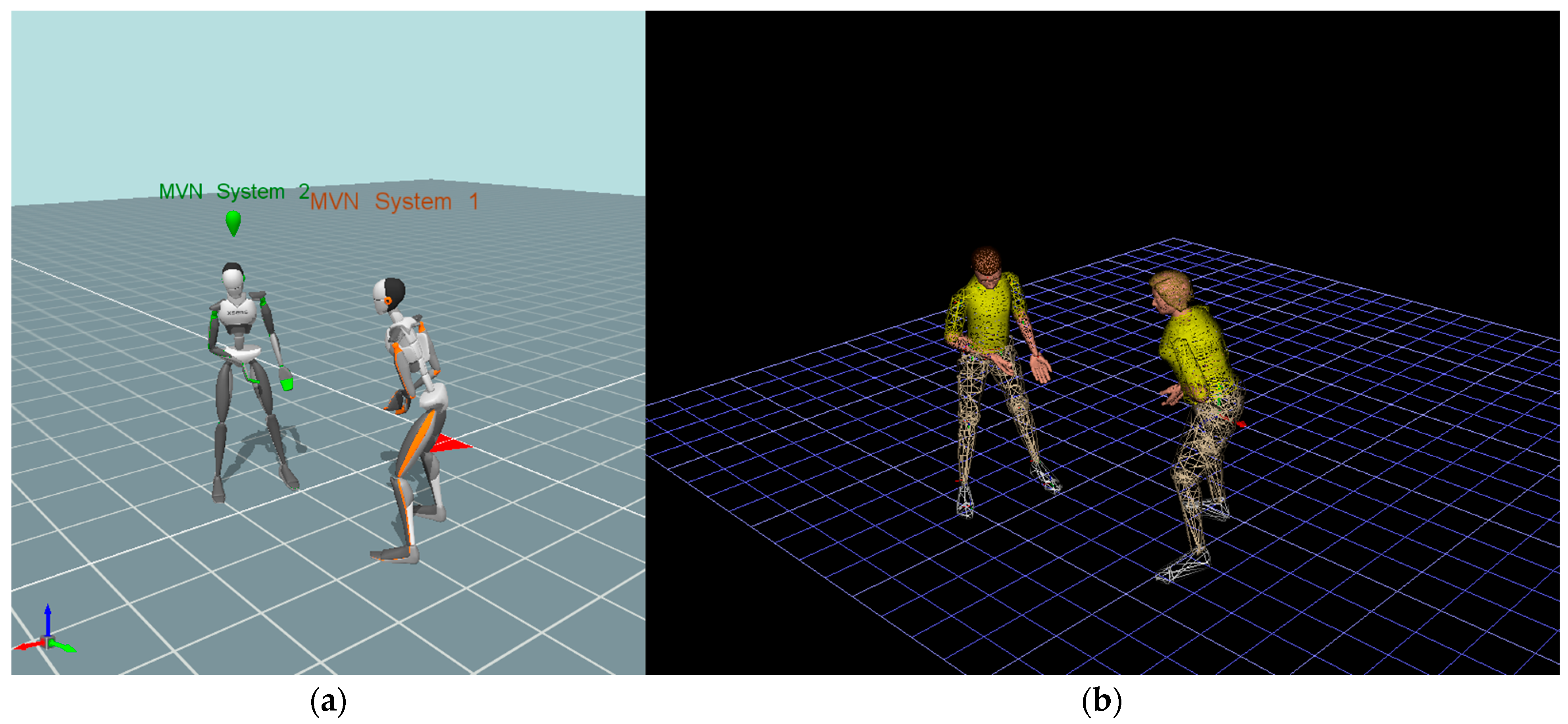
The two DHMs were synchronized by streaming motion data through a designated network port, allowing the Siemens Jack model to replicate the participant’s real-time movements. This integration enabled realistic human motion simulation and biomechanical analysis, including the assessment of lower back loading during task performance, as shown in Figure 1.
Previous studies have demonstrated the applicability of Jack for healthcare ergonomics and have shown good agreement between simulated and measured human movement [25]. Furthermore, inverse dynamics–based musculoskeletal models have been extensively validated against electromyography and experimental joint moment measurements, demonstrating their ability to provide reliable estimates of internal biomechanical loading. EMG-driven optimization approaches have reported a mean correlation coefficient of approximately 0.74 between predicted and measured muscle activation patterns, indicating good agreement between model predictions and physiological measurements [26].

2.4. Task and Pose

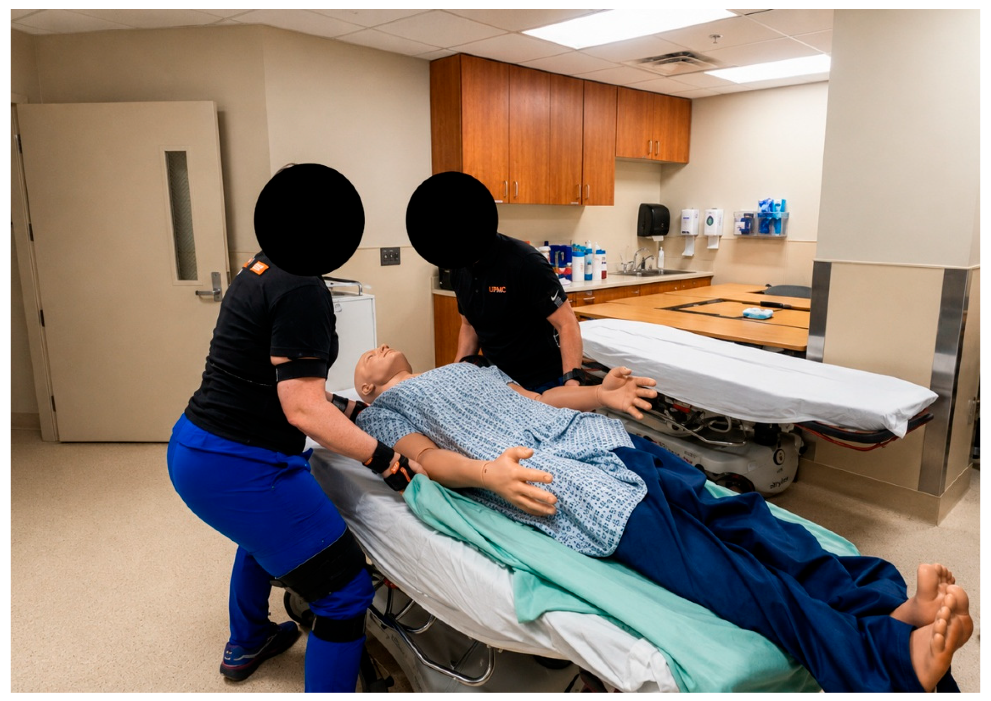
Before data collection and testing, the experimental environment was set up using a patient mannequin placed on a stretcher. The mannequin, representing a standardized 75 kg patient, was positioned closer to the foot end of the stretcher rather than the head, approximately within the lower third of the bed. To begin each trial, one healthcare provider stood on the right side of the mannequin and the other on the left. Both participants simultaneously grasped the sheet underneath the mannequin and lifted in coordination to reposition the patient upward on the stretcher. Each pair repeated the task three times, as shown in Figure 2. The average kinematic data (joint angles) and kinetic data (lower back forces) across the three trials were calculated and used to evaluate the participants’ risk of musculoskeletal injury.
The body posture corresponding to the maximum measured hand force was selected for biomechanical analysis because previous studies have demonstrated that lumbar compression and shear forces typically reach their highest values during peak loading conditions in manual material handling tasks, making these instants representative of periods of high biomechanical exposure during manual material handling tasks [27,28,29]. In the present study, synchronization between the force gauge and motion capture data confirmed that peak hand forces occurred during the initial pulling phase. Therefore, the posture associated with the maximum external force was selected as a representative high-exposure phase for biomechanical analysis during the boosting task, as shown in Figure 1. Initial kinematic variables at this phase, including trunk inclination, hip angles, and knee flexion, were extracted and analyzed.

2.5. Data Analysis

Biomechanical loading at the L4–L5 spinal segment, including compressive forces as well as anterior–posterior (AP) and lateral shear forces, estimated using the Siemens Jack Low Back Analysis module, which is based on established inverse dynamics and anthropometric biomechanical principles for predicting spinal loading from body posture, external forces, and segmental kinematics [27].
The force exerted during the pulling task was measured using a digital force gauge (SF-500, Wenzhou, Zhejiang, China) attached to the bed sheet and held with both hands by the participant throughout the task. Each pair of participants took turns using the force gauge to record their individual pulling forces. The direction of the applied hand force was defined as the instantaneous direction of patient mannequin movement during the pulling phase. Because the pulling direction changed continuously throughout the dynamic movement, the force vector applied in the Siemens Jack model corresponded to the selected synchronized posture at the time of the maximum measured hand pulling force. After the synchronized posture was imported into Siemens Jack, the orientation of the applied hand force was defined by the software based on the posture of the hand segments in the selected frame. Because participants used a symmetrical two-handed grip, the measured pulling force was assumed to be equally distributed between the left and right hands, and one-half of the measured force was applied to each hand in the Siemens Jack module. The posture corresponding to the maximum measured pulling force was synchronized with the motion capture data using video recordings and was subsequently analyzed in Jack. This posture was selected as a representative high-exposure phase for biomechanical analysis during the pulling task.
For the selected posture, trunk angle, hip flexion/extension, and knee flexion angles were recorded for each participant across all trials. These joint kinematics were then used to examine correlations (r) between body posture and L4–L5 loading. Additionally, differences in spinal forces and joint angles between male and female participants were assessed using independent-samples t-tests, with statistical significance set at p < 0.05. The Kolmogorov–Smirnov test was performed to assess normality.
Additionally, 95% confidence intervals were calculated for all outcome measures. A Linear Mixed-Effects Model (LME) was used to evaluate differences in spinal loading among team compositions (Male–Male, Female–Female, and Mixed-gender). To further determine whether gender independently influenced spinal loading beyond anthropometric differences, an Analysis of Covariance (ANCOVA) was conducted with body height and body weight included as covariates.
Additionally, to quantify the practical magnitude of the variance explained by our statistical frameworks, Cohen’s f 2 was calculated as the primary metric of effect size for each overall ANCOVA regression model. Cohen’s f 2 is defined mathematically based on the coefficient of determination ( R 2 ) and was calculated using Equation (1):
f 2 = R 2 1 R 2

3. Results

3.1. Forces and Joints

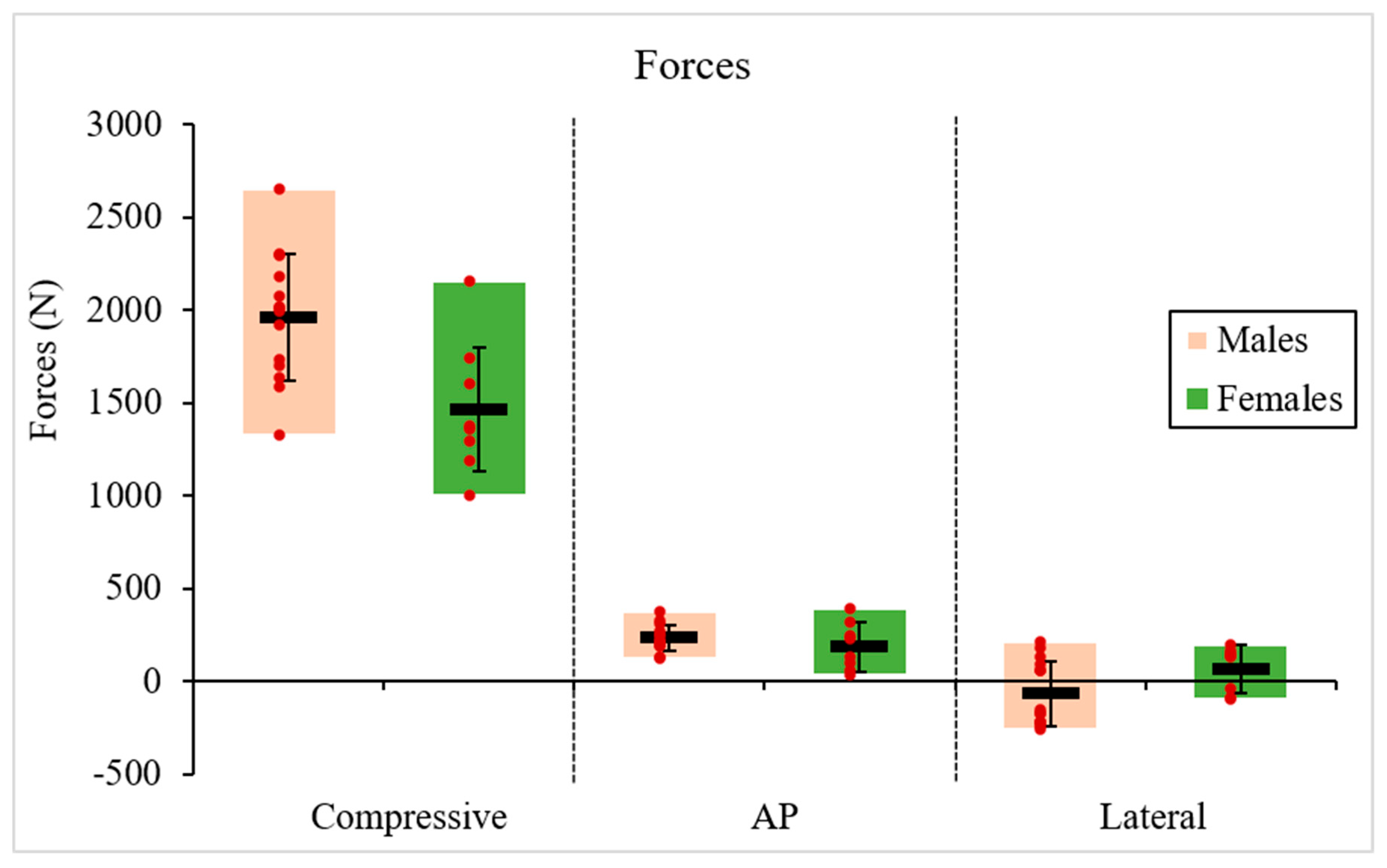
In the bed-boosting task, compressive forces among participants ranged from 1002.7 N to 2650.8 N, AP shear forces ranged from 33.2 N to 392.2 N, and lateral forces ranged from −254.1 N to 218.6 N. The positive lateral values indicate force acting toward the right side of body, and the negative lateral values indicate to the left. The average compressive, AP, and lateral forces were 1961.2 N, 235.8 N, and −64.7 N for males, and 1466.2 N, 188.9 N, and 69.9 N for females, respectively as shown in Figure 3. Negative values for lateral shear force indicate left-directed forces, whereas positive values indicate right-directed forces. These directional forces arise because the two participants stand on opposite sides of the bed while performing the boosting task, resulting in lateral loading that reflects their standing positions and pulling directions.
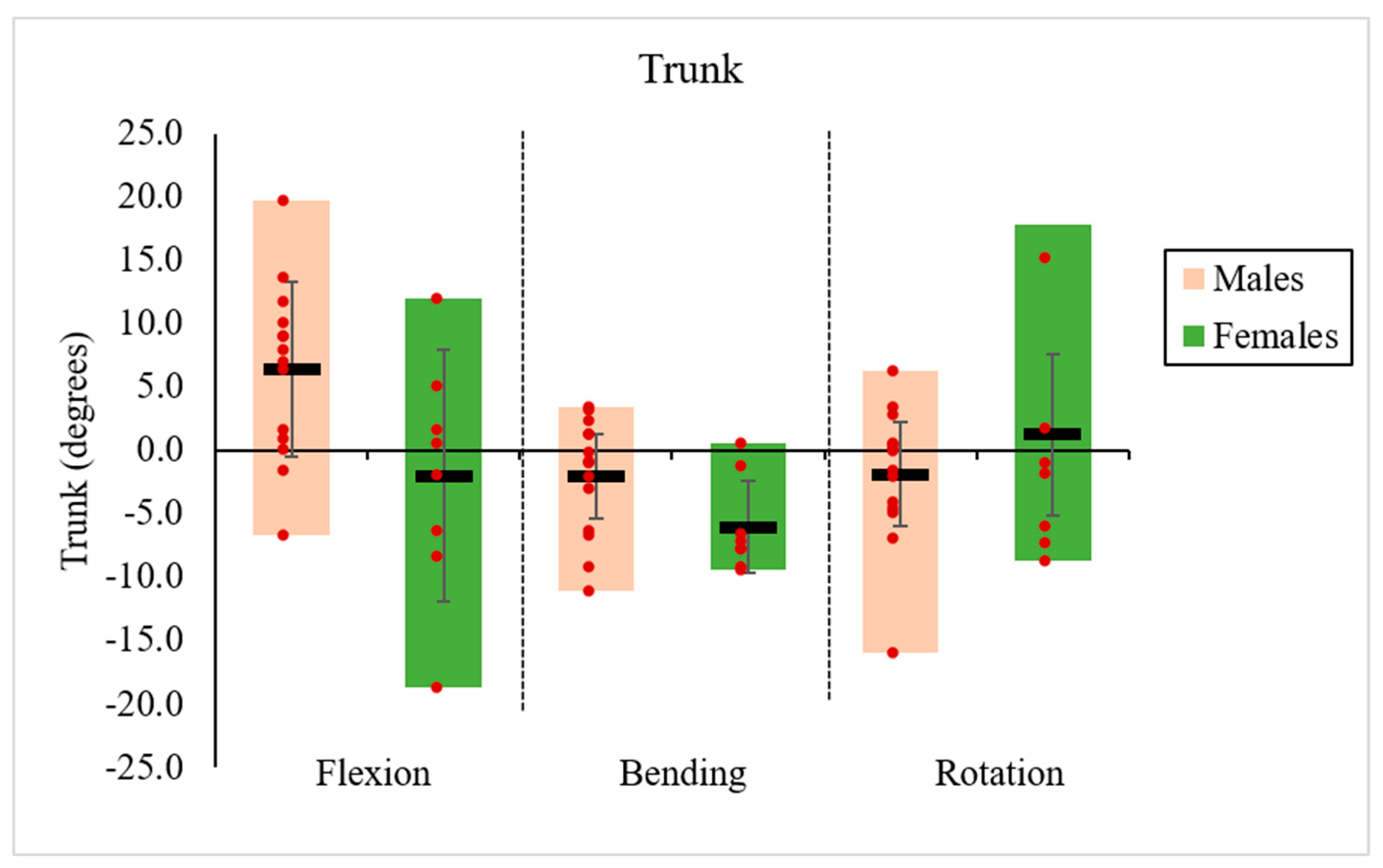
In this task, trunk angles ranged from −18.7° to 19.7° for flexion/extension, −11.1° to 3.4° for lateral bending, and −15.9° to 17.8° for axial rotation. Positive values represent trunk flexion, right lateral bending, and rightward rotation, whereas negative values indicate trunk extension, left lateral bending, and leftward rotation. The average trunk joint angles for flexion/extension, bending, and axial rotation were 6.4°, −2.1°, and −1.9° for males, and −2.0°, −6.0°, and 1.3° for females, respectively, as shown in Figure 4.
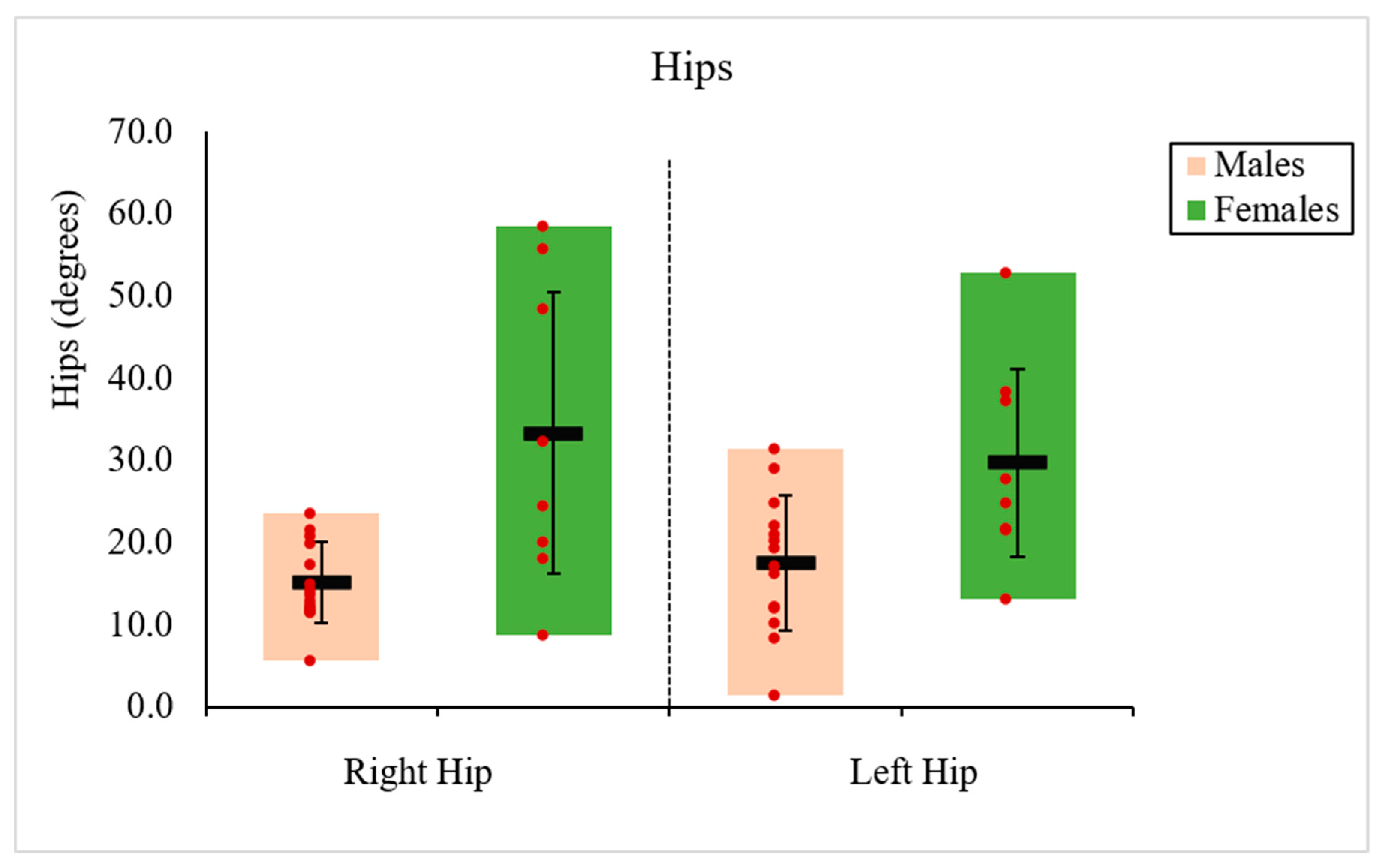
For the hip angles, right hip flexion ranged from 5.7° to 58.5°, and left hip flexion ranged from 1.4° to 52.9°. The average angles of the right and left hips were 15.2° and 17.6° for males, and 33.3° and 29.7° for females, respectively, as shown in Figure 5.
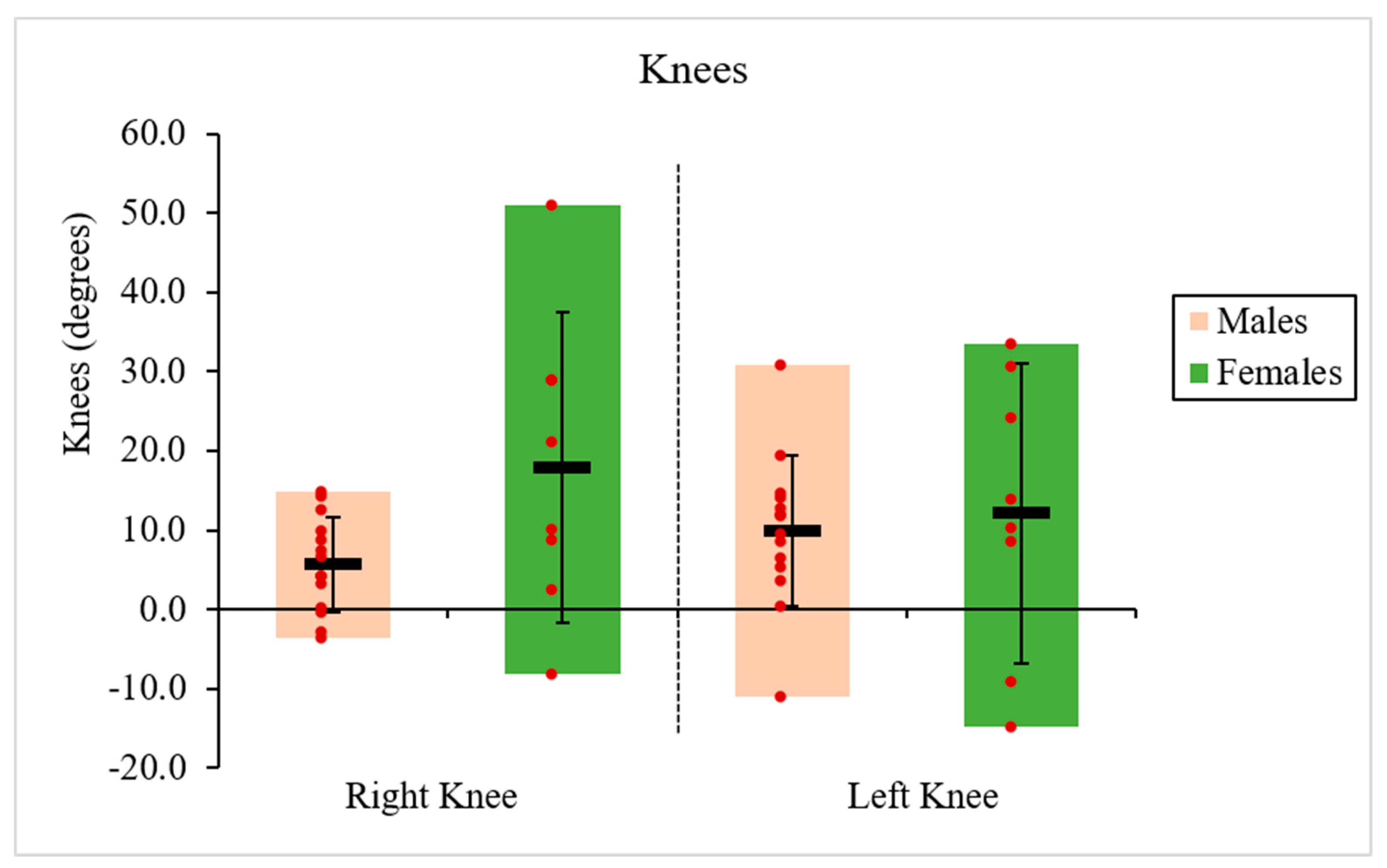
Similarly, for the knee angles, right knee ranged from −3.6° to 51.1°, and left knee ranged from −11.1° to 33.4°. The positive knee values indicate flexion, and the negative values indicate extension. The average angles of the right and left knees were 5.7° and 9.9° for males, and 17.9° and 12.1° for females, respectively as shown in Figure 6. The positive knee values indicate flexion, and the negative values indicate extension movement.

3.2. p Values

There were significant differences between the male and female participants in both body height and body weight, with p-values of 0.0002 and 0.0006, respectively. Table 1 presents the p-values used to compare forces and joint angles between males and females. Variables with values below 0.05, including compressive forces, trunk flexion, right and left hip angles, and the right knee, show significant differences. In contrast, AP and lateral shear forces, trunk bending and axial rotation, and the left knee do not exhibit statistically significant differences.

3.3. Correlations

Table 2 presents the correlations between lower back forces (compressive, AP, and lateral), body height, body weight, and seven joint angles (trunk flexion/extension, trunk bending, trunk axial rotation, right and left hip angles, and right and left knee angles) during the boosting task.
In Table 2, compressive forces showed moderate to strong positive correlations with body height, body weight, trunk flexion/extension, and trunk rotation, and moderate negative correlations with the right hip and knee. AP shear force exhibited moderate correlations with the hips and knees. Lateral shear force showed moderate to strong positive correlations with body height, body weight, trunk bending, and trunk rotation. Other correlations were negligible.
Table 3 presents the correlations between hips, body height, body weights, and other joints. Body height, body weight, and trunk flexion/extension exhibited moderate to strong negative correlations with both right and left hip movements, while both hips showed strong positive correlations with both knees. The left hip is correlated with trunk bending, while the right hip shows a correlation with trunk axial rotation.

3.4. Confidence Intervals, LME, ANCOVA

The results indicated that all data, including force and joint angle measurements, were normally distributed across participants. 95% confidence intervals have been added to Table 4. A Linear Mixed-Effects Model was used to evaluate differences in spinal loading based on team composition (Male–Male, Female–Female, or Mixed-gender), while an ANCOVA was performed to examine the relationship between gender and spinal loading after controlling for body height and body weight. The statistical results in Table 5 demonstrate that gender does not remain significantly related to spinal loading once body height and weight are accounted for.

4. Discussion

The present study examined potential gender-based differences in anthropometrics, trunk and lower-limb kinematics, and lower back loading during a boost-in-bed patient transfer task. The results demonstrate significant differences between male and female participants in body height, body weight, compressive spinal force, trunk flexion/extension, and hip movements, as well as meaningful correlations between anthropometric factors and spinal loading.

4.1. Lower Back Forces in Relation to Anthropometrics and Trunk Motion

Compressive forces showed strong positive correlations with body height and body weight, which aligns with established biomechanical principles. Taller individuals experience larger moment arms, while greater body weight increases gravitational loading, both contributing to higher spinal compression [27,30].
Compressive force was also moderately associated with trunk flexion/extension and axial rotation, indicating that even small deviations from neutral posture can significantly increase spinal loading [31,32]. Increased trunk flexion enlarges the moment arm about the lumbar spine, while axial rotation contributes to spinal instability and disc loading, particularly when combined with flexion [30,33].
In contrast, the relatively weak relationship between compressive force and trunk bending likely reflects the limited lateral bending observed in the task. Nevertheless, lateral bending remains an important risk factor in asymmetric lifting due to its contribution to uneven load distribution [34].
Differences in lifting coordination were observed between male and female participants. Male participants exhibited greater trunk involvement, whereas female participants demonstrated reduced trunk flexion and greater reliance on lower-limb motion, characterized by increased hip and knee flexion. Similar movement patterns have been reported in previous studies [35,36], However, the ANCOVA results indicated that these observed differences were largely explained by body height and body weight.

4.2. Shear Forces and Their Biomechanical Determinants

Lateral shear forces were moderately to strongly correlated with body height, body weight, trunk bending, and axial rotation. These relationships indicate that asymmetric postures and lateral trunk deviations increase shear loading and may compromise spinal stability [34]. Taller and heavier individuals may be particularly susceptible due to greater inertial and gravitational effects.
AP shear forces were moderately correlated with both hip and knee motion, indicating that lower-limb kinematics contribute significantly to the regulation of shear loading at the lumbar spine. Increased hip and knee flexion appears to shift the center of mass and redistribute forces [37]. Although no significant gender differences were observed in lateral shear force magnitude, the contributing movement patterns differed. Previous studies have reported differences in postural strategies between male and female populations [38]. However, in the present study, the observed movement differences appear to be more closely associated with anthropometric characteristics and movement coordination patterns than with gender itself.
The fixed effects of a Linear Mixed-Effects Model were evaluated. These findings suggest that the gender differences in kinetic spinal loading commonly observed during collaborative lifting are largely attributable to differences in physical scale, particularly body height and mass. However, previous biomechanical studies have reported that females generally possess a wider pelvis, a greater quadriceps angle, and greater lumbar lordosis than males [39,40]. These anatomical characteristics may contribute to differences in movement strategies adopted during lifting tasks. Although the present study indicated that body height and body weight were significant contributors to the observed differences in lumbar loading, gender-related skeletal morphology was not evaluated. Future studies incorporating detailed skeletal and pelvic morphology measurements may help clarify the extent to which anatomical characteristics, beyond body height and body weight, influence lifting coordination, posture, and spinal loading [41].

4.3. Coordination Between Trunk, Hip and Knee

The interaction among trunk, hip, and knee joints reflects a coordinated, whole-body lifting strategy. A negative correlation between hip flexion and trunk flexion/extension suggests a compensatory mechanism in which increased hip motion reduces reliance on trunk movement, consistent with a hip-dominant lifting strategy [30].
Hip motion showed negative correlations with body height, body weight, and trunk flexion/extension, suggesting that individuals with larger anthropometrics or greater trunk involvement rely less on hip contribution. This trend was more pronounced in males, reinforcing the observation of a trunk-dominant strategy.
Strong positive correlations between hip and knee flexion further demonstrate coordinated lower-limb involvement. This coordination facilitates load transfer away from the spine to larger muscle groups in the lower extremities, reducing spinal stress [28].
Asymmetrical relationships, such as the left hip correlating with trunk bending and the right hip with trunk rotation, suggest task-specific coordination patterns. These asymmetries may be influenced by hand dominance or positioning and could contribute to uneven spinal loading [42], as side assignment was not formally controlled and may have affected the asymmetric correlations observed for trunk bending and trunk rotation. In contrast, the correlations for the remaining variables were generally symmetrical, suggesting that the asymmetry was limited to these specific movement patterns rather than representing a systematic bias across all measures.
Differences in movement coordination were observed between male and female participants. Female participants exhibited greater hip and knee flexion, whereas male participants relied more on trunk motion. These distinct movement strategies are likely associated with anthropometric differences between the groups and may contribute to different load distributions across body segments during lifting.

4.4. Practical Implications and Recommendations

(1) Promote Hip-Dominant Lifting Techniques: Encouraging increased hip and knee flexion can reduce reliance on trunk motion and lower spinal loading. Squat-like lifting strategies are particularly beneficial.
(2) Minimize Trunk Flexion, Bending, and Rotation: Reducing excessive trunk motion and asymmetry is critical for limiting both compressive and shear forces, especially for individuals who naturally adopt trunk-dominant strategies.
(3) Incorporate Anthropometric Considerations: Taller and heavier individuals may face greater biomechanical demands. Adjustments such as optimizing working height or using team lifting can help mitigate risk.
(4) Encourage Use of Assistive Devices: Assistive equipment and improved handling techniques can substantially reduce biomechanical loads during patient handling tasks [43].

4.5. Limitations

First, although distinct kinematic trends were observed, a larger sample size with precise anatomical characterization is required to confirm whether these behavioral strategies are independently driven by biological gender anatomy or completely explained by body scale. Second, only the initial lifting posture was analyzed, as it was considered to represent a high-exposure phase for biomechanical analysis during the boosting task; however, spinal loading varies dynamically throughout the movement, and future studies should include a more comprehensive analysis of multiple postures across the entire task. Third, a standardized 75 kg mannequin was used, which may not fully represent the wide range of patient body masses encountered in clinical settings. Fourth, electromyography (EMG) was not included in the present study; incorporating EMG measurements in future work would enable a more direct assessment of muscle activation strategies and improve understanding of neuromuscular control during patient-handling tasks.

5. Conclusions

This study highlights that spinal loading during patient handling is influenced by a complex interaction of trunk posture, lower-limb coordination, and anthropometric characteristics. Although differences in lifting mechanics were observed between male and female participants, the ANCOVA results indicate that these differences were largely explained by body height and body weight after adjustment. These findings support the development of individualized bio-ergonomic training strategies. Adapting interventions to an individual’s unique anthropometric profile and movement mechanics provides a more accurate, data-aligned approach to minimizing musculoskeletal injury risk in occupational lifting tasks.

Author Contributions

Conceptualization, X.J.; Methodology, X.J.; Software, X.G.; Validation, X.G.; Formal analysis, X.J.; Investigation, X.J.; Resources, X.J.; Writing—original draft, X.J. and X.G.; Writing—review and editing, X.G. All authors have read and agreed to the published version of the manuscript.

Funding

This work was supported by the UPMC Hamot Health Foundation and the College of Engineering and Business at Gannon University.

Institutional Review Board Statement

This study was conducted in accordance with the Declaration of Helsinki and was approved by both the Gannon University Institutional Review Board (protocol code: GUIRB-2022-9-6970) and the University of Pittsburgh Institutional Review Board (protocol code: STUDY22110153).

Informed Consent Statement

Informed consent was obtained from all subjects involved in the study.

Data Availability Statement

The original contributions presented in this study are included in the article. Further inquiries can be directed to the corresponding author.

Conflicts of Interest

The authors declare no conflict of interest.

References

  1. Anap, D.; Iyer, C.; Rao, K. Work related musculoskeletal disorders among hospital nurses in rural Maharashtra, India: A multi centre survey. Int. J. Res. Med. Sci. 2013, 1, 101–107. [Google Scholar] [CrossRef]
  2. World Health Organization (WHO). Musculoskeletal Health. Available online: https://www.who.int/news-room/fact-sheets/detail/musculoskeletal-conditions (accessed on 14 July 2022).
  3. Occupational Safety and Health Administration (OSHA). Healthcare. Safe Patient Handling. Available online: https://www.osha.gov/healthcare/safe-patient-handling (accessed on 22 April 2026).
  4. Guo, H.R.; Tanaka, S.; Cameron, L.L.; Seligman, P.J.; Behrens, V.J.; Ger, J.; Wild, D.K.; Putz-Anderson, V. Back pain among workers in the United States: National estimates and workers at high risk. Am. J. Ind. Med. 1995, 28, 591–602. [Google Scholar] [CrossRef] [PubMed]
  5. Soylar, P.; Ozer, A. Evaluation of the prevalence of musculoskeletal disorders in nurses: A systematic review. Med. Sci. 2018, 7, 479–485. [Google Scholar]
  6. Vinstrup, J.; Jakobsen, M.D.; Madeleine, P.; Andersen, L.L. Physical exposure during patient transfer and risk of back injury & low-back pain: Prospective cohort study. BMC Musculoskelet. Disord. 2020, 21, 715. [Google Scholar] [CrossRef] [PubMed]
  7. Alexopoulos, E.C.; Burdorf, A.; Kalokerinou, A. Risk factors for musculoskeletal disorders among nursing personnel in Greek hospitals. Int. Arch. Occup. Environ. Health 2003, 76, 289–294. [Google Scholar] [CrossRef] [PubMed]
  8. World Health Organization (WHO). Unsafe Patient Handling. Available online: https://www.who.int/tools/occupational-hazards-in-health-sector/unsafe-patient-handling (accessed on 22 April 2026).
  9. Naoum, S.; Mitseas, P.; Koutserimpas, C.; Spinthouri, M.; Kalomikerakis, I.; Raptis, K.; Sarafis, P.; Govina, O.; Konstantinidis, T. Musculoskeletal disorders and caring behaviors among nursing staff in greek hospitals: A prospective multicenter study. Maedica 2022, 17, 52. [Google Scholar] [CrossRef] [PubMed]
  10. Rathore, F.A.; Attique, R.; Asmaa, Y. Prevalence and perceptions of musculoskeletal disorders among hospital nurses in Pakistan: A cross-sectional survey. Cureus 2017, 9, e1001. [Google Scholar] [CrossRef] [PubMed]
  11. Letvak, S.A.; Ruhm, C.J.; Gupta, S.N. Nurses’ presenteeism and its effects on self-reported quality of care and costs. Am. J. Nurs. 2012, 112, 30–38. [Google Scholar] [PubMed]
  12. Alexopoulos, E.C.; Tanagra, D.; Detorakis, I.; Gatsi, P.; Goroyia, A.; Michalopoulou, M.; Jelastopulu, E. Knee and low back complaints in professional hospital nurses: Occurrence, chronicity, care seeking and absenteeism. Work 2011, 38, 329–335. [Google Scholar] [CrossRef] [PubMed]
  13. Bergman, R.; De Jesus, O. Patient Care Transfer Techniques; StatPearls Publishing: Treasure Island, FL, USA, 2020. [Google Scholar]
  14. Kulshrestha, A.; Singh, J. Inter-hospital and intra-hospital patient transfer: Recent concepts. Indian J. Anaesth. 2016, 60, 451–457. [Google Scholar] [CrossRef] [PubMed]
  15. Warming, S.; Juul-Kristensen, B.; Ebbehøj, N.; Schibye, B. An observation instrument for the description and evaluation of patient transfer technique. Appl. Ergon. 2004, 35, 603–614. [Google Scholar] [CrossRef] [PubMed]
  16. Ulin, S.S.; Chaffin, D.B.; Patellos, C.L.; Blitz, S.G.; Emerick, C.A.; Lundy, F.; Misher, L. A biomechanical analysis of methods used for transferring totally dependent patients. Sci. Nurs. 1997, 14, 19–27. [Google Scholar] [PubMed]
  17. Vieira, E.; Kumar, S. Safety analysis of patient transfers and handling tasks. BMJ Qual. Saf. 2009, 18, 380–384. [Google Scholar] [CrossRef]
  18. Hwang, J.; Kuppam, V.A.; Chodraju, S.S.R.; Chen, J.; Kim, J.H. Commercially available friction-reducing patient-transfer devices reduce biomechanical stresses on caregivers’ upper extremities and low back. Hum. Factors 2019, 61, 1125–1140. [Google Scholar] [CrossRef] [PubMed]
  19. Vinstrup, J.; Jakobsen, M.D.; Madeleine, P.; Andersen, L.L. Biomechanical load during patient transfer with assistive devices: Cross-sectional study. Ergonomics 2020, 63, 1164–1174. [Google Scholar] [CrossRef] [PubMed]
  20. Häske, D.; Schier, L.; Weerts, J.O.; Groß, B.; Rittmann, A.; Grützner, P.A.; Münzberg, M.; Kreinest, M. An explorative, biomechanical analysis of spine motion during out-of-hospital extrication procedures. Injury 2020, 51, 185–192. [Google Scholar] [CrossRef] [PubMed]
  21. Fray, M.; Holgate, G. A comparative force assessment of 4 methods to move a patient up a bed. In Proceedings of the Congress of the International Ergonomics Association, Florence, Italy, 26–30 August 2018; pp. 631–640. [Google Scholar]
  22. Cudejko, T.; Button, K.; Al-Amri, M. Validity and reliability of accelerations and orientations measured using wearable sensors during functional activities. Sci. Rep. 2022, 12, 14619. [Google Scholar] [CrossRef] [PubMed]
  23. Xsens. Xsens 3D Motion Tracking Technology. Available online: https://www.xsens.com/movella (accessed on 5 May 2025).
  24. Ji, X.; Gao, X. Using Dual Motion Capture to Optimize Patient Transfer Techniques and Identify Lower Back Injury Risk Factors in Healthcare Settings. Hum. Factors Healthc. 2026, 9, 100126. [Google Scholar] [CrossRef]
  25. Ji, X.; Piovesan, D.; Arenas, M.; Liu, H. Analysis of healthcare push and pull task via JACK: Predicted joint accuracy during full-body simulation. Appl. Sci. 2022, 12, 6450. [Google Scholar] [CrossRef]
  26. Michaud, F.; Lamas, M.; Lugrís, U.; Cuadrado, J. A fair and EMG-validated comparison of recruitment criteria, musculotendon models and muscle coordination strategies, for the inverse-dynamics based optimization of muscle forces during gait. J. Neuroeng. Rehabil. 2021, 18, 17. [Google Scholar] [CrossRef] [PubMed]
  27. Chaffin, D.B.; Andersson, G.B.; Martin, B.J. Occupational Biomechanics; John Wiley & Sons: Hoboken, NJ, USA, 2006. [Google Scholar]
  28. Hoozemans, M.J.; Kingma, I.; de Vries, W.H.; van Dieën, J.H. Effect of lifting height and load mass on low back loading. Ergonomics 2008, 51, 1053–1063. [Google Scholar] [CrossRef] [PubMed]
  29. Marras, W.; Davis, K.; Kirking, B.; Bertsche, P. Low back disorder risk and spinal loading during patient transfer. Proc. Hum. Factors Ergon. Soc. Annu. Meet. 1998, 42, 901–905. [Google Scholar] [CrossRef]
  30. Bender, A.; Schmidt, H.; Wellner, D.L.; Duda, G.N.; Brandl, C.; Damm, P. In vivo load on knee, hip and spine during manual materials handling with two lifting techniques. J. Biomech. 2024, 163, 111963. [Google Scholar] [CrossRef] [PubMed]
  31. Marras, W.S.; Lavender, S.A.; Leurgans, S.E.; Rajulu, S.L.; Allread, S.G.; Fathallah, F.A.; Ferguson, S.A. The role of dynamic three-dimensional trunk motion in occupationally-related. Spine 1993, 18, 617–628. [Google Scholar] [CrossRef] [PubMed]
  32. McGill, S. Low Back Disorders: Evidence-Based Prevention and Rehabilitation; Human Kinetics: Champaign, IL, USA, 2025. [Google Scholar]
  33. Callaghan, J.P.; McGill, S.M. Low back joint loading and kinematics during standing and unsupported sitting. Ergonomics 2001, 44, 280–294. [Google Scholar] [CrossRef] [PubMed]
  34. Marras, W.S.; Davis, K.G.; Heaney, C.A.; Maronitis, A.B.; Allread, W.G. The influence of psychosocial stress, gender, and personality on mechanical loading of the lumbar spine. Spine 2000, 25, 3045–3054. [Google Scholar] [CrossRef] [PubMed]
  35. van der Kruk, E. BIASMECHANICS: Does an unconscious bias still persist in biomechanics, positioning males as the default in human research? A meta-analysis on the Journal of Biomechanics 2024 publications. J. Biomech. 2025, 181, 112560. [Google Scholar] [CrossRef] [PubMed]
  36. Tamer, S.; Silva, D.D.O.; Pazzinatto, M.F.; Crossley, K.M.; McCarthy, B.J.; Savage, M.; Patterson, B.E.; Low, J.; Mentiplay, B.F. Are running biomechanics different between females and males with knee injuries? A systematic review and meta-analysis. Gait Posture 2026, 128, 110187. [Google Scholar] [CrossRef] [PubMed]
  37. McGill, S.M.; Norman, R.W.; Cholewicki, J. A simple polynomial that predicts low-back compression during complex 3-D tasks. Ergonomics 1996, 39, 1107–1118. [Google Scholar] [CrossRef] [PubMed]
  38. Akinoğlu, B.; Paköz, B.; Shehu, S.U.; Kocahan, T.; Weiss, K.; Rosemann, T.; Knechtle, B. Evaluation of Athletes’ Gender-Related Postural Differences. Percept. Mot. Ski. 2025, 132, 534–547. [Google Scholar]
  39. Whitcome, K.K.; Shapiro, L.J.; Lieberman, D.E. Fetal load and the evolution of lumbar lordosis in bipedal hominins. Nature 2007, 450, 1075–1078. [Google Scholar] [CrossRef] [PubMed]
  40. Bailey, J.F.; Sparrey, C.J.; Been, E.; Kramer, P.A. Morphological and postural sexual dimorphism of the lumbar spine facilitates greater lordosis in females. J. Anat. 2016, 229, 82–91. [Google Scholar] [CrossRef] [PubMed]
  41. Lindbeck, L.; Kjellberg, K. Gender differences in lifting technique. Ergonomics 2001, 44, 202–214. [Google Scholar] [CrossRef] [PubMed]
  42. Davis, K.; Marras, W. The effects of motion on trunk biomechanics. Clin. Biomech. 2000, 15, 703–717. [Google Scholar] [CrossRef]
  43. Fray, M.; Davis, K.G. Effectiveness of safe patient handling equipment and techniques: A review of biomechanical studies. Hum. Factors 2024, 66, 2283–2322. [Google Scholar] [PubMed]
Figure 1. The synchronous movements captured by (a) Xsens motion capture and implemented in (b) Jack software simulation.
Tae 02 00013 g001
Figure 2. The body configuration of the two providers positioned on opposite sides of the patient while holding the sheet to execute the lift.
Tae 02 00013 g002
Figure 3. The lower back forces exerted by male and female participants. The error bars illustrate the range of values for each gender. The bold horizontal line at the center of each bar represents the mean value, and the vertical error bars indicate the standard deviation.
Tae 02 00013 g003
Figure 4. The trunk angles of flexion/extension, bending and axial rotation for male and female participants.
Tae 02 00013 g004
Figure 5. The hip angles of flexion for male and female participants.
Tae 02 00013 g005
Figure 6. The knee angles of flexion/extension for male and female participants.
Tae 02 00013 g006
Table 1. The p values of forces and joints between genders.
Compressive (N)AP (N)Lateral (N)Trunk Flexion (°)Trunk Bending (°)
p-Values0.0050.2760.0730.0260.051
Trunk Rotation (°)Right Hip (°)Left Hip (°)Right Knee (°)Left Knee (°)
p-Values0.3490.0020.0120.0330.701
Table 2. The correlation between forces, body height, body weight, and joint angles. Moderate to strong relationships are highlighted in bold.
Body Height Body WeightTrunk Flexion Trunk Bending Trunk Rotation Right HipLeft HipRight Knee Left Knee
Compressive0.7640.5120.4030.0100.347−0.430−0.171−0.359−0.066
AP0.2650.014−0.1780.0580.1510.3720.3980.4730.448
Lateral0.5320.5960.2010.7040.409−0.0680.390−0.1680.143
Table 3. The correlation between hips, body height, body weight, and other joints. Moderate to strong relationships are highlighted in bold.
Body Height Body Weight Trunk FlexionTrunk BendingTrunk RotationRight KneeLeft Knee
Right Hip−0.411−0.566−0.6240.0930.6080.8540.460
Left Hip−0.288−0.478−0.5110.4210.1440.7050.659
Table 4. Linear mixed-effects model results for lower back forces and joint angles including the pair composition. FF baseline refers to the Female–Female baseline.
Dependent VariableEffectβ (Estimate)95% CIp ValueEffect Size
Compressive Force (N)Intercept [FF Baseline]1710.2[1243.8    2176.6]<0.0013.0
Pair: Male–Male8.0[−525.61    541.61]0.98
Pair: Mixed124.8[−381.62    631.21]0.61
AP Shear Force (N)Intercept [FF Baseline]158.2[2.3366    314.02]0.0470.39
Pair: Male–Male60.5[−118.06        239]0.48
Pair: Mixed70.7[−98.532     240.02]0.39
Lateral (N)Intercept [FF Baseline]176.1[99.036     253.2]<0.0010.76
Pair: Male–Male−29.4[−118.22    59.398]0.49
Pair: Mixed−18.5[−102.34    65.428]0.13
Trunk Flexion (°)Intercept [FF Baseline]5.07[−8.426     18.567]0.440.84
Pair: Male–Male−2.73[−18.16     12.709]0.71
Pair: Mixed−1.38[−16.035     13.271]0.84
Trunk Bend (°)Intercept [FF Baseline]6.5[1.5162     11.478]0.0140.35
Pair: Male–Male−1.49[−7.2328     4.2444]0.59
Pair: Mixed−2.45[−7.8738     2.9664]0.35
Trunk Rotation (°)Intercept [FF Baseline]6.4[−0.704       13.511]0.070.46
Pair: Male–Male−3.33[−11.518       4.859]0.40
Pair: Mixed−0.1[−7.838       7.631]0.98
Right Hip (°)Intercept [FF Baseline]25.1[7.4236       42.726]0.0080.84
Pair: Male–Male−11.3[−31.67       9.0041]0.26
Pair: Mixed1.50[−17.71       20.70]0.54
Left Hip (°)Intercept [FF Baseline]17.4[1.7404       33.127]0.030.44
Pair: Male–Male0.65[−17.43       18.73]0.94
Pair: Mixed7.92[−9.154       25.002]0.34
Right Knee (°)Intercept [FF Baseline]12.26[−6.993      31.517]0.200.28
Pair: Male–Male−6.4[−28.587      15.782]0.55
Pair: Mixed0.34[−20.611      21.297]0.97
Left Knee (°)Intercept [FF Baseline]6.82[−14.483      28.131]0.510.26
Pair: Male–Male4.6[−19.82      29.014]0.70
Pair: Mixed4.06[−19.091      27.201]0.72
Table 5. ANCOVA analysis to explore the relationship between gender and spinal loading after height and weight control.
Dependent Variableβ (Estimate)95% CIp Value
Compressive Force (N)−0.88[−434.57        432.82]0.997
AP Shear (N)55.5[−89.60        200.66]0.432
Lateral Shear (N)−48.62[−121.91       24.68]0.180
Disclaimer/Publisher’s Note: The statements, opinions and data contained in all publications are solely those of the individual author(s) and contributor(s) and not of MDPI and/or the editor(s). MDPI and/or the editor(s) disclaim responsibility for any injury to people or property resulting from any ideas, methods, instructions or products referred to in the content.