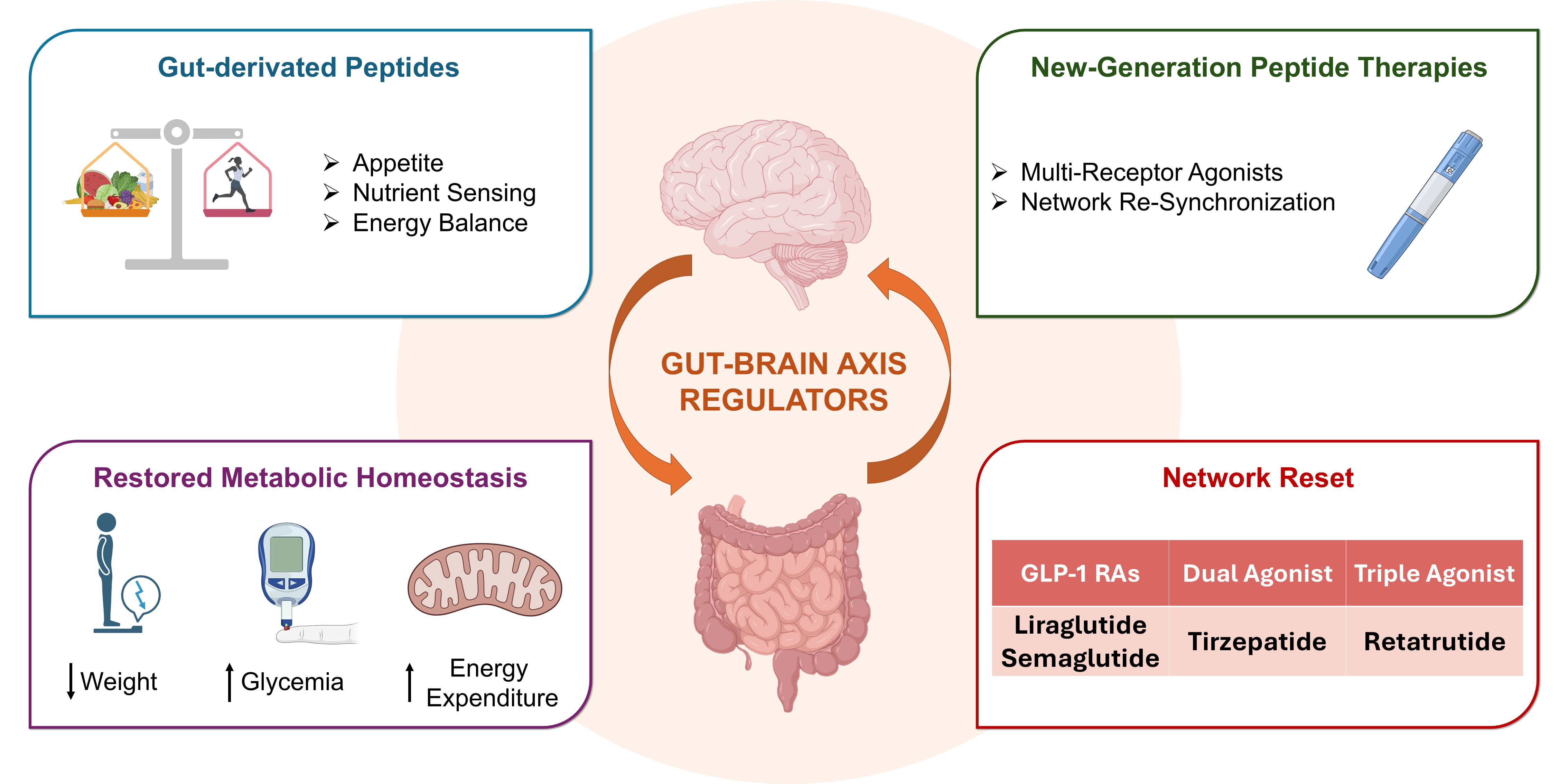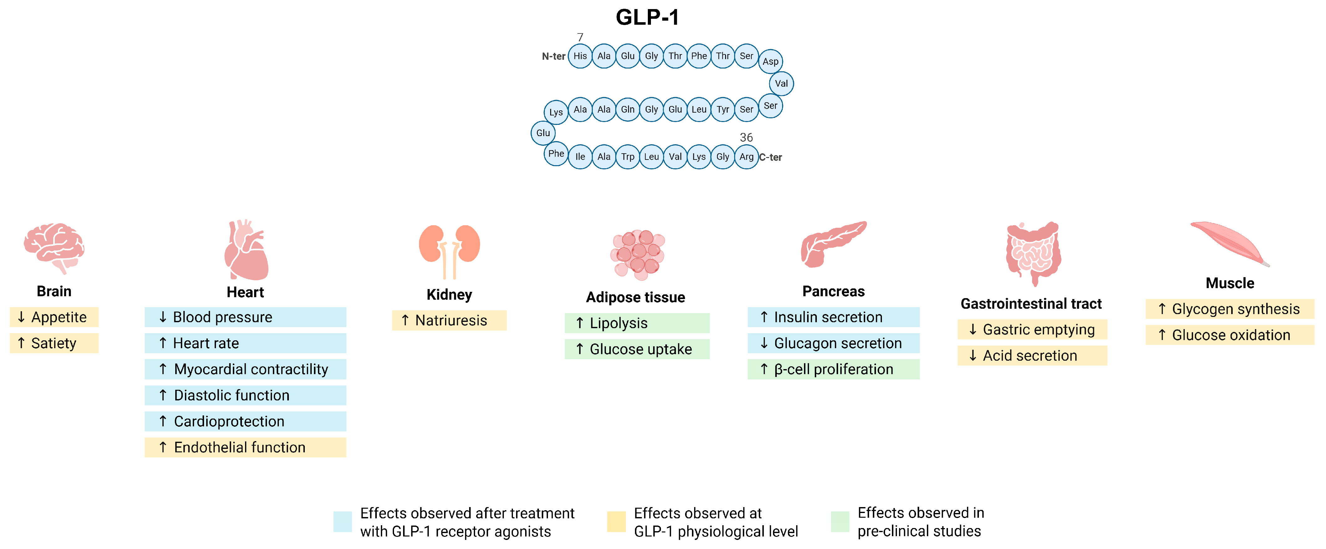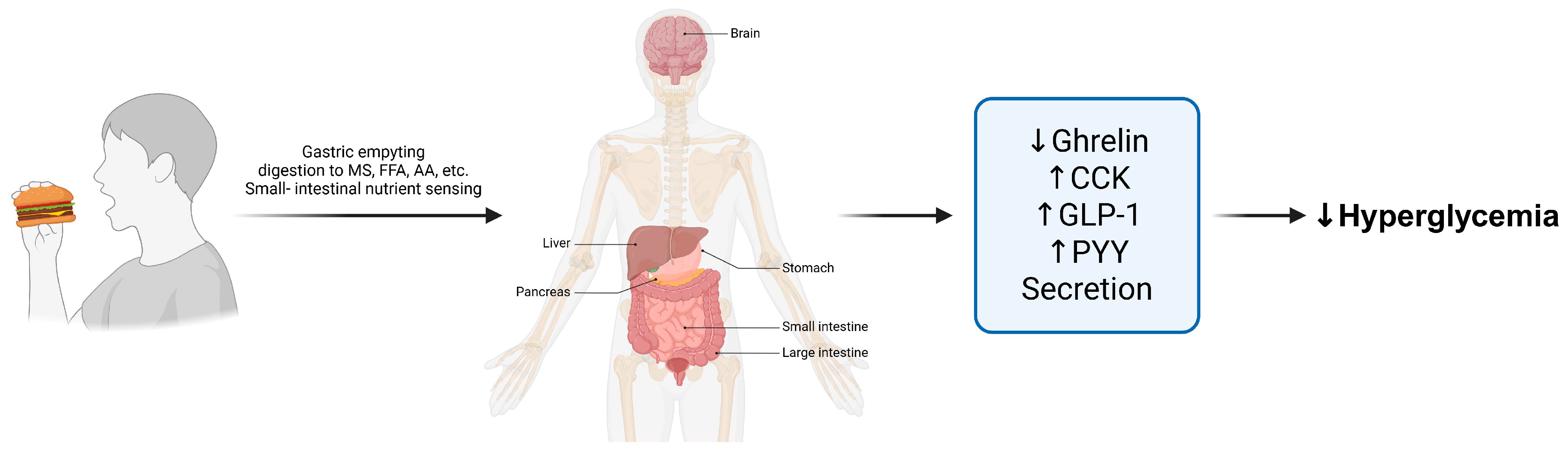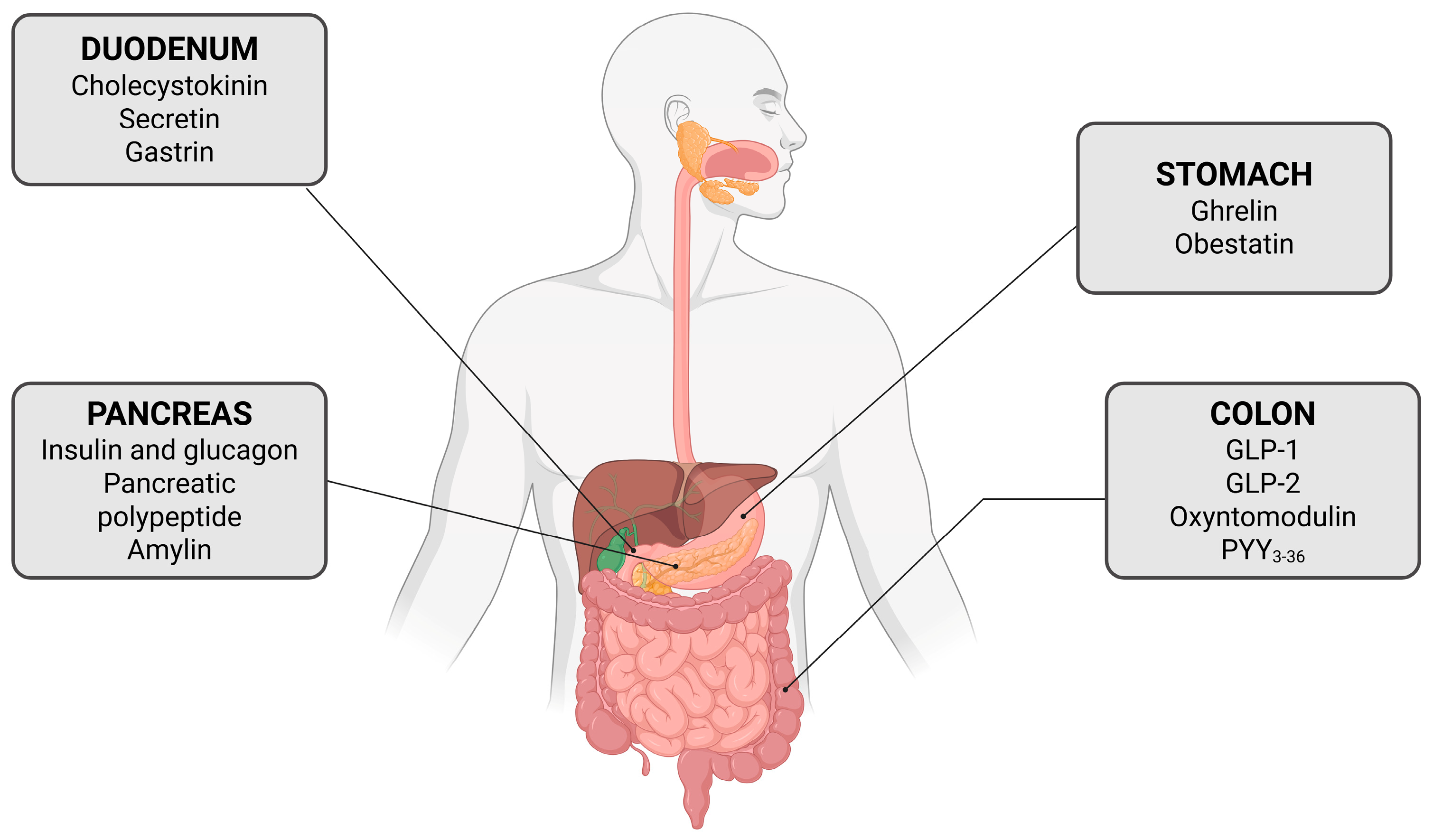

The Bioenergetic Architecture of Metabolic Regulation: From Gut–Brain Signalling to Next-Generation Peptide Pharmacology
 , , , , and
Abstract
1. Introduction
2. The Gut–Brain Axis
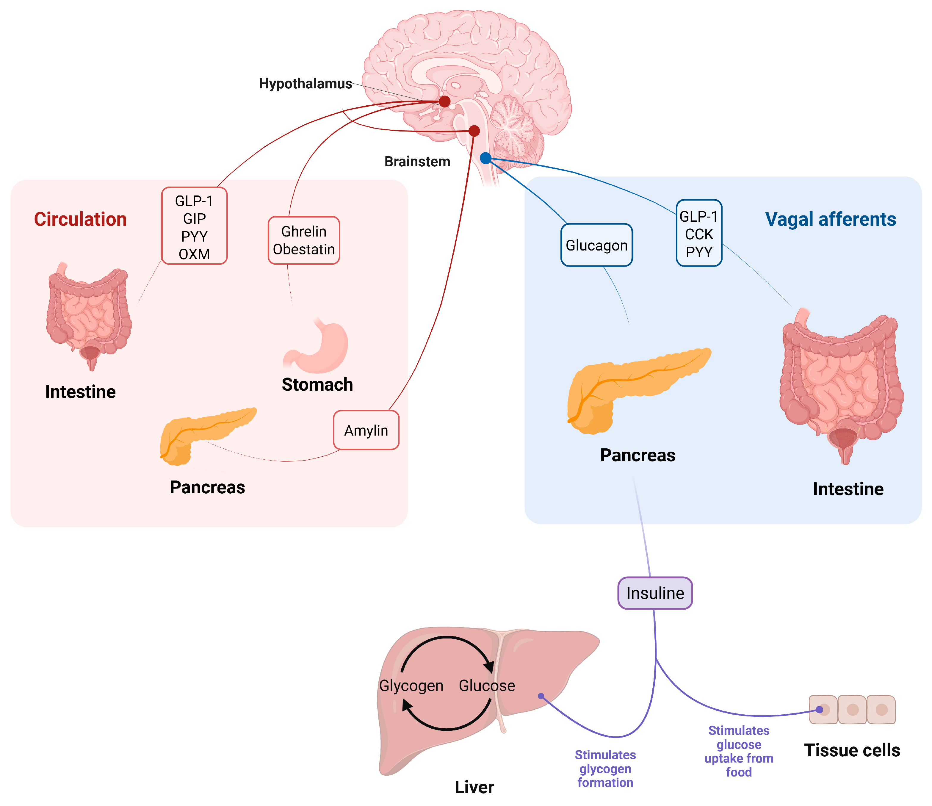
2.1. Enteroendocrine Cell Function and Local-Systemic Signalling Integration
2.2. Central and Pancreatic Integration of Peptide-Derived Signals
2.3. Bioenergetic Regulation and Pathophysiological Disruption of the Gut–Brain–Pancreas Axis
3. Key Peptides Beyond the Incretins
3.1. Ghrelin: The Hunger Hormone
3.2. Peptide YY and Obestatin: The Postprandial Restraint Signals
3.3. Amylin: The Synchronizer of Metabolic Timing
3.4. Cholecystokinin: The Rapid Satiation Trigger
3.5. Other Emerging Peptides: Beyond Classical Gut Hormones
4. Therapeutic Implications and Combinatorial Rationale
4.1. From Single-Target to Network-Oriented Therapy
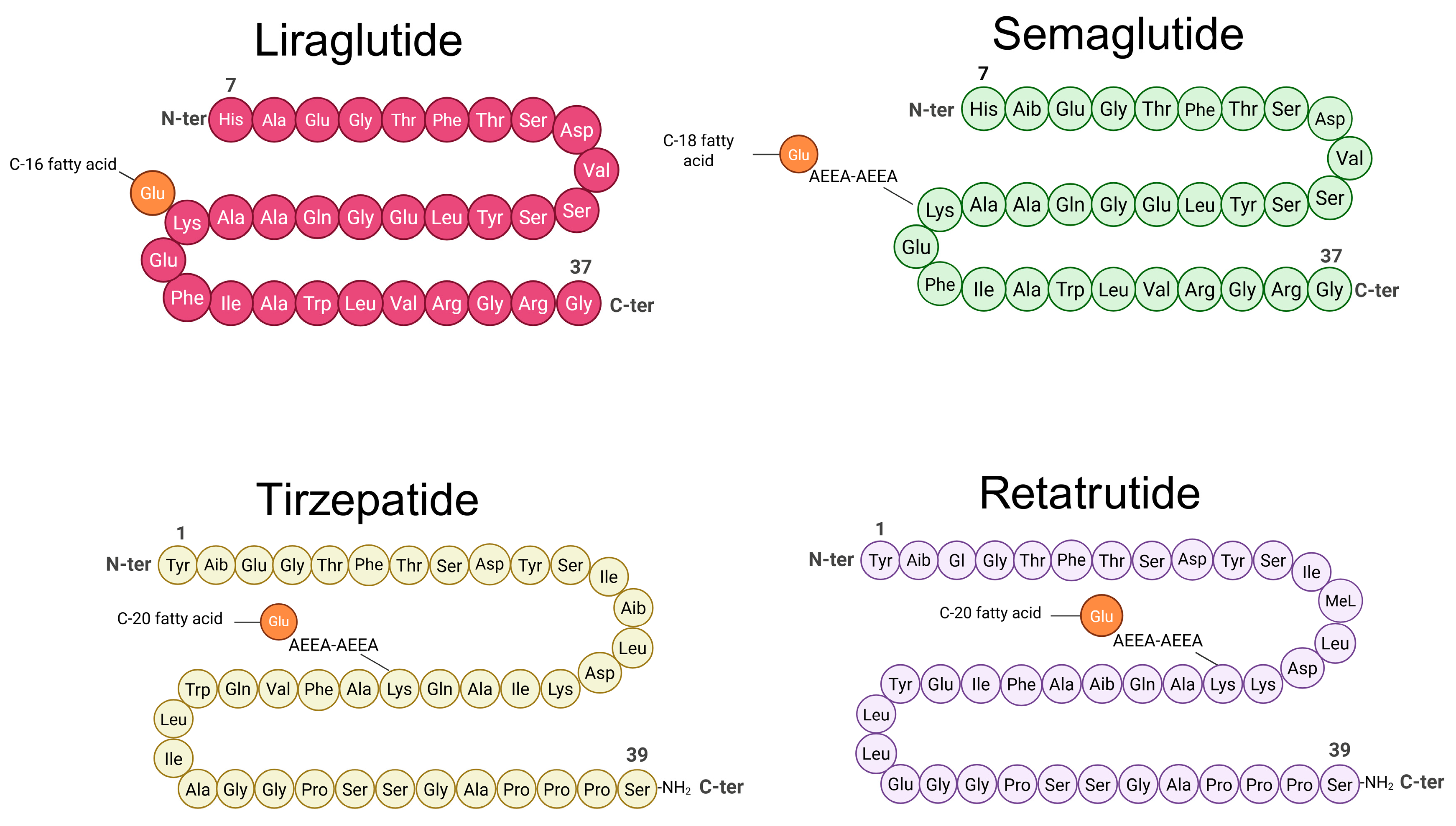
4.2. Multi-Peptide and Dual/Triple Agonist Strategies
4.3. Peptide Stabilisation, Delivery, and Molecular Design
4.4. Main Limitation of Therapeutic Peptides
5. Gut–Brain–Pancreas Peptide Signalling and Mitochondrial Bioenergetics
6. Conclusions and Future Perspectives
Author Contributions
Funding
Institutional Review Board Statement
Informed Consent Statement
Data Availability Statement
Conflicts of Interest
Abbreviations
| GLP-1 Ras | glucagon-like peptide-1 receptor agonists |
| EECs | intestinal enteroendocrine cells |
| GIP | gastric inhibitory peptide |
| PYY | peptide tyrosine tyrosine |
| CCK | cholecystokinin |
| POMC | proopiomelanocortin |
| AgRP | agouti-related neuropeptide |
| NTS | nucleus of the solitary tract |
| AMPK | AMP-activated protein kinase |
| mTOR | mechanistic target of rapamycin |
| PGC-1α | peroxisome proliferator-activated receptor-gamma coactivator |
| ATP | adenosine triphosphate |
| HPA | hypothalamic–pituitary–adrenal |
| OXM | oxyntomodulin |
| GPCRs | G protein-coupled receptors |
| SGLT1 | sodium-glucose co-transporter 1 |
| ENS | enteric nervous system |
| CART | cocaine and amphetamine-regulated transcript |
| NPY | neuropeptide Y |
| PVN | paraventricular |
| VMH | ventromedial |
| LH | lateral hypothalamus |
| PKA | protein kinase A |
| GHS-R1a | ghrelin receptor |
| Y2R | presynaptic Y2 receptors |
| IAPP | islet amyloid polypeptide |
| CT | calcitonin |
| AMYRs | amylin receptors |
| CTR | calcitonin receptor |
| RAMPs | receptor activity-modifying proteins |
| BAT | brown adipose tissue |
| GDF15 | growth differentiation factor 15 |
| T2DM | type 2 diabetes mellitus |
| GIPR | gastric inhibitory polypeptide receptor |
| Aib | α-aminoisobutyric acid |
| RYGB | Roux-en-Y gastric bypass |
| SNAC | salcaprozate sodium |
| TZDs | thiazolidinediones |
References
- Muzurović, E.M.; Volčanšek, Š.; Tomšić, K.Z.; Janež, A.; Mikhailidis, D.P.; Rizzo, M.; Mantzoros, C.S. Glucagon-like peptide-1 receptor agonists and dual glucose-dependent insulinotropic polypeptide/glucagon-like peptide-1 receptor agonists in the treatment of obesity/metabolic syndrome, prediabetes/diabetes and non-alcoholic fatty liver disease—Current evidence. J. Cardiovasc. Pharmacol. Ther. 2022, 27, 10742484221146371. [Google Scholar]
- Zhang, J.; Sanan, S.; Csanalosi, M.; Zheng, C.; Pfeiffer, A.F.H. Novel dual and triple agonists targeting GLP-1, GIP, Glucagon, and GDF15 for type 2 diabetes and obesity management. Endocrinology 2025, 166, bqaf130. [Google Scholar] [CrossRef]
- Romaní-Pérez, M.; Bullich-Vilarrubias, C.; López-Almela, I.; Liébana-García, R.; Olivares, M.; Sanz, Y. The microbiota and the gut–brain axis in controlling food intake and energy homeostasis. Int. J. Mol. Sci. 2021, 22, 5830. [Google Scholar] [CrossRef] [PubMed]
- Myers, M.G., Jr.; Affinati, A.H.; Richardson, N.; Schwartz, M.W. Central nervous system regulation of organismal energy and glucose homeostasis. Nat. Metab. 2021, 3, 737–750. [Google Scholar] [CrossRef]
- Kiba, T. Recent advancements in the relationship between the autonomic nervous system and the pancreas, encompassing the regulation of regeneration and apoptosis. Cells 2025, 14, 1371. [Google Scholar] [CrossRef] [PubMed]
- Gribble, F.M.; Reimann, F. Nutrient sensing in the gut and the regulation of appetite. Curr. Opin. Endocr. Metab. Res. 2022, 23, 100318. [Google Scholar] [CrossRef]
- Lu, V.B.; Gribble, F.M.; Reimann, F. Nutrient-induced cellular mechanisms of gut hormone secretion. Nutrients 2021, 13, 883. [Google Scholar] [CrossRef]
- Brüning, J.C.; Fenselau, H. Integrative neurocircuits that control metabolism and food intake. Science 2023, 381, eabl7398. [Google Scholar] [CrossRef]
- Cheng, W.; Ndoka, E.; Maung, J.N.; Pan, W.; Rupp, A.C.; Rhodes, C.J.; Olson, D.P.; Myers, M.G., Jr. NTS Prlh overcomes orexigenic stimuli and ameliorates dietary and genetic forms of obesity. Nat. Commun. 2021, 12, 5175. [Google Scholar] [CrossRef]
- Urva, S.; Coskun, T.; Loh, M.T.; Du, Y.; Thomas, M.K.; Gurbuz, S.; Haupt, A.; Benson, C.T.; Hernandez-Illas, M.; D’Alessio, D.A. LY3437943, a novel triple GIP, GLP-1, and glucagon receptor agonist in people with type 2 diabetes: A phase 1b, multicentre, double-blind, placebo-controlled, randomised, multiple-ascending dose trial. Lancet 2022, 400, 1869–1881. [Google Scholar] [CrossRef]
- Jastreboff, A.M.; Aronne, L.J.; Ahmad, N.N.; Wharton, S.; Connery, L.; Alves, B.; Kiyosue, A.; Zhang, S.; Liu, B.; Bunck, M.C. Tirzepatide once weekly for the treatment of obesity. N. Engl. J. Med. 2022, 387, 205–216. [Google Scholar] [CrossRef] [PubMed]
- Aronne, L.J.; Horn, D.B.; le Roux, C.W.; Ho, W.; Falcon, B.L.; Gomez Valderas, E.; Das, S.; Lee, C.J.; Glass, L.C.; Senyucel, C. Tirzepatide as compared with semaglutide for the treatment of obesity. N. Engl. J. Med. 2025, 393, 26–36. [Google Scholar] [CrossRef]
- Xie, C.; Jones, K.L.; Rayner, C.K.; Wu, T. Enteroendocrine hormone secretion and metabolic control: Importance of the region of the gut stimulation. Pharmaceutics 2020, 12, 790. [Google Scholar] [CrossRef] [PubMed]
- Halling, J.F.; Pilegaard, H. PGC-1α-mediated regulation of mitochondrial function and physiological implications. Appl. Physiol. Nutr. Metab. 2020, 45, 927–936. [Google Scholar] [CrossRef]
- Chiu, Y.-H.; Lin, S.-C.A.; Kuo, C.-H.; Li, C.-J. Molecular machinery and pathophysiology of mitochondrial dynamics. Front. Cell Dev. Biol. 2021, 9, 743892. [Google Scholar] [CrossRef]
- Steinert, R.E.; Feinle-Bisset, C.; Asarian, L.; Horowitz, M.; Beglinger, C.; Geary, N. Ghrelin, CCK, GLP-1, and PYY (3–36): Secretory controls and physiological roles in eating and glycemia in health, obesity, and after RYGB. Physiol. Rev. 2017, 97, 411–463. [Google Scholar] [CrossRef]
- Koliaki, C.; Liatis, S.; Dalamaga, M.; Kokkinos, A. The implication of gut hormones in the regulation of energy homeostasis and their role in the pathophysiology of obesity. Curr. Obes. Rep. 2020, 9, 255–271. [Google Scholar] [CrossRef]
- Olson, B.; Marks, D.L.; Grossberg, A.J. Diverging metabolic programmes and behaviours during states of starvation, protein malnutrition, and cachexia. J. Cachexia Sarcopenia Muscle 2020, 11, 1429–1446. [Google Scholar] [CrossRef]
- Rohm, M.; Zeigerer, A.; Machado, J.; Herzig, S. Energy metabolism in cachexia. EMBO Rep. 2019, 20, e47258. [Google Scholar] [CrossRef]
- De Silva, A.; Bloom, S.R. Gut hormones and appetite control: A focus on PYY and GLP-1 as therapeutic targets in obesity. Gut Liver 2012, 6, 10. [Google Scholar] [CrossRef] [PubMed]
- Browning, K.N.; Travagli, R.A. Central nervous system control of gastrointestinal motility and secretion and modulation of gastrointestinal functions. Compr. Physiol. 2014, 4, 1339–1368. [Google Scholar] [CrossRef] [PubMed]
- Stengel, A.; Taché, Y. Neuroendocrine control of the gut during stress: Corticotropin-releasing factor signaling pathways in the spotlight. Annu. Rev. Physiol. 2009, 71, 219–239. [Google Scholar] [CrossRef]
- Singh, S.C.; Vayyat, S.; Mishra, P. Gastrointestinal system and its neurophysiology. In Brain and Organ Communication; Elsevier: Amsterdam, The Netherlands, 2025; pp. 177–194. [Google Scholar]
- Yusta, B.; Baggio, L.L.; Koehler, J.; Holland, D.; Cao, X.; Pinnell, L.J.; Johnson-Henry, K.C.; Yeung, W.; Surette, M.G.; Bang, K.W.A. GLP-1R agonists modulate enteric immune responses through the intestinal intraepithelial lymphocyte GLP-1R. Diabetes 2015, 64, 2537–2549. [Google Scholar] [CrossRef] [PubMed]
- Kaelberer, M.M.; Buchanan, K.L.; Klein, M.E.; Barth, B.B.; Montoya, M.M.; Shen, X.; Bohórquez, D.V. A gut-brain neural circuit for nutrient sensory transduction. Science 2018, 361, eaat5236. [Google Scholar] [CrossRef]
- Hunne, B.; Stebbing, M.J.; McQuade, R.M.; Furness, J.B. Distributions and relationships of chemically defined enteroendocrine cells in the rat gastric mucosa. Cell Tissue Res. 2019, 378, 33–48. [Google Scholar] [CrossRef] [PubMed]
- Gunawardene, A.R.; Corfe, B.M.; Staton, C.A. Classification and functions of enteroendocrine cells of the lower gastrointestinal tract. Int. J. Exp. Pathol. 2011, 92, 219–231. [Google Scholar] [CrossRef]
- Latorre, R.; Sternini, C.; De Giorgio, R.; Greenwood-Van Meerveld, B. Enteroendocrine cells: A review of their role in brain–gut communication. Neurogastroenterol. Motil. 2016, 28, 620–630. [Google Scholar] [CrossRef]
- Reimann, F.; Habib, A.M.; Tolhurst, G.; Parker, H.E.; Rogers, G.J.; Gribble, F.M. Glucose sensing in L cells: A primary cell study. Cell Metab. 2008, 8, 532–539. [Google Scholar] [CrossRef]
- Kuhre, R.E.; Frost, C.R.; Svendsen, B.; Holst, J.J. Molecular mechanisms of glucose-stimulated GLP-1 secretion from perfused rat small intestine. Diabetes 2015, 64, 370–382. [Google Scholar] [CrossRef]
- Parker, H.E.; Habib, A.M.; Rogers, G.J.; Gribble, F.M.; Reimann, F. Nutrient-dependent secretion of glucose-dependent insulinotropic polypeptide from primary murine K cells. Diabetologia 2009, 52, 289–298. [Google Scholar] [CrossRef]
- Yusta, B.; Matthews, D.; Koehler, J.A.; Pujadas, G.; Kaur, K.D.; Drucker, D.J. Localization of glucagon-like peptide-2 receptor expression in the mouse. Endocrinology 2019, 160, 1950–1963. [Google Scholar] [CrossRef]
- Richards, P.; Parker, H.E.; Adriaenssens, A.E.; Hodgson, J.M.; Cork, S.C.; Trapp, S.; Gribble, F.M.; Reimann, F. Identification and characterization of GLP-1 receptor–expressing cells using a new transgenic mouse model. Diabetes 2014, 63, 1224–1233. [Google Scholar] [CrossRef] [PubMed]
- Holst, J.J. The physiology of glucagon-like peptide 1. Physiol. Rev. 2007, 87, 1409–1439. [Google Scholar] [CrossRef]
- Müller, T.D.; Finan, B.; Bloom, S.R.; D’Alessio, D.; Drucker, D.J.; Flatt, P.R.; Fritsche, A.; Gribble, F.; Grill, H.J.; Habener, J.F. Glucagon-like peptide 1 (GLP-1). Mol. Metab. 2019, 30, 72–130. [Google Scholar] [CrossRef] [PubMed]
- Adriaenssens, A.; Broichhagen, J.; De Bray, A.; Ast, J.; Hasib, A.; Jones, B.; Tomas, A.; Burgos, N.F.; Woodward, O.; Lewis, J. Hypothalamic and brainstem glucose-dependent insulinotropic polypeptide receptor neurons employ distinct mechanisms to affect feeding. JCI Insight 2023, 8, e164921. [Google Scholar] [CrossRef] [PubMed]
- De Bray, A.; Roberts, A.G.; Armour, S.; Tong, J.; Huhn, C.; Gatin-Fraudet, B.; Roßmann, K.; Shilleh, A.H.; Jiang, W.; Figueredo Burgos, N.S. Fluorescent GLP1R/GIPR dual agonist probes reveal cell targets in the pancreas and brain. Nat. Metab. 2025, 7, 1536–1549. [Google Scholar] [CrossRef]
- Nauck, M.A.; Quast, D.R.; Wefers, J.; Pfeiffer, A.F.H. The evolving story of incretins (GIP and GLP-1) in metabolic and cardiovascular disease: A pathophysiological update. Diabetes Obes. Metab. 2021, 23, 5–29. [Google Scholar] [CrossRef]
- Gribble, F.M.; Reimann, F. Function and mechanisms of enteroendocrine cells and gut hormones in metabolism. Nat. Rev. Endocrinol. 2019, 15, 226–237. [Google Scholar] [CrossRef]
- Meek, C.L.; Lewis, H.B.; Reimann, F.; Gribble, F.M.; Park, A.J. The effect of bariatric surgery on gastrointestinal and pancreatic peptide hormones. Peptides 2016, 77, 28–37. [Google Scholar] [CrossRef] [PubMed]
- Ratner, C.; Skov, L.J.; Raida, Z.; Bächler, T.; Bellmann-Sickert, K.; Le Foll, C.; Sivertsen, B.; Dalbøge, L.S.; Hartmann, B.; Beck-Sickinger, A.G. Effects of peripheral neurotensin on appetite regulation and its role in gastric bypass surgery. Endocrinology 2016, 157, 3482–3492. [Google Scholar] [CrossRef] [PubMed]
- D’Agostino, G.; Lyons, D.J.; Cristiano, C.; Burke, L.K.; Madara, J.C.; Campbell, J.N.; Garcia, A.P.; Land, B.B.; Lowell, B.B.; Dileone, R.J. Appetite controlled by a cholecystokinin nucleus of the solitary tract to hypothalamus neurocircuit. eLife 2016, 5, e12225. [Google Scholar] [CrossRef]
- Hickey, J.W.; Becker, W.R.; Nevins, S.A.; Horning, A.; Perez, A.E.; Zhu, C.; Zhu, B.; Wei, B.; Chiu, R.; Chen, D.C. Organization of the human intestine at single-cell resolution. Nature 2023, 619, 572–584. [Google Scholar] [CrossRef] [PubMed]
- Barton, J.R.; Londregan, A.K.; Alexander, T.D.; Entezari, A.A.; Covarrubias, M.; Waldman, S.A. Enteroendocrine cell regulation of the gut-brain axis. Front. Neurosci. 2023, 17, 1272955. [Google Scholar] [CrossRef]
- Valassi, E.; Scacchi, M.; Cavagnini, F. Neuroendocrine control of food intake. Nutr. Metab. Cardiovasc. Dis. 2008, 18, 158–168. [Google Scholar] [CrossRef]
- Tu, L.; Fukuda, M.; Tong, Q.; Xu, Y. The ventromedial hypothalamic nucleus: Watchdog of whole-body glucose homeostasis. Cell Biosci. 2022, 12, 71. [Google Scholar] [CrossRef]
- Holt, M.K. The ins and outs of the caudal nucleus of the solitary tract: An overview of cellular populations and anatomical connections. J. Neuroendocrinol. 2022, 34, e13132. [Google Scholar]
- Bonaz, B.; Sinniger, V.; Pellissier, S. The vagus nerve in the neuro-immune axis: Implications in the pathology of the gastrointestinal tract. Front. Immunol. 2017, 8, 1452. [Google Scholar] [CrossRef]
- Marroncini, G.; Naldi, L.; Martinelli, S.; Amedei, A. Gut–liver–pancreas axis crosstalk in health and disease: From the role of microbial metabolites to innovative microbiota manipulating strategies. Biomedicines 2024, 12, 1398. [Google Scholar] [CrossRef] [PubMed]
- Kong, C.-C.; Cheng, J.-D.; Wang, W. Neurotransmitters regulate β cells insulin secretion: A neglected factor. World J. Clin. Cases 2023, 11, 6670. [Google Scholar] [CrossRef] [PubMed]
- Schuit, F.C.; Huypens, P.; Heimberg, H.; Pipeleers, D.G. Glucose sensing in pancreatic β-cells: A model for the study of other glucose-regulated cells in gut, pancreas, and hypothalamus. Diabetes 2001, 50, 1–11. [Google Scholar] [CrossRef]
- Zheng, Z.; Zong, Y.; Ma, Y.; Tian, Y.; Pang, Y.; Zhang, C.; Gao, J. Glucagon-like peptide-1 receptor: Mechanisms and advances in therapy. Signal Transduct. Target. Ther. 2024, 9, 234. [Google Scholar] [CrossRef]
- Chung, J.Y.; Jeong, J.-H.; Song, J. Resveratrol modulates the gut-brain axis: Focus on glucagon-like peptide-1, 5-HT, and gut microbiota. Front. Aging Neurosci. 2020, 12, 588044. [Google Scholar]
- Coll, A.P.; Yeo, G.S.H. The hypothalamus and metabolism: Integrating signals to control energy and glucose homeostasis. Curr. Opin. Pharmacol. 2013, 13, 970–976. [Google Scholar] [CrossRef]
- Tsatsoulis, A.; Mantzaris, M.D.; Bellou, S.; Andrikoula, M. Insulin resistance: An adaptive mechanism becomes maladaptive in the current environment—An evolutionary perspective. Metabolism 2013, 62, 622–633. [Google Scholar]
- Genchi, V.A.; D’Oria, R.; Palma, G.; Caccioppoli, C.; Cignarelli, A.; Natalicchio, A.; Laviola, L.; Giorgino, F.; Perrini, S. Impaired leptin signalling in obesity: Is leptin a new thermolipokine? Int. J. Mol. Sci. 2021, 22, 6445. [Google Scholar] [CrossRef]
- Bhusal, A.; Rahman, M.H.; Suk, K. Hypothalamic inflammation in metabolic disorders and aging. Cell. Mol. Life Sci. 2022, 79, 32. [Google Scholar]
- Ward, Z.J.; Bleich, S.N.; Cradock, A.L.; Barrett, J.L.; Giles, C.M.; Flax, C.; Long, M.W.; Gortmaker, S.L. Projected US state-level prevalence of adult obesity and severe obesity. N. Engl. J. Med. 2019, 381, 2440–2450. [Google Scholar] [PubMed]
- Camilleri, M.; Acosta, A. Newer pharmacological interventions directed at gut hormones for obesity. Br. J. Pharmacol. 2024, 181, 1153–1164. [Google Scholar] [PubMed]
- Lu, J.; Liu, P.; Cai, M.; Lv, T.; Zhang, M.; Yin, K.; Cheng, J.; Zhang, G. Recent Progress in the Pharmacotherapy for Obesity. Eur. J. Pharmacol. 2025, 1002, 177850. [Google Scholar] [CrossRef] [PubMed]
- Richards, P.; Thornberry, N.A.; Pinto, S. The gut–brain axis: Identifying new therapeutic approaches for type 2 diabetes, obesity, and related disorders. Mol. Metab. 2021, 46, 101175. [Google Scholar] [CrossRef]
- d’Afflitto, M.; Upadhyaya, A.; Green, A.; Peiris, M. Association between sex hormone levels and gut microbiota composition and diversity—A systematic review. J. Clin. Gastroenterol. 2022, 56, 384–392. [Google Scholar] [CrossRef]
- Aukan, M.I.; Coutinho, S.; Pedersen, S.A.; Simpson, M.R.; Martins, C. Differences in gastrointestinal hormones and appetite ratings between individuals with and without obesity—A systematic review and meta-analysis. Obes. Rev. 2023, 24, e13531. [Google Scholar] [PubMed]
- Jiao, Z.-T.; Luo, Q. Molecular mechanisms and health benefits of ghrelin: A narrative review. Nutrients 2022, 14, 4191. [Google Scholar] [CrossRef] [PubMed]
- Cummings, D.E.; Purnell, J.Q.; Frayo, R.S.; Schmidova, K.; Wisse, B.E.; Weigle, D.S. A preprandial rise in plasma ghrelin levels suggests a role in meal initiation in humans. Diabetes 2001, 50, 1714–1719. [Google Scholar] [CrossRef]
- da Boitb Marta, C.D.K.; Novelleb, G.; de Morentinb Rubén, P.B.M.; Lópeza, N.M. New insights in ghrelin orexigenic effect. Pituit. Today II New Mol. Physiol. Clin. Asp. 2010, 38, 196–205. [Google Scholar]
- Koliaki, C.; Kokkinos, A.; Tentolouris, N.; Katsilambros, N. The effect of ingested macronutrients on postprandial ghrelin response: A critical review of existing literature data. Int. J. Pept. 2010, 2010, 710852. [Google Scholar] [CrossRef] [PubMed]
- Wang, B.; Cheng, K.K.-Y. Hypothalamic AMPK as a mediator of hormonal regulation of energy balance. Int. J. Mol. Sci. 2018, 19, 3552. [Google Scholar] [CrossRef]
- Broglio, F.; Arvat, E.; Benso, A.; Gottero, C.; Muccioli, G.; Papotti, M.; Lely, A.J.v.d.; Deghenghi, R.; Ghigo, E. Ghrelin, a natural GH secretagogue produced by the stomach, induces hyperglycemia and reduces insulin secretion in humans. J. Clin. Endocrinol. Metab. 2001, 86, 5083. [Google Scholar] [CrossRef]
- Akalu, Y.; Molla, M.D.; Dessie, G.; Ayelign, B. Physiological effect of ghrelin on body systems. Int. J. Endocrinol. 2020, 2020, 1385138. [Google Scholar] [CrossRef]
- Tong, J.; Prigeon, R.L.; Davis, H.W.; Bidlingmaier, M.; Kahn, S.E.; Cummings, D.E.; Tschöp, M.H.; D’Alessio, D. Ghrelin suppresses glucose-stimulated insulin secretion and deteriorates glucose tolerance in healthy humans. Diabetes 2010, 59, 2145–2151. [Google Scholar] [CrossRef]
- Lv, Y.; Liang, T.; Wang, G.; Li, Z. Ghrelin, a gastrointestinal hormone, regulates energy balance and lipid metabolism. Biosci. Rep. 2018, 38, BSR20181061. [Google Scholar] [CrossRef]
- Lin, Y.; Liang, Z.; He, L.; Yang, M.; Liu, D.; Gu, H.F.; Liu, H.; Zhu, Z.; Zheng, H.; Li, L. Gut ghrelin regulates hepatic glucose production and insulin signaling via a gut-brain-liver pathway. Cell Commun. Signal. 2019, 17, 8. [Google Scholar] [PubMed]
- Abbott, C.R.; Monteiro, M.; Small, C.J.; Sajedi, A.; Smith, K.L.; Parkinson, J.R.C.; Ghatei, M.A.; Bloom, S.R. The inhibitory effects of peripheral administration of peptide YY3–36 and glucagon-like peptide-1 on food intake are attenuated by ablation of the vagal–brainstem–hypothalamic pathway. Brain Res. 2005, 1044, 127–131. [Google Scholar] [PubMed]
- Varin, E.M.; Mulvihill, E.E.; Baggio, L.L.; Koehler, J.A.; Cao, X.; Seeley, R.J.; Drucker, D.J. Distinct neural sites of GLP-1R expression mediate physiological versus pharmacological control of incretin action. Cell Rep. 2019, 27, 3371–3384. [Google Scholar] [CrossRef]
- Brierley, D.I.; de Lartigue, G. Reappraising the role of the vagus nerve in GLP-1-mediated regulation of eating. Br. J. Pharmacol. 2022, 179, 584–599. [Google Scholar]
- Plamboeck, A.; Veedfald, S.; Deacon, C.F.; Hartmann, B.; Wettergren, A.; Svendsen, L.B.; Meisner, S.; Hovendal, C.; Vilsbøll, T.; Knop, F.K. The effect of exogenous GLP-1 on food intake is lost in male truncally vagotomized subjects with pyloroplasty. Am. J. Physiol.-Gastrointest. Liver Physiol. 2013, 304, G1117–G1127. [Google Scholar]
- Deschaine, S.L.; Leggio, L. From “hunger hormone” to “it’s complicated”: Ghrelin beyond feeding control. Physiology 2022, 37, 5–15. [Google Scholar] [CrossRef]
- Yanagi, S.; Sato, T.; Kangawa, K.; Nakazato, M. The homeostatic force of ghrelin. Cell Metab. 2018, 27, 786–804. [Google Scholar] [CrossRef]
- Adrian, T.E.; Ferri, G.L.; Bacarese-Hamilton, A.J.; Fuessl, H.S.; Polak, J.M.; Bloom, S.R. Human distribution and release of a putative new gut hormone, peptide YY. Gastroenterology 1985, 89, 1070–1077. [Google Scholar] [CrossRef] [PubMed]
- Karra, E.; Chandarana, K.; Batterham, R.L. The role of peptide YY in appetite regulation and obesity. Physiol. Soc. 2009, 587, 19–25. [Google Scholar]
- Batterham, R.L.; Cohen, M.A.; Ellis, S.M.; Le Roux, C.W.; Withers, D.J.; Frost, G.S.; Ghatei, M.A.; Bloom, S.R. Inhibition of food intake in obese subjects by peptide YY3–36. N. Engl. J. Med. 2003, 349, 941–948. [Google Scholar] [CrossRef] [PubMed]
- Le Roux, C.W.; Bloom, S.R. Peptide YY, appetite and food intake. Proc. Nutr. Soc. 2005, 64, 213–216. [Google Scholar] [CrossRef]
- Batterham, R.L.; Cowley, M.A.; Small, C.J.; Herzog, H.; Cohen, M.A.; Dakin, C.L.; Wren, A.M.; Brynes, A.E.; Low, M.J.; Ghatei, M.A. Gut hormone PYY3-36 physiologically inhibits food intake. Nature 2002, 418, 650–654. [Google Scholar] [CrossRef]
- Cherbut, C.; Ferrier, L.; Rozé, C.; Anini, Y.; Blottière, H.; Lecannu, G.; Galmiche, J.-P. Short-chain fatty acids modify colonic motility through nerves and polypeptide YY release in the rat. Am. J. Physiol.-Gastrointest. Liver Physiol. 1998, 275, G1415–G1422. [Google Scholar] [CrossRef]
- Lin, H.C.; Neevel, C.; Chen, J.H. Slowing intestinal transit by PYY depends on serotonergic and opioid pathways. Am. J. Physiol.-Gastrointest. Liver Physiol. 2004, 286, G558–G563. [Google Scholar]
- Cox, H.M. Neuropeptide Y receptors; antisecretory control of intestinal epithelial function. Auton. Neurosci. 2007, 133, 76–85. [Google Scholar] [CrossRef]
- Schmidt, J.B.; Gregersen, N.T.; Pedersen, S.D.; Arentoft, J.L.; Ritz, C.; Schwartz, W.; Astrup, J.J.H.A.; Sjödin, A. Effects of PYY3-36 and GLP-1 on energy intake, energy expenditure and appetite in overweight men 2. Am. J. Physiol. Endocrinol. Metab. 2014, 306, E1248–E1256. [Google Scholar] [CrossRef]
- Cegla, J.; Cuenco, J.; Minnion, J.; Ghourab, S.; Hostomska, K.; Tan, T.; Bloom, S. Pharmacokinetics and pharmacodynamics of subcutaneously administered PYY3–36 and its analogues in vivo. Lancet 2015, 385, S28. [Google Scholar] [CrossRef] [PubMed]
- Oertel, M.; Ziegler, C.G.; Kohlhaas, M.; Nickel, A.; Kloock, S.; Maack, C.; Sequeira, V.; Fassnacht, M.; Dischinger, U. GLP-1 and PYY for the treatment of obesity: A pilot study on the use of agonists and antagonists in diet-induced rats. Endocr. Connect. 2024, 13, e230398. [Google Scholar] [PubMed]
- Zhang, J.V.; Ren, P.-G.; Avsian-Kretchmer, O.; Luo, C.-W.; Rauch, R.; Klein, C.; Hsueh, A.J.W. Obestatin, a peptide encoded by the ghrelin gene, opposes ghrelin’s effects on food intake. Science 2005, 310, 996–999. [Google Scholar] [CrossRef]
- Laitakari, A.; Liu, L.; Frimurer, T.M.; Holst, B. The zinc-sensing receptor GPR39 in physiology and as a pharmacological target. Int. J. Mol. Sci. 2021, 22, 3872. [Google Scholar] [CrossRef] [PubMed]
- Grunddal, K.V.; Diep, T.A.; Petersen, N.; Tough, I.R.; Skov, L.J.; Liu, L.; Buijink, J.D.A.; Mende, F.; Jin, C.; Jepsen, S.L. Selective release of gastrointestinal hormones induced by an orally active GPR39 agonist. Mol. Metab. 2021, 49, 101207. [Google Scholar] [CrossRef] [PubMed]
- Chartrel, N.; Alvear-Perez, R.; Leprince, J.; Iturrioz, X.; Goazigo, A.R.-L.; Audinot, V.; Chomarat, P.; Cogé, F.; Nosjean, O.; Rodriguez, M. Comment on “Obestatin, a peptide encoded by the ghrelin gene, opposes ghrelin’s effects on food intake”. Science 2007, 315, 766. [Google Scholar] [CrossRef] [PubMed]
- Gourcerol, G.; Tache, Y. Obestatin–a ghrelin-associated peptide that does not hold its promise to suppress food intake and motility. Neurogastroenterol. Motil. 2007, 19, 161–165. [Google Scholar] [CrossRef] [PubMed]
- Granata, R.; Settanni, F.; Gallo, D.; Trovato, L.; Biancone, L.; Cantaluppi, V.; Nano, R.; Annunziata, M.; Campiglia, P.; Arnoletti, E. Obestatin promotes survival of pancreatic β-cells and human islets and induces expression of genes involved in the regulation of β-cell mass and function. Diabetes 2008, 57, 967–979. [Google Scholar] [CrossRef]
- Baragli, L.; Grande, C.; Gesmundo, I.; Settanni, F.; Taliano, M.; Gallo, D.; Gargantini, E.; Ghigo, E.; Granata, R. Obestatin enhances in vitro generation of pancreatic islets through regulation of developmental pathways. PLoS ONE 2013, 8, e64374. [Google Scholar] [CrossRef]
- Konarska, K.; Cieszkowski, J.; Warzecha, Z.; Ceranowicz, P.; Chmura, A.; Kuśnierz-Cabala, B.; Gałązka, K.; Kowalczyk, P.; Miskiewicz, A.; Konturek, T.J. Treatment with obestatin—A ghrelin gene-encoded peptide—Reduces the severity of experimental colitis evoked by trinitrobenzene sulfonic acid. Int. J. Mol. Sci. 2018, 19, 1643. [Google Scholar] [CrossRef]
- Granata, R.; Gallo, D.; Luque, R.M.; Baragli, A.; Scarlatti, F.; Grande, C.; Gesmundo, I.; Córdoba-Chacón, J.; Bergandi, L.; Settanni, F. Obestatin regulates adipocyte function and protects against diet-induced insulin resistance and inflammation. FASEB J. 2012, 26, 3393–3411. [Google Scholar] [CrossRef]
- Bower, R.L.; Hay, D.L. Amylin structure–function relationships and receptor pharmacology: Implications for amylin mimetic drug development. Br. J. Pharmacol. 2016, 173, 1883–1898. [Google Scholar] [CrossRef]
- Cao, J.; Belousoff, M.J.; Liang, Y.-L.; Johnson, R.M.; Josephs, T.M.; Fletcher, M.M.; Christopoulos, A.; Hay, D.L.; Danev, R.; Wootten, D. A structural basis for amylin receptor phenotype. Science 2022, 375, eabm9609. [Google Scholar] [CrossRef]
- Volčanšek, Š.; Koceva, A.; Jensterle, M.; Janež, A.; Muzurović, E. Amylin: From Mode of Action to Future Clinical Potential in Diabetes and Obesity. Diabetes Ther. 2025, 16, 1207–1227. [Google Scholar] [CrossRef]
- Dehestani, B.; Stratford, N.R.S.; le Roux, C.W. Amylin as a future obesity treatment. J. Obes. Metab. Syndr. 2021, 30, 320. [Google Scholar] [CrossRef]
- Boyle, C.N.; Zheng, Y.; Lutz, T.A. Mediators of amylin action in metabolic control. J. Clin. Med. 2022, 11, 2207. [Google Scholar] [CrossRef] [PubMed]
- Lutz, T.A. The role of amylin in the control of energy homeostasis. Am. J. Physiol.-Regul. Integr. Comp. Physiol. 2010, 298, R1475–R1484. [Google Scholar] [CrossRef]
- Zhang, Y.; Sowers, J.R.; Ren, J. Targeting autophagy in obesity: From pathophysiology to management. Nat. Rev. Endocrinol. 2018, 14, 356–376. [Google Scholar] [CrossRef] [PubMed]
- Mukherjee, N.; Lin, L.; Contreras, C.J.; Templin, A.T. β-Cell death in diabetes: Past discoveries, present understanding, and potential future advances. Metabolites 2021, 11, 796. [Google Scholar] [CrossRef]
- Westermark, P.; Andersson, A.; Westermark, G.T. Islet amyloid polypeptide, islet amyloid, and diabetes mellitus. Physiol. Rev. 2011, 91, 795–826. [Google Scholar] [CrossRef]
- Sasahara, K. Membrane-mediated amyloid deposition of human islet amyloid polypeptide. Biophys. Rev. 2018, 10, 453–462. [Google Scholar] [CrossRef] [PubMed][Green Version]
- Reinehr, T.; De Sousa, G.; Niklowitz, P.; Roth, C.L. Amylin and its relation to insulin and lipids in obese children before and after weight loss. Obesity 2007, 15, 2006–2011. [Google Scholar] [CrossRef]
- Smith, D.; Mamo, J. Islet amyloid polypeptide (amylin) modulates chylomicron metabolism in rats. Clin. Exp. Pharmacol. Physiol. 2000, 27, 345–351. [Google Scholar] [CrossRef]
- Zheng, N.; Chen, R.; Yang, L.; Xu, P.; Wang, X.; Sun, X.; Lin, Y.; Qiu, J.; Su, X.-x.; Wang, Y. Engineering of an Ultralong-Acting and Nonaggregating Dual Amylin and Calcitonin Receptor Agonist with Durable Efficacy in Obesity and Diabetes. J. Med. Chem. 2025, 68, 24701–24717. [Google Scholar] [CrossRef]
- Mack, C.M.; Soares, C.J.; Wilson, J.K.; Athanacio, J.R.; Turek, V.F.; Trevaskis, J.L.; Roth, J.D.; Smith, P.A.; Gedulin, B.; Jodka, C.M. Davalintide (AC2307), a novel amylin-mimetic peptide: Enhanced pharmacological properties over native amylin to reduce food intake and body weight. Int. J. Obes. 2010, 34, 385–395. [Google Scholar] [CrossRef]
- Guerreiro, L.H.; Guterres, M.F.A.N.; Melo-Ferreira, B.; Erthal, L.C.S.; da Silva Rosa, M.; Lourenço, D.; Tinoco, P.; Lima, L.M.T.R. Preparation and characterization of PEGylated amylin. AAPS PharmSciTech 2013, 14, 1083–1097. [Google Scholar] [CrossRef] [PubMed]
- Bhattachar, S.N.; Tham, L.-S.; Tidemann-Miller, B.; Briere, D.A.; Qu, H.; Haupt, A.; Pratt, E.J.; Mather, K.J. 882-P: Eloralintide, a Selective, Long-Acting Amylin Receptor Agonist for Obesity—Phase 1 Proof of Concept. Diabetes 2025, 74, 882-P. [Google Scholar] [CrossRef]
- Billings, L.K.; Hsia, S.; Bays, H.; Tidemann-Miller, B.; O’Hagan, J.; San Tham, L.; Butler, A.; Kazda, C.; Mather, K.J.; Coskun, T. Eloralintide, a selective amylin receptor agonist for the treatment of obesity: A 48-week phase 2, multicentre, double-blind, randomised, placebo-controlled trial. Lancet 2025, 406, 2631–2643. [Google Scholar] [CrossRef] [PubMed]
- Briere, D.A.; Qu, H.; Lansu, K.; He, M.M.; Moyers, J.S.; Coskun, T.; Long, A.; Allen, D.; O’Farrell, L.; Bowen, B. Eloralintide (LY3841136), a novel amylin receptor agonist for the treatment of obesity: From discovery to clinical proof of concept. Mol. Metab. 2025, 102, 102271. [Google Scholar] [CrossRef]
- Hay, D.L.; Chen, S.; Lutz, T.A.; Parkes, D.G.; Roth, J.D. Amylin: Pharmacology, physiology, and clinical potential. Pharmacol. Rev. 2015, 67, 564–600. [Google Scholar] [CrossRef]
- Rehfeld, J.F. Cholecystokinin—From local gut hormone to ubiquitous messenger. Front. Endocrinol. 2017, 8, 47. [Google Scholar] [CrossRef]
- Albrechtsen, N.J.W.; Kuhre, R.E.; Deacon, C.F.; Holst, J.J. Targeting the intestinal L-cell for obesity and type 2 diabetes treatment. Expert Rev. Endocrinol. Metab. 2014, 9, 61–72. [Google Scholar] [CrossRef]
- van der Klaauw, A.A. Neuropeptides in obesity and metabolic disease. Clin. Chem. 2018, 64, 173–182. [Google Scholar] [CrossRef]
- Okonkwo, O.; Zezoff, D.; Adeyinka, A. Biochemistry, Cholecystokinin; StatPearls Publishing: Treasure Island, FL, USA, 2018. [Google Scholar]
- Cao, S.-G.; Wu, H.; Cai, Z.-Z. Dose-dependent effect of ghrelin on gastric emptying in rats and the related mechanism of action. Kaohsiung J. Med. Sci. 2016, 32, 113–117. [Google Scholar] [CrossRef] [PubMed]
- Schier, L.A.; Spector, A.C. The functional and neurobiological properties of bad taste. Physiol. Rev. 2019, 99, 605–663. [Google Scholar] [CrossRef] [PubMed]
- Wang, L.; Barachina, M.D.; Martínez, V.; Wei, J.Y.; Taché, Y. Synergistic interaction between CCK and leptin to regulate food intake. Regul. Pept. 2000, 92, 79–85. [Google Scholar] [CrossRef]
- Owyang, C.; Heldsinger, A. Vagal control of satiety and hormonal regulation of appetite. J. Neurogastroenterol. Motil. 2011, 17, 338. [Google Scholar] [CrossRef]
- Ashby, D.; Bloom, S.R. Recent progress in PYY research—An update report for 8th NPY meeting. Peptides 2007, 28, 198–202. [Google Scholar] [CrossRef]
- Cox, H.M. Peptide YY: A neuroendocrine neighbor of note. Peptides 2007, 28, 345–351. [Google Scholar] [CrossRef]
- Dockray, G.J. Cholecystokinin. Curr. Opin. Endocrinol. Diabetes Obes. 2012, 19, 8–12. [Google Scholar] [CrossRef]
- Saito, M.; Matsushita, M.; Yoneshiro, T.; Okamatsu-Ogura, Y. Brown adipose tissue, diet-induced thermogenesis, and thermogenic food ingredients: From mice to men. Front. Endocrinol. 2020, 11, 222. [Google Scholar] [CrossRef]
- Kalafatakis, K.; Triantafyllou, K. Contribution of neurotensin in the immune and neuroendocrine modulation of normal and abnormal enteric function. Regul. Pept. 2011, 170, 7–17. [Google Scholar] [CrossRef] [PubMed]
- Barchetta, I.; Baroni, M.G.; Melander, O.; Cavallo, M.G. New insights in the control of fat homeostasis: The role of neurotensin. Int. J. Mol. Sci. 2022, 23, 2209. [Google Scholar] [CrossRef]
- Boeckxstaens, G.; Camilleri, M.; Sifrim, D.; Houghton, L.A.; Elsenbruch, S.; Lindberg, G.; Azpiroz, F.; Parkman, H.P. Fundamentals of neurogastroenterology: Physiology/motility–sensation. Gastroenterology 2016, 150, 1292–1304. [Google Scholar] [CrossRef]
- Gereau, G.B.; Garrison, S.K.D.; McElligott, Z.A. Neurotensin and energy balance. J. Neurochem. 2023, 166, 189–200. [Google Scholar] [CrossRef]
- Acosta, F.M.; Örling, J.; Salmi, M.; Virtanen, K.A. The interplay between organized lymphoid structures and thermogenic adipose tissue. Trends Endocrinol. Metab. 2025. [Google Scholar] [CrossRef]
- Ramirez-Virella, J.; Leinninger, G.M. The role of central neurotensin in regulating feeding and body weight. Endocrinology 2021, 162, bqab038. [Google Scholar] [CrossRef]
- Gasbjerg, L.S.; Nielsen, C.K.; Suppli, M.P.; Grøndahl, M.F.G.; Holst, J.J.; Knop, F.K.; Jeppesen, P.B.; Lund, A.B. Proglucagon-derived peptides: Human physiology and therapeutic potential. Physiol. Rev. 2026, 106, 529–586. [Google Scholar] [CrossRef]
- Holst, J.J.; Albrechtsen, N.J.W.; Gabe, M.B.N.; Rosenkilde, M.M. Oxyntomodulin: Actions and role in diabetes. Peptides 2018, 100, 48–53. [Google Scholar] [CrossRef]
- Lafferty, R.A.; O’Harte, F.P.M.; Irwin, N.; Gault, V.A.; Flatt, P.R. Proglucagon-derived peptides as therapeutics. Front. Endocrinol. 2021, 12, 689678. [Google Scholar] [CrossRef] [PubMed]
- Laurila, S.; Sun, L.; Lahesmaa, M.; Schnabl, K.; Laitinen, K.; Klén, R.; Li, Y.; Balaz, M.; Wolfrum, C.; Steiger, K. Secretin activates brown fat and induces satiation. Nat. Metab. 2021, 3, 798–809. [Google Scholar] [CrossRef] [PubMed]
- Schnabl, K.; Li, Y.; U-Din, M.; Klingenspor, M. Secretin as a satiation whisperer with the potential to turn into an obesity-curbing knight. Endocrinology 2021, 162, bqab113. [Google Scholar] [CrossRef] [PubMed]
- Psilopanagioti, A.; Nikou, S.; Papadaki, H. Nucleobindin-2/nesfatin-1 in the human hypothalamus is reduced in obese subjects and colocalizes with oxytocin, vasopressin, melanin-concentrating hormone, and cocaine-and amphetamine-regulated transcript. Neuroendocrinology 2019, 108, 190–200. [Google Scholar] [CrossRef]
- Stengel, A.; Taché, Y. Nesfatin-1—Role as possible new potent regulator of food intake. Regul. Pept. 2010, 163, 18–23. [Google Scholar] [CrossRef]
- Li, Z.; Gao, L.; Tang, H.; Yin, Y.; Xiang, X.; Li, Y.; Zhao, J.; Mulholland, M.; Zhang, W. Peripheral effects of nesfatin-1 on glucose homeostasis. PLoS ONE 2013, 8, e71513. [Google Scholar] [CrossRef]
- Lahane, G.P.; Dhar, A. Nesfatin-1 peptide protects rat renal epithelial cells against high glucose and H2O2 induced injury via inhibition of oxidative stress, apoptosis, and fibrosis. Peptides 2023, 165, 171013. [Google Scholar] [CrossRef] [PubMed]
- Wang, Z.Z.; Chen, S.C.; Zou, X.B.; Tian, L.L.; Sui, S.H.; Liu, N.Z. Nesfatin-1 alleviates acute lung injury through reducing inflammation and oxidative stress via the regulation of HMGB1. Eur. Rev. Med. Pharmacol. Sci. 2020, 24, 5071–5081. [Google Scholar] [PubMed]
- Jena, J.; García-Peña, L.M.; Pereira, R.O. The roles of FGF21 and GDF15 in mediating the mitochondrial integrated stress response. Front. Endocrinol. 2023, 14, 1264530. [Google Scholar] [CrossRef] [PubMed]
- Johann, K.; Kleinert, M.; Klaus, S. The role of GDF15 as a myomitokine. Cells 2021, 10, 2990. [Google Scholar] [CrossRef]
- Benichou, O.; Coskun, T.; Gonciarz, M.D.; Garhyan, P.; Adams, A.C.; Du, Y.; Dunbar, J.D.; Martin, J.A.; Mather, K.J.; Pickard, R.T. Discovery, development, and clinical proof of mechanism of LY3463251, a long-acting GDF15 receptor agonist. Cell Metab. 2023, 35, 274–286. [Google Scholar] [CrossRef]
- Breit, S.N.; Tsai, V.W. Metabolic Messenger: Growth differentiation factor 15. Nat. Metab. 2025, 7, 1732–1744. [Google Scholar] [CrossRef]
- Jorsal, T.; Rungby, J.; Knop, F.K.; Vilsbøll, T. GLP-1 and amylin in the treatment of obesity. Curr. Diabetes Rep. 2016, 16, 1. [Google Scholar] [CrossRef]
- Ueno, H.; Nakazato, M. Mechanistic relationship between the vagal afferent pathway, central nervous system and peripheral organs in appetite regulation. J. Diabetes Investig. 2016, 7, 812–818. [Google Scholar] [CrossRef]
- Kentish, S.J.; Page, A.J. The role of gastrointestinal vagal afferent fibres in obesity. J. Physiol. 2015, 593, 775–786. [Google Scholar] [CrossRef]
- Campos, C.A.; Shiina, H.; Silvas, M.; Page, S.; Ritter, R.C. Vagal afferent NMDA receptors modulate CCK-induced reduction of food intake through synapsin I phosphorylation in adult male rats. Endocrinology 2013, 154, 2613–2625. [Google Scholar] [CrossRef]
- Tsuboi, T.; Silva Xavier, G.d.; Holz, G.G.; Jouaville, L.S.; Thomas, A.P.; Rutter, G.A. Glucagon-like peptide-1 mobilizes intracellular Ca2+ and stimulates mitochondrial ATP synthesis in pancreatic MIN6 beta-cells. Biochem. J. 2003, 369, 287–299. [Google Scholar] [CrossRef]
- Tomas, E.; Stanojevic, V.; Habener, J.F. GLP-1-derived nonapeptide GLP-1 (28–36) amide targets to mitochondria and suppresses glucose production and oxidative stress in isolated mouse hepatocytes. Regul. Pept. 2011, 167, 177–184. [Google Scholar] [CrossRef]
- Morales, P.E.; Torres, G.; Sotomayor-Flores, C.; Peña-Oyarzún, D.; Rivera-Mejías, P.; Paredes, F.; Chiong, M. GLP-1 promotes mitochondrial metabolism in vascular smooth muscle cells by enhancing endoplasmic reticulum–mitochondria coupling. Biochem. Biophys. Res. Commun. 2014, 446, 410–416. [Google Scholar] [CrossRef]
- Lim, Y.A.; Rhein, V.; Baysang, G.; Meier, F.; Poljak, A.; Raftery, M.J.; Guilhaus, M.; Ittner, L.M.; Eckert, A.; Götz, J. Aβ and human amylin share a common toxicity pathway via mitochondrial dysfunction. Proteomics 2010, 10, 1621–1633. [Google Scholar] [CrossRef] [PubMed]
- Atlante, A.; Valenti, D. Mitochondrial complex I and β-amyloid peptide interplay in Alzheimer’s disease: A critical review of new and old little regarded findings. Int. J. Mol. Sci. 2023, 24, 15951. [Google Scholar] [CrossRef] [PubMed]
- Bharadwaj, P.; Wijesekara, N.; Liyanapathirana, M.; Newsholme, P.; Ittner, L.; Fraser, P.; Verdile, G. The link between type 2 diabetes and neurodegeneration: Roles for amyloid-β, amylin, and tau proteins. J. Alzheimer’s Dis. 2017, 59, 421–432. [Google Scholar] [CrossRef] [PubMed]
- Shome, G.; Mondal, R.; Deb, S.; Roy, J.; Mandal, A.K.; Benito-León, J. Bridging Pancreatic Amyloidosis and Neurodegeneration: The Emerging Role of Amylin in Diabetic Dementia. Int. J. Mol. Sci. 2025, 26, 5021. [Google Scholar] [CrossRef]
- Veselov, I.M.; Vinogradova, D.V.; Maltsev, A.V.; Shevtsov, P.N.; Spirkova, E.A.; Bachurin, S.O.; Shevtsova, E.F. Mitochondria and oxidative stress as a link between Alzheimer’s disease and diabetes mellitus. Int. J. Mol. Sci. 2023, 24, 14450. [Google Scholar] [CrossRef]
- Santoro, A.; Buonocore, M.; Grimaldi, M.; Napolitano, E.; D’Ursi, A.M. Monitoring the Conformational Changes of the Aβ (25–35) Peptide in SDS Micelles: A Matter of Time. Int. J. Mol. Sci. 2023, 24, 971. [Google Scholar] [CrossRef]
- Skaanning, L.K.; Santoro, A.; Skamris, T.; Martinsen, J.H.; D’Ursi, A.M.; Bucciarelli, S.; Vestergaard, B.; Bugge, K.; Langkilde, A.E.; Kragelund, B.B. The non-fibrillating N-terminal of α-synuclein binds and co-fibrillates with heparin. Biomolecules 2020, 10, 1192. [Google Scholar] [CrossRef]
- Santoro, A.; Ricci, A.; Rodriquez, M.; Buonocore, M.; D’Ursi, A.M. A Structural Effect of the Antioxidant Curcuminoids on the Aβ (1–42) Amyloid Peptide. Antioxidants 2025, 14, 53. [Google Scholar] [CrossRef]
- Willard, F.S.; Douros, J.D.; Gabe, M.B.N.; Showalter, A.D.; Wainscott, D.B.; Suter, T.M.; Capozzi, M.E.; van der Velden, W.J.C.; Stutsman, C.; Cardona, G.R. Tirzepatide is an imbalanced and biased dual GIP and GLP-1 receptor agonist. JCI Insight 2020, 5, e140532. [Google Scholar] [CrossRef]
- Sun, B.; Willard, F.S.; Feng, D.; Alsina-Fernandez, J.; Chen, Q.; Vieth, M.; Ho, J.D.; Showalter, A.D.; Stutsman, C.; Ding, L. Structural determinants of dual incretin receptor agonism by tirzepatide. Proc. Natl. Acad. Sci. USA 2022, 119, e2116506119. [Google Scholar] [CrossRef]
- Zhao, F.; Zhou, Q.; Cong, Z.; Hang, K.; Zou, X.; Zhang, C.; Chen, Y.; Dai, A.; Liang, A.; Ming, Q. Structural insights into multiplexed pharmacological actions of tirzepatide and peptide 20 at the GIP, GLP-1 or glucagon receptors. Nat. Commun. 2022, 13, 1057. [Google Scholar] [CrossRef]
- Zhang, J.; Wei, J.; Lai, W.; Sun, J.; Bai, Y.; Cao, H.; Guo, J.; Su, Z. Focus on Glucagon-like Peptide-1 Target: Drugs Approved or Designed to Treat Obesity. Int. J. Mol. Sci. 2025, 26, 1651. [Google Scholar] [CrossRef] [PubMed]
- Day, J.W.; Ottaway, N.; Patterson, J.T.; Gelfanov, V.; Smiley, D.; Gidda, J.; Findeisen, H.; Bruemmer, D.; Drucker, D.J.; Chaudhary, N. A new glucagon and GLP-1 co-agonist eliminates obesity in rodents. Nat. Chem. Biol. 2009, 5, 749–757. [Google Scholar] [CrossRef]
- Evers, A.; Haack, T.; Lorenz, M.; Bossart, M.; Elvert, R.; Henkel, B.; Stengelin, S.; Kurz, M.; Glien, M.; Dudda, A. Design of novel exendin-based dual glucagon-like peptide 1 (GLP-1)/glucagon receptor agonists. J. Med. Chem. 2017, 60, 4293–4303. [Google Scholar] [CrossRef] [PubMed]
- Pocai, A. G protein-coupled receptors and obesity. Front. Endocrinol. 2023, 14, 1301017. [Google Scholar] [CrossRef] [PubMed]
- Cryer, P.E. Minireview: Glucagon in the pathogenesis of hypoglycemia and hyperglycemia in diabetes. Endocrinology 2012, 153, 1039–1048. [Google Scholar] [CrossRef] [PubMed]
- Svoboda, M.; Tastenoy, M.; Vertongen, P.; Robberecht, P. Relative quantitative analysis of glucagon receptor mRNA in rat tissues. Mol. Cell. Endocrinol. 1994, 105, 131–137. [Google Scholar] [CrossRef] [PubMed]
- Geary, N.; Kissileff, H.R.; Pi-Sunyer, F.X.; Hinton, V. Individual, but not simultaneous, glucagon and cholecystokinin infusions inhibit feeding in men. Am. J. Physiol.-Regul. Integr. Comp. Physiol. 1992, 262, R975–R980. [Google Scholar] [CrossRef]
- Artasensi, A.; Pedretti, A.; Vistoli, G.; Fumagalli, L. Type 2 diabetes mellitus: A review of multi-target drugs. Molecules 2020, 25, 1987. [Google Scholar] [CrossRef]
- Song, G.; Yang, D.; Wang, Y.; De Graaf, C.; Zhou, Q.; Jiang, S.; Liu, K.; Cai, X.; Dai, A.; Lin, G. Human GLP-1 receptor transmembrane domain structure in complex with allosteric modulators. Nature 2017, 546, 312–315. [Google Scholar] [CrossRef]
- van Witteloostuijn, S.B.; Dalbøge, L.S.; Hansen, G.; Midtgaard, S.R.; Jensen, G.V.; Jensen, K.J.; Vrang, N.; Jelsing, J.; Pedersen, S.L. GUB06-046, a novel secretin/glucagon-like peptide 1 co-agonist, decreases food intake, improves glycemic control, and preserves beta cell mass in diabetic mice. J. Pept. Sci. 2017, 23, 845–854. [Google Scholar] [CrossRef]
- Tanday, N.; English, A.; Lafferty, R.A.; Flatt, P.R.; Irwin, N. Benefits of sustained upregulated unimolecular GLP-1 and CCK receptor signalling in obesity-diabetes. Front. Endocrinol. 2021, 12, 674704. [Google Scholar] [CrossRef]
- Hornigold, D.C.; Roth, E.; Howard, V.; Will, S.; Oldham, S.; Coghlan, M.P.; Blouet, C.; Trevaskis, J.L. A GLP-1: CCK fusion peptide harnesses the synergistic effects on metabolism of CCK-1 and GLP-1 receptor agonism in mice. Appetite 2018, 127, 334–340. [Google Scholar] [CrossRef]
- Irwin, N.; Pathak, V.; Flatt, P.R. A novel CCK-8/GLP-1 hybrid peptide exhibiting prominent insulinotropic, glucose-lowering, and satiety actions with significant therapeutic potential in high-fat–fed mice. Diabetes 2015, 64, 2996–3009. [Google Scholar] [CrossRef] [PubMed]
- Samms, R.J.; Cosgrove, R.; Snider, B.M.; Furber, E.C.; Droz, B.A.; Briere, D.A.; Dunbar, J.; Dogra, M.; Alsina-Fernandez, J.; Borner, T. GIPR agonism inhibits PYY-induced nausea-like behavior. Diabetes 2022, 71, 1410–1423. [Google Scholar] [CrossRef]
- Borner, T.; De Jonghe, B.C.; Hayes, M.R. The antiemetic actions of GIP receptor agonism. Am. J. Physiol.-Endocrinol. Metab. 2024, 326, E528–E536. [Google Scholar] [CrossRef]
- James-Okoro, P.-P.; Lewis, J.E.; Gribble, F.M.; Reimann, F. The role of GIPR in food intake control. Front. Endocrinol. 2025, 16, 1532076. [Google Scholar] [CrossRef] [PubMed]
- Coskun, T.; Urva, S.; Roell, W.C.; Qu, H.; Loghin, C.; Moyers, J.S.; O’Farrell, L.S.; Briere, D.A.; Sloop, K.W.; Thomas, M.K. LY3437943, a novel triple glucagon, GIP, and GLP-1 receptor agonist for glycemic control and weight loss: From discovery to clinical proof of concept. Cell Metab. 2022, 34, 1234–1247. [Google Scholar] [CrossRef]
- Sánchez-Garrido, M.A.; Brandt, S.J.; Clemmensen, C.; Müller, T.D.; DiMarchi, R.D.; Tschöp, M.H. GLP-1/glucagon receptor co-agonism for treatment of obesity. Diabetologia 2017, 60, 1851–1861. [Google Scholar] [CrossRef]
- Nauck, M.A.; D’Alessio, D.A. Tirzepatide, a dual GIP/GLP-1 receptor co-agonist for the treatment of type 2 diabetes with unmatched effectiveness regrading glycaemic control and body weight reduction. Cardiovasc. Diabetol. 2022, 21, 169. [Google Scholar] [CrossRef]
- Mathiesen, D.S.; Bagger, J.I.; Bergmann, N.C.; Lund, A.; Christensen, M.B.; Vilsbøll, T.; Knop, F.K. The effects of dual GLP-1/GIP receptor agonism on glucagon secretion—A review. Int. J. Mol. Sci. 2019, 20, 4092. [Google Scholar] [CrossRef] [PubMed]
- Svane, M.S.; Jørgensen, N.B.; Bojsen-Møller, K.N.; Dirksen, C.; Nielsen, S.; Kristiansen, V.B.; Toräng, S.; Wewer Albrechtsen, N.J.; Rehfeld, J.F.; Hartmann, B. Peptide YY and glucagon-like peptide-1 contribute to decreased food intake after Roux-en-Y gastric bypass surgery. Int. J. Obes. 2016, 40, 1699–1706. [Google Scholar] [CrossRef]
- Simoneau, M. Appetite Hormones Following Roux-en-Y Gastric Bypass: What Is the Magnitude of Change with Time? Ph.D. Thesis, University of Ottawa, Ottawa, ON, Canada, 2023. [Google Scholar]
- Khoo, B.; Tan, T.M.-M. Combination gut hormones: Prospects and questions for the future of obesity and diabetes therapy. J. Endocrinol. 2020, 246, R65–R74. [Google Scholar] [CrossRef]
- Huang, X.; Liu, J.; Peng, G.; Lu, M.; Zhou, Z.; Jiang, N.; Yan, Z. Gut hormone multi-agonists for the treatment of type 2 diabetes and obesity: Advances and challenges. J. Endocrinol. 2024, 262, e230404. [Google Scholar] [CrossRef] [PubMed]
- Tan, T.; Behary, P.; Tharakan, G.; Minnion, J.; Al-Najim, W.; Albrechtsen, N.J.W.; Holst, J.J.; Bloom, S.R. The effect of a subcutaneous infusion of GLP-1, OXM, and PYY on energy intake and expenditure in obese volunteers. J. Clin. Endocrinol. Metab. 2017, 102, 2364–2372. [Google Scholar] [CrossRef]
- Behary, P.; Tharakan, G.; Alexiadou, K.; Johnson, N.; Wewer Albrechtsen, N.J.; Kenkre, J.; Cuenco, J.; Hope, D.; Anyiam, O.; Choudhury, S. Combined GLP-1, oxyntomodulin, and peptide YY improves body weight and glycemia in obesity and prediabetes/type 2 diabetes: A randomized, single-blinded, placebo-controlled study. Diabetes Care 2019, 42, 1446–1453. [Google Scholar] [CrossRef]
- Skarbaliene, J.; Secher, T.; Jelsing, J.; Neerup, T.S.R.; Billestrup, N.; Fosgerau, K. The anti-diabetic effects of GLP-1-gastrin dual agonist ZP3022 in ZDF rats. Peptides 2015, 69, 47–55. [Google Scholar] [CrossRef] [PubMed]
- Zhao, S.; Yan, Z.; Du, Y.; Li, Z.; Tang, C.; Jing, L.; Sun, L.; Yang, Q.; Tang, X.; Yuan, Y. A GLP-1/glucagon (GCG)/CCK2 receptors tri-agonist provides new therapy for obesity and diabetes. Br. J. Pharmacol. 2022, 179, 4360–4377. [Google Scholar] [CrossRef] [PubMed]
- Grimaldi, M.; Santoro, A.; Buonocore, M.; Crivaro, C.; Funicello, N.; Sublimi Saponetti, M.; Ripoli, C.; Rodriquez, M.; De Pasquale, S.; Bobba, F. A new approach to supramolecular structure determination in pharmaceutical preparation of self-assembling peptides: A case study of lanreotide Autogel. Pharmaceutics 2022, 14, 681. [Google Scholar] [CrossRef]
- Petraglia, F.M.; Giordano, S.; Santoro, A. Functional Hydrogels in Bone Tissue Engineering: From Material Design to Translational Applications. Biologics 2026, 6, 2. [Google Scholar] [CrossRef]
- Santoro, A.; Ferrara, Y.V.; De Angelis, A. Therapeutic strategies in cystinosis: A focus on cysteamine and beyond. Exp. Mol. Pathol. 2025, 144, 104995. [Google Scholar] [CrossRef]
- Buonocore, M.; Santoro, A.; Grimaldi, M.; Covelli, V.; Firoznezhad, M.; Rodriquez, M.; Santin, M.; D’Ursi, A.M. Structural analysis of a simplified model reproducing SARS-CoV-2 S RBD/ACE2 binding site. Heliyon 2022, 8, e11568. [Google Scholar] [CrossRef]
- Liu, H.; Xiao, H.; Lin, S.; Zhou, H.; Cheng, Y.; Xie, B.; Xu, D. Effect of gut hormones on bone metabolism and their possible mechanisms in the treatment of osteoporosis. Front. Pharmacol. 2024, 15, 1372399. [Google Scholar] [CrossRef]
- Xu, Y.; Drucker, D.J.; Traverso, G.; Beloqui, A. Innovative molecules and delivery technologies enabling the future of GLP-1-based therapies. Endocr. Rev. 2025, 47, bnaf027. [Google Scholar] [CrossRef]
- Orlandin, A.; Guryanov, I.; Giraldo, C.; Biondi, B.; Santoro, A.; Macis, M.; Cabri, W.; D’Ursi, A.M.; Glisenti, A.; Formaggio, F. Improving Solid-State Properties of the Side-Chain Peptide Building Blocks for the Synthesis of GLP-1 Analogs through Their Complexation with Metal Cations. Org. Process Res. Dev. 2025, 29, 785–793. [Google Scholar] [CrossRef]
- Knudsen, L.B.; Lau, J. The discovery and development of liraglutide and semaglutide. Front. Endocrinol. 2019, 10, 155. [Google Scholar] [CrossRef]
- Barredo-Vacchelli, G.R.; Rodríguez, J.A.; Eloy, J.A.; Camperi, S.A. A novel method for liraglutide synthesis and purification. Pept. Sci. 2024, 116, e24351. [Google Scholar] [CrossRef]
- Kohler, A.; Jülke, E.-M.; Stichel, J.; Beck-Sickinger, A.G. Comparison of protocols to test peptide stability in blood plasma and cell culture supernatants. ACS Pharmacol. Transl. Sci. 2024, 7, 3618–3625. [Google Scholar] [CrossRef] [PubMed]
- Dinsmore, T.C.; Liu, J.; Miao, J.; Ünsal, Ö.; Sürmeli, D.; Beinborn, M.; Lin, Y.S.; Kumar, K. Potent and Protease Resistant Azapeptide Agonists of the GLP-1 and GIP Receptors. Angew. Chem. Int. Ed. 2024, 63, e202410237. [Google Scholar] [CrossRef] [PubMed]
- Lee, A.A.; Khotina, V.A.; Kashirskikh, D.A.; Voronko, O.E.; Gasanov, V.A.o.; Vasiliev, A.V. Incretin-Based Therapies Through the Decades: Molecular Innovations and Clinical Impact. Med. Sci. 2025, 13, 269. [Google Scholar] [CrossRef] [PubMed]
- Lau, J.; Bloch, P.; Schäffer, L.; Pettersson, I.; Spetzler, J.; Kofoed, J.; Madsen, K.; Knudsen, L.B.; McGuire, J.; Steensgaard, D.B. Discovery of the once-weekly glucagon-like peptide-1 (GLP-1) analogue semaglutide. J. Med. Chem. 2015, 58, 7370–7380. [Google Scholar] [CrossRef]
- Jepsen, M.M.; Christensen, M.B. Emerging glucagon-like peptide 1 receptor agonists for the treatment of obesity. Expert Opin. Emerg. Drugs 2021, 26, 231–243. [Google Scholar] [CrossRef]
- Kruse, T.; Hansen, J.L.; Dahl, K.; Schaffer, L.; Sensfuss, U.; Poulsen, C.; Schlein, M.; Hansen, A.M.K.; Jeppesen, C.B.; Dornonville de la Cour, C. Development of cagrilintide, a long-acting amylin analogue. J. Med. Chem. 2021, 64, 11183–11194. [Google Scholar] [CrossRef]
- D’Ascanio, A.M.; Mullally, J.A.; Frishman, W.H. Cagrilintide: A long-acting amylin analog for the treatment of obesity. Cardiol. Rev. 2024, 32, 83–90. [Google Scholar] [CrossRef]
- Min, J.S.; Jo, S.J.; Lee, S.; Kim, D.Y.; Kim, D.H.; Lee, C.B.; Bae, S.K. A Comprehensive Review on the Pharmacokinetics and Drug−Drug Interactions of Approved GLP-1 Receptor Agonists and a Dual GLP-1/GIP Receptor Agonist. Drug Des. Dev. Ther. 2025, 19, 3509–3537. [Google Scholar] [CrossRef]
- Kommu, S.; Whitfield, P. Semaglutide. In StatPearls [Internet]; StatPearls Publishing: Treasure Island, FL, USA, 2024. [Google Scholar]
- Eggleton, J.S.; Jialal, I. Thiazolidinediones; StatPearls Publishing: Treasure Island, FL, USA, 2019. [Google Scholar]
- Yau, H.; Rivera, K.; Lomonaco, R.; Cusi, K. The future of thiazolidinedione therapy in the management of type 2 diabetes mellitus. Curr. Diabetes Rep. 2013, 13, 329–341. [Google Scholar] [CrossRef]
- Bettge, K.; Kahle, M.; Abd El Aziz, M.S.; Meier, J.J.; Nauck, M.A. Occurrence of nausea, vomiting and diarrhoea reported as adverse events in clinical trials studying glucagon-like peptide-1 receptor agonists: A systematic analysis of published clinical trials. Diabetes Obes. Metab. 2017, 19, 336–347. [Google Scholar] [CrossRef]
- Franks, A.S.; Lee, P.H.; George, C.M. Pancreatitis: A potential complication of liraglutide? Ann. Pharmacother. 2012, 46, 1547–1553. [Google Scholar] [CrossRef]
- Knezevich, E.; Crnic, T.; Kershaw, S.; Drincic, A. Liraglutide-associated acute pancreatitis. Am. J. Health-Syst. Pharm. 2012, 69, 386–389. [Google Scholar] [CrossRef] [PubMed]
- Smits, M.M.; Van Raalte, D.H. Safety of semaglutide. Front. Endocrinol. 2021, 12, 645563. [Google Scholar] [CrossRef]
- Shu, Y.; He, X.; Wu, P.; Liu, Y.; Ding, Y.; Zhang, Q. Gastrointestinal adverse events associated with semaglutide: A pharmacovigilance study based on FDA adverse event reporting system. Front. Public Health 2022, 10, 996179. [Google Scholar] [CrossRef] [PubMed]
- Azab, M.; Pasina, L. Semaglutide: Nonarteritic Anterior Ischemic Optic Neuropathy in the FDA adverse event reporting system–A disproportionality analysis. Obes. Res. Clin. Pract. 2025, 19, 77–79. [Google Scholar] [CrossRef]
- Stevens, H.; de la Paz, M.; Cooper, B.; Bhattacharya, R. Long-term use of semaglutide and risk of diabetic retinopathy progression. Endocr. Metab. Sci. 2024, 15, 100168. [Google Scholar] [CrossRef]
- Abbass, N.J.; Nahlawi, R.; Shaia, J.K.; Allan, K.C.; Kaelber, D.C.; Talcott, K.E.; Singh, R.P. The effect of semaglutide and GLP-1 RAs on risk of nonarteritic anterior ischemic optic neuropathy. Am. J. Ophthalmol. 2025, 274, 24–31. [Google Scholar] [CrossRef] [PubMed]
- Meng, Z.; Yang, M.; Wen, H.; Zhou, S.; Xiong, C.; Wang, Y. A systematic review of the safety of tirzepatide-a new dual GLP1 and GIP agonist-is its safety profile acceptable? Front. Endocrinol. 2023, 14, 1121387. [Google Scholar] [CrossRef]
- Zhang, Z.; Li, J.; Zheng, J.; Liang, Y.; Ma, L.; Su, L. Tirzepatide safety in type 2 diabetes: A disproportionality analysis of adverse events using the FDA FAERS database. Endocr. Connect. 2025, 14, e250205. [Google Scholar] [CrossRef]
- Katsi, V.; Koutsopoulos, G.; Fragoulis, C.; Dimitriadis, K.; Tsioufis, K. Retatrutide—A Game Changer in Obesity Pharmacotherapy. Biomolecules 2025, 15, 796. [Google Scholar] [CrossRef] [PubMed]
- Abdrabou Abouelmagd, A.; Abdelrehim, A.M.; Bashir, M.N.; Abdelsalam, F.; Marey, A.; Tanas, Y.; Abuklish, D.M.; Belal, M.M. Efficacy and safety of retatrutide, a novel GLP-1, GIP, and glucagon receptor agonist for obesity treatment: A systematic review and meta-analysis of randomized controlled trials. Bayl. Univ. Med. Cent. Proc. 2025, 38, 291–303. [Google Scholar] [CrossRef] [PubMed]
- Jastreboff, A.M.; Kaplan, L.M.; Frías, J.P.; Wu, Q.; Du, Y.; Gurbuz, S.; Coskun, T.; Haupt, A.; Milicevic, Z.; Hartman, M.L. Triple–hormone-receptor agonist retatrutide for obesity—A phase 2 trial. N. Engl. J. Med. 2023, 389, 514–526. [Google Scholar] [CrossRef] [PubMed]
- Bain, S.C.; Min, T. A new class of glucose-lowering therapy for type 2 diabetes: The latest development in the incretin arena. Lancet 2023, 402, 504–505. [Google Scholar] [CrossRef]
- Campbell, J.E.; Drucker, D.J. Pharmacology, physiology, and mechanisms of incretin hormone action. Cell Metab. 2013, 17, 819–837. [Google Scholar] [CrossRef]
- Liu, Z.; Chen, Y.; Su, Y.; Peng, Y.; Xu, F.; Yao, B.; Liang, H.; Lin, B.; Xu, W. GLP-1 receptor agonist protects glucose-stimulated insulin secretion in pancreatic β-cells against lipotoxicity via PPARδ/UCP2 pathway. Cell. Mol. Life Sci. 2025, 82, 375. [Google Scholar] [CrossRef]
- Vogt, É.L.; Kowaltowski, A.J. GLP-1, Pancreatic β-Cells, and Insulin Secretion: What We Know and Where We Need to Go. Diabetes 2026, 75, 403–413. [Google Scholar] [CrossRef]
- Steven, S.; Kuntic, M.; Münzel, T.; Daiber, A. Modern antidiabetic therapy by sodium-glucose cotransporter 2 inhibitors, glucagon-like peptide 1 receptor agonists, and dipeptidyl peptidase 4 inhibitors against cardiovascular diseases. Pharmacol. Rev. 2025, 77, 100082. [Google Scholar] [CrossRef]
- Yip, J.M.X.; Chiang, G.S.H.; Lee, I.C.J.; Lehming-Teo, R.; Dai, K.; Dongol, L.; Wang, L.Y.-T.; Teo, D.; Seah, G.T.; Lehming, N. Mitochondria and the repurposing of diabetes drugs for off-label health benefits. Int. J. Mol. Sci. 2025, 26, 364. [Google Scholar] [CrossRef]
- Li, W.; Chang, M.; Qiu, M.; Chen, Y.; Zhang, X.; Li, Q.; Cui, C. Exogenous obestatin decreases beta-cell apoptosis and alfa-cell proliferation in high fat diet and streptozotocin induced type 2 diabetic rats. Eur. J. Pharmacol. 2019, 851, 36–42. [Google Scholar] [CrossRef]
- Bhowmick, D.C.; Kudaibergenova, Z.; Burnett, L.; Jeremic, A.M. Molecular mechanisms of amylin turnover, misfolding and toxicity in the pancreas. Molecules 2022, 27, 1021. [Google Scholar] [CrossRef]
- Clemen, R.; Fuentes-Lemus, E.; Bekeschus, S.; Davies, M.J. Oxidant-modified amylin fibrils and aggregates alter the inflammatory profile of multiple myeloid cell types, but are non-toxic to islet β cells. Redox Biol. 2023, 65, 102835. [Google Scholar] [CrossRef]
- Son, J.W.; le Roux, C.W.; Blüher, M.; Nauck, M.A.; Lim, S. Novel GLP-1-based medications for type 2 diabetes and obesity. Endocr. Rev. 2025, bnaf036. [Google Scholar] [CrossRef]
- Havel, P.J. Peripheral signals conveying metabolic information to the brain: Short-term and long-term regulation of food intake and energy homeostasis. Exp. Biol. Med. 2001, 226, 963–977. [Google Scholar] [CrossRef] [PubMed]
- Yang, C.; Chen, H.; Huang, X.; Zhou, L.; Yuan, X. GDF15: An emerging disease target and biomarker of metabolic diseases. J. Endocrinol. Investig. 2025, 48, 2563–2576. [Google Scholar] [CrossRef] [PubMed]
- Xu, M.; Lv, D.; Wei, H.; Li, Z.; Jin, S.; Liu, Q.; Zhang, Y.; Liu, Y. Effects of antidiabetic agents on lipid metabolism of skeletal muscle: A narrative review. Diabetes Obes. Metab. 2025, 27, 1693–1707. [Google Scholar] [CrossRef]
- Lanzillotta, S.; Rolfi, L.R.; Zulli, B.; Barone, E. Metabolic breakdown: Linking insulin resistance and mitochondrial dysfunction to neurodegeneration in Alzheimer’s disease. Neural Regen. Res. 2025, 21, 2227–2237. [Google Scholar] [CrossRef] [PubMed]
- Miedzybrodzka, E.L.; Gribble, F.M.; Reimann, F. Targeting the Enteroendocrine System for Treatment of Obesity. In From Obesity to Diabetes; Springer: Berlin/Heidelberg, Germany, 2022; pp. 487–513. [Google Scholar]
- Sternini, C.; Anselmi, L.; Rozengurt, E. Enteroendocrine cells: A site of ‘taste’in gastrointestinal chemosensing. Curr. Opin. Endocrinol. Diabetes Obes. 2008, 15, 73–78. [Google Scholar] [CrossRef]
- Chuyue, D.Y.; Xu, Q.J.; Chang, R.B. Vagal sensory neurons and gut-brain signaling. Curr. Opin. Neurobiol. 2020, 62, 133–140. [Google Scholar] [CrossRef]
- Kang, M.Y.; Oh, T.J.; Cho, Y.M. Glucagon-like peptide-1 increases mitochondrial biogenesis and function in INS-1 rat insulinoma cells. Endocrinol. Metab. 2015, 30, 216. [Google Scholar] [CrossRef] [PubMed]
- Tsai, V.W.W.; Husaini, Y.; Sainsbury, A.; Brown, D.A.; Breit, S.N. The MIC-1/GDF15-GFRAL pathway in energy homeostasis: Implications for obesity, cachexia, and other associated diseases. Cell Metab. 2018, 28, 353–368. [Google Scholar] [CrossRef] [PubMed]
- Sabatini, P.V.; Frikke-Schmidt, H.; Arthurs, J.; Gordian, D.; Patel, A.; Rupp, A.C.; Adams, J.M.; Wang, J.; Beck Jørgensen, S.; Olson, D.P. GFRAL-expressing neurons suppress food intake via aversive pathways. Proc. Natl. Acad. Sci. USA 2021, 118, e2021357118. [Google Scholar] [CrossRef]
- Garvey, W.T.; Blüher, M.; Osorto Contreras, C.K.; Davies, M.J.; Winning Lehmann, E.; Pietiläinen, K.H.; Rubino, D.; Sbraccia, P.; Wadden, T.; Zeuthen, N. Coadministered cagrilintide and semaglutide in adults with overweight or obesity. N. Engl. J. Med. 2025, 393, 635–647. [Google Scholar] [CrossRef]
- Baggio, L.L.; Kim, J.-G.; Drucker, D.J. Chronic exposure to GLP-1r agonists promotes homologous GLP-1 receptor desensitization in vitro but does not attenuate GLP-1r–dependent glucose homeostasis in vivo. Diabetes 2004, 53, S205–S214. [Google Scholar] [CrossRef] [PubMed]
- Nauck, M.A.; Kemmeries, G.; Holst, J.J.; Meier, J.J. Rapid tachyphylaxis of the glucagon-like peptide 1–induced deceleration of gastric emptying in humans. Diabetes 2011, 60, 1561–1565. [Google Scholar] [CrossRef] [PubMed]



| Peptide | Origin | Main Receptor(s) | Central Effects | Peripheral Effects | Therapeutic Relevance |
|---|---|---|---|---|---|
| Ghrelin | Stomach, hypothalamus, pituitary | GHS-R1a | Activates NPY/AgRP neurons; hunger signal | ↑ hepatic glucose output; ↓ insulin secretion; ↑ adipogenesis | Obesity (antagonism); cachexia/anorexia (agonism) [69,70,71,72,73] |
| PYY (PYY3-36) | L-cells in distal ileum and colon | Y2R | Reduces appetite; disinhibits POMC neurons | ↓ gastric emptying; ↓ intestinal motility | Obesity; Metabolic syndrome [85,86,87,127,128] |
| Obestatin | Preproghrelin-derived peptide | GPR39 (not confirmed) | Controversial anorexigenic effect | ↑ β-cell survival; ↑ β-like cell generation (in vitro) | Metabolic resilience; β-cell protection [96,97,98] |
| Amylin (IAPP) | β-cells | AMYRs | Activates AP/NTS → hypothalamic satiety pathways | ↓ gastric emptying; ↓ postprandial glucagon; ↓ adiposity; ↑ energy expenditure; | T2DM; Obesity [103,104,105,106,107,108,109,110,111] |
| CCK | Intestinal L-cells and neurons | CCK1R CCK2R | Rapid satiation; integrates hindbrain-forebrain pathways | Regulates pancreatic enzyme secretion; gallbladder contraction; inhibits gastric emptying; postprandial thermogenesis | Obesity; Digestive regulation [122,123,124,125,126,129,130] |
| Peptide | Origin | Main Receptor(s) | Central Effects | Peripheral Effects | Therapeutic Relevance |
|---|---|---|---|---|---|
| Neurotensin | N-cells in distal small intestine | NTRs | Modulates dopaminergic and neuromodulatory feeding circuits; thermoregulation | ↓ intestinal transit; ↑ lipid digestion | Metabolic regulation [131,132,133,134,135] |
| Oxyntomodulin (OXM) | L-cells, proglucagon-derived; co-secreted with GLP-1 | GLP-1R GCGR | Appetite suppression | ↑ energy expenditure; | Obesity [137,138,139] |
| Secretin | S-cells | Secretin-R | BAT activation; increases energy expenditure | ↑ pancreatic bicarbonate secretion | Metabolic modulation; Thermogenesis [140,141] |
| Nesfatin-1 | Nucleobindin-2 (GI tract and hypothalamus) | Not fully defined | Reduces food intake via melanocortin pathways; improves glucose homeostasis centrally | Anti-inflammatory and antioxidant (in rodents) | Obesity; Insulin resistance [142,143,144] |
| GDF15 | Stress-induced mitokine | GFRAL-RET | Strong appetite suppression (in rodents); stress-related energy regulation (in non-human primates) | Long-range negative-feedback on metabolic overload | Obesity; Metabolic stress [147,148,149] |
| Peptide Drug | Endogenous Analogue | Receptor(s) | Key Clinical Evidence | Main Limitations |
|---|---|---|---|---|
| Liraglutide | GLP-1 | GLP-1R | HbA1c reduction and moderate weight loss; CV risk reduction | Acute pancreatitis; GI adverse effects; Lower efficacy vs. newer agents [206,217,218] |
| Semaglutide | GLP-1 | GLP-1R | Marked weight loss; Once-weekly dosing; CV benefit in outcome trials | GI intolerance in titration; Gallbladder disease risk; Rare ocular safety signals [209,220,222] |
| Tirzepatide | GIP/GLP-1 | GIPR GLP-1R | Greater weight loss vs. GLP-1Ras; Superior glycemic control; Improved tolerability via GIP co-activation; | Limited long-term data; GI side effects; Adaptive neuroendocrine responses unclear [166,167,168,187,226] |
| Retatrutide | GCG/GIP/GLP-1 | GCGR GIPR GLP-1R | Profound weight loss (early trials); Improved glycemia; Increased energy expenditure via GCGR | Long-term safety unknown; Theoretical GCGR-related risks [227,228] |
Disclaimer/Publisher’s Note: The statements, opinions and data contained in all publications are solely those of the individual author(s) and contributor(s) and not of MDPI and/or the editor(s). MDPI and/or the editor(s) disclaim responsibility for any injury to people or property resulting from any ideas, methods, instructions or products referred to in the content. |
© 2026 by the authors. Licensee MDPI, Basel, Switzerland. This article is an open access article distributed under the terms and conditions of the Creative Commons Attribution (CC BY) license.
Share and Cite
Rega, M.; Petraglia, F.M.; D’Ursi, L.; Buonocore, M.; Criscuolo, D.; Santoro, A. The Bioenergetic Architecture of Metabolic Regulation: From Gut–Brain Signalling to Next-Generation Peptide Pharmacology. Clin. Bioenerg. 2026, 2, 5. https://doi.org/10.3390/clinbioenerg2010005
Rega M, Petraglia FM, D’Ursi L, Buonocore M, Criscuolo D, Santoro A. The Bioenergetic Architecture of Metabolic Regulation: From Gut–Brain Signalling to Next-Generation Peptide Pharmacology. Clinical Bioenergetics. 2026; 2(1):5. https://doi.org/10.3390/clinbioenerg2010005
Chicago/Turabian StyleRega, Miriana, Francesco Maria Petraglia, Luisa D’Ursi, Michela Buonocore, Diego Criscuolo, and Angelo Santoro. 2026. "The Bioenergetic Architecture of Metabolic Regulation: From Gut–Brain Signalling to Next-Generation Peptide Pharmacology" Clinical Bioenergetics 2, no. 1: 5. https://doi.org/10.3390/clinbioenerg2010005
APA StyleRega, M., Petraglia, F. M., D’Ursi, L., Buonocore, M., Criscuolo, D., & Santoro, A. (2026). The Bioenergetic Architecture of Metabolic Regulation: From Gut–Brain Signalling to Next-Generation Peptide Pharmacology. Clinical Bioenergetics, 2(1), 5. https://doi.org/10.3390/clinbioenerg2010005