Abstract
Poor drug entrapment, burst release, and variable drug release profiles are the most critical challenges associated with biodegradable-polymer-based microspheres. In this study, biodegradable-polymer-based microspheres were used to entrap an antiplatelet drug, eptifibatide, using a single-emulsion solvent evaporation method. Critical challenges associated with biodegradable-polymer-based microspheres were addressed by incorporating different additives in the drug or polymer phase. Additives such as hydroxy propyl beta cyclodextrins (HPβCD), carboxy methyl cellulose sodium (Na CMC), and trehalose were added to the drug phase to evaluate their impact on the entrapment and stability of eptifibatide. The effect of the addition of additives such as polyvinyl alcohol (PVA), polyethylene glycol-400 (PEG-400), and methoxy polyethylene glycol phospholipid dimyristoyl phosphatidylethanolamine (mPEG-2000-DMPE, Na) to the polymer phase on the release profile of eptifibatide was evaluated. The inclusion of HPβCD resulted in good drug entrapment and helped control the initial unwanted burst release. Including Na CMC increased eptifibatide entrapment from 75% to 95%. Trehalose helped prevent the degradation of eptifibatide during lyophilization, and including PVA and PEG-400 reduced the lag phase and led to a controlled-release profile. Thus, including additives in the formulation can effectively improve the drug load and address issues associated with biodegradable-polymer-based microspheres.
1. Introduction
Many proteins and peptides have been discovered with improved understanding and technological progress in biotechnology. Many are being used to treat incurable or chronic diseases. The market for proteins and peptides is growing at a fast pace and will soon surpass the market for smaller molecules. However, systemic proteases, structural changes, short half-lives, opsonization, and damage to labile side groups affect the bioavailability of proteins and peptides, so very few reach the market [1,2]. Oral delivery of these molecules is ineffective due to their poor stability in the gastric fluid, highlighting the need for a specialized drug delivery system and a sophisticated manufacturing process. The parenteral route, using hypodermic needles and syringes, is still the best and most preferred route for administering these therapeutic proteins. However, formulating a drug delivery system for the parenteral delivery of proteins and peptides is a major challenge [3]. Hence, novel approaches are explored to successfully deliver proteins and peptides parenterally. Different novel delivery systems, such as liposomes, lipid vesicles, lipid emulsions, implants, and microspheres, have been developed to successfully deliver these therapeutic proteins, each having its own advantages and complications. Among them, biodegradable-polymer-based microspheres have received considerable attention. Microspheres serve as protective enclosures for therapeutic proteins, thus preventing their exposure to external hydrolytic and enzymatic activity. Long-acting microspheres could improve patient compliance, acting as a controlled drug delivery system that delivers the drug for one month with a single injection instead of multiple injections. Moreover, when properly educated, patients can administer the injections themselves without visiting a healthcare provider, just like diabetes patients take insulin injections. With the availability of biodegradable polymers used to formulate these microspheres, more interest has been generated, which has further led to more research on these novel drug delivery systems [4]. The solvent evaporation method has been the most widely used for formulating poly-(dl-lactide-co-glycolide) (PLGA) microspheres due to its easy scalability without the need for sophisticated manufacturing equipment. However, although it is easy to perform, this method is associated with problems such as drug loss, unwanted burst release, and sometimes irregular release profiles. The stability of these proteins and peptides during manufacturing is another challenge [5].
Microspheres are known for having low drug entrapment and need an optimized manufacturing method for a desirable drug load [6]. With current fabrication techniques, the proteins in the solution state can easily leak into the external aqueous phase, causing unacceptably low encapsulation efficiency. Most of the drug is lost during manufacturing, requiring drug overages. Regulatory authorities do not prefer such overages and demand a strong justification for using them [7]. The impact of the processing parameters during the manufacturing of the microspheres has already been evaluated [8]. However, the effect of the inclusion of additives during manufacturing needs to be assessed.
Drug degradation in the formulation during storage and fabrication is expected with biodegradable-polymer-based microspheres. Biodegradable polymers undergo hydrolytic degradation into lactic and glycolic acids, which create an acidic environment inside the microspheres, causing the degradation of acid-labile proteins and peptides [9]. Biodegradable-polymer-based microspheres might protect the enclosed drug, but polymer degradation might reverse/neutralize this protective effect if the drug is acid-labile. Hence, for acid-labile medications, there is a need to create an environment where the drug will remain stable during and after fabrication. Using excessive amounts of organic solvents during manufacturing creates further problems. Thus, achieving good drug stability during manufacturing and storage is challenging with these delivery systems [10]. Including additives in the formulation that bind to the therapeutic proteins, the degradation products of the polymer, or the polymer itself, would be beneficial in improving and addressing the challenges with these biodegradable-polymer-based microspheres [11].
Another challenge associated with these polymeric microspheres is continuous zero-order release without an initial burst release. Unwanted burst release, as occurs with biodegradable polymeric microspheres, is quite common. If not controlled, this unwanted burst release might cause toxicity. Sometimes, the burst release is followed by a significantly low release called the lag phase, which can lead to therapeutic ineffectiveness [12,13,14]. Releasing the drug all at once instead of releasing it in a controlled manner is contrary to the aim of controlled drug delivery.
Thus, to find suitable alternatives, it was essential to undertake this study and understand the impact of adding additives to PLGA microspheres and fully exploit the potential of these controlled-release formulations.
Eptifibatide is an antiplatelet drug that inhibits platelet glycoprotein (GP) IIb/IIIa receptors and prevents the cross-linking of the receptor GP IIb/IIIa on adjacent platelets by adhesive protein fibrinogen [15]. It binds to the Gp IIb/IIIa receptor with high specificity because of the structural compatibility of the KGD (Lys-Gly-Asp) sequence and shows very rapid reversibility [16]. Eptifibatide is indicated for the treatment of acute coronary syndrome (ACS) for the medical management of unstable angina (UA) and non-ST elevation myocardial infarction. It has also been approved for patients undergoing percutaneous coronary intervention (PCI) with intracoronary stenting [17]. Despite being treated with GP IIb/IIIa inhibitors, some patients require an emergency coronary artery bypass graft (CABG). This increases the risk of bleeding and further complicates the treatment due to the prolonged action of GP IIb/IIIa inhibitors and the initiation of the surgical procedure within a few hours of ending the treatment. Three GP IIb/IIIa inhibitors, abciximab, eptifibatide, and tirofiban, were approved by the U.S. Food and Drug Administration. Eptifibatide, a peptide, and tirofiban, a nonpeptide, are associated with fewer bleeding problems due to their immediate reversible action [18]. Tirofiban HCl is a nonpeptide that also exhibits immediately reversible action. Therefore, when a CABG is needed, eptifibatide and tirofiban are preferred over abciximab, as they are associated with fewer bleeding episodes [19]. Post-surgery, these patients are prescribed antiplatelets/anticoagulants for extended periods. Parenteral treatment with antiplatelets/anticoagulants is more effective than oral treatment [19]. However, parenteral treatment requires the patient to be confined to the hospital, and self-administration is not possible. Thus, a controlled-release formulation of eptifibatide would offer the best alternative from a safety and patient compliance point of view. In this study, a controlled-release formulation of eptifibatide was fabricated using PLGA microspheres with different additives to improve the drug’s encapsulation, stability, and in vitro release profile. The additives that were chosen to increase the loading of eptifibatide included HPβCD and Na CMC. Trehalose was selected to improve stability and prevent eptifibatide degradation during the manufacturing of microspheres. Additives such as PEG-400, mPEG-2000-DMPE Na, and PVA were chosen to evaluate their impact on the release profile of the entrapped eptifibatide. Each additive was assessed separately. The goal was to determine the best concentration of each of these additives.
2. Materials and Methods
Poly (d, l-lactide-co-glycolide) (PLGA) (Resomer RG752H), with a copolymer ratio of 75:25 and inherent viscosity in the range of 0.16–0.24 dL/g (Batch No. D150500554), was bought from Evonik Polymers, Rheinfelden, Germany. Poly (-vinyl alcohol) (PVA) (Mw 13–23 kDa, 87–89% hydrolyzed, Batch No. K44535053) was provided by Sigma-Aldrich Co. (Milwaukee, WI, USA), and used as received. Eptifibatide (Batch No. RD000X), the drug selected for this research, was obtained as a gift sample from USV Pharmaceuticals, Mumbai, India. D-Mannitol (Batch No. 4V01583) and Na CMC (Batch No. 0000247031) were bought from AppliChem GmbH, Darmstadt, Germany. HPβCD (Batch No. 20160217) was obtained as a gift sample from Shandong Binzhou Zhiywan, Biotech Co. Ltd., Binzhou, China. Trehalose (Batch No. 2107009-2076) was provided as a gift sample by DFE Pharma, Cuddalore, India. mPEG-2000-DMPE, Na (Batch No. 588200-2200098-01), was supplied as a gift sample by Lipoid AG, Steinhausen, Switzerland. PEG-400 (Batch No. VIA-389060) was provided as a gift sample by Merck India Pvt Ltd (Mumbai, India). Dichloromethane (DCM) (Batch No. K53462149). Dimethyl formamide (DMFA) (Batch No. STBJ9963), methanol (Batch No. I1155008), and all other chemicals and reagents were supplied by Merck Chemicals (Mumbai, India) and used as received. Five-micron cellulose nitrate filter paper (Lot No. 037561) and 0.45 µ polyvinylidene fluoride (PVDF) filters (Lot No. 954245) were purchased from Advanced Microdevices Pvt. Ltd., Ambala Cantt, India.
2.1. Preparation of Microspheres
A single-emulsion solvent evaporation method was used to formulate eptifibatide microspheres. A weighed amount of eptifibatide was dissolved in 1 mL of dimethyl formamide (DMFA). A weighed amount of the polymer (PLGA 75:25) was dissolved in 1–2 mL of dichloromethane (DCM). The drug solution was added to the polymer solution to form an internal organic phase, which was added to the external aqueous phase, with 0.25% polyvinyl alcohol (PVA) added as a surfactant. The internal organic phase was added to the external aqueous phase via an inline homogenizer run at 500 ± 50 rpm. The resultant dispersion of microspheres was then subjected to a solvent evaporation cycle with a gradual increase in temperature to 35 °C for about 2–3 h to aid the evaporation of organic solvents. After solvent evaporation, the dispersion of microspheres was filtered using a Buckner funnel fitted with 5-micron cellulose nitrate filter paper. The microspheres retained over the filter paper were washed with 500 mL of water for injection (WFI) and were collected in a glass Petri plate. The microspheres thus obtained were suspended in D-mannitol solution in a plate and lyophilized. After lyophilization, the microspheres were collected from the plate and placed in glass vials sealed with rubber bungs for use. Hydrophilic and lipophilic additives were added to either the drug or polymer phase. About 10 batches were manufactured with various compositions with a batch size of 10 vials.
2.1.1. The Inclusion of Additives in the Drug Phase
Three additives, HPβCD, trehalose, and Na CMC, were chosen to be added to the drug phase. These additives were studied in three different ratios relative to the amount of eptifibatide to be loaded. The amount of solvent used to dissolve the drug and additives was adjusted to prevent any precipitation of the dissolved components. Table 1 provides details of the trials conducted with the inclusion of additives in the drug phase.

Table 1.
Details of trials conducted with the inclusion of additives in the drug phase.
2.1.2. The Inclusion of Additives in the Polymer Phase
Three additives, namely, mPEG-2000-DMPE, Na; PVA; and PEG-400, were chosen to be added to the polymer phase. These additives were also studied at three different ratios relative to the amount of polymer used. Again, the amount of solvent used to dissolve the polymer and additives was adjusted to prevent any precipitation of the dissolved components and keep the solution’s viscosity low/manageable. PVA was included as an additive in addition to the 0.25% PVA already being used in the external aqueous phase as a surfactant. Table 2 provides the details of trials conducted with the inclusion of additives in the polymer phase.

Table 2.
Details of trials conducted with the inclusion of additives in the polymer phase.
2.2. Evaluation of Microspheres
The developed microspheres were evaluated for the following physicochemical properties.
2.2.1. Percent Drug Entrapment
High-performance liquid chromatography was used to perform an assay (total drug content and free drug content) on eptifibatide microspheres.
Preparation of Sample Solution (Total Drug Content)
The preparation of the sample solution involved dispersing microspheres in an organic solvent, followed by the extraction of the drug in an aqueous buffer. As per the label claim, 12.96 mg of eptifibatide is present in 87–200 mg of microspheres. Thus, about 104 mg of microspheres was weighed and dissolved in 10 mL of DCM. The solution was transferred to a separating funnel, and 10 mL of extraction buffer was added. The separating funnel was then mounted on a mechanical shaker (150 rpm) and shaken for 15 min. After 15 min of shaking, another 10 mL of extraction buffer was added, followed by additional shaking for 15 min. This step was performed thrice (i.e., a total of about 30 mL of extraction buffer was used). The separating funnel was allowed to stand, the DCM layer was removed, and the aqueous layer was transferred to a beaker. This solution was filtered using a 0.45 μ polyvinylidene fluoride (PVDF) membrane filter, and the drug content was analyzed spectrophotometrically at 219 nm. The extraction buffer used to extract the drug from the DCM consisted of acetate buffer pH 4.1. The HPLC system used was a Waters ACQUITY UPLC with a photodiode array (PDA) detector. The column used was a Thermo hypersil BDS column C18 (250 mm × 4.6 mm) with I.D.5 µ particle size. The injection volume was 20 µL, with a run time of 6 min. The mobile phase consisted of a mixture of acetonitrile (ACN) and 50 mM sodium dihydrogen orthophosphate, with the pH adjusted to 2.2 with orthophosphoric acid (OPA) (25:75 v/v) in isocratic mode with a flow rate of 1 mL/min.
The percentage drug entrapment efficiency was calculated using the following equation:
Preparation of Test Solution (Free Drug Content)
The same procedure as described above was used in this step. Thus, about 104 mg of microspheres was weighed and dispersed in 10 mL of extraction buffer. The solution was transferred to a separating funnel and then mounted on a mechanical shaker (150 rpm) and shaken for 15 min. After 15 min of shaking, another 10 mL of extraction buffer was added, followed by shaking again for 15 min. This step was conducted three times (i.e., 30 mL of extraction buffer was used). Each vial of the developed formulation had about 12.96 mg (i.e., 13 mg) of eptifibatide. The fill volume of each vial is based on the assay of lyophilized bulk powder. The formula used to calculate the % drug content of the bulk is detailed below.
2.2.2. Particle Size Analysis
A particle size analysis of drug-loaded eptifibatide microspheres was performed using laser diffraction with a Malvern Mastersizer 3000+ lab model (Catalog No. NC2226413), supplied by Malvern Panalytical, Worcestershire, UK. About 30 mg of microspheres was weighed accurately and added to a beaker holding 200 mL of purified water. The dispersion was then ultrasonicated for a few seconds to loosen the aggregated particles. The Malvern Mastersizer probe was inserted and adjusted to achieve the desired level of obscuration.
2.2.3. In Vitro Drug Release (Accelerated In Vitro Dissolution)
Dissolution tests were performed using a mechanical shaking water bath, shaken in the horizontal field. The temperature of the bath was maintained at 52 ± 0.5 °C, and the shaking speed was set at 50 ± 5 rpm. Then, 80 mg of eptifibatide microspheres, equivalent to one unit dose of microspheres, was weighed and added to clean and empty glass vials, where they were reconstituted with 1 mL of suitable diluent to form a suspension of microspheres. About 200 mL of buffer was added to a Schott glass bottle with a lid. The reconstituted suspension was withdrawn from the vial and introduced into the Schott glass bottle by injection, and the lid was closed. This Schott glass bottle was then placed in the mechanical shaking water bath. The dissolution medium consisted of a lactic acid buffer with pH 2.2. About 1 mL of the sample was removed and replaced with fresh buffer at predetermined times. The sample was then analyzed to determine the percentage of drug release using the HPLC method, as described earlier. The time points chosen for accelerated in vitro release testing were 0, 0.25, 0.5, 1, 8, 12, 24, 36, 48, and 72 h.
2.2.4. Surface Morphology
Morphological characterization of the microspheres was conducted using scanning electron microscopy (SEM) at high and low resolutions. About 1–2 mg of microspheres was transferred to a coating stub and then coated under vacuum with conductive material (gold) for a few minutes to achieve the required thickness. The coated particles were then observed under a scanning electron microscope (model JSM-6390, made by JEOL, Tokyo, Japan).
2.2.5. Drug Stability
The drug’s stability was evaluated during manufacturing and on the shelf by comparing the currently marketed conventional injection of eptifibatide in liquid form with PLGA microspheres containing trehalose as an additive. Both dosage forms were lyophilized, and the percent drug degradation/drop in the assay was evaluated after lyophilization. Short-term stability under ambient conditions (25 °C/60% RH) was also tested for up to 3 months.
3. Results and Discussion
3.1. Percentage Entrapment Efficiency
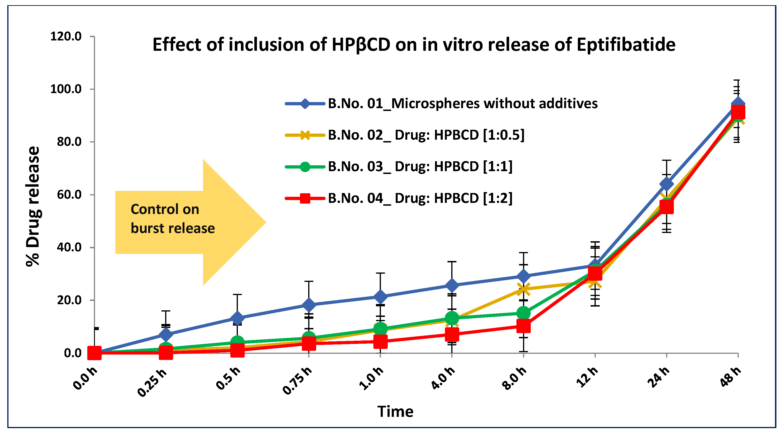
Complexing the drug with HPβCD resulted in good entrapment (>85%) at all the ratios studied. Its presence also helped reduce the free drug content. HPβCD not only led to good drug entrapment but also controlled the unwanted initial burst release. For many years, HPβCD has been known to enhance solubility for poorly soluble drugs [20]. This cyclic oligosaccharide comprises many dextrose units of (α-1,4)-linked α-D-glucopyranose. This structure gives it a lipophilic center and hydrophilic outer surface. The inclusion of HPβCD helped improve the drug entrapment from 75% to 89% during manufacturing. This encapsulation enhancement can be attributed to HPβCD forming a strong bond with eptifibatide by enclosing it in its hydrophobic central core. HPβCD formed a barrier and prevented the partition of eptifibatide into the external phase. This bonding also prevented the initial burst release of eptifibatide, as is observed in Figure 1 [21]. Please note that no interaction study between the drug and HPβCD were performed. Table 3 and Table 4 detail the analytical data for trials conducted with additives in the drug phase and with additives in the polymer phase, respectively.
Figure 1.
Comparison of in vitro release profiles of eptifibatide microspheres with different ratios of drug to HPβCD.

Table 3.
Analytical data for trials conducted to formulate eptifibatide microspheres with the inclusion of additives in the drug phase.

Table 4.
Analytical data for trials conducted to formulate eptifibatide microspheres with the inclusion of additives in the polymer phase.
Including Na CMC also provided good entrapment; however, little effect on the release profile was observed. Na CMC is a viscosity-increasing agent. Its inclusion in the eptifibatide solution caused an increase in the viscosity of the solution and lowered the partitioning of eptifibatide into the external aqueous phase, thus improving its entrapment. Microspheres are particulate drug delivery systems that must be reconstituted with a diluent before administration. Including Na CMC in the formulation prevents the aggregation of the microspheres because of its negative charge. Thus, the microsphere form a uniform suspension and can be quickly injected to avoid syringeability and injectability issues. Na CMC is also known to have wound-healing activity. Its inclusion as an additive can provide a beneficial effect when immediate wound healing is needed [22]. As such, no impact was observed on drug entrapment with the inclusion of trehalose. Please note that the determination of zeta potential to measure the negative charge on the microspheres was not performed in this study.
3.2. Particle Size Analysis
Little difference was observed in the particle size of the formulated microspheres when different additives were included. Table 3 and Table 4 detail the particle size data from trials conducted with additives included in the drug phase and with additives included in the polymer phase, respectively. The particle size did not change based on whether the different additives were added to the drug or polymer phase. The amounts of solvent used to dissolve both the drug and additives in the drug phase and in the polymer phase were adjusted to prevent any precipitation of the dissolved components and to keep the viscosity of the solution low/manageable. Maintaining the same solution viscosity and the same homogenization speed likely resulted in microspheres with similar particle sizes.
3.3. In Vitro Accelerated Drug Release
The inclusion of HPβCD in the drug phase helped control the initial burst, as shown in Figure 1. The drug’s release profile from microspheres is biphasic [23]. The initial release, which occurred immediately after injection, was due to the loosely bound drug on the surface and in the superficial pores of the microspheres, i.e., the free drug content. The second phase of release, which was more controlled and gradual, was due to the degradation of the polymer [23]. The presence of HPβCD in the formulation reduced the burst release and made it more gradual compared to the batch without HPβCD, likely because of the complex/bonds formed between the drug and HPβCD. A study on the interaction between the drug and HPβCD was not performed. Including Na CMC and trehalose did not significantly affect the release profile; however, improved drug loading was achieved when Na CMC was used.
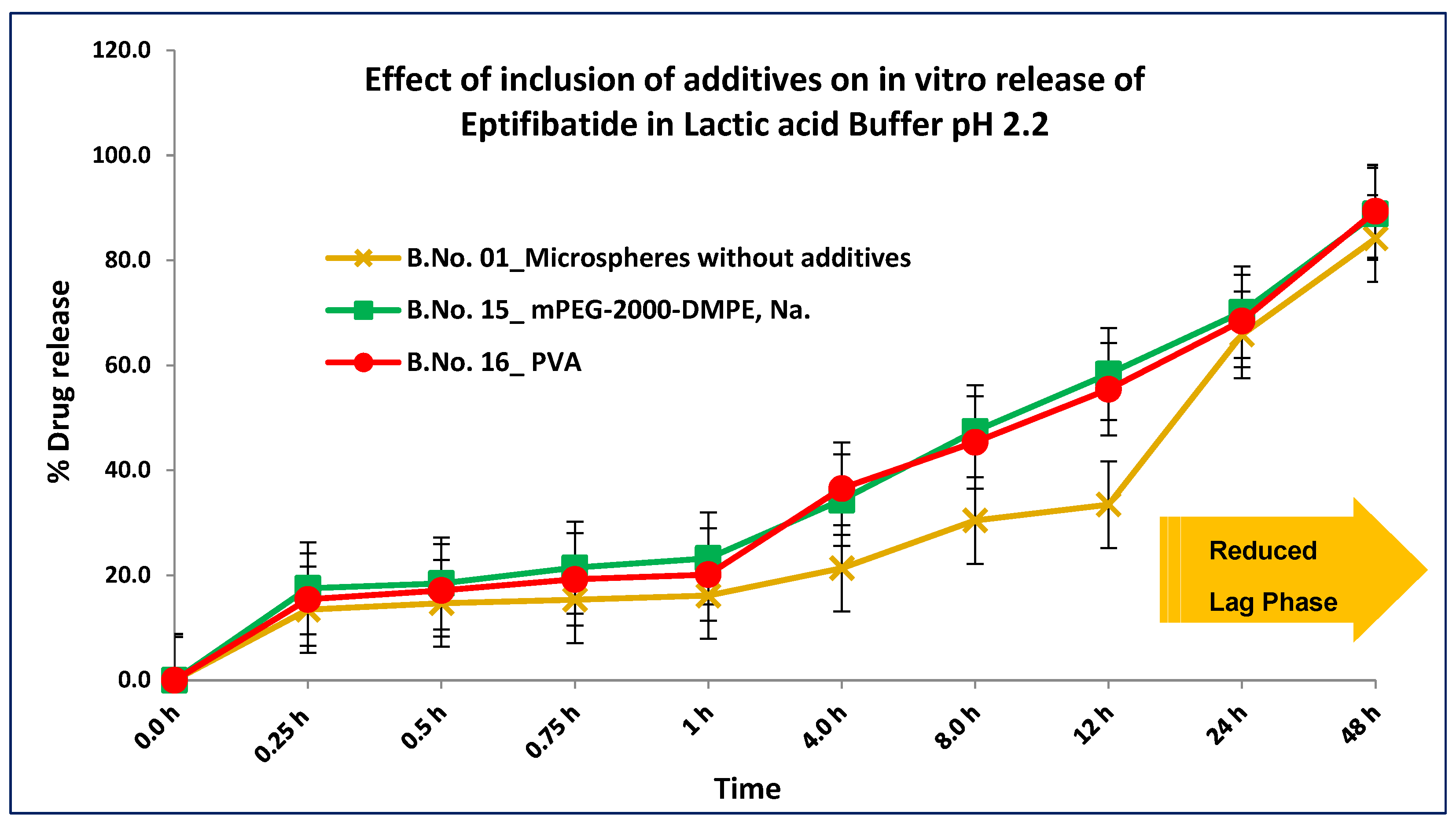
PLGA polymers are hydrophobic and show a long lag time during in vitro release [24]. PVA is used as a surfactant in the solvent evaporation method. Complexing this surfactant with a biodegradable polymer affected the wettability of the polymer and significantly improved the initial lag in the release profile (see Figure 2). The presence of PVA reduced the lag phase by increasing polymer wetting. Including mPEG-2000-DMPE, Na had a positive effect by controlling the release at later time points. mPEG-2000-DMPE, Na is known for its use in conjugation with proteins due to its single hydroxyl group, which can bind to other groups [25]. It is well known for its use in liposomes to prolong their presence in the blood. PEG 400 is a plasticizer in oral, topical, and parenteral formulations, and its inclusion also improved the stability and release profile of polymeric microparticles [26]. PEG 400, which does not promote microbial growth, can prove helpful in formulations that need to have a low bioburden or be sterile [27]. It is used in amounts of up to 30% v/v in parenteral formulations. PVA and PEG 400, both plasticizers, helped wet the polymer. The lag phase that is usually observed with these polymers was reduced, which is beneficial.
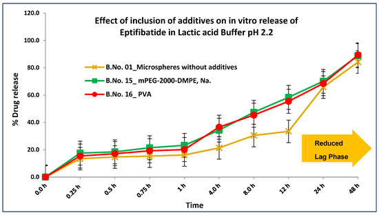
Figure 2.
Comparison of in vitro release profiles of eptifibatide microspheres: effect of inclusion of additives on lag phase.
3.4. Surface Morphology
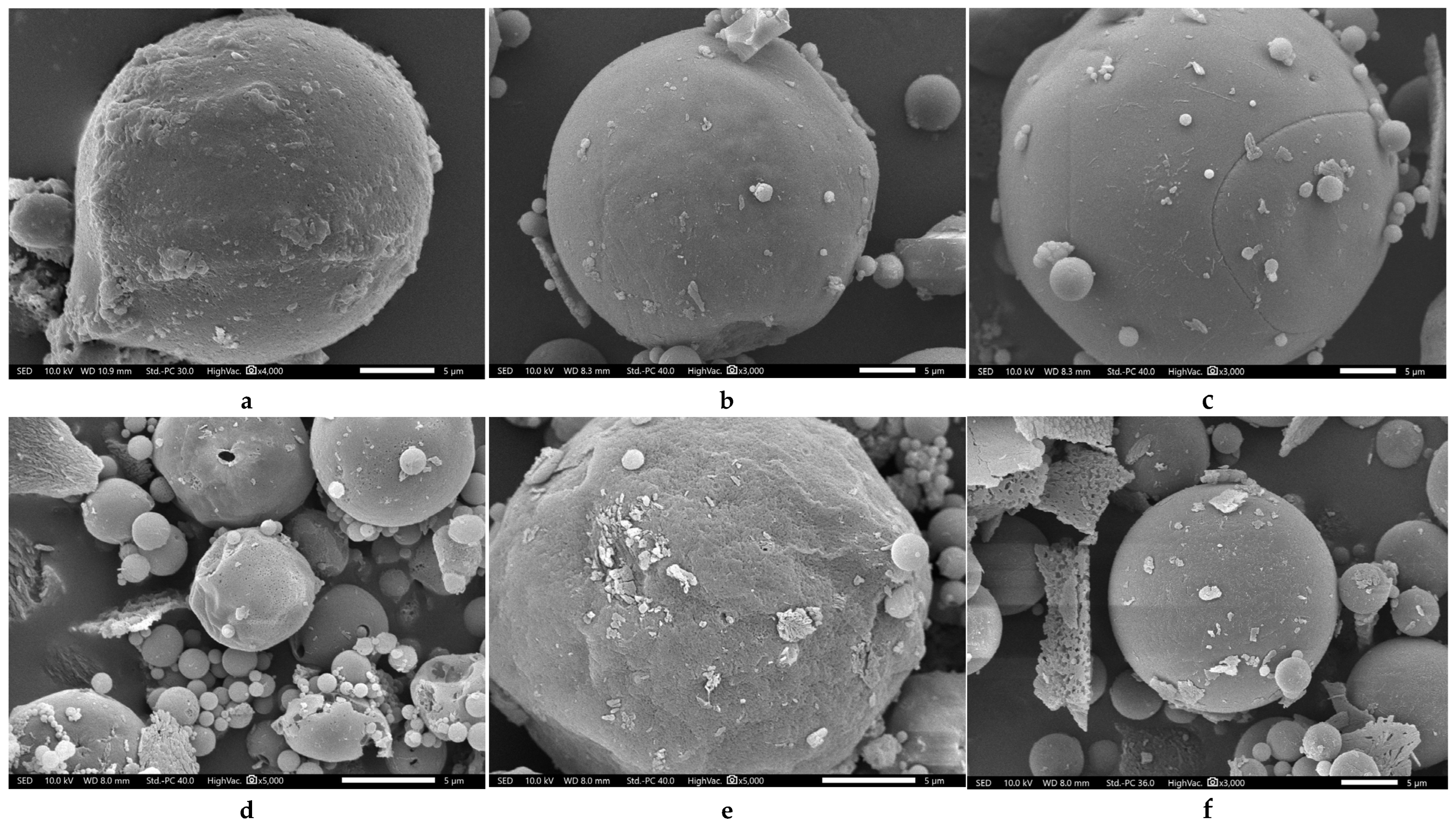
Microspheres formulated using HPβCD were slightly porous, presumably because HPβCD partitioned along with the polymer into the external phase due to the hydrophilic outer core (Figure 3a). Trehalose and Na CMC thus did not have an effect on the surface morphology of the formulated microspheres, as they were both confined to the interior and must not have partitioned into the external phase during formulation (Figure 3b,c). On the other hand, microspheres formulated using PVA and PEG400 were more porous, as these additives functioned as wetting agents and allowed the immediate ingress of the external aqueous phase into the microspheres (Figure 3d,e). mPEG-2000-DMPE, Na, being lipophilic, remained confined and bonded with the polymer, resulting in less porous microspheres (Figure 3f).
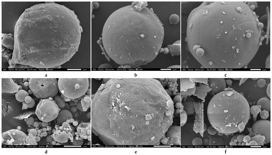
Figure 3.
SEM images of eptifibatide microspheres formulated using different additives (scale bar: 5 µm). (a) HPβCD, giving a more porous surface; (b) trehalose, with a less porous surface; (c) NaCMC, again with a less porous surface; (d) PVA, with a highly porous surface; (e) PEG 400, again with a highly porous surface; and (f) mPEG-2000-DMPE, Na, with a much less porous surface.
3.5. Drug Stability
Maintaining the activity of the entrapped drug is of utmost importance when formulating PLGA microspheres, especially when the drug to be entrapped is a protein or peptide. During the formulation of these PLGA microspheres, the drug is exposed to harsh conditions, such as organic solvents, excessive attrition, and high temperatures. It is essential to monitor and control the manufacturing procedure and use appropriate ingredients to prevent the denaturation of the entrapped protein. Degradation of the drug also occurs due to the acidic microenvironment created by the degradation of PLGA polymers. The addition of additives is one of the ways by which the denaturation of entrapped drugs can be prevented. The addition of additives in the correct phase is essential. Two additives proved to stabilize eptifibatide and provide other beneficial effects. In addition to reducing the burst effect, HPβCD stabilized the entrapped eptifibatide by preventing its exposure to organic solvents, excessive temperature, and attrition used during manufacturing and the acidic microenvironment. Trehalose helped to prevent the denaturation of eptifibatide during the lyophilization phase by preventing aggregation. Trehalose is a non-reducing disaccharide with two glucose molecules linked by an α, α–1,1 glycosidic bond and has been used as a stabilizing agent in pharmaceutical products [28]. Trehalose’s glass transition temperature (Tg) is between 110 °C and 120 °C, which is exceptionally high compared with other sugars. Eptifibatide is a peptide known to undergo structural changes due to organic solvents and harsh manufacturing conditions, including lyophilization. It was decided to evaluate the advantage of including trehalose in the formulation. The presence of trehalose in the formulation did not have much effect on the drug loading. However, its presence stabilized eptifibatide, which was evaluated by performing lyophilization of the conventional eptifibatide injection liquid form and comparing it with eptifibatide-loaded microspheres containing trehalose. The currently marketed liquid injection of eptifibatide must be stored at 2–8 °C, so the lyophilized dry powder for the solution was evaluated for the percent drop in drug content. As shown in Table 5, a significant drop in drug content was observed after lyophilization of the conventional injection (Batch No. 23) when it was lyophilized without the addition of trehalose, whereas a slight drop of 2% was observed in Batch No. 22 when it was lyophilized in the presence of trehalose. Thus, the presence of trehalose during lyophilization proved to stabilize eptifibatide. However, when the same batch was compared with microspheres containing trehalose (Batch No. 8), a drop in drug content was observed after 3 months. This is supported by the fact that the drug is enclosed inside the microspheres, and additional stability is provided by trehalose. Thus, eptifibatide microspheres can be stored at ambient temperatures, in contrast to the conventional injection, which requires storage at 2–8 °C.
Although not discussed in the main body of this research paper, the residual solvent content of the formulated microspheres is an important consideration, as the drug is known to undergo structural changes in the presence of certain solvents. Relevant data are provided in the Supplementary Materials accompanying this publication for interested readers.

Table 5.
Percent drug degradation in stability studies conducted at ambient temperature.
Table 5.
Percent drug degradation in stability studies conducted at ambient temperature.
| Details of Additives | MSP | MSP, Trehalose | Con. Inj, Trehalose | Con. Inj, Trehalose | Con. Inj. | ||||||||
|---|---|---|---|---|---|---|---|---|---|---|---|---|---|
| Batch No. | 1 | 8 | 22 | 22 | 23 | ||||||||
| Stability Storage Conditions | 25 °C and 60% RH | 25 °C and 60% RH | 25 °C and 60% RH | Not applicable | Not applicable | ||||||||
| Stability Time Point | Initial | 1 M | 3 M | Initial | 1 M | 3 M | Initial | 1 M | 3 M | BL | AL | BL | AL |
| Drug Content (%w/w) | 96.3 | 95.3 | 95.1 | 97.4 | 96.8 | 96.4 | 95.1 | 90.1 | 88.6 | 97.2 | 95.1 | 98.3 | 83.1 |
MSP: plain microspheres without any additives; MSP, Trehalose: microspheres containing trehalose as an additive in the drug phase; Con. Inj. Trehalose: conventional liquid injection containing trehalose added during lyophilization. 1 M and 3 M: 1 and 3 months; Con. Inj.: conventional liquid injection without trehalose during lyophilization; BL and AL: before and after lyophilization.
4. Conclusions
Biodegradable-polymer-based microspheres are novel drug delivery systems with enormous potential to successfully deliver therapeutic proteins and peptides. However, several challenges have limited their application. The inclusion of additives proved to address some of these challenges, such as drug loading, drug release, and drug stability. The inclusion of HP-β-CD did result in good loading and also helped to significantly reduce the initial unwanted burst release. A similar effect on drug loading was observed when Na CMC was used. The inclusion of trehalose imparted stability to the drug during manufacturing and lyophilization. Eptifibatide-loaded microspheres as a drug delivery system were found to be stable when stored at ambient temperature, in contrast to the conventional liquid formulation, which requires storage in cold conditions. Including additives such as PVA; PEG 400; and mPEG-2000-DMPE, Na proved to help reduce the initial lag observed with microspheres and facilitated a gradual release at later time points, thus providing a desired release profile.
This research demonstrates that by using suitable additives in the drug phase or the polymer phase, long-known challenges associated with microspheres as drug delivery systems can be addressed to some extent. Using the appropriate manufacturing process and additives can provide a positive outcome and is an excellent practical approach to increasing the drug load, drug stability, and release profile. More research in this direction is needed to maximally utilize the full potential of these drug delivery systems. This approach can also be tested with other controlled and targeted drug delivery systems.
Supplementary Materials
The following supporting information can be downloaded at: https://www.mdpi.com/article/10.3390/jpbi2020008/s1, Table S1: Residual solvent content for trials conducted using the inclusion of additives.
Author Contributions
Conceptualization, A.K.; investigation, A.K.; methodology, A.K.; supervision, B.P.; validation, B.P.; writing—original draft preparation, A.K.; writing—review and editing, B.P. All authors have read and agreed to the published version of the manuscript.
Funding
This research received neither internal nor external funding.
Institutional Review Board Statement
Not applicable.
Informed Consent Statement
Not applicable.
Data Availability Statement
The dataset presented in this article is not readily available because the studies were conducted at an R&D center. As per company policy, raw data cannot be made available unless on reasonable request. Request to access the dataset should be directed to the corresponding author.
Acknowledgments
The authors are thankful to BSVL and NMIMS for the administrative and lab support provided during the research work, which is part of the project of the doctoral degree [29] of Anand Kyatanwar under the supervision of Bala Prabhakar.
Conflicts of Interest
Anand Kyatanwar is working as a Senior Principal Scientist at Bharat Serums and Vaccines Ltd. R&D Center. This project was an independent work of Anand Kyatanwar and is not related to any ongoing company projects. The authors declare no conflicts of interest.
References
- Lee, A.C.-L.; Harris, J.L.; Khanna, K.K.; Hong, J.-H. A Comprehensive Review on Current Advances in Peptide Drug Development and Design. Int. J. Mol. Sci. 2019, 20, 2383. [Google Scholar] [CrossRef]
- Saeed, S.; Irfan, M.; Naz, S.; Liaquat, M.; Jahan, S.; Hayat, S. Routes and barriers associated with protein and peptide drug delivery system. J. Pak. Med. Assoc. 2021, 71, 2032–2039. [Google Scholar] [CrossRef]
- Ibeanu, N.; Egbu, R.; Onyekuru, L.; Javaheri, H.; Khaw, P.T.; Williams, G.R.; Brocchini, S.; Awwad, S. Injectables and Depots to Prolong Drug Action of Proteins and Peptides. Pharmaceutics 2020, 12, 999. [Google Scholar] [CrossRef] [PubMed]
- Su, Y.; Zhang, B.; Sun, R.; Liu, W.; Zhu, Q.; Zhang, X.; Wang, R.; Chen, C. PLGA-based biodegradable microspheres in drug delivery: Recent advances in research and application. Drug Deliv. 2021, 28, 1397–1418. [Google Scholar] [CrossRef] [PubMed]
- Huang, L.; Wang, S.; Yin, Z. Study in the stabilization of proteins encapsulated in PLGA delivery system: Effects of additives on protein encapsulation, release, and stability. J. Drug Deliv. Sci. Technol. 2022, 73, 103436. [Google Scholar] [CrossRef]
- Matsumoto, A.; Matsukawa, Y.; Nishioka, Y.; Harada, M.; Horikiri, Y.; Yamahara, H. A new method of preparing TRH derivative-loaded poly(dl-lactide-coglycolide) microspheres based on a solid solution system. Drug Discov. Ther. 2008, 2, 45–51. [Google Scholar]
- Li, W.; Chen, J.; Zhao, S.; Huang, T.; Ying, H.; Trujillo, C.; Molinaro, G.; Zhou, Z.; Jiang, T.; Liu, W.; et al. High drug-loaded microspheres enabled by controlled in-droplet precipitation promote functional recovery after spinal cord injury. Nat. Commun. 2022, 13, 1262. [Google Scholar] [CrossRef]
- Parikh, R.H.; Parikh, J.R.; Dubey, R.R.; Soni, H.N.; Kapadia, K.N. Poly(D,L-Lactide-Co-Glycolide) microspheres containing 5-fluorouracil: Optimization of process parameters. AAPS PharmSciTech 2003, 4, 14–21. [Google Scholar] [CrossRef]
- Ding, A.G.; Schwendeman, S.P. Acidic Microclimate pH Distribution in PLGA Microspheres Monitored by Confocal Laser Scanning Microscopy. Pharm. Res. 2008, 25, 2041–2052. [Google Scholar] [CrossRef]
- Park, K.; Otte, A.; Sharifi, F.; Garner, J.; Skidmore, S.; Park, H.; Jhon, Y.K.; Qin, B.; Wang, Y. Formulation composition, manufacturing process, and characterization of poly(lactide-co-glycolide) microparticles. J. Control. Release 2021, 329, 1150–1161. [Google Scholar] [CrossRef]
- Zhang, Y.; Sophocleous, A.M.; Schwendeman, S.P. Inhibition of Peptide Acylation in PLGA Microspheres with Water-soluble Divalent Cationic Salts. Pharm. Res. 2009, 26, 1986–1994. [Google Scholar] [CrossRef][Green Version]
- Kang, F.; Singh, J. Effect of additives on the release of a model protein from PLGA microspheres. AAPS PharmSciTech 2001, 2, 86–92. [Google Scholar] [CrossRef]
- Lin, X.; Yang, H.; Su, L.; Yang, Z.; Tang, X. Effect of size on the in vitro/in vivo drug release and degradation of exenatide-loaded PLGA microspheres. J. Drug Deliv. Sci. Technol. 2018, 45, 346–356. [Google Scholar] [CrossRef]
- Zheng, C.; Liang, W. A one-step modified method to reduce the burst initial release from PLGA microspheres. Drug Deliv. 2010, 17, 77–82. [Google Scholar] [CrossRef]
- Srikanth, S.; Ambrose, J.A. Ambrose Pathophysiology of Coronary Thrombus Formation and Adverse Consequences of Thrombus During PCI. Curr. Cardiol. Rev. 2012, 8, 168–176. [Google Scholar] [CrossRef]
- Iqbal, A.M.; Lopez, R.A.; Hai, O. “Antiplatelet Medications,” StatPearls [Internet]; StatPearls Publishing: Treasure Island, FL, USA, 2025. Available online: https://pubmed.ncbi.nlm.nih.gov/30725747/ (accessed on 15 February 2025).
- Fischell, T. Eptifibatide: The evidence for its role in the management of acute coronary syndromes. Core Evid. 2009, 4, 49–65. [Google Scholar] [CrossRef]
- King, S.; Short, M.; Harmon, C. Glycoprotein IIb/IIIa inhibitors: The resurgence of tirofiban. Vasc. Pharmacol. 2016, 78, 10–16. [Google Scholar] [CrossRef]
- Eikelboom, J.W.; Hirsh, J. Combined antiplatelet and anticoagulant therapy: Clinical benefits and risks. J. Thromb. Haemost. 2007, 5, 255–263. [Google Scholar] [CrossRef]
- Malanga, M.; Szemán, J.; Fenyvesi, É.; Puskás, I.; Csabai, K.; Gyémánt, G.; Fenyvesi, F.; Szente, L. ‘Back to the Future’: A New Look at Hydroxypropyl Beta-Cyclodextrins. J. Pharm. Sci. 2016, 105, 2921–2931. [Google Scholar] [CrossRef]
- Carneiro, S.B.; Costa Duarte, F.Í.; Heimfarth, L.; Siqueira Quintans, J.D.S.; Quintans-Júnior, L.J.; Veiga Júnior, V.F.D.; De Lima, A.N. Cyclodextrin–Drug Inclusion Complexes: In Vivo and In Vitro Approaches. Int. J. Mol. Sci. 2019, 20, 642. [Google Scholar] [CrossRef]
- Basu, P.; Narendrakumar, U.; Arunachalam, R.; Devi, S.; Manjubala, I. Characterization and Evaluation of Carboxymethyl Cellulose-Based Films for Healing of Full-Thickness Wounds in Normal and Diabetic Rats. ACS Omega 2018, 3, 12622–12632. [Google Scholar] [CrossRef] [PubMed]
- Fredenberg, S.; Wahlgren, M.; Reslow, M.; Axelsson, A. The mechanisms of drug release in poly(lactic-co-glycolic acid)-based drug delivery systems—A review. Int. J. Pharm. 2011, 415, 34–52. [Google Scholar] [CrossRef] [PubMed]
- Butreddy, A.; Gaddam, R.P.; Kommineni, N.; Dudhipala, N.; Voshavar, C. PLGA/PLA-Based Long-Acting Injectable Depot Microspheres in Clinical Use: Production and Characterization Overview for Protein/Peptide Delivery. Int. J. Mol. Sci. 2021, 22, 8884. [Google Scholar] [CrossRef] [PubMed]
- Pasut, G.; Paolino, D.; Celia, C.; Mero, A.; Joseph, A.S.; Wolfram, J.; Cosco, D.; Schiavon, O.; Shen, H.; Fresta, M. Polyethylene glycol (PEG)-dendron phospholipids as innovative constructs for the preparation of super stealth liposomes for anticancer therapy. J. Control. Release 2015, 199, 106–113. [Google Scholar] [CrossRef]
- Péan, J.; Boury, F.; Venier-Julienne, M.; Menei, P.; Proust, J.; Benoit, J. Why does PEG 400 co-encapsulation improve NGF stability and release from PLGA biodegradable microspheres? Pharm. Res. 1999, 16, 1294–1299. [Google Scholar] [CrossRef]
- Chirife, J.; Herszage, L.; Joseph, A.; Bozzini, J.P.; Leardini, N.; Kohn, E.S. In vitro antibacterial activity of concentrated polyethylene glycol 400 solutions. Antimicrob. Agents Chemother. 1983, 24, 409–412. [Google Scholar] [CrossRef]
- Jain, N.K.; Roy, I. Effect of trehalose on protein structure. Protein Sci. 2009, 18, 24–36. [Google Scholar] [CrossRef]
- Kyatanwar, A. Development of Controlled Release Parenteral Formulation of An Antiplatelet Drug Eptifibatide. Ph.D. Thesis, Narsee Monjee Institute of Management Studies, Mumbai, India, June 2023. [Google Scholar]
Disclaimer/Publisher’s Note: The statements, opinions and data contained in all publications are solely those of the individual author(s) and contributor(s) and not of MDPI and/or the editor(s). MDPI and/or the editor(s) disclaim responsibility for any injury to people or property resulting from any ideas, methods, instructions or products referred to in the content. |
© 2025 by the authors. Licensee MDPI, Basel, Switzerland. This article is an open access article distributed under the terms and conditions of the Creative Commons Attribution (CC BY) license (https://creativecommons.org/licenses/by/4.0/).

