Article

Urinary Neutrophil Gelatinase-Associated Lipocalin as a Predictor of COVID-19 Mortality in Hospitalized Patients

1
Clinic for Infectious Diseases, University Hospital Centre Osijek, 31000 Osijek, Croatia
2
Department of Infectology and Dermatovenerology, Faculty of Medicine Osijek, J. J. Strossmayer University of Osijek, 31000 Osijek, Croatia
3
Faculty of Medicine Osijek, J. J. Strossmayer University of Osijek, 31000 Osijek, Croatia
4
Department of Medical Statistics and Medical Informatics, Faculty of Medicine Osijek, J. J. Strossmayer University of Osijek, 31000 Osijek, Croatia
5
Clinic for Pediatrics, University Hospital Centre Osijek, 31000 Osijek, Croatia
6
Department of Pediatrics, Faculty of Medicine Osijek, J. J. Strossmayer University of Osijek, 31000 Osijek, Croatia
7
Faculty of Dental Medicine and Health Osijek, J. J. Strossmayer University of Osijek, 31000 Osijek, Croatia
8
Polyclinic LabPlus, 31000 Osijek, Croatia
9
Department of Chemistry, Biochemistry and Clinical Chemistry, Faculty of Medicine Osijek, J. J. Strossmayer University of Osijek, 31000 Osijek, Croatia
*
Authors to whom correspondence should be addressed.
Acta Microbiol. Hell. 2024, 69(4), 224-235; https://doi.org/10.3390/amh69040021
Submission received: 6 September 2024 / Revised: 28 September 2024 / Accepted: 15 October 2024 / Published: 17 October 2024

Abstract

:
Neutrophil gelatinase-associated lipocalin (NGAL) is a protein secreted by activated neutrophils and certain tissues. The aim of this study was to investigate the prognostic role of urinary neutrophil gelatinase-associated lipocalin (uNGAL) in hospitalized COVID-19 patients with regard to mortality. The prospective observational cohort study involved 86 hospitalized adult COVID-19 patients. Patients’ urine samples were collected upon admission and 48–72 h after admission. General anamnesis and clinical status information were obtained from medical records. Within 24 h of sample collection, urine aliquots were centrifuged, the supernatant was separated, and samples were frozen at −80 °C until uNGAL concentration measurement, which was performed within two years of sample collection. The patients were categorized into groups based on disease outcome (survived/deceased). Data analysis was performed to identify predictive factors and diagnostic indicators for the unfavorable outcome group using logistic regression and ROC curve methods. Logistic regression associated age, cardiomyopathy, invasive mechanical ventilation, and uNGAL concentration (follow-up sampling) with mortality. ROC analysis identified uNGAL concentration (follow-up sampling) as an indicator of mortality, with a cut-off value of >23.8 ng/mL. This study concludes that there is an association between disease outcome and uNGAL concentration in COVID-19 patients. However, understanding the dynamics of uNGAL concentration during COVID-19 remains limited, which is crucial for its effective clinical application.

1. Introduction

Coronavirus disease 2019 (COVID-19) is an illness caused by severe acute respiratory syndrome coronavirus 2 (SARS-CoV-2) which is primarily transmitted via respiratory routes. This virus is characterized by its rapid spread and pandemic potential [1,2]. Before the World Health Organization declared an end to the global COVID-19 emergency in May 2023, the disease had caused an estimated 15 million excess deaths in 2020 and 2021 alone [3]. Considering that SARS-CoV-2 continues to emerge sporadically, but globally, throughout the year and is likely to persist in the population, further research is required to gather useful knowledge about COVID-19.
Neutrophil gelatinase-associated lipocalin (NGAL) is a protein secreted by activated neutrophils and certain tissues, including renal tubules, cardiomyocytes, lungs, liver, stomach, intestines, adipocytes, macrophages, and others [4,5,6,7]. NGAL has been most extensively studied as an early and highly sensitive biomarker of acute kidney injury, detectable in both serum and urine [4,5,6,7]. The primary advantage of NGAL in detecting acute kidney injury is its rapid increase in serum and urine levels, which can be seen approximately 24 h before the rise in creatinine levels, a marker commonly used in clinical practice [4,5,6,7]. During infection, NGAL acts as an acute phase reactant and promotes an anti-inflammatory response. In addition to infections, NGAL concentrations are altered in acute and chronic metabolic, malignant, and cardiovascular diseases [4,8].
Previous studies have connected elevated levels of NGAL with worse clinical outcomes or the development of coagulopathy in patients suffering from COVID-19 [9,10,11,12]. Other studies have shown that NGAL is a reliable biomarker for the development of acute kidney injury during COVID-19 [13,14,15]. Contrary to these findings, NGAL has not proven to be an early marker for the development of kidney failure in cohorts of critically ill COVID-19 patients. However, higher NGAL concentrations may suggest that the interval from NGAL concentration measurement to the development of acute kidney injury is shorter than in patients with lower NGAL concentrations [16]. Some studies do not associate elevated NGAL levels with the prediction of negative outcomes or the development of acute kidney injury in COVID-19 patients [17,18].
This study was designed as a prospective observational cohort study and aimed to investigate the prognostic significance of urinary NGAL in COVID-19 patients with regard to early mortality. Our hypothesis is that elevated urinary concentrations of NGAL in hospitalized patients are positively correlated with mortality in COVID-19 patients.

2. Participants and Methods

2.1. Ethical Approval

The study was conducted at the Faculty of Medicine Osijek and the University Hospital Centre Osijek, Croatia. Ethical approval for this research was obtained from the Ethics Committee of the Faculty of Medicine Osijek. Before participating in the study, patients or their legal representatives provided informed consent (written consent after disclosure), and the research was conducted in accordance with the Declaration of Helsinki.

2.2. Participants

Study participants were patients aged 18 and older who were initially hospitalized at the Clinic for Infectious Diseases of the University Hospital Centre Osijek from 30 December 2021 to 12 February 2022. The inclusion criteria for the study were hospitalization, clinical characteristics consistent with COVID-19, and a positive reverse transcription polymerase chain reaction (RT-PCR) test result for SARS-CoV-2 from a nasopharyngeal swab sample. Patients who did not survive 72 h after admission were excluded from the study, as were those who were transferred to the Intensive Care Unit (ICU) or discharged to home care within the first 72 h of hospitalization (exclusion criteria). To detect differences in continuous variables between two measurements with an effect size (f) of 0.25, a test power of 0.95, and a significance level of 0.05, the minimum sample size was determined to be 54 patients. Additionally, for regression analysis with a test power of 80%, the minimum required sample size was 56 patients. Consequently, the established minimum sample size for the study was 56 patients (G*Power, 3.1.9.7, Heinrich-Heine-Universität Düsseldorf, Düsseldorf, Germany).
After selecting patients who fulfilled the inclusion criteria and excluding those who met the exclusion criteria, the study ultimately included 86 patients. All patients were observed from hospital admission until discharge or death.

2.3. Methods

2.3.1. Sampling

All the following sampling, preparation, and processing of samples, as well as radiological examinations, were conducted at the University Hospital Centre Osijek or at the Faculty of Medicine, University of Osijek, Croatia.
Before hospital admission, all patients underwent a nasopharyngeal swab, and SARS-CoV-2 RNA was detected using the RT-PCR method. After patient admission, random urine samples were immediately collected from each participant in the study. A follow-up random urine sample was also collected 48 to 72 h after the initial sample.
Urine samples were collected in a URINtainer® (F. L. Medical s.r.l., Torreglia, Italy) and transferred to a centrifuge tube. Urine aliquots were centrifuged for 15 min at 13,000× g. The supernatant was separated, the samples were packaged and labeled in accordance with applicable local, national, and international regulations for the transport of clinical samples and infectious substances, and then frozen at −80 °C, until measurement of uNGAL concentration, which was performed within two years of sample collection.

2.3.2. Chemiluminescent Microparticle Immunoassay

uNGAL concentrations were measured in the supernatant with a chemiluminescent microparticle immunoassay (CMIA) using an Alinity i immunoassay analyzer (Abbott Diagnostics, Lake Forest, IL, USA) following the manufacturer’s instructions. Throughout the experimental work, all mandatory laboratory safety and health procedures were strictly followed.

2.3.3. Radiological Examinations

Furthermore, upon admission to hospital care, a chest X-ray was performed for each patient included in the study. The purpose of the chest X-ray was to confirm or exclude pneumonia radiologically, within the limitations of this imaging method.

2.3.4. Patient Data Collection

In addition to these measurements, data were collected from the patients’ medical records, including age, sex, comorbidities (classified into the following groups after data collection: type 2 diabetes mellitus, arterial hypertension, cardiomyopathy, atrial fibrillation, chronic lung diseases, chronic kidney diseases), clinical signs and symptoms at admission, systolic and diastolic blood pressure, peripheral blood oxygen saturation, pulse rate, respiratory rate, length of hospitalization, need for ICU treatment at any point during hospitalization, and disease outcome (discharge home in clinically stable condition or death). Additionally, the need for conventional oxygen therapy, high-flow nasal cannula (HFNC), and/or invasive mechanical ventilation (IMV) during hospitalization was documented. Disease severity was calculated according to the World Health Organization criteria [19].
For each patient, the mean arterial pressure (MAP) was calculated using the formula:
MAP = (systolic blood pressure + 2 × diastolic blood pressure)/3
The body mass index (BMI) was also calculated using the formula: BMI = body mass/(height2).

2.4. Statistical Methods

Categorical data were presented as absolute and relative frequencies. Differences in categorical variables were assessed using the Chi-square test or Fisher’s exact test, as appropriate. The normality of continuous variables was evaluated with the Shapiro–Wilk test. Continuous data were summarized using the median and interquartile range (IQR). Differences between two independent groups for continuous variables were analyzed using the Mann–Whitney U test. Logistic regression analysis (bivariate and multivariate, employing a stepwise method) was conducted to identify independent factors associated with the outcome. In addition, Cox and Snell R2 was calculated to assess the proportion of variance explained by the predictors. Although limited by values less than 1, this measure provides a conservative estimate of model fit. For comparison, the Nagelkerke R2 was also reported, which adjusts the Cox and Snell value to span the full range from 0 to 1. The receiver operating characteristic (ROC) curve was utilized to determine the optimal threshold, the area under the curve (AUC), and the specificity and sensitivity of the parameters tested. The Youden Index was employed to identify the optimal cut-off, balancing sensitivity and specificity to maximize overall diagnostic accuracy. All p-values were two-tailed, and statistical significance was set at an alpha level of 0.05. Statistical analyses were conducted using MedCalc® software version 22.018 (MedCalc Software Ltd., Ostend, Belgium; https://www.medcalc.org, accessed on 20 August 2024) and SPSS version 23.0 (IBM Corp., Armonk, NY, USA; released 2015).
The research report was prepared following the guidelines for reporting research results in biomedicine and health (EQUATOR Network) [20].

3. Results

3.1. Clinical Features of Patients

This cohort consisted primarily of female patients (57%). The most common conditions were chronic cardiac diseases, with hypertension being the most prevalent (72%), followed by cardiomyopathy (24%). Patients most frequently reported cough (85%), fever (75%), and general weakness (67%). Nearly all patients (95%) had pneumonia at admission (Table S1).
Regarding outcomes, 58 patients (67%) recovered. Approximately one-quarter of the patients (24%) were admitted to the ICU (Table 1).

3.2. Factors Associated with Mortality

Death outcome was associated with critical disease (χ2 test, p < 0.001), as well as with cardiomyopathy (χ2 test, p = 0.03) and age (Table 2 and Table 3).
uNGAL concentration was higher in patients who did not survive, with statistical significance found only in follow-up sampling concentrations (Mann–Whitney U test, p < 0.001) (Table 4). uNGAL demonstrated multiple correlations with other observed variables across the observed subgroups (Table S2).

3.3. Bivariate and Multivariate Logistic Regression with ROC Analysis

To assess the impact of uNGAL, age, comorbidities, and complications on mortality in hospitalized COVID-19 patients, both bivariate and multivariate logistic regression analyses were performed. Independent factors were clinically significant variables that showed a change concerning mortality. The results of the bivariate analysis are presented in Table 5. Using multivariate logistic regression (stepwise method), a significant model for predicting mortality was established. Factors that increase the likelihood of mortality included older age (OR = 1.12), higher follow-up concentration of uNGAL (OR = 1.01), and the necessity for IMV (OR = 159.4). This model explains between 50% (according to Cox and Snell R2) and 70% (according to Nagelkerke R2) of the variance in mortality and correctly classifies 90% of cases.
The ROC curve method was chosen as a simple way to assess the differences in individual factors between patient groups with respect to mortality, based on specificity and sensitivity. To evaluate the diagnostic value of variables that previous tests identified as significant predictors of a negative outcome, the ROC curve calculation was used. This method incrementally adjusts the values distinguishing between patients who survived and those who died. The cut-off point was tailored for each patient group to determine, using the ROC curve, the value that best differentiates the groups and maximizes both sensitivity and specificity. The ROC analysis identified one significant diagnostic indicator for mortality: uNGAL concentration in follow-up sampling (AUC = 0.717) (Table 6 and Figure 1).

4. Discussion

This research examined the prognostic significance of uNGAL in hospitalized patients with COVID-19. Notably, elevated uNGAL concentrations (>23.8 mg/mL) measured during follow-up demonstrated prognostic value concerning death outcome, whereas uNGAL concentrations from samples collected at admission did not. This suggests that the research hypothesis, which stated that elevated urinary concentrations of NGAL in hospitalized patients are positively correlated with mortality in COVID-19 patients, was partially accepted. Although this study has some limitations, which are discussed in detail in the following sections, we believe its scientific contribution lies in providing insight into the potential predictive role of uNGAL in COVID-19 mortality, depending on the timing of uNGAL concentration measurement throughout the disease course. This is in contrast to earlier studies, which assessed the relationship between NGAL and COVID-19 mortality without considering the time variable. Furthermore, no papers have been published from this specific region, and while there are some studies addressing the possible connection between COVID-19 mortality and NGAL, such studies remain scarce.
The observed patients were primarily older individuals, with a median age of 77 years. Both sexes were represented, with a higher proportion of female patients, comprising 57% of the cases. A considerable number (86%) of patients had at least one comorbidity, with cardiovascular comorbidities being the most common. Nearly all (95% of patients) developed pneumonia, predominantly bilateral, which often required oxygen supplementation. Considering that pneumonia is one of the leading causes of hospitalization among infectious disease patients in general, and particularly among COVID-19 patients, these results are not surprising, given that the observed group consisted of hospitalized patients. Previous studies consistently identify older age and the presence of comorbidities as significant risk factors for hospitalization among COVID-19 patients [21,22,23,24]. Almost all hospitalized patients, as expected, suffered from severe and critical forms of COVID-19 according to the World Health Organization classification [19], with the critical form of the disease almost always proving fatal. The need for IMV also proved fatal, which is expected since patients requiring IMV are typically in advanced stages of the disease.
In this study, uNGAL was found to be significantly associated with mortality, but this association was observed with uNGAL levels measured during follow-up sampling, not at admission. Furthermore, uNGAL was identified as a predictor (in both bivariate and multivariate regression, as well as in ROC analysis) of mortality, with all analyses using NGAL concentrations from follow-up sampling. These findings suggest that uNGAL levels may increase only when clinical deterioration occurs and the disease approaches its peak. Similarly, a study on critically ill COVID-19 patients found that higher NGAL levels could predict a shorter time to the development of acute kidney injury and disease worsening [16]. This suggests that elevated NGAL values are not necessarily an early biological indicator of disease severity but rather predict that severe disease will develop rapidly from the time of measurement. These results do not imply that NGAL is not useful in evaluating COVID-19; on the contrary, they suggest that NGAL has a limited predictive window. Therefore, it is important to direct further research towards understanding the dynamics of NGAL increase to better interpret the obtained findings.
Another question that arises is whether elevated levels of NGAL in severe cases of COVID-19 are caused by kidney damage or by the release of NGAL from neutrophils. Currently, this remains speculative. Some studies link elevated NGAL concentrations during COVID-19 to the development of acute kidney failure [13,14,15], while others associate them with mortality [9,11,12,25]. The answer likely lies somewhere in between. Given NGAL’s expression in multiple tissues, its elevated concentrations may potentially arise from any of these sources—activated neutrophils, renal tubules, cardiomyocytes, lungs, liver, stomach, intestines, adipocytes, macrophages, and others [4,5,6,7]. Studies that could help answer these questions include mRNA expression analyses exploring potential NGAL synthesis in the kidney tissue of patients with SARS-CoV-2. However, such studies are expensive, impractical, and require invasive diagnostics (biopsy). Those that have been conducted involve a modest number of patients and show conflicting results [26,27]. Nevertheless, given the activation of neutrophils and numerous studies indicating increased neutrophil levels in severe forms of COVID-19 [28,29,30,31], it is likely that a significant portion of the elevated NGAL concentrations can be attributed to neutrophils. Furthermore, since the patients in this study were hospitalized, there was an opportunity to access creatinine values and diuresis data (for all patients with significantly elevated serum creatinine), which were initially recorded as part of routine clinical evaluation. These data were considered for inclusion in the study. However, a preliminary analysis to determine how many patients met the criteria for acute kidney injury (AKI) based on the Kidney Disease: Improving Global Outcomes (KDIGO) guidelines [32] revealed that only two patients met the criteria. Due to this modest result, further statistical analysis involving AKI was not pursued, as meaningful conclusions could not be drawn from such a small sample size. This information further supports the hypothesis that elevated NGAL concentrations may be attributed to neutrophil release during COVID-19, and also that uNGAL is a potential biomarker of disease outcome, not only for AKI.
The unanswered question that persists is the dynamics of uNGAL concentration and the timing of its peak release from tissues in COVID-19. Studies involving large patient cohorts are needed to fully clarify this issue.
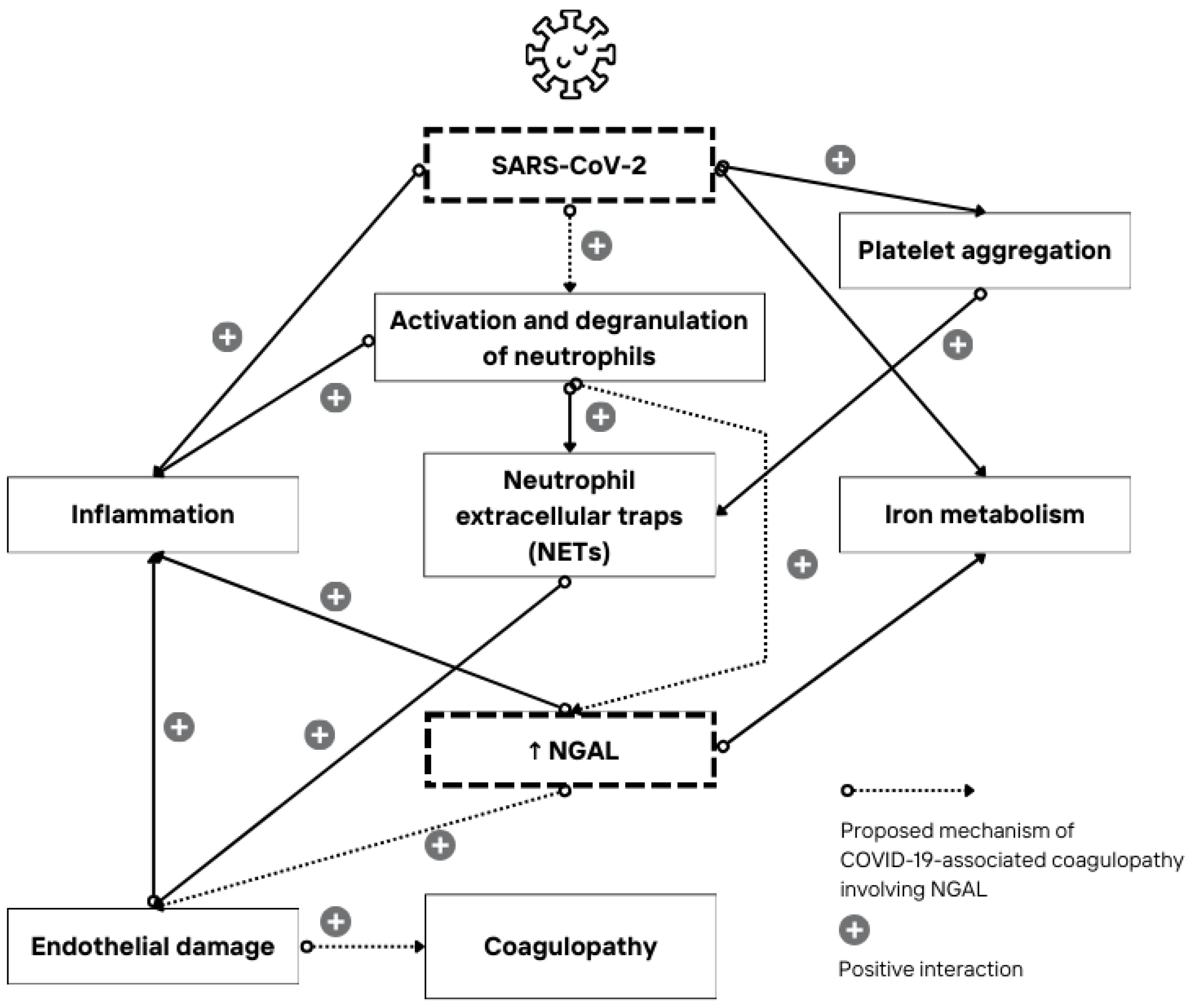
During a SARS-CoV-2 infection, NGAL could also contribute to the hypercoagulable state known as COVID-19-induced coagulopathy (CIC) [10], as shown in Figure 2. Evidence suggests that the SARS-CoV-2 spike protein itself influences the increase in NGAL concentrations, as exhibited in a study that measured levels of gelatinases in the renal cortex after spike protein administration in mice [10]. Additionally, studies on sepsis have demonstrated that during a systemic inflammatory response, increased endothelial damage is associated with higher NGAL concentrations [33]. This endothelial damage could also be caused by neutrophil extracellular traps (NETs) released from activated neutrophils, which likely act synergistically with NGAL, leading to further damage to the blood vessel endothelium and promoting CIC [10,33]. Recent research shows that SARS-CoV-2 may promote platelet aggregation and the subsequent binding of these aggregates with NETs, which can then adhere to the endothelium or circulate freely in the blood [34]. This highlights the important role of platelets in CIC and their interaction with neutrophils through complex mechanisms [34].
NGAL is a siderophoric protein that transports iron into the cell, while free NGAL, which is not bound to iron, induces the transport of iron extracellularly [4]. The interaction between NGAL and iron has also been observed during bacterial infections, where NGAL prevents bacteria from obtaining iron by sequestering siderophores [4]. This reduction in available iron decreases the bacteria’s ability to grow and multiply since iron is necessary for bacterial proliferation [4]. Given NGAL’s role in iron metabolism during bacterial infections and beyond, it is expected that NGAL will also interact with iron metabolism in COVID-19. This potential interaction in COVID-19 should be studied further, as the exact mechanism is not yet known. In the case of COVID-19, patients typically show lower serum iron levels along with increased levels of serum ferritin and hepcidin, especially in the more severe forms of the disease [35,36]. Considering the earlier statements about NGAL and its elevation in COVID-19, the exact pathophysiological pathway could potentially involve NGAL, and this should be recognized in future studies concerning this mechanism.
Figure 2. Interaction mechanism of SARS-CoV-2, NGAL, and COVID-19 coagulopathy. During severe infection, SARS-CoV-2 promotes the activation and degranulation of neutrophils, leading to increased release of NGAL and the formation of neutrophil extracellular traps (NETs). Additionally, SARS-CoV-2 may induce platelet aggregation, with the aggregates binding to NETs, which can then adhere to the endothelium. NGAL release also contributes to endothelial damage in blood vessels, which then leads to further cytokine release. The released cytokines, along with mechanical damage to the endothelium, lead to a hypercoagulable state. These factors contribute to an inflammatory response, triggering additional secretion of pro-inflammatory cytokines that can worsen the hypercoagulable condition. Furthermore, NGAL is involved in iron metabolism during and beyond the infection, which may also contribute to oxidative stress and endothelial damage [8,10,34,37,38,39,40]. This is just one potential mechanism of COVID-19-induced coagulopathy, which likely arises from the combined effects of multiple factors.
Amh 69 00021 g002
This research is primarily limited by the number of patients. Additionally, the study may be subject to potential bias due to confounding factors, as most of the observed patients had comorbidities that could theoretically influence uNGAL levels, such as obesity or chronic diseases. It was not possible to exclude these factors from this study, given that COVID-19 patients requiring hospitalization typically have pre-existing chronic conditions.
If a similar study was conducted with a larger sample size, it would be advisable to monitor patients hospitalized on the same day of illness onset to ensure a more reliable understanding of uNGAL concentration dynamics. Since this study was conducted at a single center, it was not expected that such a sampling method would provide a sufficient number of patients for comprehensive analysis. Additionally, a group of negative controls should be included to evaluate differences in uNGAL levels between COVID-19 patients and healthy controls, which this study currently lacks. Further multicenter research with larger sample sizes is needed, and this study can certainly serve as a foundation.
Another limitation of this study is the lack of follow-up on patients after hospitalization; there was no assessment of late morbidity, mortality, or clinical status following hospital discharge. Specifically, uNGAL levels could also be measured after hospital discharge, once symptoms have resolved. This would allow researchers to determine whether concentrations return to baseline values, aiding in a better understanding of the impact of comorbidities and in addressing this potential confounding bias.

5. Conclusions

In terms of NGAL and COVID-19, this study concludes that there is an association between uNGAL concentrations and disease outcomes. However, there is still a lack of understanding regarding the dynamics of NGAL concentration changes during SARS-CoV-2 infection, which is crucial for its appropriate clinical utility and application. Further research is needed to address this gap.

Supplementary Materials

The following supporting information can be downloaded at: https://www.mdpi.com/article/10.3390/amh69040021/s1, Table S1: Baseline demographic and clinical characteristics of participants; Table S2: Association of uNGAL with age, BMI, length of hospitalization, and MAP among all patients and subgroups by disease outcome, severity, and respiratory complications.

Author Contributions

Conceptualization: L.Š., K.K., D.L. and S.M.; methodology: L.Š., B.G., N.P., B.B. and S.M.; data curation: L.Š., M.Z. and B.G.; formal analysis and investigation: L.Š., K.K. and N.P.; writing—original draft preparation: L.Š., M.Z., B.G., K.K., N.P. and B.B.; writing—review and editing: D.L. and S.M.; resources: L.Š., B.B. and S.M.; supervision: D.L. and S.M. All authors have read and agreed to the published version of the manuscript.

Funding

This research received no external funding.

Institutional Review Board Statement

The study was conducted according to the guidelines of the Declaration of Helsinki, and approved by the Institutional Ethics Committee of the Faculty of Medicine Osijek (KLASA: 641-01/24-01/04, URBROJ: 2158-61-46-24-135, 24 May 2024).

Informed Consent Statement

Informed consent was obtained from all subjects involved in the study or from their legal representatives.

Data Availability Statement

The data that support the findings of this study are available from the corresponding author, S.M., upon reasonable request.

Acknowledgments

We extend our gratitude to Ira Rešetar for their diligent proofreading, which greatly improved the clarity and precision of the manuscript.

Conflicts of Interest

The authors declare no conflicts of interest.

References

  1. Ravi, V.; Saxena, S.; Panda, P.S. Basic virology of SARS-CoV 2. Indian J. Med. Microbiol. 2022, 40, 182–186. [Google Scholar] [CrossRef]
  2. Hu, B.; Guo, H.; Zhou, P.; Shi, Z.L. Characteristics of SARS-CoV-2 and COVID-19. Nat. Rev. Microbiol. 2021, 19, 141–154. [Google Scholar] [CrossRef]
  3. Msemburi, W.; Karlinsky, A.; Knutson, V.; Aleshin-Guendel, S.; Chatterji, S.; Wakefield, J. The WHO estimates of excess mortality associated with the COVID-19 pandemic. Nature 2023, 613, 130–137. [Google Scholar] [CrossRef]
  4. Romejko, K.; Markowska, M.; Niemczyk, S. The Review of Current Knowledge on Neutrophil Gelatinase-Associated Lipocalin (NGAL). Int. J. Mol. Sci. 2023, 24, 10470. [Google Scholar] [CrossRef]
  5. Bolignano, D.; Donato, V.; Coppolino, G.; Campo, S.; Buemi, A.; Lacquaniti, A.; Buemi, M. Neutrophil gelatinase-associated lipocalin (NGAL) as a marker of kidney damage. Am. J. Kidney Dis. 2008, 52, 595–605. [Google Scholar] [CrossRef]
  6. Lentini, P.; De Cal, M.; Clementi, A.; D’Angelo, A.; Ronco, C. Sepsis and AKI in ICU Patients: The Role of Plasma Biomarkers. Crit. Care Res. Pract. 2012, 2012, 856401. [Google Scholar] [CrossRef]
  7. Yoon, S.Y.; Kim, J.S.; Jeong, K.H.; Kim, S.K. Acute Kidney Injury: Biomarker-Guided Diagnosis and Management. Medicina 2022, 58, 340. [Google Scholar] [CrossRef]
  8. Buonafine, M.; Martinez-Martinez, E.; Jaisser, F. More than a simple biomarker: The role of NGAL in cardiovascular and renal diseases. Clin. Sci. 2018, 132, 909–923. [Google Scholar] [CrossRef]
  9. Menez, S.; Moledina, D.G.; Thiessen-Philbrook, H.; Wilson, F.P.; Obeid, W.; Simonov, M.; Yamamoto, Y.; Corona-Villalobos, C.P.; Chang, C.; Garibaldi, B.T.; et al. Prognostic Significance of Urinary Biomarkers in Patients Hospitalized With COVID-19. Am. J. Kidney Dis. 2022, 79, 257–267.e1. [Google Scholar] [CrossRef]
  10. Singh, M.; Pushpakumar, S.; Zheng, Y.; Smolenkova, I.; Akinterinwa, O.E.; Luulay, B.; Tyagi, S.C. Novel mechanism of the COVID-19 associated coagulopathy (CAC) and vascular thromboembolism. Npj Viruses 2023, 1, 3. [Google Scholar] [CrossRef]
  11. Racovitan, D.; Hogeweg, M.; Doevelaar, A.A.; Seidel, M.; Rohn, B.; Bettag, S.; Rieckmann, S.; Babel, N.; Seibert, F.S.; Westhoff, T.H. Urinary biomarkers to predict acute kidney damage and mortality in COVID-19. Clin. Nephrol. 2023, 99, 161–171. [Google Scholar] [CrossRef]
  12. Abers, M.S.; Delmonte, O.M.; Ricotta, E.E.; Fintzi, J.; Fink, D.L.; Almeida de Jesus, A.A.; Zarember, K.A.; Alehashemi, S.; Oikonomou, V.; Desai, J.V.; et al. An immune-based biomarker signature is associated with mortality in COVID-19 patients. JCI Insight 2021, 6, e144455. [Google Scholar] [CrossRef]
  13. He, L.; Zhang, Q.; Li, Z.; Shen, L.; Zhang, J.; Wang, P.; Wu, S.; Zhou, T.; Xu, Q.; Chen, X.; et al. Incorporation of Urinary Neutrophil Gelatinase-Associated Lipocalin and Computed Tomography Quantification to Predict Acute Kidney Injury and In-Hospital Death in COVID-19 Patients. Kidney Dis. 2021, 7, 120–130. [Google Scholar] [CrossRef]
  14. Larcher, R.; Bargnoux, A.S.; Badiou, S.; Besnard, N.; Brunot, V.; Daubin, D.; Platon, L.; Benomar, R.; Amalric, M.; Dupuy, A.-M.; et al. Acute kidney injury in critical COVID-19 patients: Usefulness of urinary biomarkers and kidney proximal tubulopathy. Ren. Fail. 2023, 45, 2292152. [Google Scholar] [CrossRef]
  15. Paranjpe, I.; Jayaraman, P.; Su, C.-Y.; Zhou, S.; Chen, S.; Thompson, R.; Del Valle, D.M.; Kenigsberg, E.; Zhao, S.; Jaladanki, S.; et al. Proteomic characterization of acute kidney injury in patients hospitalized with SARS-CoV2 infection. Commun. Med. 2023, 3, 81. [Google Scholar] [CrossRef]
  16. Casas-Aparicio, G.; Alvarado-de la Barrera, C.; Escamilla-Illescas, D.; León-Rodríguez, I.; Del Río-estrada, P.M.; Calderón-Dávila, N.; González-Navarro, M.; Olmedo-Ocampo, R.; Castillejos-López, M.; Figueroa-Hernández, L.; et al. Role of Urinary Kidney Stress Biomarkers for Early Recognition of Subclinical Acute Kidney Injury in Critically Ill COVID-19 Patients. Biomolecules 2022, 12, 275. [Google Scholar] [CrossRef]
  17. Filev, R.; Lyubomirova, M.; Hristova, J.; Bogov, B.; Kalinov, K.; Svinarov, D.; Rostaing, L. Serum and Urinary Biomarkers in COVID-19 Patients with or without Baseline Chronic Kidney Disease. J. Pers. Med. 2023, 13, 382. [Google Scholar] [CrossRef]
  18. Lablad, Y.; Vanhomwegen, C.; De Prez, E.; Antoine, M.H.; Hasan, S.; Baudoux, T.; Nortier, J. Longitudinal Follow-Up of Serum and Urine Biomarkers Indicative of COVID-19-Associated Acute Kidney Injury: Diagnostic and Prognostic Impacts. Int. J. Mol. Sci. 2023, 24, 16495. [Google Scholar] [CrossRef]
  19. Clinical Management of COVID-19: Living Guideline. Available online: https://pubmed.ncbi.nlm.nih.gov/35917394/ (accessed on 25 September 2024).
  20. EQUATOR Network|Enhancing the QUAlity and Transparency of Health Research. Available online: https://www.equator-network.org/ (accessed on 7 July 2024).
  21. Vahey, G.M.; McDonald, E.; Marshall, K.; Martin, S.W.; Chun, H.; Herlihy, R.; Tate, J.E.; Kawasaki, B.; Midgley, C.M.; Alden, N.; et al. Risk factors for hospitalization among persons with COVID-19-Colorado. PLoS ONE 2021, 16, e0256917. [Google Scholar] [CrossRef]
  22. Velásquez García, H.A.; Adu, P.A.; Harrigan, S.; Wilton, J.; Rasali, D.; Binka, M.; Sbihi, H.; Smolina, K.; Janjua, N.Z. Risk factors for COVID-19 hospitalization after COVID-19 vaccination: A population-based cohort study in Canada. Int. J. Infect. Dis. 2023, 127, 116–123. [Google Scholar] [CrossRef]
  23. Gao, Y.-d.; Ding, M.; Dong, X.; Zhang, J.-j.; Kursat Azkur, A.; Azkur, D.; Gan, H.; Sun, Y.-l.; Fu, W.; Li, W.; et al. Risk factors for severe and critically ill COVID-19 patients: A review. Allergy 2021, 76, 428–455. [Google Scholar] [CrossRef]
  24. Dessie, Z.G.; Zewotir, T. Mortality-related risk factors of COVID-19: A systematic review and meta-analysis of 42 studies and 423,117 patients. BMC Infect. Dis. 2021, 21, 855. [Google Scholar] [CrossRef]
  25. Bezerra, G.F.; Meneses, G.C.; Albuquerque, P.L.; Lopes, N.C.; Santos, R.S.; Da Silva, J.C.; Mota, S.M.; Guimarães, R.R.; Guimarães, F.R.; Guimarães, Á.R.; et al. Urinary tubular biomarkers as predictors of death in critically ill patients with COVID-19. Biomark. Med. 2022, 16, 681–692. [Google Scholar] [CrossRef]
  26. Xu, K.; Shang, N.; Levitman, A.; Corker, A.; Kudose, S.; Yaeh, A.; Neupane, U.; Stevens, J.; Sampogna, R.; Mills, A.M.; et al. Elevated Neutrophil Gelatinase-Associated Lipocalin Is Associated With the Severity of Kidney Injury and Poor Prognosis of Patients With COVID-19. Kidney Int. Rep. 2021, 6, 2979–2992. [Google Scholar] [CrossRef]
  27. Volbeda, M.; Jou-Valencia, D.; van den Heuvel, M.C.; Knoester, M.; Zwiers, P.J.; Pillay, J.; Berger, S.P.; van der Voort, P.H.J.; Zijlstra, J.G.; van Meurs, M.; et al. Comparison of renal histopathology and gene expression profiles between severe COVID-19 and bacterial sepsis in critically ill patients. Crit. Care 2021, 25, 202. [Google Scholar] [CrossRef]
  28. Battaglini, D.; Lopes-Pacheco, M.; Castro-Faria-Neto, H.C.; Pelosi, P.; Rocco, P.R.M. Laboratory Biomarkers for Diagnosis and Prognosis in COVID-19. Front. Immunol. 2022, 13, 857573. [Google Scholar] [CrossRef]
  29. Velavan, T.P.; Meyer, C.G. Mild versus severe COVID-19: Laboratory markers. Int. J. Infect. Dis. 2020, 95, 304–307. [Google Scholar] [CrossRef]
  30. Hedayati-Ch, M.; Ebrahim-Saraie, H.S.; Bakhshi, A. Clinical and immunological comparison of COVID-19 disease between critical and non-critical courses: A systematic review and meta-analysis. Front. Immunol. 2024, 15, 1341168. [Google Scholar] [CrossRef]
  31. Krishna Reddy, C.H.; Achari, P.K.; Nisha, B.; Radha, A.R. Significance of Laboratory Markers in Predicting the Severity of COVID-19 in the Central Reserve Police Force Front-line Workers with a Review of Literature. Indian J. Public Health 2022, 66, 512–515. [Google Scholar] [CrossRef]
  32. Goyal, A.; Daneshpajouhnejad, P.; Hashmi, M.F.; Bashir, K.; John, B.K. Acute Kidney Injury (Nursing); StatPearls Publishing: Treasure Island, FL, USA, 2023. [Google Scholar]
  33. Sikora, J.P.; Karawani, J.; Sobczak, J. Neutrophils and the Systemic Inflammatory Response Syndrome (SIRS). Int. J. Mol. Sci. 2023, 24, 13469. [Google Scholar] [CrossRef]
  34. Sciaudone, A.; Corkrey, H.; Humphries, F.; Koupenova, M. Platelets and SARS-CoV-2 During COVID-19: Immunity, Thrombosis, and Beyond. Circ. Res. 2023, 132, 1272–1289. [Google Scholar] [CrossRef]
  35. Suriawinata, E.; Mehta, K.J. Iron and iron-related proteins in COVID-19. Clin. Exp. Med. 2023, 23, 969–991. [Google Scholar] [CrossRef]
  36. Mahroum, N.; Alghory, A.; Kiyak, Z.; Alwani, A.; Seida, R.; Alrais, M.; Shoenfeld, Y. Ferritin—From iron, through inflammation and autoimmunity, to COVID-19. J. Autoimmun. 2022, 126, 102778. [Google Scholar] [CrossRef]
  37. Macdonald, S.P.J.; Bosio, E.; Neil, C.; Arendts, G.; Burrows, S.; Smart, L.; Brown, S.G.A.; Fatovich, D.M. Resistin and NGAL are associated with inflammatory response, endothelial activation and clinical outcomes in sepsis. Inflamm. Res. 2017, 66, 611–619. [Google Scholar] [CrossRef]
  38. McKenna, E.; Wubben, R.; Isaza-Correa, J.M.; Melo, A.M.; Mhaonaigh, A.U.; Conlon, N.; O’Donnell, J.S.; Cheallaigh, C.N.; Hurley, T.; Stevenson, N.J.; et al. Neutrophils in COVID-19: Not Innocent Bystanders. Front. Immunol. 2022, 13, 864387. [Google Scholar] [CrossRef]
  39. Liu, J.T.; Song, E.; Xu, A.; Berger, T.; Mak, T.W.; Tse, H.-F.; Law, I.K.; Huang, B.; Liang, Y.; Vanhoutte, P.M.; et al. Lipocalin-2 deficiency prevents endothelial dysfunction associated with dietary obesity: Role of cytochrome P450 2C inhibition. Br. J. Pharmacol. 2012, 165, 520–531. [Google Scholar] [CrossRef]
  40. Song, E.; Fan, P.; Huang, B.; Deng, H.-B.; Cheung, B.M.Y.; Félétou, M.; Vilaine, J.-P.; Villeneuve, N.; Xu, A.; Vanhoutte, P.M.; et al. Deamidated lipocalin-2 induces endothelial dysfunction and hypertension in dietary obese mice. J. Am. Heart Assoc. 2014, 3, e000837. [Google Scholar] [CrossRef]
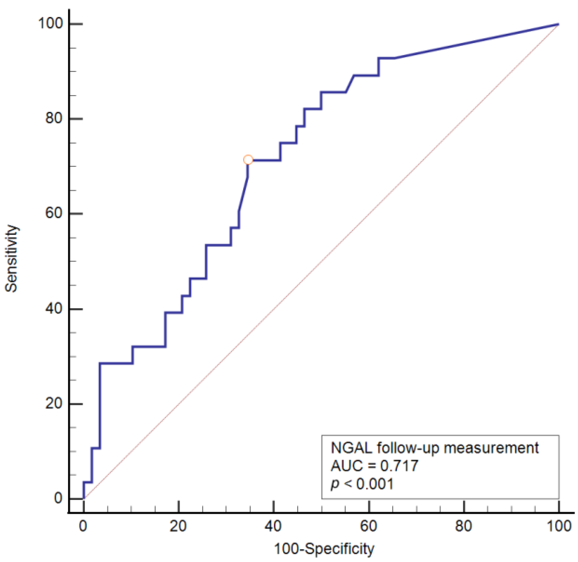
Figure 1. ROC analysis of sensitivity, specificity, and cut-off values of follow-up urinary neutrophil gelatinase-associated lipocalin (uNGAL) concentration with respect to death outcome.
Amh 69 00021 g001
Table 1. The distribution of patients according to disease outcome and according to admission to the ICU at some point during hospitalization.
Number (%) of Patients
Disease outcome
Recovery58 (67)
Death outcome28 (33)
Admission to the ICU
ICU21 (24)
Non-ICU65 (76)
ICU—Intensive Care Unit.
Table 2. The association of sex, disease severity, and comorbidities with disease outcome.
Number (%) of Patients Regarding Outcomep *
SurvivedDeath OutcomeTotal
Sex
Male26 (45)11 (39)37 (43)0.63
Female32 (55)17 (61)49 (57)
Disease severity
Moderate5 (9)05 (6)<0.001
Severe52 (90)11 (39)63 (73)
Critical1 (1)17 (61)18 (21)
Comorbidities
Type 2 diabetes mellitus14 (24)5 (18)19 (22)0.51
Hypertension38 (66)24 (86)62 (72)0.05
Cardiomyopathy10 (17)11 (39)21 (24)0.03
Atrial fibrillation3 (5)5 (18)8 (9)0.11
Chronic lung disease5 (9)3 (11)8 (9)0.75
Chronic kidney disease2 (3)1 (4)3 (4)>0.99
* χ2 test; Fisher’s exact test.
Table 3. Differences in age, length of hospitalization, mean arterial pressure, and body mass index with regard to disease outcome.
Median (Interquartile Range)Difference95% CIp *
SurvivedDeath Outcome
Age (years)71 (62–80)80 (76–86)84–140.001
Length of hospitalization (days)8 (6–12)11 (7–15)20–40.10
MAP (mmHg)83.3 (80–93.3)81.7 (73.3–88.3)−3.3−10–00.12
BMI (kg/m2)26.14 (24.6–28.7)28.6 (23.8–32.2)0.97−2.85–4.990.55
* Mann–Whitney U test (Hodges–Lehmann median difference); CI—confidence interval; MAP—mean arterial pressure; BMI—body mass index.
Table 4. Differences in measured concentrations of uNGAL at admission and during follow-up sampling in relation to disease outcome.
Median (Interquartile Range)Difference95% CIp *
SurvivedDeath Outcome
At admission
uNGAL (ng/mL)21.2 (10.6–41.3)27.6 (15.4–91.8)5.2−2–17.20.21
Follow-up sampling
uNGAL (ng/mL)15.6 (9–40.7)34.5 (19.9–103.2)17.76.4–36.90.001
* Mann–Whitney U test (Hodges–Lehmann median difference); CI—confidence interval; uNGAL—urinary neutrophil gelatinase-associated lipocalin.
Table 5. Prediction of mortality probability (bivariate and multivariate logistic regression).
βWaldp ValueOdds Ratio95% CI
Bivariate regression
Age0.078.480.0041.071.02–1.12
Cardiomyopathy1.094.570.032.971.09–8.6
IMV4.6018.4<0.00199.812.2–817.5
uNGAL (at admission)0.0010.630.431.0010.99–1.003
uNGAL (follow-up sampling)0.014.300.031.011.001–1.03
Multivariate regression
Age0.116.330.011.121.03–1.22
uNGAL (follow-up sampling)0.13.880.041.011.001–1.03
IMV5.0717.4<0.001159.414.7–1728.5
Constant−141.059.010.003
β—regression coefficient; CI—confidence interval; IMV—invasive mechanical ventilation; uNGAL—neutrophil gelatinase-associated lipocalin.
Table 6. ROC curve values of the observed variables with respect to death outcome.
AUC95% CISensitivitySpecificityCut-OffYouden Indexp Value
uNGAL (follow-up sampling) (ng/mL)0.7170.610–0.80971.465.5>23.80.370<0.001
AUC—area under the curve; CI—confidence interval; uNGAL—neutrophil gelatinase-associated lipocalin.
Disclaimer/Publisher’s Note: The statements, opinions and data contained in all publications are solely those of the individual author(s) and contributor(s) and not of MDPI and/or the editor(s). MDPI and/or the editor(s) disclaim responsibility for any injury to people or property resulting from any ideas, methods, instructions or products referred to in the content.