Case Report

Elevated Alpha-Fetoprotein in Hypothyroidism

1
Department of Medicine, Surgery and Health Sciences, University of Trieste, 34129 Trieste, Italy
2
Institute for Maternal and Child Health IRCCS “Burlo Garofolo”, 34137 Trieste, Italy
3
Primary Care Pediatrician, Azienda Sanitaria Universitaria Friuli Centrale, 33100 Udine, Italy
4
Department of Pediatrics, Azienda Sanitaria Universitaria Integrata Giuliano-Isontina, 34074 Monfalcone, Italy
5
SC Laboratorio Unico, Ospedale Maggiore, Azienda Sanitaria Universitaria Giuliano-Isontina, 34125 Trieste, Italy
*
Author to whom correspondence should be addressed.
LabMed 2025, 2(4), 24; https://doi.org/10.3390/labmed2040024
Submission received: 17 September 2025 / Revised: 8 November 2025 / Accepted: 20 November 2025 / Published: 25 November 2025

Abstract

Alpha-fetoprotein (AFP) is a biomarker commonly used in the diagnosis of various malignancies but may also be elevated in non-neoplastic conditions, including hypothyroidism. We report the case of a 3-year-old girl with Down syndrome (DS) and newly diagnosed hypothyroidism, who presented with a hypoechoic oval lesion adjacent to the thymic parenchyma on ultrasound and markedly elevated AFP levels (169.2 ng/mL). Further investigations, including MRI, excluded the presence of germ cell tumors. Following initiation of levothyroxine therapy, AFP levels normalized in parallel with thyroid function. No evidence of malignancy was detected despite the initial suspicion. This case underscores the association between elevated AFP and hypothyroidism, highlighting the importance of evaluating thyroid status in patients with increased AFP to avoid unnecessary oncological investigations. In particular, elevated AFP in the context of hypothyroidism and DS warrants careful thyroid assessment and follow-up to prevent redundant diagnostic procedures and reduce patient and family anxiety. Thyroid function testing should be considered before extensive oncological evaluation in children with elevated AFP.

Graphical Abstract

1. Introduction

Alpha-fetoprotein (AFP) is a well-established biomarker for malignancies of endodermal origin, including germ cell tumors, hepatoblastoma, and hepatocellular carcinoma [1]. However, AFP is a tumor-associated rather than a tumor-specific protein, and elevated concentrations may also be observed in several non-neoplastic conditions [Hanif]. These include physiological states such as pregnancy and infancy, as well as pathological conditions such as liver disease, liver regeneration, and ataxia–telangiectasia [1,2,3,4,5,6,7]. Primary hypothyroidism has likewise been reported as a potential cause of AFP elevation, although the underlying mechanisms remain unclear [8,9]. In this context, we describe the case of a young girl with trisomy 21/Down syndrome (DS) and newly diagnosed hypothyroidism who presented with markedly increased AFP levels, initially raising concerns for malignancy.

2. Case Presentation

A 3-year-old Bengali girl with DS, diagnosed at birth, was referred after the incidental detection of hypothyroidism. Her past medical history included corrective cardiac surgery at the age of two for an atrial septal defect, followed by persistent pulmonary hypertension managed with phosphodiesterase-5 inhibitors. Previous thyroid function tests had consistently been within the normal range.
During a routine well-child visit, her pediatrician noted isolated bradycardia (heart rate 50–55 bpm) and significant weight gain (3 kg over eight months). Laboratory evaluation revealed severe primary hypothyroidism, with markedly elevated thyroid-stimulating hormone (TSH > 489 μIU/mL; reference range 0.79–5.85) and low thyroid hormone levels (free thyroxine, fT4 < 2.5 pg/mL; reference 6.1–10.6; free triiodothyronine, fT3 1.9 pg/mL; reference 2.6–4.0). There was no family history of thyroid disease. Treatment with levothyroxine (50 μg/day; 3.5 μg/kg/day) was initiated, and the patient was referred to the pediatric endocrinology unit for further assessment.
Two days after starting therapy, repeat testing confirmed persistent hypothyroidism (TSH > 489 μIU/mL; fT4 4.3 pg/mL; fT3 2.5 pg/mL). Autoimmune thyroiditis was suggested by markedly elevated anti-thyroid antibodies (anti-thyroid peroxidase 6354 IU/mL; reference < 9; anti-thyroglobulin 28.4 IU/mL; reference < 4) and by thyroid ultrasonography, which showed diffuse enlargement with hypoechoic echotexture and increased vascularity on Doppler imaging. In addition, a hypoechoic oval lesion (approximately 20 × 8 × 11 mm) with a solid echostructure was detected adjacent to the thymic parenchyma (Figure 1).
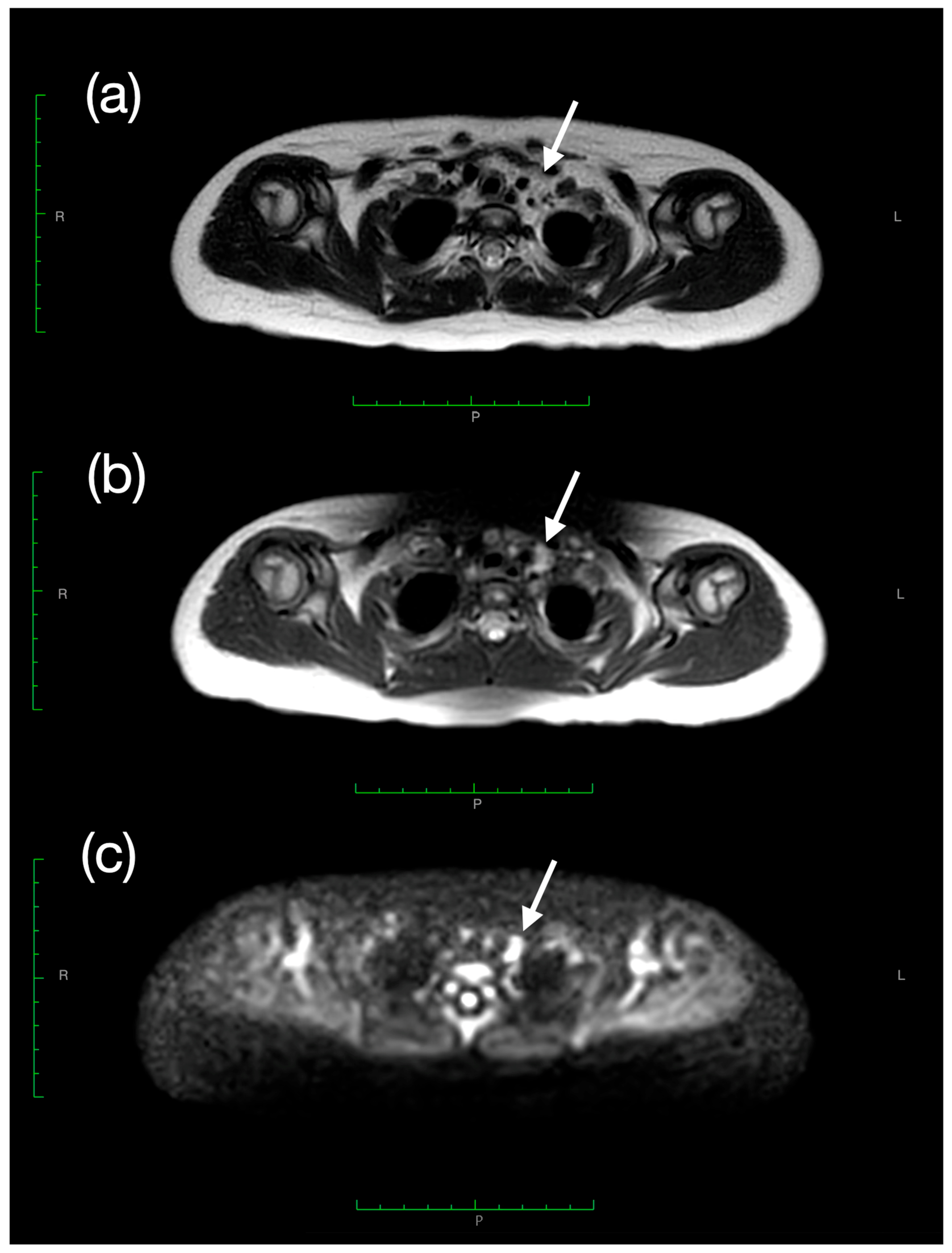
Given these findings, the patient was referred to the pediatric oncology unit. Tumor marker testing revealed elevated alpha-fetoprotein (AFP 169.2 ng/mL; reference range 0.8–5.0), with normal liver enzyme levels. To exclude germ cell tumors, a whole-body MRI under sedation was performed 10 days after the initiation of levothyroxine therapy. The scan showed no pathological masses but revealed several solid nodules in the paratracheal and laterocervical regions (levels 2R and 2L), one of which was contiguous with the upper left margin of the thymus, consistent with lymph nodes (Figure 2).
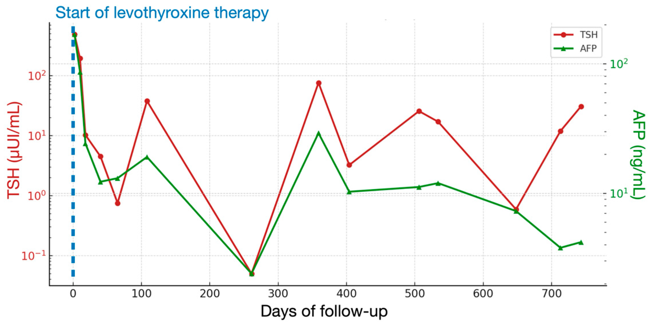
Blood tests repeated over the following weeks showed a gradual normalization of AFP levels, occurring in parallel with the restoration of thyroid function (Table 1, Figure 3).
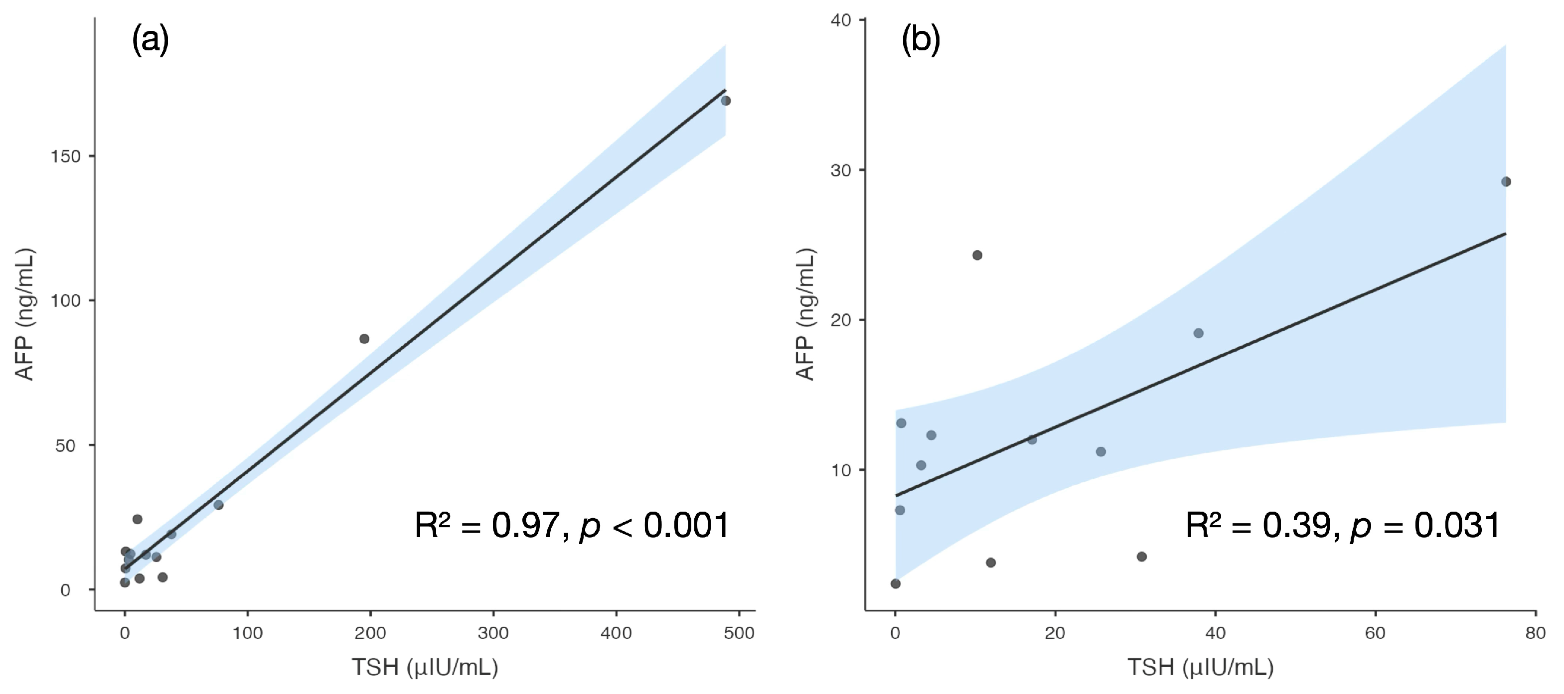
Linear regression analysis was performed to explore the relationship between AFP and thyroid function parameters using a simple linear model (AFP as the dependent variable. TSH or fT4 as independent variables) based on 14 serial measurements collected from day 0 to day 743 and shown in Table 1. Analyses were performed using Jamovi software (version 2.3.28.0). The analysis demonstrated a significant direct correlation between AFP and TSH (R2 = 0.97, p < 0.001) (Figure 4a), and between AFP and fT4 (R2 = 0.50. p = 0.005). When the two extreme values at days 2 and 10 were excluded, no significant relationship was observed between AFP and fT4 (R2 < 0.01, p = 0.78), while the model fit for TSH decreased (R2 = 0.39, p = 0.031) (Figure 4b), confirming that high TSH values correspond to high AFP values. whereas the relationship became weaker as TSH normalized.
To rule out the rare possibility of Van Wyk–Grumbach syndrome (the triad of hypothyroidism, isosexual precocious puberty, and ovarian mass) [10], pelvic ultrasonography and gonadal function tests were performed. The results excluded precocious puberty and ovarian involvement (undetectable 17β-estradiol; LH 0.2 mIU/mL; FSH 4.9 mIU/mL; no ovarian masses and prepubertal morphology of the uterus and ovaries).
Seven months after the initiation of levothyroxine therapy, the patient was clinically well and asymptomatic, with AFP levels fully normalized. During the subsequent two years of follow-up, mild transient increases in AFP were observed whenever therapy adjustments were required due to impaired thyroid function. Semi-annual ultrasound examinations showed no changes in the size or characteristics of the lymph nodes in the thymic area. Given the consistent clinical, biochemical, and imaging stability, cytological or histological evaluation was not pursued.
Written informed consent has been obtained from the patient’s parents to publish this paper.

3. Discussion

Autoimmune hypothyroidism occurs more frequently in children with DS than in the general pediatric population. with prevalence rates reported from 5% up to 30–40%, especially for subclinical forms, and typically onset at a younger age (often in the first years of life) [11,12,13]. While the association between hypothyroidism and elevated AFP was first described over three decades ago, more recent evidence links congenital and acquired hypothyroidism in children to marked AFP elevations.
The remarkable aspect of this case was the incidental finding of a cervical mass on thyroid ultrasound associated with markedly elevated AFP, a biomarker of germ cell tumors, whose incidence is increased in individuals with DS [14]. In our patient, the absence of suspicious lesions on MRI combined with AFP normalization parallel to the restoration of thyroid function strongly supports a causal relationship.
AFP is a glycoprotein produced during gestation primarily by the fetal yolk sac and liver. Serum levels are therefore markedly elevated at birth, gradually decreasing to 10–20 ng/mL during the first year of life. By the age of two years, mean values are around 8 ng/mL (95.5% CI, 0.8–87), but adult reference values are not yet reached [15]. To date, no studies have systematically evaluated AFP concentrations in children older than two years or in those with DS.
Increased AFP is a well-recognized biomarker in endodermal-derived malignancies, such as germ cell tumors, hepatoblastoma, and hepatocellular carcinoma. Nevertheless, AFP should be interpreted with caution because values are age-dependent and because AFP is a tumor-associated rather than a tumor-specific protein [1,2,3,4,5,6,7,8,9] (Table 2). Although levels above 500 ng/mL are rarely associated with benign conditions, tumor marker assessment must always be integrated with clinical and imaging findings [16].
The association between hypothyroidism and AFP elevation was first described in 1989, when Hashimoto and Matsubara reported elevated tumor markers, including AFP, in adult women with hypothyroidism, with direct correlation to TSH and inverse correlation to thyroid hormones [8]. Notably, AFP values in that cohort (6 ± 0.3 ng/mL) were significantly lower than those observed in our case. In contrast, a subsequent study reported AFP levels ranging from 166 to 695 ng/mL in children with congenital hypothyroidism (mean age 7.5 ± 6.5 years), compared to 25–30 ng/mL in healthy controls, again with significant correlation to TSH and inverse correlation to fT4 and age [9].
The recent literature has significantly expanded our understanding of thyroid-hormone-dependent regulation of AFP. In hepatocytes and hepatocellular carcinoma models, triiodothyronine (T3) acting through thyroid hormone receptors (TRs) downregulates AFP transcription both directly—via TR binding to negative thyroid response elements in the AFP promoter—and indirectly, by repressing the long non-coding RNA taurine upregulated gene 1 (TUG1), which positively regulates AFP expression and tumor cell proliferation [17,18,19]. In parallel, T3-dependent gene regulation in other tissues involves chromatin-level mechanisms, including changes in histone acetylation and DNA methylation, highlighting the potential for epigenetic regulation of thyroid-responsive genes [20].
At the post-transcriptional level, miR-122 is one of the most abundant liver-enriched microRNAs and a key regulator of hepatocyte differentiation and metabolic homeostasis. In AFP-producing gastric cancer and hepatocellular carcinoma, miR-122 expression in tissue and plasma correlates with AFP levels and malignant potential, and circulating miR-122 improves the diagnostic performance of AFP as a biomarker [21,22]. These converging data support a mechanistic framework in which impaired thyroid hormone signaling may facilitate AFP upregulation by removing TR-mediated transcriptional repression and perturbing non-coding RNA networks (such as TUG1- and microRNA-dependent pathways), although direct evidence in primary hypothyroidism remains limited.
The presence of DS in this case deserves separate consideration. In DS pregnancies, maternal serum AFP levels are about 70% lower than normal, reflecting impaired placental transfer and altered glycoform distribution rather than reduced fetal hepatic production [23,24,25]. In older children, however, the liver becomes the predominant AFP source, and hepatic maturation is strongly dependent on T3 availability.
In DS, global epigenetic dysregulation—partly driven by the overexpression of chromosome 21 genes such as dual-specificity tyrosine phosphorylation–regulated kinase 1A (DYRK1A) and amyloid precursor protein (APP)—affects DNA methylation patterns and chromatin remodeling, altering the expression of developmental and metabolic genes. DYRK1A hyperdosage has been shown to interfere with histone modification and to dysregulate chromatin-remodeling complexes, while APP overexpression contributes to aberrant methylation and oxidative stress–related epigenetic changes [1,25].
The coexistence of severe T3 deficiency and DS-related epigenetic instability may therefore promote the reactivation of a fetal hepatic gene program, explaining higher AFP levels compared with non-DS hypothyroid children.
Our findings confirm that primary hypothyroidism alone can cause AFP elevation—even in children with Down syndrome and in the absence of precocious puberty. Clinicians should therefore interpret AFP results with caution and always assess thyroid status before initiating extensive oncological investigations. Given the growing evidence of endocrine involvement, both congenital and acquired hypothyroidism should be systematically considered in the differential diagnosis of unexplained AFP elevation in pediatric patients (Table 3). Early evaluation of thyroid function may prevent unnecessary oncological work-up in selected cases. Close follow-up after levothyroxine initiation is essential, as normalization of thyroid function can resolve AFP abnormalities and spare patients and families undue anxiety and invasive procedures.

Author Contributions

Conceptualization, V.C. and G.T. (Gianluca Tornese); investigation, V.K., F.M.M., A.B., D.D. and L.Z.; data curation, V.C.; supervision, A.F. and G.T. (Gianluca Tamaro); funding acquisition, G.T. (Gianluca Tornese); formal analysis: G.T. (Gianluca Tornese); writing—original draft preparation, V.C., V.K., F.M.M., A.B., D.D. and L.Z.; writing—review and editing, A.F., G.T. (Gianluca Tamaro) and G.T. (Gianluca Tornese); All authors have read and agreed to the published version of the manuscript.

Funding

This work was supported by the Italian Ministry of Health, through the contribution given to the Institute for Maternal and Child Health IRCCS Burlo Garofolo, Trieste, Italy (RC 16/24).

Institutional Review Board Statement

Not applicable.

Informed Consent Statement

Written informed consent has been obtained from the patient’s parents to publish this paper.

Data Availability Statement

The original contributions presented in this study are included in the article. Further inquiries can be directed to the corresponding author.

Conflicts of Interest

The authors declare no conflicts of interest.

Abbreviations

The following abbreviations are used in this manuscript:
AFPAlpha-fetoprotein
FSHFollicle-Stimulating Hormone
fT3free Triiodothyronine
fT4free Thyroxine
LHLuteinizing Hormone
MRIMagnetic Resonance Imaging
TSHThyroid-Stimulating hormone

References

  1. Głowska-Ciemny, J.; Szymański, M.; Kuszerska, A.; Rzepka, R.; von Kaisenberg, C.S.; Kocyłowski, R. Role of Alpha-Fetoprotein (AFP) in Diagnosing Childhood Cancers and Genetic-Related Chronic Diseases. Cancers 2023, 15, 4302. [Google Scholar] [CrossRef] [PubMed]
  2. Hanif, H.; Ali, M.J.; Susheela, A.T.; Yeo, Y.H.; Lee, Y.-T.; Zhu, Y.; Yang, J.D. Update on the Applications and Limitations of Alpha-Fetoprotein for Hepatocellular Carcinoma. World J. Gastroenterol. 2022, 28, 216–229. [Google Scholar] [CrossRef]
  3. Matonóg, A.; Drosdzol-Cop, A. Alpha-Fetoprotein Level in Fetuses, Infants, and Children with Ovarian Masses: A Literature Review. Front. Endocrinol. 2024, 15, 1307619. [Google Scholar] [CrossRef] [PubMed]
  4. Yeo, Y.H.; Lee, Y.-T.; Tseng, H.-R.; Zhu, Y.; You, S.S.; Agopian, V.G.; Yang, J.D. Alpha-Fetoprotein: Past, Present, and Future. Hepatol. Commun. 2024, 8, e0422. [Google Scholar] [CrossRef] [PubMed]
  5. Demir, H.; Hızal, G.; Uslu Kızılkan, N.; Gürakan, F.; Talim, B.; Coşkun, T.; Kale, G.; Yüce, A. Serum Alpha-Fetoprotein Levels in Neonatal Cholestasis. Turk. J. Pediatr. 2013, 55, 152–157. [Google Scholar]
  6. Chen, Y.; Zhao, Y.; Feng, L.; Zhang, J.; Zhang, J.; Feng, G. Association between Alpha-Fetoprotein and Metabolic Syndrome in a Chinese Asymptomatic Population: A Cross-Sectional Study. Lipids Health Dis. 2016, 15, 85. [Google Scholar] [CrossRef]
  7. Braga-Neto, P.; Dutra, L.A.; Pedroso, J.L.; Barsottini, O.G. Alpha-Fetoprotein as a Biomarker for Recessive Ataxias. Arq. Neuropsiquiatr. 2010, 68, 953–955. [Google Scholar] [CrossRef]
  8. Hashimoto, T.; Matsubara, F. Changes in the Tumor Marker Concentration in Female Patients with Hyper-, Eu-, and Hypothyroidism. Endocrinol. Jpn. 1989, 36, 873–879. [Google Scholar] [CrossRef]
  9. Shawky, R.M.; Abd El-Fattah, S.; El-Din Azzam, M.E.; Rafik, M.M.; Osman, A. Alpha-Fetoprotein in Screening for Congenital Hypothyroidism. East. Mediterr. Health J. 2001, 7, 171–180. [Google Scholar] [CrossRef]
  10. Gupta, J.; Lin-Su, K. Van Wyk–Grumbach Syndrome in a Female Pediatric Patient with Trisomy 21: A Case Report. Int. J. Pediatr. Endocrinol. 2020, 2020, 2. [Google Scholar] [CrossRef]
  11. Amr, N.H. Thyroid Disorders in Subjects with Down Syndrome: An Update. Acta Biomed. 2018, 89, 132–139. [Google Scholar] [CrossRef]
  12. Szeliga, K.; Antosz, A.; Skrzyńska, K.; Kalina-Faska, B.; Januszek-Trzciakowska, A.; Gawlik, A. Subclinical Hypothyroidism as the Most Common Thyroid Dysfunction Status in Children with Down’s Syndrome. Front. Endocrinol. 2022, 12, 782865. [Google Scholar] [CrossRef]
  13. Dalrymple, R.A. Thyroid Disorder in Children and Young People with Down Syndrome: DSMIG Guideline Review. Arch. Dis. Child. Educ. Pract. 2022, 107, 34–35. [Google Scholar] [CrossRef] [PubMed]
  14. Satgé, D.; Sommelet, D.; Geneix, A.; Nishi, M.; Malet, P.; Vekemans, M. A tumour profile in Down syndrome. Am. J. Med. Genet. 1998, 78, 207–216. [Google Scholar] [CrossRef]
  15. Blohm, M.E.; Vesterling-Hörner, D.; Calaminus, G.; Göbel, U. Alpha 1-fetoprotein (AFP) reference values in infants up to 2 years of age. Pediatr. Hematol. Oncol. 1998, 15, 135–142. [Google Scholar] [CrossRef]
  16. Van Houwelingen, L.; Sandoval, J.A. Alpha-fetoprotein in malignant pediatric conditions. In Proof and Concepts in Rapid Diagnostic Tests and Technologies; Saxena, S.K., Ed.; IntechOpen: London, UK, 2016. [Google Scholar]
  17. Caturla, M.; Van Reeth, T.; Drèze, P.; Szpirer, J.; Szpirer, C. The thyroid hormone down-regulates the mouse alpha-foetoprotein promoter. Mol. Cell. Endocrinol. 1997, 135, 139–145. [Google Scholar] [CrossRef] [PubMed]
  18. Lin, Y.-H.; Wu, M.-H.; Huang, Y.-H.; Yeh, C.-T.; Lin, K.-H. TUG1 is a regulator of AFP and serves as prognostic marker in non-hepatitis B, non-hepatitis C hepatocellular carcinoma. Cells 2020, 9, 262. [Google Scholar] [CrossRef]
  19. Huang, P.-S.; Chang, C.-C.; Wang, C.-S.; Lin, K.-H. Functional roles of non-coding RNAs regulated by thyroid hormones in liver cancer. Biomed. J. 2021, 44, 272–284. [Google Scholar] [CrossRef]
  20. Forini, F.; Nicolini, G.; Pitto, L.; Iervasi, G. Novel Insight Into the Epigenetic and Post-transcriptional Control of Cardiac Gene Expression by Thyroid Hormone. Front. Endocrinol. 2019, 10, 601. [Google Scholar] [CrossRef]
  21. Maruyama, S.; Furuya, S.; Shiraishi, K.; Shimizu, H.; Akaike, H.; Hosomura, N.; Kawaguchi, Y.; Amemiya, H.; Kawaida, H.; Sudo, M.; et al. miR-122-5p as a Novel Biomarker for Alpha-Fetoprotein-Producing Gastric Cancer. World J. Gastrointest. Oncol. 2018, 10, 344–350. [Google Scholar] [CrossRef]
  22. Fang, Y.; Yan, D.; Wang, L.; Zhang, J.; He, Q. Circulating microRNAs (miR-16, miR-22, miR-122) expression and early diagnosis of hepatocellular carcinoma. J. Clin. Lab. Anal. 2022, 36, e24541. [Google Scholar] [CrossRef] [PubMed]
  23. Kronquist, K.E.; Dreazen, E.; Keener, S.L.; Nicholas, T.W.; Crandall, B.F. Reduced Fetal Hepatic Alpha-Fetoprotein Levels in Down’s Syndrome. Prenat. Diagn. 1990, 10, 739–751. [Google Scholar] [CrossRef]
  24. Guibourdenche, J.; Leguy, M.-C.; Pidoux, G.; Hebert-Schuster, M.; Laguillier, C.; Anselem, O.; Grangé, G.; Bonnet, F.; Tsatsaris, V. Biochemical Screening for Fetal Trisomy 21: Pathophysiology of Maternal Serum Markers and Involvement of the Placenta. Int. J. Mol. Sci. 2023, 24, 7669. [Google Scholar] [CrossRef] [PubMed]
  25. Chen, Y.; Chen, Y.; Ning, W.; Zhang, W.; Li, L.; Wang, X.; Yin, Y.; Zhang, H. Diagnostic value of maternal alpha-fetoprotein variants in second-trimester biochemical screening for trisomy 21 and 18. Sci. Rep. 2022, 12, 13605. [Google Scholar] [CrossRef] [PubMed]
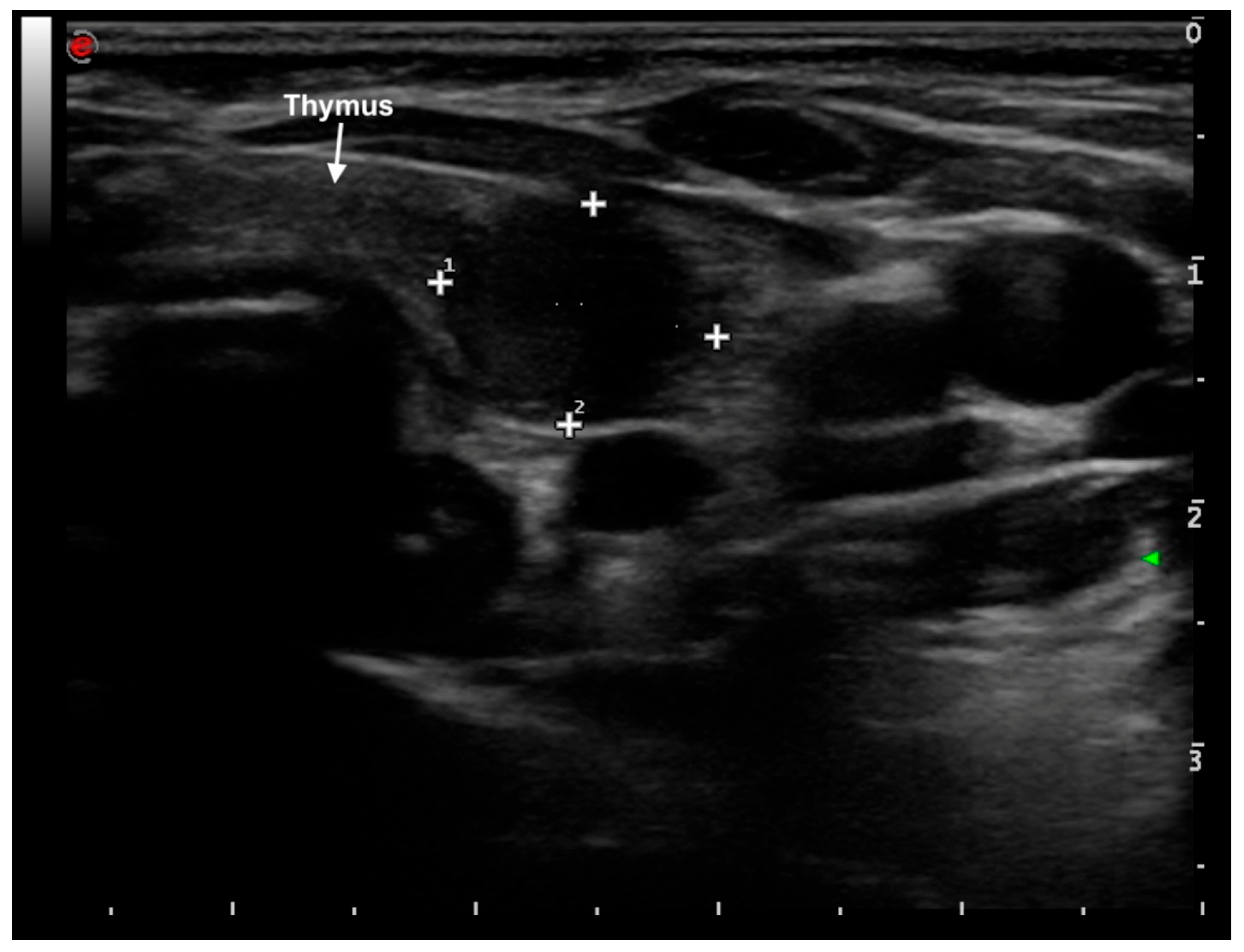
Figure 1. Transverse high-frequency linear ultrasound scan at the left jugular region showing a hypoechoic oval lesion (11.6 × 9.1 mm) located within the thymic parenchyma, medial to the internal jugular vein and anterior to the carotid artery. The arrow indicates the thymus; crosses (+) mark the margins of the lesion used for measurement; the numbers (1, 2) correspond to the orthogonal diameters of the lesion.
Labmed 02 00024 g001
Figure 2. Axial magnetic resonance imaging of the neck (slice thickness 3 mm; TR 4691, TE 100): (a) Axial T2-weighted sequence showing a mildly hyperintense solid tissue within the left jugular region (arrow). (b) Axial SPAIR sequence confirming the same finding with fat suppression (arrow). (c) Axial DWIBS sequence demonstrating restricted diffusion in the same 11 mm jugular lesion, corresponding to the ultrasound finding and compatible with a lymph node (arrow).
Labmed 02 00024 g002
Figure 3. Trend of thyroid-stimulating hormone (TSH, μIU/mL; red line) and alpha-fetoprotein (AFP, ng/mL; green line) after the start of levothyroxine therapy, during follow-up. Both parameters showed a parallel trend, with AFP progressively normalizing as thyroid function improved under levothyroxine therapy. Both axes are plotted on a logarithmic scale to allow comparison of parameters with different orders of magnitude.
Labmed 02 00024 g003
Figure 4. Scatterplots showing the relationship between alpha-fetoprotein (AFP) and thyroid stimulating hormone (TSH) levels: (a) the full dataset including all 12 serial measurements; (b) excluding the two extreme TSH values recorded at days 2 and 10. Shaded areas represent 95% confidence intervals.
Labmed 02 00024 g004
Table 1. Blood test results during the patient’s follow-up: thyroid-stimulating hormone (TSH), free triiodothyronine (fT3), free thyroxine (fT4) and alpha-fetoprotein (AFP). Normal values (n.v.) are reported in brackets.
DayTSH (μUI/mL)
(n.v. 0.79–5.85)
fT3 (pg/mL)
(n.v. 2.6–4.0)
fT4 (pg/mL)
(n.v. 6.1–10.6)
AFP (ng/mL)
(n.v. 0.8–5.0)
Levothyroxine Dose
(mcg/kg/day)
0>489.001.9<2.5-3.5
2>489.002.54.3169.13.5
10194.903.76.286.73.5
1810.254.314.324.33.5
404.504.211.912.32.5
650.754.314.413.11.7
10837.893.49.119.12.2
14240.143.27.1-2.7
1787.783.411.1-2.7
2310.693.415.2-2.1
2610.052.210.92.41.9
35976.303.07.529.22.6
4043.243.110.310.32.6
43938.643.67.7-2.8
4715.633.210.6-2.6
50625.683.19.111.23.2
53417.083.610.912.03.6
5860.782.314.4-2.6
6480.594.613.77.32.1
71311.94-8.93.81.7
7350.411.47.3-1.5
74330.782.810.74.21.7
7622.162.713.0-1.7
Serum AFP, TSH, fT3. and fT4 were measured using chemiluminescent immunoassays with paramagnetic particles (Access Immunoassay Systems. DxI 800 analyzer; Beckman Coulter, Brea, CA, USA). Calibration was performed every 28 days for all assays, except for TSH, which is calibrated every 63 days according to the manufacturer’s specifications. The assays are routinely monitored for potential interferences including hemolysis, lipemia, and icterus. For thyroid hormones, biotin-independent reagents were used to minimize analytical bias. The AFP assay showed no interference from common endogenous or exogenous substances at the tested concentrations (acetaminophen, aspirin, bilirubin, hemoglobin, triglycerides, hCG, prednisolone, and others), as indicated by the manufacturer’s data. Interference testing for heterophile antibodies and rheumatoid factor was not required, as reagent blocking agents are incorporated in the assay design and serial measurements showed consistent results. Reference intervals were those validated for the pediatric population in our laboratory: AFP 0.8–5.0 ng/mL, TSH 0.79–5.85 µIU/mL, fT3 2.6–4.0 pg/mL, fT4 6.1–10.6 pg/mL.
Table 2. Causes of elevated alpha-fetoprotein (AFP) in children.
CategoryExamples
PhysiologicalPregnancy (maternal source)
Infancy (first year of life)
Prematurity
HepaticAcute or chronic hepatitis
Liver regeneration
Cirrhosis
Neonatal cholestasis
Biliary atresia
Inherited metabolic liver diseases (e.g., tyrosinemia type I, citrin deficiency)
Metabolic dysfunction-associated steatotic liver disease (MASLD)
Genetic syndromesAtaxia-telangiectasia
Fanconi anemia
Bloom syndrome
Nijmegen breakage syndrome
Other DNA repair disorders
Endocrine/MetabolicCongenital or acquired hypothyroidism
Metabolic syndromes
Van Wyk–Grumbach syndrome
TumoralGerm cell tumors (yolk sac tumor, mixed germ cell tumor)
Hepatoblastoma
Hepatocellular carcinoma
Iatrogenic/Treatment-relatedLiver regeneration after chemotherapy or partial hepatectomy
Anabolic steroid therapy
Androgen-secreting tumors or exogenous androgen exposure
Table 3. Suggested clinical algorithm for managing hypothyroid children with incidental alpha-fetoprotein (AFP) elevation.
Confirm hypothyroidism and analytical validity
-
Verify elevated TSH with low FT4 and initiate levothyroxine replacement.
-
Confirm AFP result with repeat testing (preferably using the same assay) to exclude laboratory error or interference.
-
Review age-specific AFP reference ranges and exclude physiological causes (infancy, prematurity).
Reassess AFP after thyroid normalization
-
Re-evaluate AFP levels 4–6 weeks after achieving euthyroidism.
-
If AFP decreases in parallel with normalization of thyroid function → likely thyroid-related functional elevation; continue routine follow-up.
If AFP remains >2–3× upper limit of normal (ULN) or increases:
-
Proceed to targeted imaging: Abdominal ultrasound (first-line) ± MRI if findings are equivocal.
-
Evaluate for hepatic or germ cell tumors, or metabolic/genetic syndromes if clinical signs are suggestive.
Immediate imaging and referral at baseline
-
If hepatomegaly, palpable mass, systemic symptoms,
-
or AFP > 1000 ng/mL or no decline after thyroid hormone replacement.
-
Refer promptly to pediatric hepatology/oncology for further assessment.
Follow-up
-
If AFP normalizes → continue thyroid monitoring only.
-
If persistently elevated → consider genetic evaluation (e.g., ataxia-telangiectasia, Fanconi anemia).
Disclaimer/Publisher’s Note: The statements, opinions and data contained in all publications are solely those of the individual author(s) and contributor(s) and not of MDPI and/or the editor(s). MDPI and/or the editor(s) disclaim responsibility for any injury to people or property resulting from any ideas, methods, instructions or products referred to in the content.