Article

Optimisation of Ex Vivo Peripheral Blood Mononuclear Cell Culture and DNA Double Strand Break Repair Kinetics

by
Holly Hosking
*,
Wayne Pederick
,
Paul Neilsen
and
Andrew Fenning
School of Health, Medical and Applied Sciences, Central Queensland University, Bruce Highway, North Rockhampton, QLD 4701, Australia
*
Author to whom correspondence should be addressed.
These authors contributed equally to this work.
LabMed 2024, 1(1), 5-13; https://doi.org/10.3390/labmed1010003
Submission received: 14 June 2024 / Revised: 12 September 2024 / Accepted: 13 September 2024 / Published: 28 September 2024
(This article belongs to the Collection Feature Papers in Laboratory Medicine)

Abstract

:
The assessment and modelling of DNA double-strand break damage and repair is widely investigated throughout the literature. This optimisation study investigated the requirement of cell proliferation prior to treatment with chemotherapeutic agents to damage DNA and the optimal window of analysis for DNA double-strand break repair measurements with γ-H2AX. Peripheral blood mononuclear cells were collected from healthy volunteers and incubated with phytohaemagglutinin at final concentrations of 0, 0.25, 0.5, 1, 2.5, 5 and 10 µg/mL for 0, 24, 48, 72 and 168 h at 37 °C, 5% CO2, and proliferation was measured via spectrometry (MTS assay). This study, detailed in this methodology paper, found that peripheral blood mononuclear cells must be proliferated prior to the chemical induction of DNA double-strand breaks. The window for assessment of early DNA double-strand break repair was determined to be one hour after removal of the DNA damaging agent.

1. Introduction

The use of γ-H2AX for the quantification of DNA double-strand breaks is gaining traction throughout the literature. However, the methodology varies between publications, with no clear standard assessment for DNA double-strand break repair. This variation in methodology is partly due to the varied applications of γ-H2AX and the analysed tissues. Clinically, γ-H2AX may be used to monitor patients’ exposure to radiation and predict patients at risk of normal tissue toxicity and hypersensitivity reactions following radiation treatment [1]. The use of radiation also encompasses issues with the bystander effect on cells which were not intended to be damaged [2]. This phenomenon induces damage to nearby cells, which may result in changes in cell proliferation, translation, gene expression, and cell death and apoptosis [2]. Furthermore, the unavailability of sources of radiation in diagnostic and research laboratories limits the accessibility of DNA repair studies.
In contrast, chemotherapeutic DNA-damaging agents can be removed from the culture, making them a viable option for use in DNA repair studies. However, many of these chemotherapeutic agents require cellular proliferation to function, adding a further complexity to the development of DNA double-strand break repair assays. The literature also uses a range of chemotherapeutic agents and doses in the assessment of DNA double-strand breaks, which also contributes to the lack of a standardised method for the assessment of DNA double-strand breaks [3,4]. The literature is highly supportive of the use of a proliferative agent to stimulate lymphocyte proliferation prior to treatment with chemotherapeutics in DNA damage studies [5]. However, the dose and incubation time of proliferative agent treatment varies throughout the literature. Within the literature, the doses of phytohaemagglutinin (PHA) range from 0.5 µg/mL to 50 µg/mL [6,7,8,9,10]. The incubation times also range from 48 h to 72 h [3,6,10]. To identify the optimal incubation time and dose of PHA for the proliferation of PBMC prior to DNA-damaging infliction with chemotherapeutic agent use, this experimental work modelled the etoposide incubation doses and incubation times from the literature [11,12,13,14], which were congruent with the preliminary work with HeLa cells within our laboratory.
Furthermore, much of the available literature that uses chemotherapeutics to damage DNA often use immortalised cell lines which do not require stimulation for proliferation to occur [11,12,13]. Gamma-H2AX has been used to assess DNA damage in blood cells in a recent study by Vlachogiannis, Ntouros [15], which found resolution in both ageing and biological sex. Vlachogiannis, Ntouros [15] also compared the results to the comet assay and found that they were comparable. However, their study only focused on a static cell population and should be extended into a dynamic study of DNA damage and repair following a challenge condition with a chemotherapeutic. This highlights a major gap in the methodology for the assessment of DNA double-strand break repair response as there is no clear indication of the requirement to stimulate primary cells prior to use in DNA double-strand break repair assessment and no evidence of how ageing and biological sex affect DNA double-strand break repair in a dynamic cell population.
There are many technical aspects to consider when assessing DNA double-strand break repair. Firstly, as PBMC stimulation methods vary between studies, it was necessary to determine the requirement of PBMC stimulation prior to treatment with DNA damaging agents. As such, this experimental work investigated whether the ex vivo stimulation of PBMC proliferation is required prior to the assessment of DNA double-strand breaks and their repair response. In the development of a standardised method for the assessment of DNA double-strand break repair within PBMCs, this explorative work evaluated a range of stimulant doses and incubation times and the essential requirement of stimulation to observe DNA double-strand breaks in culture and the quantitative assessment of proliferation.
The inclusion of a DNA repair window within the methodology is crucial when assessing DNA repair capacity; however, reported repair windows vary from 15 min to one week, showing evidence that there is no conclusion on a standard repair time for DNA damage repair studies [16,17]. Within this methodology paper and ongoing research, this testing platform has been used to investigate the first hour of DNA double-strand break repair. This methodology paper will detail the optimisations and technical considerations for assessing DNA damage within a cell culture. Here, we explore the requirements of PBMC growth stimulation and methods of DNA damage induction via chemotherapy exposure.
There were three aims of this study: The first was to determine if stimulation of proliferation is required for use in DNA damage studies that utilise chemotherapeutic agents. The second aim was to determine the concentration of stimulant required for proliferation, and the final aim was to define the optimal stimulant incubation time required for proliferation.

2. Materials and Methods

2.1. Reagents

All reagents were purchased from Sigma-Aldrich (Melbourne, VIC, Australia) unless otherwise specified.

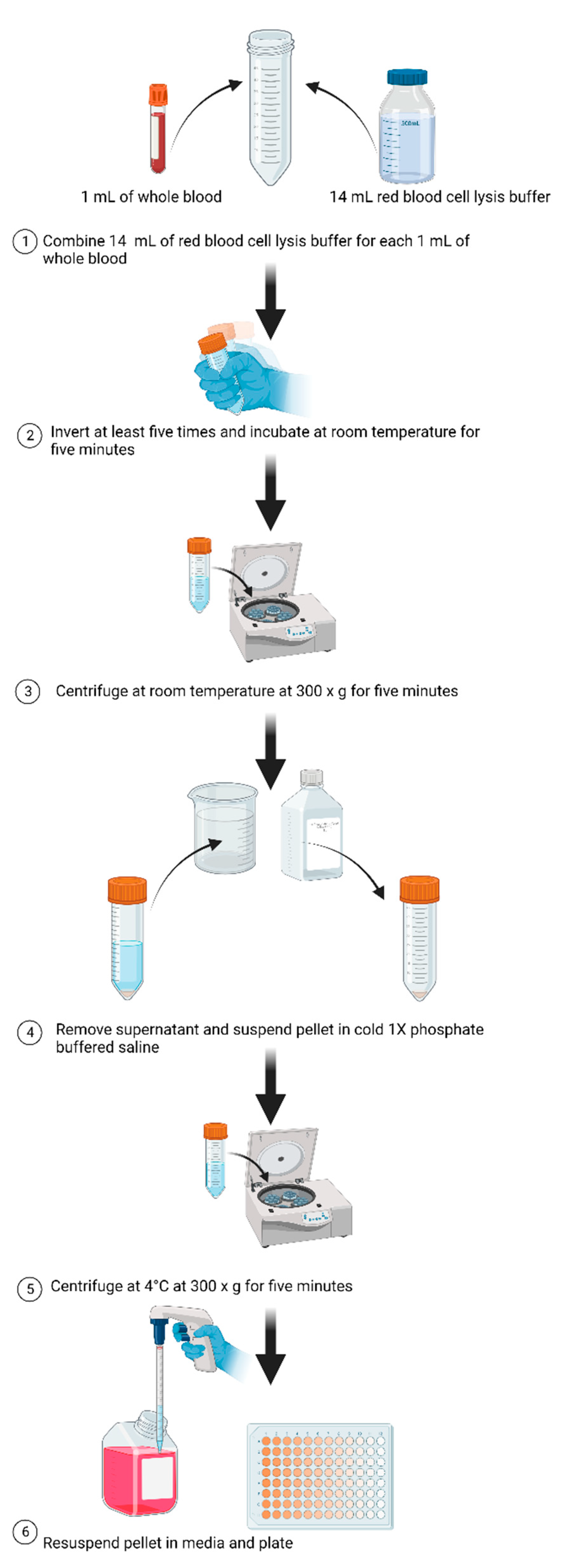
2.2. Preparation of Blood Samples

Twenty millilitres of whole blood was collected in ethylenediaminetetraacetic acid (EDTA) tubes from one participant that was used for the optimisations. The blood was distributed equally between two EDTA tubes and stored at room temperature before red blood cell (RBC) lysis processing.
As depicted in Figure 1, 1 mL blood samples were placed into 50 mL centrifuge tubes with 14 mL of cold RBC lysis buffer (0.16 M NH4Cl (Sigma-Aldrich (Australia) A9434)), 10 mM KHCO3 (Sigma-Aldrich (Australia) (60339)) and 0.13 mM EDTA (Sigma-Aldrich (Australia) (E8008)), dissolved in sterile H2O) [18]. The samples were inverted at least five times before incubation at room temperature for five minutes. After incubation, the samples were centrifuged at 300× g for five minutes at room temperature. Important: if a pellet did not form, the sample was not sufficiently mixed. In this case, repeat the inversion mixing process before repeating the centrifuge step. The supernatant was then removed, and the pellets suspended in 5 mL of cold 1× phosphate buffered saline (PBS) (Sigma-Aldrich (Australia) (D1408)) for every 1 mL of blood. The samples were then centrifuged at 300× g for 5 min at 4 °C. The pelleted cells were then suspended in 1 mL of RPMI 1640 media (Sigma-Aldrich (Australia) (R0883)) and tubes collated with double the volume of collated resuspension mix added. The samples were then plated, stimulated with 5 µg/mL PHA (Sigma-Aldrich (Australia) (L2769), and incubated at 37 °C and 5% CO2 for 72 h.

2.3. Proliferation

Peripheral blood mononuclear cells (PBMCs) were plated at a density of 5 × 10⁵ cells/mL for all assessments of PBMC stimulation. Increasing concentrations of PHA (final concentrations of 0, 0.25, 0.5, 1, 2.5, 5 and 10 µg/mL) were added, and the samples were incubated for 0, 24, 48, 72 and 168 h at 37 °C, 5% CO2. Following each incubation period, the proliferation was assessed via spectrometry and flow cytometry.

2.4. Assessment of Proliferation

This study utilised the MTS (3-(4,5-dimethylthiazol-2-yl)-5-(3-carboxymethoxyphenyl)-2-(4-sulfophenyl)-2H-tetrazolium) assay described by Mani, Johnson [19] with modifications to assess cellular metabolic activity as a measurement of cellular proliferation and viability. In brief, following removal of the DNA damaging agent, each sample was resuspended in 50 µL of media and supplemented 10 µL CellTiter 96 Aqueous One Solution Reagent (MTS reagent) (G3582) (Promega, Madison, WI, USA) for 4 h at 37 °C, 5% CO2. Absorbance was then measured at 490 and 630 nm on a SPECTROstar Omega spectrometer (BMG Labtech, Ortenberg, Germany) [19,20].

2.5. DNA Damage and Repair

The samples were treated with 25 µM etoposide (Cayman Chemicals (product number 12092), Ann Arbor, MI, USA) for two hours; washed with PBS twice; and then, were incubated for 0, 1, 2, 3 or 4 h with fresh media. All samples were centrifuged for five minutes at 400× g and washed with PBS twice. The samples were then treated with 4% paraformaldehyde (Sigma-Aldrich (Australia) (P6148) for 15 min at 4 °C and centrifuged for five minutes to pellets. As the paraformaldehyde treatment caused splaying, all centrifuge steps following the paraformaldehyde treatment were at 700× g. The samples were then washed with PBS twice before the samples were resuspended in 70% ethanol overnight at 4 °C. The next day, the samples were centrifuged to pellet cells and washed with PBS twice to remove the ethanol. The samples were then permeabilised with 100 µL of 0.1% Triton X-100 (Triton X-100 (Sigma-Aldrich (Melbourne, VIC, Australia) (product number X100)), 1X PBS) for five minutes. Note: the samples must not be incubated for more than five minutes; otherwise, the cellular integrity is destroyed by the permeabilisation.
The sample was then centrifuged at 1300× g for 5 min and washed twice with fluorescence-activated cell sorting (FACS) buffer (1X PBS, 0.5% bovine serum albumin (Sigma-Aldrich (Melbourne, VIC, Australia) (A4503)), 0.05% sodium azide) [3]. The samples were then centri-fuged at 700× g for 5 min to remove the supernatant and incubated in 200 µL of a blocking buffer (3% bovine serum albumin in Milli-Q water) at room temperature for 30 min. After incubation, the samples were centrifuged. To prevent drying of the cells during pooling of the samples, the supernatant was only removed in individual wells just before adding 100 µL of conjugated A488 anti-phospho-histone H2AX (γ-H2AX) antibody (anti-phospho histone H2A.X (Ser139), clone JBW301, Alexa Fluor® 488 conjugate antibody) (Merck Life Science, Melbourne, VIC, Australia). The 100 µL was mixed with the well before being transferred to the next duplicate well. Each sample contained the pellets of 4 wells from a 96-well plate resuspended in 100 µL of conjugated A488 anti-phospho-histone H2AX (γ-H2AX) antibody at a 1:600 concentration (1% bovine serum albumin, Milli-Q, anti-phospho histone H2A.X (Ser139), clone JBW301, Alexa Fluor® 488 conjugate antibody). The samples were incubated in the dark at room temperature for two hours before being centrifuged, washed with PBS twice and resuspended in 300 µL of the FACS solution. For flow cytometry analysis, the samples were transferred to 1.5 mL Eppendorf tubes.

2.6. Statistical Analysis

All the data were collected from a single participant under ethical approval from the Central Queensland Human Research Ethics Committee (CQUHREC clearance number 0000021074; date of approval April 2018). All experiments were conducted on a single day with dual or triplicate technical replicates, unless otherwise stated. All FACS analyses were set to a threshold of 10,000 minimum cells. The data were analysed on GraphPad Prism 10.1 software (San Diego, CA, USA) using a one-way ANOVA and mixed effects model. The data are presented as mean ± SD, and statistical significance was considered at p < 0.05.

3. Results

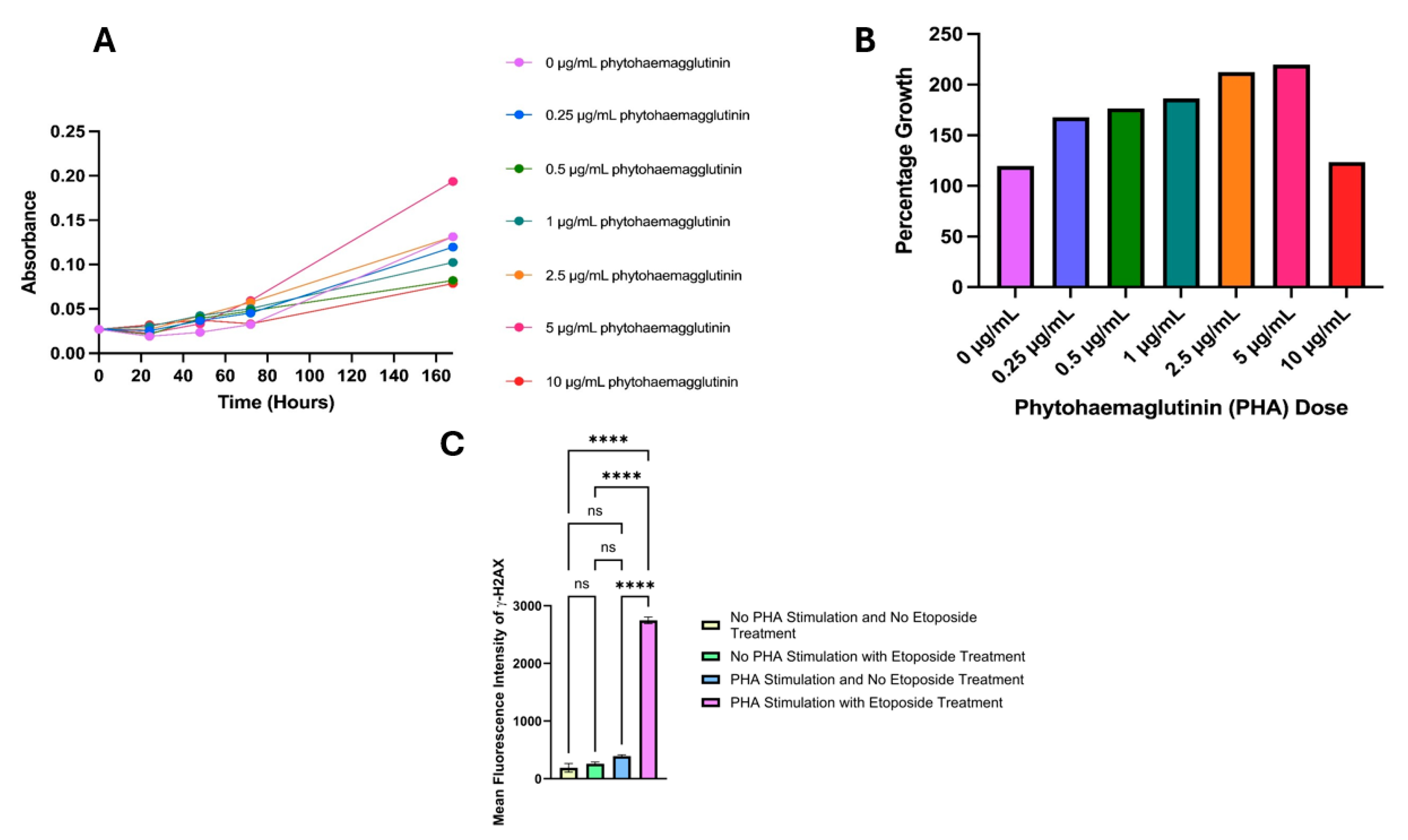
3.1. Proliferation Dose Selection

Chemotherapeutic agents such as etoposide are unable to induce DNA damage in quiescent cell populations such as PBMCs. Indeed, the mean fluorescence intensity of γ-H2AX had no significant changes after exposure of quiescent PBMCs to etoposide (Figure 2C). Hence, PHA was used to stimulate proliferation in PBMCs. This optimisation explored the use of seven doses of PHA (0, 0.25 µg/mL, 0.5 µg/mL, 1 µg/mL, 2.5 µg/mL, 5 µg/mL and 10 µg/mL) over five incubation periods (0 h, 24 h, 48 h, 72 h and 168 h) (Figure 2A). The data shown below depict the absorbance at each time point across one experiment; significant cell clumping was observed in the 168 h sample, and all 10 µg/mL samples and were excluded for consideration in further experiments. Seventy-two hours was selected due to its common use in the literature, its reduced clumping and being the most suitable for the experimental design [21,22] (Figure 2B). The addition of PHA did not significantly increase the γ-H2AX fluorescence levels (Figure 2C). However, the addition of etoposide to PHA-stimulated PBMCs significantly increase γ-H2AX fluorescence. This suggests that the stimulation of PBMCs is a requirement to predispose them to the genotoxic effects of etoposide.

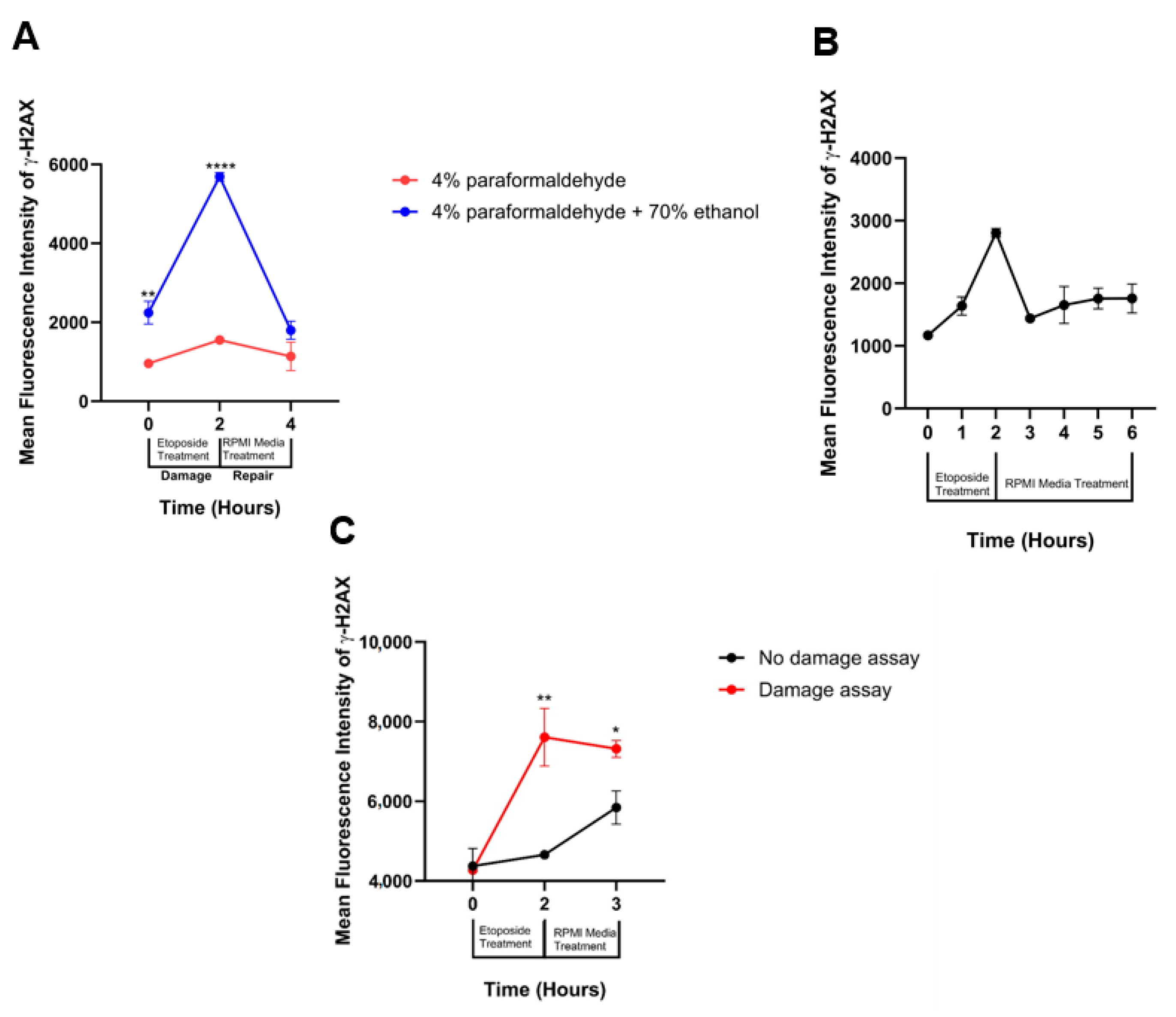
3.2. Fixation Selection

Previous observational work within our laboratory in HeLa cells determined etoposide to be the best DNA double-strand break-inducing agent for use in DNA damage and repair assessments as the other tested chemotherapeutic agents (mitomycin C and cisplatin) caused widespread cell death at low doses. Etoposide is also frequently used in the literature for the induction of DNA damage within PBMC [11,14]. Our work also investigated two methods of fixation: 4% paraformaldehyde only and 4% paraformaldehyde with 70% ethanol in overnight incubation (Figure 3A). A significantly higher response to the two-hour etoposide treatment was seen when the sample was fixed with 4% paraformaldehyde for 15 min followed by 70% ethanol overnight incubation.

3.3. DNA Damage Repair Window Selection

In our optimisation experiments, we also evaluated the DNA repair kinetic window. These experiments assessed the mean fluorescence intensity of γ-H2AX across six sampling times, which included a baseline sample that had not been exposed to any DNA damaging agents; samples incubated with etoposide for two hours (peak DNA damage); and samples that were incubated at 37 °C in fresh RPMI 1640 media (Sigma-Aldrich (Melbourne, VIC, Australia) for one, two, three or four hours (Figure 3B). As the mean fluorescence intensity of γ-H2AX returned to near baseline levels at three hours, further experiments were concluded following one-hour fresh media incubation (three-hour total experiment time).
To ensure the DNA damage seen throughout the assay was not due to the experimental conditions, an additional experiment was conducted, where the assay was run without the addition of etoposide. While there was an increase in the percentage change in γ-H2AX, it was concluded that this increase did not affect the DNA damage response induced by etoposide (Figure 3C).

4. Discussion

Previous research has proven that γ-H2AX and COMET assays are comparable in the assessment of DNA double-strand break repair; however, this assessment has been in a static cell population [5]. In this study, we analysed the proliferation response of PBMC after treatment with PHA at final concentrations of 0, 0.25, 0.5, 1, 2.5, 5 and 10 µg/mL over periods of 0, 24, 48, 72 and 168 h. The most effective duration of PHA treatment was determined to be 72 h due to high proliferation without the severe cellular clumping observed in the 168 h sample. Given the significant morphological changes that occur in PBMCs during PHA treatment, it important to control for the additional non-specific background fluorescence due to these changes in cell size and shape during cellular expansion of the PBMCs. Figure 2C shows that there were no significant changes in γ-H2AX fluorescence as result of PHA; however, it was a requirement to activate these cells for etoposide to have an effect on these cells.
Further experiments showed that 72 h was most suitable for the experimental design as it followed other published experimental models [21,22]. Furthermore, this methodological development study determined that PBMC proliferation is critical for DNA damage and repair studies as our experiments proved that significant DNA damage occurs when PBMCs are proliferated prior to treatment with etoposide compared to samples that were not treated with the proliferative agent (Figure 2C). Therefore, we propose that our standardised method for the analysis of DNA damage after PBMC proliferation is adopted.
This study also demonstrated that decreasing the incubation time with 4% paraformaldehyde from overnight to 15 min followed by an overnight incubation with 70% ethanol significantly improved the mean fluorescence intensity of γ-H2AX. Our study also demonstrated that one-hour incubation with fresh RPMI media is sufficient to see substantial changes in the mean fluorescence intensity of γ-H2AX (Figure 3B). Additionally, this methodology demonstrates that extended repair incubation is not required when quantifying DNA double-strand breaks via γ-H2AX.

5. Conclusions

Within this study, methods of assessing the DNA double-strand break repair response following treatment with the DNA damaging agent etoposide were developed as a standard method for assessing dynamic DNA double-strand break repair within PBMC. This study found that effective modelling and assessment of DNA double-strand breaks within PBMC with γ-H2AX requires the use of a proliferation stimulant. In this case, we recommend the use of PHA at a final concentration of 5 µg/mL. The DNA damaging agent (etoposide) is also required to be removed prior to analyses of DNA damage, with two-hour exposure sufficient to induce a significant double-strand break damage response. This study also determined that one hour after removal of etoposide and incubation with fresh media is a suitable period for the assessment of DNA double-strand break repair. These findings show that for an effective assessment of DNA double-strand break repair with γ-H2AX in PBMC, PBMCs should be stimulated with PHA at a final concentration of 5 µg/mL for 72 h prior to treatment with etoposide for two hours and assessment of repair in the hour following fresh RPMI 1640 media incubation.

Author Contributions

Conceptualisation, H.H., W.P., P.N. and A.F.; methodology, H.H., P.N. and A.F.; software, H.H. and A.F.; validation, H.H. and P.N.; formal analysis, H.H. and A.F.; investigation, H.H.; resources, H.H., W.P., P.N. and A.F.; data curation, H.H. and A.F.; writing—original draft preparation, H.H.; writing—review and editing, P.N. and A.F.; visualisation, H.H.; supervision, W.P., P.N. and A.F.; project administration, H.H. and A.F.; funding acquisition, P.N. and A.F. All authors have read and agreed to the published version of the manuscript.

Funding

This research was supported by an Australian Government Research Training Program (RTP) Scholarship awarded to the first author (H.H.).

Institutional Review Board Statement

This study was conducted in accordance with the Declaration of Helsinki and approved by the Central Queensland Human Research Ethics Committee (CQUHREC clearance number 0000021074; date of approval April 2018) for studies involving humans.

Informed Consent Statement

Informed consent was obtained from all subjects involved in this study.

Data Availability Statement

The datasets presented in this article are not readily available because the data are part of an ongoing study. Requests to access the datasets should be directed to the corresponding author.

Conflicts of Interest

The authors declare no conflicts of interest.

References

  1. Raavi, V.; Perumal, V.; Paul, S.F. Potential application of γ-H2AX as a biodosimetry tool for radiation triage. Mutat. Res./Rev. Mutat. Res. 2021, 787, 108350. [Google Scholar] [CrossRef] [PubMed]
  2. Najafi, M.; Fardid, R.; Hadadi, G.; Fardid, M. The mechanisms of radiation-induced bystander effect. J. Biomed. Phys. Eng. 2014, 4, 163. [Google Scholar]
  3. Stefanou, D.T.; Bamias, A.; Episkopou, H.; Kyrtopoulos, S.A.; Likka, M.; Kalampokas, T.; Photiou, S.; Gavalas, N.; Sfikakis, P.P.; Dimopoulos, M.A.; et al. Aberrant DNA damage response pathways may predict the outcome of platinum chemotherapy in ovarian cancer. PLoS ONE 2015, 10, e0117654. [Google Scholar] [CrossRef] [PubMed]
  4. Lampert, E.J.; Cimino-Mathews, A.; Lee, J.S.; Nair, J.; Lee, M.J.; Yuno, A.; An, D.; Trepel, J.B.; Ruppin, E.; Lee, J.M. Clinical outcomes of prexasertib monotherapy in recurrent BRCA wild-type high-grade serous ovarian cancer involve innate and adaptive immune responses. J. Immunother. Cancer 2020, 8, e000516. [Google Scholar] [CrossRef]
  5. Hosking, H.; Pederick, W.; Neilsen, P.; Fenning, A. Considerations for the Use of the DNA Damage Marker γ-H2AX in Disease Modeling, Detection, Diagnosis, and Prognosis. Aging Cancer 2024. [Google Scholar] [CrossRef]
  6. Jiao, J.; Zhao, X.; Hou, R.; Wang, Y.; Chang, W.; Liang, N.; Liu, Y.; Xing, J.; Cao, Y.; Li, X.; et al. Comparison of two commonly used methods for stimulating T cells. Biotechnol. Lett. 2019, 41, 1361–1371. [Google Scholar] [CrossRef]
  7. Beliakova-Bethell, N.; Hezareh, M.; Wong, J.K.; Strain, M.C.; Lewinski, M.K.; Richman, D.D.; Spina, C.A. Relative efficacy of T cell stimuli as inducers of productive HIV-1 replication in latently infected CD4 lymphocytes from patients on suppressive cART. Virology 2017, 508, 127–133. [Google Scholar] [CrossRef]
  8. Lin, Z.; Huang, Y.; Jiang, H.; Zhang, D.; Yang, Y.; Geng, X.; Li, B. Functional differences and similarities in activated peripheral blood mononuclear cells by lipopolysaccharide or phytohemagglutinin stimulation between human and cynomolgus monkeys. Ann. Transl. Med. 2021, 9, 257. [Google Scholar] [CrossRef] [PubMed]
  9. Smith-Norowitz, T.A.; Abdelmajid, H.; Yin, C.; Choice, S.; Joks, R.; Kohlhoff, S. Effect of Dimethyl Sulfoxide on B Cells in PHA-Stimulated PBMC from Adult Subjects. A Pilot Study. Ann. Clin. Lab. Sci. 2023, 53, 126–129. [Google Scholar]
  10. Chuchawankul, S.; Khorana, N.; Poovorawan, Y. Piperine inhibits cytokine production by human peripheral blood mononuclear cells. Genet. Mol. Res. 2012, 11, 617–627. [Google Scholar] [CrossRef]
  11. Bruni, E.; Reichle, A.; Scimeca, M.; Bonanno, E.; Ghibelli, L. Lowering etoposide doses shifts cell demise from caspase-dependent to differentiation and caspase-3-independent apoptosis via DNA damage response, inducing AML culture extinction. Front. Pharmacol. 2018, 9, 1307. [Google Scholar] [CrossRef] [PubMed]
  12. Wei, F.; Hao, P.; Zhang, X.; Hu, H.; Jiang, D.; Yin, A.; Wen, L.; Zheng, L.; He, J.Z.; Mei, W.; et al. Etoposide-induced DNA damage affects multiple cellular pathways in addition to DNA damage response. Oncotarget 2018, 9, 24122. [Google Scholar] [CrossRef] [PubMed]
  13. Sunter, N.J.; Cowell, I.G.; Willmore, E.; Watters, G.P.; Austin, C.A. Role of Topoisomerase II in DNA Damage Response following IR and Etoposide. J. Nucleic Acids 2010, 2010, 710589. [Google Scholar] [CrossRef] [PubMed]
  14. Menendez, D.; Anand, J.R.; Murphy, C.C.; Bell, W.J.; Fu, J.; Slepushkina, N.; Buehler, E.; Martin, S.E.; Lal-Nag, M.; Nitiss, J.L.; et al. Etoposide-induced DNA damage is increased in p53 mutants: Identification of ATR and other genes that influence effects of p53 mutations on Top2-induced cytotoxicity. Oncotarget 2022, 13, 332. [Google Scholar] [CrossRef] [PubMed]
  15. Vlachogiannis, N.I.; Ntouros, P.A.; Pappa, M.; Kravvariti, E.; Kostaki, E.G.; Fragoulis, G.E.; Papanikolaou, C.; Mavroeidi, D.; Bournia, V.-K.; Panopoulos, S.; et al. Chronological Age and DNA Damage Accumulation in Blood Mononuclear Cells: A Linear Association in Healthy Humans after 50 Years of Age. Int. J. Mol. Sci. 2023, 24, 7148. [Google Scholar] [CrossRef] [PubMed]
  16. Al Nasrallah, N.; Zhou, H.; Smith, P.A.; Sears, C.R. DNA Repair Capacity for Personalizing Risk and Treatment Response−Assay Development and Optimization in Human Peripheral Blood Mononuclear Cells (PBMCs). DNA Repair 2022, 111, 103274. [Google Scholar] [CrossRef] [PubMed]
  17. Schumann, S.; Scherthan, H.; Pfestroff, K.; Schoof, S.; Pfestroff, A.; Hartrampf, P.; Hasenauer, N.; Buck, A.K.; Luster, M.; Port, M.; et al. DNA damage and repair in peripheral blood mononuclear cells after internal ex vivo irradiation of patient blood with 131 I. Eur. J. Nucl. Med. Mol. Imaging 2022, 49, 1447–1455. [Google Scholar] [CrossRef] [PubMed]
  18. Liu, X.; Quan, N. Immune cell isolation from mouse femur bone marrow. Bio-Protocol 2015, 5, e1631. [Google Scholar] [CrossRef]
  19. Mani, J.; Johnson, J.; Hosking, H.; Walsh, K.; Neilsen, P.; Naiker, M. In vitro cytotoxic properties of crude polar extracts of plants sourced from Australia. Clin. Complement. Med. Pharmacol. 2022, 2, 100022. [Google Scholar] [CrossRef]
  20. Mani, J.; Johnson, J.; Hosking, H.; Hoyos, B.E.; Walsh, K.B.; Neilsen, P.; Naiker, M. Bioassay guided fractionation protocol for determining novel active compounds in selected Australian flora. Plants 2022, 11, 2886. [Google Scholar] [CrossRef]
  21. Norian, R.; Delirezh, N.; Azadmehr, A. (Eds.) Evaluation of proliferation and cytokines production by mitogen-stimulated bovine peripheral blood mononuclear cells. In Veterinary Research Forum; Faculty of Veterinary Medicine, Urmia University: Urmia, Iran, 2015. [Google Scholar]
  22. Mandarapu, R.; Ajumeera, R.; Venkatesan, V.; Prakhya, B.M. Proliferation and TH1/TH2 cytokine production in human peripheral blood mononuclear cells after treatment with cypermethrin and mancozeb in vitro. J. Toxicol. 2014, 2014, 308286. [Google Scholar] [CrossRef] [PubMed]
Figure 1. Red blood cell lysis. Original artwork created with BioRender.com.
Labmed 01 00003 g001
Figure 2. The treatment of peripheral blood mononuclear cells (PBMCs) with phytohaemagglutinin (PHA) across various concentrations and incubation times. (A) The samples were treated with final concentrations of 0, 0.25, 0.5, 1, 2.5, 5 and 10 µg/mL PHA for 0, 24, 48, 72 and 168 h at 37 °C, 5% CO2. The optimal time for PHA treatment was 72 h. (B) The optimal dose of PHA at 72 h was determined to be 5 µg/mL due to having the maximum percent growth with minimal clumping. (C) Determination of the need for cell proliferation with PHA prior to the treatment. These results show that the proliferation with PHA and treatment with etoposide significantly increased the average γ-H2AX fluorescence per cell compared to samples that were not treated with PHA or etoposide, samples treated with etoposide only or samples treated with PHA only. However, there was no significant difference between samples not treated with PHA or etoposide and samples only treated with PHA, which shows that PHA does not cause DNA damage. The cell numbers were controlled at 10,000 cells per sample. Non-significant; ns, p < 0.0001; ****.
Labmed 01 00003 g002
Figure 3. The determination of a DNA damage fixation method and repair window in PBMC following etoposide treatment. (A) A comparison of 4% paraformaldehyde and 4% paraformaldehyde with 70% ethanol for the fixation of cells. The combination of 4% paraformaldehyde with 70% ethanol showed optimal average fluorescence of γ-H2AX and was chosen as the fixation method for the final assay. (B) The sample was incubated with etoposide for 2 h and then washed twice with PBS to remove the etoposide and incubated with fresh RPMI media for 1, 2, 3 or 4 h to determine the optimised DNA repair window of 1 h. As minimal change was seen after 1 h incubation with fresh media (3 h time point), this was selected for use in the final DNA damage assay. (C) Determination of the effect of the assay on DNA damage. A significantly higher mean fluorescence intensity was seen in samples that were treated with etoposide, showing that there is minimal effect from the DNA damage assay (centrifuging and washing steps) on DNA damage. p < 0.05; *, p < 0.01; **, p < 0.0001; ****.
Labmed 01 00003 g003
Disclaimer/Publisher’s Note: The statements, opinions and data contained in all publications are solely those of the individual author(s) and contributor(s) and not of MDPI and/or the editor(s). MDPI and/or the editor(s) disclaim responsibility for any injury to people or property resulting from any ideas, methods, instructions or products referred to in the content.

