Article

Molecular Characterization of MDR and XDR Clinical Strains from a Tertiary Care Center in North India by Whole Genome Sequence Analysis

1
Department of Microbiology, Jawaharlal Nehru Medical College and Hospital, Aligarh Muslim University, Aligarh 202002, India
2
Maharaja Suhel Dev Autonomous State Medical College, Mahrishi Balark Hospital, Bahraich 271801, India
3
Central Research Laboratory, Kempegowda Institute of Medical Sciences and Research Centre, Bangalore 560013, India
*
Author to whom correspondence should be addressed.
J. Oman Med. Assoc. 2024, 1(1), 29-47; https://doi.org/10.3390/joma1010005
Submission received: 2 August 2024 / Revised: 14 September 2024 / Accepted: 18 September 2024 / Published: 24 September 2024

Abstract

:
Whole genome sequencing (WGS) has the potential to greatly enhance AMR (Anti-microbial Resistance) surveillance. To characterize the prevalent pathogens and dissemination of various AMR-genes, 73 clinical isolates were obtained from blood and respiratory tract specimens, were characterized phenotypically by VITEK-2 (bioMerieux), and 23 selected isolates were genotypically characterized by WGS (Illumina). AST revealed high levels of resistance with 50.7% XDR, 32.9% MDR, and 16.4% non-MDR phenotype. A total of 11 K. pneumoniae revealed six sequence types, six K-locus, and four O-locus types, with ST437, KL36, and O4 being predominant types, respectively. They carried ESBL genes CTX-M-15 (90.9%), TEM-1D (72.7%), SHV-11 (54.5%), SHV-1, SHV-28, OXA-1, FONA-5, and SFO-1; NDM-5 (72.7%) and 63.6%OXA48-like carbapenamases; 90.9%OMP mutation; dfrA12, sul-1, ermB, mphA, qnrB1, gyrA831, and pmrB1 for other groups. Virulence gene found were Yerisiniabactin (90.9%), aerobactin, RmpADC, and rmpA2. Predominant plasmid replicons were Col(pHAD28), IncFII, IncFIB(pQil), and Col440. A total of seven XDR A. baumannii showed single MLST type(2) and single O-locus type(OCL-1); with multiple AMR-genes: blaADC-73, blaOXA-66, blaOXA-23, blaNDM-1, gyrA, mphE, msrE, and tetB. Both S. aureus tested were found to be ST22, SCCmec IVa(2B), and spa type t309; multiple AMR-genes: blaZ, mecA, dfrC, ermC, and aacA-aphD. Non-MDR Enterococcus faecalis sequenced was ST 946, with multiple virulence genes. This study documents for the first-time prevalent virulence genes and MLST types, along with resistance genes circulating in our center.

1. Introduction

Globally, infections are the leading cause of mortality, morbidity, and prolonged hospitalization. Infectious diseases such as bloodstream infections (BSIs) and pneumonia are potentially lethal conditions, particularly in healthcare settings and in intensive care units (ICU). Gram-negative bacilli, such as Klebsiella pneumoniae, Escherichia coli, Pseudomonas aeruginosa, Acinetobacter baumannii, and other nil-fermenters are increasingly reported from these invasive and nosocomial infections [1]. Gram-positive bacteria isolated from BSI include Staphylococcus species, which is also the most common BSI pathogen overall, and Enterococcus species. Moreover, methicillin-resistant S. aureus (MRSA) is increasingly isolated from ICU patients and poses significant risk, especially to immunocompromised persons [1,2].
Aggravating the problem is the propensity of these notorious bacteria to develop resistance against the antimicrobial agents used for treating these infections. Because of the worldwide dissemination of antimicrobial resistance (AMR) and the lack of novel therapeutic options available, AMR has been declared a “global public health concern” by various important international bodies, such as the World Health Organization (WHO), Infectious Diseases Society of America (IDSA), and Centers for Disease Control and Prevention (CDC) [3,4]. Broadly, 700,000 deaths per year globally are due to AMR, which is estimated to further reach up to 10 million by the year 2050 [5]. Moreover, an insufficient supply of alternative drugs and a high prevalence of resistant pathogens in low and middle-income countries [6,7] lead to a pronounced impact of AMR in resource-poor settings such as ours.
Hence intelligent surveillance of AMR infections is crucial for effective policy making. Characterization of pathogens plays a pivotal role in the epidemiology of infectious diseases, providing information critical for identification, tracking, and infection control. Thus, a proper understanding of the resistance mechanism, route of transmission, and epidemiology of infectious agents helps in the reduction of the spread of AMR agents.
Molecular surveillance can provide additional value over phenotypic methods to national surveillance systems. Bacterial DNA sequence information helps in a better understanding of AMR pathogens. Whole genome sequencing (WGS) is one such powerful tool for molecular surveillance and can characterize resistance genes, virulence, as well as transmissibility, and thus can be used in outbreak investigation to deduce the origin and transmission route of AMR agents. Furthermore, WGS helps in determining different AMR mechanisms, as well as in the identification of complex mechanisms, like efflux pump up-regulation or changes in membrane permeability, which are difficult to infer just by standard procedures. WGS provides unprecedented scope to study genomic content as well as corroborate the frequency and diversity of clusters [8]. This study aimed to characterize the prevalent pathogens circulating in our hospital, which is a tertiary care center in North India, and to explore the dissemination of various AMR genes, multilocus sequence types, and virulence genes in resistant pathogens.

2. Material and Methods

2.1. Place of Study and Ethical Consideration

This is a preliminary exploratory study conducted in the Department of Microbiology of a tertiary care center in North India for a basic understanding of prevalent pathogens circulating in the hospital. The clinical isolates were characterized phenotypically by preliminary identification and antibiotic susceptibility testing in the Department of Microbiology, and genotypically by WGS conducted by the Central Research Laboratory, India. This research was conducted in accordance with the ethical standards of the institute. This study was approved by the Institutional Ethical Committee (Ref: IECJNMC/1504).

2.2. Sample Processing and Bacterial Isolation

A total of 73 Bacterial isolates were obtained from blood and respiratory tract specimens received for routine culture and sensitivity testing in the Bacteriology and Enteric laboratory of the Department of Microbiology over a period of 2 months. A total of 30 blood samples were collected in BacT/ALERT (bioMérieux, Inc., Durham, NC, USA) bottles with aseptic precautions. A total of 38 respiratory tract specimens included endotracheal aspirate, broncho-alveolar lavage, and sputum. Blood culture was identified as positive by the automated system, and respiratory tract specimens were inoculated onto routine culture media: 5% sheep blood agar, chocolate agar, and MacConkey agar, and were processed as per laboratory protocol.

2.3. Antimicrobial Sensitivity and Supplementary Testing

All clinical isolates were processed by the VITEK-2 (bioMérieux, Inc., Durham, NC, USA) automated system for identification as well as antimicrobial sensitivity. All isolates were stored at −20 °C for further characterization. Sensitivity results were interpreted as per CLSI 2022 guidelines [9].
Certain AMR phenotypes further underwent confirmatory tests. For detection of methicillin resistance in Staphylococcus aureus, isolates were tested by disk diffusion method using 30 µg cefoxitin disk (HiMedia, Maharashtra, India). As per CLSI 2022 guidelines [9], zones of inhibition of size ≤ 21 mm were considered positive, and ≥22 mm were considered negative for mecA-mediated resistance. All Staphylococcus aureus and Enterococcus species were screened for vancomycin resistance by vancomycin agar screen using a fixed concentration of 6 µg/mL vancomycin (HiMedia, Maharashtra, India). According to CLSI 2022 guidelines [9], >1 colony signifies presumptive resistance and should be further confirmed by MIC testing.
Colistin resistance was tested by colistin broth disk elution (CBDE). Standardized inoculum of isolates was inoculated in four concentrations of 0 µg/mL (growth control), 1 µg/mL, 2 µg/mL, and 4 µg/mL of colistin (HiMedia, Maharashtra, India) and incubated overnight. As per CLSI 2022 guidelines, colistin MIC of ≤2 µg/mL was considered as intermediate, and ≥4 µg/mL was considered resistant [9].

2.4. AMR Phenotype

According to the antimicrobial susceptibility test results, isolates were further classified as non-MDR, MDR, XDR, or PDR phenotype according to definitions given by CDC expert group [10]. A multidrug-resistant (MDR) organism is defined as a bacterial isolate that is not susceptible to at least one agent in three or more antimicrobial classes. The bacterial isolate that is not susceptible to at least one agent in all but two or fewer antimicrobial classes is defined as extensively drug-resistant (XDR). When the isolate is found not susceptible to all the antimicrobial agents in all antimicrobial classes, they are considered pan-drug resistant (PDR).

2.5. Clinical History

Relevant clinical data was also collected such as patient characteristics, presenting complaints and diagnosis, presence of fever, leukocyte count, intubated or not, and outcome of the patient.

2.6. Molecular Characterization

These clinical isolates were then sent to the Central Research Laboratory, India for further characterization by whole genome sequencing. Genomic DNA extracted from pure colonies was processed for whole genome sequencing using the Illumina MiSeq platform. Library preparation involved shearing the DNA, end repair, A-tailing, and adapter ligation using the NEBNext Ultra II FS DNA Library Prep Kit [11], followed by PCR amplification and size selection. Library quality was assessed using the Agilent TapeStation system, and subsequent paired-end sequencing with a read length of 250 bp was performed using the MiSeq Reagent Kit v2. Contigs were generated of the strains which passed sequence quality control with the help of SPAdes version 3.14 [12], and annotated by Prokka version 1.5 [13]. ARIBA tool version 2.14.4 was used for the identification of MLST, AMR, and virulence genes [14].

3. Results

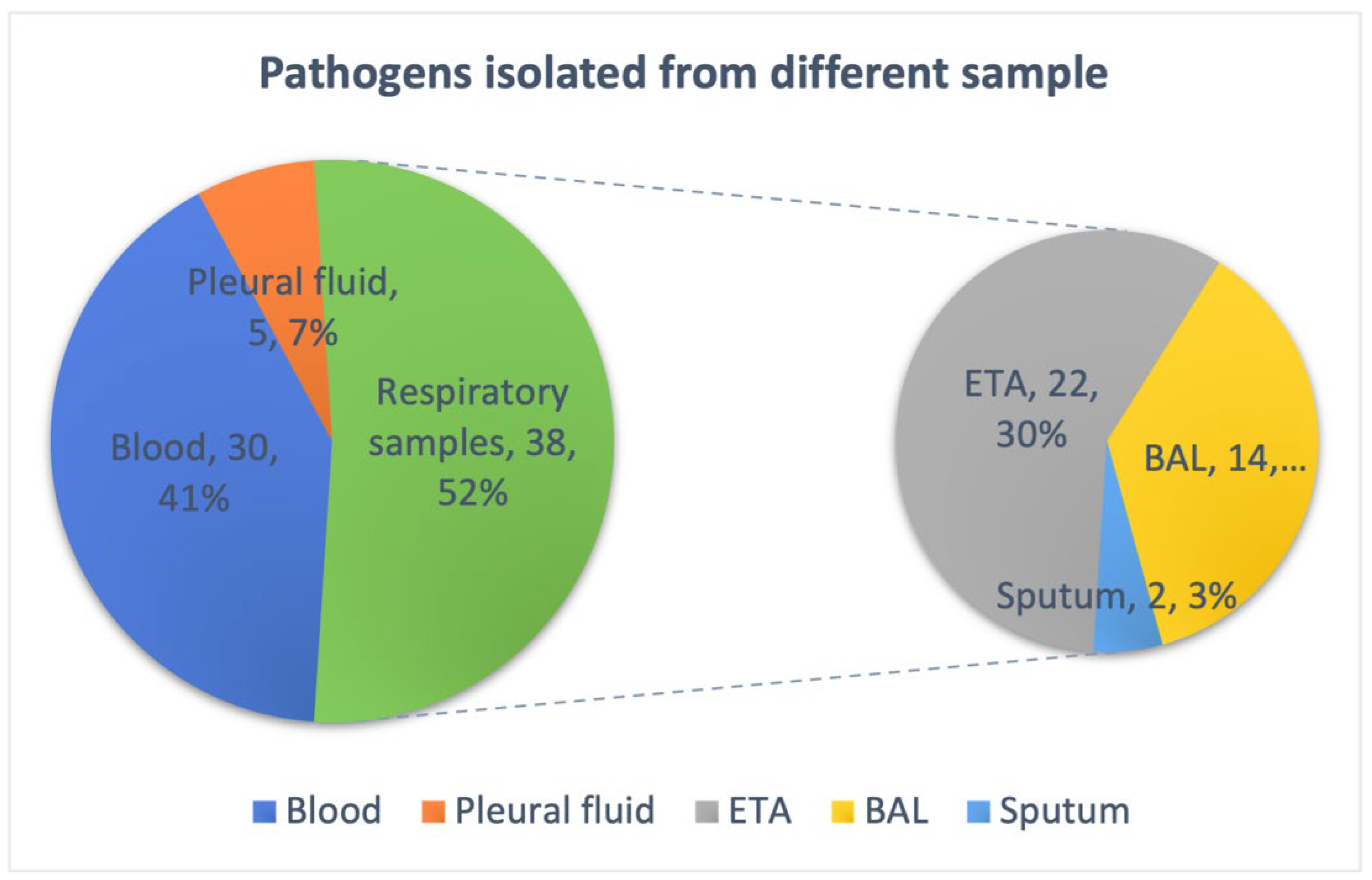
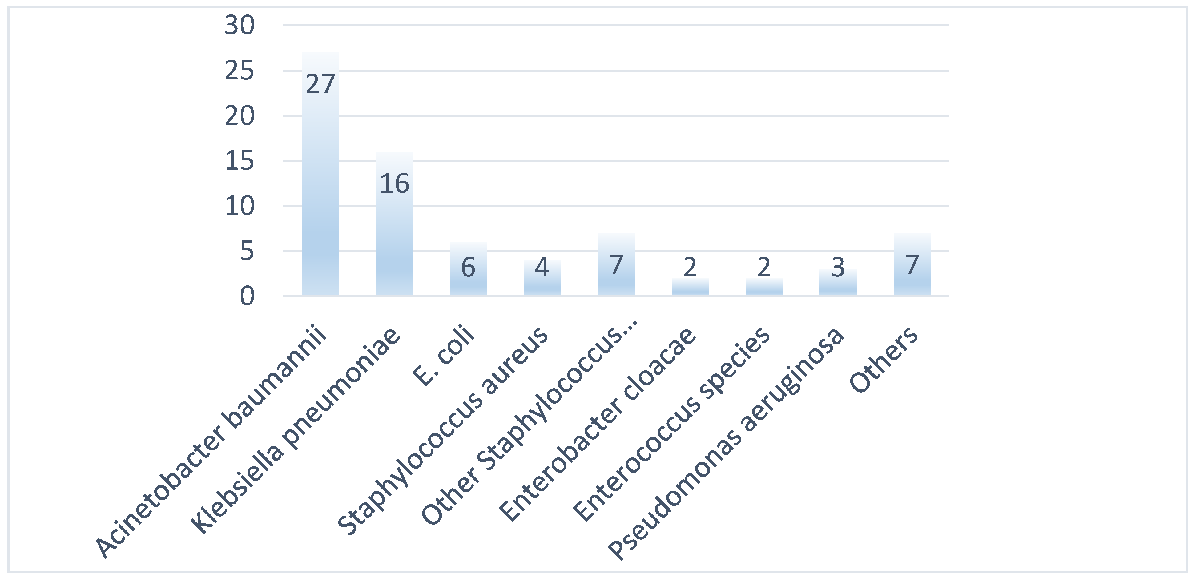
3.1. Bacterial Isolates and Phenotypic Characterization
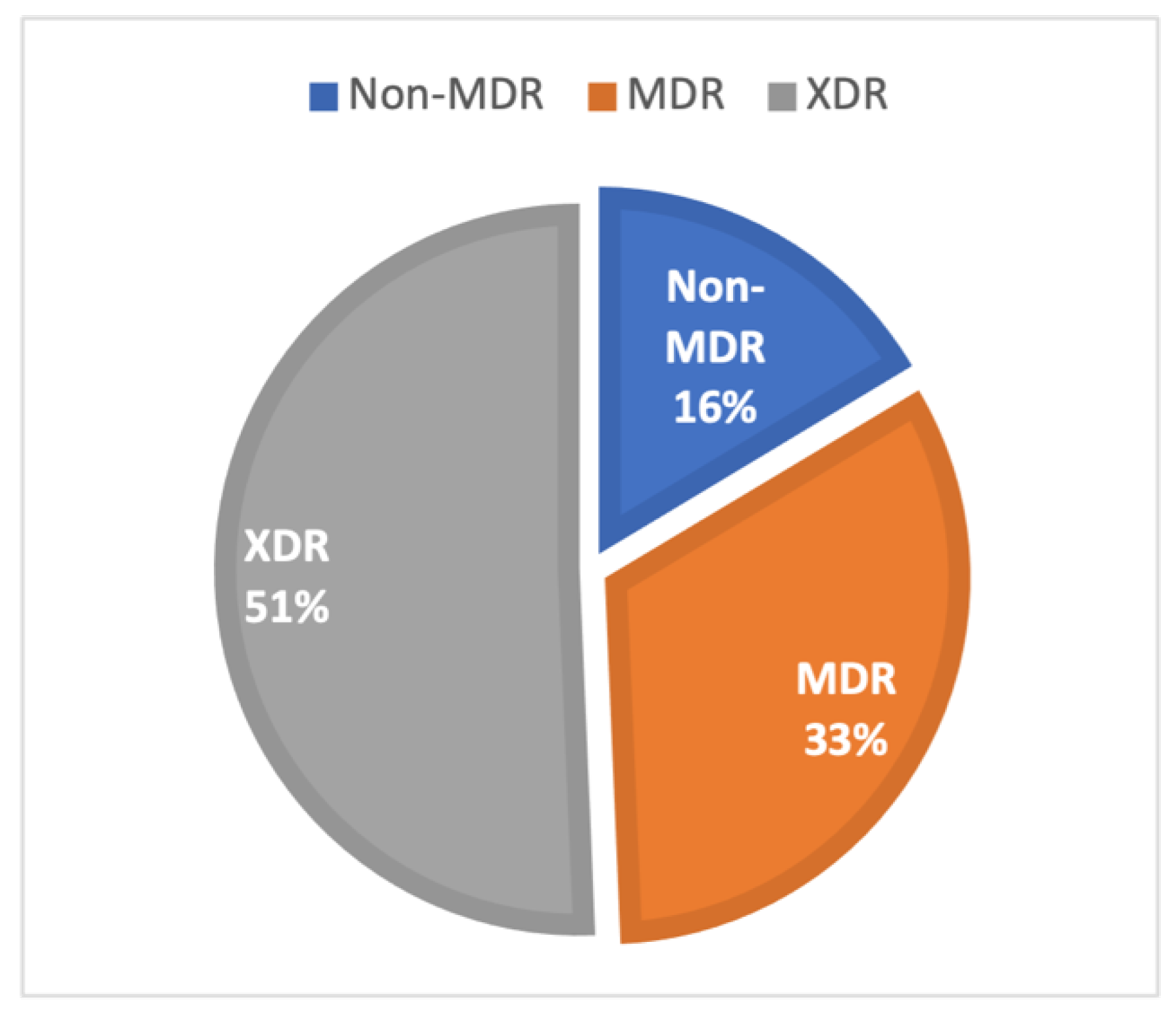
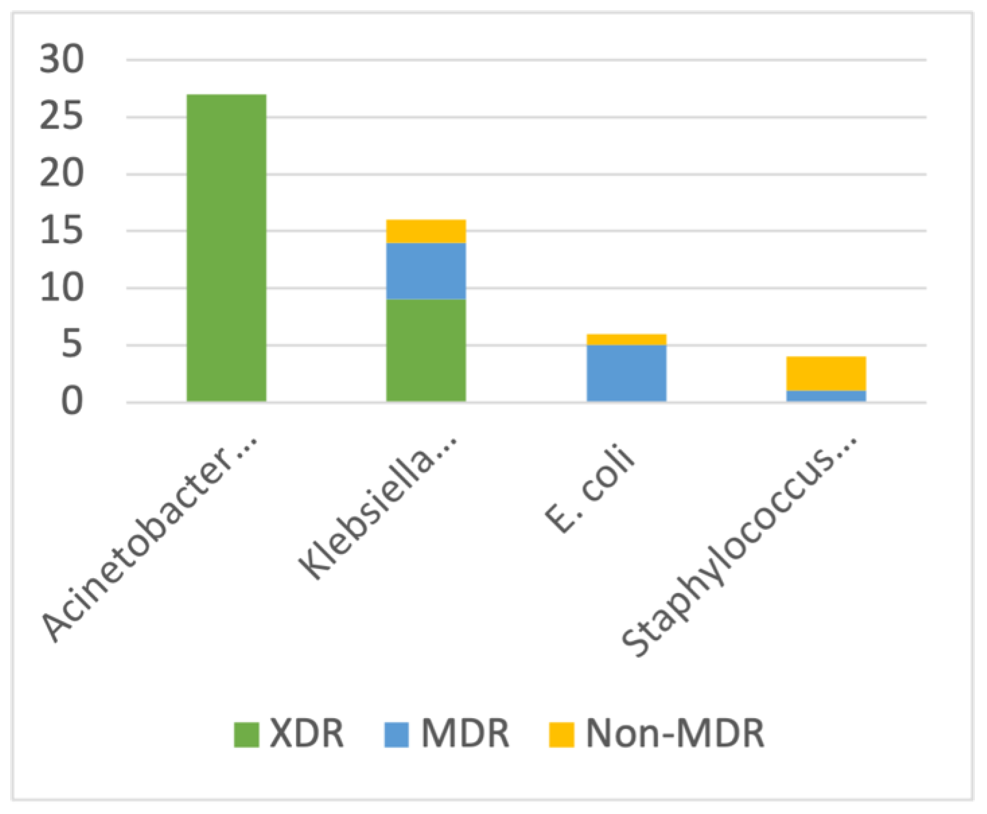
The study included 73 clinical isolates, out of which 30 (41%) were from blood samples, five (7%) from pleural fluid, and 38 (52%) from respiratory tract specimens, which included 22 (30%) endotracheal aspirate, 14 (19%) broncho-alveolar lavage, and two (3%) sputum samples (Figure 1).
Out of the 73 clinical isolates, the majority (29, 39.7%) were from pediatrics, including neonatology, the rest from medicine alliance (26, 35.6%) and surgery alliance (18, 24.7%). Almost half (49.3%) of all isolates were from critical care units.
Bacterial distribution of 73 isolates showed that the maximum was Acinetobacter baumannii complex (27, 37%), followed by Klebsiella pneumoniae (16, 21.9%), then other Staphylococcus species (7, 9%), Escherichia coli (6, 8.2%), Staphylococcus aureus (4, 5.5%), Pseudomonas aeruginosa (3, 4%), Enterobacter cloacae complex (2, 2.7%), Enterococcus species (2, 2.7%), and others, as depicted in Figure 2.

3.2. AMR Phenotypes

Antimicrobial susceptibility tests (Table 1 and Table 2) revealed high levels of resistance with 37 isolates (50.7%) to be XDR phenotype, 24 (32.9%) MDR, and 12 (16.4%) non-MDR phenotype (Figure 3). From critical care units, 78.4% (29/37) isolates were found to be XDR.
It was observed that all of the 27 Acinetobacter baumannii (100%) were of XDR phenotype, with one being possibly PDR, showing resistance to at least one of the β-lactams, carbapenems, aminoglycosides, quinolones, β-lactam/inhibitor combinations, and other clinical antimicrobial agents (Table 1, Figure 4). A possible explanation could be that most of these isolates were from critical care units (81.5%) and the majority were from respiratory tract specimens (92.6%).
Out of 16 Klebsiella pneumoniae included in the study, nine (56.3%) were found to be XDR, five (31.2%) MDR, and two non-MDR (Figure 4). XDR isolates were susceptible to colistin only and resistant to cephalosporins, carbapenems, β-lactam/inhibitor combinations, fluoroquinolones, aminoglycosides, and other antibiotic groups, whereas, five MDR isolates were found susceptible to amikacin, gentamicin, tigecycline, and cotrimoxazole, along with colistin (Table 1).
Amongst six E. coli isolates, five were found to be MDR and one non-MDR phenotype. The most observed resistance phenotypes for E. coli isolates were against ceftriaxone, cefuroxime, cefotaxime, ciprofloxacin, and trimethoprim/sulfamethoxazole, with all six isolates being resistant to all these, followed by gentamicin (4/6), amoxicillin/clavulanate (4/6), and piperacillin/tazobactam (3/6). The most susceptible antibiotics were colistin (6/6), amikacin (5/6), and imipenem and meropenem (4/6).
All four Staphylococcus aurei were detected as resistant to penicillin, oxacillin, cefoxitin, ciprofloxacin, and erythromycin, and three were resistant to clindamycin and gentamicin (Table 2). All four were susceptible to vancomycin, teicoplanin, linezolid, tigecycline, and daptomycin.

3.3. Clinical Data of Isolates with Molecular Characterization

Clinical data, antimicrobial usage, period of stay in ICU, and patient outcome were analyzed in 23 samples whose isolates were molecularly characterized (Table 3). Two patients yielded the same organisms from two different samples, one from blood and one from endotracheal aspirate. It was observed that seven patients (33.3%) were from pediatric units, and overall, 15 (71.4%) patients were from critical care units. A total of 47.6% were male and 52.3% females. A total of 71.4% had a fever, and 80.9% had raised counts. The majority of the patients (76.2%) expired and only three patients (14.2%) had favorable outcomes.

3.4. Molecular Analysis

Out of the total isolates sent for WGS, 23 isolates, which include 11 Klebsiella pneumoniae, seven Acinetobacter baumannii, two Staphylococcus aureus, two Escherichia coli, and one Enterococcus faecalis, were eventually revived and analyzed for sequencing and the rest were excluded due to various technical reasons. From their whole genome sequences analysis, AMR determinants, capsular types, multilocus sequence types, plasmid replicon types, and virulence genes were determined.

3.4.1. Klebsiella Pneumoniae

Sequencing analysis of 11 Klebsiella pneumoniae revealed seven sequence types, with the predominant type being ST437 (5, 45.4%), others types were ST15 (2, 18.2%), and (1, 9%) each of ST231, ST16, ST15, ST987, and ST147. Capsular diversity analysis revealed seven K-locus and four O-locus types. Seven K-locus types were distributed as KL36 (4, 36.4%), KL51 (2, 18.2%), KL112 (2, 18.2%), and (1, 9%) each of KL52, KL81, and KL39. KL36 was detected in ST437 isolates, KL112 in ST 15, KL51 in ST147 and ST 231, KL81 in ST16, and KL39 in ST985. Four O-locus types identified were O4 (4, 36.4%), O1/O2V2 (3, 27.3%), O1/O2V1 (2, 18.2%), and OL101 (2, 18.2%). All four isolates with O4 serotypes were detected in ST437 [Table 4].
All sequenced Klebsiella pneumoniae isolates had a varied combination of resistance genes to different antibiotic classes. Among beta-lactamases, 90.9% were found to harbor CTX-M-15, 72.7% TEM-1D, and 54.5% with SHV-11 AMR gene. Other beta-lactamases genes detected were SHV-1, SHV-28, SHV-187, OXA-1, FONA-5, and SFO-1. Apart from this, 72.7% of isolates had NDM-5 and 63.6% had OXA48-like as major carbapenamases. OXA-181 and OXA-232 are the commonly found variants of OXA48-like carbapenamases and were found in 36.4% and 27.3% of isolates, respectively. NDM-1 was also detected in one isolate. We also found outer membrane protein (OMP) mutation in 90.9% of all isolates, as OmpK36GD in 45.5% and OmpK36TD in 45.5% of isolates.
Amongst 11 Klebsiella pneumoniae, ST985 was the only isolate in which FONA-5, SFO-1, and NDM-1 gene were detected, and unlike all other isolates, ST985 lacks CTX-M-15 and OmpK mutation (Table 4). All patients from whom these Klebsiella pneumoniae were isolated succumbed to their disease and expired, except one with ST985, which improved and was later discharged (Table 3).
Apart from beta-lactams, resistance genes were also found for other classes of antibiotics, such as dfrA12 (54.5%), dfrA30 (27.3%) for trimethoprim resistance; sul-1 (90.9%) for sulphonamide; ermB (72.7%) and mphA (90.9%) for macrolide; arr-2 (36.4%) for rifampicin, and qnrB1 (45.5%) for fluoroquinolone resistance. For fluoroquinolone, the predominant mutations observed were ParC-80I (81.8%) and GyrA-83I (63.6%), others were GyrA-83F, GyrA-87N, and ParC-84K. Colistin resistance by pmrB1 was also present in one isolate (ST437). Overall, we report excellent concordance with phenotypic and genotypic AMR.
The predominant plasmid replicons detected in the 11 K. pneumoniae isolates sequenced were Col(pHAD28) (10, 90.9%), IncFII (9, 81.8%), IncFIB(pQil) (7, 63.6%), Col440II (6, 54.5%), and Col440I (5, 45.5%). Other plasmid replicons present were ColpVC, IncFIB(K), IncFII, IncL, ColKP3, ColRNAI, IncFIB(pKPHS1), IncFII(pKP91), IncFII(K), IncHI1B(pNDM-MAR), repB, IncFIB(K), IncFIB(pNDM-Mar), IncR, IncX3, IncFIA, IncFII(pAMA1167-NDM-5), FIA(pBK30683), IncFIB(AP001918), and IncFII(pRSB107). Among virulence genes, the majority of isolates had yerisiniabactin (90.9%). Other genes responsible for virulence found were aerobactin, RmpADC, and rmpA2.

3.4.2. Acinetobacter baumannii

Sequencing analysis of seven XDR Acinetobacter baumannii showed single multi-locus sequence type (MLST) type (2), single O-locus type (OCL-1), and different K-locus types (KL9, KL234, KL49, and KL4). The majority of isolates were found harboring multiple AMR genes conferring resistance to different antibiotic classes, such as to beta-lactams via blaADC-73 (83.3%), blaOXA-66 (100%), blaOXA-23 (100%), and blaNDM-1 (66.7%); to aminoglycosides by ant(3″)-IIa (100%), aph(3″)-Ib (100%), aph(6)-Id (100%), armA (100%), and aph(3′)-Ia (66.7%); phenicoles: catB8 (50%); tetracyclines: tetB (100%); sulfonamides: sul1 and sul2; macrolides and lincosamide: mphE (100%) and msrE (100%); and fluoroquinolone: gyrA (100%) and parC. However, we did not identify any MDR efflux pump in any of our isolates. All these XDR isolates were obtained from respiratory tract specimens from patients admitted to critical care units and expired during their ICU stay.

3.4.3. Escherichia coli

Two isolates that were sequenced were isolated from blood samples. One was identified as sequence type 53 and the other a novel type. For different antibiotic classes, AMR genes detected were aadA2, rmtB1, aadA5, aph(3″)-Ib, aph(6)-Id, and aac(3)-IId for aminoglycosides; blaOXA-181, blaNDM-5, blaTEM-1, and blaCTX-M-15 for beta-lactams; mph(A) and erm(B) for macrolides; qnrS1 for quinolone; sul1 and sul2 for sulfonamides; tet(B) and tet(A) for tetracyclines; and dfrA12 and dfrA17 for trimethoprim. Genotypic AMR was in concordance with the in-silico resistance pattern in all antibiotic classes.
Some virulence genes were detected in ST53, but not in isolates with novel sequence types, such as iron uptake gene iucA, iucB, iucC, iucD, iroB, iroC, iroD, iroE, iroN, irp1, and irp2; P fimbriae genes: papA, papB, papC, papD, papE, papF, papG, papH, papI, papJ, papK, and papX; hyaluronidases: hlyA, hlyB, hlyC, and hlyD; hemin receptor genes: chuA, chuS, chuT, chuU, chuV, chuW, chuX, and chuY; Pyelonephritis-associated pili: papA, papB, papC, papD, papE, papF, papG, papH, papI, papJ, papK, and papX; yersiniabactin (Ybt), an additional siderophore which binds copper and other non-iron metal ions and found in extra-intestinal pathogenic E. coli: ybtA, ybtE, ybtP, ybtQ, ybtS, ybtT, ybtU, and ybtX. However, enteroaggregative heat-stable toxin (coded by astA) was found in the novel type but not in the ST53 isolate. Virulence genes found in both isolates were enterobactin: entA, entB, entC, entD, entE, entF, and entS; ferric enterobactin: fepA, fepB, fepC, fepD, and fepG; type 1 fimbriae: fimA, fimB, fimC, fimD, fimE, fimF, fimG, fimH, and fimI; fibroblast growth factor receptor: flgE, flgG, flgH, flgJ, and flgM; general secretory pathway: gspC, gspD, gspE, gspF, gspG, gspH, gspI, gspJ, gspK, gspL, and gspM; flagellar component cheA and cheD.

3.4.4. Staphylococcus aureus

In two isolates of Staphylococcus aureus sequenced and analyzed, SCCmec IVa(2B) was identified as the main SCCmec type (staphylococcal cassette chromosome mec), sequence type ST22, and spa (S. aureus protein A) type t309 in both isolates. Various combination of genotypes found was blaZ (β-lactams resistance), mecA (methicillin-resistance determinant), ermC (erythromycin ribosome methylase gene for macrolides-lincosamides-streptogramin), aacA-aphD (aminoglycosides), and dfrC (trimethoprim). However, the far-1 gene, responsible for fusidic acid resistance, as well as the mupirocin-resistance gene mupA was not detected in our study. Vancomycin resistance genes were also not detected which was in accordance with the phenotypic susceptibility of all isolates to vancomycin.
Regarding virulence factors, both isolates were positive for Panton-Valentine leukocidin (PVL) gene (lukF-PV + lukS-PV), as well as enterotoxin gene cluster (egc, consisting of seg, sei, sel, sem, sen, seo, and seu), toxic shock toxin gene (tst1), staphylokinase (sak), gamma hemolysin (hlgA, hlgB, and hlgC) and other immune evasion complex (IEC) genes (such as scn and sak) [15,16].
Both of these Staphylococcus aurei were isolated from blood samples of ward patients (non-critical) and both had favorable outcomes and showed improvement after treatment.

3.4.5. Enterococcus faecalis

One isolate was sequenced and analyzed, which was isolated from the blood sample of a hospitalized neonate and was determined to be of multilocus sequence type ST 946. The isolate was non-MDR, showing phenotypic antibiotic resistance against aminoglycoside, lincosamide/macrolide/streptogramin, quinolone, streptothricin, and tetracycline. Antimicrobial resistance genes conferring resistance to aminoglycosides (ant(6)-Ia, aph(3′)-IIIa and aac(6′)-Ie/aph(2″)-Ia), lincosamides (lnu(G) and lsa(A)), lincosamides/macrolides/streptogramin (erm(B)), streptothricin (sat4), quinolone (parC_S80I, gyrA_S83I), and tetracyclines (tet(L) and tet(M)) were identified. No van genes were detected, which conferred phenotypic susceptibility to vancomycin as well.
Virulence genes detected include genes responsible for the adhesion of pathogens, such as esp (enterococcal surface protein), efaA (endocarditis-specific antigen A), ace (collagen-binding proteins), ebp pilli (ebpA, ebpB, and ebpC); invasins like gelE (gelatinase) and sprE; and genes for biofilm production such as bopD and fsrC. Different capsule encoding genes detected were cpsA, cpsB, cpsC, cpsD, cpsE, cpsF, cpsG, cpsH, cpsI, cpsJ, and cpsK.

4. Discussion

The emergence and dissemination of resistance against anti-microbials is a serious threat to public health globally. Infections caused by MDR pathogens are difficult to treat and cause prolonged hospitalization and higher mortality. On one hand, bacterial pathogens, such as Klebsiella pneumoniae, Pseudomonas aeruginosa, Acinetobacter baumannii, and other nil-fermenters have the propensity to colonize admitted patients and hospital environment and thus act as reservoirs of infections and are frequent sources of healthcare-associated infections [1]. On the other hand, certain bacteria present naturally low-level susceptibility to certain drugs, irrespective of past antibiotic exposure and not due to horizontal gene transfer. A few examples of this phenomenon, also known as intrinsic resistance, are macrolides and vancomycin for gram-negative bacilli, and colistin for gram-positive bacteria [17]. Moreover, extensive antimicrobial usage in hospitals leads to selective pressure, causes extrinsic resistance, and helps in the further dissemination of AMR.
One such AMR pathogen Klebsiella pneumoniae, has been reported as an important agent of nosocomial infections, especially in immunocompromised patients and ICUs [18,19]. The determination of mechanisms of resistance in these isolates plays an important role in developing effective treatment guidelines.
Sequencing analysis of 11 Klebsiella pneumoniae in this study revealed seven sequence types, with the predominant type being ST437 (5, 45.4%). Other types were ST15 (2, 18.2%), and (1, 9%) each of ST231, ST16, ST15, ST987, and ST147. However, ST258, the most prevailing CRKP worldwide, was not found in our study. This is in accordance with published data from India [20]. The predominant type, ST437, is a global epidemic clone and has been reported in Europe, America, and Asia, including India. This ST is related to the dominant epidemic multidrug-resistant clonal complex CC11 [21]. Another high-risk type found globally, usually found with XDR strains [22], ST147 was also found in our study. Yet another CRKP clone, ST231, which has been reported in India, and several other Southeast Asian and European countries, was also found in our study [20]. Reports documenting these MDR clones from the country emphasize the gravity of the worrisome state of AMR in the subcontinent.
Capsular diversity analysis of 11 Klebsiella pneumoniae in our study revealed seven K-locus and four O-locus types. Seven K-locus types were distributed as KL36 (4, 36.4%), KL51 (2, 18.2%), KL112 (2, 18.2%), and (1, 9%) each of KL52, KL81, and KL39 in this study. Various K-loci reported from the Indian subcontinent are KL1, KL2, KL5, KL20, KL17, KL51, KL54, KL57, and KL64 [23]. The four O-locus types identified were O4 (4, 36.4%), O1/O2V2 (3, 27.3%), O1/O2V1 (2, 18.2%), and OL101 (2, 18.2%). Serotypes most frequently isolated were O1, O2, and O3, with almost 80% of infectious diseases associated with these serotypes [24].
In our study, all sequenced Klebsiella pneumoniae isolates had a varied combination of resistance genes to different antibiotic classes. Among beta-lactamases, 90.9% were found to harbor CTX-M-15, 72.7% TEM-1D, and 54.5% with SHV-11 AMR gene. OXA-181 and OXA-232 were the commonly found variants of OXA48-like carbapenamases and were found in 36.4% and 27.3% of isolates. NDM-1 was also detected in one isolate. We also found outer membrane protein (OMP) mutation as OmpK36GD in 45.5% of isolates and OmpK36TD in 45.5% of isolates. These mutations are known to constrict the porin channel by insertion mutations of TD/GD amino acids, resulting in disruption of outer membrane protein. Overall, OmpK mutations were found in 90.9% of all isolates. Phenotypic resistance to carbapenems in isolates in our study can be explained by the presence of carbapenamases genes (blaOXA and blaNDM) and a combination of ESBLs and inactivation of porins OmpK35/36.
A study in India by Veeraraghavan B. et al. (2017) also reported CTM-15 as a major ESBL gene similar to ours. Also, they reported blaNDM and blaOXA48-like both in 28%, blaNDM in 19%, and blaOXA48-like in 13% [25]. As has been reported previously, in India, carbapenem resistance is predominantly due to NDM and OXA-48-like, although OXA-48-like are emerging [26]. Similar to this, we found 72.7% NDM-5 and 63.6% OXA48-like in our study. Bhatia M et al. reported 77.8% of blaOXA-48-like genes, and 55.6% blaNDM-1/5 in North India [27]. Notably, another carbapenamases gene found predominantly in Colombia and other European countries, blaKPC genes, was not detected in our study [28,29].
Acquisition of agents of horizontal gene transfer such as plasmids and mobile genetic elements leads to further extension of AMR burden. A proper understanding of these agents could help in effective policy-making to stop the dissemination of AMR genes. The predominant plasmid replicons detected in this study were Col(pHAD28) (10, 90.9%), IncFII (9, 81.8%), IncFIB(pQil) (7, 63.6%), Col440II (6, 54.5%), and Col440I (5, 45.5%).
Siderophores are iron acquisition systems essential for bacterial growth. Siderophore systems found frequently in Enterobacteriaceae are yersiniabactin, enterobactin, and aerobactin [30]. In this study, the virulence factor gene, yerisiniabactin, was present in 90.9% of isolates. Another important virulence factor known for hypervirulent K. pneumoniae, aerobactin, was also found in few. This finding is in agreement with other published data in India [27]. Further virulence genes found in our study were RmpADC and rmpA2. These are carried by virulence plasmids and known to play a critical role in virulence expression by regulating capsular polysaccharide overproduction, along with the hypermucoviscosity phenotype of hypervirulent K. pneumoniae (hvKP) [31].
Another gram-negative pathogen is Acinetobacter baumannii, which is known to cause nosocomial infections, particularly in intensive care units, and is associated with high mortality [32]. In this study, 92.6% of Acinetobacter baumannii were from respiratory tract specimens, 81.5% were from critical care units, and all isolates (100%) were of XDR phenotype, with one being possibly PDR. Similar to our study, high isolation rates of A. baumannii from bronchopulmonary specimens have been previously reported from across the globe [33,34]. The tendency of the bacteria to colonize the medical devices and biofilm formation helps in the setting of lower respiratory tract infections.
In this study, all seven XDR Acinetobacter baumannii isolates sequenced showed single multilocus sequence type (MLST) type (2), single O-locus type (OCL-1), and different K-locus types (KL9, KL234, KL49, and KL4). This is in agreement with different international studies which also reported ST/2 as the sequence type most frequently found circulating in the Southeast Asian region including Thailand, Myanmar, Singapore, Vietnam, and Malaysia [35]. Furthermore, ST/2 was also the most dominant type found globally [36].
The majority of isolates were found harboring multiple AMR genes, conferring resistance to different antibiotic classes. Similar to our study, MDR and XDR A. baumannii are reported from all over the world in clinical isolates, especially from critical care units [37]. Antibiotic efflux pump-encoding genes are the well-documented mechanism of resistance in A. baumannii [35]. However, we did not identify any MDR efflux pumps in any of our isolates.
Carbapenems are generally considered as drugs of choice for the MDR A. baumannii isolates. However, in recent decades, higher rates of carbapenem-resistant A. baumannii (CRAB) infections have been reported, particularly nosocomial BSI and pneumonia. Class D oxacillinase enzymes are documented to be the main genotype in clinical A. baumannii isolates. We found all sequenced isolates were harboring blaOXA-66 and blaOXA-23. blaOXA-23-like gene is a dominant resistant determinant in the Asian subcontinent and has also been reported worldwide [38]. The presence of such XDR strains in clinical samples, and that too from ICUs, is unsettling. This condones the necessity of strict adherence to infection control practices.
Yet another significant bacterial pathogen known to cause nosocomial infections is Staphylococcus aureus. Particularly, methicillin-resistant S. aureus (MRSA) poses a significant risk, especially for immunocompromised persons [2]. MRSA strains express resistance to β-lactam antibiotics because of reduced binding affinity to β-lactams by altered penicillin-binding protein (PBP2a) which is coded by the mec gene (mecA, mecB, and mecC). These methicillin resistance (mecA–C) genes are carried by the mobile genetic element SCCmec (staphylococcal cassette chromosome mec) [39,40]. SCCmec can be classified as SCCmec type I to SCCmec XI [41]. In our study, SCCmec IVa(2B) was identified as the main SCCmec type in both isolates, which is reported as the most common SCC type in another report in North India as well [42].
Apart from MLST and SCCmec identification, another method of MRSA typing is the sequencing of the highly polymorphic repeat region of S. aureus protein A (spa) gene. The identification of spa clusters helps in differentiating between relapse and re-infection and may be used as a quick method for the study of MRSA epidemiology, particularly in a hospital setting [43]. In this study, two isolates of Staphylococcus aureus tested were found to be of the same sequence type (ST22), and spa type t309. This ST22 is a common sequence type reported from all over India as well as globally [44].
Enterococcus faecalis, a common gut flora, is notoriously known for causing various nosocomial infections, which can be attributed to its inherent property of intrinsic resistance to most antibiotics, virulence factors, and horizontal transfers of resistance genes [45,46]. The multilocus sequence type ST946 of one nosocomial Enterococcus faecalis determined in this study was once described in a clinical isolate from a urine sample of a hospitalized patient in Bangladesh in the year 2018 [47]. To the authors’ knowledge, the sequence types and virulence genes of Enterococcus spp. reported in this study are the first reported enterococcal genome sequences isolated in North India.
We conclude that numerous resistant pathogens are circulating in our hospital, carrying important resistance and virulence determinants. The presence of global epidemic clone ST437, and other high-risk MDR clones such as ST147 in K. pneumoniae, as well as high dissemination of AMR genes, notably 90.9% CTX-M-15, 72.7% TEM-1D, 54.5% SHV-11, 72.7% NDM-5, and 63.6%OXA48-like carbapenamases emphasize the gravity of the worrisome state of AMR. Moreover, all Acinetobacter baumannii isolates were found to be XDR and the majority were from critical care units. Furthermore, all sequenced isolates showed single MLST type (2) which is the most dominant type found globally, and all were harboring blaOXA-66 and blaOXA-23. The presence of such XDR strains in clinical samples, and that too from ICUs, is unsettling. Both S. aureus tested were found to be of the same ST22, SCCmec IVa(2B), and spa type t309. This study lays the foundation for future research, providing a basis for further exploration and investigation into sequence types and virulence genes along with resistance genes of MDR and XDR clinical strains.
The limitation of this study is the small sample size and the short duration. However, this study provides a preliminary understanding of the genetic characteristics of circulating strains, paving the way for more in-depth and comprehensive studies in our hospital. WGS information can also help in the assessment of possible outbreaks in the institution. Nonetheless, a notable advantage of the study is the availability of clinical data, antibiotic history, and outcome of the patient along with the sequence types of bacterial isolates.

5. Conclusions

This study elucidated the molecular characteristics of antimicrobial resistance, and documents for the first time prevalent virulence genes and multilocus sequence types (MLST), along with resistance genes, circulating in our center. This study accentuates the desire for genomic surveillance of MDR and XDR pathogens that can contribute to developing effective infection control policies and treatment guidelines based on integrating phenotypic and genotypic methods.

Author Contributions

Conceptualization, A.S. and F.K.; Data curation, U.T.; Formal analysis, U.T.; Investigation, B.B.; Methodology, U.T. and S.W.K.; Project administration, A.S.; Resources, G.N.; Supervision, F.K.; Visualization, A.A.; Writing—original draft, U.T. and S.A.; Writing—review & editing, A.S. All authors have read and agreed to the published version of the manuscript.

Funding

This research received no external funding.

Institutional Review Board Statement

Ethical approval was obtained from the Institutional Ethical Committee, Jawaharlal Nehru Medical College and Hospital, Faculty of Medicine, Aligarh Muslim University, Aligarh, India (Ref: IECJNMC/1504).

Informed Consent Statement

Not applicable.

Data Availability Statement

The original contributions presented in the study are included in the article.

Conflicts of Interest

The authors declare that the research was conducted in the absence of any commercial or financial relationships that could be construed as potential conflicts of interest.

References

  1. Gaynes, R.; Edwards, J.R. National Nosocomial Infections Surveillance System Overview of nosocomial infections caused by gram-negative bacilli. Clin. Infect. Dis. 2005, 41, 848–854. [Google Scholar] [CrossRef] [PubMed]
  2. Samia, N.I.; Robicsek, A.; Heesterbeek, H.; Peterson, L.R. Methicillin-resistant staphylococcus aureus nosocomial infection has a distinct epidemiological position and acts as a marker for overall hospital-acquired infection trends. Sci. Rep. 2022, 12, 17007. [Google Scholar] [CrossRef] [PubMed]
  3. World Health Organization. Global Antimicrobial Resistance Surveillance System: Manual for Early Implementation; World Health Organization: Geneva, Switzerland, 2015; Available online: https://iris.who.int/handle/10665/188783 (accessed on 29 January 2024).
  4. Spellberg, B.; Srinivasan, A.; Chambers, H.F. New Societal Approaches to Empowering Antibiotic Stewardship. JAMA 2016, 315, 1229–1230. [Google Scholar] [CrossRef] [PubMed]
  5. O’Neill, J. Tackling Drug-Resistant Infections Globally: Final Report and Recommendations; Antimicrobial Resistance; Wellcome Trust: London, UK, 2016. [Google Scholar]
  6. Alividza, V.; Mariano, V.; Ahmad, R.; Charani, E.; Rawson, T.M.; Holmes, A.H.; Castro-Sánchez, E. Investigating the impact of poverty on colonization and infection with drug-resistant organisms in humans: A systematic review. Infect. Dis. Poverty 2018, 7, 76. [Google Scholar] [CrossRef] [PubMed]
  7. Jasovský, D.; Littmann, J.; Zorzet, A.; Cars, O. Antimicrobial resistance-a threat to the world’s sustainable development. Ups. J. Med. Sci. 2016, 121, 159–164. [Google Scholar] [CrossRef]
  8. NIHR Global Health Research Unit on Genomic Surveillance of AMR. Whole-genome sequencing as part of national and international surveillance programmes for antimicrobial resistance: A roadmap. BMJ Glob. Health 2020, 5, e002244. [CrossRef]
  9. Lewis, J.S., II. Performance Standards for Antimicrobial Susceptibility Testing, 33rd ed.; M100Ed33; Clinical & Laboratory Standards Institute: Wayne, PA, USA, 2024; Available online: https://clsi.org/standards/products/microbiology/documents/m100/ (accessed on 27 March 2023).
  10. Magiorakos, A.-P.; Srinivasan, A.; Carey, R.B.; Carmeli, Y.; Falagas, M.E.; Giske, C.G.; Harbarth, S.; Hindler, J.F.; Kahlmeter, G.; Olsson-Liljequist, B.; et al. Multidrug-resistant, extensively drug-resistant and pandrug-resistant bacteria: An international expert proposal for interim standard definitions for acquired resistance. Clin. Microbiol. Infect. 2012, 18, 268–281. [Google Scholar] [CrossRef]
  11. Underwood, A. GHRU (Genomic Surveillance of Antimicrobial Resistance) Retrospective 1 Bioinformatics Methods. 2020. Available online: https://protocols.io/view/ghru-genomic-surveillance-of-antimicrobial-resista-bpn6mmhe (accessed on 14 September 2024).
  12. Bankevich, A.; Nurk, S.; Antipov, D.; Gurevich, A.A.; Dvorkin, M.; Kulikov, A.S.; Lesin, V.M.; Nikolenko, S.I.; Pham, S.; Prjibelski, A.D.; et al. SPAdes: A new genome assembly algorithm and its applications to single-cell sequencing. J. Comput. Biol. 2012, 19, 455–477. [Google Scholar] [CrossRef] [PubMed]
  13. Seemann, T. Prokka: Rapid prokaryotic genome annotation. Bioinformatics 2014, 30, 2068–2069. [Google Scholar] [CrossRef]
  14. Hunt, M.; Mather, A.E.; Sánchez-Busó, L.; Page, A.J.; Parkhill, J.; Keane, J.A.; Harris, S.R. ARIBA: Rapid antimicrobial resistance genotyping directly from sequencing reads. Microb. Genom. 2017, 3, e000131. [Google Scholar] [CrossRef]
  15. Monecke, S.; Coombs, G.; Shore, A.C.; Coleman, D.C.; Akpaka, P.; Borg, M.; Chow, H.; Ip, M.; Jatzwauk, L.; Jonas, D.; et al. A field guide to pandemic, epidemic and sporadic clones of methicillin-resistant Staphylococcus aureus. PLoS ONE 2011, 6, e17936. [Google Scholar] [CrossRef] [PubMed]
  16. Monecke, S.; Slickers, P.; Ehricht, R. Assignment of Staphylococcus aureus isolates to clonal complexes based on microarray analysis and pattern recognition. FEMS Immunol. Med. Microbiol. 2008, 53, 237–251. [Google Scholar] [CrossRef] [PubMed]
  17. Olivares Pacheco, J.A.; Bernardini, A.; Garcia-Leon, G.; Corona, F.; Sanchez, M.B.; Martinez, J.L. The intrinsic resistome of bacterial pathogens. Front. Microbiol. 2013, 4, e00103. [Google Scholar] [CrossRef]
  18. Asensio, A.; Oliver, A.; González-Diego, P.; Baquero, F.; Pérez-Díaz, J.C.; Ros, P.; Cobo, J.; Palacios, M.; Lasheras, D.; Cantón, R. Outbreak of a multiresistant Klebsiella pneumoniae strain in an intensive care unit: Antibiotic use as risk factor for colonization and infection. Clin. Infect. Dis. 2000, 30, 55–60. [Google Scholar] [CrossRef]
  19. Jarvis, W.R.; Munn, V.P.; Highsmith, A.K.; Culver, D.H.; Hughes, J.M. The epidemiology of nosocomial infections caused by Klebsiella pneumoniae. Infect. Control 1985, 6, 68–74. [Google Scholar] [CrossRef] [PubMed]
  20. Shankar, C.; Mathur, P.; Venkatesan, M.; Pragasam, A.K.; Anandan, S.; Khurana, S.; Veeraraghavan, B. Rapidly disseminating blaOXA-232 carrying Klebsiella pneumoniae belonging to ST231 in India: Multiple and varied mobile genetic elements. BMC Microbiol. 2019, 19, 137. [Google Scholar] [CrossRef]
  21. Sahoo, S.; Sahoo, R.K.; Dixit, S.; Behera, D.U.; Subudhi, E. NDM-5-carrying Klebsiella pneumoniae ST437 belonging to high-risk clonal complex (CC11) from an urban river in eastern India. 3 Biotech 2023, 13, 139. [Google Scholar] [CrossRef]
  22. Lee, C.-R.; Lee, J.H.; Park, K.S.; Kim, Y.B.; Jeong, B.C.; Lee, S.H. Global Dissemination of Carbapenemase-Producing Klebsiella pneumoniae: Epidemiology, Genetic Context, Treatment Options, and Detection Methods. Front. Microbiol. 2016, 7, 895. [Google Scholar] [CrossRef]
  23. Wyres, K.L.; Nguyen, T.N.T.; Lam, M.M.C.; Judd, L.M.; van Vinh Chau, N.; Dance, D.A.B.; Ip, M.; Karkey, A.; Ling, C.L.; Miliya, T.; et al. Genomic surveillance for hypervirulence and multi-drug resistance in invasive Klebsiella pneumoniae from South and Southeast Asia. Genome Med. 2020, 12, 11. [Google Scholar] [CrossRef]
  24. Follador, R.; Heinz, E.; Wyres, K.L.; Ellington, M.J.; Kowarik, M.; Holt, K.E.; Thomson, N.R. The diversity of Klebsiella pneumoniae surface polysaccharides. Microb. Genom. 2016, 2, e000073. [Google Scholar] [CrossRef]
  25. Veeraraghavan, B.; Shankar, C.; Karunasree, S.; Kumari, S.; Ravi, R.; Ralph, R. Carbapenem resistant Klebsiella pneumoniae isolated from bloodstream infection: Indian experience. Pathog. Glob. Health 2017, 111, 240–246. [Google Scholar] [CrossRef] [PubMed]
  26. Anandan, S.; Damodaran, S.; Gopi, R.; Bakthavatchalam, Y.D.; Veeraraghavan, B. Rapid Screening for Carbapenem Resistant Organisms: Current Results and Future Approaches. J. Clin. Diagn. Res. 2015, 9, DM01–DM03. [Google Scholar] [CrossRef]
  27. Bhatia, M.; Shamanna, V.; Nagaraj, G.; Sravani, D.; Gupta, P.; Omar, B.J.; Chakraborty, D.; Ravikumar, K.L. Molecular characterisation of carbapenem-resistant Klebsiella pneumoniae clinical isolates: Preliminary experience from a tertiary care teaching hospital in the Himalayas. Trans. R. Soc. Trop. Med. Hyg. 2022, 116, 655–662. [Google Scholar] [CrossRef] [PubMed]
  28. David, S.; Reuter, S.; Harris, S.R.; Glasner, C.; Feltwell, T.; Argimon, S.; Abudahab, K.; Goater, R.; Giani, T.; Errico, G.; et al. Epidemic of carbapenem-resistant Klebsiella pneumoniae in Europe is driven by nosocomial spread. Nat. Microbiol. 2019, 4, 1919–1929. [Google Scholar] [CrossRef] [PubMed]
  29. Saavedra, S.Y.; Bernal, J.F.; Montilla-Escudero, E.; Arévalo, S.A.; Prada, D.A.; Valencia, M.F.; Moreno, J.; Hidalgo, A.M.; García-Vega, Á.S.; Abrudan, M.; et al. Complexity of Genomic Epidemiology of Carbapenem-Resistant Klebsiella pneumoniae Isolates in Colombia Urges the Reinforcement of Whole Genome Sequencing-Based Surveillance Programs. Clin. Infect. Dis. 2021, 73, S290–S299. [Google Scholar] [CrossRef] [PubMed]
  30. Lawlor, M.S.; O’Connor, C.; Miller, V.L. Yersiniabactin Is a Virulence Factor for Klebsiella pneumoniae during Pulmonary Infection. Infect. Immun. 2007, 75, 1463–1472. [Google Scholar] [CrossRef] [PubMed]
  31. Ovchinnikova, O.G.; Treat, L.P.; Teelucksingh, T.; Clarke, B.R.; Miner, T.A.; Whitfield, C.; Walker, K.A.; Miller, V.L. Hypermucoviscosity Regulator RmpD Interacts with Wzc and Controls Capsular Polysaccharide Chain Length. mBio 2023, 14, e0080023. [Google Scholar] [CrossRef]
  32. Kurihara, M.N.L.; de Sales, R.O.; da Silva, K.E.; Maciel, W.G.; Simionatto, S. Multidrug-resistant Acinetobacter baumannii outbreaks: A global problem in healthcare settings. Rev. Soc. Bras. Med. Trop. 2020, 53, e20200248. [Google Scholar] [CrossRef]
  33. Alkasaby, N.M.; El Sayed Zaki, M. Molecular Study of Acinetobacter baumannii Isolates for Metallo-β-Lactamases and Extended-Spectrum-β-Lactamases Genes in Intensive Care Unit, Mansoura University Hospital, Egypt. Int. J. Microbiol. 2017, 2017, e3925868. [Google Scholar] [CrossRef]
  34. Gautam, D.; Dolma, K.G.; Khandelwal, B.; Goyal, R.K.; Mitsuwan, W.; Pereira, M.d.L.G.; Klangbud, W.K.; Gupta, M.; Wilairatana, P.; Siyadatpanah, A.; et al. Acinetobacter baumannii in suspected bacterial infections: Association between multidrug resistance, virulence genes, & biofilm production. Indian J. Med. Res. 2023, 158, 439. [Google Scholar] [CrossRef]
  35. Wareth, G.; Linde, J.; Nguyen, N.H.; Nguyen, T.N.M.; Sprague, L.D.; Pletz, M.W.; Neubauer, H. WGS-Based Analysis of Carbapenem-Resistant Acinetobacter baumannii in Vietnam and Molecular Characterization of Antimicrobial Determinants and MLST in Southeast Asia. Antibiotics 2021, 10, 563. [Google Scholar] [CrossRef] [PubMed]
  36. Hamidian, M.; Nigro, S.J. Emergence, molecular mechanisms and global spread of carbapenem-resistant Acinetobacter baumannii. Microb. Genom. 2019, 5, e000306. [Google Scholar] [CrossRef] [PubMed]
  37. Pormohammad, A.; Mehdinejadiani, K.; Gholizadeh, P.; Nasiri, M.J.; Mohtavinejad, N.; Dadashi, M.; Karimaei, S.; Safari, H.; Azimi, T. Global prevalence of colistin resistance in clinical isolates of Acinetobacter baumannii: A systematic review and meta-analysis. Microb. Pathog. 2020, 139, 103887. [Google Scholar] [CrossRef]
  38. Evans, B.A.; Amyes, S.G.B. OXA β-Lactamases. Clin. Microbiol. Rev. 2014, 27, 241–263. [Google Scholar] [CrossRef] [PubMed]
  39. Liu, J.; Chen, D.; Peters, B.M.; Li, L.; Li, B.; Xu, Z.; Shirliff, M.E. Staphylococcal chromosomal cassettes mec (SCCmec): A mobile genetic element in methicillin-resistant Staphylococcus aureus. Microb. Pathog. 2016, 101, 56–67. [Google Scholar] [CrossRef]
  40. Hiramatsu, K.; Katayama, Y.; Yuzawa, H.; Ito, T. Molecular genetics of methicillin-resistant Staphylococcus aureus. Int. J. Med. Microbiol. 2002, 292, 67–74. [Google Scholar] [CrossRef]
  41. Haaber, J.; Penadés, J.R.; Ingmer, H. Transfer of Antibiotic Resistance in Staphylococcus aureus. Trends Microbiol. 2017, 25, 893–905. [Google Scholar] [CrossRef]
  42. Bhatia, M.; Shamanna, V.; Nagaraj, G.; Sravani, D.; Gupta, P.; Omar, B.J.; Singh, A.; Rani, D.; Ravikumar, K.L. An insight into whole genome sequencing data of methicillin-resistant Staphylococcus aureus circulating in a teaching hospital in North India. Indian J. Med. Microbiol. 2023, 44, 100365. [Google Scholar] [CrossRef]
  43. Satta, G.; Ling, C.L.; Cunningham, E.S.; McHugh, T.D.; Hopkins, S. Utility and limitations of Spa-typing in understanding the epidemiology of staphylococcus aureus bacteraemia isolates in a single University Hospital. BMC Res. Notes 2013, 6, 398. [Google Scholar] [CrossRef]
  44. Abrudan, M.I.; Shamanna, V.; Prasanna, A.; Underwood, A.; Argimón, S.; Nagaraj, G.; Di Gregorio, S.; Govindan, V.; Vasanth, A.; Dharmavaram, S.; et al. Novel multidrug-resistant sublineages of Staphylococcus aureus clonal complex 22 discovered in India. mSphere 2023, 8, e00185-23. [Google Scholar] [CrossRef]
  45. Armin, S.; Fallah, F.; Karimi, A.; Rashidan, M.; Shirdust, M.; Azimi, L. Genotyping, antimicrobial resistance and virulence factor gene profiles of vancomycin resistance Enterococcus faecalis isolated from blood culture. Microb. Pathog. 2017, 109, 300–304. [Google Scholar] [CrossRef] [PubMed]
  46. Freitas, A.R.; Tedim, A.P.; Francia, M.V.; Jensen, L.B.; Novais, C.; Peixe, L.; Sánchez-Valenzuela, A.; Sundsfjord, A.; Hegstad, K.; Werner, G.; et al. Multilevel population genetic analysis of vanA and vanB Enterococcus faecium causing nosocomial outbreaks in 27 countries (1986–2012). J. Antimicrob. Chemother. 2016, 71, 3351–3366. [Google Scholar] [CrossRef] [PubMed]
  47. Jolley, K.A.; Bray, J.E.; Maiden, M.C.J. Open-access bacterial population genomics: BIGSdb software, the PubMLST.org website and their applications. Wellcome Open Res. 2018, 3, 124. [Google Scholar] [CrossRef] [PubMed]
Figure 1. Distribution of bacterial isolates in different samples.
Joma 01 00005 g001
Figure 2. Bacterial species isolated from clinical samples.
Joma 01 00005 g002
Figure 3. Distribution of AMR phenotypes in clinical isolates.
Joma 01 00005 g003
Figure 4. MDR, XDR, and non-MDR distribution in different bacterial isolates.
Joma 01 00005 g004
Table 1. Antimicrobial sensitivity rates of isolated gram-negative bacilli.
Gram-Negative BacilliSusceptibility Rates %(n)
No. of IsolatesCeftriaxoneCeftazidimeAmoxicillin ClavulanateGentamicinCiprofloxacinTrimethoprim/SulfamethoxazoleCefepimePiperacillin TazobactamCefoperazone SulbactamAmikacinMeropenemImipenemTigecyclineColistin
Acinetobacter baumannii complex27---3.7 (1)0 (0)19.2 (5)0
(0)
0
(0)
0
(0)
-0
(0)
0
(0)
7.4 (2)96.3 (26)
Klebsiella pneumoniae1612.5
(2)
-12.5 (2)18.8 (3)0
(0)
18.8 (3)12.5 (2)12.5 (2)12.5 (2)18.8 (3)12.5 (2)0
(0)
18.8 (3)87.5 (14)
Escherichia coli #60-120003354346
Pseudomonas aeruginosa #3-2-32-232323-3
Citrobacter species #20-0100020220-2
Enterobacter cloacae complex #21--101111111-2
# % not calculated of isolates less than 10.
Table 2. Antimicrobial sensitivity rates of isolated gram-positive cocci.
Gram-Positive CocciSusceptibility Rates (n)
Isolates (n)PenicillinOxacillinCefoxitinGentamicinHigh-Level GentamicinCiprofloxacinLevofloxacinTetracyclineTrimethoprim/SulfamethoxazoleErythromycinClindamycinVancomycinTeicoplaninLinezolidTigecyclineDaptomycin
Staphylococcus aureus40001-00320144444
CONS (coagulase negative staphylococcus)77706-7745017777-
Enterococcus species20---1------2-22
Table 3. Clinical history of WGS isolates (n = 23).
OrganismSequence TypeSampleAgeSexHistory: DiagnosisChief ComplaintFeverTLCAntibiotic HistoryIntubatedOutomeICU Length of Stay (Days)
ABA2BAL2.5 YMResp Failure, ShockFast Breathing, Abnormal MovementYes16,500TAXIM/AK; MRP/AKNOExpired3
ABA2ETA18 YFT2DM, Diabetic Ketoacidosis No18,000MRP, PITYESExpired7
ABA2Blood
ABA2ETA18 YFTBMUnconsciousNo16,600PITYESExpired6
ABA2ETA28 YMARDS, Broncho PneumoniaSOB, FeverYes12,000PIT, MOXIFLOX, MRP, LZYESExpired12
ABA2ETA56 YFPneumonia; Post-Op Ileal PerforationSOB, Infiltration on CXRYes12,000IMP-CL, METRON, MOXIYESExpired15
ABA2ETA80 YMPneumonia; Ischemic CVART Side Paralyzed, Pleural Effusion No16,000PITYESExpired16
ECONovelBlood23 YFSepsis, DICFeverYes4700MRP, PITYESExpired-
ECO53Blood69 YMDengue, Ascites, SplenomegalyFever, Pain AbdYes20,000PITNOExpired-
ENT946Blood2 mMBPN, Anaemia; CysticercosisCough, SOBYes14,000AK; MRP, VANCOYESExpired4
KPN437BloodD7FBronchiolitis, EOSFeverYes29,000AK, MRPNOExpired3
KPN437Blood10 mFTOF with Cyanotic Spell, PneumoniaFever, SOB, B/L Infiltrate, ThrombocytopeniaYes15,000CTR, MRP, VANCOYESExpired4
KPN231BAL1 YMRt Pneumonia; F/U/C ARM (Stoma Closure)Fever, SOBYes18,000MRP, METRO, TGC, FLUCONNOExpired23
KPN437BAL11 mM Resp DistressYes22,000 NOExpired2
KPN16BAL4 mMK/C/O Down’s, AV-Defect; BPNResp DistressYes19,500MRP, VANCOYESExpired7
KPN15Blood72 YFPost-Op TKR, K/C/O Syst HTN No36,700MRPYESLAMA7
KPN985Blood28 YMSepsis, Obstructive Hydrocephalus, UTIFever, UnconsciousnessYes25,000CAZ, MRP, VANCOYESImproved5
KPN147ETA63 YFARDS, ShockAltered SensoriumYes7000PITYESExpired8
KPN437Blood48 YFPost-Op 4th Ventricle MeningiomaHydrocephalusNo9000MRP/VANCO; CIS/AKYESExpired10
KPN437ETA
SAU22Blood42 YFSepsisFever, Alt Senso, HypotensionYes8500MRP, PITNOImproved-
SAU22Blood50 YMCAD/NSTEMI/ASFever, Chest PainYes60,000AMC, PIT, CefpodoximeNOImproved-
Abbreviations: ABA = Acinetobacter baumannii complex, ECO = Escherichia coli, ENT = Enterococcus faecalis, KPN = Klebsiella pneumoniae, SAU = Staphylococcus aureus; BAL = Broncho-alveolar lavage, ETA = Endotracheal aspirate; Age: m = months, Y = years; M = Male, F = Female; T2DM = Type 2 Diabetes Mellitus, TBM = Tubercular Meningitis, ARDS = Acute Respiratory Distress Syndrome, CVA = cardio-vascular accident, DIC = Disseminated Intravascular Coagulation, BPN = Bronchopneumonia, EOS = Early onset Sepsis, TOF = Tetralogy of Fallot, F/U/C = Follow-up case of, ARM = Anorectal Malformation, TKR = Total Knee Replacement, HTN = Hypertension, CAD = Coronary Artery Disease, NSTEMI = Non-ST-elevation Myocardial Infarction, AS = Aortic Stenosis; SOB = Shortness of Breath, CXR = Chest X-ray, B/L = Bilateral, UTI = Urinary Tract Infection, ALT SENSO = Altered sensorium; TLC = Total leukocyte Count; TAXIM = Cefotaxime, AK = Amikacin, MRP = Meropenem, PIT = Piperacillin + Tazobactam, LZ = Linezolid, MOXI = Moxifloxacin, IMP-CL = Imipenem + Cilastatin, VANCO = Vancomycin, CTR = Ceftriaxone, TGC = Tigecycline, FLUCON = Fluconazole, METRO = Metronidazole, CAZ = Ceftazidime, CIS = Ceftriaxone + Sulbactam, AMC = Amoxycillin + Clavulunate.
Table 4. Distribution of virulence factor genes, AMR genes, and plasmid repertoire across different STs (sequence types) of 11 Klebsiella pneumoniae isolates tested.
STISOLATESVirulence GenesCapsular DiversityAMR GenesPlasmids
YersiniabactinColibactinAerobactinSalmochelinRmpADCrmpA2K_locusO_locusAGly_acquiredCol_acquiredFcyn_acquiredFlq_acquiredGly_acquiredMLS_acquiredPhe_acquiredRif_acquiredSul_acquiredTet_acquiredTgc_acquiredTmt_acquiredBla_acquiredBla_ESBLBla_Carb_acquiredBla_chrOmp_mutationsCol_mutationsFlq_mutations
ST4375500000KL36 (4), KL52 (1)O4 (4), OL101 (1)rmtB (4),
aph3-Ia (1)
--qnrS1 (1)-ermB (4); mphA (4)-arr-2 (1)sul1 (4)--dfrA30 (3), dfrA12 (1)TEM-1D (4)CTX-M-15 (5)NDM-5 (4); OXA-48 (2); OXA-181 (2)SHV-11 (5)OmpK36GD (5)PmrB (1)GyrA-83I (5); GyrA-87N (2); ParC-80I (5)Col(pHAD28) (5); IncFII (5); Col440II (4); Col440I (3),
ST152200000KL112 (2)O1/O2v1 (2)aac(6′)-Ib; aadA2; rmtB; rmtF--qnrB1 (2)-ermB; mphA (2)-arr-2 (1)sul1 (2)--dfrA12 (2)TEM-1D (2)CTX-M-15 (2)NDM-5; OXA-232 (2)SHV-28 (2)OmpK35-17%; OmpK36TD-GyrA-83F; GyrA-87A; ParC-80ICol(pHAD28) (2); IncFII (2); Col440I (2); Col440II (2); ColKP3; ColRNAI; IncFIB(pKPHS1); IncFII(pKP91)
ST1471100000KL51O1/O2v2aac(6′)-Ib; aadA3; rmtF--qnrB1-mphA-arr-2sul1--dfrA12-CTX-M-15
-
NDM-5; OXA-181SHV-11OmpK35-76%; OmpK36TD-GyrA-83I; ParC-80ICol(pHAD28); ColpVC; IncFIB(K); IncFII
ST161000000KL81OL101aac(3)-IIa; aac(6′)-Ib-cr; aadA2; rmtB; strA; strB--qnrB1; qnrS1-ermB; mphACatB4-sul1; sul2tet(A)-dfrA12; dfrA14OXA-1; TEM-1DCTX-M-15
-
NDM-5; OXA-181SHV-1OmpK35-6%; OmpK36TD-GyrA-83F; GyrA-87N; ParC-84KCol(pHAD28); IncX3; IncFIA
ST2311101000KL51O1/O2v2aac(6′)-Ib; aadA2; rmtF----ermB; mphAcatA1arr-2sul1--dfrA12TEM-1DCTX-M-15
-
OXA-232SHV-1OmpK35-30%; OmpK36TD-GyrA-83I; ParC-80ICol(pHAD28); IncFIB(pQil); ColKP3; IncFII(K); IncFIA
ST9851101111KL39O1/O2v2aac(3)-IId; aph(3′)-VI; rmtB; rmtC--qnrB1-mphA--sul1---FONA-5SFO-1
-
NDM-1SHV-187---IncFII; IncFIB(pQil); IncFII(K) IncHI1B(pNDM-MAR); repB
Disclaimer/Publisher’s Note: The statements, opinions and data contained in all publications are solely those of the individual author(s) and contributor(s) and not of MDPI and/or the editor(s). MDPI and/or the editor(s) disclaim responsibility for any injury to people or property resulting from any ideas, methods, instructions or products referred to in the content.

