Abstract
Background: Achilles tendon (AT) thickening reflects cumulative low-density lipoprotein cholesterol (LDL-C) exposure. The Japan Atherosclerosis Society (JAS) explicitly includes AT thickness as a diagnostic criterion for familial hypercholesterolemia (FH) in adults, whereas internationally, it is not a standard diagnostic measure. However, the clinical significance of AT thickening in pediatric populations remains unclear. Methods: We conducted a single-center, retrospective, observational study involving pediatric patients (11–18 years old) with suspected FH through standardized universal lipid screening across Kagawa Prefecture, Japan. Genetic testing confirmed FH through pathogenic variants in the LDLR, PCSK9, or APOB genes. The AT thickness was measured using a standardized ultrasonography protocol. We assessed associations between the FH status, cumulative LDL-C levels, and AT thickness. Results: In the pediatric patients, no significant difference in the AT thickness was observed between the FH and non-FH groups (median 4.4 vs. 4.5 mm; p = 0.570). Cumulative LDL-C was higher in the FH group, while no clear association between cumulative LDL-C and AT thickness was apparent in either group. Conclusions: In this single-center, retrospective study of pediatric patients identified through standardized universal lipid screening, no significant differences were found in AT thickness between FH and non-FH groups although cumulative LDL-C levels were higher in the FH group. Given methodological limitations (small sample size, selection bias, and residual confounding related to statin therapy and growth), these findings should be interpreted as exploratory rather than confirmatory. Regardless of genotype, early risk management may be warranted.
1. Introduction
Familial hypercholesterolemia (FH) is a common genetic disorder affecting approximately 1 in 300 individuals [1]. Patients with FH are exposed to elevated plasma levels of LDL-C from birth, significantly increasing their risk of developing coronary artery disease [2]. Consequently, early diagnosis and prophylactic treatment are crucial for this population, especially in pediatric patients [3].
Tendon xanthomas, including Achilles tendon (AT) thickening, are associated with FH [4]. AT thickening is a characteristic finding in adult FH [4]. Internationally, AT thickening is considered a form of tendon xanthomas associated with FH. However, its measurement is not established as a standard diagnostic criterion in commonly used international guidelines, such as Simon Broome or the Dutch Lipid Clinic Network criteria [5]. In contrast, in Japan, the measurement of AT thickness is explicitly included as one of the diagnostic criteria for adult FH according to the Japan Atherosclerosis Society (JAS) guidelines, updated in 2022 [6]. The JAS also suggested measuring AT thickness using both soft radiography and ultrasonography [6]. Notably, Achilles tendon ultrasonography is a noninvasive diagnostic tool that can be performed repeatedly, offering advantages not only for adults but also for pediatric patients, particularly owing to the lack of radiation exposure.
AT thickening reflects cumulative LDL-C levels associated with atherosclerosis in adults [7]. Pediatric patients generally have fewer clinical signs of atherosclerosis because AT thickening is influenced by cumulative LDL-C level, making it difficult to detect in children with FH [8,9]. However, in children with homozygous FH, xanthomas are known to develop from an early age, indicating that lipid accumulation is already progressing [10,11]. Additionally, intima-media thickening can be observed as early as 7.5 years of age [3], suggesting that AT thickening may also begin during childhood. Michikura et al. have reported AT thickening even in adolescents and suggested that measuring AT thickness may be more sensitive in detecting early signs of atherosclerosis than measuring carotid intima-media thickness [7]. Collectively, these findings imply that pediatric patients with FH may already exhibit early subclinical atherosclerotic changes. However, previous studies about the measurement of AT thickness by ultrasonography have primarily focused on adult FH patients, and similar research on pediatric FH patients is limited. Given the importance of early diagnosis and the critical role of AT thickness in assessing cardiovascular risk, determining whether AT thickness can serve as a diagnostic marker in pediatric patients with hypercholesterolemia, including those with FH, is essential. However, the precise onset of AT thickening in pediatric patients with hypercholesterolemia remains unclear.
Since 2012, Kagawa Prefecture has been conducting universal lipid screenings for early detection of pediatric dyslipidemia [12,13]. Each year, these health examinations cover more than 7000 schoolchildren aged 9 or 10 years. Children with LDL-C levels equal to or above 140 mg/dL are advised to visit their primary care providers for further evaluation. Those without a clear diagnosis based on current criteria are subsequently referred to one of four specialized hospitals for detailed reassessment and genetic testing after comprehensive consultations. Therefore, this study was conducted as a retrospective exploratory observational study. The present study aimed to assess (1) whether there is a difference in AT thickness measured by ultrasonography between pediatric patients aged 11–18 years with genetically confirmed FH and those without FH pathogenic variants, who were identified through universal lipid screening due to suspected FH based on elevated LDL-C levels, and (2) whether cumulative LDL-C levels are correlated with AT thickness in this pediatric population, as has been previously reported in adults.
2. Materials and Methods
2.1. Study Design
This retrospective observational study was conducted at a single center. Reporting followed STROBE; for transparency, we also referenced relevant STARD items (participant flow and reference standard). Patients were selected through Kagawa Prefecture’s unique universal lipid screening system, which is currently the only prefecture-wide standardized comprehensive lipid screening program for pediatric dyslipidemia implemented in Japan, targeting children aged 9–10 years. Genetic testing for pediatric patients suspected of FH was conducted at our facility and collaborating institutions; however, all measurements of AT thickness by ultrasonography were performed exclusively at our facility.
2.2. Ethical Considerations
The study protocol was approved by the Ethics and Human Genome Committees of Kagawa University (Approval nos. Heisei 30-187 and Heisei 30-059). All procedures adhered to the ethical standards set by the committee for Human Experimentation. The study was conducted in accordance with the principles of the Declaration of Helsinki. Written informed consent for genetic testing and the measurement of AT thickness was obtained from at least one parent of each pediatric patient.
2.3. Study Population and Selection Criteria
This study focused on pediatric patients selected through the universal screening system in Kagawa Prefecture [12,13]. Pediatric patients were defined as individuals aged ≤18 years [14]. Although universal screening targeted children aged 9–10 years, genetic testing for suspected FH was occasionally performed at younger ages, such as 6 years, or older ages up to approximately 18 years, depending on clinical suspicion and timing of referral. Patients identified with LDL-C levels ≥ 140 mg/dL, corresponding to values above the 95th percentile for the pediatric population [13], were referred to our facility and collaborating institutions by family physicians for genetic testing involving 21 dyslipidemia-related Mendelian genes. All measurements of AT thickness by ultrasonography were performed exclusively at our facility. Participants were excluded if measurements of AT thickness were not performed within the designated study period despite obtaining initial consent, if consent from participants or their parents/guardians was lacking, or if measurements of AT thickness were not performed due to patients’ inability to visit our facility. Normal-lipid profile controls were not included, as this study specifically aimed to evaluate differences within a pediatric hyperlipidemic population identified by universal lipid screening. In accordance with the JAS pediatric FH guidelines [4,9], and as implemented in the Kagawa universal screening program [12,13], patients with potential secondary causes of hyper-LDL cholesterolemia, such as hypothyroidism, diabetes, nephrotic syndrome, or cholestatic liver disease, were excluded at local medical institutions prior to referral for genetic testing and ultrasonography. Due to the retrospective nature of the study, detailed demographic and clinical data for excluded patients were unavailable.
At the time of genetic testing, we re-evaluated each patient’s lipoprotein profile, including LDL-C, high-density lipoprotein cholesterol (HDL-C), triglycerides (TG), and total cholesterol (TC). The highest LDL-C level measured at or before genetic testing was defined as “maximum LDL-C.” Additionally, LDL-C levels were measured at each AT thickness by ultrasonographic evaluation. The cumulative LDL-C exposure for each patient was calculated according to the method reported by Harada-Shiba et al. [15], together with a previous report [7] and the LDL-C accumulation simulation application developed by the JAS (https://www.fh-ldl-c-app.com/ (accessed on 6 July 2025)). For patients who started statin therapy before AT measurement, cumulative LDL-C was calculated as follows: (age at genetic testing × maximum LDL-C) + (age at statin initiation − age at genetic testing) × LDL-C at statin initiation + (age at AT thickness measurement − age at statin initiation) × LDL-C at AT thickness measurement. For patients who had not started statin therapy, cumulative LDL-C was calculated as follows: (age at genetic testing × maximum LDL-C) + (age at AT thickness measurement − age at genetic testing) × LDL-C at AT thickness measurement.
For patients who underwent multiple AT thickness measurements, only the most recent measurement was used for analysis. Lipid profiles were measured from blood samples collected in serum-separating tubes, with the majority obtained approximately 3 h after meals, in accordance with pediatric health check-up protocols in Kagawa Prefecture [16]. Measurements were performed on an AU5800 analyzer (Hitachi High-Technologies, Tokyo, Japan). TC and TG were measured using enzymatic methods. HDL-C was determined using a direct enzymatic method (selective inhibition), and LDL-C using a direct enzymatic method (selective solubilization). According to the laboratory protocol, direct LDL-C measurement would have been applied when triglyceride levels were ≥400 mg/dL. However, no patient in this study exceeded this threshold, and therefore all LDL-C values were calculated using the Friedewald formula.
2.4. Genetic Testing
The reference standard for the diagnosis of FH was genetic testing, conducted at Kanazawa University between January 2018 and October 2024. This process involved the use of next-generation sequencing (NGS) and multiplex ligation-dependent probe amplification (MLPA) platforms to evaluate their genotypes. In this study, targeted-capture NGS analysis was performed using the Illumina MiSeq platform (Illumina, San Diego, CA, USA). When large exon-level deletions or duplications, which are difficult to detect by NGS alone, were clinically suspected, MLPA (SALSA MLPA Probemix P062 LDLR kit, MRC Holland, Amsterdam, the Netherlands) was performed [17]. Specifically, we sequenced the coding regions of the LDL receptor (LDLR: NM_000527.5), apolipoprotein B (APOB: NM_000384.3), LDL receptor adaptor protein 1(LDLRAP1: NM_015627.3), and pro-protein convertase subtilisin/kexin type 9 (PCSK9: NM_174936.4), as previously described [18]. Additionally, we assessed copy number variations in the LDLR locus using the eXome Hidden Markov Model [19]. We determined the pathogenicity of the identified variants through multidisciplinary sessions involving genetic specialists based on the guidelines of the American College of Medical Genetics and Genomics (ACMG) [20]. Patients were classified as having genetically confirmed FH if pathogenic variants were identified in LDLR, PCSK9, or APOB, and as non-FH if no pathogenic variants were detected in these three genes, regardless of whether variants in other genes were detected or not. This combined approach is generally estimated to identify pathogenic variants in approximately 60–80% of clinically diagnosed FH cases, based on previous reports from Japan and Europe [21,22].
2.5. Measurement of AT Thickness by Ultrasonography
Achilles tendon ultrasonography was selected due to its noninvasive nature, absence of radiation exposure, and high feasibility in pediatric populations. Measurements of AT thickness by ultrasonography were performed exclusively at our facility between October 2022 and March 2025. AT thickness measurements were obtained based on previous reports [23,24]. The ankles were positioned at 90° in a kneeling position at the bedside. After complete ultrasound imaging of the Achilles tendon, the maximum thickness and width were measured at the most hypertrophic region in the short-axis view. The area was then delineated on the long-axis view, and the maximum thickness along this axis was measured. Measurements were taken bilaterally, and the maximal AT thickness value was used as the final value in our analyses.
The measurements were performed twice for each patient to ensure reliability, and the mean value was used for analysis. Intra-observer reliability was assessed by repeated measurements in a subset of patients, demonstrating a high intraclass correlation coefficient (ICC = 0.88). No history of Achilles tendon rupture was noted, and no calcification or low-echoic regions were observed during measurement. The testing equipment and ultrasound probes comprised a LOGIQ e device (GE Healthcare, Chicago, IL, USA) paired with a high-frequency linear probe with a frequency range of 5–13 MHz. All ultrasonographic evaluations were performed by a single experienced sonographer, who was blinded to the genetic testing results to avoid any potential bias.
2.6. Treatment Information
Lipid-lowering therapy was initiated according to the JAS guidelines in patients whose LDL-C levels exceeded 180 mg/dL [9]. Medications used included pitavastatin, rosuvastatin, atorvastatin, and ezetimibe. The median treatment duration prior to the measurement of AT thickness by ultrasonography was approximately 1 year (range: <1–6 years). Dosages and LDL-C response varied among patients.
2.7. Statistical Analysis
Patient characteristics were summarized using median values with interquartile ranges (IQR) for continuous variables (age, height, weight, BMI, LDL-C, cumulative LDL-C, HDL-C, triglycerides, and total cholesterol) and frequencies (%) for categorical variables (sex and statin treatment). Comparisons of continuous variables between FH and non-FH groups were conducted using the Mann–Whitney U test, while differences in categorical variables were evaluated using the Chi-square test.
No multivariable models including statin exposure were prespecified; therefore, no adjustment for statin treatment was performed. Because initiation timing, dose, duration, and adherence varied across participants, potential residual confounding by statin exposure may remain. The study was not powered or designed to estimate the direct effect of statin therapy on AT thickness. Exploratory scatter plots were prepared; regression lines and group × slope interaction tests were not performed due to limited power and potential confounding. Spearman’s rank correlation was calculated as an exploratory measure of monotonic association between cumulative LDL-C and Achilles tendon thickness.
Statistical analyses were performed using SPSS software (version 29.0, Chicago, IL, USA) and R statistical software (version 4.5.0, R Foundation for Statistical Computing, Vienna, Austria). Statistical significance was defined as a p-value < 0.05. Normality and linearity assumptions were checked using visual inspection of scatter plots and distribution histograms. Due to the limited sample size and non-normal distributions of LDL-C exposure, non-parametric analyses (Mann–Whitney U test, Spearman’s rank correlation) were primarily employed.
3. Results
3.1. Study Population and Patient Selection
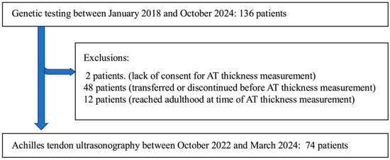
A total of 136 pediatric patients underwent genetic testing at our facility between January 2018 and October 2024. Among these, 2 patients were excluded due to a lack of consent for AT thickness measurement, 48 patients were excluded because they transferred to other facilities or discontinued clinic visits before the AT thickness measurement could be performed within the designated study period, and 12 patients were excluded because they had reached adulthood by the time of AT measurement. Ultimately, 74 pediatric patients were included in the final analysis, as illustrated in the STARD flow diagram (Figure 1). Genetic testing identified 31 patients with FH and 43 patients without FH. No homozygous FH cases were identified; all FH-positive patients were heterozygous. FH group included with LDLR variants (24 detected through NGS and 3 using MLPA), 3 with PCSK9 variant, and 1 with APOB variants (Supplementary Table S1). A substantial proportion of initially screened patients (62 of 136) were excluded from the final analysis, primarily due to logistical reasons such as transfer to other facilities, loss to follow-up, or reaching adulthood prior to measurement. Table 1 summarizes the baseline characteristics of the 74 patients included in the study. There were no significant differences between the two groups in terms of the age, sex, height, body weight, HDL-C, TG, or TC levels. However, patients in the FH group had significantly lower Body Mass Index (BMI) (median 18.5 vs. 20.2 kg/m2, p = 0.017), higher maximum LDL-C levels (202 vs. 173 mg/dL, p < 0.001), higher cumulative LDL-C levels (3098 vs. 2310 mg·years/dL, p < 0.001), and a higher prevalence of statin treatment (77% vs. 26%, p < 0.001) compared with the non-FH group.

Figure 1.
Flowchart illustrating patient enrollment and exclusions.

Table 1.
Baseline characteristics of pediatric patients stratified by genetic diagnosis.
3.2. Comparison of AT Thickness Between FH and Non-FH Groups
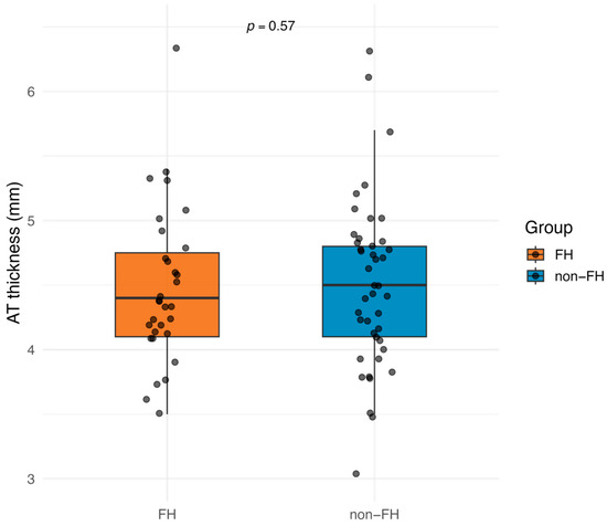
The AT thickness, measured by ultrasonography, showed no detectable difference between the FH and non-FH groups (median 4.4 mm [interquartile range (IQR) 4.1–4.8 mm] vs. 4.5 mm [IQR 4.1–4.8 mm]; Mann–Whitney U test, p = 0.570). Figure 2 illustrates the distribution of AT thickness values in both groups.

Figure 2.
Comparison of AT thickness measured by ultrasonography between FH and non-FH groups. Data are shown as median (horizontal line) and interquartile range (box). Group differences were assessed using the Mann–Whitney U test (p = 0.570). FH: familial hypercholesterolemia; ATT: Achilles tendon thickness.
3.3. Association Between Cumulative LDL-C and AT Thickness
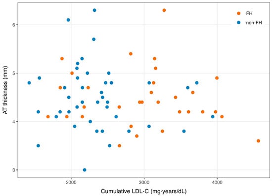
Figure 3 presents exploratory scatter plots of cumulative LDL-C versus AT thickness in FH and non-FH groups. No clear association was apparent in either group. Given the limited sample size and potential residual confounding—including heterogeneous statin exposure—formal linear regression and group × slope interaction tests were not performed.

Figure 3.
Exploratory scatter plots of cumulative LDL-C vs. AT thickness in FH and non-FH groups (no fitted lines). FH: familial hypercholesterolemia; AT: Achilles tendon; LDL-C: low-density lipoprotein cholesterol.
4. Discussion
4.1. Interpretation of Main Findings
Achilles tendon ultrasonography offers a tangible visualization of early atherosclerotic changes in adults with FH, as demonstrated in previous studies where approximately 39% of young adult FH patients exhibited detectable AT thickening [7]. Given its noninvasive nature, simplicity, and absence of radiation exposure, AT measurement might also serve as a potentially useful tool for repeated assessments and early cardiovascular risk evaluation in pediatric FH patients. However, direct pediatric evidence remains limited and warrants further investigation.
The absence of detectable differences in AT thickness between pediatric FH and non-FH patients may partly reflect physiological fluctuations in LDL-C levels during childhood. Particularly, LDL-C levels physiologically decline in childhood [25,26], which may potentially confound the accurate assessment of cumulative LDL-C. Therefore, identifying the maximum LDL-C level prior to physiological reductions may be crucial for accurately evaluating the relationship between cumulative LDL-C and AT thickness in pediatric populations. Physiological LDL-C reductions during childhood represent a distinct pediatric feature. In adult populations, such physiologically fluctuating periods typically constitute a minor fraction of cumulative lifetime LDL-C exposure. However, in pediatric populations, these periods may account for a substantial proportion of lifetime exposure at the time of assessment, potentially explaining why our study failed to detect measurable correlations between cumulative LDL-C and AT thickness. Although our results did not demonstrate significant differences or correlations between cumulative LDL-C exposure and AT thickness, we cannot exclude the possibility that other factors, such as physiological fluctuations in LDL-C during childhood and variations in statin treatment, may have influenced these findings. Further research is necessary to clarify the precise relationship between cumulative LDL-C exposure and AT thickness in pediatric populations.
Even among patients with similar cumulative LDL-C levels, the pathophysiological significance may vary considerably depending on whether the LDL-C elevation is mild or severe at different ages. For instance, prolonged exposure to mild LDL-C elevation during late adolescence may differ substantially from brief but severe LDL-C elevation at a younger age. Thus, weighing the maximum LDL-C value may be essential for accurately evaluating the relationship between cumulative LDL-C and AT thickness in pediatric populations. Supporting this perspective, Domanski et al. reported that the risk of incident cardiovascular disease events depends on cumulative prior exposure to LDL-C, and importantly, the timing of LDL-C exposure significantly modifies this risk [27]. Specifically, the same LDL-C exposure accumulated at a younger age resulted in a greater increase in risk compared with accumulation at an older age. These findings highlight the importance of considering both the duration and intensity of LDL-C exposure, especially during early life, in assessing atherosclerotic cardiovascular risk.
Another reason is that differences in statin treatment practices may also have influenced our results. According to JAS guidelines, statin therapy is recommended for pediatric FH patients with LDL-C levels exceeding 180 mg/dL [9]. However, at our institution, statin treatment is often initiated even at LDL-C levels between 140 and 179 mg/dL. In contrast, non-FH patients with LDL-C levels in this range typically receive dietary interventions alone [28]. Such earlier initiation of statins in FH patients likely reduces cumulative LDL-C exposure, potentially masking differences in AT thickness between the FH and non-FH groups. It is important to note that our primary analysis was designed to investigate associations between the AT thickness, genetic diagnosis of FH, and cumulative LDL-C exposure, rather than directly assessing the impact of statin treatment. We did not include statin treatment as a covariate in multivariable models; given substantial heterogeneity in initiation timing, duration, dosage, and adherence, residual confounding by statin exposure is likely, and our study was not designed or powered to estimate its direct effect on AT thickness.
Nevertheless, genetic testing remains the gold standard for definitive FH diagnosis and etiologic classification, with utility in both family-based cascade screening and universal lipid screening of children presenting with isolated high LDL-C. It informs management and family risk stratification. In summary, our findings suggest that the diagnostic utility of Achilles tendon thickness and cumulative LDL-C burden in pediatric populations is limited; therefore, these results should be interpreted as exploratory rather than confirmatory.
4.2. Population-Specific Considerations
Several patients in the non-FH group exhibited AT thickness exceeding typical adult FH diagnostic thresholds. These cases were identified through universal screening, and our institutional experience suggests that intensive sports activities could induce non-specific AT thickening in pediatric populations. Thus, interpreting AT thickness measurements in universally screened pediatric patients requires careful consideration. Indeed, Cassel et al. demonstrated that increased AT and Patellar tendon thickness in adolescent athletes compared to non-athletes could be shown [29]. Additionally, the concept of “cholesterol-years”, as proposed by Shapiro and Bhatt, represents cumulative LDL-C exposure calculated as the product of LDL-C concentration and the duration of exposure [30]. This concept underscores the critical role of cumulative exposure as a pivotal determinant of atherosclerotic risk, supporting the relevance of our cumulative LDL-C calculations used in the present study. Therefore, it is plausible that the AT thickening observed in our non-FH group could be attributed, at least in part, to the intensive sports activities common in pediatric populations, rather than solely reflecting pathological lipid accumulation.
The pediatric population identified through universal lipid screening likely represents a milder phenotype compared with those selected through cascade screening. Previous reports indicated AT thickening in adolescents with FH; however, our inability to detect such differences may reflect the milder phenotype of our study cohort. Indeed, previous research has reported significant AT thickening in adolescents, even those as young as teenagers, within a clinically diagnosed FH cohort [7]. In contrast, our pediatric population, identified through universal lipid screening, did not demonstrate detectable differences in the AT thickness between the FH and non-FH groups. These contrasting findings may be attributed to differences in patient selection methods, screening procedures, or severity of FH phenotypes. Further studies are required to clarify the characteristics of pediatric populations identified through universal lipid screening.
Moreover, pediatric populations are inherently heterogeneous due to variations in growth, body composition [31,32], and pubertal development, all of which can affect AT thickness measurements. Thus, subgroup analyses considering these developmental factors are warranted in future studies. Physiological variability due to rapid growth and pubertal changes during adolescence could have increased data variability, masking small differences in AT thickness between the FH and non-FH groups. We did not perform multivariable adjustment; therefore, residual confounding by age, height, BMI, and pubertal stage may remain. Given that AT thickness may vary significantly during different pubertal stages, this lack of detailed developmental data represents a potential limitation. Future studies should comprehensively incorporate growth-related developmental parameters. Although physiological growth fluctuations and variations in physical activity were hypothesized as potential confounders, it is important to acknowledge that we did not collect detailed data on pubertal stages or quantified physical activity levels. Therefore, these interpretations remain speculative and require further verification in future studies.
4.3. Study Limitations
4.3.1. Single-Center Design and Selection Bias
This was a single-center, retrospective observational study, which inherently limits the generalizability of the findings. Moreover, a substantial exclusion rate (62 of 136 patients, approximately 45%) due to logistical factors, patient transfers, or loss to follow-up might have introduced significant selection bias. The absence of detailed clinical and demographic information for these excluded patients further restricts our ability to assess differences between the included and excluded groups. Future studies should aim to reduce exclusion rates and ensure detailed data collection to enhance the external validity and applicability of the findings.
4.3.2. Statistical Power and Sample Size
Because this study was retrospective and exploratory, no formal prospective sample size calculation was conducted. A previous post hoc calculation suggested that approximately 25 participants per group would be sufficient to detect a minimal detectable difference of 0.51 mm in AT thickness (SD ≈ 0.63 mm) with 80% power at a two-sided α = 0.05. However, that calculation assumed equal allocation and normally distributed data. In our actual study, the groups were unequally allocated (FH: n = 31, non-FH: n = 43), which slightly reduces power compared with an ideal equal split.
Using the concept of effective sample size, an allocation of 30:45 provides an efficiency equivalent to approximately 36 participants per group under equal allocation. Furthermore, the efficiency of non-parametric tests relative to t-based methods is at worst 0.864 [33]. Thus, our total sample size of 74 behaves like an effective sample of ~62 under non-parametric testing. For a standardized effect size of d ≈ 0.81 (difference 0.51 mm, SD 0.63 mm), the power was ~88% at the two-sided 0.05 level, and remained above 80% for somewhat smaller effect sizes (d ≈ 0.72).
Therefore, despite the unequal allocation and reliance on non-parametric methods, our study retained sufficient statistical power (>80%) to detect clinically meaningful differences in AT thickness between FH and non-FH groups. The absence of statistically significant differences in our results suggests that any true biological difference, if present, may be smaller than these detectable thresholds.
4.3.3. Variability in Statin Treatment
Variability in the timing, duration, and intensity of statin treatment represented a significant confounding factor. Statin therapy was disproportionately more common in the FH group (77% vs. 26%), with considerable variation in treatment duration (median: 1 year; range: 1–6 years). Previous randomized controlled trials have demonstrated that intensive LDL-C lowering, particularly to levels below current clinical targets, can effectively stabilize or even reverse atherosclerotic plaques in patients with acute coronary syndrome [34]. Although our study specifically focused on Achilles tendon thickness rather than coronary plaques, these findings highlight the substantial biological impact of LDL-C reduction therapy on atherosclerotic tissues, suggesting that variability in statin administration might have significantly influenced cumulative LDL-C exposure and potentially affected our results. Future prospective studies should rigorously control the timing, dosage, and duration of statin therapy.
4.3.4. Pediatric Population Heterogeneity
Currently, pediatric-specific normative reference values for Achilles tendon thickness are lacking, complicating interpretation in this population. Pediatric populations exhibit significant heterogeneity in terms of physical growth, body composition, and pubertal development, all of which could influence AT thickness measurements. Our limited sample size precluded meaningful subgroup analyses (e.g., age, sex, pubertal stage), further restricting our ability to interpret findings across different pediatric subpopulations. Our single-center study limits external validity, highlighting the need for future multicenter collaborative studies. Because polygenic FH cannot be identified by the current genetic testing approach, some true FH cases may have been misclassified as non-FH. This possibility represents an additional limitation of our study. Overall, these limitations suggest that the clinical utility of AT thickness and cumulative LDL-C as diagnostic tools for FH in children is limited, and our findings should be interpreted with caution as exploratory observations. In addition, the potential inclusion of polygenic FH cases within the non-FH group may have further attenuated detectable differences. Furthermore, even children without detectable FH variants but with markedly elevated LDL-C from early childhood may be at comparable cardiovascular risk, raising concern for undertreatment in this group.
4.4. Future Directions
Given these limitations, our findings should be interpreted as exploratory. Any future validation should be considered only where rigorous control of confounding is feasible, and the feasibility of establishing pediatric-specific reference values remains a major challenge. Therefore, while AT thickness measurement may have potential utility, further studies should be carefully designed and interpreted within these constraints.
5. Conclusions
Among pediatric patients identified through universal lipid screening, no significant differences in AT thickness were detected between FH and non-FH groups, although cumulative LDL-C levels were higher in the FH group and showed no clear correlation with AT thickness. However, due to methodological limitations such as the small sample size, selection bias, and potential confounding by statin therapy and growth-related factors, these findings should be interpreted cautiously. Given these limitations, our findings should be interpreted as exploratory. Any future validation should be considered only where rigorous control of confounding is feasible.
Supplementary Materials
The following supporting information can be downloaded at https://www.mdpi.com/article/10.3390/lipidology2030015/s1. Supplementary Table S1: List of pathogenic variants identified in pediatric patients with FH in this study.
Author Contributions
Conceptualization: T.I. (Tomoko Inoue) and K.M.; Methodology: T.I. (Tomoko Inoue); Software: T.I. (Tomoko Inoue); Validation: T.I. (Tomoko Inoue), K.M., and T.M.; Formal analysis: T.I. (Tomoko Inoue); Investigation: T.I. (Tomoko Inoue), K.M., R.T., K.N., S.K., T.I. (Takashi Iwase), H.Y.F., T.K., H.T., and M.T.; Resources: T.I. (Tomoko Inoue), K.M., R.T., K.N., S.K., T.I. (Takashi Iwase), H.Y.F., T.K., H.T., and M.T.; Data curation: T.I. (Tomoko Inoue) and R.T.; Writing—Original draft preparation: T.I. (Tomoko Inoue); Writing—Review and editing: K.M. and T.M.; Visualization: T.I. (Tomoko Inoue); Supervision: K.M. and T.M.; Project administration: T.M. All authors have read and agreed to the published version of the manuscript.
Funding
This research received no external funding.
Institutional Review Board Statement
The study was conducted in accordance with the Declaration of Helsinki and approved by the Ethics and Human Genome Committees of Kagawa University (Approval nos. Heisei 30-187 (approval date: 15 August 2018) and Heisei 30-059 (approval date: 6 March 2019)).
Informed Consent Statement
Written informed consent was obtained from all pediatric patients and their legal guardians involved in the study, specifically including consent for genetic testing, ultrasonographic measurement of Achilles tendon thickness, and publication of anonymized clinical data.
Data Availability Statement
The datasets generated and/or analyzed during the current study are not publicly available due to privacy and ethical restrictions, but are available from the corresponding author upon reasonable request.
Acknowledgments
We would like to express our sincere gratitude to Akane Yabuchi and Tetsuo Maruyama for their valuable support and assistance throughout this study.
Conflicts of Interest
The authors declare no conflicts of interest.
Abbreviations
The following abbreviations are used in this manuscript:
| FH | Familial Hypercholesterolemia |
| AT | Achilles Tendon |
| LDL-C | Low-Density Lipoprotein Cholesterol |
| HDL-C | High-Density Lipoprotein Cholesterol |
| TG | Triglycerides |
| TC | Total Cholesterol |
| IQR | Interquartile Range |
| NGS | Next Generation Sequencing |
| MLPA | Multiplex Ligation-dependent Probe Amplification |
| JAS | Japan Atherosclerosis Society |
| BMI | Body Mass Index |
References
- Fourgeaud, M.; Lebreton, L.; Belabbas, K.; Zaouiche, S.; Fouache, E.; Carreau, V.; Antigny, F.; Benlian, P.; Bruckert, E.; Farnier, M.; et al. Phenotypic and genotypic characterization of familial hypercholesterolemia in French adult and pediatric populations. J. Clin. Lipidol. 2022, 16, 298–305. [Google Scholar] [CrossRef] [PubMed]
- Hu, P.; Dharmayat, K.I.; Stevens, C.A.T.; Sharabiani, M.T.A.; Jones, R.S.; Watts, G.F.; Genest, J.; Ray, K.K.; Vallejo-Vaz, A.J. Prevalence of Familial Hypercholesterolemia among the General Population and Patients with Atherosclerotic Cardiovascular Disease: A Systematic Review and Meta-Analysis. Circulation 2020, 141, 1742–1759. [Google Scholar] [CrossRef] [PubMed]
- Wiegman, A.; Gidding, S.S.; Watts, G.F.; Chapman, M.J.; Ginsberg, H.N.; Cuchel, M.; Ose, L.; Averna, M.; Boileau, C.; Borén, J.; et al. Familial hypercholesterolaemia in children and adolescents: Gaining decades of life by optimizing detection and treatment. Eur. Heart J. 2015, 36, 2425–2437. [Google Scholar] [CrossRef] [PubMed]
- Harada-Shiba, M.; Arai, H.; Ishigaki, Y.; Ishibashi, S.; Okamura, T.; Ogura, M.; Dobashi, K.; Nohara, A.; Bujo, H.; Miyauchi, K.; et al. Guidelines for diagnosis and treatment of familial hypercholesterolemia 2017. J. Atheroscler. Thromb. 2018, 25, 751–770. [Google Scholar] [CrossRef] [PubMed]
- Tsouli, S.G.; Kiortsis, D.N.; Argyropoulou, M.I.; Mikhailidis, D.P.; Elisaf, M.S. Pathogenesis, detection and treatment of Achilles tendon xanthomas. Eur. J. Clin. Investig. 2005, 35, 236–244. [Google Scholar] [CrossRef] [PubMed]
- Harada-Shiba, M.; Arai, H.; Ohmura, H.; Okazaki, H.; Sugiyama, D.; Tada, H.; Dobashi, K.; Matsuki, K.; Minamino, T.; Yamashita, S.; et al. Guidelines for the diagnosis and treatment of adult familial hypercholesterolemia 2022. J. Atheroscler. Thromb. 2023, 30, 558–586. [Google Scholar] [CrossRef]
- Michikura, M.; Ogura, M.; Hori, M.; Matsuki, K.; Makino, H.; Fujioka, S.; Shishikura, D.; Hoshiga, M.; Harada-Shiba, M.; Ogawa, M.; et al. Association of Achilles tendon thickness with lipid profile and carotid IMT in patients with familial hypercholesterolemia. Atherosclerosis 2025, 403, 119173. [Google Scholar] [CrossRef]
- Nordestgaard, B.G.; Chapman, M.J.; Humphries, S.E.; Ginsberg, H.N.; Masana, L.; Descamps, O.S.; Wiklund, O.; Hegele, R.A.; Raal, F.J.; Defesche, J.C.; et al. Familial hypercholesterolaemia is underdiagnosed and undertreated in the general population: Guidance for clinicians to prevent coronary heart disease: Consensus statement of the European Atherosclerosis Society. Eur. Heart J. 2013, 34, 3478–3490. [Google Scholar] [CrossRef] [PubMed]
- Harada-Shiba, M.; Ohtake, A.; Sugiyama, D.; Tada, H.; Dobashi, K.; Matsuki, K.; Minamino, T.; Yamashita, S.; Yamamoto, Y. Guidelines for the diagnosis and treatment of pediatric familial hypercholesterolemia 2022. J. Atheroscler. Thromb. 2023, 30, 531–557. [Google Scholar] [CrossRef] [PubMed]
- Cuchel, M.; Raal, F.J.; Hegele, R.A.; Al-Rasadi, K.; Arca, M.; Averna, M.; Bruckert, E.; Freiberger, T.; Gaudet, D.; Harada-Shiba, M.; et al. 2023 Update on European Atherosclerosis Society Consensus Statement on Homozygous Familial Hypercholesterolaemia: New treatments and clinical guidance. Eur. Heart J. 2023, 44, 2277–2291. [Google Scholar] [CrossRef] [PubMed]
- Cuchel, M.; Bruckert, E.; Ginsberg, H.N.; Raal, F.J.; Santos, R.D.; Hegele, R.A.; Kuivenhoven, J.A.; Nordestgaard, B.G.; Descamps, O.S.; Steinhagen-Thiessen, E.; et al. Homozygous familial hypercholesterolaemia: New insights and guidance for clinicians to improve detection and clinical management. Eur. Heart J. 2014, 35, 2146–2157. [Google Scholar] [CrossRef] [PubMed]
- Fu, H.Y.; Matsunaga, K.; Inoue, T.; Tani, R.; Funatsuki, K.; Iwase, T.; Kondo, S.; Nishioka, K.; Ito, S.; Sasaki, T.; et al. Improved efficiency of the clinical diagnostic criteria for familial hypercholesterolemia in children: A comparison of the Japan Atherosclerosis Society guidelines of 2017 and 2022. J. Atheroscler. Thromb. 2024, 31, 1048–1057. [Google Scholar] [CrossRef] [PubMed]
- Matsunaga, K.; Mizobuchi, A.; Fu, H.Y.; Ishikawa, S.; Tada, H.; Kawashiri, M.-A.; Yokota, I.; Sasaki, T.; Ito, S.; Kunikata, J.; et al. Universal screening for familial hypercholesterolemia in children in Kagawa, Japan. J. Atheroscler. Thromb. 2022, 29, 839–849. [Google Scholar] [CrossRef] [PubMed]
- Strouse, P.J.; Trout, A.T.; Offiah, A.C. Editors’ notebook: What is ‘pediatric’? Pediatr. Radiol. 2022, 52, 2241–2242. [Google Scholar] [CrossRef] [PubMed]
- Bujo, H.; Takahashi, K.; Saito, Y.; Maruyama, T.; Yamashita, S.; Matsuzawa, Y.; Ishibashi, S.; Shionoiri, F.; Yamada, N.; Kita, T. Multicenter study to determine the diagnostic criteria for heterozygous familial hypercholesterolemia in Japan. J. Atheroscler. Thromb. 2004, 11, 146–151. [Google Scholar] [CrossRef]
- Kagawa Prefecture Health and Welfare Department. Kagawa Health Checkups for the Prevention of Lifestyle-Related Diseases in Children: Manual. Revised March 2021. Available online: https://www.pref.kagawa.lg.jp/kenkosomu/seikatsushukanbyo/manual2021.html (accessed on 26 August 2025).
- Mabuchi, H.; Nohara, A.; Noguchi, T.; Kobayashi, J.; Kawashiri, M.A.; Tada, H.; Nakanishi, C.; Mori, M.; Yamagishi, M.; Inazu, A.; et al. Molecular genetic epidemiology of homozygous familial hypercholesterolemia in the Hokuriku district of Japan. Atherosclerosis 2011, 214, 404–407. [Google Scholar] [CrossRef]
- Tada, H.; Kawashiri, M.-A.; Nomura, A.; Teramoto, R.; Hosomichi, K.; Nohara, A.; Inazu, A.; Mabuchi, H.; Tajima, A.; Yamagishi, M.; et al. Oligogenic familial hypercholesterolemia, LDL cholesterol, and coronary artery disease. J. Clin. Lipidol. 2018, 12, 1436–1444. [Google Scholar] [CrossRef] [PubMed]
- Yamamoto, T.; Shimojima, K.; Ondo, Y.; Imai, K.; Chong, P.F.; Kira, R.; Amemiya, M.; Saito, A.; Okamoto, N. Challenges in detecting genomic copy number aberrations using next-generation sequencing data and the eXome Hidden Markov Model: A clinical exome-first diagnostic approach. Hum. Genome Var. 2016, 3, 16025. [Google Scholar] [CrossRef] [PubMed]
- Richards, S.; Aziz, N.; Bale, S.; Bick, D.; Das, S.; Gastier-Foster, J.; Grody, W.W.; Hegde, M.; Lyon, E.; Spector, E.; et al. Standards and Guidelines for the Interpretation of Sequence Variants: A Joint Consensus Recommendation of the American College of Medical Genetics and Genomics and the Association for Molecular Pathology. Genet. Med. 2015, 17, 405–424. [Google Scholar] [CrossRef]
- Tada, H.; Nomura, A.; Yoshimura, K.; Kawashiri, M.-A.; Takamura, M.; Nohara, A.; Harada-Shiba, M.; Yamagishi, M. Genetic Testing for Familial Hypercholesterolemia in the General Population: A Nationwide Study in Japan. J. Clin. Lipidol. 2024, 18, 123–132. [Google Scholar] [CrossRef]
- Benn, M.; Watts, G.F.; Tybjærg-Hansen, A.; Nordestgaard, B.G. Mutations Causative of Familial Hypercholesterolaemia: Screening of 98,098 Individuals from the Copenhagen General Population Study Estimated a Prevalence of 1 in 217. Eur. Heart J. 2016, 37, 1384–1394. [Google Scholar] [CrossRef]
- Michikura, M.; Ogura, M.; Yamamoto, M.; Sekimoto, M.; Fuke, C.; Hori, M.; Arai, K.; Kihara, S.; Hosoda, K.; Yanagi, K.; et al. Achilles tendon ultrasonography for diagnosis of familial hypercholesterolemia among Japanese subjects. Circ. J. 2017, 81, 1879–1885. [Google Scholar] [CrossRef] [PubMed]
- Michikura, M.; Ogura, M.; Matsuki, K.; Yamaoka, M.; Makino, H.; Harada-Shiba, M. Risk assessment for cardiovascular events using Achilles tendon thickness and softness and intima-media thickness in familial hypercholesterolemia. J. Atheroscler. Thromb. 2024, 31, 1607–1619. [Google Scholar] [CrossRef] [PubMed]
- Dobashi, K. Changes in serum cholesterol in childhood and its tracking to adulthood. J. Atheroscler. Thromb. 2022, 29, 5–7. [Google Scholar] [CrossRef] [PubMed]
- Banderali, G.; Capra, M.E.; Biasucci, G.; Stracquadaino, R.; Viggiano, C.; Pederiva, C. Detecting familial hypercholesterolemia in children and adolescents: Potential and challenges. Ital. J. Pediatr. 2022, 48, 115. [Google Scholar] [CrossRef] [PubMed]
- Gidding, S.S.; Champagne, M.A.; de Ferranti, S.D.; Defesche, J.; Ito, M.K.; Knowles, J.W.; McCrindle, B.; Raal, F.; Rader, D.; Santos, R.D.; et al. The Agenda for Familial Hypercholesterolemia: A Scientific Statement From the American Heart Association. J. Am. Coll. Cardiol. 2020, 76, 2431–2455. [Google Scholar] [CrossRef] [PubMed]
- Horton, A.E.; Martin, A.C.; Srinivasan, S.; Justo, R.N.; Poplawski, N.K.; Sullivan, D.; Brett, T.; Chow, C.K.; Nicholls, S.J.; Pang, J.; et al. Integrated guidance to enhance the care of children and adolescents with familial hypercholesterolaemia: Practical advice for the community clinician. J. Paediatr. Child Health 2022, 58, 1297–1312. [Google Scholar] [CrossRef] [PubMed]
- Cassel, M.; Intziegianni, K.; Risch, L.; Müller, S.; Engel, T.; Mayer, F. Physiological tendon thickness adaptation in adolescent elite athletes: A longitudinal study. Front. Physiol. 2017, 8, 795. [Google Scholar] [CrossRef] [PubMed]
- Shapiro, M.D.; Bhatt, D.L. “Cholesterol-Years” for ASCVD Risk Prediction and Treatment. J. Am. Coll. Cardiol. 2020, 76, 1517–1520. [Google Scholar] [CrossRef] [PubMed]
- Zheng, Y.; Liang, J.; Zeng, D.; Tan, W.; Yang, L.; Lu, S.; Yao, W.; Yang, Y.; Liu, L. Association of body composition with pubertal timing in children and adolescents from Guangzhou, China. Front. Public Health 2022, 10, 943886. [Google Scholar] [CrossRef] [PubMed]
- Drole Torkar, A.; Plesnik, E.; Groselj, U.; Battelino, T.; Kotnik, P. Carotid intima-media thickness in healthy children and adolescents: Normative data and systematic literature review. Front. Cardiovasc. Med. 2020, 7, 597768. [Google Scholar] [CrossRef] [PubMed]
- Hodges, J.L.; Lehmann, E.L. The efficiency of some nonparametric competitors of the t-test. Ann. Math. Stat. 1956, 27, 324–335. [Google Scholar] [CrossRef]
- Chen, Z.; Lu, G. Lowering low-density lipoprotein cholesterol targets to below 1.0 mmol/L in acute coronary syndrome patients: A potential new standard. Cardiol. Plus 2024, 9, 10159. [Google Scholar] [CrossRef]
Disclaimer/Publisher’s Note: The statements, opinions and data contained in all publications are solely those of the individual author(s) and contributor(s) and not of MDPI and/or the editor(s). MDPI and/or the editor(s) disclaim responsibility for any injury to people or property resulting from any ideas, methods, instructions or products referred to in the content. |
© 2025 by the authors. Licensee MDPI, Basel, Switzerland. This article is an open access article distributed under the terms and conditions of the Creative Commons Attribution (CC BY) license (https://creativecommons.org/licenses/by/4.0/).