Article

Effects of 5% Caffeine Ultrasonophoresis on Gynoid Lipodystrophy—A Randomized Controlled Study

by
Diana Santos Cabral
1 and
Andreia Noites
2,3,*
1
Department of Physiotherapy, ULSTMAD, Local Health Unit of Trás-os-Montes and Alto, E.P.E., Av. da Noruega, Lordelo, 5000-508 Vila Real, Portugal
2
Center for Rehabilitation Research (CIR), ESS, Polytechnic of Porto, Rua Dr. António Bernardino de Almeida 400, 4200-072 Porto, Portugal
3
Department of Physiotherapy, School of Health, Polytechnic Institute of Porto, Rua Dr. António Bernardino de Almeida 400, 4200-072 Porto, Portugal
*
Author to whom correspondence should be addressed.
Lipidology 2025, 2(3), 13; https://doi.org/10.3390/lipidology2030013
Submission received: 4 April 2025 / Revised: 6 June 2025 / Accepted: 30 June 2025 / Published: 18 July 2025

Abstract

Introduction: Gynoid lipodystrophy (GL) affects most women, manifesting itself from puberty to adulthood. Its multifactorial etiology generates controversy in the literature about the most suitable treatment. Several methods are used, from the smallest to the most invasive, in the search for an effective fight against the severity of GL. The positive effect of ultrasound therapy (US) in decreasing subcutaneous adipose tissue is in increasing the skin permeability of pharmacological molecules, and it has aroused interest in the effect of a combination of the two techniques on the severity of GL. However, the results of this technique associated with an exercise program are unknown. Objective(s): To analyze the effect of three sessions of US + 5% caffeine in association with the realization of an exercise program, in females, on the level of severity of GL in the gluteal region and on the posterior proximal third part of the thigh. Methods: A total of 36 healthy women, aged between 18 and 55, who were considered to have GL, were randomly allocated in two experimental groups and one placebo group. The placebo group (PG) performed only physical exercise during the study. Experimental group 1 (EP1) performed US with 5% caffeine alongside a physical exercise protocol and experimental group 2 (EP2) performed US with a conventional US gel alongside a physical exercise protocol. The three groups completed three intervention sessions over 3 weeks, with one session per week. In addition to the level of severity assessed by the Cellulite Several Scale (CSS), anthropometric measures, body composition, and lipid profile of the participants were evaluated. The first assessment was carried out before the intervention (M0) and the last assessment after the three interventions (M1). The results were analyzed using the ANOVA test. The Tukey test was used for multiple comparisons of the groups in all variables, except for those related to the CSS, where the Kruskal–Wallis test was used with a significance level of 0.05. Results: A total of 29 women completed the study. There was a significant decrease inside the PG related to triglycerides (p = 0.012). In M1, all groups started to present median values below 200 mg of triglycerides. In cholesterol, a significant reduction was observed in all groups (p = 0.05). On the gluteal level at 5 cm, there was a decrease in EP1 and EP2 between M0 and M1 with p = 0.006 and p = 0.002, respectively. On the CSS there were no significant differences between groups or between moments. Conclusions: Three sessions of 5% caffeine and US in association with a physical exercise protocol have no effect on reducing the level of severity of GL.

1. Introduction

Gynoid lipodystrophy, commonly known as cellulite, is a structural, inflammatory, and biochemical disorder of the subcutaneous tissue that primarily affects women after puberty, significantly impacting their self-esteem and quality of life [1,2]. Its etiology involves genetic, hormonal, and lifestyle factors, including microcirculation impairment, adipocyte hypertrophy, and chronic inflammation [3,4]. The structure of female connective tissue contributes to the development of GL, differing from the pattern observed in men [2,3,4,5].
Various methods have been used to minimize GL, such as topical treatments containing caffeine and retinol, physical therapies including massage, radiofrequency, cryolipolysis, and US, as well as more invasive medical procedures such as carboxytherapy, mesotherapy, and surgery [6,7]. US has shown effectiveness in reducing localized fat by disrupting adipocyte membranes with minimal damage to surrounding structures, but there is no consensus on the optimal application parameters [8]. Caffeine is widely used in anti-cellulite cosmetics due to its lipolytic action, which prevents fat accumulation and stimulates microcirculation and collagen production [9]. Its potential slimming effect has been demonstrated in studies [9,10]. Pires-de-Campos et al. in 2008 [10] explored the effect of gel application on swine, using hypodermis gel with caffeine (5%, water-in-water) and gel with caffeine and US daily for 15 days. The results showed a significant reduction in the thickness of the subcutaneous adipose tissue, as well as damage of the adipocytes, consequently decreasing the number of these cells.
Aerobic exercise is another key factor in reducing body fat, as it increases fatty acid oxidation rates by five to ten times compared to resting levels and promotes triglyceride lipolysis in adipose tissue [11]. Studies indicate that lipid oxidation is most effective during exercise sessions lasting between 30 and 60 min at moderate intensity (40–60% of heart rate reserve), ideally below 60%, and performed regularly while engaging large muscle groups [11,12]. Thus, physical exercise may induce changes in adipose tissue by enhancing lipolysis and fatty acid oxidation, potentially influencing GL severity [12].
The combination of US with physical exercise has been shown to improve subcutaneous fat mobilization, possibly due to increased norepinephrine release from sympathetic nerves in white adipose tissue [13]. Catecholamines such as epinephrine and norepinephrine are known for their lipolytic properties, which directly affect adipocytes [13]. Given that US disrupts adipocyte membranes, releasing fatty acids, the combination of these two techniques may offer a safer and more effective treatment approach [14,15].
Due to the lack of studies evaluating the effects of caffeine on GL, its association with US, and the increasing concern with body image, this study aims to fill this gap. The primary objective is to compare the effects of three US sessions, with and without caffeine, combined with an exercise program on GL severity in the gluteal region and the proximal third of the thigh using the Cellulite Several Scale (CSS). Secondary objectives include assessing variations in perimetric measurements of the proximal third of the thigh and gluteal region, analyzing changes in total cholesterol and triglyceride levels at two time points (M0 and M1), and examining bioimpedance variations in participants. This study aims to contribute to optimizing available treatments by providing more robust evidence regarding the efficacy of combined approaches in managing GL.

2. Materials and Methods

2.1. Study Design

The present study is experimental, randomized, and controlled, comprising the following three groups: experimental group 1 (EP1) with n = 10, experimental group 2 (EP2) with n = 10, and the placebo group (PG) with n = 9. All three groups followed the same physical exercise protocol (PEF). The caffeine gel used was a compounded product with 5% caffeine and Carbopol gel qbp.
In EP1, three US sessions were carried out using a 5% caffeine gel; in EP2, three US sessions were performed using water-based ultrasound gel; and in the PG, only the physical exercise component was undertaken, with a 5% caffeine gel applied during the three sessions while the US device remained switched off. Each participant was assigned a unique code for the random allocation into groups. The distribution of participants across the groups was conducted randomly by drawing folded paper slips with the assigned codes from an opaque bag. This process was carried out by a Giesta Rehabilitation Centre (C.R.G.) staff member who was independent of the study. The principal investigator was unaware of the participants’ allocation. The guidelines and criteria were recommended by CONSORT.

2.2. Sample

Female participants were voluntarily recruited to participate in the study. They were aged between 18 and 55 years and were recruited at the C.R.G., Chaves, Portugal, through internet advertising, posters, and the dissemination of the research during Physical Medicine and Rehabilitation consultations, as well as during physiotherapy and clinical Pilates sessions.
The inclusion criteria for the study were as follows: all female individuals aged between 18 and 55 years who considered themselves to suffer from GL in the posterior region of the proximal third of the thigh and/or the gluteal region, and who expressed a willingness to participate. Additionally, participants who scored between 1 and 3 on the CSS were eligible.
The exclusion criteria included individuals with cardiac pathology; cancer patients and those in remission within the first 5 years; pregnant women or those up to one year postpartum, or individuals intending to become pregnant during the evaluation; participants who had undergone any intervention for GL in the previous 3 months; participants with obesity (BMI ≥ 30 kg/m2) or those planning to initiate any dietary changes during the study; individuals taking medications (corticosteroids and non-steroidal anti-inflammatory drugs); participants with difficulties in understanding the procedure; and those with osteosynthesis material in the area of US application.

2.3. Instruments

Three questionnaires were used to characterize the sample, focusing on sociodemographic characteristics, anthropometric measurements, physical activity, and dietary frequency. Easily replicable assessments were conducted to measure total cholesterol and triglyceride levels, as well as the perimeters of the gluteal region and the proximal third of the right thigh of each participant. Additionally, photographic records were collected for later evaluation by a panel of experts using the CSS.

2.3.1. Sociodemographic Questionnaire

A questionnaire was developed to characterize the sample, consisting of 13 short-answer questions with an estimated completion time of approximately 5 min. The objective was to collect information on each participant’s age and medical history, including the presence of any pathologies, disease remission, medication use, previous surgeries, dietary habits, pregnancy status or attempts to conceive, breastfeeding, recent childbirth (within the past year), ongoing treatments for GL (oral, topical, or other), and whether they considered themselves affected by GL.

2.3.2. Short Version of the International Physical Activity Questionnaire (IPAQ)

The short version of the IPAQ has been validated for the Portuguese population [16], with a validity of 0.49, Spearman’s correlation coefficient of 0.77, and an intraclass correlation coefficient (ICC) of 0.83.

2.3.3. Semi-Quantitative Food Frequency Questionnaire (FFQ)

This has been adapted and validated for the Portuguese population by the Department of Hygiene and Epidemiology at the Faculty of Medicine of the University of Porto [17], with an average correlation coefficient of 0.54 for dietary intake over the previous 12 months. The reproducibility of the questionnaire showed an average correlation of 0.57 for 22 nutrients [16,17].

2.3.4. Bioimpedance Scale

This was used to assess body weight, muscle mass (kg), fat mass (%), and perimetry. To obtain measurements a Tanita® BC 601 601 bioimpedance scale was used (Tanita Corporation, Tokyo, Japan).

2.3.5. Measuring Tape

A COMED® was used to assess height and body perimeters (Comed S.A.S., Strasbourg, France), with an accuracy of 1 mm up to a maximum length of 2 m. It demonstrated an intraclass correlation coefficient (ICC) of 0.98 when compared to X-ray measurements [18].

2.3.6. Cellulite Severity Scale CSS

The CSS has an intraclass correlation coefficient (ICC) > 0.7 and has reliability, with Cronbach’s alpha ranging from 0.851 to 0.989 [19].
Each variable is scored between 0 and 3, with the total sum classifying cellulite severity as:
Mild (1–5 points);
Moderate (6–10 points);
Severe (11–15 points) [19,20].
For assessment, photographic records of the gluteal region and the proximal third of the thigh were taken using a Nikon D3200 camera, with a 24.2-megapixel resolution and an ISO sensitivity range of 100 to 6400.

2.3.7. Lipid Profile Meter

Total cholesterol and triglyceride levels were assessed using a Menarini MULTICARE IN device (A. Menarini Diagnostics, Pisa, Italy).
Regarding precision and repeatability, the average measurement imprecision is <5%, with an average coefficient of 4.66% when compared to laboratory-based measurements. Reproducibility also shows an average imprecision of <5%, with a mean coefficient of 4.62%, based on a series of laboratory measurements (MULTICARE IN user manual).

2.3.8. Borg Scale

This scale showed an average intraclass correlation coefficient (ICC) of 0.62 when compared to heart rate and 0.64 when compared to the percentage of maximum oxygen uptake [21].

2.3.9. Heart Rate Monitor

The device demonstrated an intraclass correlation coefficient of 0.99 during exercise when compared to an electrocardiogram, indicating excellent accuracy with an error margin of ±1% or ±1 beats per minute (bpm) (Polar®, 2013, Polar Electro Oy, Kempele, Finland).

2.4. Methods

Sample Characterization: The sample characterization questionnaire was completed by participants in the week before data collection began. The IPAQ was provided and filled out by each participant during the first assessment and then returned to the principal investigator. The FFQ was given during the first session and completed at home by the participants, with the results later sent and processed using Food Processor Plus software [16].
The principal investigator entered all questionnaire data into a computer without internet access, ensuring the protection of participants’ data.
Data Collection: All participants were assessed individually in a private room at C.R.G. to ensure privacy, and the room was under consistent temperature and lighting conditions during both evaluation moments. The study objectives and procedures were thoroughly explained to each participant.
During the assessment, each participant remained in an upright standing position, looking straight ahead, with their upper limbs crossed in front of the abdominal region, barefoot, and with their lower limbs positioned hip-width apart.
The first evaluation (M0) was conducted before the interventions began, and the final evaluation (M1) took place after the last intervention. This procedure was applied to all three groups. During M0 and M1, photographic records of the gluteal and thigh regions were taken, along with assessments of body composition, anthropometric measurements, and lipid profile evaluation.
Assessment of GL Severity: The numerical results from the CSS were used to assign a severity score to each participant. This evaluation was conducted by a panel of experts consisting of three physiotherapists specializing in dermatofunctional physiotherapy, using photographic records. The experts were blinded to the participants’ group allocation and the timing of the assessment.
Photographic records were taken from both the frontal and sagittal planes, with and without muscle contraction, and with skin pinching. The camera was positioned at a fixed distance of 100 cm, while the height was adjusted according to each participant’s specifics. The base of the camera was aligned with the coccyx using a tripod. An ISO setting of 1600 was selected.
Assessment of Body Composition and Anthropometric Measurements: Participants were instructed to wear only underwear, remain barefoot, and remove any metallic objects whenever possible during the evaluation.
To obtain values for body mass index (BMI), body fat percentage, and muscle mass in kilograms, participants stepped onto the scale barefoot, ensuring their feet were positioned over the electrodes. They maintained an upright posture, holding the electrodes parallel to the floor with their arms extended along their body while looking straight ahead, following the TANITA company guidelines [22].
Each measurement was taken twice, and the average value was calculated. For height measurement, the measuring tape was fixed to the wall and participants stood barefoot with their back against the wall, keeping their feet hip-width apart.
The previously mentioned measurements, along with those taken at 5 cm, 10 cm, and 15 cm below the gluteal fold, as well as 5 cm, 10 cm, and 15 cm below the posterior inferior iliac crest, were recorded at the end of the expiratory phase at tidal volume, during apnea [11].
Lipid Profile Assessment: Capillary blood collection was performed to assess the lipid profile 10 min before the first session (M0) and immediately after the final session (M1).
The collection was carried out by a Senior Diagnostic and Therapeutic Technician in Clinical Analysis and Public Health, holding professional registration number C-038489015. The procedure involved a puncture on the fourth finger of the right hand using a disposable lancet. A capillary blood drop was collected onto a test strip inserted into a Menarini MULTICARE IN lipid profile measuring device, which displayed the total cholesterol value on the screen.
The same procedure was repeated on the left hand to assess triglyceride levels.
The recorded values for total cholesterol and triglycerides were logged in a table corresponding to each participant’s code. After data collection, all used test strips and lancets were discarded in a biological waste container.
Calculation of Training Heart Rate and Perceived Exertion: All participants followed an exercise protocol in which their theoretical maximum heart rate (HRmax) was determined using Tanaka’s Equation [23].
After five minutes, once heart rate stabilization was achieved, the value was measured and recorded [11].
Before starting aerobic exercise, the Borg scale was explained to participants to help them maintain an intensity level between light (9) and somewhat hard (11) [11].
Intervention Protocol: The sample collection took place between May and July 2020 at C.R.G. In each session, participants allocated to GE1, GE2, and PG underwent a specific treatment protocol.
In GE1, a US device was applied in combination with a 5% caffeine gel. The caffeine used was acquired from a pharmacy that formulated a 5% caffeine compound with Carbopol gel.
In GE2, the same procedure was replicated, but the 5% caffeine gel was replaced with a water-based US gel composed of: water, carbomer, glycerin, triethanolamine, and phenoxyethanol (and) decylene glycol (and) glycol, CI 42090.
In PG, the ultrasound device was applied with an intensity of zero, using the 5% caffeine gel, and the previously mentioned protocol was replicated.
At the end of each session, all three groups followed the prescribed exercise protocol.
A total of three intervention sessions were conducted, with a one-week interval between each session, completing the data collection for each intervention cycle within three weeks.
Ultrasound Protocol: The equipment used was an ultrasound device, Zimmer® Soleo Sono model (Zimmer MedizinSysteme GmbH, Munich, Germany), set to a frequency of 3 MHz and an intensity of 1.5 W/cm2 in continuous mode.
During the procedure, participants adopted a prone position. The technique was applied to the posterior thigh and gluteal region using circular movements. The application lasted 10 min per limb, covering both limbs; however, parameter evaluation was performed on the right limb.
Exercise Protocol: The exercise protocol was carried out immediately after the treatment session, lasting a total of 30 min. A treadmill from the BH brand, model i.Magna RC G65091 (Beistegui Hermanos S.A. (BH Fitness), Vitoria-Gasteiz, Spain) was used. To monitor heart rate, a heart rate monitor was placed on the participants.
The exercise began with a five-minute warm-up, walking on the treadmill, gradually increasing speed until reaching the target heart rate. Between the 5th and 25th minute, participants walked at a speed that allowed them to maintain their heart rate within the target zone. They were encouraged to maintain a relaxed and comfortable posture, avoiding breath-holding during the exercise. Throughout the session, they were asked about their physical condition, including any signs of fatigue or dizziness.
Upon reaching level 9 on the Borg scale, participants were encouraged to maintain that intensity. At the 25th minute, the speed was gradually reduced to allow for a return to rest, completing the final five minutes of the protocol.
Ethics: This study was conducted with the approval of the Ethics Committee of ESS-IPP (No. E009472020).
The use of the QFA questionnaire for this study was authorized by the author of its validation, with adaptations made for the Portuguese population.
Statistics: Statistical analysis was performed using IBM SPSS Statistics software (version 24.0; IBM Corp, Armonk, NY, USA) with a significance level of 0.05.
The ANOVA test, followed by multiple comparisons using Tukey’s test, was used to compare groups across all variables, except for those related to the CSS, where the Kruskal–Wallis test was applied.
To compare different time points within each group, a paired t-test was used. For comparisons involving BMI, as well as variables related to the CSS, the Wilcoxon test was applied. The choice between parametric and non-parametric tests was based on the assumption of normality, which was verified using the Shapiro–Wilk test.
Mean and standard deviation were used as descriptive statistics for all variables, except for those related to the CSS where the median and interquartile range were reported.

3. Results

3.1. Sample Characterization

The initial population comprised 43 participants, of which 7 were excluded for not meeting the inclusion criteria, as 3 participants were over 55 years of age, 1 participant was breastfeeding, 2 participants were trying to become pregnant, and 1 participant was in remission from cancer. The initial sample consisted of 36 participants and 2 of the participants in the first session chose not to participate in the study due to lack of availability, 1 patient discovered that the blood collection was not possible, 1 participant was not compatible with the location to participate in the study, and 3 did not attend the last session. The study was applied to 29 participants. Figure 1 shows the “Sample composition diagram” according to the Consort 2010 flowchart.
Table 1 contains the sample characterization, where it is possible to verify that no significant differences were observed between the groups with regard to age, weight, height, BMI, and level of physical activity (p > 0.05). As for the food frequency questionnaire, it can be observed that EP2 presented significantly higher values of carbohydrates and sugar compared to EP1 (p = 0.04 and p = 0.03, respectively). In the other variables, no significant differences were observed between the groups (p > 0.05).

3.2. Results of the Effects of the Intervention

Table 2 shows the results of the comparison between groups and between time points. Regarding the lipid profile and the variables collected by the bioimpedance scale, no significant differences were observed between the groups. The mean and standard deviation values were used for all variables. Although the values in the BMI variable, a non-parametric test, should have been presented as median and interquartile deviation, for simplicity’s sake the mean and standard deviation were used to facilitate comparison. In an intra-group analysis, a significant decrease in triglycerides was observed in the PG (p = 0.01). It is worth noting that in M1 all groups began to present mean values of less than 200 mg of triglycerides. A significant reduction in cholesterol was observed in all groups (p < 0.05), with mean values of less than 200 mg being obtained in M1. The percentage of adipose tissue, muscle tissue, weight, and BMI did not show significant changes between time points in any group.
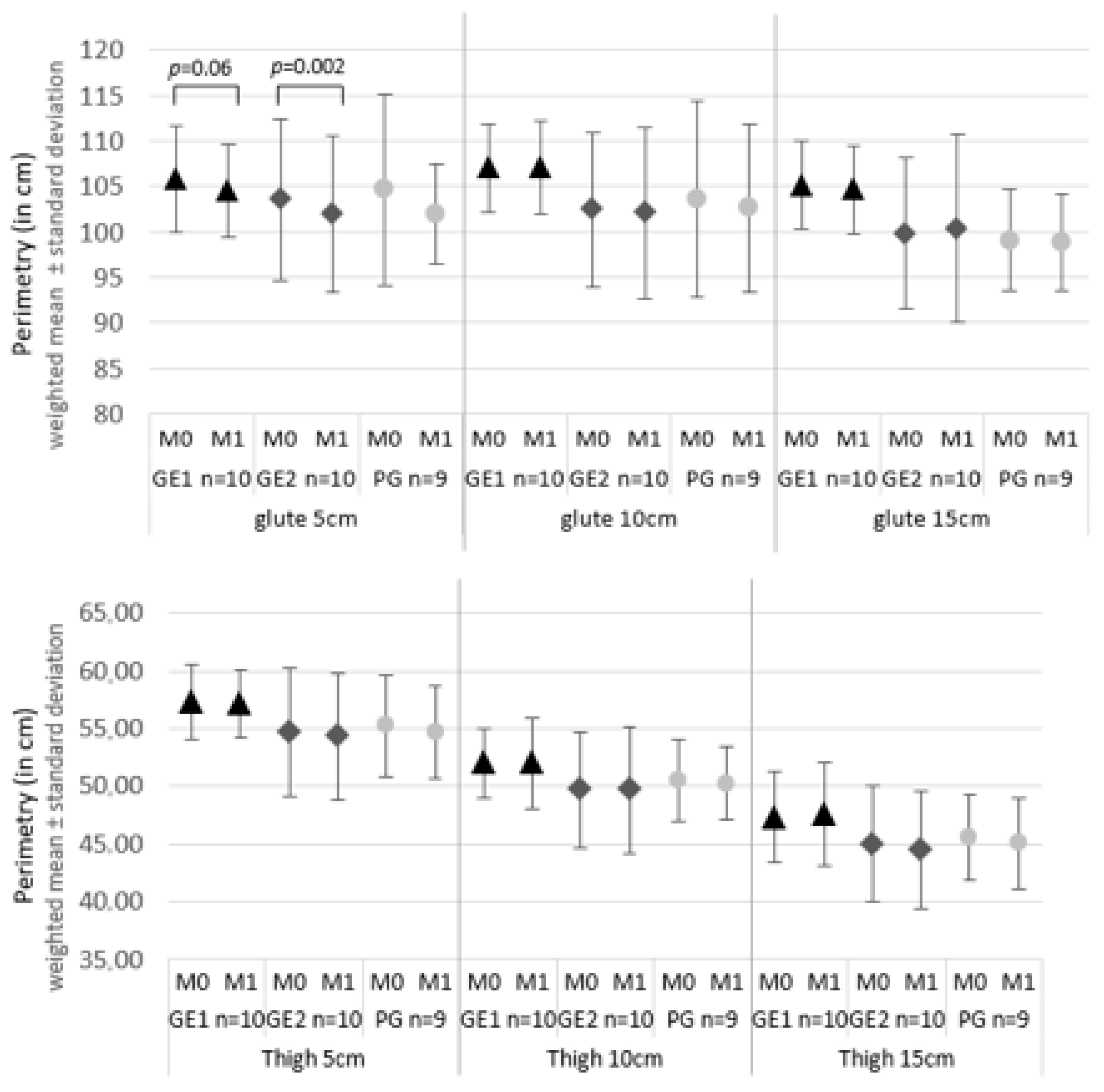
Regarding perimetry, there was no significant reduction between the groups. At the gluteal level at 5 cm, there was a decrease in GE1 and GE2 between M0 and M1, with p = 0.06 and p = 0.002, respectively. At the thigh level, there were no significant values between the evaluation moments. Figure 2 illustrates the perimetry values performed.
Regarding the CSS scale, no significant differences were observed between the groups, nor between the moments, regarding the final score, as well as each of its domains, as can be seen in Table 3.

4. Discussion

The main objective of this study was to evaluate the effect of ultrasound (US) with 5% caffeine on the severity of GL. The combination of US with drugs enhances their skin permeability [24]. Caffeine is used for its lipolytic effect on adipose tissue [8], potentially reducing hypodermis thickness due to the decrease in adipocytes [25]. In vitro tests indicate that US increases caffeine molecule permeability while maintaining skin integrity [25]. The use of 5% caffeine is due to this concentration being commonly found in commercial gels [24]. US was standardized for a predetermined period of 10 min, based on an estimated calculation of 1 min per cm2 of treatment area. The US intensity of 1.5 W/cm2 was based on its effects in promoting physiological responses to injury and increasing transcutaneous drug absorption, particularly caffeine [24,25,26]. As a characterization variable, dietary patterns were evaluated through the QFA. At baseline, GE2 presented significant differences in carbohydrate and sugar intake compared to the other groups. However, these differences were not reflected in anthropometric measurements and were considered within normal ranges. The discrepancies between groups might be attributed to self-reported data, which can lead to classification errors since people tend to report values that align with social expectations [27].
Regarding lipid profile results, a decrease was observed between M0 and M1 in all three groups. A study by the authors of [28] reported an immediate increase in cholesterol levels after US intervention, reflecting the release of cholesterol into the bloodstream. In this study, such an increase was not observed, which might be explained by the physical activity performed by all three groups. This is supported by the results from the PG, which did not undergo US but showed a significant reduction in triglyceride levels between M0 and M1 through exercise alone. The physical exercise performed was of moderate intensity (45 to 55% of resting heart rate) for 30 min. According to the American College of Sports Medicine (2022), this type of exercise promotes adipose tissue reduction through fatty acid oxidation and the release of hormones such as catecholamines and growth factors, which enhance lipolysis and subsequently reduce fat tissue [10,11,12,13,14,15,16,17,18,19,20,21,22,23,24,25,26,27,28,29]. This should be performed regularly, involving large muscle groups whenever possible [10,11]. The combination of physical exercise with US may be beneficial as fat tissue reduction is directly linked to GL reduction. Moreover, exercise boosts the metabolism of fatty acids released by US [29]. Physical activity increases the body’s energy demand, leading to lipolysis through the release of catecholamines from the sympathetic nervous system and adrenal glands, which recruit triglycerides and hydrolyze them into fatty acids and glycerol [30,31]. Lipolysis is stimulated by circulating catecholamines combined with low insulin concentration, accelerated blood flow, and high vasomotor activity, resulting in fatty acid oxidation which may lead to reduced measurements [31]. Regarding perimeter measurements, a reduction in the gluteal region was observed between M0 and M1 in the two experimental groups, GE1 and GE2, specifically at the 5 cm mark. No perimeter changes were found in the PG, which can be explained by the lipolytic effect of US. US induces fat mobilization by increasing catecholamine secretion from white adipose tissue [12]. These results found a reduction in subcutaneous adipose tissue thickness in thighs exposed to US [12]. The study evaluated thigh subcutaneous tissue thickness in individuals who underwent US combined with physical exercise for 10 consecutive days, with US applied to one thigh. Regarding the CSS, no significant differences were observed between groups or time points in the final score or its domains. These results contradict studies [10] by Pires-de-Campos et al., which evaluated the effect of caffeine on swine hypodermis morphology when applied topically or combined with US over 15 days. Pires-de-Campos [10] observed a significant reduction in subcutaneous adipose tissue thickness. However, the evaluation was conducted histologically on post-mortem skin segments, making this procedure unfeasible to replicate. The differing data may be due to the study being conducted on swine, while the present study involved humans over a different duration (3 weeks, one session per week). Furthermore, the CSS results contradict data from Hexel [7], who demonstrated a positive relationship between ultrasound use and GL severity reduction. However, these differences may stem from the number of sessions conducted—12 sessions over 6 weeks by Hexel and colleagues. Their sample consisted of women with moderate to severe CSS grades, which may explain the differences in results as this study lacked stratified randomization by severity due to a smaller sample size. Future studies should ensure sample randomization is stratified by CSS severity grades. Dietary habits and physical activity assessments were conducted at the start of the study, with maintenance recommended throughout. However, these were not monitored at M1, leaving room for potential changes. The limitation of monitoring physical activity only on technique application days should also be noted. One of the widely adopted global guidelines, advised by the American College of Sports Medicine [11], recommends that adults engage in moderate-intensity physical activity for 30 min or more at least 5 days a week or achieve 450 to 750 MET minutes/week of combined moderate and vigorous activity. Initial assessments using the IPAQ showed that participants did not meet ACSM recommendations, all remaining below 361 MET minutes/week. Bioimpedance variables showed no changes between M0 and M1, possibly due to the study’s 3-week duration. Future studies should consider a longer intervention protocol with more weekly sessions. Additionally, more subjective measures are suggested, using IPAQ and QFA questionnaires at both the start and end of the study to account for confounding factors such as physical activity levels and dietary frequency.

5. Conclusions

It is concluded that three sessions of 5% caffeine and US in association with the performance of an exercise protocol does not have an effect on reducing the severity of GL. The protocol under discussion does not have a significant effect on the lipid profile values, however physical exercise presents a decrease in triglyceride values. Further studies are recommended to corroborate the findings of the present study for caffeine in reducing GL, ideally conducted over a longer duration to determine whether the observed values change or remain consistent over time.

Author Contributions

Conceptualization, D.S.C.; methodology, D.S.C.; software, D.S.C.; validation, D.S.C. and A.N.; formal analysis, D.S.C.; investigation, D.S.C.; resources, D.S.C.; data curation, D.S.C.; writing—original draft preparation, D.S.C.; writing—review and editing, D.S.C. and A.N.; visualization, D.S.C.; supervision, A.N.; project administration, D.S.C. All authors have read and agreed to the published version of the manuscript.

Funding

This research received no external funding.

Institutional Review Board Statement

The study was conducted in accordance with the Declaration of Helsinki and approved by the Ethics Committee of ESCOLA SUPERIOR de SAÚDE, POLITÉCNICO DO PORTO, protocol code E009472020 on 20/02/2020.

Informed Consent Statement

Informed consent was obtained from all subjects involved in the study.

Data Availability Statement

The data that support the findings of this study are available from the corresponding author upon reasonable request.

Conflicts of Interest

The authors declare no conflicts of interest.

Abbreviations

The following abbreviations are used in this manuscript:
ACSMAmerican College of Sports Medicine
BpmBeats per minute
BMIBody Mass Index
Cm Centimeters
C.R.G.Giesta Rehabilitation Centre
CSSCellulite Several Scale
EP1Experimental Group 1
EP2Experimental Group 2
FFQSemi-Quantitative Food Frequency Questionnaire
GLGynoid Lipodystrophy
HRmaxMaximum Heart Rate
ICCIntraclass Correlation Coefficient
IPAQInternational Physical Activity Questionnaire
KgKilograms
M0Before the intervention
M1After the three interventions
MHzMega hertz
PEFPhysical Exercise Protocol
PGPlacebo Group
USUltrasound Therapy
W/cm2Watt per centimeter square

References

  1. Bauer, J.; Hoq, N.; Mulcahy, J.; Tofail, S.A.M.; Gulshan, F.; Silien, C.; Podbielska, H.; Akbar, M. Implementation of artificial intelligence and non-contact infrared. EPMA J. 2020, 11, 17–29. [Google Scholar] [CrossRef] [PubMed]
  2. Dupont, E. An integral topical gel for cellulite reduction: Results from a double-blind, randomized. Clin. Cosmet. Investig. Dermatol. 2014, 7, 73–88. [Google Scholar] [CrossRef] [PubMed]
  3. Almeida, M.D.L.C.; Serrano, C.S.; Sánchez, E.M.M.; Mohedo, E.D.; Moriana, G.C.; Salas, M.R. The efficacy of capacitive radio-frequency diathermy in reducing. J. Cosmet. Laser Ther. 2014, 16, 214–224. [Google Scholar] [CrossRef] [PubMed]
  4. Kruglikov, I. The Pathophysiology of Cellulite: Can the Puzzle Eventually Be Solved? J. Cosmet. Dermatol. Sci. Appl. 2012, 2, 1–7. [Google Scholar] [CrossRef]
  5. Nürnberg, F.M.A. So-Called Cellulite: An Invented Disease. J. Dermatol. Surg. Oncol. 1978, 4, 221–229. [Google Scholar] [CrossRef] [PubMed]
  6. Atamoros, F.M.P.; Pérez, D.A.; Sigall, D.A.; Romay, A.A.Á.; Gastelum, J.A.B.; Salcedo, J.A.d.l.P.; Salgado, P.E.E.; Palacios, G.J.G.; Guerrero-Gonzalez, G.A.; De la Cerda, R.M.; et al. Evidence-based treatment for gynoid lipodystrophy: A review. J. Cosmet. Dermatol. 2018, 17, 977–983. [Google Scholar] [CrossRef] [PubMed]
  7. Hexsel, D.; Camozzato, F.O.; Silva, A.F.; Siega, C. Acoustic wave therapy for cellulite, body shaping and fat reduction. J. Cosmet. Laser Ther. 2017, 19, 165–173. [Google Scholar] [CrossRef] [PubMed]
  8. Kania, B.; Goldberg, D.J. Cryolipolysis: A promising nonsurgical technique for localized fat reduction. J. Cosmet. Dermatol. 2023, 22 (Suppl. S3), 1–7. [Google Scholar] [CrossRef] [PubMed]
  9. Herman, A.H. Caffeine’s Mechanisms of Action and Its. Ski. Pharmacol. Physiol. 2013, 26, 8–14. [Google Scholar] [CrossRef] [PubMed]
  10. Pires-de-Campos, M.S.M.; Leonardi, G.R.; Chorilli, M.; Spadari-Bratfisch, R.C.; Polacow, M.L.O.; Grassi-Kassisse, D.M. The effect of topical caffeine on the morphology of swine hypodermis as measured by ultrasound. J. Cosmet. Dermatol. 2008, 7, 232–237. [Google Scholar] [CrossRef] [PubMed]
  11. Jeppesen, J.; Kiens, B. Regulation and limitations to fatty acid oxidation. J. Physiol. 2012, 590, 1059–1068. [Google Scholar] [CrossRef] [PubMed]
  12. Gary, L. ACSM’s Guidelines for Exercise Testing and Prescription; Wolters Kluwer Health: Philadelphia, PA, USA, 2022. [Google Scholar]
  13. Miwa, H.; Kino, M.; Han, L.-K.; Takaoka, K.; Tsujita, T.; Furuhata, H.; Sugiyama, M. Effect of ultrasound application on fat mobilization. Pathophysiology 2002, 9, 13–19. [Google Scholar] [CrossRef] [PubMed]
  14. Ormsbee, M.J.; Thyfault, J.P.; Johnson, E.A.; Kraus, R.M.; Choi, M.D.; Hickner, R.C. Fat metabolism and acute resistance exercise in trained men. J. Appl. Physiol. 2007, 102, 1767–1772. [Google Scholar] [CrossRef] [PubMed]
  15. Otero-Díaz, B.; Rodríguez-Flores, M.; Sánchez-Muñoz, V.; Monraz-Preciado, F.; Ordoñez-Ortega, S.; Becerril-Elias, V.; Baay-Guzmán, G.; Obando-Monge, R.; García-García, E.; Palacios-González, B.; et al. Exercise induces white adipose tissue browning across the weight spectrum in humans. Front. Physiol. 2018, 9, 1781. [Google Scholar] [CrossRef] [PubMed]
  16. Craig, C.L.; Marshall, A.L.; Sjöström, M.; Bauman, A.E.; Booth, M.L.; Ainsworth, B.E.; Pratt, M.; Ekelund, U.L.; Yngve, A.; Sallis, J.F.; et al. International physical activity questionnaire: 12-country reliability and validity. Med. Sci. Sports Exerc. 2003, 35, 1381–1395. [Google Scholar] [CrossRef] [PubMed]
  17. Lopes, C.; Torres, D.; Oliveira, A.; Severo, M.; Alarcão, V.; Guiomar, S.; Mota, J.; Teixeira, P.; Ramos, E.; Rodrigues, S.; et al. Inquérito Alimentar Nacional e de Atividade Física, IAN-AF-2015-2016: Relatório metodológico. 2017. Available online: www.ian-af.up.pt (accessed on 6 June 2025).
  18. Lopes, C.; Aro, A.; Azevedo, A.; Ramos, E.; Barros, H. Intake and adipose tissue composition of fatty acids and risk of myocardial infarction in a male Portuguese community sample. J. Am. Diet. Assoc. 2007, 107, 276–286. [Google Scholar] [CrossRef] [PubMed]
  19. Gogia, P.P.; Braatz, J.H. Validity and reliability of leg length measurements. J. Orthop. Sports Phys. Ther. 1986, 8, 185–188. [Google Scholar] [CrossRef] [PubMed]
  20. Chen, M.J. Criterion-related validity of the Borg ratings of perceived exertion scale in healthy individuals: A meta-analysis. J. Sports Sci. 2002, 20, 873–899. [Google Scholar] [CrossRef] [PubMed]
  21. Lintsi, M.K. Comparison of hand-to-hand bioimpedance and anthropometry equations versus dual-energy X-ray absorptiometry for the assessment of body fat percentage in 17–18-year-old conscripts. Clin. Physiol. Funct. Imaging 2004, 24, 85–90. [Google Scholar] [CrossRef] [PubMed]
  22. Tanaka, H.; Monahan, K.D.; Seals, D.R. Predicted Maximal Heart Revisited. J. Am. Coll. Cardiol. 2001, 37, 153–156. [Google Scholar] [CrossRef] [PubMed]
  23. Tassinary, J.A.; Rogeri, L.N.; Schmitt, B.; Marder, F.; Sinigaglia, G.; Herbele, J.; Hilgemann, M.; Stülp, S. Efeito do ultrassom terapêutico na liberação, permeação e retenção de ácido kójico em sistema de difusão vertical. Acta Biomed. Bras. 2011, 9, 8–16. [Google Scholar] [CrossRef]
  24. Chorilli, M.; Tamascia, P.; Rossim, C.; Salgado, H. Ensaios biológicos para avaliação de segurança de produtos cosméticos. Rev. Cienc. Farm. Basica E Apl. 2009, 30, 19–30. [Google Scholar]
  25. Leung, M.C.; Ng, G.Y.; Yip, K.K. Effect of ultrasound on acute inflammation of transected medial. Arch. Phys. Med. Rehabil. 2004, 85, 963–966. [Google Scholar] [CrossRef] [PubMed]
  26. Santos, J.; Leitão-Correia, F.; Sousa, M.J.; Leão, C. Dietary Restriction and Nutrient Balance in Aging. Oxidative Med. Cell. Longev. 2015, 2016, 4010357. [Google Scholar] [CrossRef] [PubMed]
  27. Presotto, L.; Rogeri, L.N.; Sinigaglia, G.; Bitencourt, S.; Tassinary, J.A.F. Aesthetic nonthermal ultrasound and electric current combination therapy for body sculpting. J. Cosmetol. 2018, 2, 000109. [Google Scholar]
  28. Lafontan, M.; Langin, D. Lipolysis and lipid mobilization in human adipose tissue. Prog. Lipid Res. 2009, 48, 275–297. [Google Scholar] [CrossRef] [PubMed]
  29. Glisezinski, I.D. Mobilisation des lipides du tissu adipeux au cours de l’exercice physique. Sci. Sports 2007, 22, 280–285. [Google Scholar] [CrossRef]
  30. Ahmadian, M.; Wang, Y.; Sul, H.S. Lipolysis in adipocytes. Int. J. Biochem. Cell Biol. 2010, 42, 555–559. [Google Scholar] [CrossRef] [PubMed]
  31. Stich, V.; de Glisezinski, I.; Berlan, M.; Bulow, J.; Galitzky, J.; Harant, I.; Suljkovicova, H.; Lafontan, M.; Rivière, D.; Crampes, F. Adipose Tissue Lipolysis Is Increased during a Repeated Bout of Aerobic Exercise. J. Appl. Physiol. 2000, 88, 1277–12833. [Google Scholar] [CrossRef] [PubMed]
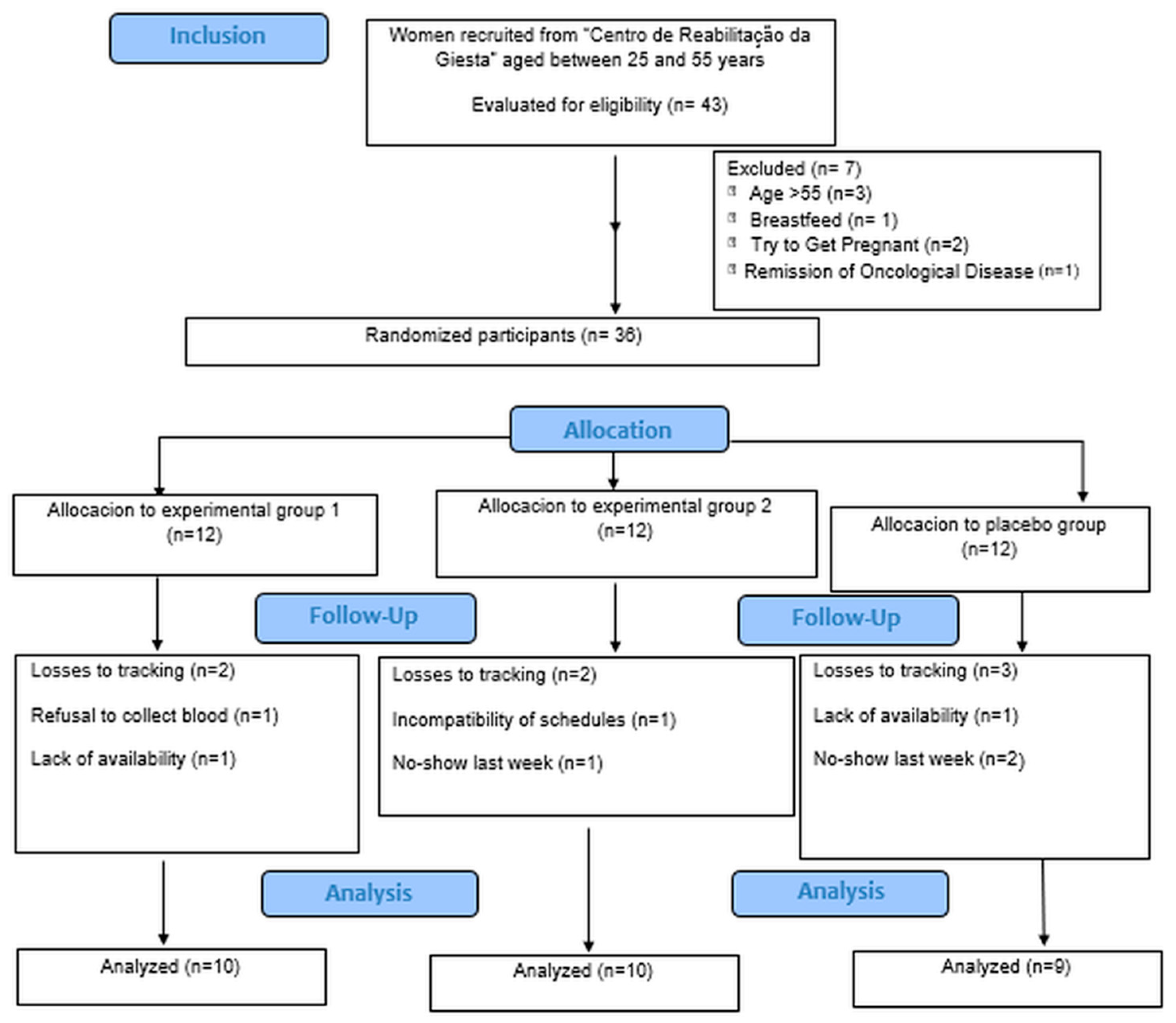
Figure 1. Sample composition diagram.
Lipidology 02 00013 g001
Figure 2. Perimetry values performed.
Lipidology 02 00013 g002
Table 1. Characterization of sample variables by group.
GE1 (n = 10)GE2 (n = 10)PG (n = 9)Difference Groups
AverageSDAverageSDAverageSDValuer p*
Age (years)35.2012.5439.4010.3432.7811.820.46
Height (cm)163.306.40165.605.19168.787.140.18
Weight (kg)69.096.8166.6411.5063.776.700.42
BMI (kg/cm2)25.912.1124.243.7022.523.400.08
IPAQ
MET min/week1.600.702.200.791.890.780.23
QFA
Calories (kcal)1791.87855.672339.23742.261790.04656.310.22
Protein78.0542.15119.6152.0487.4026.910.09
Carbohydrates190.0585.69310.59123.91180.5786.160.01 a
Fat
Total79.6344.54122.49115.2282.6129.280.38
Satured20.3011.0431.4615.6921.519.110.11
Monosaturated37.0421.6858.8470.8740.3214.870.51
Polysaturated16.3210.5823.1823.8314.194.770.43
Cholesterol252.61136.21364.7996.97294.52123.610.13
Fibers23.1412.0133.5223.0820.028.020.17
Sugar77.0827.30145.6372.6892.7752.070.02 b
Alcohol4.343.211.212.142.823.410.08
Caffeine65.4741.9458.6243.5735.4831.670.25
Post-hoc analysis
(a) GC > GE: p = 0.03; GC > GP: p = 0.02 (b) GC > GE: p = 0.02
Legend: Values expressed as means and standard deviation (SD); value p*, proof value GE1: experimental group 1 (US + caffeine 5%); GE2: experimental group 2 (US + US gel); PG: placebo group (US off + caffeine 5%); BMI: body mass index; IPAC: international physical activity questionnaire (met min/week); FFQ: food frequency questionnaire; cm: centimeters; kg: kilograms; ANOVA test used to compare groups.
Table 2. The results of the comparison between groups and between time points.
GE1 (n = 10)GE2 (n = 10)PG (n = 9)Difference Groups
AverageSDAverageSDAverage9SDValue p
Lipid profile Triglycerides (mg/dL)
M0220.6093.36161.2047.89235.0080.020.10
M1176.0062.90188.2049.71188.7863.020.86
Value p0.230.080.01 *
Cholesterol (mg/dL)
M0204.2049.33221.0062.51204.5644.540.73
M1173.1055.85199.7045.58189.7832.190.44
Value p<0.0010.040.04
Bioimpedance Adipose tissue %
M031.473.5628.418.6228.223.580.40
M131.074.0928.408.6227.763.990.45
Value p0.200.980.31
Muscle tissue (kg)
M044.673.2244.393.3044.244.170.97
M144.843.2444.353.0145.004.400.92
Value p0.710.920.17
Weight (kg)
M069.096.8166.6411.5063.776.700.42
M166.638.7864.569.7564.078.230.80
Value p0.110.090.83
BMI (kg/m2)
M025.912.1124.243.7022.523.400.08
M125.013.2623.513.2122.583.490.29
Value p0.110.100.91
Legend: Values expressed as means and standard deviation (SD); value p, proof value. M0: Initial evaluation moment; M1: final moment of evaluation; GE1: experimental group 1 (US + caffeine 5%); GE2: experimental group 2 (US + US gel); PG: placebo group (US off + caffeine 5%); BMI muscle mass index; kg: Kilogram; cm2: square centimeter; mg: milligram. *: Test wilcoxon. An ANOVA test was used, a Wilcoxon test was used, and a t-test was used for the significant value.
Table 3. Mean and standard deviation of the CSS.
GE1 (n = 10)GE2 (n = 10)PG (n = 9)Difference Groups
AverageSDAverageSDAverageSDValue p
CSS—total score
M02.500.502.001.002.000.500.060
M12.000.632.000.502.000.500.188
Value p0.250.500≈1.000
Sub categories Number of depressions
M02.001.002.000.632.000.500.645
M11.501.002.000.501.000.500.590
Value p0.500≈1.0000.500
Depth
M02.000.632.000.631.000.250.101
M12.000.632.000.501.000.250.148
Value p0.375≈1.0000.500
Appearance
M02.001.001.501.001.000.500.671
M11.500.631.500.631.000.500.809
Value p0.25≈1.000≈1.000
Nurnberger
M02.000.632.000.502.000.500.128
M12.001.001.500.631.000.750.191
Value p0.500≈1.0000.250
Sagging
M02.001.001.501.002.000.500.836
M12.001.001.500.631.000.500.357
Value p≈1.000≈1.0000.500
Legend: Values expressed as means and standard deviation (SD); value p, proof value; GE1: experimental group 1 (US + caffeine 5%); GE2: experimental group 2 (US + US gel); PG: placebo group (US off + caffeine 5%); p-value: intergroup value, Kruskall-Wallis test used for differences between groups and Wilcoxon between moments.
Disclaimer/Publisher’s Note: The statements, opinions and data contained in all publications are solely those of the individual author(s) and contributor(s) and not of MDPI and/or the editor(s). MDPI and/or the editor(s) disclaim responsibility for any injury to people or property resulting from any ideas, methods, instructions or products referred to in the content.