Article

Lipid-Functionalized Electrospun Chitosan Gauze Performs Comparably to Standard of Care in Contaminated Complex Trauma Model

by
Ezzuddin Abuhussein
1,
Luke J. Tucker
2,
Andie R. Tubbs
1,
Lauren B. Priddy
2 and
Jessica Amber Jennings
2,*
1
Department of Biomedical Engineering, The University of Memphis, 3806 Norriswood Ave, Memphis, TN 38152, USA
2
Department of Agricultural & Biological Engineering, Mississippi State University, 130 Creelman St, Mississippi State, MS 39762, USA
*
Author to whom correspondence should be addressed.
Lipidology 2025, 2(2), 7; https://doi.org/10.3390/lipidology2020007
Submission received: 24 October 2024 / Revised: 4 February 2025 / Accepted: 1 April 2025 / Published: 6 April 2025

Abstract

:
(1) Background: Musculoskeletal trauma from combat wounds, accidents, or surgeries is highly associated with infections and hospitalization. The current “gold standard” for such injuries when access to hospitals is limited is administering antibiotics and opioids; however, they are not ideal treatments due to their contributions to antibiotic resistance and the opioid epidemic. Electrospun chitosan acylated with lipids and loaded with hydrophobic drugs has been shown to release the therapeutics systemically and to prevent infections. (2) Methods: Electrospun chitosan membranes (ESCMs) were fabricated and acylated using decanoyl chloride. FTIR was used to confirm acylation through the presence of ester bonds and acyl chains. ESCMs were loaded with the quorum-sensing molecule cis-2-decenoic acid (C2DA) and the local anesthetic bupivacaine and then implanted in rat femurs for 3 days. Afterward, the rats were euthanized, and CFUs were measured on retrieved bone, tissue, and treatment material. (3) Conclusions: While ESCMs prevented bacterial growth on the surface of the material, controls outperformed treatment groups. This is possibly due to bupivacaine’s role in inhibiting sodium channels, which favors the production of Th2-type cytokines associated with immune response suppression. Furthermore, ESCMs provide a large surface area for bacteria to grow on and form bridges between nanofibers.

1. Introduction

Severe musculoskeletal trauma from injury, orthopedic surgery, or combat-related injury is associated with biofilm growth and infections that often lead to long-term disability. Sixty-five percent of all bacterial infections are a result of biofilms, especially Staphylococcus aureus (S. aureus) and Staphylococcus epidermidis (S. epidermidis), as they are the most common biofilm-forming bacteria in such injuries [1]. S. aureus is also the most common cause of acute and chronic osteomyelitis as the bacteria has been shown to decrease osteoblast viability and increase bone resorption [2]. This phenomenon contributes to implant-associated osteomyelitis, which can progress into chronic osteomyelitis even after the sanitization of the wound, removal of hardware or debris, and preemptive antibiotic administration [3]. Currently, the “gold standard” of treating such infections is the use of gauze to protect the wound and the administration of oral antibiotics along with opioids for pain management. However, high concentrations of antibiotics are needed to combat biofilm, and opioids are addictive and contribute to the opioid epidemic [4]. Therefore, there is a need for a novel approach to combat biofilm and simultaneously provide pain relief without relying on addictive substances or on antibiotics that might contribute to the development of antibiotic-resistant bacteria.
The formation of biofilm is considered an adaptive survival mechanism of bacteria in hostile environments, whereby planktonic bacteria attach to surfaces and establish an extracellular matrix composed of water and extracellular substances (EPSs) [5]. Persister cells that exist within the biofilm form a barrier and allow bacterial cells to disperse from the biofilm to colonize new inhabitable surfaces. Persister cells can also require a 10–1000-times increase in antibiotic concentration compared with their planktonic counterparts, and they act as a physical barrier against neutrophils [6]. One method of combating persister cells is by activating them. There has been evidence that mannitol can reverse the dormant state of persister cells by activating bacterial metabolism [7]. Similarly, carbon source transitions, a stress response in bacteria, can also lead to the activation of respiratory responses in bacteria, causing a high metabolism in the cells [8]. Diffusible signaling factors (DSFs) are a group of quorum-sensing molecules produced by bacterial strains and are involved in cell-to-cell communication. They are composed of medium or long fatty acids that have been shown to disperse or inhibit biofilm. An example of such molecules is the lipid cis-2-decenoic acid (C2DA), which is produced by P. aeruginosa to signal to bacterial cells in the colony to form or disperse [9]. Unlike other metabolites, C2DA activates persister cells, inhibits the growth of S. aureus biofilm in vitro at concentrations as low as 125 µg/mL [9,10], and has also demonstrated efficacy against S. aureus in a murine model of Periprosthetic Joint Infection (PJI) [11]. Local anesthetics can serve as an alternative to opioids for pain relief; they can be loaded hydrophobically onto ESCMs and have reportedly been able to help prevent infections and alleviate pain simultaneously [12]. Bupivacaine specifically has the potential to be antimicrobial, especially when combined with C2DA at concentrations as low as 5 mg [13]. Lastly, when loaded onto ESCMs, bupivacaine systemically elutes out of the nanofibrous structure, allowing for a prolonged exposure to the therapeutic [14].
Chitosan, a derivative of chitin, is a polysaccharide obtained from shellfish that has been widely used in drug delivery due to its intrinsic antimicrobial properties conferred by the charged amine group on the molecule, which disrupts the integrity of microbial cell walls [15]. Chitosan can be electrospun to form nanofibers, which resemble the extracellular matrix and increase the surface area of the material, thus allowing for the encapsulation of hydrophobic molecules and the systemic release of loaded therapeutics [16]. For the encapsulation of hydrophobic molecules, electrospun chitosan membranes (ESCMs) are acylated with lipids. Carbon tails of the molecules used in the process make the fibers hydrophobic. A number of studies showed that grafting butyryl groups on ESCMs protects the fibers and preserves the mechanical integrity of the ESCMs [17]. Others have employed the same method using different lipids, such as acyl chlorides [18].
This work explores the efficacy of an alternative to antibiotics and opioids to combat infections caused by orthopedic trauma to the musculoskeletal system. This treatment constitutes a prehospital, temporary treatment for military and civilian populations in remote areas where hospital care is not immediately available. We fabricated electrospun chitosan that is chemically modified with decanoic acid for the local drug delivery of C2DA and the local anesthetic Bupivacaine in an acute osteomyelitis rat model. The model was designed to test the hypothesis that chitosan loaded with C2DA and bupivacaine would reduce the number of colony-forming units (CFUs) 3 days after implanting the materials into the contaminated wound.

2. Materials and Methods

2.1. Materials

Chitosan flakes (86% DDA) were purchased from ChitoLytic (Ajax, ON, Canada). Trifluoroacetic acid was purchased from ThermoFisher (Waltham, MA, USA; catalog number: A12198.22). Dichloromethane was purchased from Sigma-Aldrich (Burlington, MA, USA; catalog number: 270997). Decanoyl chloride was purchased from ThermoFisher (Burlington, MA, USA; catalog number: A19486.22). Pyridine was purchased from ThermoFisher (Burlington, MA, USA; catalog number: 270970). C2DA was purchased from Cayman Chemicals. Bupivacaine was purchased from Alfa Aesar (Shanghai, China; catalog number: J62835). Celox gauze was purchased from Medtrade Products Ltd. (Crewe, UK).

2.2. Preparation of Electrospun Chitosan Materials

Nanofibrous chitosan materials were electrospun with 86% degree of deacetylation (DDA), 221 kDA chitosan (ChitoLytic) at 5.5% (m/v) in a 70% (v/v) trifluoroacetic acid (TFA)–30% (v/v) dichloromethane (DCM) solution at 26 kV. Membranes were spun to 16 cm diameter with ~0.7 mm thickness (12 mL solution). Briefly, chitosan was dissolved in the TFA/DCM solution for ~14 h, and then the solution was vortexed and centrifuged to remove any undissolved chitosan particles. The solution was ejected from a 20-gauge needle with a flow rate between 0.01 and −0.03 mL/minute. The voltage was maintained within the range of 14–26 kV. Fibers were collected on a sheet of non-stick aluminum foil 25 cm (about 9.84 in) away from the tip of the needle that was attached to a motor rotating at 8 RPM to ensure random distribution. The temperature was maintained at 24–28 Celsius (room temperature) and the humidity was maintained between 40% and 60%.

2.3. Chloride Treatment

Electrospun chitosan was used to create ¼ in × 1 in rectangular pieces weighing 150 mg each, which were then modified using decanoyl chloride [19]. Chitosan was reacted with decanoyl chloride in the presence of pyridine for 1.5 h, and then rinsed with acetone, 70% ethanol, and deionized water at room temperature. The modified chitosan rectangles were then freeze-dried for 24 h (FreeZone 2.5, Kansas, MO, USA).
The chitosan materials were then loaded with either 35 mg/mL C2DA, 40 mg/mL bupivacaine, or nothing. For loading, chitosan samples were submerged in a 200-proof ethanol solution containing the therapeutic agents. The unloaded chitosan was also placed in 200-proof ethanol so that all materials received the same treatment.

2.4. Fourier Transform Infrared Spectroscopy (FTIR)

FTIR spectra of as-spun chitosan nanofibers and acylated nanofibers were recorded in Attenuated Total Reflectance (ATR) mode using a PerkinElmer Frontier FT-IR spectrometer (Waltham, MA, USA) to confirm acylation. The analysis was performed using a diamond crystal at room temperature (25 °C), a range of 500 cm−1 to 4000 cm−1, and a resolution of 4 cm−1.

2.5. Scanning Electron Microscopy (SEM)

The surface morphology and the nanofibrous structure were inspected using scanning electron microscopy (Nova NANOSEM 650 FEI™, Hillsboro, OR, USA). Samples were placed on flat metal stubs and sputter-coated with a 5.2 nm gold–palladium coating prior to scanning (EMS Quorum/Q 150T ES plus, Quorum, UK). Both modified ESCMs and Celox Gauze were imaged at 1000× and 2500× magnification.

2.6. Preparation of Commercial Materials and Sterilization

Celox gauze (Medtrade Products Ltd., UK) was purchased from the manufacturer and was used as the standard-of-care control. Celox gauze is composed of chitosan in a granular structure and is typically used as a hemostatic agent. Celox gauze, along with the modified and loaded chitosan, was kept in a sterile environment and under UV light following submersion in ethanol. The materials were placed in autoclave bags and delivered to the surgery room.

2.7. Surgical Procedures

All animal research performed in this paper was approved by and monitored by the Institutional Animal Care and Use Committee (IACUC) of Mississippi State University under protocol number IACUC-23-177. An established composite-tissue infection model was re-used for the purpose of this study [20]. To recreate a contaminated hardware situation, an S. aureus infection was achieved by placing an orthopedic screw containing approximately 5 × 10⁴ CFUs of GFP-expressing S. aureus into a mid-diaphyseal, bicortical femoral defect in Sprague Dawley female rats. The surgical procedure consisted of taking 13-week old, CD female rats, creating a 0.889 mm bicortical defect, contaminating the screw at about 1 × 10⁸ CFUs/mL, removing the screw after five minutes, and placing 150 mg of the given treatment material in and around the femoral defect (Figure 1). The treatment groups consisted of no treatment/control, Celox gauze (commercial-grade, standard of care), ESCMs, ESCMs + C2DA (35 mg/mL), ESCMs + bupivacaine (40 mg/mL), and ESCMs + bupivacaine (40 mg/mL) + C2DA (35 mg/mL) (Table 1). The treatment materials were filled into and wrapped around the defect. Three days after the surgery, the animals were euthanized using CO2 inhalation.

2.8. Colony Forming Units

Samples were collected from the animals in a sterile environment and individually placed in 50 mL tubes with 10 mL of PBS. Scissors and bone rongeur were used to further break down the tissue before it was vortexed for 60 s. The solution was diluted and spread onto aerobic Petrifilm™. CFU counts were quantified after 24 h.

2.9. Statistical Analysis

Statistical analysis was performed using GraphPad Prism 9 software (GraphPad Software Incorporation, La Jolla, CA, USA). Normality and equal variance tests revealed that the sample size was too small for parametric tests. The CFU data were analyzed using Kruskal–Wallis, where p < 0.05 was considered significant.

3. Results

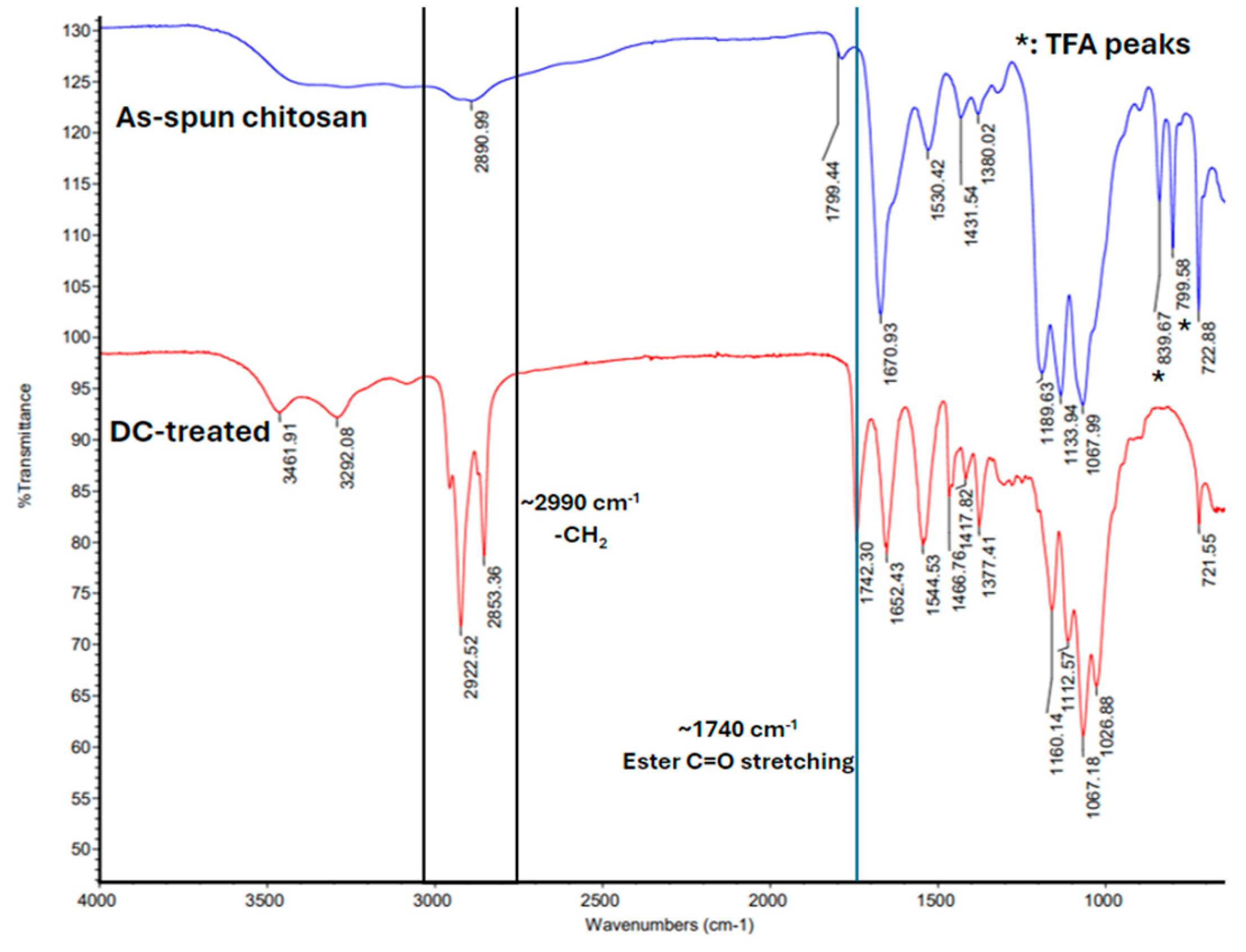
FTIR (Figure 2) confirms successful acylation of ESCMs with decanoyl chloride, as demonstrated by the presence of ester bonds (~1740 cm−1 stretch) and acyl chains (~2900 cm−1 peaks) in the decanoyl chloride-treated (DC-treated) membranes, which are absent in the as-spun chitosan membranes. Peaks at ~2922 cm−1 and ~2853 cm−1 correspond to C-H bond stretching vibrations, indicating the incorporation of the fatty carbon tails on chitosan after acylation. The stretch at ~1740 cm−1 corresponds to C=O ester bond stretching and confirms that acylation led to ester linkage formation between the fatty acid and the hydroxyl groups on chitosan.
As-spun chitosan contains trifluoroacetic acid (TFA) salts, used in chitosan processing and signified by the peaks marked with asterisks in Figure 2. The washing steps following the acylation reaction successfully removed the salts, as indicated by the absence of those peaks in the acylated chitosan membranes, improving the purity of the acylated membranes.
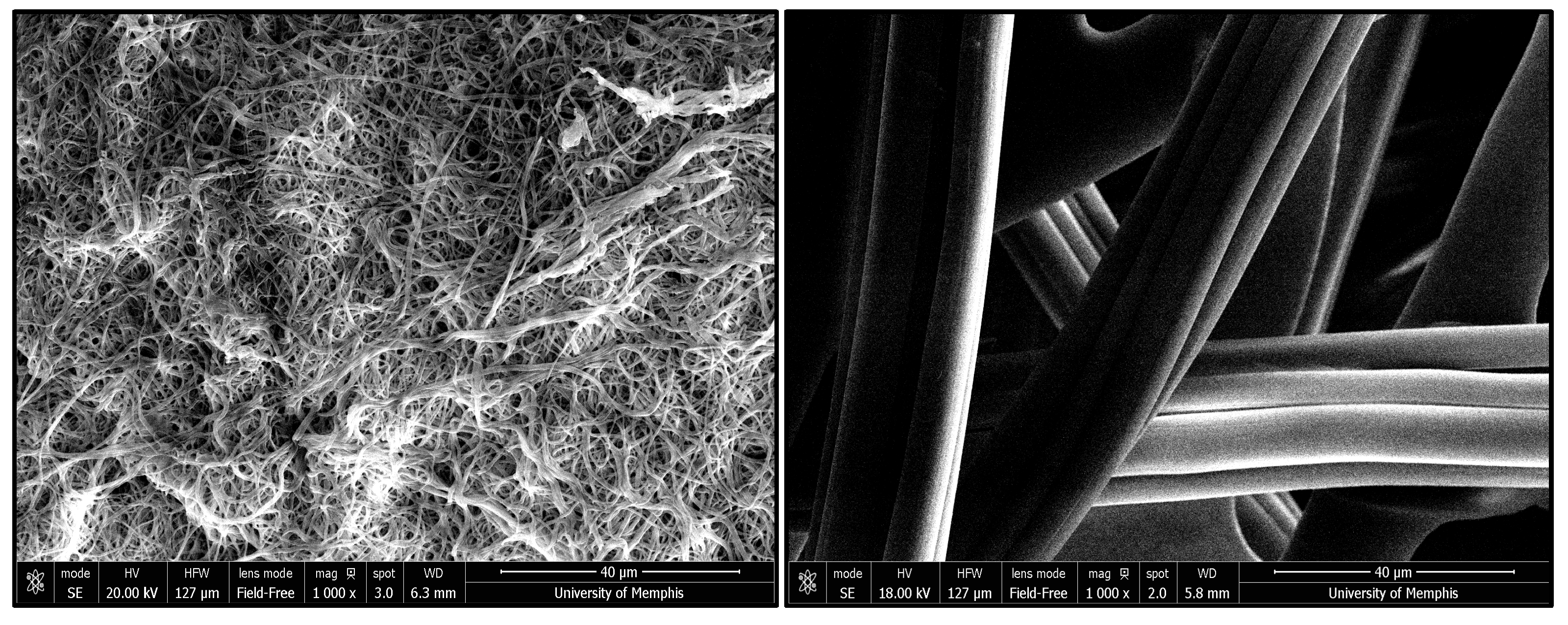
The surface morphology of ESCMs and Celox gauze is shown in Figure 3. The images depict nearly uniform nanofibers and minimal swelling. At the same magnification, the gauze can be seen to possess larger fiber diameters (microns) than the ESCMs.
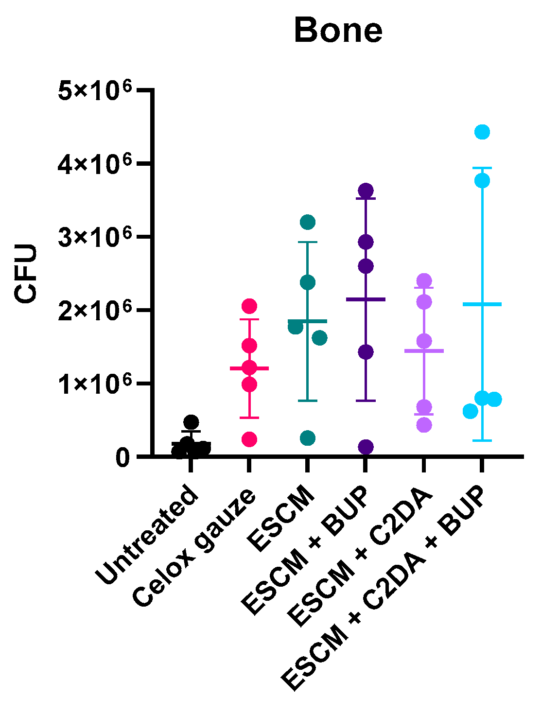
In bone (Figure 4), there was high variability within the data in each group except for the untreated group; the untreated bacterial control had fewer bacterial colonies compared with all other groups. While no significant differences exist between any of the treatment groups, the ESCMs loaded with bupivacaine and C2DA had more consistent CFU counts compared with the other groups. Full dataset for CFU counts are provided in Supplementary Table S1 (S1).
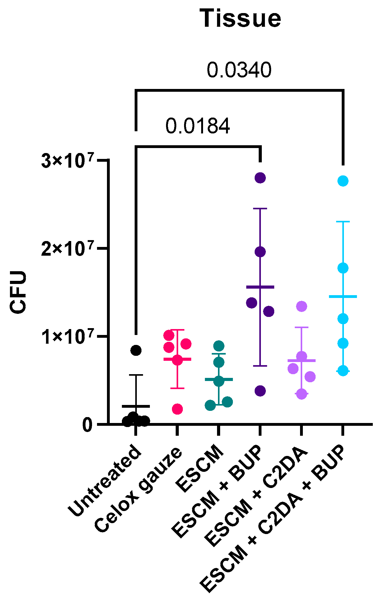
In tissue (Figure 5), significant differences were found between the untreated control group and the ESCMs loaded with bupivacaine (p = 0.0184), and ESCMs loaded with bupivacaine and C2DA (p = 0.0236). Celox gauze, ESCMs, and ESCMs loaded with C2DA had fewer bacterial colonies overall compared with the groups loaded with bupivacaine.
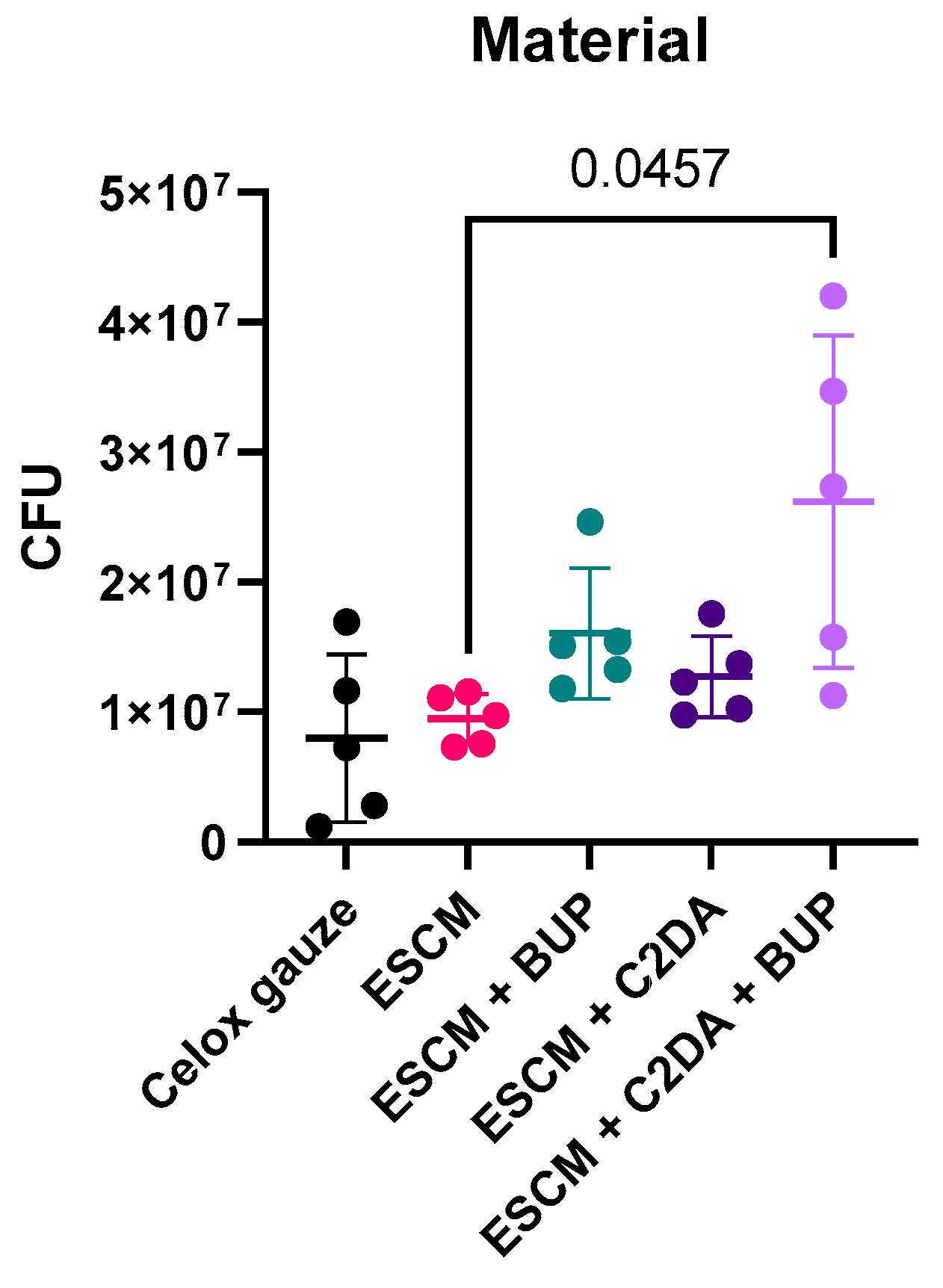
CFU counts on the material (Figure 6) show that there were higher CFU counts in the experimental groups compared to the Celox gauze, but no statistically significant differences were found. Furthermore, both ESCMs and ESCMs loaded with C2DA outperformed the dual-loaded group (ESCMs + BUP + C2DA), with significant differences found between ESCMs and ESCMs + BUP + C2DA (p = 0.0457).
Overall, there were no significant reductions in CFU counts as a result of the treatment materials when compared with the untreated bacterial control or the standard of care (Celox gauze). Groups containing bupivacaine had significantly higher bacterial growth compared with the untreated group in tissue. All materials degraded or showed signs of degradation after the 3-day implantation.

4. Discussion

Local drug delivery of antimicrobial molecules is considered an effective method of preventing bacterial growth in traumatic wounds. The critical point of bacterial growth and infection formation is bacterial adhesion and the establishment of biofilm on the tissue or implant surface [21]. Therefore, preventing bacterial adhesion is considered an effective method of infection prevention after a traumatic injury. Moreover, such traumatic injuries are often treated by administering oral antibiotics and administering opioids to alleviate the pain; however, the overuse of antibiotics and opioids contributes to the emergence of antimicrobial resistance and to the opioid epidemic.
The quorum-sensing molecule C2DA has previously been shown to inhibit planktonic and biofilm bacterial growth, to disperse existing biofilm, and to eradicate existing biofilm [19]. Furthermore, the use of local anesthetics has been shown to be effective in alleviating pain, and bupivacaine has potential synergism with fatty acids such as C2DA [22]. For instance, 5 mg of bupivacaine has been shown to significantly increase the zone of inhibition against Methicillin-resistant S. aureus when combined with 250 µg of C2DA, compared with a paper disk control [13]. This work aimed to explore the efficacy of ESCMs loaded with C2DA, ESCMs loaded with bupivacaine, and ESCMs loaded with both therapeutics against S. aureus in an acute osteomyelitis in vivo model.
The current work shows that the experimental groups enhanced bacterial growth compared with the no-treatment control. This could be due to the introduction of a natural material with a large surface area that allowed the bacteria to attach and proliferate. Lencova et al. reported that electrospun nanofibers with a large diameter and high permeability promote biofilm formation since bacteria require space for the formation of clusters and bridges between the fibers [23]. In a different study, it was found that larger fiber diameters of polystyrene allowed S. aureus to proliferate within the fibrous material, and the highest proliferation occurred when the fibers were close in diameter to the size of the bacterial cells [24]. Although the materials ESCMs and ESCMs loaded with C2DA had significantly fewer bacterial colonies compared with ESCMs loaded with bupivacaine and C2DA, those groups still had more bacterial colonies than the untreated group in tissue and bone. This further indicates that the nanofibers could have allowed the bacteria to proliferate in this model.
Furthermore, bupivacaine-containing groups had a particularly increased bacterial growth and high variability in bone, material, and tissue. This could be due to bupivacaine containing an amino–amide scaffold that highly inhibits sodium channels [25]. This inhibition shifts the production of cytokines from helper T cells from Th1-type cytokines to Th2-type cytokines. The presence of excess Th2 is known to counteract Th1 bactericidal action and leads to cell immune response suppression [26].
According to the theory of attachment points, cells with a size smaller than the surface texture exhibit stronger adhesion and have more readily accessible attachment sites. Small bacteria, such as S. aureus, have more attachment points on surfaces available to them compared with larger bacteria like E. coli, and are more likely to attach to fibers if the diameter of the fibers is comparable in size with the bacterial cells. Therefore, one approach to limiting bacterial growth would be to reduce the fiber diameter of the electrospun materials and confirm that the size of the fibers is less than 1 μm, which is the average size of staphylococcal cells [27]. This creates an electrospun material with low density and high permeability where bacteria cannot form clusters on fibers or bridges across fibers. Another approach would be to have thicker fibers with high density and low permeability which limits the space that bacteria can grow into [24,28]. In contrast to these findings, Choi et al. reported that the ESCMs modified with lipids and loaded with C2DA and bupivacaine achieved a 10-fold reduction in S. aureus biofilm viability and about 4-fold reduction in planktonic viability in vitro [14]. This indicates that while the combinations are bactericidal, they behave differently in an animal model with complex metabolic processes and immune responses.
In a similar model in tibiae, rats received stainless steel rods coated with S. aureus biofilm (ATCC 29213, a slime-producing variant) as well as a bacterial suspension in the defect to simulate an infection. Although the study used common antibiotics, neither tobramycin nor vancomycin was effective against biofilm, but vancomycin was somewhat efficacious against the infection [29]. Further investigation of the model used contaminated implants without the addition of bacterial suspension during surgery. The results demonstrate that osteomyelitis was present at the end of the 21-day treatment period with the administration of antibiotics (cefuroxime, vancomycin, and tobramycin) daily over the 21-day period [30]. This demonstrates that (1) the addition of bacterial suspension is not necessary and (2) a lower bacterial load should be used to simulate acute osteomyelitis. Lucke et al. tested three different concentrations of an S. aureus inoculum: 106, 103, and 102 CFUs/10 µL, with simultaneous insertion of titanium wires. Four weeks after implantation, it was found that 102 CFUs/10 µL is enough to induce acute osteomyelitis [31]. However, K-wires were inserted into the medullary cavity of the tibia along its length for the duration of the study to simulate the insertion of a sterile implant.
Electrospun chitosan-based materials and composites are being explored as alternatives to antibiotics and opioids to combat orthopedic trauma. For instance, studies on chitosan/polycaprolactone composite membranes augmented with silver nanoparticles (AgNPs) have demonstrated a balance between biocompatibility and broad-spectrum antimicrobial activity [32]. Silver is known to be an effective antimicrobial; however, its uses are limited due to its known tissue toxicity [33]. Bagheri et al. examined the effects of chitosan/polyethylene oxide (PEO) loaded with zinc oxide nanoparticles against S. aureus and two Gram-negative strains (Escherichia coli, and Pseudomonas aeruginosa). Their findings demonstrate that the composite is effective against these common pathogens in comparison with a negative control as measured by a zone inhibition test, but there was more inhibition of S. Aureus than Gram-negative strains [34].

5. Conclusions

Chitosan has shown promising potential in drug delivery applications for antimicrobial properties. This study examined the efficacy of ESCMs embedded with an anti-biofilm fatty acid and a local anesthetic in preventing bacterial growth in an acute osteomyelitis rodent model. The results highlight a need to examine the role of nanofiber morphology in bacterial cell proliferation and the role of bupivacaine in modulating cytokines and sodium channels. Future work for this research should examine the effects of using a lower inoculum to attain the minimal concentration of bacteria required to simulate acute osteomyelitis. Furthermore, a positive antibiotic control could be added directly to the contaminated defect and loaded onto ESCMs to determine the role of ESCMs in biofilm formation. Lastly, C2DA could be added to the defect directly to assess the efficacy of C2DA on biofilm formation in this model without the presence of ESCMs.

Supplementary Materials

The following supporting information can be downloaded at: https://www.mdpi.com/article/10.3390/lipidology2020007/s1, Table S1: Full dataset for CFU counts.

Author Contributions

Conceptualization, J.A.J. and L.B.P.; methodology, E.A., J.A.J., L.B.P., and L.J.T.; software, E.A. and L.J.T.; validation, J.A.J., L.B.P., and E.A.; formal analysis, L.J.T. and E.A.; investigation, L.B.P. and J.A.J.; resources, J.A.J. and L.B.P.; data curation, L.J.T. and E.A.; writing—original draft preparation, E.A. and A.R.T.; writing—review and editing, E.A., J.A.J., L.B.P., and A.R.T.; visualization, J.A.J. and L.B.P.; supervision, J.A.J. and L.B.P.; project administration, J.A.J. and L.B.P.; funding acquisition, J.A.J. and L.B.P. All authors have read and agreed to the published version of the manuscript.

Funding

This research was funded through the Peer Reviewed Medical Research Program (PRMRP), award number W81XWH-21-1-0193.

Institutional Review Board Statement

This study was approved by the Institutional Animal Care and Use Committee (IACUC). Protocol number: #IACUC-23-177, 22 June 2023.

Informed Consent Statement

Not applicable.

Data Availability Statement

The authors confirm that the data supporting the findings of this study are available within the article.

Acknowledgments

We would like to thank Rabeta Yeasmin, Josh Bush, Jermiah Tate, and Pavel Qaladize for assisting with the surgical procedures and preparing the treatment materials.

Conflicts of Interest

The authors declare no conflicts of interest. The funders had no role in the study’s design, collection, or analyses or in the interpretation of data, writing of the manuscript, or decision to publish the results.

References

  1. Nana, A.; Nelson, S.B.; McLaren, A.; Chen, A.F. What’s new in musculoskeletal infection: Update on biofilms. J. Bone Jt. Surg. 2016, 98, 1226–1234. [Google Scholar] [CrossRef]
  2. Sanchez, C.J.; Ward, C.L.; Romano, D.R.; Hurtgen, B.J.; Hardy, S.K.; Woodbury, R.L.; Trevino, A.V.; Rathbone, C.R.; Wenke, J.C. Staphylococcus aureus biofilms decrease osteoblast viability, inhibits osteogenic differentiation, and increases bone resorption in vitro. BMC Musculoskelet. Disord. 2013, 14, 187. [Google Scholar]
  3. Zimmerli, W.; Sendi, P. Orthopaedic biofilm infections. Apmis 2017, 125, 353–364. [Google Scholar] [CrossRef] [PubMed]
  4. Phillips, J.K.; Ford, M.A.; Bonnie, R.J. (Eds.) Pain Management and the Opioid Epidemic: Balancing Societal and Individual Benefits and Risks of Prescription Opioid Use; National Academies Press: Washington, DC, USA, 2017; Available online: https://nap.nationalacademies.org/catalog/24781/pain-management-and-the-opioid-epidemic-balancing-societal-and-individual (accessed on 25 August 2024).
  5. Mahto, K.U.; Vandana; Priyadarshanee, M.; Samantaray, D.P.; Das, S. Bacterial biofilm and extracellular polymeric substances in the treatment of environmental pollutants: Beyond the protective role in survivability. J. Clean. Prod. 2022, 379, 134759. [Google Scholar] [CrossRef]
  6. Olsen, I. Biofilm-specific antibiotic tolerance and resistance. Eur. J. Clin. Microbiol. Infect. Dis. 2015, 34, 877–886. [Google Scholar]
  7. Žiemytė, M.; Carda-Diéguez, M.; Rodríguez-Díaz, J.C.; Ventero, M.P.; Mira, A.; Ferrer, M.D. Real-time monitoring of Pseudomonas aeruginosa biofilm growth dynamics and persister cells’ eradication. Emerg. Microbes Infect. 2021, 10, 2062–2075. [Google Scholar]
  8. Amato, S.M.; Orman, M.A.; Brynildsen, M.P. Metabolic Control of Persister Formation in Escherichia coli. Mol. Cell 2013, 50, 475–487. [Google Scholar] [CrossRef]
  9. Davies, D.G.; Marques, C.N.H. A Fatty Acid Messenger Is Responsible for Inducing Dispersion in Microbial Biofilms. J. Bacteriol. 2009, 191, 1393–1403. [Google Scholar] [CrossRef]
  10. Jennings, J.A.; Courtney, H.S.; Haggard, W.O. Cis-2-decenoic Acid Inhibits S. aureus Growth and Biofilm In Vitro: A Pilot Study. Clin. Orthop. Relat. Res. 2012, 470, 2663–2670. [Google Scholar] [CrossRef]
  11. Harris, M.A.; Beenken, K.E.; Smeltzer, M.S.; Haggard, W.O.; Jennings, A.J. Phosphatidylcholine coatings deliver local antimicrobials and reduce infection in a murine model: A preliminary study. Clin. Orthop. Relat. Res. 2017, 475, 1847–1853. [Google Scholar] [CrossRef]
  12. Faustova, M.; Nazarchuk, O.; Dmytriiev, D.; Babina, Y.; Nazarchuk, H.; Dudar, A. The effect of local anesthetics against planktonic forms and film formation of S. aureus strains and its dependence on antiseptics activity. Front. Microbiol. 2023, 14, 1199899. [Google Scholar] [CrossRef] [PubMed]
  13. Harrison, Z.L.; Bumgardner, J.D.; Fujiwara, T.; Baker, D.L.; Jennings, J.A. In vitro evaluation of loaded chitosan membranes for pain relief and infection prevention. J. Biomed. Mater. Res. Part B Appl. Biomater. 2021, 109, 1735–1743. [Google Scholar] [CrossRef] [PubMed]
  14. Choi, L.R.; Harrison, Z.; Montgomery, E.C.; Bush, J.R.; Abuhussein, E.; Bumgardner, J.D.; Fujiwara, T.; Jennings, J.A. Chitosan Membranes Stabilized with Varying Acyl Lengths Release Cis-2-Decenoic Acid and Bupivacaine at Controlled Rates and Inhibit Pathogenic Biofilm. Front. Biosci. 2024, 29, 108. [Google Scholar] [CrossRef] [PubMed]
  15. Ardean, C.; Davidescu, C.M.; Nemeş, N.S.; Negrea, A.; Ciopec, M.; Duteanu, N.; Negrea, P.; Duda-Seiman, D.; Musta, V. Factors influencing the antibacterial activity of chitosan and chitosan modified by functionalization. Int. J. Mol. Sci. 2021, 22, 7449. [Google Scholar] [CrossRef]
  16. Shekarforoush, E.; Ajalloueian, F.; Zeng, G.; Mendes, A.C.; Chronakis, I.S. Electrospun xanthan gum-chitosan nanofibers as delivery carrier of hydrophobic bioactives. Mater. Lett. 2018, 228, 322–326. [Google Scholar] [CrossRef]
  17. Wu, C.; Su, H.; Tang, S.; Bumgardner, J.D. The stabilization of electrospun chitosan nanofibers by reversible acylation. Cellulose 2014, 21, 2549–2556. [Google Scholar] [CrossRef]
  18. Yeasmin, R.; Abuhussein, E.; Perez, F.; Fujiwara, T.; Bumgardner, J.D.; Jennings, J.A. Pyridine catalyzed acylation of electrospun chitosan membranes by C6-C12 acyl chlorides: Effect of reaction time and chain length. Carbohydr. Polym. Technol. Appl. 2024, 7, 100443. [Google Scholar] [CrossRef]
  19. Wells, C.M.; Coleman, E.C.; Yeasmin, R.; Harrison, Z.L.; Kurakula, M.; Baker, D.L.; Bumgardner, J.D.; Fujiwara, T.; Jennings, J.A. Synthesis and Characterization of 2-Decenoic Acid Modified Chitosan for Infection Prevention and Tissue Engineering. Mar. Drugs 2021, 19, 556. [Google Scholar] [CrossRef]
  20. Cobb, L.H.; Park, J.; Swanson, E.A.; Beard, M.C.; McCabe, E.M.; Rourke, A.S.; Seo, K.S.; Olivier, A.K.; Priddy, L.B. CRISPR-Cas9 modified bacteriophage for treatment of Staphylococcus aureus induced osteomyelitis and soft tissue infection. PLoS ONE 2019, 14, e0220421. [Google Scholar] [CrossRef]
  21. Lamret, F.; Colin, M.; Mongaret, C.; Gangloff, S.C.; Reffuveille, F. Antibiotic tolerance of Staphylococcus aureus biofilm in periprosthetic joint infections and antibiofilm strategies. Antibiotics 2020, 9, 547. [Google Scholar] [CrossRef]
  22. Heffernan, J.M.; McLaren, A.C.; Glass, C.M.; Overstreet, D.J. Extended release of bupivacaine from temperature-responsive hydrogels provides multi-day analgesia for postoperative pain. Pain Med. 2023, 24, 113–121. [Google Scholar] [CrossRef] [PubMed]
  23. Lencova, S.; Svarcova, V.; Stiborova, H.; Demnerova, K.; Jencova, V.; Hozdova, K.; Zdenkova, K. Bacterial biofilms on polyamide nanofibers: Factors influencing biofilm formation and evaluation. ACS Appl. Mater. Interfaces 2020, 13, 2277–2288. [Google Scholar] [CrossRef] [PubMed]
  24. Abrigo, M.; Kingshott, P.; McArthur, S.L. Electrospun polystyrene fiber diameter influencing bacterial attachment, proliferation, and growth. ACS Appl. Mater. Interfaces 2015, 7, 7644–7652. [Google Scholar] [CrossRef] [PubMed]
  25. Tochitsky, I.; Jo, S.; Andrews, N.; Kotoda, M.; Doyle, B.; Shim, J.; Talbot, S.; Roberson, D.; Lee, J.; Haste, L.; et al. Inhibition of inflammatory pain and cough by a novel charged sodium channel blocker. Br. J. Pharmacol. 2021, 178, 3905–3923. [Google Scholar] [CrossRef]
  26. Wang, Y.-C.; He, F.; Feng, F.; Liu, X.-W.; Dong, G.-Y.; Qin, H.-Y.; Hu, X.-B.; Zheng, M.-H.; Liang, L.; Feng, L.; et al. Notch signaling determines the m1 versus m2 polarization of macrophages in antitumor immune responses. Cancer Res. 2010, 70, 4840–4849. [Google Scholar] [CrossRef]
  27. Monteiro, J.M.; Fernandes, P.B.; Vaz, F.; Pereira, A.R.; Tavares, A.C.; Ferreira, M.T.; Pereira, P.M.; Veiga, H.; Kuru, E.; VanNieuwenhze, M.S.; et al. Cell shape dynamics during the staphylococcal cell cycle. Nat. Commun. 2015, 6, 8055. [Google Scholar] [CrossRef]
  28. Kim, T.; Kwon, S.; Lee, J.; Lee, J.S.; Kang, S. A metallic anti-biofouling surface with a hierarchical topography containing nanostructures on curved micro-riblets. Microsystems Nanoeng. 2022, 8, 1–14. [Google Scholar] [CrossRef]
  29. Gracia, E.; Laclériga, A.; Monzón, M.; Leiva, J.; Oteiza, C.; Amorena, B. Application of a rat osteomyelitis model to comparein vivoandin vitrothe antibiotic efficacy against bacteria with high capacity to form biofilms. J. Surg. Res. 1998, 79, 146–153. [Google Scholar] [CrossRef]
  30. Monzón, M.; García-Álvare, F.; Laclériga, A.; Gracia, E.; Leiva, J.; Oteiza, C.; Amorena, B. A simple infection model using pre-colonized implants to reproduce rat chronic Staphylococcus aureus osteomyelitis and study antibiotic treatment. J. Orthop. Res. 2001, 19, 820–826. [Google Scholar] [CrossRef]
  31. Lucke, M.; Schmidmaier, G.; Sadoni, S.; Wildemann, B.; Schiller, R.; Stemberger, A.; Haas, N.P.; Raschke, M. A new model of implant-related osteomyelitis in rats. J. Biomed. Mater. Res. Part B Appl. Biomater. Off. J. Soc. Biomater. Jpn. Soc. Biomater. Aust. Soc. Biomater. Korean Soc. Biomater. 2003, 67, 593–602. [Google Scholar]
  32. Korniienko, V.; Husak, Y.; Diedkova, K.; Varava, Y.; Grebnevs, V.; Pogorielova, O.; Bērtiņš, M.; Korniienko, V.; Zandersone, B.; Ramanaviciene, A.; et al. Antibacterial potential and biocompatibility of chitosan/polycaprolactone nanofibrous membranes incorporated with silver nanoparticles. Polymers 2024, 16, 1729. [Google Scholar] [CrossRef] [PubMed]
  33. Burdușel, A.-C.; Gherasim, O.; Grumezescu, A.M.; Mogoantă, L.; Ficai, A.; Andronescu, E. Biomedical applications of silver nanoparticles: An up-to-date overview. Nanomaterials 2018, 8, 681. [Google Scholar] [CrossRef] [PubMed]
  34. Bagheri, M.; Validi, M.; Gholipour, A.; Makvandi, P.; Sharifi, E. Chitosan nanofiber biocomposites for potential wound healing applications: Antioxidant activity with synergic antibacterial effect. Bioeng. Transl. Med. 2022, 7, e10254. [Google Scholar] [CrossRef] [PubMed]
Figure 1. A summary of the surgical procedure.
Lipidology 02 00007 g001
Figure 2. FTIR spectra of as-spun ESCMs and ESCMs acylated with decanoyl chloride (DC-treated). Ester bonds are indicated by the ~1740 cm−1 stretch, and acyl chains are indicated by peaks at ~2900 cm−1 in the DC-treated membranes, which are absent in the as-spun chitosan membranes. Asterisks indicate trifluoroacetic acid (TFA) salts, which are absent in the DC-treated membranes.
Lipidology 02 00007 g002
Figure 3. SEM images of modified ESCMs (left) and Celox gauze (right) at 1000× magnification.
Lipidology 02 00007 g003
Figure 4. After three days, CFU counts were measured in retrieved bone. Groups’ means were compared with the means of other groups. Statistical differences are displayed in figures as p-values (p < 0.05, n = 5). Significance was determined by ANOVA with Tukey’s Multiple Comparisons Test.
Lipidology 02 00007 g004
Figure 5. After three days, CFU counts were measured in retrieved tissue. Groups’ means were compared with the means of other groups. Statistical differences are displayed in figures as p-values. (p < 0.05, n = 5). Significance was determined by ANOVA with Tukey’s Multiple Comparisons Test.
Lipidology 02 00007 g005
Figure 6. After three days, CFU counts were measured in treatment material. Groups’ means were compared with the means of other groups. Statistical differences are displayed in figures as p-values. (p < 0.05, n = 5). Significance was determined by ANOVA with Tukey’s Multiple Comparisons Test.
Lipidology 02 00007 g006
Table 1. Treatment groups applied to rats after defect creation.
Treatment GroupsMaterial Mass (mg)Sample Size (n)
Celox1505
ESCMs150
ESCMs + C2DA150
ESCMs + Bup150
ESCMs + C2DA + Bup150
No treatment0
Disclaimer/Publisher’s Note: The statements, opinions and data contained in all publications are solely those of the individual author(s) and contributor(s) and not of MDPI and/or the editor(s). MDPI and/or the editor(s) disclaim responsibility for any injury to people or property resulting from any ideas, methods, instructions or products referred to in the content.