Review

Regulation of Urate Homeostasis by Membrane Transporters

by
Tappei Takada
1,*,†,
Hiroshi Miyata
1,†,
Yu Toyoda
1,2,
Akiyoshi Nakayama
2,
Kimiyoshi Ichida
3 and
Hirotaka Matsuo
2
1
Department of Pharmacy, The University of Tokyo Hospital, Tokyo 113-8655, Japan
2
Department of Integrative Physiology and Bio-Nano Medicine, National Defense Medical College, Tokorozawa 359-8513, Japan
3
Department of Pathophysiology, Tokyo University of Pharmacy and Life Science, Hachioji 192-0392, Japan
*
Author to whom correspondence should be addressed.
These authors equally contributed to this work.
Gout Urate Cryst. Depos. Dis. 2024, 2(2), 206-219; https://doi.org/10.3390/gucdd2020016
Submission received: 4 April 2024 / Revised: 15 June 2024 / Accepted: 18 June 2024 / Published: 19 June 2024

Abstract

:
Uric acid is the final purine metabolite in humans. Serum urate levels are regulated by a balance between urate production, mainly in the liver, and its excretion via the kidneys and small intestine. Given that uric acid exists as a urate anion at physiological pH 7.4, membrane transporters are required to regulate urate homeostasis. In the kidney, urate transporter 1, glucose transporter 9, and organic anion transporter 10 contribute to urate reabsorption, whereas sodium-dependent phosphate transport protein 1 would be involved in urate excretion. Other transporters have been suggested to be involved in urate handling in the kidney; however, further evidence is required in humans. ATP-binding cassette transporter G2 (ABCG2) is another urate transporter, and its physiological role as a urate exporter is highly demonstrated in the intestine. In addition to urate, ABCG2 regulates the behavior of endogenous substances and drugs; therefore, the functional inhibition of ABCG2 has physiological and pharmacological effects. Although these transporters explain a large part of the urate regulation system, they are not sufficient for understanding the whole picture of urate homeostasis. Therefore, numerous studies have been conducted to find novel urate transporters. This review provides the latest evidence of urate transporters from pathophysiological and clinical pharmacological perspectives.

1. Introduction

Humans and other higher primates synthesize uric acid as the final purine metabolite. The loss of uricase (Uox) and extensive urate reabsorption from the glomerular filtrate result in plasma urate levels approximately 10 times higher in humans than in most other mammals (reviewed in [1]). Approximately two-thirds of uric acid, which is mainly generated in the liver, is excreted via urine as urate, and approximately one-third is excreted via feces. Hyperuricemia is caused by the increased synthesis and/or decreased excretion of urate and is classified into different types based on its etiology, overproduction, and/or underexcretion. Recently, based on the discovery of ATP-binding cassette transporter G2 (ABCG2) as an intestinal urate exporter, extrarenal underexcretion type hyperuricemia was defined as described later [2,3,4]. Hyperuricemia is one of the most important risk factors of gout; therefore, urate-lowering therapy is recommended for patients with tophi and/or frequent acute gout flares (reviewed in [5]).
Conversely, hypouricemia is caused by increased excretion of urate mainly due to its decreased reabsorption from the glomerular filtrate or aberrant metabolism of purines. Hypouricemia could be associated with several diseases, such as exercise-induced acute kidney injury and some neurodegenerative diseases such as Alzheimer’s disease and Parkinson’s disease [6,7,8,9], which may be partially explained by the antioxidant property of urate [10]. Therefore, maintaining serum urate levels within the appropriate range is clinically important. In this context, the concept of “dysuricemia,” which combines hyperuricemia and hypouricemia, has recently been proposed (reviewed in [11,12]).
Urate circulates between organs through the bloodstream for the maintenance of urate homeostasis throughout the whole body, a process that requires cell transporters for urate. Because urate is a water-soluble compound that exists as an anion under physiological conditions, it cannot passively pass through the cell membrane, which consists of a lipid bilayer. Therefore, membrane transporters are essential for the regulation of urate homeostasis. In this review, we summarize the regulatory mechanisms of urate homeostasis in the body and the role of urate transporters based on the latest findings from pathophysiological and clinical pharmacological perspectives (Figure 1).
URAT1, OAT10, and OAT4 localized on the apical membrane of the renal proximal tubule epithelial cells reabsorb urate from glomerular filtrate in exchange for appropriate organic anions. The reabsorbed urate is transported into the blood stream primarily via the urate transporter GLUT9, located on the basolateral membrane of the proximal tubule epithelial cells. The urate from the blood is thought to be transported via the basolateral transporters OAT1 and OAT3 into the proximal tubule epithelial cells, and finally secreted into the tubular lumen via the apical transporters ABCG2, ABCC4, NPT1, and NPT4.

2. Urate Transporters Are Classified into Two Superfamilies

Transporters are a group of membrane proteins that regulate the movement of substances across biological membranes. Transporters can be classified into two superfamilies: the ABC superfamily and the solute carrier (SLC) superfamily. Mammalian ABC transporters utilize the energy derived from ATP hydrolysis and are mostly involved in transporting various substances out of cells or into cellular organelles (reviewed in [13]). In contrast, members of the SLC superfamily use the ion gradient formed by ATP hydrolysis as a driving force or promote passive diffusion; thus, SLC transporters can transport substrates out of and into cells (reviewed in [14]). During the regulation of urate (re)absorption and excretion, urate is incorporated into cells from one side of the plasma membrane and secreted to the other. During this, ABC and SLC transporters, which have different transport properties, are cooperatively involved in regulating urate homeostasis.

3. How Have the Transporters Involved in the Regulation of Urate Disposition Been Discovered?

Since the discovery of urate transporter 1 (URAT1) in 2002 [15], which is described below, as a transporter responsible for urate reabsorption from glomerular filtrate in exchange for intracellular monocarboxylate anion, many studies have been actively conducted to identify transporters that regulate the kinetics of urate in the body. Genetic analyses have been useful in searching for urate transporters that function in vivo in humans. URAT1 is a causative gene for hereditary type 1 renal hypouricemia (RHUC1), characterized by lower serum urate levels and higher urate excretion into the urine [15]. In addition to information on the causative genes of these inherited diseases, genome-wide association study (GWAS), which compares the frequency of single nucleotide polymorphisms (SNPs) between case and control populations, has been widely used to search for urate transporters (reviewed in [16]).
GWAS is a genetic approach used to analyze the genome-wide associations between SNPs in genome sequences and the phenotypes of interest (reviewed in [17]). To identify the regulators of urate homeostasis, SNPs are analyzed in patients with gout or hyperuricemia and in healthy individuals, and their frequencies are compared between the two groups to identify genes with statistically significant differences. However, in GWAS, a significant difference in the frequency of SNPs not directly associated with the phenotype of interest may be observed because of a linkage disequilibrium, in which one SNP is inherited in conjunction with another SNP in the genomic vicinity (reviewed in [17]). Therefore, to identify urate transporters, selecting transporter(-like) proteins among the genes detected in GWAS and verifying their direct urate transport activity through in vitro experiments using cultured cells and/or membrane vesicle transport systems is important. Several transporters that play a role in the regulation of urate kinetics described below have been shown to be important in humans through such genetic approaches and in vitro transport experiments [4,15,18,19,20,21,22,23].

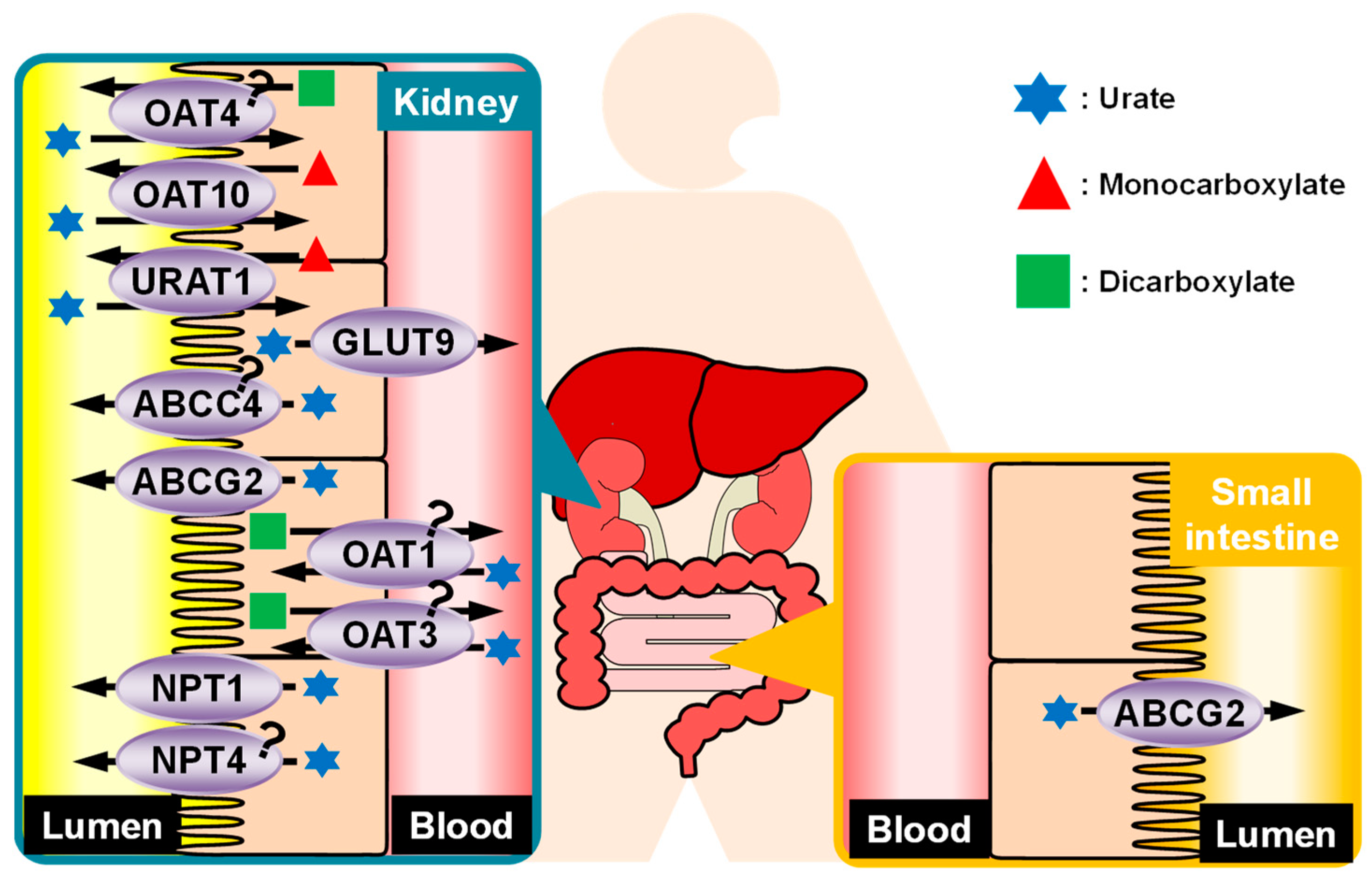
4. Regulation of Urate Kinetics in the Kidney

The kidneys account for approximately two-thirds of urate excretion in the body. The kidney is responsible for blood purification and maintenance of body fluid homeostasis by filtering plasma components through the glomeruli and reabsorbing the necessary nutrients and ions in various segments from the renal tubules to the collecting ducts. Some waste products in the blood of our body are secreted via cells of kidney tubules and finally excreted through urine. Reabsorption and cellular secretion are also important for controlling the urinary excretion of urate. Approximately 90% of the urate filtered via the glomeruli is reabsorbed before reaching the collecting ducts, and only approximately 10% is excreted from the body. Transporters play important roles in urate reabsorption and secretion in the kidneys.

4.1. Reabsorption of Urate from Glomerular Filtrate

As mentioned above, approximately 90% of the urate from glomerular filtrate is reabsorbed, and transporters involved in this process have a significant effect on urate kinetics. Some of the transporters involved in the reabsorption process are targets of uricosuric drugs used in clinical practice. Therefore, they are important not only from physiological perspective but also from pharmacological and clinical perspectives. Some urate reabsorption transporters are expressed in human renal tubules. URAT1 and OAT10 are localized on the apical membrane of the renal tubules, and GLUT9 is localized on the basolateral membrane of the renal tubules. OAT4’s physiological importance in urate regulation requires further investigation.

4.1.1. URAT1/SLC22A12

URAT1, encoded by the SLC22A12 gene and mapped to human chromosome 11q13.3, was first identified as a urate transporter involved in the regulation of urate kinetics [15]. It was identified based on its nucleotide sequence similarity with OAT4 and as the causative gene of RHUC1 [15]. Additionally, several GWAS have shown an association between SNPs in SLC22A12 and serum urate levels [24,25,26]. URAT1 belongs to a subfamily of organic anion transporters within the large SLC22 family. Functional characterization through expression in Xenopus oocytes showed the urate transport activity of URAT1 [15].
URAT1 is responsible for urate reabsorption from glomerular filtrate through exchange with intracellular monocarboxylates, which are incorporated into epithelial cells by the sodium-coupled monocarboxylate transporter (SMCT) 1/SLC5A8 and SMCT2/SLC5A12 [27,28] at the apical membrane, on the luminal side of renal proximal tubular cells. In addition, in vitro experiments have shown that uricosuric drugs such as benzbromarone, probenecid, losartan [15], verinurad [29], and dotinurad [30], which decrease serum urate levels by promoting urate excretion into the urine, inhibit the urate transport activity of URAT1, indicating the importance of URAT1 as a drug target for hyperuricemia treatment. A recent in vitro study also suggested that tranilast, an anti-inflammatory drug, inhibited URAT1 in higher concentrations than the plasma unbound concentrations observed in clinical dosages [31]. URAT1 is not only a pharmacological target, but is also involved in the development of hyperuricemia as an adverse effect of drugs. Pyrazinecarboxylic acid (PZA; an active metabolite of the antituberculosis drug pyrazinamide) and small amounts of salicylate can enhance URAT1 function by serving as substrates for exchange transport of urate, resulting in hyperuricemia [15]. Recent advances in functional regulation of URAT1 have suggested the role of sodium/glucose cotransporter (SGLT) 2 [32] and insulin for the regulation of expression [33] but not on the functional regulation of URAT1 [34]. Toyoki et al. reported the roles of insulin in regulating Urat1 expression in a streptozocin-induced diabetic rat model lacking insulin secretion from pancreatic beta cells. This study showed that the loss of insulin decreased Urat1 protein levels in the kidney, which were recovered by insulin administration, whereas the SGLT2 inhibitor ipragliflozin did not affect Urat1 levels [33]. In contrast, Novikov et al. showed that the genetic loss of Sglt2 and administration of the SGLT2 inhibitor canagliflozin increased the fractional excretion of urate (FEUA) in a mouse model, while the uricosuric effect of canagliflozin was not observed in Urat1 knockout (KO) mice [32]. Although the molecular mechanisms underlying the association between URAT1 and insulin/SGLT2 remains unclear, they might explain the relationship between diabetes and hyperuricemia [34,35,36,37].

4.1.2. Glucose Transporter (GLUT) 9/SLC2A9

GLUTs are members of the hexose transporters family. A GWAS for hyperuricemia and gout in the Framingham cohort first identified the SLC2A9 locus [38]. GLUT9, encoded by the SLC2A9 gene (mapped to human chromosome 4p16.1), was initially characterized in vitro [39,40,41] as a glucose/fructose transporter. However, further studies on GLUT9 failed to reproduce GLUT9 as either [42]. GLUT9 expression is almost ubiquitous, with higher levels in the liver, kidney, brain, placenta, lung, and peripheral leukocytes [43]; it is suggested that its expression level is regulated by the insulin and PI3K/AKT signaling pathway [34,44]. Like URAT1, GLUT9 is inhibited by high concentrations of uricosuric drugs like benzbromarone, tranilast, and probenecid in Xenopus oocyte expression systems [31]. In contrast, unlike URAT1, anti-uricosuric agents like PZA, nicotinate, and salicylate had no effect on GLUT9 activity [31]. Multiple GWAS have shown that variations in the SLC2A9 gene have the single most prominent genetic influence on serum urate level [25,45,46,47]. Patients with type 2 renal hypouricemia (RHUC2) who have no URAT1 mutations were found to have missense mutations in GLUT9, which decreased the urate transport activity in vitro [18]. The relationship between GLUT9 loss-of-function mutations and the complete absence of renal reabsorption of filtered urate with approximately 150% FEUA in patients with hypouricemia [48] clearly indicates the role of GLUT9 in urate reabsorption in the renal tubules.
GLUT9 exists in two isoforms with distinct N-terminal domains: GLUT9a/GLUT9L (540 residues) is trafficked to the basolateral membrane and GLUT9b/GLUT9S (511 residues) to the apical membrane, as reported in Madin–Darby Canine Kidney cells [18,39]. With increasing concentrations of extracellular potassium, stimulation of their urate transport activity (140–150-fold) indicates that it is induced by changes in membrane potential [42]. Both isoforms have urate transport activity. However, considering that intratubular urate lithiasis occurs in Glut9 KO mice [49], which suggested urate accumulation in tubular cells, GLUT9a, localized at the basolateral membrane, is believed to play an important role in vivo. Collectively, URAT1 reabsorbs urate from urine into cells at the apical membrane, and then GLUT9a on the basolateral membrane secretes it into the blood.

4.1.3. Organic Anion Transporter (OAT) 10/SLC22A13

OATs comprise a family of transporters responsible for the transport of organic anions, and URAT1/SLC22A12 belongs to this family. OAT10, encoded by the SLC22A13 gene (mapped to human chromosome 3p22.2), has been characterized in vitro as a low-affinity urate transporter and high-affinity nicotinate transporter [50]. The recent study implied that insulin signaling might affect the urate transport activity of OAT10 [34]. OAT10’s physiological role in human urate homeostasis remained unclear. Therefore, to clarify the role of OAT10 in the regulation of urate kinetics, the presence of genetic mutations in all exons of OAT10 was analyzed in Japanese patients with gout and healthy participants [51]. The results showed that rs117371763 (c.1129C>T; p.R377C) mutation was significantly associated with a lower risk of gout, and functional analyses revealed that p.R377C is functionally null. Subsequent analyses showed that OAT10 is expressed on the apical membrane of proximal tubules [20] and that populations heterozygous for the OAT10 c.1129C>T mutation (1129CT) have a higher urinary urate excretion rate and lower serum urate levels than populations with 1129CC (homozygous for the WT allele) [20]. In another report, the rs72542450 (c.47G>A; p.R16H) mutation detected in a cohort of patients with hyperuricemia and gout decreased the urate transport activity of OAT10 by approximately 20% [22]. These results indicated that OAT10, similar to URAT1, is involved in urate reabsorption in the apical membrane of the proximal tubules. It is suggested that OAT10 functions could be modulated by the existence of nearby monocarboxylate transporter 1/SLC16A1 [52], in addition to the genetic effects; functional modifications of OAT10 by other proteins may be important for OAT10-urate regulation. Interestingly, in vitro experiments have shown that losartan, an angiotensin II receptor antagonist, which promotes urate excretion in clinical practice, inhibits urate transport via OAT10 [20]. Although the uricosuric effect of losartan was thought to be based on URAT1, the above report indicates that losartan inhibits OAT10 more efficiently than URAT1. In another report, the urate transport activity of OAT10 was demonstrated to be inhibited by high concentrations of tranilast, PZA, nicotinate, and salicylate in Xenopus oocyte expression systems [31]. These data suggested that OAT10, as a urate reabsorption transporter, may be important from a pharmacological perspective.

4.1.4. OAT4/SLC22A11

OAT4, encoded by the SLC22A11 gene (mapped to human chromosome 11q13.1), is located very close to SLC22A12, which encodes URAT1. OAT4 was first reported as an organic anion/dicarboxylate exchanger expressed on the apical membrane of renal tubules [53], then characterized as a modest urate transporter [54] in vitro. The urate transport activity of OAT4 was reported to be inhibited by high concentrations of benzbromarone, probenecid, and tranilast [31] and might be enhanced by insulin signaling [34]. The relationship between SNPs in SLC22A11 and hyperuricemia/gout has been reported in GWAS [25,47,55,56]. Although most SNPs in these reports were located in introns or intergenic regions, some missense (rs141159367; c.5C>T; p.A2V, rs75933978; c.1028G>T; p.R343L) or nonsense (rs35008345; c.142C>T; p.R48X) polymorphisms were identified [56]. Moreover, rs144573306 (c.1556C>T; p.P519L) was recently identified in cohort of patients with hyperuricemia and gout, impairing the urate transport activity of OAT4 by approximately 30% [22]. Further functional analyses of OAT4 variants and genetic analyses would contribute to a deeper understanding of OAT4’s role in urate homeostasis.

4.2. Secretion of Urate into the Renal Tubule

As mentioned above, most of the urate filtered by the glomeruli is reabsorbed and returned to the blood. However, the existence of a urate secretion system is suggested based on a report showing that urate clearance could exceed inulin clearance after sulfinpyrazone and mannitol administration [57]. Similar to reabsorption, urate transporters play an important role in the secretion process. This section discusses the roles of OAT1, OAT3, and sodium-dependent phosphate transport protein (NPT) 1, NPT4, ABCG2, and ABCC4.

4.2.1. OAT1/SLC22A6 and OAT3/SLC22A8

OAT1 and OAT3 are closely related organic anion transporters with a 51% amino acid sequence identity [58]. The genes for OAT1 (SLC22A6 gene) and OAT3 (SLC22A8 gene) are tandemly arrayed on human chromosome 11q12.3 [59]. OAT1 [60] and OAT3 [61] are both localized on the basolateral membrane of the human kidney proximal tubule cells and are involved in transporting amphiphilic organic anions and uncharged molecules [62]. OAT1 and OAT3 are organic anion/dicarboxylate exchangers [62] exhibiting urate transport activities in vitro [21]. An in vitro study suggested that benzbromarone, a clinically used uricosuric agent, inhibited the urate transport activities of both OAT1 and OAT3 [30]. However, benzbromarone’s half-maximal inhibitory concentration values against OAT1 and OAT3 were higher than those against URAT1 [30], suggesting that benzbromarone had less inhibitory activity against OAT1 and OAT3. The recent study implied that insulin signaling might affect the urate transport activity of OAT1 and OAT3 [34]. Considering their localization and urate transport activities, OAT1 and OAT3 are believed to participate in urate excretion in the kidneys. Recent research has shown that a rare non-synonymous variant of the SLC22A8 gene (rs45566039; c.445C>T; p.R149C), found in a cohort of patients with hyperuricemia and gout, abolished the urate transport activities of OAT3 [21]. Further accumulation of in vivo evidence, followed by functional analyses, will improve our understanding of the roles of OAT1 and OAT3 in urate homeostasis.

4.2.2. NPT1/SLC17A1 and NPT4/SLC17A3

The NPT family was originally identified as phosphate transporters that transport inorganic phosphate in a sodium-dependent manner (reviewed in [63]). However, the affinity of the NPT family transporters for inorganic phosphate is not so high; therefore, their main role is now thought to be that of organic anion transporters [63]. Although NPT1, encoded by the SLC17A1 gene on human chromosome 6p22.2, was shown to have voltage-dependent urate excretion activity in vitro [64], results of genetics analyses regarding serum urate levels and/or gout were inconsistent among studies for NPT1 [65,66,67,68]. Therefore, the physiological role of NPT1 in the regulation of urate kinetics remained unclear. In this context, the frequency of NPT1 gene polymorphism rs1165196 (c.806T>C; p.I269T) has been investigated in patients with gout with classified disease types [19]. The results showed that p.I269T did not affect the risk of gout as a whole or in renal overload gout; however, it decreased the risk of renal underexcretion gout. In addition, in vitro experiments have shown that the p.I269T mutation increases the urate transport activity of NPT1 [19], suggesting that NPT1 is responsible for urate secretion into the urine in humans. In contrast, the NPT1 p.I269T polymorphism is known to be in linkage disequilibrium with SNPs in SLC17A3 gene [38]. The SLC17A3 gene is located on human chromosome 6p22.2 and codes NPT4, which shows urate transport activity in vitro [69]. This indicates that individuals with NPT1 p.I269T polymorphism are likely to have a polymorphism in NPT4, suggesting that NPT4 may also contribute to a lower risk of urate underexcretion gout. Further studies focusing on the urate transport activity of NPT4 will help to clarify its physiological role in urate regulation.

4.2.3. Multidrug Resistance-Associated Protein (MRP) 4/ABCC4
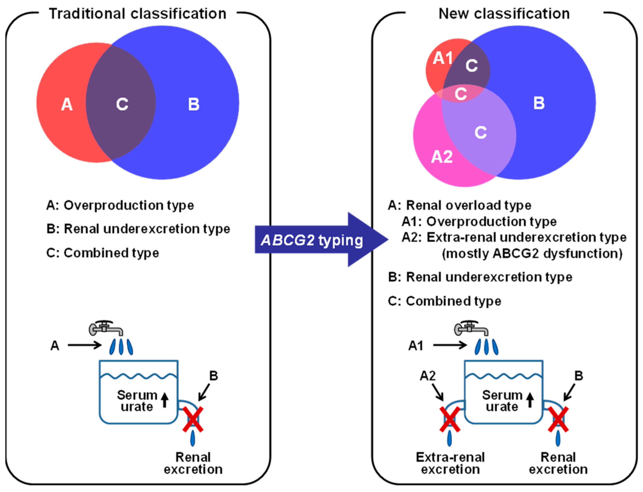
ABCC4, localized on the apical membrane of the proximal tubule [70], is an MRP/ABCC subfamily member of ATP-binding cassette transporters. ABCC4 pumps out various endogenous and xenobiotic organic anionic compounds, such as eicosanoids and conjugated steroids in addition to urate and cyclic nucleotides [71]. The ABCC4 gene is located on human chromosome 13q32.1. A genetic variant of MRP4 reduced urate efflux activity (rs972711951; c.3107C>T; p.P1036L) in individuals of Western Polynesian ancestry by approximately 30% and increased the risk of gout [23]. These in vitro and in vivo studies suggest ABCC4 functions as a urate efflux transporter in the apical membrane of proximal tubule. Further in vivo evidence of the role of ABCC4 in urate handling is required to improve disease management and treatment.

5. BCRP/ABCG2

ABCG2, also known as breast cancer resistance protein (BCRP), is a drug transporter that was initially identified as an anticancer drug resistance gene product in breast cancer-derived cell lines [72,73] or in the placenta [74]. The ABCG2 gene is located on human chromosome 4q22.1. It is abundantly expressed in the apical membranes of the kidney, liver, small intestine, blood–brain barrier, and placenta [34,75] and plays an important role in drug excretion and biological defense by pumping substrate drugs from the inside to the outside of cells by utilizing energy from ATP hydrolysis. ABCG2 is also a transporter whose function shows interindividual variations derived from several SNPs. Among these, rs2231142 (c.421C>A; p.Q141K) polymorphism, which reduces protein levels by half due to instability in the nucleotide-binding domain [76], has been reported to affect the clinical disposition of substrate drugs, such as rosuvastatin [77,78].

5.1. ABCG2 as a Urate Transporter

ABCG2 has attracted attention for its function as a drug transporter since its discovery; however, its role in the regulation of urate disposition has been revealed through genomic approaches. The association between ABCG2 and gout was first reported in 2004 [79], and many subsequent GWAS reports have demonstrated the relationship between ABCG2 SNPs and hyperuricemia and/or gout. To clarify the physiological role of ABCG2, its urate transport activity was evaluated by in vitro experiments with membrane vesicles of mammalian cells [4] and Xenopus oocytes [3]. Notably, urate was found to be a substrate for ABCG2. Moreover, by combining genetic and in vitro functional analyses, decreased ABCG2 function was found to be a major risk factor for hyperuricemia and gout [3,4].
Experiments were conducted using mice to elucidate the physiological role of ABCG2 in urate homeostasis. Considering that mice, unlike humans, possess a urate-metabolizing enzyme (uricase, Uox), the serum urate levels of Abcg2 KO and WT mice were compared under continuous administration of a Uox inhibitor, oxonate. The results showed that the serum urate levels of Abcg2 KO mice were higher than those of WT mice, similar to those observed in humans. Furthermore, Abcg2 deficiency did not decrease urate excretion into the urine but increased it, while urate excretion from the small intestine dramatically decreased [2]. This result was surprising since ABCG2 is known to be expressed in the kidney and is reported to be involved in the excretion of substrate compounds into the urine at the apical membrane. However, recent studies using Abcg2 p.Q140K mice (corresponding to human ABCG2 p.Q141K) [80] and Abcg2 KO mice with acute gout attack [81] strongly support the physiological importance of the intestinal secretion of urate via the ABCG2-mediated pathway.
The conventional classification of gout/hyperuricemia involves the overproduction type (increased synthesis), renal underexcretion type (decreased urinary excretion), and combined type (a mixture of the two). However, the discovery of ABCG2 as a urate transporter has revealed the existence of hyperuricemia that does not belong to either type but is caused by decreased excretion of urate from the intestinal tract. Based on these findings, the current classification of gout/hyperuricemia includes extra-renal underexcretion hyperuricemia, in which intestinal urate excretion is low because of ABCG2 dysfunction. The extra-renal underexcretion type and overproduction type are sometimes considered to be the renal overload type because distinguishing them clinically is impossible with easy examinations; this classification is considered useful when considering treatment strategies (Figure 2) [2].
The regulation of urate kinetics by ABCG2, primarily as an exporter in the small intestine, has been clarified. However, genetic polymorphisms resulting in decreased ABCG2 function have been shown to be risk factors for not only renal-loaded hyperuricemia, which is caused by reduced urate excretion from the small intestine, but also renal underexcretion hyperuricemia [82]. This suggests that ABCG2 may also be involved in urate excretion via urine at the apical membranes of tubular epithelial cells [83].

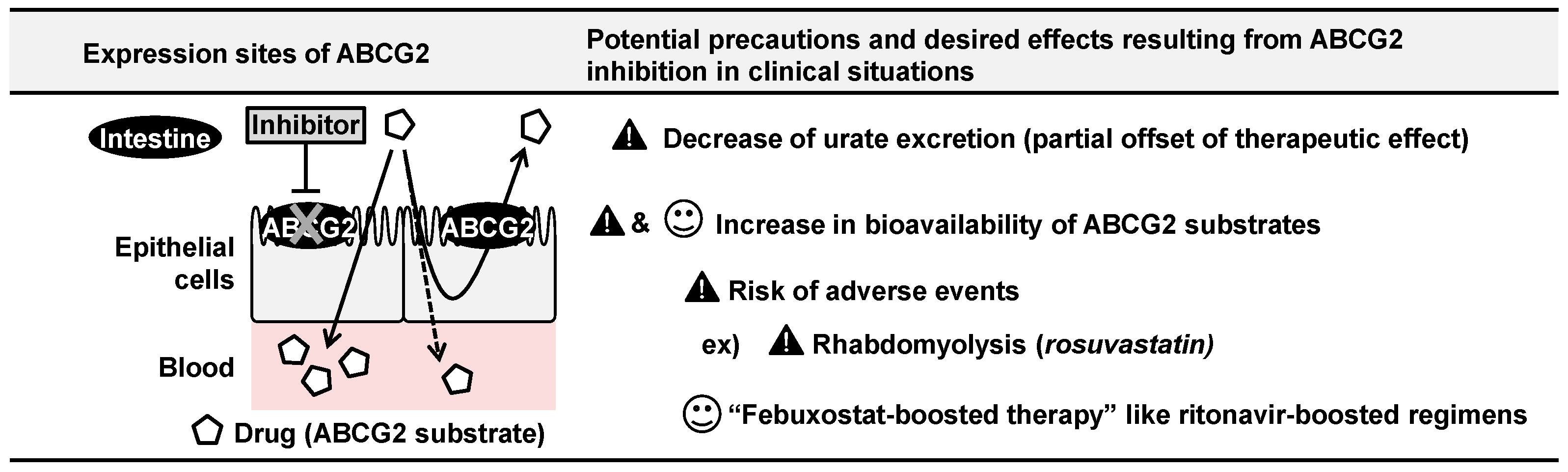
5.2. ABCG2 Inhibition by Febuxostat and Its Clinical Implications

Given that ABCG2 was identified as a drug transporter before its discovery as a urate regulator, it is possible that ABCG2 may interact with drugs that alter serum urate levels. Therefore, the effects of drugs that influence serum urate levels in clinical practice on ABCG2-mediated urate transport were examined in vitro [84]. The results showed that febuxostat, a clinically used drug for hyperuricemia (xanthine oxidoreductase inhibitor), strongly inhibited ABCG2 at a clinically achievable concentration range.
Because febuxostat has strong urate-lowering activity via the inhibition of urate synthesis, an increase in serum urate levels by ABCG2 inhibition was not observed. However, part of the urate-lowering effect of febuxostat may be attenuated by ABCG2 inhibition.
Subsequently, a drug–drug interaction study of febuxostat with rosuvastatin, a well-known substrate drug for ABCG2, was conducted in humans [85]. ABCG2 contributes to the reduction of rosuvastatin absorption by expelling it to the luminal side during intestinal absorption. In theory, if febuxostat inhibits ABCG2 in humans, the intestinal absorption of rosuvastatin should be higher when both drugs are co-administered. The drug–drug interaction study reported results supporting this possibility [85], suggesting that febuxostat can inhibit intestinal ABCG2 in humans. Therefore, when ABCG2 substrate drugs, including anticancer drugs, are used in combination with febuxostat, attention must be given to the unexpected adverse effects caused by increased blood concentrations. However, intentional coadministration of oral ABCG2 substrate drugs with febuxostat may be beneficial in some cases; keeping blood concentrations at a lower dose may be possible like in a CYP3A-mediated ritonavir boost, or so to speak, “febuxostat-boosted therapy” [84,86] (Figure 3). Although ABCG2 is expressed in the kidneys [83], no reports suggest the renal complications of febuxostat relating the inhibition of renal ABCG2.

5.3. ABCG2 Transports Uremic Toxins and other Endogenous Substrates

Urate is a uremic toxin that accumulates in patients with kidney disease, and recent studies have shown that ABCG2 is involved in the excretion of indoxyl sulfate, another well-known uremic toxin [87]. When Abcg2 KO or WT mice were administered with adenine to induce renal damage, Abcg2 KO mice drastically accumulated indoxyl sulfate in the plasma due to its decreased excretion into the urine and feces compared to WT mice. Surprisingly, Abcg2 deficiency resulted in a significant reduction in the survival rate 57 days after adenine administration (Abcg2 KO mice: 31% vs. WT mice: 91%). As renal impairment is a risk factor for increased serum urate levels, considering the inhibitory effects of candidate compounds on ABCG2 function in the development of future urate-lowering drugs remains advisable, considering their effects on both urate-lowering potential and prognosis. Inhibition of ABCG2 by drugs has been reported to affect indoxyl sulfate excretion [88]. Recently, in addition to urate, indoxyl sulfate, porphyrin [89], and riboflavin [90], other potentially important substrates of ABCG2 have been identified, such as melatonin metabolites [91] and itaconic acid [92]. However, the (patho)physiological significance of ABCG2-mediated transport of these substances remains to be addressed.

6. Conclusions

In this review, we outlined the regulatory mechanisms of urate kinetics, especially focusing on the role of urate transporters. However, the transporters described in this review do not completely explain the regulation of urate disposition and excretion. For example, in the excretion of urate from the small intestine, ABCG2 has been identified as an exporter that functions in the apical membrane on the luminal side. However, other transporters involved in the uptake of urate on the opposite side, the basolateral (vascular) membrane, remain unknown. Information on transporters involved in the regulation of urate kinetics in the liver, the largest urate-producing organ, is also still scarce. Although Glut12/Slc2a12 has been recently suggested to be involved in the uptake of urate from the blood into the liver of mice [93], the role of GLUT12 in the regulation of human urate kinetics is currently unknown and needs further clarification. GWAS is a useful tool for identifying physiologically important urate transporters; however, there is a methodological limitation in that genes with rare or no mutations cannot be targeted. Therefore, developing studies based on different perspectives beyond GWAS will be necessary.

Author Contributions

Conceptualization, T.T. and H.M. (Hiroshi Miyataand); Writing—Original Draft Preparation, T.T. and H.M. (Hiroshi Miyataand); Writing—Review and Editing, all authors; Supervision, T.T. All authors have read and agreed to the published version of the manuscript.

Funding

This study was supported by JSPS KAKENHI (Grant Numbers [21K15293 and 24K18304 to H. Mi.], [22K19369, 22KK0152, and 24H00672 to T. T.] and [21H03350 and 23K21618 to Y. T.]). T. T. received research grants from “Takeda Science Foundation”, “Gout and uric acid foundation of Japan”, and “Chugai Foundation for Innovative Drug Discovery Science”. A. N. received a research grant from “Suzuken Memorial Foundation”. The funders had no role in the writing of the manuscript.

Data Availability Statement

Not applicable.

Acknowledgments

The authors would like to acknowledge Hiroshi Suzuki and Nariyoshi Shinomiya for previous mentorships and encouragement.

Conflicts of Interest

T.T. and K.I. received fees for advisory boards and speaking fees from Mochida Pharmaceutical and Fuji Yakuhin. None of the companies played any role in the manuscript preparation. The remaining authors declare that they have no conflicts of interest regarding the contents of this article.

References

  1. Mandal, A.K.; Mount, D.B. The molecular physiology of uric acid homeostasis. Annu. Rev. Physiol. 2015, 77, 323–345. [Google Scholar] [CrossRef]
  2. Ichida, K.; Matsuo, H.; Takada, T.; Nakayama, A.; Murakami, K.; Shimizu, T.; Yamanashi, Y.; Kasuga, H.; Nakashima, H.; Nakamura, T.; et al. Decreased extra-renal urate excretion is a common cause of hyperuricemia. Nat. Commun. 2012, 3, 764. [Google Scholar] [CrossRef]
  3. Woodward, O.M.; Köttgen, A.; Coresh, J.; Boerwinkle, E.; Guggino, W.B.; Köttgen, M. Identification of a urate transporter, ABCG2, with a common functional polymorphism causing gout. Proc. Natl. Acad. Sci. USA 2009, 106, 10338–10342. [Google Scholar] [CrossRef]
  4. Matsuo, H.; Takada, T.; Ichida, K.; Nakamura, T.; Nakayama, A.; Ikebuchi, Y.; Ito, K.; Kusanagi, Y.; Chiba, T.; Tadokoro, S.; et al. Common defects of ABCG2, a high-capacity urate exporter, cause gout: A function-based genetic analysis in a Japanese population. Sci. Transl. Med. 2009, 1, 5ra11. [Google Scholar] [CrossRef]
  5. Dalbeth, N.; Choi, H.K.; Joosten, L.A.B.; Khanna, P.P.; Matsuo, H.; Perez-Ruiz, F.; Stamp, L.K. Gout. Nat. Rev. Dis. Primers 2019, 5, 69. [Google Scholar] [CrossRef]
  6. Ye, B.S.; Lee, W.W.; Ham, J.H.; Lee, J.J.; Lee, P.H.; Sohn, Y.H.; Alzheimer’s Disease Neuroimaging Initiative. Does serum uric acid act as a modulator of cerebrospinal fluid Alzheimer’s disease biomarker related cognitive decline? Eur. J. Neurol. 2016, 23, 948–957. [Google Scholar] [CrossRef]
  7. Schwarzschild, M.A.; Schwid, S.R.; Marek, K.; Watts, A.; Lang, A.E.; Oakes, D.; Shoulson, I.; Ascherio, A.; Hyson, C.; Gorbold, E.; et al. Serum urate as a predictor of clinical and radiographic progression in Parkinson disease. Arch. Neurol. 2008, 65, 716–723. [Google Scholar] [CrossRef]
  8. Ascherio, A.; LeWitt, P.A.; Xu, K.; Eberly, S.; Watts, A.; Matson, W.R.; Marras, C.; Kieburtz, K.; Rudolph, A.; Bogdanov, M.B.; et al. Urate as a predictor of the rate of clinical decline in Parkinson disease. Arch. Neurol. 2009, 66, 1460–1468. [Google Scholar] [CrossRef]
  9. Liu, B.; Shen, Y.; Xiao, K.; Tang, Y.; Cen, L.; Wei, J. Serum uric acid levels in patients with multiple sclerosis: A meta-analysis. Neurol. Res. 2012, 34, 163–171. [Google Scholar] [CrossRef]
  10. Ames, B.N.; Cathcart, R.; Schwiers, E.; Hochstein, P. Uric acid provides an antioxidant defense in humans against oxidant- and radical-caused aging and cancer: A hypothesis. Proc. Natl. Acad. Sci. USA 1981, 78, 6858–6862. [Google Scholar] [CrossRef]
  11. Nakayama, A.; Kurajoh, M.; Toyoda, Y.; Takada, T.; Ichida, K.; Matsuo, H. Dysuricemia. Biomedicines 2023, 11, 3169. [Google Scholar] [CrossRef]
  12. Otani, N.; Ouchi, M.; Mizuta, E.; Morita, A.; Fujita, T.; Anzai, N.; Hisatome, I. Dysuricemia-A New Concept Encompassing Hyperuricemia and Hypouricemia. Biomedicines 2023, 11, 1255. [Google Scholar] [CrossRef]
  13. Borst, P.; Elferink, R.O. Mammalian ABC transporters in health and disease. Annu. Rev. Biochem. 2002, 71, 537–592. [Google Scholar] [CrossRef]
  14. Hediger, M.A.; Clémençon, B.; Burrier, R.E.; Bruford, E.A. The ABCs of membrane transporters in health and disease (SLC series): Introduction. Mol. Asp. Med. 2013, 34, 95–107. [Google Scholar] [CrossRef]
  15. Enomoto, A.; Kimura, H.; Chairoungdua, A.; Shigeta, Y.; Jutabha, P.; Cha, S.H.; Hosoyamada, M.; Takeda, M.; Sekine, T.; Igarashi, T.; et al. Molecular identification of a renal urate anion exchanger that regulates blood urate levels. Nature 2002, 417, 447–452. [Google Scholar] [CrossRef]
  16. Leask, M.P.; Merriman, T.R. The genetic basis of urate control and gout: Insights into molecular pathogenesis from follow-up study of genome-wide association study loci. Best Pract. Res. Clin. Rheumatol. 2021, 35, 101721. [Google Scholar] [CrossRef]
  17. Uffelmann, E.; Huang, Q.Q.; Munung, N.S.; de Vries, J.; Okada, Y.; Martin, A.R.; Martin, H.C.; Lappalainen, T.; Posthuma, D. Genome-wide association studies. Nat. Rev. Methods Primers 2021, 1, 59. [Google Scholar] [CrossRef]
  18. Matsuo, H.; Chiba, T.; Nagamori, S.; Nakayama, A.; Domoto, H.; Phetdee, K.; Wiriyasermkul, P.; Kikuchi, Y.; Oda, T.; Nishiyama, J.; et al. Mutations in glucose transporter 9 gene SLC2A9 cause renal hypouricemia. Am. J. Hum. Genet. 2008, 83, 744–751. [Google Scholar] [CrossRef]
  19. Chiba, T.; Matsuo, H.; Kawamura, Y.; Nagamori, S.; Nishiyama, T.; Wei, L.; Nakayama, A.; Nakamura, T.; Sakiyama, M.; Takada, T.; et al. NPT1/SLC17A1 is a renal urate exporter in humans and its common gain-of-function variant decreases the risk of renal underexcretion gout. Arthritis Rheumatol. 2015, 67, 281–287. [Google Scholar] [CrossRef]
  20. Toyoda, Y.; Kawamura, Y.; Nakayama, A.; Morimoto, K.; Shimizu, S.; Tanahashi, Y.; Tamura, T.; Kondo, T.; Kato, Y.; Ichida, K.; et al. OAT10/SLC22A13 Acts as a Renal Urate Re-Absorber: Clinico-Genetic and Functional Analyses with Pharmacological Impacts. Front. Pharmacol. 2022, 13, 842717. [Google Scholar] [CrossRef]
  21. Vávra, J.; Mančíková, A.; Pavelcová, K.; Hasíková, L.; Bohatá, J.; Stibůrková, B. Functional Characterization of Rare Variants in OAT1/ SLC22A6 and OAT3/ SLC22A8 Urate Transporters Identified in a Gout and Hyperuricemia Cohort. Cells 2022, 11, 1063. [Google Scholar] [CrossRef]
  22. Vávra, J.; Pavelcová, K.; Mašínová, J.; Hasíková, L.; Bubeníková, E.; Urbanová, A.; Mančíková, A.; Stibůrková, B. Examining the Association of Rare Allelic Variants in Urate Transporters SLC22A11, SLC22A13, and SLC17A1 with Hyperuricemia and Gout. Dis. Markers 2024, 2024, 5930566. [Google Scholar] [CrossRef]
  23. Tanner, C.; Boocock, J.; Stahl, E.A.; Dobbyn, A.; Mandal, A.K.; Cadzow, M.; Phipps-Green, A.J.; Topless, R.K.; Hindmarsh, J.H.; Stamp, L.K.; et al. Population-Specific Resequencing Associates the ATP-Binding Cassette Subfamily C Member 4 Gene with Gout in New Zealand Māori and Pacific Men. Arthritis Rheumatol. 2017, 69, 1461–1469. [Google Scholar] [CrossRef]
  24. Tin, A.; Woodward, O.M.; Kao, W.H.; Liu, C.T.; Lu, X.; Nalls, M.A.; Shriner, D.; Semmo, M.; Akylbekova, E.L.; Wyatt, S.B.; et al. Genome-wide association study for serum urate concentrations and gout among African Americans identifies genomic risk loci and a novel URAT1 loss-of-function allele. Hum. Mol. Genet. 2011, 20, 4056–4068. [Google Scholar] [CrossRef]
  25. Kolz, M.; Johnson, T.; Sanna, S.; Teumer, A.; Vitart, V.; Perola, M.; Mangino, M.; Albrecht, E.; Wallace, C.; Farrall, M.; et al. Meta-analysis of 28,141 individuals identifies common variants within five new loci that influence uric acid concentrations. PLoS Genet. 2009, 5, e1000504. [Google Scholar] [CrossRef]
  26. Karns, R.; Zhang, G.; Sun, G.; Rao Indugula, S.; Cheng, H.; Havas-Augustin, D.; Novokmet, N.; Rudan, D.; Durakovic, Z.; Missoni, S.; et al. Genome-wide association of serum uric acid concentration: Replication of sequence variants in an island population of the Adriatic coast of Croatia. Ann. Hum. Genet. 2012, 76, 121–127. [Google Scholar] [CrossRef]
  27. Lu, Y.; Nakanishi, T.; Tamai, I. Functional cooperation of SMCTs and URAT1 for renal reabsorption transport of urate. Drug Metab. Pharmacokinet. 2013, 28, 153–158. [Google Scholar] [CrossRef]
  28. Thangaraju, M.; Ananth, S.; Martin, P.M.; Roon, P.; Smith, S.B.; Sterneck, E.; Prasad, P.D.; Ganapathy, V. c/ebpdelta Null mouse as a model for the double knock-out of slc5a8 and slc5a12 in kidney. J. Biol. Chem. 2006, 281, 26769–26773. [Google Scholar] [CrossRef]
  29. Tan, P.K.; Liu, S.; Gunic, E.; Miner, J.N. Discovery and characterization of verinurad, a potent and specific inhibitor of URAT1 for the treatment of hyperuricemia and gout. Sci. Rep. 2017, 7, 665. [Google Scholar] [CrossRef]
  30. Taniguchi, T.; Ashizawa, N.; Matsumoto, K.; Saito, R.; Motoki, K.; Sakai, M.; Chikamatsu, N.; Hagihara, C.; Hashiba, M.; Iwanaga, T. Pharmacological Evaluation of Dotinurad, a Selective Urate Reabsorption Inhibitor. J. Pharmacol. Exp. Ther. 2019, 371, 162–170. [Google Scholar] [CrossRef]
  31. Mandal, A.K.; Mercado, A.; Foster, A.; Zandi-Nejad, K.; Mount, D.B. Uricosuric targets of tranilast. Pharmacol. Res. Perspect. 2017, 5, e00291. [Google Scholar] [CrossRef]
  32. Novikov, A.; Fu, Y.; Huang, W.; Freeman, B.; Patel, R.; van Ginkel, C.; Koepsell, H.; Busslinger, M.; Onishi, A.; Nespoux, J.; et al. SGLT2 inhibition and renal urate excretion: Role of luminal glucose, GLUT9, and URAT1. Am. J. Physiol. Ren. Physiol. 2019, 316, F173–F185. [Google Scholar] [CrossRef]
  33. Toyoki, D.; Shibata, S.; Kuribayashi-Okuma, E.; Xu, N.; Ishizawa, K.; Hosoyamada, M.; Uchida, S. Insulin stimulates uric acid reabsorption via regulating urate transporter 1 and ATP-binding cassette subfamily G member 2. Am. J. Physiol. Ren. Physiol. 2017, 313, F826–F834. [Google Scholar] [CrossRef]
  34. Mandal, A.K.; Leask, M.P.; Estiverne, C.; Choi, H.K.; Merriman, T.R.; Mount, D.B. Genetic and Physiological Effects of Insulin on Human Urate Homeostasis. Front. Physiol. 2021, 12, 713710. [Google Scholar] [CrossRef]
  35. Quiñones Galvan, A.; Natali, A.; Baldi, S.; Frascerra, S.; Sanna, G.; Ciociaro, D.; Ferrannini, E. Effect of insulin on uric acid excretion in humans. Am. J. Physiol. 1995, 268, E1–E5. [Google Scholar] [CrossRef]
  36. Muscelli, E.; Natali, A.; Bianchi, S.; Bigazzi, R.; Galvan, A.Q.; Sironi, A.M.; Frascerra, S.; Ciociaro, D.; Ferrannini, E. Effect of insulin on renal sodium and uric acid handling in essential hypertension. Am. J. Hypertens. 1996, 9, 746–752. [Google Scholar] [CrossRef]
  37. Ter Maaten, J.C.; Voorburg, A.; Heine, R.J.; Ter Wee, P.M.; Donker, A.J.; Gans, R.O. Renal handling of urate and sodium during acute physiological hyperinsulinaemia in healthy subjects. Clin. Sci. 1997, 92, 51–58. [Google Scholar] [CrossRef]
  38. Dehghan, A.; Köttgen, A.; Yang, Q.; Hwang, S.J.; Kao, W.L.; Rivadeneira, F.; Boerwinkle, E.; Levy, D.; Hofman, A.; Astor, B.C.; et al. Association of three genetic loci with uric acid concentration and risk of gout: A genome-wide association study. Lancet 2008, 372, 1953–1961. [Google Scholar] [CrossRef]
  39. Augustin, R.; Carayannopoulos, M.O.; Dowd, L.O.; Phay, J.E.; Moley, J.F.; Moley, K.H. Identification and characterization of human glucose transporter-like protein-9 (GLUT9): Alternative splicing alters trafficking. J. Biol. Chem. 2004, 279, 16229–16236. [Google Scholar] [CrossRef]
  40. Manolescu, A.R.; Augustin, R.; Moley, K.; Cheeseman, C. A highly conserved hydrophobic motif in the exofacial vestibule of fructose transporting SLC2A proteins acts as a critical determinant of their substrate selectivity. Mol. Membr. Biol. 2007, 24, 455–463. [Google Scholar] [CrossRef]
  41. Doblado, M.; Moley, K.H. Facilitative glucose transporter 9, a unique hexose and urate transporter. Am. J. Physiol. Endocrinol. Metab. 2009, 297, E831–E835. [Google Scholar] [CrossRef]
  42. Anzai, N.; Ichida, K.; Jutabha, P.; Kimura, T.; Babu, E.; Jin, C.J.; Srivastava, S.; Kitamura, K.; Hisatome, I.; Endou, H.; et al. Plasma urate level is directly regulated by a voltage-driven urate efflux transporter URATv1 (SLC2A9) in humans. J. Biol. Chem. 2008, 283, 26834–26838. [Google Scholar] [CrossRef]
  43. Phay, J.E.; Hussain, H.B.; Moley, J.F. Cloning and expression analysis of a novel member of the facilitative glucose transporter family, SLC2A9 (GLUT9). Genomics 2000, 66, 217–220. [Google Scholar] [CrossRef]
  44. Liu, Y.; Han, Y.; Huang, C.; Feng, W.; Cui, H.; Li, M. Xanthoceras sorbifolium leaves alleviate hyperuricemic nephropathy by inhibiting the PI3K/AKT signaling pathway to regulate uric acid transport. J. Ethnopharmacol. 2024, 327, 117946. [Google Scholar] [CrossRef]
  45. Vitart, V.; Rudan, I.; Hayward, C.; Gray, N.K.; Floyd, J.; Palmer, C.N.; Knott, S.A.; Kolcic, I.; Polasek, O.; Graessler, J.; et al. SLC2A9 is a newly identified urate transporter influencing serum urate concentration, urate excretion and gout. Nat. Genet. 2008, 40, 437–442. [Google Scholar] [CrossRef]
  46. Cummings, N.; Dyer, T.D.; Kotea, N.; Kowlessur, S.; Chitson, P.; Zimmet, P.; Blangero, J.; Jowett, J.B. Genome-wide scan identifies a quantitative trait locus at 4p15.3 for serum urate. Eur. J. Hum. Genet. 2010, 18, 1243–1247. [Google Scholar] [CrossRef]
  47. Tin, A.; Marten, J.; Halperin Kuhns, V.L.; Li, Y.; Wuttke, M.; Kirsten, H.; Sieber, K.B.; Qiu, C.; Gorski, M.; Yu, Z.; et al. Target genes, variants, tissues and transcriptional pathways influencing human serum urate levels. Nat. Genet. 2019, 51, 1459–1474. [Google Scholar] [CrossRef]
  48. Dinour, D.; Gray, N.K.; Campbell, S.; Shu, X.; Sawyer, L.; Richardson, W.; Rechavi, G.; Amariglio, N.; Ganon, L.; Sela, B.A.; et al. Homozygous SLC2A9 mutations cause severe renal hypouricemia. J. Am. Soc. Nephrol. 2010, 21, 64–72. [Google Scholar] [CrossRef]
  49. Preitner, F.; Bonny, O.; Laverrière, A.; Rotman, S.; Firsov, D.; Da Costa, A.; Metref, S.; Thorens, B. Glut9 is a major regulator of urate homeostasis and its genetic inactivation induces hyperuricosuria and urate nephropathy. Proc. Natl. Acad. Sci. USA 2009, 106, 15501–15506. [Google Scholar] [CrossRef]
  50. Bahn, A.; Hagos, Y.; Reuter, S.; Balen, D.; Brzica, H.; Krick, W.; Burckhardt, B.C.; Sabolic, I.; Burckhardt, G. Identification of a new urate and high affinity nicotinate transporter, hOAT10 (SLC22A13). J. Biol. Chem. 2008, 283, 16332–16341. [Google Scholar] [CrossRef]
  51. Higashino, T.; Morimoto, K.; Nakaoka, H.; Toyoda, Y.; Kawamura, Y.; Shimizu, S.; Nakamura, T.; Hosomichi, K.; Nakayama, A.; Ooyama, K.; et al. Dysfunctional missense variant of OAT10/SLC22A13 decreases gout risk and serum uric acid levels. Ann. Rheum. Dis. 2020, 79, 164–166. [Google Scholar] [CrossRef]
  52. Ohtsu, N.; Ohgaki, R.; Jin, C.; Xu, M.; Okanishi, H.; Takahashi, R.; Matsui, A.; Kishimoto, W.; Ishiguro, N.; Kanai, Y. Functional coupling of organic anion transporter OAT10 (SLC22A13) and monocarboxylate transporter MCT1 (SLC16A1) influencing the transport function of OAT10. J. Pharmacol. Sci. 2022, 150, 41–48. [Google Scholar] [CrossRef]
  53. Ekaratanawong, S.; Anzai, N.; Jutabha, P.; Miyazaki, H.; Noshiro, R.; Takeda, M.; Kanai, Y.; Sophasan, S.; Endou, H. Human organic anion transporter 4 is a renal apical organic anion/dicarboxylate exchanger in the proximal tubules. J. Pharmacol. Sci. 2004, 94, 297–304. [Google Scholar] [CrossRef]
  54. Hagos, Y.; Stein, D.; Ugele, B.; Burckhardt, G.; Bahn, A. Human renal organic anion transporter 4 operates as an asymmetric urate transporter. J. Am. Soc. Nephrol. 2007, 18, 430–439. [Google Scholar] [CrossRef]
  55. Köttgen, A.; Albrecht, E.; Teumer, A.; Vitart, V.; Krumsiek, J.; Hundertmark, C.; Pistis, G.; Ruggiero, D.; O’Seaghdha, C.M.; Haller, T.; et al. Genome-wide association analyses identify 18 new loci associated with serum urate concentrations. Nat. Genet. 2013, 45, 145–154. [Google Scholar] [CrossRef]
  56. Sinnott-Armstrong, N.; Tanigawa, Y.; Amar, D.; Mars, N.; Benner, C.; Aguirre, M.; Venkataraman, G.R.; Wainberg, M.; Ollila, H.M.; Kiiskinen, T.; et al. Genetics of 35 blood and urine biomarkers in the UK Biobank. Nat. Genet. 2021, 53, 185–194. [Google Scholar] [CrossRef]
  57. Gutman, A.B.; Yu, T.F.; Berger, L. Tubular secretion of urate in man. J. Clin. Investig. 1959, 38, 1778–1781. [Google Scholar] [CrossRef]
  58. Henjakovic, M.; Hagos, Y.; Krick, W.; Burckhardt, G.; Burckhardt, B.C. Human organic anion transporter 2 is distinct from organic anion transporters 1 and 3 with respect to transport function. Am. J. Physiol. Ren. Physiol. 2015, 309, F843–F851. [Google Scholar] [CrossRef]
  59. Race, J.E.; Grassl, S.M.; Williams, W.J.; Holtzman, E.J. Molecular cloning and characterization of two novel human renal organic anion transporters (hOAT1 and hOAT3). Biochem. Biophys. Res. Commun. 1999, 255, 508–514. [Google Scholar] [CrossRef]
  60. Hosoyamada, M.; Sekine, T.; Kanai, Y.; Endou, H. Molecular cloning and functional expression of a multispecific organic anion transporter from human kidney. Am. J. Physiol. 1999, 276, F122–F128. [Google Scholar] [CrossRef]
  61. Cha, S.H.; Sekine, T.; Fukushima, J.I.; Kanai, Y.; Kobayashi, Y.; Goya, T.; Endou, H. Identification and characterization of human organic anion transporter 3 expressing predominantly in the kidney. Mol. Pharmacol. 2001, 59, 1277–1286. [Google Scholar] [CrossRef]
  62. Nigam, S.K.; Bush, K.T.; Martovetsky, G.; Ahn, S.Y.; Liu, H.C.; Richard, E.; Bhatnagar, V.; Wu, W. The organic anion transporter (OAT) family: A systems biology perspective. Physiol. Rev. 2015, 95, 83–123. [Google Scholar] [CrossRef]
  63. Reimer, R.J. SLC17: A functionally diverse family of organic anion transporters. Mol. Asp. Med. 2013, 34, 350–359. [Google Scholar] [CrossRef]
  64. Iharada, M.; Miyaji, T.; Fujimoto, T.; Hiasa, M.; Anzai, N.; Omote, H.; Moriyama, Y. Type 1 sodium-dependent phosphate transporter (SLC17A1 Protein) is a Cl(-)-dependent urate exporter. J. Biol. Chem. 2010, 285, 26107–26113. [Google Scholar] [CrossRef]
  65. Stark, K.; Reinhard, W.; Grassl, M.; Erdmann, J.; Schunkert, H.; Illig, T.; Hengstenberg, C. Common polymorphisms influencing serum uric acid levels contribute to susceptibility to gout, but not to coronary artery disease. PLoS ONE 2009, 4, e7729. [Google Scholar] [CrossRef]
  66. Urano, W.; Taniguchi, A.; Anzai, N.; Inoue, E.; Kanai, Y.; Yamanaka, M.; Endou, H.; Kamatani, N.; Yamanaka, H. Sodium-dependent phosphate cotransporter type 1 sequence polymorphisms in male patients with gout. Ann. Rheum. Dis. 2010, 69, 1232–1234. [Google Scholar] [CrossRef]
  67. Yang, Q.; Köttgen, A.; Dehghan, A.; Smith, A.V.; Glazer, N.L.; Chen, M.H.; Chasman, D.I.; Aspelund, T.; Eiriksdottir, G.; Harris, T.B.; et al. Multiple genetic loci influence serum urate levels and their relationship with gout and cardiovascular disease risk factors. Circ. Cardiovasc. Genet. 2010, 3, 523–530. [Google Scholar] [CrossRef]
  68. Hollis-Moffatt, J.E.; Phipps-Green, A.J.; Chapman, B.; Jones, G.T.; van Rij, A.; Gow, P.J.; Harrison, A.A.; Highton, J.; Jones, P.B.; Montgomery, G.W.; et al. The renal urate transporter SLC17A1 locus: Confirmation of association with gout. Arthritis Res. Ther. 2012, 14, R92. [Google Scholar] [CrossRef]
  69. Jutabha, P.; Anzai, N.; Kimura, T.; Taniguchi, A.; Urano, W.; Yamanaka, H.; Endou, H.; Sakurai, H. Functional Analysis of Human Sodium-Phosphate Transporter 4 (NPT4/SLC17A3) Polymorphisms. J. Pharmacol. Sci. 2011, 115, 249–253. [Google Scholar] [CrossRef]
  70. van Aubel, R.A.M.H.; Smeets, P.H.E.; Peters, J.G.P.; Bindels, R.J.M.; Russel, F.G.M. The MRP4/ABCC4 gene encodes a novel apical organic anion transporter in human kidney proximal tubules: Putative efflux pump for urinary cAMP and cGMP. J. Am. Soc. Nephrol. 2002, 13, 595–603. [Google Scholar] [CrossRef]
  71. Russel, F.G.; Koenderink, J.B.; Masereeuw, R. Multidrug resistance protein 4 (MRP4/ABCC4): A versatile efflux transporter for drugs and signalling molecules. Trends Pharmacol. Sci. 2008, 29, 200–207. [Google Scholar] [CrossRef]
  72. Doyle, L.A.; Yang, W.; Abruzzo, L.V.; Krogmann, T.; Gao, Y.; Rishi, A.K.; Ross, D.D. A multidrug resistance transporter from human MCF-7 breast cancer cells. Proc. Natl. Acad. Sci. USA 1998, 95, 15665–15670. [Google Scholar] [CrossRef]
  73. Miyake, K.; Mickley, L.; Litman, T.; Zhan, Z.; Robey, R.; Cristensen, B.; Brangi, M.; Greenberger, L.; Dean, M.; Fojo, T.; et al. Molecular cloning of cDNAs which are highly overexpressed in mitoxantrone-resistant cells: Demonstration of homology to ABC transport genes. Cancer Res. 1999, 59, 8–13. [Google Scholar]
  74. Allikmets, R.; Schriml, L.M.; Hutchinson, A.; Romano-Spica, V.; Dean, M. A human placenta-specific ATP-binding cassette gene (ABCP) on chromosome 4q22 that is involved in multidrug resistance. Cancer Res. 1998, 58, 5337–5339. [Google Scholar]
  75. Vlaming, M.L.; Lagas, J.S.; Schinkel, A.H. Physiological and pharmacological roles of ABCG2 (BCRP): Recent findings in Abcg2 knockout mice. Adv. Drug Deliv. Rev. 2009, 61, 14–25. [Google Scholar] [CrossRef]
  76. Woodward, O.M.; Tukaye, D.N.; Cui, J.; Greenwell, P.; Constantoulakis, L.M.; Parker, B.S.; Rao, A.; Köttgen, M.; Maloney, P.C.; Guggino, W.B. Gout-causing Q141K mutation in ABCG2 leads to instability of the nucleotide-binding domain and can be corrected with small molecules. Proc. Natl. Acad. Sci. USA 2013, 110, 5223–5228. [Google Scholar] [CrossRef]
  77. Zhang, W.; Yu, B.N.; He, Y.J.; Fan, L.; Li, Q.; Liu, Z.Q.; Wang, A.; Liu, Y.L.; Tan, Z.R.; Fen, J.; et al. Role of BCRP 421C>A polymorphism on rosuvastatin pharmacokinetics in healthy Chinese males. Clin. Chim. Acta 2006, 373, 99–103. [Google Scholar] [CrossRef]
  78. Keskitalo, J.E.; Zolk, O.; Fromm, M.F.; Kurkinen, K.J.; Neuvonen, P.J.; Niemi, M. ABCG2 polymorphism markedly affects the pharmacokinetics of atorvastatin and rosuvastatin. Clin. Pharmacol. Ther. 2009, 86, 197–203. [Google Scholar] [CrossRef]
  79. Cheng, L.S.; Chiang, S.L.; Tu, H.P.; Chang, S.J.; Wang, T.N.; Ko, A.M.; Chakraborty, R.; Ko, Y.C. Genomewide scan for gout in taiwanese aborigines reveals linkage to chromosome 4q25. Am. J. Hum. Genet. 2004, 75, 498–503. [Google Scholar] [CrossRef]
  80. Hoque, K.M.; Dixon, E.E.; Lewis, R.M.; Allan, J.; Gamble, G.D.; Phipps-Green, A.J.; Halperin Kuhns, V.L.; Horne, A.M.; Stamp, L.K.; Merriman, T.R.; et al. The ABCG2 Q141K hyperuricemia and gout associated variant illuminates the physiology of human urate excretion. Nat. Commun. 2020, 11, 2767. [Google Scholar] [CrossRef]
  81. Zhao, T.; Cao, L.; Lin, C.; Xu, R.; Du, X.; Zhou, M.; Yang, X.; Wan, W.; Zou, H.; Zhu, X. Intestinal uric acid excretion contributes to serum uric acid decrease during acute gout attack. Rheumatology 2023, 62, 3984–3992. [Google Scholar] [CrossRef]
  82. Matsuo, H.; Nakayama, A.; Sakiyama, M.; Chiba, T.; Shimizu, S.; Kawamura, Y.; Nakashima, H.; Nakamura, T.; Takada, Y.; Oikawa, Y.; et al. ABCG2 dysfunction causes hyperuricemia due to both renal urate underexcretion and renal urate overload. Sci. Rep. 2014, 4, 3755. [Google Scholar] [CrossRef]
  83. Huls, M.; Brown, C.D.; Windass, A.S.; Sayer, R.; van den Heuvel, J.J.; Heemskerk, S.; Russel, F.G.; Masereeuw, R. The breast cancer resistance protein transporter ABCG2 is expressed in the human kidney proximal tubule apical membrane. Kidney Int. 2008, 73, 220–225. [Google Scholar] [CrossRef]
  84. Miyata, H.; Takada, T.; Toyoda, Y.; Matsuo, H.; Ichida, K.; Suzuki, H. Identification of Febuxostat as a New Strong ABCG2 Inhibitor: Potential Applications and Risks in Clinical Situations. Front. Pharmacol. 2016, 7, 518. [Google Scholar] [CrossRef]
  85. Lehtisalo, M.; Keskitalo, J.E.; Tornio, A.; Lapatto-Reiniluoto, O.; Deng, F.; Jaatinen, T.; Viinamäki, J.; Neuvonen, M.; Backman, J.T.; Niemi, M. Febuxostat, But Not Allopurinol, Markedly Raises the Plasma Concentrations of the Breast Cancer Resistance Protein Substrate Rosuvastatin. Clin. Transl. Sci. 2020, 13, 1236–1243. [Google Scholar] [CrossRef]
  86. Toyoda, Y.; Takada, T.; Suzuki, H. Inhibitors of Human ABCG2: From Technical Background to Recent Updates with Clinical Implications. Front. Pharmacol. 2019, 10, 208. [Google Scholar] [CrossRef]
  87. Takada, T.; Yamamoto, T.; Matsuo, H.; Tan, J.K.; Ooyama, K.; Sakiyama, M.; Miyata, H.; Yamanashi, Y.; Toyoda, Y.; Higashino, T.; et al. Identification of ABCG2 as an Exporter of Uremic Toxin Indoxyl Sulfate in Mice and as a Crucial Factor Influencing CKD Progression. Sci. Rep. 2018, 8, 11147. [Google Scholar] [CrossRef]
  88. Taniguchi, T.; Omura, K.; Motoki, K.; Sakai, M.; Chikamatsu, N.; Ashizawa, N.; Takada, T.; Iwanaga, T. Hypouricemic agents reduce indoxyl sulfate excretion by inhibiting the renal transporters OAT1/3 and ABCG2. Sci. Rep. 2021, 11, 7232. [Google Scholar] [CrossRef]
  89. Jonker, J.W.; Buitelaar, M.; Wagenaar, E.; Van Der Valk, M.A.; Scheffer, G.L.; Scheper, R.J.; Plosch, T.; Kuipers, F.; Elferink, R.P.; Rosing, H.; et al. The breast cancer resistance protein protects against a major chlorophyll-derived dietary phototoxin and protoporphyria. Proc. Natl. Acad. Sci. USA 2002, 99, 15649–15654. [Google Scholar] [CrossRef]
  90. van Herwaarden, A.E.; Wagenaar, E.; Merino, G.; Jonker, J.W.; Rosing, H.; Beijnen, J.H.; Schinkel, A.H. Multidrug transporter ABCG2/breast cancer resistance protein secretes riboflavin (vitamin B2) into milk. Mol. Cell Biol. 2007, 27, 1247–1253. [Google Scholar] [CrossRef]
  91. Álvarez-Fernández, L.; Gomez-Gomez, A.; Haro, N.; García-Lino, A.M.; Álvarez, A.I.; Pozo, O.J.; Merino, G. ABCG2 transporter plays a key role in the biodistribution of melatonin and its main metabolites. J. Pineal Res. 2023, 74, e12849. [Google Scholar] [CrossRef]
  92. Chen, C.; Zhang, Z.; Liu, C.; Sun, P.; Liu, P.; Li, X. ABCG2 is an itaconate exporter that limits antibacterial innate immunity by alleviating TFEB-dependent lysosomal biogenesis. Cell Metab. 2024, 36, 498–510.e411. [Google Scholar] [CrossRef]
  93. Toyoda, Y.; Takada, T.; Miyata, H.; Matsuo, H.; Kassai, H.; Nakao, K.; Nakatochi, M.; Kawamura, Y.; Shimizu, S.; Shinomiya, N.; et al. Identification of GLUT12/SLC2A12 as a urate transporter that regulates the blood urate level in hyperuricemia model mice. Proc. Natl. Acad. Sci. USA 2020, 117, 18175–18177. [Google Scholar] [CrossRef]
Figure 1. Urate transporters that regulate serum urate levels in humans. ABCC4, ATP-binding cassette transporter C4; ABCG2, ATP-binding cassette transporter G2; GLUT9, glucose transporter 9; NPT1, sodium-dependent phosphate transport protein 1; NPT4, sodium-dependent phosphate transport protein 4; OAT1, organic anion transporter 1; OAT3, organic anion transporter 3; OAT4, organic anion transporter 4; OAT10, organic anion transporter 10; URAT1, urate transporter 1.
Gucdd 02 00016 g001
Figure 2. Classification of hyperuricemia considering ABCG2 functions [2].
Gucdd 02 00016 g002
Figure 3. Potential effects of ABCG2 inhibition in vivo [84,86]. Schematic illustration of potential precautions and putative application of ABCG2 inhibitors. ABCG2, ATP-binding cassette transporter G2.
Gucdd 02 00016 g003
Disclaimer/Publisher’s Note: The statements, opinions and data contained in all publications are solely those of the individual author(s) and contributor(s) and not of MDPI and/or the editor(s). MDPI and/or the editor(s) disclaim responsibility for any injury to people or property resulting from any ideas, methods, instructions or products referred to in the content.

