Review

Transglutaminase2: An Enduring Enzyme in Diabetes and Age-Related Metabolic Diseases

by
Neera Yadav
1,2,* and
Sun-Yeou Kim
1
1
College of Pharmacy, Gachon University, #191, Hambakmoero, Yeonsu-gu, Incheon 21936, Republic of Korea
2
School of Medicine, Kyung Hee University, Dongdaemun-gu, Seoul 02447, Republic of Korea
*
Author to whom correspondence should be addressed.
Kinases Phosphatases 2024, 2(1), 67-92; https://doi.org/10.3390/kinasesphosphatases2010005
Submission received: 22 December 2023 / Revised: 20 February 2024 / Accepted: 20 February 2024 / Published: 21 February 2024

Abstract

:
Tissue transglutaminase2 (TG2) has emerged as a key enigmatic protein in the development of various metabolic and age-related diseases. It catalyzes covalent cross-linking of countless proteins and provides strength to the extracellular matrix and resistance to proteolytic degradation via different pathways, including NF-kβ, TGF-β and PI3K/Akt as the major signaling pathways. The etiology of diabetes and associated diseases has been found to be linked to unbalanced TG2 activity that may not only result in impaired or delayed wound healing in diabetics but also worsen degenerative and metabolic disease conditions. TG2 is usually overexpressed in diabetes, fibrosis, cancer, and neurodegenerative disorders. These TG2-linked diseases are usually associated with prolonged activation of inflammatory pathways. Therefore, reducing the inflammatory mechanisms and improving tissue remodeling appear to be the main treatment strategies to exterminate TG2-linked diseases. The present review aims to deliver a detailed overview of the existing understanding of TG2 in diabetes and associated diseases’ progression, as well as treatment strategies to regulate TG2 tightly and its potential clinical applications. Our research endorses the notion that TG2 can serve as an effective early-stage diagnostic biomarker for metabolic diseases and a therapeutic target for the development of potential drug.

1. Introduction

Being a global epidemic chronic disease, diabetes affects an estimated 529 million patients worldwide as of 2021, and this number is expected to rise to 1310 million by 2050 [1]. Delayed wound healing and diabetic foot ulcer (DFU) are the most frequent and deadliest skin problems of diabetic patients. These are usually characterized by chronic wound status due to postponed or no closure as a result of prolonged inflammation, reduced growth factors production, enhanced matrix metalloproteinases (MMPs) production, and unrestrained proteolysis of extracellular matrix (ECM) proteins. All these processes establish an imbalance between ECM protein accumulation, and their degradation, usually mediated by MMPs [2]. Clinically, it is estimated that nearly 15% of diabetic patients suffer from DFU in their lifetime. It becomes even worse in some patients where limb amputation remains the only solution. It is estimated that over 1 million lower limb amputations are being carried out worldwide yearly that costs approximately USD 242 billion [3,4]. Moreover, diabetes prolongs other age-related health complications, including coronary artery disease (CAD), glaucoma, fibroproliferative diseases, diabetic peripheral neuropathy, kidney failure, and delayed wound healing.
Subsequently, diabetes has been associated with additional issues such as microbial infections, a delay in the fibrogenesis process, and collagen deposition [5]. Wound healing, an innate mechanism, starts naturally when any damage to the skin occurs and it involves several steps, including hemostasis, inflammation, angiogenesis (proliferation), and maturation (remodeling) [6]. These processes are overlapping and progress in an orderly manner. In a non-diabetic person, wound healing occurs in definite time, whereas in a diabetic person, the formation of mature granulation tissue is stalled and inflammation is prolonged; therefore, the wound healing process is delayed, causing a chronic wound [7]. ECM being the major component of the dermal skin layer plays an important role in wound healing. It is composed of innumerable proteins performing different functions in a synchronized manner. One of these proteins is the tissue TG2, which provides stability and resistance to the ECM against proteolytic degradation by catalyzing the covalent cross-linking of proteins. TG2 is a widely distributed, multifunctional, ubiquitously expressed member of the Ca2+-dependent cross-linking transglutaminase family of enzymes [8]. TG2 is overexpressed in numerous chronic and age-related diseases, including diabetes [9,10]. Its exceptional property of executing enzymatic as well as non-enzymatic functions makes it different from other enzymes. TG2 is unique in its specialized biochemical, structural, and functional aspects, ubiquitous tissue distribution and subcellular localization, and substrate specificity. It is found intracellularly as well as extracellularly and catalyzes protein cross-linking by Ca2+-dependent post-translational modifications [11]. TG2 can change its subcellular localization and biological actions according to the requirement of the cell type and stimuli. TG2 is usually localized in the ECM, plasma membrane, cytosol, mitochondria, recycling endosomes, and nucleus [12]. TG2 affects cell adhesion functions by binding to fibronectin (Fn) and integrins [13,14]. In cells, TG2 is directly or indirectly associated with various biological processes, including cellular proliferation and development, apoptosis, ECM maintenance, cell survival, cell-to-cell adhesion, receptor-mediated endocytosis, autophagy, disease development, and wound healing [15]. In a normal cellular environment, TG2 is found in active and inactive states, both where malfunctioned TG2 makes the cells experience a diseased state that can progress to celiac disease, inflammatory disease, tissue fibrosis, neurodegenerative diseases, nephropathy, cardio vascular diseases (CVD), and cancers [16]. Additionally, TG2 can be successfully detected in complex serum samples, indicating potential diagnostic uses. Thus, investigations into the critical role of TG2 in chronic and degenerative diseases are necessary over the course of the disease’s evolution. This review embraces thorough investigation of TG2 contribution to chronic disease development, as well as an examination of its role as a therapeutic target in diabetes and a number of age-related metabolic disease diagnoses, prognoses, and treatment options.

2. The Transglutaminase Enzyme Family

TGs (EC 2.3.2.13) are Ca2+-dependent enzymes that catalyze the formation of a covalent link between the free amine group and the γ-carboxamide group of certain proteins or peptides. They were initially discovered in mammalian liver homogenates in the 1950s. In particular, TGs mediate the formation of Nε-(-glutamyl)-lysine isopeptide bond formation by mediating the acyl-transfer between glutamine residues and primary amines from lysine residues, which gives the ECM stability and rigidity [17]. Stable and insoluble macromolecules are produced as a result of isopeptide cross-linking by TGs between the free amine group in peptide-bound lysine and the γ-carboxamide group of glutamine residues. TGs are connected to a wide range of biological processes, such as blood coagulation, wound healing, and programmed cell death [18]. Additionally, some TGs, including TG2, function as kinase, protein disulfide isomerase, GTPases, G proteins, and as an adaptor protein to perform calcium-dependent or independent enzymatic activity [19]. Nine isoforms of the TG family are currently recognized, including TG1, TG2, TG3, TG4, TG5, TG6, TG7, factor XIII (FXIII), and the erythrocyte membrane protein band 4.2. Except for protein 4.2, all TG isoforms include active enzymes that catalyze post-translational protein modifications via the cross-linking of lysine and glutamine residues to create Nε-(glutamyl)-lysine isopeptide bonds [20]. Keratinocyte TG (TG1) functions as a water-resistant barrier and offers prevention of pathogens [21]. The most widely expressed and researched isoform of tissue transglutaminase, TG2, is present in fibroblasts, vascular endothelium, smooth muscle cells, ECM of a variety of tissues, and the arterial walls [22,23,24]. While TG4 is present in seminal plasma and the prostate [25], epidermal TG (TG3) is widely expressed in the mucosa, brain, small intestine, and skin prostatic and glandular fluids [19]. However, the functional level of TG5, a relatively recent addition to the TG family, has not yet been completely described [26]. TG6 has been found in the lungs and testes of humans, as well as in mouse brains [27]. The function of TG7, also known as TGZ, which is expressed in the brain, testicles, and lungs, is yet unknown [18,19]. Transglutaminase FXIII has been linked to ECM turnover, wound healing, and the regulation of the inflammatory response following ischemic injury, indicating that it may have a positive impact on wound healing after an infarct. It is present in the extracellular space, the cytoplasm of different cells, and the plasma [28]. Coagulation factor XIII, also known as plasmatic FXIII, circulates as an inactive enzyme precursor that needs to be activated by thrombin in order to play a significant role in the production of clots during the last stages of blood coagulation [29]. Tissue distribution and the role of transglutaminase family members in different pathological conditions have been presented in Table 1.

3. TG2 Expression and Aging

TG2 may be found in intracellular compartments of almost all tissues, blood, and extracellular spaces of human body. Being a stress responsive enzyme, its expression and functional activities change with aging and stress conditions. The ability of TG2 to cross-link several kinds of collagen results in formation of less ordered and more protease-resistant collagen that is indicative of its function in pathologies as opposed to normal ECM formation commonly seen with aging. Studies report that enhanced TG2 activity is linked to human arterial stiffness. Elevations in ε-(γ-glutamyl)-lysine (EGGL) and TG2 cross-link have been found in the artery walls of elderly individuals in comparison to younger ones. This has been found to correspond to a 75% rise in TG2 activity from a young age to being elderly, which may be responsible for an altered barrier function of aged skin [38]. Reduced S-nitrosylation of TG2 and thereby elevated TG2 activity increases matrix cross-linking that cause aging-related vascular stiffness in the elderly. Therefore, TG2 inhibition may offer a treatment target for isolated systolic hypertension and age-related vascular stiffness [39]. Moreover, higher levels of TG2 expression have been detected in the cerebrospinal fluid of Alzheimer’s and Huntington’s disease patients [40,41]. With aging, kidney fibrosis may be a natural consequence of increased TG2 and endostatin, an antiangiogenic factor, as well as a cumulative effect of other age-related stress proteins [42]. The expression and release of TG2 from mast cells is crucial to the pathophysiology of chronic spontaneous urticaria. Higher levels of TG2 activity were detected in serum samples from patients with chronic spontaneous urticaria as a result of colocalization of TG2 and mast cell surface marker c-kit. Moreover, human mast cells attained from peripheral blood and cord blood activated with IgE showed higher TG2 activity compared to their non-activated counterparts [43]. Furthermore, TG2 can be triggered by UV light and mediate acute cutaneous inflammation in response to UV irradiation by producing inflammatory cytokines [44]. High TG2 activity and cross-links have been observed in human osteoarthritis and inflammatory myopathies associated with aging [45,46]. However, a study conducted on skin biopsies of psoriatic patients indicated that elevated TG2 is involved in psoriatic disease development but it was not associated with the subtype, duration, or stage of disease [47].

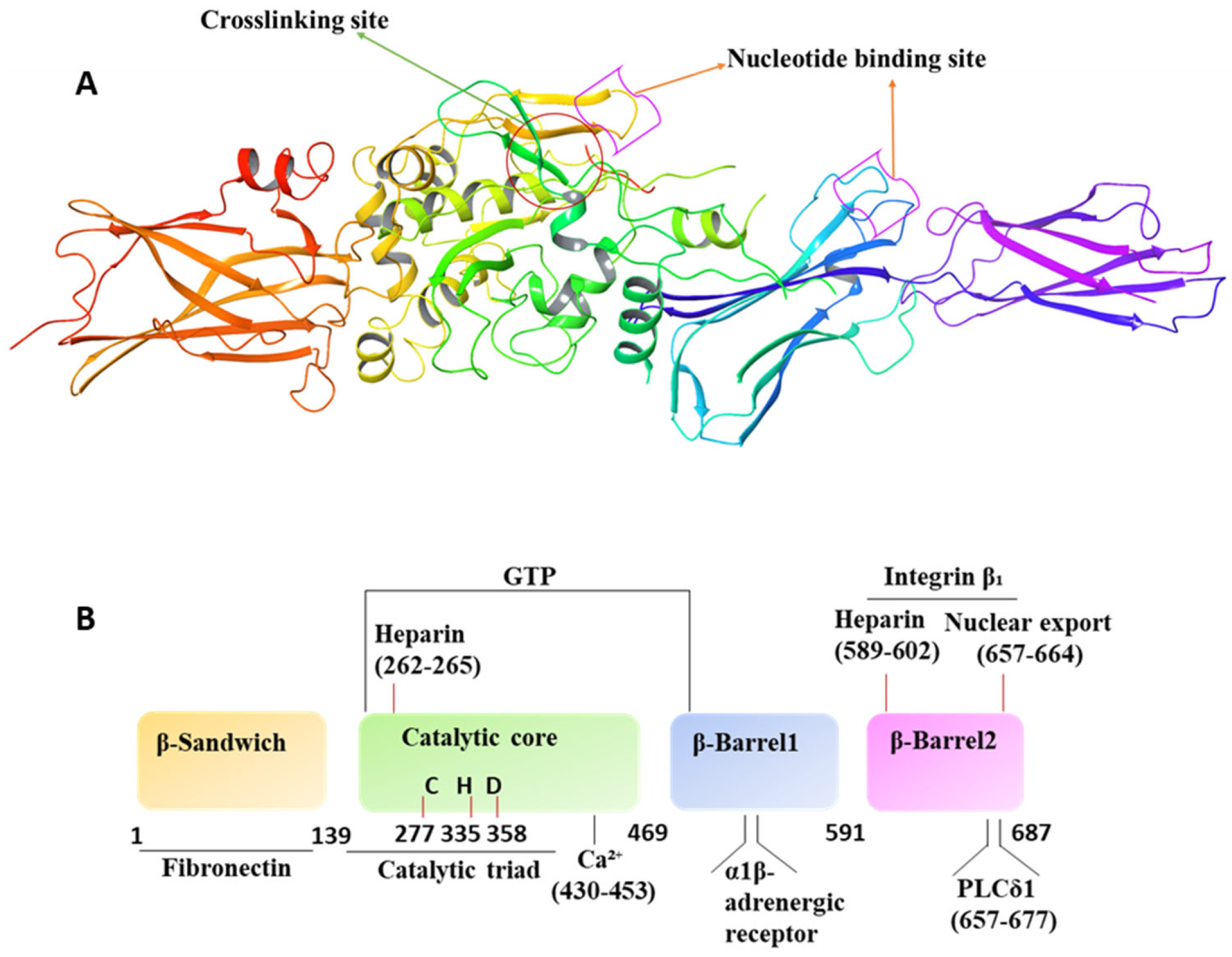
4. Molecular/Structural Features and Operation Mechanism

As a new member of the TG family with both calcium-dependent and calcium-independent activation, TG2 has emerged as a distinctive, highly complex, multifunctional isoform. This pleiotropic enzyme has a variety of localizations, including the cell membrane (both the inner and outer side) and the ECM, in addition to the cytosol and its organelles [10,48]. The miscellaneous functions of this protein are determined by its structural conformation. There are 687 amino acid residues in the TG2 protein’s primary structure. Four different structural domains make up the native 80 kDa protein: an N-terminal β-sandwich domain; a catalytic core domain made up of α-helices and β-sheets; a first barrel; and a C-terminal β-barrel [49,50] (Figure 1). These domains contain the functional binding sites of TG2. Specifically, a Fn binding site is present in the N-terminal β-sandwich domain. The substrate-binding pocket, the catalytic triad residues of cysteine, histidine, and aspartic acid, a binding site for GTP, binding sites for Ca2+, and a binding site for heparin are all found in the catalytic core. While the second β-barrel has interaction sites for phospholipase Cδ1 (PLCδ1), integrin, and heparin, the first β-barrel has a binding pocket for GTP and interactions with the α1B-adrenergic receptor [49,50].
The cytosolic free Ca2+ and GTP concentrations are close to 100 nM and 100 µM, respectively, which means that TG2 lacks enzymatic cross-linking (transamidase) activity under normal physiological conditions [51]. A high level of calcium in the environment induces a conformational change that opens up the folded form and allosterically activate TG2 and induces its cross-linking activity by making its catalytic core readily accessible to the enzyme substrates [52]. The allosteric inhibitor GTP, which holds the protein in a closed shape, interferes with this activation process. The G protein signaling activity of TG2 is activated in the closed state, allowing TG2 to transduce signals from adrenergic receptors by activating PLCδ1 [53]. The open confirmation of TG2, which is present in the ECM as a result of a relatively high extracellular calcium level, has frequently been tacitly or explicitly linked to cardiac stiffness and fibrosis due to its cross-linking activity. The concentrations of Ca2+ in ECM and cytosol are approximately 1.2 mM and 100 nM, respectively. Higher ECM Ca2+ can improve the binding of Ca2+ to the Ca2+-binding sites on the catalytic core of TG2 that induces open conformation, allowing easy accessibility of the active sites for cross-linking activity in smooth muscle cells of vascular tissues. Cross-linking reactions progress through the transfer of γ-carboxamide group of glutamine residue to the amino group of a peptide-bound lysine residue resulting in the formation of NƐ-(γ-glutamyl)-lysine isopeptide bond, a covalent bond. The formation of disproportionate covalent bonds results in excessive accumulation of cross-linked collagen, resulting in stability, rigidity, and stiffness of the myocardial ECM, ultimately leading to fibrosis. As a result, it is assumed that TG2 inhibitors that aim at and lock TG2 in an open conformation may provide some fibrosis protection. Contrarily, transmembrane signaling cascades benefit greatly from TG2’s closed shape. TG2 functions as a G proteinα (Ghα) in cellular signaling and promotes cell viability due to its GTP binding activity in the closed conformation [10]. The existence of TG2 in both open and closed conformations have significant practical ramifications since cutting-edge treatment techniques targeting this protein might target the action of either form [54]. This strategy has considerable potential for designing TG2 inhibitors that could be an important addition to the arsenal of treatments for diabetes and other age-related diseases.
The majority of mammalian tissues contain the stress-inducible gene TG2, but only have transient activity in the ECM due to their vulnerability to oxidation [55,56]. However, constant upregulation of the TG2 gene over time may result in sustained activity of this protein. Due to its cross-linking activity, TG2 catalyzes Ca2+-dependent covalent modification of protein-bound glutamine through inter- or intramolecular cross-linking, deamidation and aminylation reactions [57,58]. Numerous intracellular and extracellular proteins have been identified as TG2 substrates for these processes [59]. In the ECM, TG2 initiates the cross-linking of structural proteins like collagen, Fn, fibrinogen, and laminin, creating a scaffold that is both stiffer and more stable [60] (Figure 2). The first step in the inter- and intramolecular cross-linking processes is the acylation of a protein-bound glutamine residue at the active cysteine site of TG2, which releases ammonia and creates a thioester intermediate between TG2 and glutamine. In the next transamidation step, isopeptide bond is generated by the attack of thioester intermediate by a primary amine such as lysine. Aminolation takes place when a primary amine substrate, such as polyamines, is present. This incorporation of the substrate into the target protein is accomplished via the production of a glutamyl-amine bond. Water functions as a nucleophile in the deamidation reaction, attacking the thioester intermediate and changing the glutamine residue into a glutamate residue [61].

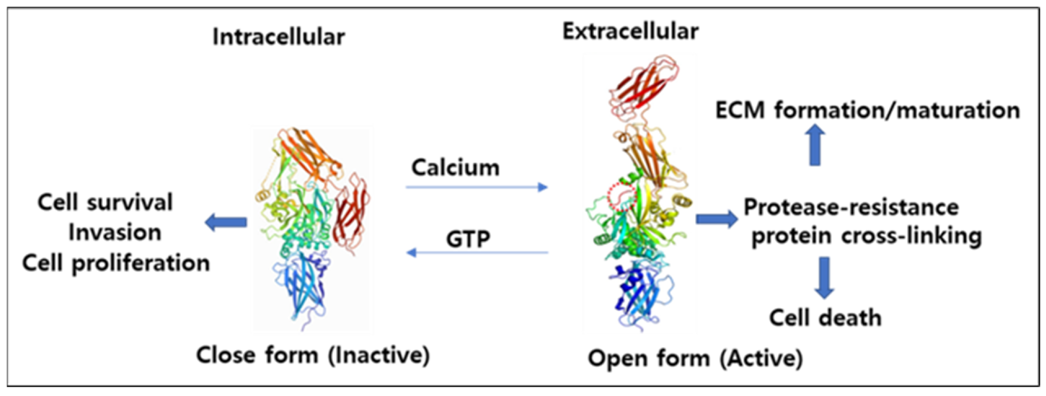
4.1. Mechanism of Activation and Inhibition

Human TG2 is composed of four distinct globular domains: one NH2-terminal β-sandwich (contains fibronectin and integrin binding sites); a catalytic core (contains the catalytic triads Cys277, His335, and Asp358); and two COOH-terminal β-barrel domains with the second barrel domain containing a phospholipase C binding sequence [62]. TG2 also contains a unique GTP/GDP binding site, located in the cleft between the first β-barrel and the catalytic core. There are three monomers, chain A, chain B, and chain C, in the asymmetric unit. Chain A is autonomously positioned, while chains B and C form a symmetric dimer. Intriguingly, all the three chains are identical and contain GTP. Calcium binding to TG2 increases its protein cross-linking activity, whereas GTP acts as an endogenous inhibitor of its protein cross-linking activity. GTP inactivates TG2 by promoting the transition to the compact conformation, while calcium activates TG2 by promoting expanded conformation [54]. Active TG2 is usually found in ECM due to the high concentration of calcium ions, whereas in intracellular environment, it is usually found in inactive form due to the low concentration of calcium [55].

4.2. Interconversion of Active and Inactive Forms of TG2

TG2 activity is greatly affected by the redox status of the cell. Usually, TG2 activity is increased in a reduced environment. TG2 consists of a redox-sensitive cysteine triad comprising of Cys230, Cys370, and Cys371, where Cys370 can form disulfide bonds with both Cys230 and Cys371 that can deactivate TG2 [55]. Cys230 is considered a TG2-specific redox sensor that helps in Cys370–Cys371 disulfide bond formation via an intermediate Cys230–Cys370 disulfide bond that is common in open TG2 form (active form). Cys230–Cys370 disulfide bond is found in the TG2/ATP complex, but not in the TG2/GDP complex [63], whereas the Cys370–Cys371 disulfide bond is neither in the TG2/ATP nor TG2/GDP complex. TG2 can be activated in the absence of GTP and the Cys230–Cys370 disulfide bond can be formed even under highly reduced conditions in the presence of GTP. It has been reported that the Cys230–Cys370 disulfide bond can be formed under highly reduced conditions of 5 mM DTT in the TG2/GTP complex. Two more amino acid residues, Cys336 and Cys277, at an active site may undergo disulfide bond formation, preventing TG2 activity under oxidative conditions [64].

4.3. Interaction with GTP

Tae-Ho Jang et al. reported the monomeric form of TG2 through the multiangle light scattering (MALS) technique. According to this study, the theoretical molecular weight of the monomeric TG2, with the C terminal His-tag and GTP, was 78.52 kDa, whereas the experimental molecular weight was 78.57 kDa with a polydispersity of 1.0. Based on these data, it is evident that GTP-bound TG2 exists as a monomer in aqueous medium. They further confirmed that GTP was in the GTP binding pockets formed by two β-barrel domains and the catalytic domain, and all four domains are well orientated [64]. Two β-barrel domains obscure the catalytic triad, indicating that this structure is a typical closed form of TG2. Various GTP binding small G proteins require Mg2+ for binding with the phosphate group of GTP, but TG2 does not require Mg2+. Instead, it contains several positively charged amino acids, such as Arg476, Arg478, and Arg580, surrounding the negatively charged phosphate group of the GTP. Arg580 has the strongest interaction with GTP containing two H bonds, whereas Arg476 and Arg478 forms H bonds with γ-phosphate of GTP. Arg580 mutation obliterates GTP binding and makes the transamidase activity of the protein insensitive to inhibition by GTP [65]. Hydrophobic interactions formed by Phe174, Met483, and Val479 of TG2 also stabilize the guanine moiety of GTP.
The majority of the amino acids of the first β-chain of the first β-barrel domain and the loop that links it to the next β-chain are involved in binding with the GTP, whereas only two residues of the catalytic core, Lys173 and Phe174, are involved in the binding with GTP. GTP (but not ATP) binds more strongly to TG2 by H bonds formed by Ser482 and Tyr583. This tight interaction of GTP to TG2 is the only reason why it can more effectively impede the TG2 activity than ATP. Location of purine moiety remains almost same in TG2/ATP and TG2/GDP complexes, whereas Arg478 is in the interior portion to form a stable H bond with β-phosphate of GDP and Arg478 moves outside in formation of the TG2/GTP and TG2/ATP complex. Furthermore, the location of Arg478 is distinctive at the TG2/GTP complex and the movement of Arg478 in TG2/GTP and TG2/ATP complex occurs due to sterical collision with the γ-phosphate group. The side chain of Arg476 was located inside on the TG2/GDP and TG2/ATP complexes, whereas it was located outside of the TG2/GTP complex [64].

4.4. Inactivation of Human TG2 by ERp57 Protein

Different cellular redox factors are obligatory for turning “on” or “off” the TG2. For example, oxidants like glutathione, cystine, hydrogen peroxide, and redox cofactor protein thioredoxin-1 (TRX) present in endoplasmic reticulum (ER) are known to initiate the TG2 activation mechanism. Meanwhile, ER-inhabitant protein 57 (ERp57), an endoplasmic protein that is also present in the extracellular environment, inactivates TG2. It is also known as PDIA3 and is a member of the protein disulphide isomerase (PDI) family of redox proteins involved in promoting the oxidative inactivation of reduced TG2. This protein is mainly involved in the folding of newly translated proteins. It has been observed that ERp57 colocalizes with extracellular TG2 in cultured HUVEC cells and inactivates TG2 by oxidizing it. The oxidized TG2 is catalytically inactive, and it is activated upon reduction by TRX via a thiol-disulfide exchange mechanism.
ERp57 specificity for TG2 is higher compared to other secreted redox proteins, such as quiescin sulfhydryl oxidase 1 (QSOX1), PDI, TRX, and ERp72. It is observed that higher TG2 activity is required for nephropathy in animal models of renal scarring [66] and ERp57 is vital in ECM build-up in animal models of renal fibrosis [67]. It is interesting to know the significant role of disulfide bond switch in TG2-mediated post-translational redox regulatory mechanism that is allosterically and reversibly regulated by two distinct proteins (ERp57 and TRX). Usually, basal TG2 level in most organs’ ECM remain low, which suggests that some potent oxidation mechanisms must exist to inactivate TG2 together with its export. Downregulation of ERp57 gene by interfering RNA (siRNA) led to a concomitant increase in extracellular TG2 activity but addition of exogenous ERp57 resulted in opposite effect [68]. It is suggested that the rise in ERp57 export may have progressed to downregulate aberrantly activated TG2 in the ECM of patients with renal fibrosis.

4.5. TG2 Kinase Activity

First identified in cell membrane fractions of T47D breast cancer cells as an enzyme that phosphorylated insulin-like growth factor-binding protein-3 (IGFBP-3), TG2 activity was later revealed to have a physiological role as a kinase that was established by using its specific quenchers [69]. TG2 phosphorylation by PKA increases its kinase activity but decreases transamidating activity [70]. A recent study demonstrated retinoblastoma (Rb) as the substrate of serine/threonine kinase activity of TG2. Ser780 residue on Rb is phosphorylated by TG2 that may be repressed by high Ca2+ level [71]. Another study showed that TG2 kinase activity induced phosphorylation of the IGFBP-3 in human breast cancer cell membranes. It phosphorylates Ser/Thr residues, but not Tyr in IGFBP-3 [69]). Cystamine and Ca2+ serve as inhibitor of kinase activity of TG2. In contrast to the monomeric IGFBP-3 substrate, TG2-cross-linked IGFBP-3 appeared to be very weakly phosphorylated in the presence of Ca2+. Another substrate of TG2 kinase activity is p53 tumor suppressor protein. Ser/Thr kinase activity of TG2 mediates phosphorylation of p53 and histones H1-4 in nucleus indicating that TG2 can control the structure and function of chromatin [72]. TG2-induced phosphorylation of p53 residues Ser15 and Ser20 might obstruct Mdm2 binding, indicating that this TG2-dependent process might promote apoptosis [73]. TG2 facilitates the adrenergic activation of extracellular signal-regulated kinases (ERK) and their regulatory kinases (MEK) in cardiomyocytes suggesting maneuver of more networks through which TG2 can cross-talk with numerous signaling pathways [74].

5. TG2 Roles under Normal and Diseased States

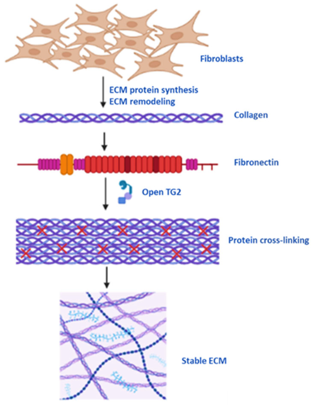
5.1. Protein Cross-Linking, Cell Membrane and ECM Stability

Conformation of countless proteins is modified by TG2-mediated formation of intramolecular cross-links and covalently linked oligomers and polymers [10]. Strengthening and hardening of ECM is the consequence of efficient cross-linking and inhibition of proteolytic degradation of ECM fibrils because of transamidation activity of TG2 to form protease-resistant inter- and intramolecular isopeptide bonds [75]. TG2-mediated cross-linking of the ECM fibrils to the latent TGF-β1 binding protein-1 (LTBP-1) result in inactive transforming growth factor (TGF)-β1 that can be activated by mechanical forces when the LTBP-1 is bound to a rigid, deformation-resistant ECM [76,77]. In fibrosis, increased deposition, dysregulated expression and cross-linking of ECM is the main reason of loss of functions of vital organs. Therefore, TG2 and TGF-β1 can serve as the targets for the treatment of fibrotic diseases. TG2 is crucial in maintaining ECM stability normally as well as in wound healing via different pathways. Scar tissue stroma formation is accomplished through binding of TGF-β1 to the ECM and forming cross-links initiated by TG2 [78].
The cell membrane has several proteins integral/transmembrane, as well as associated or peripheral depending on their location. Membrane proteins constitute about one third of the total proteins in a cell and serve as molecular targets for more than half of all drugs [79]. Two of these proteins are the integrin β1 or CD29 and fibronectin that with the help of TG2 make cells anchor into the ECM [80]. Integrin is composed of integrin α1 and α2 to form an integrin complex that functions as collagen receptors. Integrins are linking proteins between actin filaments and ECM. As a member of ubiquitous transmembrane receptors, integrin β1 play an important role in many signaling pathways, operating between the ECM and cytoplasmic domains including cell–cell and cell–matrix interactions. It also has a crucial role in skin protection and cutaneous wound repair. The knocking out of integrin β1 in keratinocytes results in wound-healing defects [81], whereas integrin β1 overexpression significantly enhances the wound-healing function of adipose-derived stem cells (ADSCs) via enhanced activation of the PI3K/AKT pathway [82].
TG2 interacts with integrins β1, β3, and β5/or syndecan-4 by attaching to the fibronectin/gelatin type I modules FnI of the collagen binding site on the Fn molecule, an ECM component. The gelatin-binding domain of Fn is 45kDa in N-terminal β-sandwich motif of TG2 structure (known as 45Fn). It forms specific and high-affinity interaction with TG2, possibly through negatively charged residues of its 45Fn domain [83]. Integrin and Syndecan-4 signaling is managed either directly or indirectly. Direct mechanism involves the formation of Fn-TG2, β-integrins, and syndecan-4 complex, whereas the indirect mechanism involves integrin cytoplasmic tail activation through Syndecan-4 stimulated PKCα or downstream convergence of integrin and syndecan signaling at p190RhoGAP [30,31]. Fn is another one of the most abundant structural ECM proteins that constitute the provisional matrix following cutaneous wounding. It facilitates cell migration, signaling, and survival [32]. Fn provides a scaffold for collagen deposition and subsequent ECM remodeling. Later, this fibronectin-rich matrix is replaced by a more mature matrix [33]. TG2 colocalizes with integrin β1 and Fn in the ECM to form adhesion complexes. About 5–40% of integrin β1 are in complex with TG2 and nearly the entire cell-membrane-bound TG2 forms a 1:1 complex with integrins [34], which is indicative of a prominent role of TG2 at the cell membrane.
The TG2 functions of binding to cell membrane receptors, mediating Intracellular signaling and cross-linking can stimulate wound healing in diabetic foot ulcer. TG2 acts as an integrin-binding co-receptor for Fn and plays a critical role in communications between cell and matrix to facilitate cell adhesion and migration. The β1 integrin-Fn complex is strengthened by interactions with TG2. Through its N-terminus domain, TG2 interacts with Fn stabilizing integrin complexes that regulate cell adherence to the matrix. In addition to the integrin-Fn binding site, Fn has an alternate binding site for TG2 that facilitates the formation of TG2-Fn-complexes. Consequently, Fn may serve as a valuable molecular target in DFU treatment. However, high and continuous Fn expression in DFUs might impede the normal healing process that can be resumed by transforming excess newly synthesized fn into the establishment of a mature matrix. Additionally, improved angiogenesis and release of transforming growth factor-3 (TGF-3) and vascular endothelial growth factor (VEGF) also speed up diabetic wound healing.

5.2. TG2 as an Inflammatory Biomarker

The expression of TGs is ubiquitous, and they are involved in numerous physiopathological conditions. Several studies have demonstrated that inflammatory disorders have higher TG2 expression levels. In most of the cases, TG2 has been observed to create an unsuitable protein aggregate that may be sufficiently cytotoxic to cause inflammation and/or apoptosis. TG2 is linked to the pathogenic development and the production of autoantibodies in various conditions, including celiac disease and rheumatoid arthritis [35]. TG2 activation in liver macrophages was significantly increased in the lipopolysaccharide (LPS)-injected and cecal ligation puncture-operated mouse models of sepsis, which is suggestive of its role as a potential biomarker of inflammation and sepsis [36]. TG2 is a crucial modulator of calcification caused by interleukin-1β (IL-1β), as well as hypertrophic differentiation and calcification in particular chondrocytes. Cytokine-mediated inflammatory pathways are severely altered by TG2 activity. The hypertrophic differentiation of joint chondrocytes and IL-1-induced calcification is mediated by TG2. Increased TG2 expression has been observed in human and experimental osteoarthritis. The expression of TG2 may serve as an additional adjuvant marker to track the tissue remodeling in osteoarthritic joint tissue [37]. Reduced cartilage breakdown and higher osteophyte formation was observed in TG2 knockout mice after osteoarthritis induction, indicating a TG2 effect on joint bone and cartilage remodeling when compared to wild-type mice. The ability of TG2 to modulate latent TGF activation through transamidation appears to have a potential effect on the control of inflammatory response in osteoarthritic tissues.
All-trans retinoic acid treatment induces TG2 expression, which leads to an overexpression of TNF-α and IL-1β in developing acute promyelocytic leukemia (APL) cells. This suggests that atypically produced TG2 is a suitable target for leukemia treatment [84]. The functional involvement of TG2 in the regulation of gene expression, ROS formation, cytokine expression, adhesion and migration, and phagocytic capacity of differentiated neutrophil granulocytes has been demonstrated by TG2 silencing in NB4 cells [85]. According to one study, omalizumab treatment reduced serum TG2 activity, which had grown with the severity of the condition. Additionally, the serum TG2 activity of chronic spontaneous urticaria (CSU) was higher than that of acute urticaria (AU). They suggested that serum TG2 activity can be a more accurate indicator of disease severity in case of CSU pathogenesis. Furthermore, TG2 shows high expression and release in human mast cells and plays a significant role in the pathophysiology of CSU [43]. Genetically predisposed individuals with celiac disease (CD) produce T and B immunological responses that culminate in the production of anti-tTG immunoglobulin (Ig)A antibodies and proinflammatory cytokines, which cause chronic inflammation and gradually deteriorate the intestinal mucosa [86]. Therefore, serum TG2 activity could be a promising biomarker for chronic-inflammation-induced disease diagnosis and predicting medicine prerequisite to control a disease. There are reports indicating the relationship between TG2 and pathophysiological conditions, such as pregnancy outcomes, impaired endometrial angiogenesis in celiac disease and interstitial fibrosis, and tubular atrophy in patients post kidney transplant.

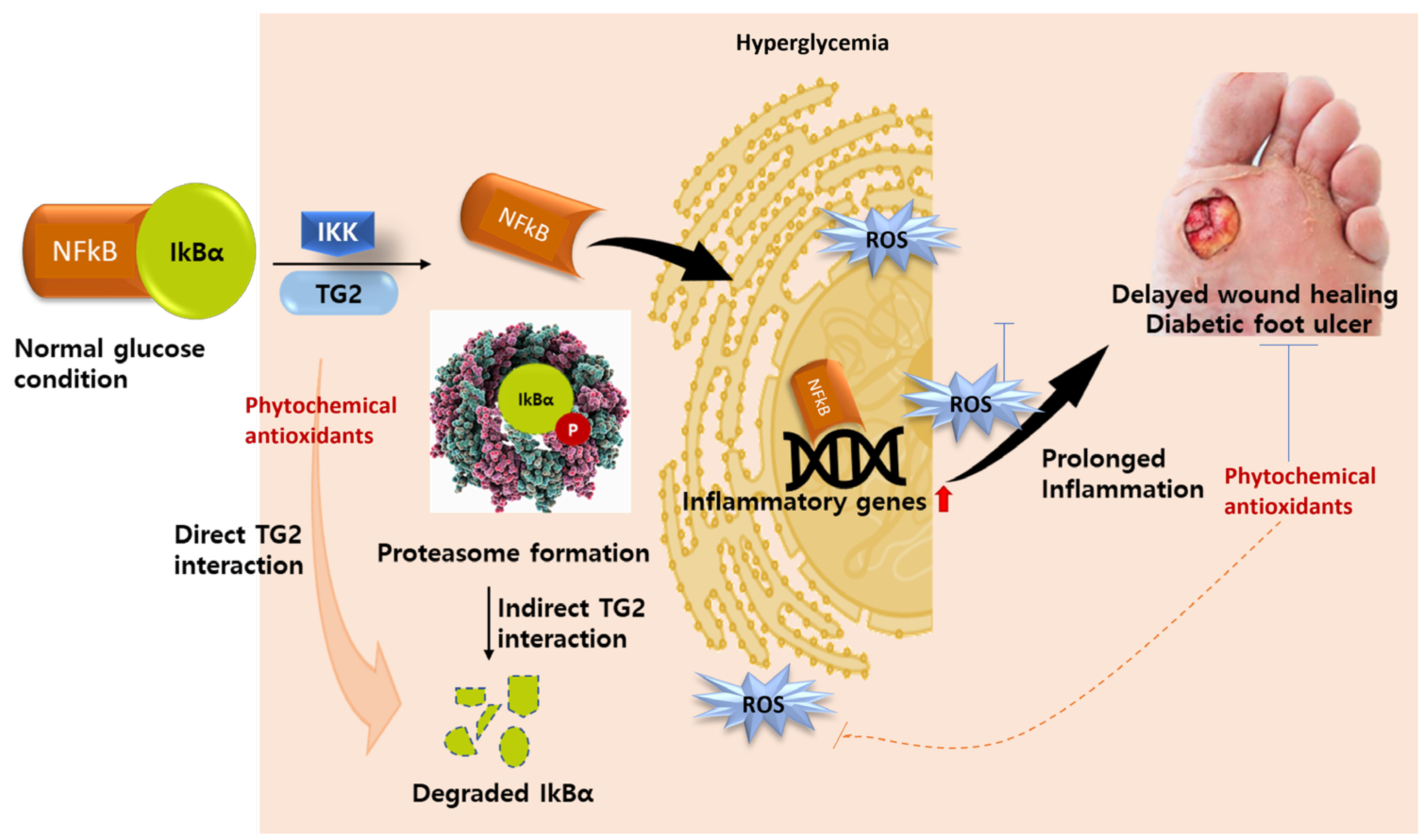
5.3. TG2 as Molecular Drug Target in Diabetes

Molecular drug targets have a specific biochemical and/or pharmacology profile and high-to-moderate drug-binding affinity to multiple additional targets. Intracellular Ca2+ and/or ROS can accelerate the activation of TG2, which is involved in a range of cellular processes, such as cell death, proliferation, differentiation, and migration, that ultimately affect cellular homeostasis (Figure 3). Therefore, TG2 can be used as a drug target in many diseases, including diabetes, fibrosis, cancer, and other chronic diseases. TG2 can serve as a potential therapeutic target for the treatment of diabetic retinopathy or TG2-associated ocular diseases. A study by Yeon-Ju Lee et al. demonstrated that in diabetic mice’s retinas, enhanced TG2 activity was observed, but this was decreased by intravitreal injection of TG2 inhibitors. In the diabetic retina, intravitreal injection of mouse TG2 siRNA also reduced hyperglycemia-induced TG2 activation and microvascular leakage. TG2 is thought to be the primary enzyme in diabetic vascular permeability and diabetic vasculopathy, and it could be a therapeutic target for diabetes complications and ophthalmic illnesses [87]. In another study, C-peptide protected endothelial cells from high-glucose-induced apoptosis by blocking intracellular ROS-mediated activation of TG2 [88]. It has been demonstrated that TG2’s transamidation activity has conflicting roles in apoptosis, either promoting or impeding it [89]. In endothelial cells, VEGF activated TG2 by increasing intracellular Ca2+ and ROS levels sequentially. Additionally, the TG2 inhibitors cystamine and monodansylcadaverine (MDC), as well as TG2 siRNA, inhibited VEGF-induced stress fiber production and adherens junction rupture. TGFβ-1 has been linked to diabetes and numerous fibrotic and scarring disorders, and TG2 inhibition by 1,3-dimethyl-2[(oxopropyl)thio]imidazolium lowers the deposition of ECM proteins caused by high glucose without altering the ECM or TGFβ-1 synthesis [90].
Normally, DFUs do not heal properly, and wound sites remain exposed to further injuries and microbial infections. Impaired wound healing makes the situation traumatic, with hospitalization and expensive treatments remaining the only options available for diabetic patients. Intriguingly, certain phytochemicals and synthetic drugs have been known to change TG2 expression in diseased conditions. Oleuropein, an extract of olive leaf, can promote the wound-healing process by modulating antioxidant capacity, fibrogenesis, and apoptotic process through TG and hydroxyproline expression that culminates in collagen deposition and re-epithelialization process [91]. Inflammation is an important event in the wound-healing process. Intracellular TG2 cross-linking during wound healing promotes inflammation through the activation of the noncanonical pathway of NFkB. In normal cellular conditions, NFkB remains inactive due to its tight association with IkBα. But under inflammatory conditions, IkBα is phosphorylated by IkB kinase (IKK) and degraded, thus releasing NFkB. Free NFkB is then translocated to the nucleus to activate downstream inflammatory genes. TG2 can either interact directly with IkBα, leading to its degradation via a proteasome-independent pathway, or indirectly polymerize IkBα, making its degradation feasible through proteasome, and thus activate NFkB during inflammation [92] (Figure 4). This knowledge can be translated into therapy for use in diabetic wound healing. Further, TG2 overexpression is known to cause the activation of TGF-β and its subsequent pathway in pulmonary fibrosis. TGF-β1-induced pulmonary fibrosis can be reduced through inhibition of TG2 and subsequent pathways by metformin [82]. TG2 is a dual-faced enzyme with contradictory roles in the instigation and progression of different diseases. TG2 overexpression exerts conflicting effects in different pathological conditions. For example, it plays an important role in cell proliferation and differentiation but mediates cell death and autophagy contingent on the kind of cell, apoptotic stimuli, and its subcellular location. Some pathological conditions, such as angiogenesis, fibroproliferative, and neurodegenerative diseases, are deteriorated as a result of TG2 overexpression, whereas in some conditions, such as diabetic wound healing, especially in diabetic foot ulcer, TG2 overexpression can be beneficial and used as a treatment strategy.
Interestingly, developing physiologically relevant immunocompetent DFU models can be used as a promising model system for understanding wound healing via theTG2 pathway [93]. Organ-on-a-chip (OoC) technology can offer suitable characteristics that could finally facilitate the idea of modeling DFU for pathophysiological studies and drug testing, including TG2 on microscale. It can serve as a turning point to develop a minimally functional immunocompetent DFU on-a-chip model, as wound healing cannot occur without a proper functioning immune response.

5.4. TG2 in Pathogenesis of Fibroproliferative Disorders

TG2 directly or indirectly is involved in the pathogenesis of pulmonary, kidney, and cardiac fibrosis, as well as different types of fibroproliferative cancers [94,95]. Fibroproliferative disorders can cause systemic organ-specific fibrotic diseases and are responsible for about 45% of mortality worldwide. Fibrotic disorders have been predominantly linked to ECM cross-linking enzymes. The alterations in ECM structure because of increased cross-linking may result in fibrosis and ultimately organ failure. TG2 is involved in many fibrogenic signaling pathways, including the AKT pathway and TGF-β/SMADs pathway [96,97]. TGF-β, an important cytokine, is one of the most important ECM regulators in pulmonary fibrosis. It can stimulate the production and inhibit the degradation of ECM proteins that can lead to deposition of ECM proteins and ultimately result in stiffened ECM and fibrotic organ development [98]. Extracellular cross-linking property of TG2 plays a crucial role in fibrotic disease development due to the storage of latent TGF-β1 in a hardened ECM and activation of profibrotic pathways that promote cell survival at the transcriptional and translational level. TG2 can induce some pro-survival factors, such as NF-κB, Bcl2, and Bcl-xL, and thus promote or restrict cell death [99]. In other words, scar tissue stroma and tumor are shaped by TG2 through ECM cross-linking with TGF-β1 and other ECM proteins, thereby presenting TGF-β1 as one of the most effective profibrotic stimuli in the progression of chronic conditions [76,78]. Intriguingly, TG2 and TGF-β1 fortify each other in developing fibrotic and tumor stroma microenvironment affecting the self-amplification of TG2 and TGF-β1 in fibrotic tissues, thus elucidating their upregulation in fibroproliferative disorders. TG2 can help in the development of fibrotic/scar cell phenotypes and tumor cell survival through its cross-linking and signaling mediator functions and intracellular signaling [100]. A study by Boroughs LK et al. reported that overexpression of TG2 in cancer cells is linked to the constitutive activation of the PI3K/Akt pro-survival pathway, either through focal adhesion kinase (FAK) activation or by forming a complex with the non-receptor tyrosine kinase c-Src and PI3K [100]. However, exogenous TG2 can inhibit tumor growth by collagen deposition and cross-linking the surrounding ECM [101,102]. Additionally, myofibroblasts play a crucial role in fibroproliferative diseases and TGF-β1 brings about the transformation of fibroblasts into myofibroblasts. They help in the accumulation of excess collagen and other fibrous proteins into the ECM with alpha-smooth muscle actin (α-SMA)-positive stress fibers expressed on its surface, thereby transforming the fibrotic matrix into a stiff, dysfunctional scar [76,77].
The role of TG2 in fibrosis pathogenesis is evidenced by the TG2 knockout mice and TG2 inhibition that protected the mice against fibrosis and reduced the fibrotic phenotype. In fibrotic tissue development, gene expression levels of ECM proteins are stimulated due to the activation of SMAD signaling by TGFβ-1, but expression of MMPs and their inhibitors (TIMPs) lead to increased matrix deposition [103]. However, inhibition of catalytic function of TG2 can reduce cardiac fibrosis in vivo [104,105]. Reduced intracellular TG2 interaction with exosome-membrane-associated syndecan-4 and reduced TGF-β1-induced myofibroblast formation results in reduced extracellular TG2 and reduced activation of fibrosis-associated genes [94]. Abnormal ECM dynamics and organization are significant drivers of abnormal cell activity and, eventually, organ failure in fibrotic disorders and malignancies. Idiopathic pulmonary fibrosis (IPF)-derived fibroblasts increase the activity of TG2 and can result in increased N-εγ-glutamyl lysine cross-links between collagen and fibronectin and thereby stiffened tissue development [95]. As scar tissue and tumor stroma have several similarities, identifying major shared pathways involved in disease causation and progression could lead to the discovery of new drug targets and the development of new medications and diagnostic tools for fibrosis. All types of heart diseases are linked to cardiac fibrosis, but there is no cure for it. Scientists have demonstrated that both TGFβ-1-induced EndMT and TGFβ-1-induced cardiofibroblast transformation into myofibroblast-like cells, as well as matrix deposition, can be attenuated by the TG2 selective inhibitor EB1-155, suggesting a new role for TG2 in regulating TGFβ-1 signaling, in addition to its role in latent TGFβ-1 activation. Myofibroblast activation and matrix deposition are two ways that TG2 contributes to cardiac fibrosis. Therefore, cardiac fibrosis can be reduced by selectively inhibiting TG2 with a small-molecule inhibitor [106]. A nanocatalyst based on multifunctional electrochemical technique for the ultrasensitive TG2 assay has been developed by Huang et al., which can either function as the TG2 substrate or be applied for signal amplification that can be used to allow high sensitivity of detection. In this method, specific glutamine-donor-peptide of TG2 is modified on the electrode and the exertion of the transamidation activity by TG2 results in the tethering of the nanocatalyst with the peptide on the electrode, producing significant changes in the electrochemical signals that can be measured in the form of TG2 accurately [107].

5.5. Relationship of Other TGs to Fibrosis

Studies suggest that association of TG3, TG4, and TG7 with fibrosis is significant yet contentious. TG3 is primarily expressed in the esophagus, hair follicles, and skin, where it cross-links terminally differentiated keratinocytes to stabilize the cornified cell membrane [108]. The majority of TG4 is found in prostate tissue where it cross-links proteins in seminal vesicle fluid, and TG7 is primarily present in the lungs, testes and brain [109,110]. TG3 has been shown to induce myocardial fibrosis and take part in cardiac remodeling as an alternative to TG2 in TG2 knockout mice. TG3 mRNA expression was shown to be elevated in the heart and aorta of these TG2 knockout mice [111,112]. TG3 plays an important role in mouse liver fibrosis and the healing process [113]. A proteome study of metabolic pathways linking inflammation and fibrosis in pediatric patients of collagenous gastritis, a rare condition characterized by augmented subepithelial collagen deposition and inflammatory infiltrates, identified that TG3 expressed in the stomach and other epithelial tissues exacerbated collagenous gastritis by its cross-linking action [114]. However, rat models of ISO-induced cardiac fibrosis did not show expression of TG3 and TG4 [115]. Studies suggest different expression patterns of TG4 in normal prostate epithelial cells and prostate cancer cells; however, this is still up for debate [116]. Defects in copulatory plug formation and seminal fluid viscosity result in decreased fertility in TG4 knockout mice [117]. Recent research demonstrates that in autoimmune polyglandular syndrome type 1, TG4 functions as a male-specific prostate autoantigen that may lead to infertility in humans. TG4 is a potential biomarker of prostate cancer development. Studies suggest that TG4 controls the invasiveness and migration of prostate cancer cells by inducing epithelial-to-mesenchymal transition [118]. TG4 overexpressing prostate cancer cells, as well as prostate cancer cells treated with exogenous TG4, resulted in decreased E-cadherin and increased N-cadherin expression that play a crucial role in epithelial-to-mesenchymal transition in cells [119]. Moreover, high levels of TG4 expressions have been linked to enhanced trans-differentiation and matrix proteins in cardiac fibroblasts [120]. However, a study conducted with an aging rat model demonstrated that TG4 reduces cross-linking and vascular stiffness in aorta with age [121]. A recent study conducted with a renal fibrosis mice model showed slightly increased TG7 expression, together with elevated TG1 and TG2 levels [122]. Another study conducted in rats detected a minor increase in TG7 expression levels in the kidney, further signifying its role in renal fibrosis [123]. An interesting study conducted using bronchopulmonary dysplasia (BPD) mouse models showed that TG7 downregulation weakened the lung development, while TGF-β mediated the hyperoxia-induced downregulation of TG7 [124]. Furthermore, TG7 has been found to be upregulated in uterine corpus endometrial carcinoma (UCEC) [125].

5.6. TG2 in Angiogenesis and Tubule Formation

In addition to the ECM’s crucial role in organ fibrosis and cancer metastasis, conditions like osteogenesis imperfecta, epidermolysis bullosa, and Ehlers-Danlos syndrome show defects in the ECM’s quality, and angiogenesis and wound healing depend on an ECM remodelling process that is properly functioning [126,127,128]. TG2 is expressed in high levels in endothelial cells and is required for endothelial cell tubule formation. However, TG2 displays pro- and antiangiogenic effects. Faye C et al. demonstrated that TG2 binding to endostatin under hypoxic conditions leading to stimulation of angiogenesis is a pro-angiogenic effect [129]. However, downregulation of TG2 has been observed in endothelial cells undergoing capillary morphogenesis in an in vitro human capillary tube formation model [130]. TG2 inhibition leads to late tubule formation, thereby delaying angiogenesis both in vitro and in vivo. TG2 regulates the activation of vascular endothelial growth factor (VEGF) signaling through activation of VEGF receptor2 (VEGFR2) and thus depositing VEGF in ECM. However, Factor XIII is already known to be involved in angiogenesis via VEGFR2 signaling [131]. Moreover, TGFβ1 is one of the factors that contribute to the endothelial–mesenchymal transition (EndMT), which results in the disruption and loss of blood vessel structures. The involvement of TG2 in matrix-bound TGFβ1 activation and the role of TGFβ1 in EndMT suggest that TG2 can exert control over angiogenesis via its functional interaction with TGFβ1. TGFβ signaling is reported to induce endothelial cells’ (ECs) proliferation, differentiation, and survival via the Alk1/Smad1/5/8 pathway. TG2 inhibition in HUVECs during tubule formation can result in the downregulation of the pro-angiogenic Smad1/5 signaling pathway and ultimately angiogenesis and tubule formation. Increased extracellular TG2 levels enhanced TGFβ1 signaling via p-Smad2/3 and Akt pathway and increased levels of mesenchymal markers, including S100A4, vimentin, SMAα, and Fn, and decreased levels of CD31 and VE-cadherin endothelial markers altered the protein profile in ECs [132]. TGFβ signaling can be prolonged by extending p-Smad signaling by cross-linking TGFβ into a collagen matrix by TG2 [133]. Endothelial TG2 function is dependent on protein cross-linking in ECM through its effect on ECM/Fn deposition, which in turn accelerates the accumulation of matrix-bound VEGF, thereby activating VEGFR2 signaling. However, TG2 inhibition can lead to a reduction in VEGFR2 signaling and ultimately the inhibition of angiogenesis in in vitro as well as in vivo models even after initiation of the tubule formation process [134].

5.7. TG2 in Progression of Neurodegenerative Diseases

TG2 plays an important role in the development of the nervous system as well as the maintenance of the homeostasis in the nervous system throughout the age. TG2 is extensively expressed in various cell types of the nervous system but is primarily enriched in neurons [135,136]. Age-related chronic neuroinflammation mediated by persistent activation of brain-resident immunocompetent cells is the primary cause in the development and progression of several neurodegenerative diseases. TGs have been shown to induce protein cross-linking in the development and progression of AD. TG2 inhibition results in reduced microglial inflammation; however, activated microglial inflammation is a hallmark of AD. Therefore, targeting microglial TG2 may help in developing therapeutics to target the microglial cells in AD by downregulating without averting complete microglial activation [137]. Knocking down TG2 in oligodendrocytes and their precursor cells (OPCs) results in reduced differentiation and maturation of OPCs, while the addition of TG2 to early oligodendrocyte-lineage neural stem cells improves differentiation process [138]. TG2 silencing in THP-1 cells (a microglial model) may be used as a therapeutic target to attenuate TNF-α production in response to amyloid-beta (Aβ), thereby reducing inflammation and progression in AD [139]. TG2 is upregulated in some glial tumors and high levels of microglia-derived cytokines encourage TG2 expression in glial tumors [140]. Tau protein, one of the many protein aggregates responsible for the neurodegeneration of brain cells, has been shown as an excellent substrate of TGs in vitro, including GGEL (γ-glutamyl-ε-lysine) cross-links in the paired helical filaments and neurofibrillary tangles of Alzheimer’s disease (AD) brains [141,142]. TG activity can induce amyloid β-protein oligomerization and aggregation [143,144] in brain cells.
Overexpression of human TG2 in the neurons of knockout mice causes increased neuronal damage in the hippocampal regions after treatment with kainic acid, but surprisingly, these mice did not show any apparent phenotype in the absence of stress [145]. Additionally, increased nuclear TG2 activity has been reported in several neurodegenerative disorders, including Huntington’s diseases (HD), AD, and Parkinson’s disease (PD) [146]. TG2-deficient HD mouse models undergo a substantial delay in motor dysfunction onset and a prolonged survival [147]. Stress-activated TG2-mediated nuclear actin re-organization and actin–cofilin covalent cross-linking elucidate a new pathogenic mechanism for TG2 hyperactivity in neurodegenerative diseases [148]. In response to TG2 knockdown, several genes in neurons are differentially expressed that primarily regulate the cytoskeletal integrity pathway, cell signaling, cell–matrix interactions, and ECM function [149]. Therefore, TG2 can be a critical therapeutic target in neurodegenerative diseases.

6. TG2 Inhibitors

Numerous transglutaminase inhibitors exist and have enabled proof-of-concept investigations in disease models, but their poor pharmacokinetic/pharmacodynamic characteristics and/or lack of selectivity for the TG2 isoform have restricted their use in clinical applications. The identification of TG2 as a possible therapeutic target in various diseases, especially diabetes and age-related disorders, has driven intense research to synthesize inhibitors that target this protein. Over the past ten years, several small molecules and peptidomimetic inhibitors of TG2 with good selectivity have been discovered, some of them produced utilizing molecular dynamics techniques [150]. However, TG2 inhibitors that are clinically useful are not currently accessible. For a number of diseases, including celiac disease, liver fibrosis, cardiac fibrosis, and neurological diseases, prospective clinical candidates are now at various phases of development and the preclinical stage [106,151,152,153]. Unfortunately, TG2 inhibitors for delayed wound healing, diabetic foot ulcer, fibroproliferative diseases, and neurodegenerative diseases have not yet undergone clinical trials.
According to their mode of inhibition, TG2 inhibitors can be divided into three categories: competitive amine inhibitors, reversible inhibitors, and irreversible inhibitors. The development of inhibitors targeting TG2 for the treatment of various diseases, particularly diabetic conditions, is attracted to irreversible inhibitors and they dominate the pipeline of development. Due to their stability and safety in living beings, competitive amine inhibitors have also garnered significant investigation as potential TG2 inhibitors. An aliphatic carbon chain with four to five saturated carbon atoms serves as the binding site for the primary amine in competitive amine inhibitors. Cystamine, putrescine, and MDC are a few of the most widely utilized competitive amine inhibitors. This class competes with natural amine substrates to reduce TG2 activity, while TG2 still remains active and transamidation still takes place [49]. Cystamine is a distinctive, non-specific TG2 inhibitor since it can inhibit TG2 through different mechanisms, including competitive inhibition. Its irreversible inhibitory mechanism, which is likely to include thiol-disulfide exchange with the nucleophilic cysteine residue in the TG2 active site, inhibits TG2 in a time-dependent way. Cystamine can effectively inhibit the activation of the TG2 extracellular pool [49,154]. Additionally, it stimulates the production of intracellular glutathione and inhibits the thiol-dependent protease caspase-3, a crucial effector in triggering cell apoptosis. The reduced form of cystamine, cystamine (2-mercaptoethylamine), also has anti-fibrotic properties, but it is less effective than cystamine and primary amines [155]. It is comparatively stable in plasma and easily absorbed into tissues via circulation. By preventing the production of inflammatory mediators like IL-6 and TNF-α, cystamine has positive effects on a variety of diseases [156,157]. In a transgenic model of HD, administration of cystamine decreased aberrant movements and tremors, enhanced survival, and upregulated transcription of neuroprotective gene. Through the prevention of apoptosis, cystamine reduced heart, brain, and liver tissue damage in an animal model predisposed to lupus [158,159].
Reversible TG2 inhibitors inhibit the activity of the enzyme by preventing substrate from accessing the active site without covalently altering the enzyme [49]. Similarly, by competing with Ca2+ for binding sites within the protein, the divalent metal ion Zn2+ can reversibly inhibit TG2 function. It was also shown that small substances, such as LDN 27219 and other GTP analogs, including GMPPCP, reversibly inhibited TG2 activity [49,54,160]. However, irreversible TG2 inhibitors, commonly referred to as “suicide inhibitors”, such as R283, Z-DON, iodoacetamide, ERW1041E, and EB-1-155, inhibit TG2 by covalently altering the enzyme and preventing the binding of its substrates. Using chemical functional groups that result in stable chemical bonds, the majority of irreversible TG2 inhibitors target the active cysteine region. For instance, the iodide moiety of iodoacetamide interacts with the TG2 active site thiol to create a persistent thioether bond with the inhibitor [161]. One of the most researched classes of TG2-irreversible inhibitors are the 3-halo-4,5-dihydroisoxazoles. Their structure is based on the naturally occurring substance acivicin, a structural homolog of glutamine that inhibits active cysteine sites. The low toxicity and excellent bioavailability of this class of inhibitors are contrasted by their low solubility at physiological pH. Recent in vivo and in vitro research revealed that the dihydroisoxazole derivative ERW1041E significantly decreased the hypoxia-induced cross-linking activity and synthesis of collagen type I and α-SMA via inhibiting TG2 activity [162,163]. SAR studies by covalent docking has demonstrated Nε-acryloyllysine piperazides and its derivatives as an irreversible inhibitor of TG2. Structure–activity relationship (SAR) analysis led to the discovery of some novel irreversible inhibitors that effectively and selectively inhibit transamidation activity of human TG2 and allosterically inhibit its GTP capacity [164]. Targeted-covalent inhibitors 3-bromo-4,5-dihydroisoxazole (DHI) and their derivatives have been demonstrated as selective TG2 inhibitors in in vivo research [165].
Researchers have revealed that selective inhibition of extracellular TG2 transamidation activity is sufficient to alter fibrotic remodeling. In this study, a TG2 inhibitory antibody that targets TG2 specifically has been developed for the first time. Four novel inhibitory epitopes in the catalytic core domain have been identified and the extracellular function of TG2 in ECM buildup has been confirmed. Clinical trials of targeted TG2 treatment for renal fibrosis have been conducted as a result of the humanization of the lead antibodies AB1, DC1, and BB7 [166]. Using TG2 antibody in conditions including diabetes, fibrosis, and scarring (where TG2 cross-links ECM proteins and the vast latent TGFβ-1) can improve selectivity, eliminate undesirable intracellular effects, and provide better TG2 inhibition. Additionally, constant pharmacokinetic characteristics and excellent specificity of antibody therapy speed up their development, which is the main reason they make up the majority of the novel therapeutic medicines now under development.
Recently, scientists have discovered that the TG2 gene is a target for epigenetic silencing in breast cancer, illuminating a molecular mechanism that results in decreased TG2 expression, and this aberrant molecular event may be a factor in invasiveness in this tumor type. It was found that in case of cancer, TG2 is commonly hypermethylated and is usually overexpressed or underexpressed in different gliomas. TG2 overexpression results in doxorubicin resistance in cancer cells. Therefore, epigenetic silencing of TG2 expression may affect the invasiveness of brain cancers [167]. TG2 has been shown as a target for epigenetic silencing in breast cancer [168]. A therapeutic strategy based on TG2 and IL-15 gene silencing in the inflamed intestine has been developed for celiac disease, suggesting that an oral microsphere formulation combining the TG2 and IL-15 RNA interference may be effective in treating celiac disease [169]. In another study, substantial downregulation of proinflammatory cytokines (TNF-α and IFN-γ) by using gelatin-based nanoformulation containing IL-15 + TG2 siRNA was detected in J774A.1 cells [170]. These studies endorse clinicians to use RNA interference via small siRNA to target genes related to chronic inflammation condition and disease progression that are directly or indirectly linked to TG2.

7. Conclusions and Perspectives

Research on the function of TG2 in diabetes and age-related diseases is a relatively recent and rapidly emerging subject. Due to its many roles in tissue remodeling and variable locations, this incredibly versatile protein has emerged as a key player in our understanding of how diabetes and related diseases develop. Although TG2 has been investigated to be involved in the progression of diabetes complications and other age-related diseases, due to its regular interconversion between active and inactive states and inhibition by vital macromolecules, its functional activity has not understood entirely. To understand the processes and signaling pathways underlying the involvement of TG2 in diabetes and related disorders, additional research is required to determine the molecular targets and importance of the enzymatic and non-enzymatic actions of TG2. A number of preclinical and clinical investigations have shown direct or indirect involvement of TG2-mediated oxidative stress, prolonged inflammation, and altered TG2 signaling in diabetes and many age-related human diseases, suggesting a vicious cycle that instills a web of complex molecular mechanisms. As a result, many individuals with diabetes and diabetic foot ulcer also exhibit cardiovascular and cognitive decline at a later stage, suggesting the crucial role of TG2 that can be used as a diagnostic biomarker for impaired wound healing in diabetic and neurodegenerative patients. Even though significant progress has been made, it is reasonable to suggest that more work must be conducted before TG2 is used as biomarker and therapeutic approach. According to the difficulties and knowledge gaps found in this review, there is a need for study in the following areas: better characterization of TG2 profibrotic vs. protective effects in the progression and outcomes of diabetes and associated diseases; elucidation of the importance of TG2 splicing variants in these diseases; better characterization of the interactions between TG2, MMPs, and TIMPs in hyperglycemic conditions, with or without TG2 inhibition; and significance of TG2 open vs. closed conformations in diabetic condition and in the pharmacological modulation.
A better understanding of TG2 kinase function in the etiology of diabetes and other age-related disorders can aid in the development of potential therapeutic approaches, providing new possibilities for innovations to generate efficient medications targeting TG2 for the management and treatment of diabetes and associated diseases. Growing data over the past ten years point TG2 as a viable therapeutic target for difficulties related to diabetes and imply that treating these complications by pharmacologically inhibiting TG2 may also have benefits for treating its associated disorders. The majority of this research, meanwhile, has only used in vitro and animal models for preclinical testing. It is advised to validate clinically viable TG2 inhibitors because moving them forward to the clinical trial stage could lead to major improvements in the management of diseases associated with hyperglycemia. To achieve these goals, extensive collaborative studies have been suggested to further understand the structural and functional activity and interaction of TG2 with various macromolecules of different signaling pathways in diabetic, as well as other age-related diseases pathophysiology, and to remove current barriers of clinically difficult diagnosis and resistance to current treatments. This will allow for the initiation of early therapy and prevention of disease progression before it worsens.

Author Contributions

Conceptualization and writing: N.Y.; image: N.Y.; editing, N.Y. and S.-Y.K. All authors have read and agreed to the published version of the manuscript.

Funding

This research did not receive any specific grant from funding agencies in the public, commercial, or not-for-profit sectors.

Institutional Review Board Statement

Not applicable.

Informed Consent Statement

Not applicable.

Data Availability Statement

No new data were created or analyzed in this study. Data sharing is not applicable to this article.

Acknowledgments

Authors are thankful to Dharmendra Kumar Yadav (Principal Scientist, Arontier Co., Ltd., 15F, Gangnam-daero 241, Seocho-gu, Seoul, Republic of Korea) for providing molecular docking.

Conflicts of Interest

The authors declare no conflict of interest.

Abbreviations

DFU Diabetic foot ulcer
MMPsMatrix metalloproteinases
ECMExtracellular matrix
TG2Transglutaminase
GTPGuanosine triphosphate
GDPGuanosine diphosphate
FXIIIFactor XIII
PLCδ1Phospholipase Cδ1
GhαG proteinα
MALSMultiangle light scattering
TRXThioredoxin
ERp57ER-resident protein 57
PDIProtein disulphide isomerase
QSOX1Quiescin sulfhydryl oxidase1
TGF-β1Transforming growth factor
FnFibronectin
ADSCsAdipose-derived stem cells
TGF-3Transforming growth factor-3
LPSLipopolysaccharide
IL-1βInterleukin-1β
CSUSpontaneous urticaria
AUAcute urticaria
CDCeliac disease
IgImmunoglobulin
ROSReactive oxygen species
VEGFVascular endothelial growth factor
MDCMonodansylcadaverine
IKKIkB kinase
OoCOrgan-on-a-chip
NFkBNuclear factor kappaB
FAKFocal adhesion kinase
α-SMAAlpha-smooth muscle actin
IPFIdiopathic pulmonary fibrosis
VEGFR2Vascular endothelial growth factor receptor2
EndMTEndothelial-mesenchymal transition
ECsEndothelial cells
OPCsOligodendrocytes precursor cells
Amyloid-beta
GGELγ-glutamyl-ε-lysine
ADAlzheimer’s diseases
HDHuntington’s diseases
PDParkinson’s disease
SARStructure–activity relationship
DHI3-bromo-4,5-dihydroisoxazole
siRNAinterfering RNA
LTBP-1Latent TGF-β1 binding protein-1
CADCoronary artery disease
UCECUterine corpus endometrial carcinoma
BPDBronchopulmonary dysplasia
RbRetinoblastoma
IGFBP-3Insulin-like growth factor-binding protein-3

References

  1. Ong, K.L.; Stafford, L.K.; McLaughlin, S.A.; Boyko, E.J.; Vollset, S.E.; Smith, A.E.; Dalton, B.E.; Duprey, J.; Cruz, J.A.; Hagins, H. Global, regional, and national burden of diabetes from 1990 to 2021, with projections of prevalence to 2050: A systematic analysis for the Global Burden of Disease Study 2021. Lancet 2023. [Google Scholar] [CrossRef] [PubMed]
  2. Kunkemoeller, B.; Kyriakides, T.R. Redox signaling in diabetic wound healing regulates extracellular matrix deposition. Antioxid. Redox Signal. 2017, 27, 823–838. [Google Scholar] [CrossRef] [PubMed]
  3. Westfall, A.E. Evaluating the Initial Management and Referral Process for Patients with Diabetic Foot Ulcers. Ph.D. Thesis, University of Kansas, Lawrence, KS, USA, 2019. [Google Scholar]
  4. Tavelli, B. Market and Business Potential Analysis of Wound Healing Products of Dermobor. Master’s Thesis, Fen Bilimleri Enstitüsü, Istanbul, Turkey, 2019. [Google Scholar]
  5. Janis, J.E.; Harrison, B. Wound healing: Part I. Basic science. Plast. Reconstr. Surg. 2016, 138, 9S–17S. [Google Scholar] [CrossRef]
  6. Chakroborty, D.; Goswami, S.; Basu, S.; Sarkar, C. Catecholamines in the regulation of angiogenesis in cutaneous wound healing. FASEB J. Off. Publ. Fed. Am. Soc. Exp. Biol. 2020, 34, 14093. [Google Scholar] [CrossRef]
  7. Liu, J.; Shu, B.; Zhou, Z.; Xu, Y.; Liu, Y.; Wang, P.; Xiong, K.; Xie, J. Involvement of miRNA203 in the proliferation of epidermal stem cells during the process of DM chronic wound healing through Wnt signal pathways. Stem Cell Res. Ther. 2020, 11, 348. [Google Scholar] [CrossRef] [PubMed]
  8. Sima, L.E.; Matei, D.; Condello, S. The Outside-In Journey of Tissue Transglutaminase in Cancer. Cells 2022, 11, 1779. [Google Scholar] [CrossRef]
  9. Doa’a, G.A.; Tranchant, C.C.; Al-Dwairi, A.; Alqudah, M.; Al-Shboul, O.; Hiram, R.; Allen, B.G.; Jaradat, S.; Alqbelat, J.; Abu-Zaiton, A.S. Implications of enigmatic transglutaminase 2 (TG2) in cardiac diseases and therapeutic developments. Biochem. Pharmacol. 2022, 201, 115104. [Google Scholar]
  10. Nurminskaya, M.V.; Belkin, A.M. Cellular functions of tissue transglutaminase. Int. Rev. Cell Mol. Biol. 2012, 294, 1–97. [Google Scholar]
  11. Shao, M.; Cao, L.; Shen, C.; Satpathy, M.; Chelladurai, B.; Bigsby, R.M.; Nakshatri, H.; Matei, D. Epithelial-to-mesenchymal transition and ovarian tumor progression induced by tissue transglutaminase. Cancer Res. 2009, 69, 9192–9201. [Google Scholar] [CrossRef]
  12. Tatsukawa, H.; Hitomi, K. Role of transglutaminase 2 in cell death, survival, and fibrosis. Cells 2021, 10, 1842. [Google Scholar] [CrossRef]
  13. Wang, Z.; Telci, D.; Griffin, M. Importance of syndecan-4 and syndecan-2 in osteoblast cell adhesion and survival mediated by a tissue transglutaminase−fibronectin complex. Exp. Cell Res. 2011, 317, 367–381. [Google Scholar] [CrossRef] [PubMed]
  14. Tempest, R.; Guarnerio, S.; Maani, R.; Cooper, J.; Peake, N. The biological and biomechanical role of transglutaminase-2 in the tumour microenvironment. Cancers 2021, 13, 2788. [Google Scholar] [CrossRef] [PubMed]
  15. Huang, L.; Xu, A.-M.; Liu, W. Transglutaminase 2 in cancer. Am. J. Cancer Res. 2015, 5, 2756. [Google Scholar] [PubMed]
  16. Caccamo, D.; Currò, M.; Ientile, R. Potential of transglutaminase 2 as a therapeutic target. Expert Opin. Ther. Targets 2010, 14, 989–1003. [Google Scholar] [CrossRef] [PubMed]
  17. Ohtsuka, T.; Ota, M.; Nio, N.; Motoki, M. Comparison of substrate specificities of transglutaminases using synthetic peptides as acyl donors. Biosci. Biotechnol. Biochem. 2000, 64, 2608–2613. [Google Scholar] [CrossRef]
  18. Duarte, L.; Matte, C.R.; Bizarro, C.V.; Ayub, M.A.Z. Transglutaminases: Part I—Origins, sources, and biotechnological characteristics. World J. Microbiol. Biotechnol. 2020, 36, 15. [Google Scholar] [CrossRef]
  19. Eckert, R.L.; Kaartinen, M.T.; Nurminskaya, M.; Belkin, A.M.; Colak, G.; Johnson, G.V.; Mehta, K. Transglutaminase regulation of cell function. Physiol. Rev. 2014, 94, 383–417. [Google Scholar] [CrossRef]
  20. Sane, D. Antibodies to tissue transglutaminase: An immune link between the gut, the coronaries and the myocardium? J. Intern. Med. 2008, 263, 1–3. [Google Scholar] [CrossRef]
  21. Aufenvenne, K.; Larcher, F.; Hausser, I.; Duarte, B.; Oji, V.; Nikolenko, H.; Del Rio, M.; Dathe, M.; Traupe, H. Topical enzyme-replacement therapy restores transglutaminase 1 activity and corrects architecture of transglutaminase-1-deficient skin grafts. Am. J. Hum. Genet. 2013, 93, 620–630. [Google Scholar] [CrossRef]
  22. Lorand, L.; Graham, R.M. Transglutaminases: Crosslinking enzymes with pleiotropic functions. Nat. Rev. Mol. Cell Biol. 2003, 4, 140–156. [Google Scholar] [CrossRef]
  23. Iismaa, S.E.; Mearns, B.M.; Lorand, L.; Graham, R.M. Transglutaminases and disease: Lessons from genetically engineered mouse models and inherited disorders. Physiol. Rev. 2009, 89, 991–1023. [Google Scholar] [CrossRef]
  24. Grenard, P.; Bates, M.K.; Aeschlimann, D. Evolution of transglutaminase genes: Identification of a transglutaminase gene cluster on human chromosome 15q15: Structure of the gene encoding transglutaminase X and a novel gene family member, transglutaminase Z. J. Biol. Chem. 2001, 276, 33066–33078. [Google Scholar] [CrossRef]
  25. Jiang, W.G.; Ablin, R.J. Prostate transglutaminase: A unique transglutaminase and its role in prostate cancer. Biomark. Med. 2011, 5, 285–291. [Google Scholar] [CrossRef] [PubMed]
  26. Candi, E.; Paradisi, A.; Terrinoni, A.; Pietroni, V.; Oddi, S.; Cadot, B.; Jogini, V.; Meiyappan, M.; Clardy, J.; Finazzi-Agro, A. Transglutaminase 5 is regulated by guanine–adenine nucleotides. Biochem. J. 2004, 381, 313–319. [Google Scholar] [CrossRef] [PubMed]
  27. Liu, Y.T.; Tang, B.S.; Lan, W.; Song, N.N.; Huang, Y.; Zhang, L.; Guan, W.J.; Shi, Y.T.; Shen, L.; Jiang, H. Distribution of transglutaminase 6 in the central nervous system of adult mice. Anat. Rec. 2013, 296, 1576–1587. [Google Scholar] [CrossRef]
  28. Nahrendorf, M.; Hu, K.; Frantz, S.; Jaffer, F.A.; Tung, C.-H.; Hiller, K.-H.; Voll, S.; Nordbeck, P.; Sosnovik, D.; Gattenlöhner, S. Factor XIII deficiency causes cardiac rupture, impairs wound healing, and aggravates cardiac remodeling in mice with myocardial infarction. Circulation 2006, 113, 1196–1202. [Google Scholar] [CrossRef]
  29. Muszbek, L.; Yee, V.C.; Hevessy, Z. Blood coagulation factor XIII: Structure and function. Thromb. Res. 1999, 94, 271–305. [Google Scholar] [CrossRef]
  30. Wang, Z.; Collighan, R.J.; Gross, S.R.; Danen, E.H.; Orend, G.; Telci, D.; Griffin, M. RGD-independent cell adhesion via a tissue transglutaminase-fibronectin matrix promotes fibronectin fibril deposition and requires syndecan-4/2 and α5β1 integrin co-signaling. J. Biol. Chem. 2010, 285, 40212–40229. [Google Scholar] [CrossRef] [PubMed]
  31. Bass, M.D.; Morgan, M.R.; Roach, K.A.; Settleman, J.; Goryachev, A.B.; Humphries, M.J. p190RhoGAP is the convergence point of adhesion signals from α5β1 integrin and syndecan-4. J. Cell Biol. 2008, 181, 1013–1026. [Google Scholar] [CrossRef]
  32. Verderio, E.A.; Telci, D.; Okoye, A.; Melino, G.; Griffin, M. A novel RGD-independent cell adhesion pathway mediated by fibronectin-bound tissue transglutaminase rescues cells from anoikis. J. Biol. Chem. 2003, 278, 42604–42614. [Google Scholar] [CrossRef]
  33. Stoffels, J.M.; Zhao, C.; Baron, W. Fibronectin in tissue regeneration: Timely disassembly of the scaffold is necessary to complete the build. Cell. Mol. Life Sci. 2013, 70, 4243–4253. [Google Scholar] [CrossRef] [PubMed]
  34. Wang, Z.; Collighan, R.J.; Pytel, K.; Rathbone, D.L.; Li, X.; Griffin, M. Characterization of heparin-binding site of tissue transglutaminase: Its importance in cell surface targeting, matrix deposition, and cell signaling. J. Biol. Chem. 2012, 287, 13063–13083. [Google Scholar] [CrossRef] [PubMed]
  35. Kim, S.-Y. Transglutaminase 2 in inflammation. Front. Biosci. Landmark 2006, 11, 3026–3035. [Google Scholar] [CrossRef]
  36. Su, T.; Qin, X.-Y.; Furutani, Y. Transglutaminase 2 as a marker for inflammation and therapeutic target in sepsis. Int. J. Mol. Sci. 2021, 22, 1897. [Google Scholar] [CrossRef] [PubMed]
  37. Tarantino, U.; Ferlosio, A.; Arcuri, G.; Spagnoli, L.G.; Orlandi, A. Transglutaminase 2 as a biomarker of osteoarthritis: An update. Amino Acids 2013, 44, 199–207. [Google Scholar] [CrossRef] [PubMed]
  38. Bains, W. Transglutaminse 2 and EGGL, the protein cross-link formed by transglutaminse 2, as therapeutic targets for disabilities of old age. Rejuvenation Res. 2013, 16, 495–517. [Google Scholar] [CrossRef]
  39. Santhanam, L.; Tuday, E.C.; Webb, A.K.; Dowzicky, P.; Kim, J.H.; Oh, Y.J.; Sikka, G.; Kuo, M.; Halushka, M.K.; Macgregor, A.M. Decreased S-nitrosylation of tissue transglutaminase contributes to age-related increases in vascular stiffness. Circ. Res. 2010, 107, 117–125. [Google Scholar] [CrossRef]
  40. Nemes, Z.; Fésüs, L.; Égerházi, A.; Keszthelyi, A.; Degrell, I.M. Nε (γ-glutamyl) lysine in cerebrospinal fluid marks Alzheimer type and vascular dementia. Neurobiol. Aging 2001, 22, 403–406. [Google Scholar] [CrossRef]
  41. Dedeoglu, A.; Kubilus, J.K.; Jeitner, T.M.; Matson, S.A.; Bogdanov, M.; Kowall, N.W.; Matson, W.R.; Cooper, A.J.; Ratan, R.R.; Beal, M.F. Therapeutic effects of cystamine in a murine model of Huntington’s disease. J. Neurosci. 2002, 22, 8942–8950. [Google Scholar] [CrossRef]
  42. Lin, C.H.S.; Chen, J.; Zhang, Z.; Johnson, G.V.; Cooper, A.J.; Feola, J.; Bank, A.; Shein, J.; Ruotsalainen, H.J.; Pihlajaniemi, T.A. Endostatin and transglutaminase 2 are involved in fibrosis of the aging kidney. Kidney Int. 2016, 89, 1281–1292. [Google Scholar] [CrossRef]
  43. Hong, G.U.; Ro, J.Y.; Bae, Y.; Kwon, I.-H.; Park, G.-H.; Choi, Y.H.; Choi, J.-H. Association of TG2 from mast cells and chronic spontaneous urticaria pathogenesis. Ann. Allergy Asthma Immunol. 2016, 117, 290–297. [Google Scholar] [CrossRef] [PubMed]
  44. Lee, S.-J.; Lee, K.B.; Son, Y.H.; Shin, J.; Lee, J.-H.; Kim, H.-J.; Hong, A.-Y.; Bae, H.W.; Kwon, M.-A.; Lee, W.J. Transglutaminase 2 mediates UV-induced skin inflammation by enhancing inflammatory cytokine production. Cell Death Dis. 2017, 8, e3148. [Google Scholar] [CrossRef]
  45. Rosenthal, A.K.; Gohr, C.M.; Uzuki, M.; Masuda, I. Osteopontin promotes pathologic mineralization in articular cartilage. Matrix Biol. 2007, 26, 96–105. [Google Scholar] [CrossRef] [PubMed]
  46. Macaione, V.; Aguennouz, M.; Mazzeo, A.; De Pasquale, M.; Russo, M.; Toscano, A.; De Luca, G.; Di Giorgio, R.M.; Vita, G.; Rodolico, C. Expression of transglutaminase 2 does not differentiate focal myositis from generalized inflammatory myopathies. Acta Neurol. Scand. 2008, 117, 393–398. [Google Scholar] [CrossRef]
  47. Su, C.C.; Su, T.R.; Lai, J.C.; Tsay, G.J.; Lin, H.K. Elevated transglutaminase-2 expression in the epidermis of psoriatic skin and its role in the skin lesion development. J. Dermatol. 2017, 44, 699–702. [Google Scholar] [CrossRef]
  48. Wang, Z.; Griffin, M. TG2, a novel extracellular protein with multiple functions. Amino Acids 2012, 42, 939–949. [Google Scholar] [CrossRef] [PubMed]
  49. Siegel, M.; Khosla, C. Transglutaminase 2 inhibitors and their therapeutic role in disease states. Pharmacol. Ther. 2007, 115, 232–245. [Google Scholar] [CrossRef]
  50. Benn, M.C.; Weber, W.; Klotzsch, E.; Vogel, V.; Pot, S.A. Tissue transglutaminase in fibrosis—More than an extracellular matrix cross-linker. Curr. Opin. Biomed. Eng. 2019, 10, 156–164. [Google Scholar] [CrossRef]
  51. Zhang, J.; Guttmann, R.P.; Johnson, G.V. Tissue transglutaminase is an in situ substrate of calpain: Regulation of activity. J. Neurochem. 1998, 71, 240–247. [Google Scholar] [CrossRef]
  52. Fok, J.Y.; Ekmekcioglu, S.; Mehta, K. Implications of tissue transglutaminase expression in malignant melanoma. Mol. Cancer Ther. 2006, 5, 1493–1503. [Google Scholar] [CrossRef]
  53. Bianchi, N.; Beninati, S.; Bergamini, C.M. Spotlight on the transglutaminase 2 gene: A focus on genomic and transcriptional aspects. Biochem. J. 2018, 475, 1643–1667. [Google Scholar] [CrossRef]
  54. Pinilla, E.; Comerma-Steffensen, S.; Prat-Duran, J.; Rivera, L.; Matchkov, V.V.; Buus, N.H.; Simonsen, U. Transglutaminase 2 inhibitor LDN 27219 age-dependently lowers blood pressure and improves endothelium-dependent vasodilation in resistance arteries. Hypertension 2021, 77, 216–227. [Google Scholar] [CrossRef]
  55. Stamnaes, J.; Pinkas, D.M.; Fleckenstein, B.; Khosla, C.; Sollid, L.M. Redox regulation of transglutaminase 2 activity. J. Biol. Chem. 2010, 285, 25402–25409. [Google Scholar] [CrossRef]
  56. Oh, K.; Park, H.-B.; Byoun, O.-J.; Shin, D.-M.; Jeong, E.M.; Kim, Y.W.; Kim, Y.S.; Melino, G.; Kim, I.-G.; Lee, D.-S. Epithelial transglutaminase 2 is needed for T cell interleukin-17 production and subsequent pulmonary inflammation and fibrosis in bleomycin-treated mice. J. Exp. Med. 2011, 208, 1707–1719. [Google Scholar] [CrossRef]
  57. Piper, J.L.; Gray, G.M.; Khosla, C. High selectivity of human tissue transglutaminase for immunoactive gliadin peptides: Implications for celiac sprue. Biochemistry 2002, 41, 386–393. [Google Scholar] [CrossRef] [PubMed]
  58. Lai, T.-S.; Lin, C.-J.; Greenberg, C.S. Role of tissue transglutaminase-2 (TG2)-mediated aminylation in biological processes. Amino Acids 2017, 49, 501–515. [Google Scholar] [CrossRef] [PubMed]
  59. Facchiano, A.; Facchiano, F. Transglutaminases and their substrates in biology and human diseases: 50 years of growing. Amino Acids 2009, 36, 599–614. [Google Scholar] [CrossRef] [PubMed]
  60. Aeschlimann, D.; Paulsson, M. Cross-linking of laminin-nidogen complexes by tissue transglutaminase. A novel mechanism for basement membrane stabilization. J. Biol. Chem. 1991, 266, 15308–15317. [Google Scholar] [CrossRef]
  61. Case, A.; Stein, R.L. Kinetic analysis of the action of tissue transglutaminase on peptide and protein substrates. Biochemistry 2003, 42, 9466–9481. [Google Scholar] [CrossRef] [PubMed]
  62. Odii, B.O.; Coussons, P. Biological functionalities of transglutaminase 2 and the possibility of its compensation by other members of the transglutaminase family. Sci. World J. 2014, 2014, 714561. [Google Scholar] [CrossRef]
  63. Han, B.-G.; Cho, J.-W.; Cho, Y.D.; Jeong, K.-C.; Kim, S.-Y.; Lee, B.I. Crystal structure of human transglutaminase 2 in complex with adenosine triphosphate. Int. J. Biol. Macromol. 2010, 47, 190–195. [Google Scholar] [CrossRef]
  64. Jang, T.-H.; Lee, D.-S.; Choi, K.; Jeong, E.M.; Kim, I.-G.; Kim, Y.W.; Chun, J.N.; Jeon, J.-H.; Park, H.H. Crystal structure of transglutaminase 2 with GTP complex and amino acid sequence evidence of evolution of GTP binding site. PLoS ONE 2014, 9, e107005. [Google Scholar] [CrossRef] [PubMed]
  65. Begg, G.E.; Holman, S.R.; Stokes, P.H.; Matthews, J.M.; Graham, R.M.; Iismaa, S.E. Mutation of a critical arginine in the GTP-binding site of transglutaminase 2 disinhibits intracellular cross-linking activity. J. Biol. Chem. 2006, 281, 12603–12609. [Google Scholar] [CrossRef] [PubMed]
  66. Huang, L.; Haylor, J.L.; Hau, Z.; Jones, R.A.; Vickers, M.E.; Wagner, B.; Griffin, M.; Saint, R.E.; Coutts, I.G.; El Nahas, A.M. Transglutaminase inhibition ameliorates experimental diabetic nephropathy. Kidney Int. 2009, 76, 383–394. [Google Scholar] [CrossRef] [PubMed]
  67. Dihazi, H.; Dihazi, G.H.; Bibi, A.; Eltoweissy, M.; Mueller, C.A.; Asif, A.R.; Rubel, D.; Vasko, R.; Mueller, G.A. Secretion of ERP57 is important for extracellular matrix accumulation and progression of renal fibrosis, and is an early sign of disease onset. J. Cell Sci. 2013, 126, 3649–3663. [Google Scholar] [PubMed]
  68. Michael, C.Y.; Melkonian, A.V.; Ousey, J.A.; Khosla, C. Endoplasmic reticulum–resident protein 57 (ERp57) oxidatively inactivates human transglutaminase 2. J. Biol. Chem. 2018, 293, 2640–2649. [Google Scholar]
  69. Mishra, S.; Murphy, L.J. Tissue transglutaminase has intrinsic kinase activity: Identification of transglutaminase 2 as an insulin-like growth factor-binding protein-3 kinase. J. Biol. Chem. 2004, 279, 23863–23868. [Google Scholar] [CrossRef] [PubMed]
  70. Mishra, S.; Murphy, L.J. Phosphorylation of transglutaminase 2 by PKA at Ser216 creates 14-3-3 binding sites. Biochem. Biophys. Res. Commun. 2006, 347, 1166–1170. [Google Scholar] [CrossRef]
  71. Mishra, S.; Melino, G.; Murphy, L.J. Transglutaminase 2 kinase activity facilitates protein kinase A-induced phosphorylation of retinoblastoma protein. J. Biol. Chem. 2007, 282, 18108–18115. [Google Scholar] [CrossRef]
  72. Mishra, S.; Murphy, L.J. The p53 oncoprotein is a substrate for tissue transglutaminase kinase activity. Biochem. Biophys. Res. Commun. 2006, 339, 726–730. [Google Scholar] [CrossRef]
  73. Mishra, S.; Saleh, A.; Espino, P.S.; Davie, J.R.; Murphy, L.J. Phosphorylation of histones by tissue transglutaminase. J. Biol. Chem. 2006, 281, 5532–5538. [Google Scholar] [CrossRef]
  74. Lee, K.-H.; Lee, N.; Lim, S.; Jung, H.; Ko, Y.-G.; Park, H.-Y.; Jang, Y.; Lee, H.; Hwang, K.-C. Calreticulin inhibits the MEK1, 2-ERK1, 2 pathway in α1-adrenergic receptor/Gh-stimulated hypertrophy of neonatal rat cardiomyocytes. J. Steroid Biochem. Mol. Biol. 2003, 84, 101–107. [Google Scholar] [CrossRef] [PubMed]
  75. Katt, W.P.; Antonyak, M.A.; Cerione, R.A. Opening up about tissue transglutaminase: When conformation matters more than enzymatic activity. Med One 2018, 3, e180011. [Google Scholar] [PubMed]
  76. Hinz, B.; McCulloch, C.A.; Coelho, N.M. Mechanical regulation of myofibroblast phenoconversion and collagen contraction. Exp. Cell Res. 2019, 379, 119–128. [Google Scholar] [CrossRef] [PubMed]
  77. Klingberg, F.; Chau, G.; Walraven, M.; Boo, S.; Koehler, A.; Chow, M.L.; Olsen, A.L.; Im, M.; Lodyga, M.; Wells, R.G. The fibronectin ED-A domain enhances recruitment of latent TGF-β-binding protein-1 to the fibroblast matrix. J. Cell Sci. 2018, 131, jcs201293. [Google Scholar] [CrossRef] [PubMed]
  78. Troilo, H.; Steer, R.; Collins, R.F.; Kielty, C.M.; Baldock, C. Independent multimerization of Latent TGFβ Binding Protein-1 stabilized by cross-linking and enhanced by heparan sulfate. Sci. Rep. 2016, 6, 34347. [Google Scholar] [CrossRef] [PubMed]
  79. Overington, J.P.; Al-Lazikani, B.; Hopkins, A.L. How many drug targets are there? Nat. Rev. Drug Discov. 2006, 5, 993–996. [Google Scholar] [CrossRef] [PubMed]
  80. Satpathy, M.; Cao, L.; Pincheira, R.; Emerson, R.; Bigsby, R.; Nakshatri, H.; Matei, D. Enhanced peritoneal ovarian tumor dissemination by tissue transglutaminase. Cancer Res. 2007, 67, 7194–7202. [Google Scholar] [CrossRef] [PubMed]
  81. Grose, R.; Hutter, C.; Bloch, W.; Thorey, I.; Watt, F.M.; Fässler, R.; Brakebusch, C.; Werner, S. A crucial role of β1 integrins for keratinocyte migration in vitro and during cutaneous wound repair. Development 2002, 129, 2303–2315. [Google Scholar] [CrossRef]
  82. Wang, Q.; Zhang, N.; Hu, L.; Xi, Y.; Mi, W.; Ma, Y. Integrin β1 in adipose-derived stem cells accelerates wound healing via activating PI3K/AKT pathway. Tissue Eng. Regen. Med. 2020, 17, 183–192. [Google Scholar] [CrossRef]
  83. Cardoso, I.; Østerlund, E.C.; Stamnaes, J.; Iversen, R.; Andersen, J.T.; Jørgensen, T.J.; Sollid, L.M. Dissecting the interaction between transglutaminase 2 and fibronectin. Amino Acids 2017, 49, 489–500. [Google Scholar] [CrossRef] [PubMed]
  84. Jambrovics, K.; Uray, I.P.; Keresztessy, Z.; Keillor, J.W.; Fésüs, L.; Balajthy, Z. Transglutaminase 2 programs differentiating acute promyelocytic leukemia cells in all-trans retinoic acid treatment to inflammatory stage through NF-κB activation. Haematologica 2019, 104, 505. [Google Scholar] [CrossRef]
  85. Csomós, K.; Német, I.; Fésüs, L.; Balajthy, Z. Tissue transglutaminase contributes to the all-trans-retinoic acid–induced differentiation syndrome phenotype in the NB4 model of acute promyelocytic leukemia. Blood J. Am. Soc. Hematol. 2010, 116, 3933–3943. [Google Scholar] [CrossRef] [PubMed]
  86. Tonutti, E.; Bizzaro, N. Antitissue Transglutaminase and Antiendomysial Antibodies. In Autoantibodies; Elsevier: Amsterdam, The Netherlands, 2014; pp. 463–470. [Google Scholar]
  87. Lee, Y.-J.; Jung, S.-H.; Kim, S.-H.; Kim, M.-S.; Lee, S.; Hwang, J.; Kim, S.-Y.; Kim, Y.-M.; Ha, K.-S. Essential Role of Transglutaminase 2 in Vascular Endothelial Growth Factor–Induced Vascular Leakage in the Retina of Diabetic Mice. Diabetes 2016, 65, 2414–2428. [Google Scholar] [CrossRef]
  88. Bhatt, M.P.; Lim, Y.-C.; Hwang, J.; Na, S.; Kim, Y.-M.; Ha, K.-S. C-peptide prevents hyperglycemia-induced endothelial apoptosis through inhibition of reactive oxygen species–mediated transglutaminase 2 activation. Diabetes 2013, 62, 243–253. [Google Scholar] [CrossRef]
  89. Park, D.; Choi, S.S.; Ha, K.-S. Transglutaminase 2: A multi-functional protein in multiple subcellular compartments. Amino Acids 2010, 39, 619–631. [Google Scholar] [CrossRef] [PubMed]
  90. Skill, N.J.; Johnson, T.S.; Coutts, I.G.; Saint, R.E.; Fisher, M.; Huang, L.; El Nahas, A.M.; Collighan, R.J.; Griffin, M. Inhibition of transglutaminase activity reduces extracellular matrix accumulation induced by high glucose levels in proximal tubular epithelial cells. J. Biol. Chem. 2004, 279, 47754–47762. [Google Scholar] [CrossRef]
  91. Al-Basher, G.; Al-Otibi, F. Biological activity of olive leaf extract and regulation of tissue transglutaminase expression in diabetic wound healing. Int. J. Pharmacol. 2018, 14, 963–972. [Google Scholar] [CrossRef]
  92. Kumar, S.; Mehta, K. Tissue transglutaminase constitutively activates HIF-1α promoter and nuclear factor-κB via a non-canonical pathway. PLoS ONE 2012, 7, e49321. [Google Scholar] [CrossRef]
  93. Ejiugwo, M.; Rochev, Y.; Gethin, G.; O’Connor, G. Toward Developing Immunocompetent Diabetic Foot Ulcer-on-a-Chip Models for Drug Testing. Tissue Eng. Part C Methods 2021, 27, 77–88. [Google Scholar] [CrossRef]
  94. Wang, Z.; Xu, W.; Kang, J.; Li, M.; Huang, J.; Ke, Q.; Kim, H.S.; Xu, B.; Kwak, S.-S. Overexpression of alfalfa Orange gene in tobacco enhances carotenoid accumulation and tolerance to multiple abiotic stresses. Plant Physiol. Biochem. 2018, 130, 613–622. [Google Scholar] [CrossRef]
  95. Philp, C.J.; Siebeke, I.; Clements, D.; Miller, S.; Habgood, A.; John, A.E.; Navaratnam, V.; Hubbard, R.B.; Jenkins, G.; Johnson, S.R. Extracellular matrix cross-linking enhances fibroblast growth and protects against matrix proteolysis in lung fibrosis. Am. J. Respir. Cell Mol. Biol. 2018, 58, 594–603. [Google Scholar] [CrossRef]
  96. Olsen, K.C.; Sapinoro, R.; Kulkarni, A.A.; Kottmann, M.; Ringo, K.; Strong, E.; Johnson, G.V.; Phipps, R.P.; Sime, P.J. Tissue transglutaminase promotes pulmonary fibrosis through multiple mechanisms including AKT activation. In A58. Animal Models of Pulmonary Fibrosis; American Thoracic Society: New York, NY, USA, 2010; p. A1963. [Google Scholar]
  97. Bhedi, C.D.; Nasirova, S.; Toksoz, D.; Warburton, R.R.; Morine, K.J.; Kapur, N.K.; Galper, J.B.; Preston, I.R.; Hill, N.S.; Fanburg, B.L. Glycolysis regulated transglutaminase 2 activation in cardiopulmonary fibrogenic remodeling. FASEB J. Off. Publ. Fed. Am. Soc. Exp. Biol. 2020, 34, 930. [Google Scholar] [CrossRef]
  98. Chen, H.-H.; Zhao, P.; Zhao, W.-X.; Tian, J.; Guo, W.; Xu, M.; Zhang, C.; Lu, R. Stachydrine ameliorates pressure overload-induced diastolic heart failure by suppressing myocardial fibrosis. Am. J. Transl. Res. 2017, 9, 4250. [Google Scholar] [PubMed]
  99. Kim, D.-S.; Park, K.-S.; Kim, S.-Y. Silencing of TGase 2 sensitizes breast cancer cells to apoptosis by regulation of survival factors. Front. Biosci. Landmark 2009, 14, 2514–2521. [Google Scholar] [CrossRef]
  100. Boroughs, L.K.; Antonyak, M.A.; Cerione, R.A. A novel mechanism by which tissue transglutaminase activates signaling events that promote cell survival. J. Biol. Chem. 2014, 289, 10115–10125. [Google Scholar] [CrossRef] [PubMed]
  101. Haroon, Z.A.; Hettasch, J.M.; Lai, T.S.; Dewhirst, M.W.; Greenberg, C.S. Tissue transglutaminase is expressed, active, and directly involved in rat dermal wound healing and angiogenesis. FASEB J. 1999, 13, 1787–1795. [Google Scholar] [CrossRef]
  102. Jones, R.; Kotsakis, P.; Johnson, T.; Chau, D.; Ali, S.; Melino, G.; Griffin, M. Matrix changes induced by transglutaminase 2 lead to inhibition of angiogenesis and tumor growth. Cell Death Differ. 2006, 13, 1442–1453. [Google Scholar] [CrossRef]
  103. Frangogiannis, N.G. Transforming growth factor–β in tissue fibrosis. J. Exp. Med. 2020, 217, e2019010. [Google Scholar] [CrossRef]
  104. Olsen, K.C.; Sapinoro, R.E.; Kottmann, R.; Kulkarni, A.A.; Iismaa, S.E.; Johnson, G.V.; Thatcher, T.H.; Phipps, R.P.; Sime, P.J. Transglutaminase 2 and its role in pulmonary fibrosis. Am. J. Respir. Crit. Care Med. 2011, 184, 699–707. [Google Scholar] [CrossRef] [PubMed]
  105. Shinde, A.V.; Su, Y.; Palanski, B.A.; Fujikura, K.; Garcia, M.J.; Frangogiannis, N.G. Pharmacologic inhibition of the enzymatic effects of tissue transglutaminase reduces cardiac fibrosis and attenuates cardiomyocyte hypertrophy following pressure overload. J. Mol. Cell. Cardiol. 2018, 117, 36–48. [Google Scholar] [CrossRef] [PubMed]
  106. Wang, Z.; Stuckey, D.J.; Murdoch, C.E.; Camelliti, P.; Lip, G.Y.; Griffin, M. Cardiac fibrosis can be attenuated by blocking the activity of transglutaminase 2 using a selective small-molecule inhibitor. Cell Death Dis. 2018, 9, 613. [Google Scholar] [CrossRef] [PubMed]
  107. Huang, Y.; Li, H.; Fan, Q.; Wang, L.; Wang, Y.; Li, G. Multifunctional nanocatalyst-based ultrasensitive detection of human tissue transglutaminase 2. Biosens. Bioelectron. 2016, 83, 85–90. [Google Scholar] [CrossRef] [PubMed]
  108. Nemes, Z.; Steinert, P.M. Bricks and mortar of the epidermal barrier. Exp. Mol. Med. 1999, 31, 5–19. [Google Scholar] [CrossRef] [PubMed]
  109. Iismaa, S.E. The prostate-specific protein, transglutaminase 4 (TG4), is an autoantigen associated with male subfertility. Ann. Transl. Med. 2016, 4 (Suppl. 1), S35. [Google Scholar] [CrossRef]
  110. Furutani, Y.; Kato, A.; Fibriani, A.; Hirata, T.; Kawai, R.; Jeon, J.-H.; Fujii, Y.; Kim, I.-G.; Kojima, S.; Hirose, S. Identification, evolution, and regulation of expression of Guinea pig trappin with an unusually long transglutaminase substrate domain. J. Biol. Chem. 2005, 280, 20204–20215. [Google Scholar] [CrossRef] [PubMed]
  111. Al-U’datt, D.a.G.; Tranchant, C.C.; Al-Husein, B.; Hiram, R.; Al-Dwairi, A.; AlQudah, M.; Al-Shboul, O.; Jaradat, S.; Alqbelat, J.; Almajwal, A. Involvement and possible role of transglutaminases 1 and 2 in mediating fibrotic signalling, collagen cross-linking and cell proliferation in neonatal rat ventricular fibroblasts. PLoS ONE 2023, 18, e0281320. [Google Scholar] [CrossRef]
  112. Chen, S.; Ma, J.; Chi, J.; Zhang, B.; Zheng, X.; Chen, J.; Liu, J. Roles and potential clinical implications of tissue transglutaminase in cardiovascular diseases. Pharmacol. Res. 2022, 177, 106085. [Google Scholar] [CrossRef]
  113. Tatsukawa, H.; Tani, Y.; Otsu, R.; Nakagawa, H.; Hitomi, K. Global identification and analysis of isozyme-specific possible substrates crosslinked by transglutaminases using substrate peptides in mouse liver fibrosis. Sci. Rep. 2017, 7, 45049. [Google Scholar] [CrossRef]
  114. Curci, D.; Dillon, S.T.; Gu, X.; Winter, H.; Libermann, T.A. Proteome-Wide Analysis Using SOMAscan Identifies and Validates Epidermal Growth Factor as a Disease Marker of Collagenous Gastritis. Gastro Hep Adv. 2022, 1, 689–702. [Google Scholar] [CrossRef]
  115. Doa’a, G.A.; Tranchant, C.C.; Alu’datt, M.; Abusara, S.; Al-Dwairi, A.; AlQudah, M.; Al-Shboul, O.; Hiram, R.; Altuntas, Y.; Jaradat, S. Inhibition of transglutaminase 2 (TG2) ameliorates ventricular fibrosis in isoproterenol-induced heart failure in rats. Life Sci. 2023, 321, 121564. [Google Scholar]
  116. An, G.; Meka, C.R.; Bright, S.P.; Veltri, R.W. Human prostate-specific transglutaminase gene: Promoter cloning, tissue-specific expression, and down-regulation in metastatic prostate cancer. Urology 1999, 54, 1105–1111. [Google Scholar] [CrossRef]
  117. Dean, M.D. Genetic disruption of the copulatory plug in mice leads to severely reduced fertility. PLoS Genet. 2013, 9, e1003185. [Google Scholar] [CrossRef] [PubMed]
  118. Jiang, W.G.; Ablin, R.J.; Kynaston, H.G.; Mason, M.D. The prostate transglutaminase (TGase-4, TGaseP) regulates the interaction of prostate cancer and vascular endothelial cells, a potential role for the ROCK pathway. Microvasc. Res. 2009, 77, 150–157. [Google Scholar] [CrossRef]
  119. Ablin, R.J.; Owen, S.; Jiang, W.G. Prostate transglutaminase (TGase-4) induces epithelial-to-mesenchymal transition in prostate cancer cells. Anticancer Res. 2017, 37, 481–487. [Google Scholar] [CrossRef]
  120. Shinde, A.V.; Dobaczewski, M.; De Haan, J.J.; Saxena, A.; Lee, K.-K.; Xia, Y.; Chen, W.; Su, Y.; Hanif, W.; Kaur Madahar, I. Tissue transglutaminase induction in the pressure-overloaded myocardium regulates matrix remodelling. Cardiovasc. Res. 2017, 113, 892–905. [Google Scholar] [CrossRef]
  121. Steppan, J.; Sikka, G.; Jandu, S.; Barodka, V.; Halushka, M.K.; Flavahan, N.A.; Belkin, A.M.; Nyhan, D.; Butlin, M.; Avolio, A. Exercise, vascular stiffness, and tissue transglutaminase. J. Am. Heart Assoc. 2014, 3, e000599. [Google Scholar] [CrossRef] [PubMed]
  122. Tatsukawa, H.; Otsu, R.; Tani, Y.; Wakita, R.; Hitomi, K. Isozyme-specific comprehensive characterization of transglutaminase-crosslinked substrates in kidney fibrosis. Sci. Rep. 2018, 8, 7306. [Google Scholar] [CrossRef] [PubMed]
  123. Burhan, I.; Furini, G.; Lortat-Jacob, H.; Atobatele, A.G.; Scarpellini, A.; Schroeder, N.; Atkinson, J.; Maamra, M.; Nutter, F.H.; Watson, P. Interplay between transglutaminases and heparan sulphate in progressive renal scarring. Sci. Rep. 2016, 6, 31343. [Google Scholar] [CrossRef]
  124. Witsch, T.J.; Niess, G.; Sakkas, E.; Likhoshvay, T.; Becker, S.; Herold, S.; Mayer, K.; Vadász, I.; Roberts, J.D.; Seeger, W. Transglutaminase 2: A new player in bronchopulmonary dysplasia? Eur. Respir. J. 2014, 44, 109–121. [Google Scholar] [CrossRef]
  125. Zhang, S.; Yao, H.-F.; Li, H.; Su, T.; Jiang, S.-H.; Wang, H.; Zhang, Z.-G.; Dong, F.-Y.; Yang, Q.; Yang, X.-M. Transglutaminases are oncogenic biomarkers in human cancers and therapeutic targeting of TGM2 blocks chemoresistance and macrophage infiltration in pancreatic cancer. Cell. Oncol. 2023, 46, 1473–1492. [Google Scholar] [CrossRef]
  126. Hielscher, A.; Ellis, K.; Qiu, C.; Porterfield, J.; Gerecht, S. Fibronectin deposition participates in extracellular matrix assembly and vascular morphogenesis. PLoS ONE 2016, 11, e0147600. [Google Scholar] [CrossRef]
  127. Hubmacher, D.; Apte, S.S. The biology of the extracellular matrix: Novel insights. Curr. Opin. Rheumatol. 2013, 25, 65. [Google Scholar] [CrossRef]
  128. Forlino, A.; Marini, J.C. Osteogenesis imperfecta. Lancet 2016, 387, 1657–1671. [Google Scholar] [CrossRef]
  129. Faye, C.; Inforzato, A.; Bignon, M.; Hartmann, D.J.; Muller, L.; Ballut, L.; Olsen, B.R.; Day, A.J.; Ricard-Blum, S. Transglutaminase-2: A new endostatin partner in the extracellular matrix of endothelial cells. Biochem. J. 2010, 427, 467–475. [Google Scholar] [CrossRef]
  130. Bell, S.E.; Mavila, A.; Salazar, R.; Bayless, K.J.; Kanagala, S.; Maxwell, S.A.; Davis, G.E. Differential gene expression during capillary morphogenesis in 3D collagen matrices: Regulated expression of genes involved in basement membrane matrix assembly, cell cycle progression, cellular differentiation and G-protein signaling. J. Cell Sci. 2001, 114, 2755–2773. [Google Scholar] [CrossRef] [PubMed]
  131. Dardik, R.; Loscalzo, J.; Inbal, A. Factor XIII (FXIII) and angiogenesis. J. Thromb. Haemost. 2006, 4, 19–25. [Google Scholar] [CrossRef] [PubMed]
  132. Wang, Z.; Perez, M.; Lee, E.-S.; Kojima, S.; Griffin, M. The functional relationship between transglutaminase 2 and transforming growth factor β1 in the regulation of angiogenesis and endothelial–mesenchymal transition. Cell Death Dis. 2017, 8, e3032. [Google Scholar] [CrossRef]
  133. Niger, C.; Beazley, K.E.; Nurminskaya, M. Induction of chondrogenic differentiation in mesenchymal stem cells by TGF-beta cross-linked to collagen-PLLA [poly (L-lactic acid)] scaffold by transglutaminase 2. Biotechnol. Lett. 2013, 35, 2193–2199. [Google Scholar] [CrossRef] [PubMed]
  134. Wang, Z.; Perez, M.; Caja, S.; Melino, G.; Johnson, T.; Lindfors, K.; Griffin, M. A novel extracellular role for tissue transglutaminase in matrix-bound VEGF-mediated angiogenesis. Cell Death Dis. 2013, 4, e808. [Google Scholar] [CrossRef] [PubMed]
  135. Perry, M.; Mahoney, S.-A.; Haynes, L. Transglutaminase C in cerebellar granule neurons: Regulation and localization of substrate cross-linking. Neuroscience 1995, 65, 1063–1076. [Google Scholar] [CrossRef]
  136. Lesort, M.; Chun, W.; Johnson, G.; Ferrante, R. Tissue transglutaminase is increased in Huntington’s disease brain. J. Neurochem. 1999, 73, 2018–2027. [Google Scholar]
  137. Maphis, N.; Xu, G.; Kokiko-Cochran, O.N.; Jiang, S.; Cardona, A.; Ransohoff, R.M.; Lamb, B.T.; Bhaskar, K. Reactive microglia drive tau pathology and contribute to the spreading of pathological tau in the brain. Brain 2015, 138, 1738–1755. [Google Scholar] [CrossRef]
  138. Espitia Pinzon, N.; Van Mierlo, H.; De Jonge, J.C.; Brevé, J.J.; Bol, J.G.; Drukarch, B.; Van Dam, A.-M.; Baron, W. Tissue transglutaminase promotes early differentiation of oligodendrocyte progenitor cells. Front. Cell. Neurosci. 2019, 13, 281. [Google Scholar] [CrossRef] [PubMed]
  139. Currò, M.; Ferlazzo, N.; Condello, S.; Caccamo, D.; Ientile, R. Transglutaminase 2 silencing reduced the beta-amyloid-effects on the activation of human THP-1 cells. Amino Acids 2010, 39, 1427–1433. [Google Scholar] [CrossRef]
  140. Gundemir, S.; Monteagudo, A.; Akbar, A.; Keillor, J.W.; Johnson, G.V. The complex role of transglutaminase 2 in glioblastoma proliferation. Neuro-Oncol. 2017, 19, 208–218. [Google Scholar] [CrossRef] [PubMed]
  141. Halverson, R.A.; Lewis, J.; Frausto, S.; Hutton, M.; Muma, N.A. Tau protein is cross-linked by transglutaminase in P301L tau transgenic mice. J. Neurosci. 2005, 25, 1226–1233. [Google Scholar] [CrossRef] [PubMed]
  142. Nemes, Z.; Devreese, B.; Steinert, P.M.; Beeumen, J.V.; Fésüs, L. Cross-linking of ubiquitin, HSP27, parkin and α-synuclein by γ-glutamyl-ε-lysine bonds in Alzheimer’s neurofibrillary tangles. FASEB J. 2004, 18, 1135–1137. [Google Scholar] [CrossRef] [PubMed]
  143. Jeitner, T.M.; Matson, W.R.; Folk, J.E.; Blass, J.P.; Cooper, A.J. Increased levels of γ-glutamylamines in Huntington disease CSF. J. Neurochem. 2008, 106, 37–44. [Google Scholar] [CrossRef] [PubMed]
  144. Hartley, D.M.; Zhao, C.; Speier, A.C.; Woodard, G.A.; Li, S.; Li, Z.; Walz, T. Transglutaminase induces protofibril-like amyloid β-protein assemblies that are protease-resistant and inhibit long-term potentiation. J. Biol. Chem. 2008, 283, 16790–16800. [Google Scholar] [CrossRef]
  145. Tucholski, J.; Roth, K.A.; Johnson, G.V. Tissue transglutaminase overexpression in the brain potentiates calcium-induced hippocampal damage. J. Neurochem. 2006, 97, 582–594. [Google Scholar] [CrossRef]
  146. Jeitner, T.M.; Pinto, J.T.; Krasnikov, B.F.; Horswill, M.; Cooper, A.J. Transglutaminases and neurodegeneration. J. Neurochem. 2009, 109, 160–166. [Google Scholar] [CrossRef]
  147. McConoughey, S.J.; Basso, M.; Niatsetskaya, Z.V.; Sleiman, S.F.; Smirnova, N.A.; Langley, B.C.; Mahishi, L.; Cooper, A.J.; Antonyak, M.A.; Cerione, R.A. Inhibition of transglutaminase 2 mitigates transcriptional dysregulation in models of Huntington disease. EMBO Mol. Med. 2010, 2, 349–370. [Google Scholar] [CrossRef]
  148. Munsie, L.; Caron, N.; Atwal, R.S.; Marsden, I.; Wild, E.J.; Bamburg, J.R.; Tabrizi, S.J.; Truant, R. Mutant huntingtin causes defective actin remodeling during stress: Defining a new role for transglutaminase 2 in neurodegenerative disease. Hum. Mol. Genet. 2011, 20, 1937–1951. [Google Scholar] [CrossRef]
  149. Yunes-Medina, L.; Paciorkowski, A.; Nuzbrokh, Y.; Johnson, G.V. Depletion of transglutaminase 2 in neurons alters expression of extracellular matrix and signal transduction genes and compromises cell viability. Mol. Cell. Neurosci. 2018, 86, 72–80. [Google Scholar] [CrossRef] [PubMed]
  150. Badarau, E.; Wang, Z.; Rathbone, D.L.; Costanzi, A.; Thibault, T.; Murdoch, C.E.; El Alaoui, S.; Bartkeviciute, M.; Griffin, M. Development of potent and selective tissue transglutaminase inhibitors: Their effect on TG2 function and application in pathological conditions. Chem. Biol. 2015, 22, 1347–1361. [Google Scholar] [CrossRef] [PubMed]
  151. Alhassan, E.; Yadav, A.; Kelly, C.P.; Mukherjee, R. Novel nondietary therapies for celiac disease. Cell. Mol. Gastroenterol. Hepatol. 2019, 8, 335–345. [Google Scholar] [CrossRef] [PubMed]
  152. Kim, S.-R.; Song, J.-H.; Ahn, J.-H.; Lee, G.-S.; Ahn, H.; Yoon, S.-I.; Kang, S.G.; Kim, P.-H.; Jeon, S.-M.; Choi, E.-J. Antiviral and anti-inflammatory activity of budesonide against human rhinovirus infection mediated via autophagy activation. Antivir. Res. 2018, 151, 87–96. [Google Scholar] [CrossRef] [PubMed]
  153. Schuppan, D.; Mäki, M.; Lundin, K.E.; Isola, J.; Friesing-Sosnik, T.; Taavela, J.; Popp, A.; Koskenpato, J.; Langhorst, J.; Hovde, Ø. A randomized trial of a transglutaminase 2 inhibitor for celiac disease. N. Engl. J. Med. 2021, 385, 35–45. [Google Scholar] [CrossRef] [PubMed]
  154. Palanski, B.A.; Khosla, C. Cystamine and disulfiram inhibit human transglutaminase 2 via an oxidative mechanism. Biochemistry 2018, 57, 3359–3363. [Google Scholar] [CrossRef] [PubMed]
  155. Jeon, J.-H.; Lee, H.-J.; Jang, G.-Y.; Kim, C.-W.; Shin, D.-M.; Cho, S.-Y.; Yeo, E.-J.; Park, S.-C.; Kim, I.-G. Different inhibition characteristics of intracellular transglutaminase activity by cystamine and cysteamine. Exp. Mol. Med. 2004, 36, 576–581. [Google Scholar] [CrossRef]
  156. Elli, L.; Ciulla, M.M.; Busca, G.; Roncoroni, L.; Maioli, C.; Ferrero, S.; Bardella, M.T.; Bonura, A.; Paliotti, R.; Terrani, C. Beneficial effects of treatment with transglutaminase inhibitor cystamine on the severity of inflammation in a rat model of inflammatory bowel disease. Lab. Investig. 2011, 91, 452–461. [Google Scholar] [CrossRef] [PubMed]
  157. Pinto, J.T.; Khomenko, T.; Szabo, S.; McLaren, G.D.; Denton, T.T.; Krasnikov, B.F.; Jeitner, T.M.; Cooper, A.J. Measurement of sulfur-containing compounds involved in the metabolism and transport of cysteamine and cystamine. Regional differences in cerebral metabolism. J. Chromatogr. B 2009, 877, 3434–3441. [Google Scholar] [CrossRef] [PubMed]
  158. Hsu, T.-C.; Chen, Y.-C.; Lai, W.-X.; Chiang, S.-Y.; Huang, C.-Y.; Tzang, B.-S. Beneficial effects of treatment with cystamine on brain in NZB/W F1 mice. Eur. J. Pharmacol. 2008, 591, 307–314. [Google Scholar] [CrossRef] [PubMed]
  159. Tzang, B.S.; Hsu, T.C.; Kuo, C.Y.; Chen, T.Y.; Chiang, S.Y.; Li, S.L.; Kao, S.H. Cystamine attenuates lupus-associated apoptosis of ventricular tissue by suppressing both intrinsic and extrinsic pathways. J. Cell. Mol. Med. 2012, 16, 2104–2111. [Google Scholar] [CrossRef] [PubMed]
  160. Aeschlimann, D.; Paulsson, M. Transglutaminases: Protein cross-linking enzymes in tissues and body fluids. Thromb. Haemost. 1994, 71, 402–415. [Google Scholar] [CrossRef] [PubMed]
  161. Kim, S.-Y. New insights into development of transglutaminase 2 inhibitors as pharmaceutical lead compounds. Med. Sci. 2018, 6, 87. [Google Scholar] [CrossRef]
  162. Choi, K.; Siegel, M.; Piper, J.L.; Yuan, L.; Cho, E.; Strnad, P.; Omary, B.; Rich, K.M.; Khosla, C. Chemistry and biology of dihydroisoxazole derivatives: Selective inhibitors of human transglutaminase 2. Chem. Biol. 2005, 12, 469–475. [Google Scholar] [CrossRef]
  163. Penumatsa, K.C.; Toksoz, D.; Warburton, R.R.; Kharnaf, M.; Preston, I.R.; Kapur, N.K.; Khosla, C.; Hill, N.S.; Fanburg, B.L. Transglutaminase 2 in pulmonary and cardiac tissue remodeling in experimental pulmonary hypertension. Am. J. Physiol. Lung Cell. Mol. Physiol. 2017, 313, L752–L762. [Google Scholar] [CrossRef]
  164. Akbar, A.; McNeil, N.M.; Albert, M.R.; Ta, V.; Adhikary, G.; Bourgeois, K.; Eckert, R.L.; Keillor, J.W. Structure–activity relationships of potent, targeted covalent inhibitors that abolish both the transamidation and GTP binding activities of human tissue transglutaminase. J. Med. Chem. 2017, 60, 7910–7927. [Google Scholar] [CrossRef]
  165. Song, M.; Hwang, H.; Im, C.Y.; Kim, S.-Y. Recent Progress in the Development of Transglutaminase 2 (TGase2) Inhibitors: Miniperspective. J. Med. Chem. 2017, 60, 554–567. [Google Scholar] [CrossRef] [PubMed]
  166. Maamra, M.; Benayad, O.M.; Matthews, D.; Kettleborough, C.; Atkinson, J.; Cain, K.; Bon, H.; Brand, H.; Parkinson, M.; Watson, P.F. Transglutaminase 2: Development of therapeutic antibodies reveals four inhibitory epitopes and confirms extracellular function in fibrotic remodelling. Br. J. Pharmacol. 2022, 179, 2697–2712. [Google Scholar] [CrossRef] [PubMed]
  167. Dyer, L.M.; Schooler, K.P.; Ai, L.; Klop, C.; Qiu, J.; Robertson, K.D.; Brown, K.D. The transglutaminase 2 gene is aberrantly hypermethylated in glioma. J. Neuro-Oncol. 2011, 101, 429–440. [Google Scholar] [CrossRef] [PubMed]
  168. Ai, L.; Kim, W.-J.; Demircan, B.; Dyer, L.M.; Bray, K.J.; Skehan, R.R.; Massoll, N.A.; Brown, K.D. The transglutaminase 2 gene (TGM2), a potential molecular marker for chemotherapeutic drug sensitivity, is epigenetically silenced in breast cancer. Carcinogenesis 2008, 29, 510–518. [Google Scholar] [CrossRef]
  169. Attarwala, H.Z.; Suri, K.; Amiji, M.M. Co-silencing of tissue transglutaminase-2 and interleukin-15 genes in a celiac disease mimetic mouse model using a nanoparticle-in-microsphere oral system. Mol. Pharm. 2021, 18, 3099–3107. [Google Scholar] [CrossRef]
  170. Attarwala, H.; Clausen, V.; Chaturvedi, P.; Amiji, M.M. Cosilencing intestinal transglutaminase-2 and interleukin-15 using gelatin-based nanoparticles in an in vitro model of celiac disease. Mol. Pharm. 2017, 14, 3036–3044. [Google Scholar] [CrossRef]
Figure 1. (A) Molecular structure of TG2; (B) detailed structure of TG2. TG2 has two C-terminal β-barrel domains (469–591 and 592–687), a catalytic core (140–454), and an N-terminal β-sandwich motif (1–139). The catalytic triad residues and the active cross-linking site are found in the catalytic core (C277, H335, D358). GTP binding takes place on the catalytic core and β-barrel 1. One heparin-binding site is present on the catalytic core and other one is on the β-barrel 2. Allosteric catalytic site activation is promoted by Ca2+ binding to five of the six Ca2+ binding sites on the catalytic core. On β-barrel 1, there is a α1β adrenergic receptor binding site, and on β-barrel 2, there is a PLCδ1 and a nuclear export signaling peptide.
Kinasesphosphatases 02 00005 g001
Figure 2. Extracellular function of TG2. Collagen and fibronectin are soluble proteins in ECM that are synthesized by fibroblasts. By regulating intracellular signaling, TG2 upregulation influences fibroblasts that play a major role in ECM stability and delayed wound healing in diabetic conditions.
Kinasesphosphatases 02 00005 g002
Figure 3. TG2 interconversion. TG2 interchanges its confirmation and activity in presence of Ca2+ and GTP.
Kinasesphosphatases 02 00005 g003
Figure 4. Mechanisms of TG2 activity in diabetic foot ulcer progression. TG2 can either interact directly with IkBα or induce proteasome formation and degrade it and set NFkB free, which translocates into the nucleus where it induces inflammatory genes responsible for prolonged inflammation condition in DFU.
Kinasesphosphatases 02 00005 g004
Table 1. The Transglutaminase enzyme family and their role in pathological conditions.
Enzyme TypeCellular LocationFunctionTissue DistributionPathological ConditionsReference
TG1Vesicles, plasma membraneWater-resistance barrier, prevention of pathogensKeratinocytesCollodion babies at birth, lifelong pronounced scaling, dramatically increased trans-epidermal water loss[21]
TG2ECM, nucleus, mitochondria, plasma membraneCell differentiation, inflammation, cell death, tissue regeneration, ECM assemblyFibroblasts, vascular endothelium, smooth muscle cells, ECM of a variety of tissues, and the arterial wallsDelayed wound healing, endothelial-basement-membrane biogenesis and enhanced fibrosis and keratinization [30,31]
TG3ECM, cell membrane, cytoplasmCell differentiationHair follicle, epidermis, brain, mucosa, small intestine, and skin prostatic and glandular fluidsFibrosis, tumor[19,32]
TG4ECM, Cell membraneCell differentiation, tissue regeneration, ECM assemblySkin, prostate tissues, fallopian tubes, vagina, adrenal gland, intestine, lungs and urinary bladderFibrosis[33]
TG5Cell membraneCell differentiation, migrationKeratinocytesKeratinization, cornification of cells[34]
TG6Cell membrane, ECM, cytoplasmCell differentiation and proliferationAstrocytes, lungs, testes, keratinocytes, central nervous system (CNS)Multiple sclerosis (MS)[27,35]
TG7Cell membrane, ECMCell differentiation and proliferationSkin epidermis, brain, testicles, lungsFibrosis[18,19]
(FXIII)ECM, cytoplasm, plasmaECM turnoverPlasmaDelayed wound healing, blood coagulation and prolonged inflammation, [28,29]
Erythrocyte membrane protein band 4.2Cytoskeleton of the RBCs Stabilizes shape and restricts deformability of erythrocytes via interaction with spectrin, protein 3, glycophorin C, ankyrin and actinBloodHereditary spherocytosis[36,37]
Disclaimer/Publisher’s Note: The statements, opinions and data contained in all publications are solely those of the individual author(s) and contributor(s) and not of MDPI and/or the editor(s). MDPI and/or the editor(s) disclaim responsibility for any injury to people or property resulting from any ideas, methods, instructions or products referred to in the content.

