Abstract
Vector-borne diseases account for over 17% of reported infectious diseases worldwide and are associated with approximately 700,000 deaths annually. The main vectors include mosquitoes, moths, sand flies, black flies, and ticks. Ticks deserve special attention because they transmit a wide range of pathogens, including viruses of major medical importance, such as tick-borne encephalitis virus (Orthoflavivirus encephalitidis) and Crimean-Congo hemorrhagic fever virus (Orthonairovirus haemorrhagiae), as well as animal-borne pathogens, such as African swine fever virus (Asfivirus haemorrhagiae). Recent advances in next-generation sequencing have expanded the ability to detect and characterize tick-borne viruses, revealing increasing viral diversity. However, for many of these viruses, aspects such as pathogenic potential, main vectors, and natural hosts remain unclear. To address this gap, we conducted an integrative literature review using the PubMed, SciELO, BVSalud, and Patuá-IEC databases. We analyzed 336 articles addressing various species of tick-borne viruses. The Flaviviridae, Phenuiviridae, and Nairoviridae families were the most frequently identified among the viral agents detected. Furthermore, we identified that as-yet-unclassified viruses have been frequently detected in different tick species, which sparks significant interest in investigating their potential interactions and public health implications. Investigating viral agents in tick populations is crucial for understanding viral diversity and assessing potential public health risks, especially in the current context of climate change.
1. Introduction
Vector-borne diseases, according to the World Health Organization (WHO), account for more than 17% of infectious diseases, causing approximately 700,000 deaths annually. In this context, agents transmitted by mosquitoes, culicoides and ticks stand out, such as Dengue virus (Orthoflavivirus denguei), Plasmodium vivax, Oropouche virus (Orthobunyavirus oropoucheense) and Tick-borne encephalitis virus (Orthoflavivirus encephalitidis), respectively [1,2].
Ticks are important vectors of infectious agents, particularly a diverse range of highly pathogenic viruses for humans, such as Orthoflavivirus encephalitidis and Orthonairovirus haemorrhagiae, and for animals, such as Asfivirus haemorrhagiae [3,4,5,6]. The viral families Flaviviridae, Phenuiviridae, Orthomyxoviridae, and Nairoviridae are the main viral families of medical and veterinary importance, with several species transmitted by ticks in a cycle involving susceptible vertebrate animals and ticks from the families Ixodidae and Argasidae, with humans serving as accidental hosts [6,7,8,9].
Arthropod vectors pose a global threat to public health, as they can spread rapidly and dynamically. Furthermore, climate change and anthropogenic activities, such as human encroachment into wild habitats, have significantly increased the dissemination of vector-borne zoonoses [10]. The recent COVID-19 (SARS-CoV-2) pandemic underscores the importance of identifying novel viruses in wild animal populations [11]. Furthermore, changes in environmental variables, caused by anthropogenic activities such as land use, affect both tick and host populations, thus promoting the spread of zoonoses, which are highly relevant to public health [5,6,8].
Currently, with the introduction of new technologies, novel viruses are being identified in tick species. Recent studies have detected a wide range of new viruses in tick pools, for which hosts, vectors, and pathogenicity remain unknown [12,13,14,15,16,17,18]. Therefore, the present study aims to conduct an integrative literature review using data from scientific databases in order to identify viruses already described in tick populations, focusing on those detected/studied through molecular techniques, viral and experimental isolation, and to discuss the impacts of climate change on tick population dynamics and the associated risks to public health.
2. Materials and Methods
2.1. Concept of the Study
This is an integrative and descriptive literature review study. Review papers, as well as other categories of scientific research, are studies that use bibliographic sources or electronic documents to compile research findings from other authors, with the aim of providing theoretical and scientific foundations for a specific purpose [19]. The integrative review is a broader methodological approach that includes both experimental and non-experimental studies to achieve a more comprehensive and complete understanding of the research topic [20]. Moreover, this research method allows for the identification of knowledge gaps and the formulation of new research questions [21].
The integrative review consists of six stages: defining the topic to be discussed; establishing inclusion and exclusion criteria; determining which information will be extracted from the selected studies; evaluating the included studies; interpreting the results; and presenting the findings [19].
2.2. Search Strategies
The bibliographic search was conducted using computerized bibliographic databases, such as the Virtual Health Library of the Evandro Chagas Institute (Patuá), National Library of Medicine (PubMed), Scientific Electronic Library Online (SciELO), and the Regional Portal of the Virtual Health Library (BVSalud), during the period from February 2024 to January 2025.
For the search process, the following descriptors were used: virus, arbovirus, isolation, infection, tick, tick-borne, as well as their equivalents in Portuguese and Spanish. To refine the search, the descriptors were combined using only the Boolean operator AND. The combinations used were: “arbovirus” AND “tick”, “virus” AND “tick”, “tick-born” AND “arbovirus”, “isolation” AND “arbovirus” AND “tick”, “isolation” AND “virus” AND “tick”, “infection” AND “arbovirus” AND “tick”, “infection” AND “virus” AND “tick”. Based on these search strategies, the retrieved articles were saved in PDF format and later organized using the Excel tool (Microsoft Office Professional Plus 2013, version 15.0). In addition, the R software (v.4.5.0) was used to generate illustrations of the results from metagenomic studies.
The review was conducted according to an adaptation of the PRISMA (Preferred Reporting Items for Systematic Reviews and Meta-Analyses) checklist guidelines, with the aim of ensuring the transparency and robustness of the selection, analysis and synthesis process of the included studies (Figure 1).
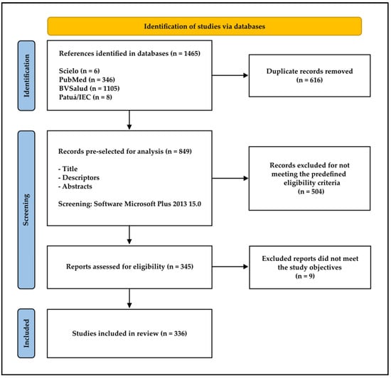
Figure 1.
Flowchart of the methodology for bibliographic data collection.
2.3. Inclusion and Exclusion Criteria
The inclusion criteria comprised original, full-text, and open-access articles; studies written in English, Portuguese, or Spanish; and publications with a complete description of the methodology used, including taxonomic identification data of ticks. Articles employing the following methodologies were included: RT-PCR, RT-qPCR, qPCR, PCR, NGS technologies, virus isolation in cell cultures from field-collected tick pools, and experimental infections in ticks and tick cell cultures. No restrictions were applied regarding the year of publication.
The exclusion criteria included review articles, editorials, opinion papers, incomplete studies, paywalled articles, dissertations and theses, articles that mentioned only tick species without providing relevant data for the research, and studies that did not present any information on viruses.
3. Results
A total of 336 articles were evaluated, including 215 studies on virus detection, both previously classified and unclassified. A summary of the main findings of viruses detected in ticks, considering only detections by molecular techniques such as PCR, qPCR, RT-PCR, RT-qPCR, RNA-seq, multiplex PCR, and conventional PCR, as well as the epidemiological significance of the virus and arthropod, is presented in Table 1 (Table S1).

Table 1.
Summary of the main virological findings in tick species of importance to public health.
Viruses of public health importance were identified in 32 viral isolation studies. A brief summary of the main viruses identified in batches of tick species of medical and veterinary importance is found in Table 2 (Table S2).
Table 2.
Viruses of medical and veterinary importance isolated from batches of tick species in cell cultures.
Among the 336 studies, forty articles were presented as experimental studies on ticks (larvae, nymphs, and adults), considering infection and experimental transmission studies. A brief summary of the main findings is found in Table 3 (Table S3). In addition, seven studies were described as experimental studies of infection in tick cells (Table 4 and Table S4).
Table 3.
Studies of experimental infection of viruses of medical and veterinary importance in ticks of great epidemiological relevance.
Table 4.
Viruses of public health importance studied in cell cultures from ticks.
High-throughput sequencing is a technology that allows the simultaneous sequencing of millions or even billions of genome fragments, resulting in a large volume of data, which may refer to complete viruses, fragmented viruses, or short viral sequences. Based on this, information from 42 articles that used high-throughput sequencing methods was analyzed using Heatmap to identify the viral families detected in tick samples. The studies used are available in the supplementary files, and a brief summary of the main tick species of public health importance used is summarized in Table 5 (Table S5).
Table 5.
The main tick species of medical and veterinary importance used in viral metagenomics studies.
Due to the large volume of data generated by high-throughput sequencing, articles that exclusively used this method were represented by a heat map to demonstrate and visualize the diversity and distribution of viral families detected in different tick species (Figure 2 and Figure 3). The families Phenuiviridae, Hepadnaviridae, Nairoviridae, Spinareo-viridae, and Virgaviridae predominate, indicating greater diversity or the number of de-tections associated with these groups (Figure 3).

Figure 2.
Heatmap of overall record abundance (number of times representatives of viral families appeared throughout the literature search), showing the relationship between viral families and tick species. Legend: Each cell indicates the number of records of viruses belonging to a given viral family (horizontal axis) detected in tick species or genera (vertical axis). Colors range from white (no records) to red and green (highest number of records), according to the intensity scale on the right.
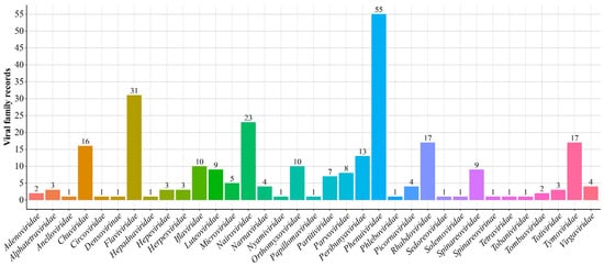
Figure 3.
Distribution of records for each detected viral family. The bar graph shows the frequency of records of different virus families (horizontal axis) identified in studies involving ticks. The vertical axis represents the total number of occurrences per viral family. The color variations between the bars correspond solely to the visual differentiation between viral families.
Based on the virological findings in ticks obtained in the reviewed scientific literature, viral agents of great importance for public health were listed, which are implicated in clinical manifestations that ranged from mild to severe cases, including deaths in humans (Table 6). The suggested severity classification was carried out based on the scientific literature consulted from the bibliographic survey.
Table 6.
Viral agents causing infections in humans.
4. Discussion
4.1. Family Flaviviridae: Orthoflavivirus
4.1.1. Tick-borne encephalitis virus: Orthoflavivirus encephalitidis
Orthoflavivirus encephalitidis is an arthropod-borne virus (arbovirus) first identified in 1937 during scientific expeditions to the Far East [117]. Historically, three subtypes have been recognized: European, Eastern, and Siberian, which exhibit broad and partially overlapping geographic distributions [118]. More recently, two additional subtypes have been described: Baikalian and Himalayan [119,120].
The vectors of Orthoflavivirus encephalitidis vary according to geographic region. For example, I. ricinus and I. persulcatus are responsible for transmission in Europe and Russia, respectively [117]. However, the virus has also been associated with other genera within the family Ixodidae, such as Hyalomma [27,66,121,122]. In addition, species of the genus Dermacentor have likewise been implicated as potential vectors of the agent [66,121,123,124].
4.1.2. Louping ill virus: Orthoflavivirus loupingi
I. ricinus is considered the natural vector of Orthoflavivirus loupingi, and the occurrence of the virus varies according to the distribution of its vectors. Sheep are highly susceptible to infection caused by the virus, often developing encephalitis, which can be fatal [125]. Additionally, the virus has been reported in horses, goats, cattle, and humans [126,127,128]. Recently, a study described inflammatory lesions and viral distribution in a naturally infected dog that exhibited progressive neurological signs leading to death [129]. Clinical manifestations in humans can be asymptomatic or symptomatic, resembling influenza-like symptoms such as fever and headache [125].
4.1.3. Powassan virus: Orthoflavivirus powassanense
Orthoflavivirus powassanense comprises two phylogenetically distinct lineages transmitted by different Ixodidae vectors. Both lineages are reported in North America (USA and Canada), with lineage I being disseminated primarily by I. cookei and I. marxi, while lineage II is transmitted by I. scapularis [32,130,131]. The virus was first isolated from Dermacentor andersoni specimens collected in Colorado [132]. Since then, the etiological agent has been isolated from I. cookei, I. scapularis, I. marxi, among others [68,133,134]. Furthermore, recent studies have demonstrated that A. americanum, D. variabilis [90], and Hae. longicornis [87] are competent vectors for the virus, as they are capable of becoming infected, transmitting it to their offspring, and infecting mammalian hosts that present clinical manifestations of infection.
In humans, Orthoflavivirus powassanense can cause severe neuroinvasive disease. Most symptomatic cases initially present with febrile illness, but neurological involvement can progress and become fatal. Approximately 10% of patients with neuroinvasive disease caused by the virus die, and survivors often suffer from neurological sequelae [134,135].
4.1.4. Kyasanur forest disease virus: Orthoflavivirus kyasanurense
Orthoflavivirus kyasanurense comprises two variants, both transmitted by ticks [136]. Lineage I, reported in the Kyasanur forest, Karnataka, India, is transmitted primarily by Haemaphysalis spinigera, while lineage II, identified in 1995 after an outbreak of hemorrhagic fever in Jeddah province, Saudi Arabia, has been reported in species of the families Argasidae and Ixodidae, specifically O. savignyi and Hy. dromedarii, respectively [64,137]. The virus has also been reported in several tick species, including Hy. marginatum s. l., D. auratus, I. petauristae, Rh. haemaphysaloides, A. lepidum, and Hae. [136,137,138,139]. The agent is pathogenic for humans, with clinical manifestations ranging from mild symptoms such as fever, headache and body aches, to more severe cases presenting hemorrhagic and neurological complications [140].
4.1.5. Omsk haemorrhagic fever virus: Orthoflavivirus omskense
Orthoflavivirus omskense was first identified in batches of D. reticulatus ticks collected in Omsk, Russia [141,142,143]. The agent is endemic in some regions of Russia, such as Omsk, Novosibirsk, Kurgan, and Tyumen [35]. Since its identification, the virus has also been reported in other tick species, such as I. persulcatus, D. reticulatus, and D. marginatus, collected in Akmola, Kazakhstan [35]. These recent findings suggest a possible expansion of the viral agent, as the pathogen was previously restricted to regions of Russia.
The clinical signs caused by infection are well documented; however, information on the pathogen’s pathogenesis in humans remains limited. The disease is characterized by a biphasic febrile illness, which can progress to hemorrhagic manifestations and, in more severe cases, neurological symptoms [144].
4.1.6. West Nile virus: Orthoflavivirus nilense
Orthoflavivirus nilense is predominantly transmitted by mosquitoes; however, the virus has also been isolated from tick species such as Rh. pulchellus and A. gemma collected in the Ijara District, Kenya [72] and Argas reflexus hermanni collected in the Nile Delta, Egypt [145]. Experimental studies involving O. moubata [86], I. ricinus [85], A. ovale, A. tigrinum, and A. tonelliae [84] suggest a potential role for ticks in virus transmission. Although these results demonstrate transmission potential, studies indicate that ticks are less efficient vectors of Orthoflavivirus nilense compared to mosquitoes [86]. However, the virus has been observed to persist within ticks for a period, suggesting that these arthropods may serve as temporary reservoirs [84,145].
4.1.7. Kadam virus: Orthoflavivirus kadamense
Orthoflavivirus kadamense was isolated from Rh. pravus ticks collected from cattle in Karamoja district, Uganda [146]. Since its discovery, new strains have been detected in ticks such as Rh. appendiculatus collected in Nairobi, Kenya [147] and Hy. dromedarii collected from a camel in Wadi Thamamah, Riyadh, Saudi Arabia [148]. The identification of antibodies against the virus in humans and cattle, together with the death of a camel from which infected ticks were collected, raises the suspicion that the viral agent may be pathogenic for camels [148,149,150]. Therefore, it is recommended that future studies be conducted to understand the ecology of the agent, as well as its pathogenic effects on public health.
4.1.8. Langat virus: Orthoflavivirus langatense
Orthoflavivirus langatense was isolated from tick batches of the genus Ixodes collected in Malaysia [151]. Subsequently, it was also isolated from Hae. papuana and I. persulcatus in Thailand and Siberia, respectively [152]. Experimental studies have evaluated the vector competence of several tick species for Orthoflavivirus langatense. For instance, the virus was found in multiple organs of Hae. longicornis, with evidence of both horizontal transmission and transmission to susceptible mice [153,154]. Studies on I. scapularis showed that the tick acquired the virus during blood feeding on infected mice, transmitted it transstadially to subsequent developmental stages, and that viral infection modulated gene expression changes in the ticks [155].
4.1.9. Usutu virus: Orthoflavivirus usutuense
The maintenance cycle of Orthoflavivirus usutuense involves several bird species as primary hosts, mosquitoes as primary vectors, especially arthropods of the genus Culex, and humans and other mammals as accidental hosts [156,157,158]. Although mosquitoes are recognized as the main vectors of this virus, a recent study demonstrated that the viral agent adapted to the cells of ticks of the species I. ricinus (IRE/CTVM19). Furthermore, the virus survived the molting process, from nymph to adult, and transmission of the agent through saliva was observed [159]. These data suggest that I. ricinus may be a competent vector for maintenance and transmission of the agent.
4.1.10. Saint Louis encephalitis virus: Orthoflavivirus louisense
Orthoflavivirus louisense is transmitted by mosquitoes, especially Culex mosquitoes [160]. Furthermore, vector competence studies conducted in Brazil on ticks for this virus [84]. The results demonstrated that ticks of the genus Amblyomma (A. ovale, A. tigrinum, and A. tonelliae) acquired the virus. Although transstadial transmission was observed, the arthropods were not competent to transmit the virus to susceptible vertebrate hosts in any case.
4.1.11. Karshi virus: Orthoflavivirus royalense
Orthoflavivirus royalense was identified in pools of O. papillipes ticks collected from Rhombomys opimus in Beshkent, Karshinsk Steppe, Uzbek Soviet Socialist Republic [161]. More recently, the viral agent was detected in ticks of the species Hy. asiaticum, Hy. scupense, and D. nuttalli collected in different regions of Xinjiang, China [162]. These findings suggest that these species may serve as potential vectors of the agent in the region. Furthermore, research has shown that the virus can infect a wide range of cells, including several human cell lines. Due to its close phylogenetic relationship with Orthoflavivirus powassanense and Orthoflavivirus kyasanurense, it is believed that Orthoflavivirus royalense can cause infections in humans [163,164]. However, to evaluate this hypothesis, further studies are needed to assess its pathogenicity in mammals.
In adult ticks, an experimental study evaluated the transmission of Orthoflavivirus royalense in ticks belonging to the genus Ornithodoros [165]. The results indicate that O. parkeri, O. sonrai, and O. tartakovskyi are important arthropods for the maintenance of the agent, as they remained vectors of the virus for long periods, transmitting it to susceptible mice.
4.2. Family Nairoviridae: Orthonairovirus
4.2.1. Crimean-Congo hemorrhagic fever virus: Orthonairovirus haemorrhagiae
Orthonairovirus haemorrhagiae is a single-stranded, negative-sense RNA virus [166]. It is a zoonotic virus maintained in a tick-vertebrate-tick cycle. The geographic distribution of Orthonairovirus haemorrhagiae overlaps with that of Hyalomma ticks, suggesting that members of this genus play a primary role in virus transmission [150,167,168]. Recently, the virus has been detected in several Hyalomma species, including Hy. marginatum, Hy. dromedarii, Hy. excavatum, Hy. anatolicum, and Hy. scupense collected from cattle in Balochistan, Pakistan [169]. However, species from other genera have also been found harboring Orthonairovirus haemorrhagiae, such as Boophilus decoloratus, Rh. (e.) evertsi, collected in regions of Senegal [170], and A. variegatum collected in Cameroon [171]. Clinical signs in animals are usually asymptomatic, but the virus is highly lethal in humans [172,173]. Therefore, it is important to expand entomovirological surveillance in ticks to prevent future outbreaks of the disease.
4.2.2. Dugbe virus: Orthonairovirus dugbeense
Orthonairovirus dugbeense was first isolated from a batch of A. variegatum ticks collected from cattle in Nigeria [174]. Since then, the virus has been isolated from several species, including A. gemma (Ijara District, Nairobi), A. lepidum (Nairobi), A. variegatum (southern Senegal), and Rh. pulchellus (Ijara District, Nairobi), leading to the belief that the agent is maintained in nature by many tick species [72,147,175]. However, we emphasize the importance of experimental studies to assess the vectorial competence of the species, since the mere identification of the agent in the arthropod does not mean that it is a vector of the pathogen. Humans are susceptible to infection by Orthonairovirus dugbeense, which usually causes a mild febrile illness [176,177,178].
4.2.3. Yezo virus: Orthonairovirus yezoense
Orthonairovirus yezoense is transmitted by ticks, with I. persulcatus as the main vector. Between 2020 and 2021, only I. persulcatus ticks tested positive for the virus in molecular assays, and only tick-derived cells demonstrated viral replication [179]. More recently, the virus was also detected in pools of I. ovatus and Hae. megaspinosa ticks collected in Hokkaido, Japan [179,180]. A recent study detected Orthonairovirus yezoense in I. persulcatus ticks collected from migratory birds in Japan; the data suggest that birds may be involved in the virus maintenance cycle. However, further studies are needed to validate this hypothesis [181].
4.2.4. Kasokero virus: Orthonairovirus kasokeroense
Orthonairovirus kasokeroense was recently detected and isolated from O. (r.) faini ticks collected from Rousettus aegyptiacus bats in Python Cave, Uganda, in 2013 and 2017 [182]. The research provides evidence supporting O. (r.) faini as the vector of Orthonairovirus kasokeroense, as no blood meal was observed in ticks, indicating that the arthropods did not feed on actively infected bats, resulting in active viral replication within the ticks. However, we emphasize the importance of experimental studies to evaluate the hypothesis.
4.2.5. Nairobi sheep disease orthonairovirus: Orthonairovirus nairobiense
Orthonairovirus nairobiense has been identified as the causative agent of the high mortality rates observed in sheep in markets in Nairobi, Kenya. The virus has been isolated from several sheep, and Rh. appendiculatus ticks have been implicated as vectors of the agent [183]. Since its discovery, the virus has also been isolated from other tick species, such as Rh. haemaphysaloides [184], Hae. longicornis [185], and A. variegatum [186], collected in India, China, and Kenya, respectively. This virus is of great importance to animal and human health, as it causes hemorrhagic gastroenteritis, fever, abortion, and high mortality in small ruminants, and febrile illness with nausea, headache, and vomiting in humans [187]. The disease has mortality rates of up to 90% in susceptible animals, resulting in significant economic losses in production systems [187,188].
4.2.6. Hazara virus: Orthonairovirus hazaraense
Orthonairovirus hazaraense was isolated from I. redikorzevi ticks collected in Kaghan Valley, Western Pakistan [189]. Although its natural host is unknown, serological evidence has been detected in rodent serum samples [190,191]. Orthonairovirus hazaraense was classified in the same serogroup as Orthonairovirus haemorrhagiae based on cross-reactivity between antibodies and antigens of the two viruses [192]. Although Orthonairovirus hazaraense is capable of inducing a disease similar to that caused by Orthonairovirus haemorrhagiae in mice, the agent has no association with human disease and is considered nonpathogenic [191,193].
4.2.7. Antu virus: Orthonairovirus antuense
Orthonairovirus antuense is another recently discovered member of the Nairoviridae family. The agent was isolated from D. silvarum tick samples collected at the China–North Korea border [194]. Although there is no evidence of infections in humans or domestic animals, the agent exhibited phylogenetic relationships with Orthonairovirus tomdiense and Orthonairovirus sakhalinense, both of which are thought to infect humans and domestic animals [195,196,197].
4.2.8. Beiji nairovirus: Norwavirus beijiense
Norwavirus beijiense was first detected in I. persulcatus collected in Heilongjiang Province, China [14]. More recently, however, the agent has been detected in I. persulcatus, I. pavlovskyi, and I. ovatus collected in Hokkaido, Fukushima, and Nagano, Japan [198]. Norwavirus beijiense has been associated with febrile illnesses in patients with a history of tick bites prior to illness onset. Additionally, the viral agent has also been detected in I. persulcatus, I. crenulatus, D. silvarum, D. nuttalli, Hae. concinna, and Hae. longicornis collected by flagging in regions of China [199]. Thus, an expansion of the agent is observed in a wide variety of species, making it extremely important to intensify entomovirological surveillance, as well as experimental studies to evaluate vector competence for the different tick species.
4.3. Family Phenuiviridae
4.3.1. SFTS virus: Dabie bandavirus: Bandavirus dabieense
Bandavirus dabieense is transmitted by ticks and is characterized by thrombocytopenia, fever, hemorrhage, and gastrointestinal symptoms [200]. The transmission cycle of Bandavirus dabieense is not fully understood. Ticks of the species Hae. longicornis are considered the primary vectors of the virus [201,202,203,204]. However, the agent has also been detected in other species collected in Korea: I. nipponensis [205,206], Hae. flava [206,207], A. testudinarium [206]; China: Hae. concinna [43,208], Rh. microplus [41], I. persulcatus, Hae. japonica, and D. silvarum [43]; Japan: Hae. hystricis [209]. The virus has also been detected in a wide range of vertebrates in China, including sheep, cattle, pigs, chickens, and dogs [210].
4.3.2. Heartland virus: Bandavirus heartlandense
Bandavirus heartlandense has been identified in A. americanum ticks collected in several areas of Missouri, implicating it as a vector [211]. Recently, the virus has been detected in A. americanum in Alabama [45], Illinois [46], and Missouri [47], and has also been isolated in Georgia [73]. A. americanum is the primary vector of the agent, but a recent study has shown that Hae. longicornis ticks can acquire and transmit the virus to vertebrates [96]. Symptoms of the disease are similar to those of ehrlichiosis and can include fever, anorexia, fatigue, nausea, and diarrhea [212].
4.3.3. Hunter island group virus: Bandavirus albatrossense
Bandavirus albatrossense was discovered after an outbreak in a colony of albatross (Thalassar-che cauta) on Albatross Island, located in northwestern Tasmania. The virus was isolated from batches of I. eudyptidis ticks collected from diseased and healthy birds. However, the maintenance cycle of the agent and its pathogenicity are unknown. Phylogenetic analyses indicated that the viral agent is closely related to Bandavirus dabieense and Bandavirus heartlandense, classifying it as a new member of the family Phenuiviridae [213].
4.3.4. Bhanja virus: Bandavirus bhanjanagarense
Bandavirus bhanjanagarense is transmitted by metastriate ixodid ticks. The first isolation of the agent occurred in 1954 from ticks of the species Hae. intermedia collected in the Ganjam district, Orissa State, India. Furthermore, the pathogen has been reported in other ixodid genera such as Haemaphysalis (Hae. punctata, Hae. sulcata), Hyalomma (Hy. marginatum, Hy. detritum, Hy. drome-darii, Hy. truncatum, Hy. asiaticum), Dermacentor (D. marginatus), Rhipicephalus (Rh. bursa, Rh. appendiculatus), Boophilus (B. decoloratus, B. annulatus, B. geigyi) and Amblyomma (A. variegatum) [214]. The viral agent does not cause clinical signs in adult animals; however, it is pathogenic for young ruminants, resulting in central nervous system impairment [215,216].
4.3.5. Dabieshan tick virus: Uukuvirus dabieshanense
Uukuvirus dabieshanense was identified in ticks of the species Hae. longicornis collected in regions of China (Hubei, Zhejiang, Beijing, and Xinjiang) [217]. Since its discovery, the virus has been reported in several tick species, including Rh. microplus, Rh. haemaphysaloides, and others in Yunnan and Guizhou provinces, southwest China [218]. Recently, molecular epidemiological investigations were conducted in ticks, and Uukuvirus dabieshanense was detected primarily in Hae. longicornis in other previously unreported provinces of China [39,218,219,220]. The high detection rate of the virus in ticks, especially Hae. longicornis, suggests that this arthropod is the main disseminator of the agent in the provinces of China and Japan [39,221,222]. Therefore, we emphasize the importance of expanding viral investigations in ticks to better understand their dynamics in nature and the risks to public health.
4.3.6. Lihan tick virus: Uukuvirus lihanense
Uukuvirus lihanense was first identified in a metagenomic study conducted on Rh. micropulus ticks collected in Yunnan Province, China. The study described the S segment (GenBank MH814975) and the L segment (GenBank MH814976); to date, there are no records of the M segment. Since its discovery, partial segments of the virus have been reported in molecular studies on ticks in Colombia (D. nitens and Rh. micropulus) and Brazil (Rh. micropulus), with all sequences showing high similarity to the sequence found in China [102,223,224]. To date, there is no clear evidence of the pathogenicity of Uukuvirus lihanense in humans, so the public health risk is unknown.
4.3.7. Kabuto mountain virus: Uukuvirus kabutoense
Uukuvirus kabutoense was isolated and characterized from batches of Hae. flava ticks collected in Hyogo, Japan. Furthermore, the study evaluated the infectivity and pathogenicity of the virus in lactating mice. The study found that high pathogenicity was maintained after three passages, demonstrating 100% mortality in this murine model [225]. Furthermore, the viral agent was also detected in a Hae. formosensis nymph collected in Wakayama Prefecture [226]. We emphasize the importance of expanding experimental research on the viral agent to assess its pathogenic potential and the risk it poses to public health.
4.3.8. Uukuniemi virus: Uukuvirus uukuniemiense
Uukuvirus uukuniemiense was isolated from ticks of the species I. ricinus, collected during a tick-borne virus investigation in Uukuniemi, Finland [227]. To date, Uukuvirus uukuniemiense has not been associated with any human disease [228]. However, its arboviral potential and pathogenicity remain unknown [229].
4.3.9. Mukawa virus: Phlebovirus mukawaense
Phlebovirus mukawaense was discovered in 2018, when scientists isolated the virus from female I. persulcatus ticks collected in Mukawa, Hokkaido, Japan [230]. Recently, the virus was detected for the first time in Heilongjiang, China, in ticks of the species I. persulcatus and Hae. concinna, representing the first report of the agent in the region [231]. In the same study, the authors isolated Phlebovirus mukawaense in Vero cells, indicating its possible pathogenicity in mammalian cells and potential to cause infections in humans. However, there are no reports of Phlebovirus mukawaense directly associated with human cases. Therefore, studies aimed at assessing its potential risk to public health are of paramount importance.
4.4. Family Orthomyxoviridae
4.4.1. Bourbon virus: Thogotovirus bourbonense
Thogotovirus bourbonense was described in 2017 after identification in a patient residing in Bourbon, Kansas, USA. Tick samples collected in Andrew, Gentry, and Nodaway, northwestern Missouri, were tested, and three pools of A. americanum were positive. The tick species A. americanum is believed to be the likely vector of Thogotovirus bourbonense [232]. Experimentally, A. americanum ticks were able to become infected and transmit the virus transstadially to the nymphal and adult stages. Vertical transmission was also observed in transstadially infected females [233]. This tick is aggressive and feeds on a variety of vertebrate species, including humans [234,235,236]. Thus, it becomes an important arthropod for the epidemiology of the agent.
4.4.2. Thogoto virus: Thogoto thogotovirus: Thogotovirus thogotoense
Thogotovirus thogotoense was discovered in two batches of ticks. The first batch contained four tick species (B. decoloratus, Rh. appendiculatus, Rh. simus, and B. evertsi), and the second batch contained only B. decoloratus (50 individuals), all collected from cattle in the Thogoto Forest, Nairobi, Kenya [237]. Since then, the virus has been reported in other tick species, such as Hae. longicornis [238,239], A. gemma, and A. lepidum [147], Kyoto City, Japan, and Nairobi, Kenya, respectively.
4.4.3. Dhori virus: Dhori thogotovirus: Thogotovirus dhoriense
Thogotovirus dhoriense was identified in ticks of the species Hy. dromedarii collected from camels in Dhori village, Kutch district, Gujarat, India. Since its discovery, the pathogen has been reported in other tick species, such as A. gemma and Rh. pulchellus, implicating them as possible vectors in regions of Kenya [147,240]. This agent has rarely been reported in humans; however, we emphasize the importance of surveillance to assess its spread among arthropod vectors to prevent future outbreaks.
4.5. Family Togaviridae
4.5.1. Semliki forest virus: Alphavirus semliki
Alphavirus semliki was isolated from ticks of the species Rh. pulchellus, A. gemma, A. lepidum, and Hy. truncatum collected from wild animals in Ijara District, Kenya [72]. Experimental studies demonstrated that the virus is lethal to mice, which developed paralysis and seizures followed by death, whereas rabbits exhibited only fever and paralysis [241,242]. Little is known about clinical symptoms in humans, although antibodies against the viral agent have been reported in South Africa and Mozambique. Furthermore, Alphavirus semliki has also been reported in Cameroon, the Democratic Republic of the Congo, the Central African Republic, Senegal, Nigeria, and Uganda [242,243].
4.5.2. Ndumu virus: Alphavirus ndumu
Alphavirus ndumu was described from batches of mosquitoes of the species Mansonia uniformis and Aedes circumluteolus, collected in Ndumu, KwaZulu-Natal, South Africa [244]. The viral agent was also isolated from other mosquito species of medical and veterinary importance, including Culex pipiens [245], Culex rubinotus, Culex cinereus, Aedes tricholabis, Aedes luridus, Anopheles rufipes, Anopheles ziemanni, among others [246,247]. Furthermore, Alphavirus ndumu was also isolated from a tick of the species Rh. pulchellus collected in Ijara District, Kenya [72]. However, the actual role of ticks in the maintenance and transmission of the virus remains unknown.
4.6. Family Asfarviridae
African swine fever virus: Asfivirus haemorrhagiae
Asfivirus haemorrhagiae has been identified in several species of the genus Ornithodoros in the regions of South Africa, Tanzania, and Madagascar [37,38,248]. Experimental studies confirm the vectorial competence of eight of these species for viral transmission, highlighting the significant epidemiological role of these ticks in maintaining the viral cycle [93,94,249,250]. Asfivirus haemorrhagiae does not pose a risk to humans, but is highly contagious among domestic and feral pigs, causing high mortality and generating substantial economic impacts on the global swine industry [4].
4.7. Family Poxviridae
4.7.1. Myxomatosum cuniculi virus: Leporipoxvirus myxoma
Leporipoxvirus myxoma has been detected in Rh. pusillus and Hy. lusitanicum collected from rabbits (Oryctolagus cuniculus) and hares (Lepus granatensis) in the provinces of Cádiz, Seville, and Badajoz, Spain [251]. Leporipoxvirus myxoma is harmless to humans; however, it is lethal to several rabbit species [252,253].
4.7.2. Milker’s node virus: Pseudocowpox virus: Parapoxvirus pseudocowpox
In 2020, Parapoxvirus pseudocowpox was detected in ticks of the species A. variegatum, Hy. (M.) rufipes and Hy. truncatum, removed from cattle in the provinces of Gourma, Kompienga, and Tapoa, located in eastern Burkina Faso [254]. The natural interaction between the virus and these tick species is unknown.
4.7.3. Bovine papular dermatitis virus: Parapoxvirus bovinestomatitis
Parapoxvirus bovinestomatitis was detected in ticks of the species A. variegatum and Hy. truncatum, removed from Cattle in Burkina Faso [254]. Other studies report the detection of Parapoxvirus genetic material in tick species such as Hy. marginatum, Hy. scupense and Rh. bursa from cattle in Corsica, France [255]. However, the role of ticks in the epidemiology of this viral agent remains unknown.
4.7.4. Lumpy skin disease virus: Capripoxvirus lumpyskinpox
Capripoxvirus lumpyskinpox, caused by Caprivirus, is a contagious disease that can be transmitted by close contact with infected animals, contaminated food and water sources, and by a wide range of biting vectors, including flies, lice, mosquitoes, and ticks [256]. The agent has been detected in ticks of the genera Rhipicephalus, Amblyomma, Hyalomma, and Dermacentor from provinces of Egypt and South Africa [257,258,259,260], and experimental studies have been conducted in some ixodid species [261]. However, further studies are needed to understand the true role of arthropods in the epidemiology of this viral agent.
4.8. Family Arenaviridae
4.8.1. Lymphocytic choriomeningitis virus: Mammarenavirus choriomeningitidis
Mammarenavirus choriomeningitidis is widely distributed in Africa, the Americas, Asia, and Europe [262,263,264]. It is a pathogen that causes significant neurological infections in humans, including encephalitis, meningitis, and congenital neurological defects [265]. The etiological agent has been detected in some tick species, such as D. silvarum and Hae. japonica collected in Jilin Province, northeastern China [266]. However, the role of ticks in the transmission of Mammarenavirus choriomeningitidis remains unknown [266,267].
4.8.2. Tacaribe virus: Mammarenavirus tacaribeense
Mammarenavirus tacaribeense was isolated from A. americanum ticks collected in Florida and showed 99.6% similarity to the original isolate [268]. No natural infections in humans have been reported; however, the isolation of the virus from mosquitoes and ticks, as well as its maintenance in vertebrate cells, suggests that the virus may use arthropods as vectors, resulting in infections in vertebrates [268,269]. However, the importance of more detailed studies to validate this hypothesis is emphasized.
4.9. Family Sedoreoviridae
4.9.1. Great Island virus: Kemerovo serocomplex: Orbivirus magninsulae
The prototype of the “Big Island virus” complex, Orbivirus magninsulae, previously classified as Big Island virus, was originally isolated from I. uriae ticks and their seabird host in 1973 in eastern Canada [270]. Since its discovery, new members have been reported, such as “Muko virus,” isolated from I. tudus ticks in Japan [271].
4.9.2. Bluetongue virus: Orbivirus caerulinguae
Orbivirus caerulinguae is widely transmitted by specimens of the genus Culicoides. Of the 1400 cataloged species, approximately 30 are known to transmit the virus [272,273]. Although members of the genus Culicoides are the main vectors of the pathogen, the agent has also been studied in ticks of different species. Serotype 8 was evaluated in five tick species: I. ricinus, I. hexagonus, D. reticulatus, Rh. bursa, and O. savignyi [274]. The research showed that arthropods of the families Ixodidae and Argasidae can become infected with the pathogen. However, further studies are needed to determine the actual role of ticks in the virus transmission cycle.
4.9.3. Murid herpesvirus 4: Rhadinovirus muridgamma4
Rhadinovirus muridgamma4 is transmitted through respiratory excretions, urine, and saliva, and vertical transmission can also occur via the transplacental route [275]. Moreover, research provides important data on the sexual transmission of the agent among mice [276,277]. Although studies indicate that the virus is maintained in a vertebrate cycle, a recent study investigated the vector competence of the tick I. ricinus for Rhadinovirus muridgamma4 [278]. The research demonstrated that ticks acquired the virus and were able to transmit it to susceptible mice, and that the agent survived molting, thereby characterizing transstadial transmission. In addition, the pathogen was transmitted to the next generation, characterizing vertical transmission. The study provides information consistent with the criteria for arthropod-borne viruses (arboviruses) [279,280]. Nevertheless, further studies are required to better understand the actual role of ticks in the transmission of this agent.
4.10. Family Spinareoviridae
4.10.1. Eyach virus: Coltivirus ixodis
Coltivirus ixodis was described from I. ricinus tick samples collected in the Federal Republic of Germany [281]. The agent was also isolated from I. ventalloi ticks collected in Mayenne, France, thus adding a potential new natural vector of the viral agent [282]. Although the study identified antibodies against Coltivirus ixodis in farmers, the pathogen has not yet been isolated from animals or humans. The identification of the agent in ticks does not confirm their status as vectors of the virus; therefore, experimental studies to assess the vectorial competence of these arthropods are of paramount importance to validate this hypothesis.
4.10.2. Colorado tick fever virus: Coltivirus dermacentoris
The primary vector of Coltivirus dermacentoris is the tick species D. andersoni [283,284]. The virus is maintained in nature through enzootic cycles between rodents and ticks, and the distribution of the etiological agent overlaps with that of its arthropod vector [283]. The viral agent has been reported in tick species such as D. albipictus, Otobius lagophilus, I. sculptus, I. spinipalpis, and Hae. leporispalustris, most of which have been removed from wild-caught vertebrates in areas of the USA [285,286].
4.10.3. Kundal virus: Kundal coltivirus: Coltivirus kundalense
Coltivirus kundalense was discovered in 2019 in a tick of the species Hy. anatolicum during surveillance for Orthonairovirus haemorrhagiae in Gujarat, India. The agent exhibited cytopathic effects in Vero CCL-81 cells, human rhabdomyosarcoma cells, and porcine stable kidney cells, but not in Pipistrellus ceylonicus bat embryo cells or Aedes albopictus cells. Although the virus shows cytopathic effects in vertebrate cell cultures, it was found to be non-pathogenic to mice [287]. Currently, it is unknown whether Coltivirus kundalense is pathogenic to humans.
4.11. Family Peribunyaviridae
Bunyamwera virus: Orthobunyavirus bunyamweraense
Orthobunyavirus bunyamweraense is maintained in nature through a cycle involving mosquitoes and susceptible vertebrates. Ae. aegypti is believed to be the primary vector of the agent [288,289]. However, the viral agent has also been isolated from tick species such as Rh. pulchellus, A. gemma, A. lepidum, and Hy. truncatum collected in Ijara District, Kenya [72]. Research suggests that ticks may contribute to the maintenance, spread, and possibly transmission of the virus.
4.12. Family Picornaviridae
Foot-and-mouth disease virus: Aphthovirus vesiculae
Foot-and-mouth disease is caused by Aphthovirus vesiculae, which is highly infectious and contagious among domestic and wild cloven-hoofed animals, with outbreaks causing substantial economic losses to the global livestock industry [290]. An unusual finding occurred in 2006 during surveillance of tick-borne arboviruses in Nairobi, Kenya. Aphthovirus vesiculae was isolated from ticks of the species Rh. pulchellus, representing the first known isolation associated with tick collections [147]. Although Aphthovirus vesiculae is not transmitted by ticks of the genus Rhipicephalus, there is evidence that the viral agent can persist for several days in ticks fed on infected animals [291].
4.13. Family Rhabdoviridae
4.13.1. Zahedan rhabdovirus: Zarhavirus zahedan
Zarhavirus zahedan is a recently discovered virus in ticks of the species Hy. anatolicum anatolicum collected in Zahedan, Iran, in 2001. However, the full study on the virus was published in 2015 [292]. In the research, the agent was able to infect mammalian Vero cells and demonstrated lethality in mice regardless of the inoculation route.
4.13.2. Barur virus: Ledantevirus barur
Ledantevirus barur was isolated from batches of Rh. pulchellus collected in Mogadishu, Somalia [293] and later from ticks of the species Hae. intermedia collected in Saga, Shimoga district, Karnataka, India [294]. The agent was isolated from Rh. pulchellus collected from animals at the Njiru and Dagoretti slaughterhouses, outside Nairobi, Kenya [147]. A recent high-throughput sequencing study detected Ledantevirus barur in Rh. appendiculatus [295]. Although the agent has not been reported in humans, identification of the virus in ticks and rodents suggests a possible maintenance cycle between hematophagous arthropods and mammals [147].
4.14. Family Paramyxoviridae
Nipah virus: Henipavirus nipahense
Henipavirus nipahense is highly pathogenic and infects a wide range of wild and domestic animals, as well as humans, in whom it can cause pulmonary or encephalitic disease with a mortality rate of 92% [296,297]. Transmission of the agent occurs through direct contact with infected pigs, as well as exposure to porcine bodily fluids [298,299,300]. The hypothesis that ticks may be involved in the agent’s transmission cycle led to a recent study evaluating infection in I. scapularis (IDE8) embryonic cells [301]. The research demonstrated the presence of viral RNA and antigens, and infectious virus was recovered. These results indicate that ixodid cells can support infection by the agent. However, further studies are required to assess the potential for viral infection in ticks.
4.15. Ungrouped
4.15.1. Alongshan virus
Recently, Alongshan virus was detected in I. persulcatus collected in the Mongolia region of China, where the agent was first described [302,303]. The primary vectors of the virus are ticks, particularly of the genus Ixodes, which is most frequently associated with the pathogen [59,104,304,305]. Recently, a study evaluated infection of Alongshan virus in ticks of the species I. ricinus and D. reticulatus. The results provide initial insights into the agent’s transmission cycle and reveal the potential of these arthropods to transmit the virus [97].
4.15.2. Yanggou tick virus
Yanggou tick virus is another member of the Jingmenvirus group, first detected in D. nuttalli ticks in 2018 (GenBank MH688529.1) collected in the Xinjiang Uyghur Autonomous Region, China. Recently, the agent was detected in D. nuttalli and D. marginatus collected in Russia [62]. Furthermore, for the first time, a study demonstrated the replication of Yanggou tick virus in cell cultures of I. ricinus (IRE/CTVM19) and Hy. anatolicum (HAE/CTVM8) ticks. These results suggest that the pathogen may adapt to different vectors or that the agent may adapt to different tick species. Yanggou tick virus and Alongshan virus are closely related and cluster separately from other viruses in the JMV group [302]. Although there are no reports of Yanggou tick virus in humans, the virus belongs to the JMV group, which includes viruses previously reported in humans, such as Alongshan virus, which generally causes mild symptoms [303,306,307].
4.15.3. Mpulungu flavivirus
Mpulungu flavivirus (GenBank LC582740.1) is a newly discovered virus in ticks of the species Rh. muhsamae collected in different areas of Zambia (Isoka, Mpulungu, and Samfya) [308]. The virus showed 83.2% identity with Ngoye virus, previously identified in Rh. evertsi evertsi and Rh. guilhoni in Senegal [309]. To date, there are no reports of Mpulungu flavivirus in humans or other vertebrates, nor in other arthropods.
4.15.4. Meaban-like virus
Meaban-like virus (GenBank KJ440085-KJ440090) was detected in O. maritimus ticks collected in the Western Mediterranean basin [310]. The detected segment is a partial 124 bp fragment corresponding to the nonstructural protein gene 5 (GenBank KJ440090.1). BLAST analysis identified approximately 95% similarity with a fragment of the NS5 gene of Orthoflavivirus meabanense, a virus first detected in O. maritimus on islands of South Brittany, France [311].
4.15.5. Phlebovirus gomselga
Phlebovirus gomselga was detected in I. persulcatus collected in the Chelyabinsk region, Russia, in 2018 [312]. Subsequently, the agent was detected again in I. persulcatus collected from the same region [313]. Phylogenetic analysis indicated that Phlebovirus gomselga clusters with tick-borne phleboviruses.
4.15.6. Phenuivirus stavropol
The first report of Stavropol virus occurred in 2020, detected in D. reticulatus ticks collected between 2011 and 2012 in Stavropol, Russia (GenBank ON920444-ON920447) [312]. The study identified a preference of Stavropol virus for D. reticulatus, although the agent was detected once in Rh. turanicus. This is an unclassified virus of the family Phenuiviridae, with no reported human infections to date, and its pathogenicity remains unknown.
4.15.7. Okutama tick virus
Okutama tick virus was first detected in Hae. flava nymphs collected from Tokyo, Japan [314]. Subsequently, the agent was also detected in adult Hae. flava ticks collected from Cape Toi and Hokuriku, Japan [221,315]. The pathogenicity in humans and animals, as well as the transmission cycle, remain unknown.
4.15.8. Shibuyunji virus
Shibuyunji virus was initially detected in Rhipicephalus (b.) sp. ticks from cattle in the Shibuyunji District, Lusaka Province, Zambia [316] and more recently in Rhipicephalus sp. ticks collected from cattle in different districts of Zambia [317]. In this study, the virus showed approximately 99% similarity with the initial report. Although there are no reports of human infections, detection in ixodid ticks infesting cat-tle raises concerns regarding potential zoonotic risk.
4.15.9. Phlebovirus-like-AYUT
A molecular study identified a fragment of the L segment of a phlebovirus, provisionally named Phlebovirus-like-AYUT, detected in Rh. sanguineus s. l. collected from a dog in Phra Nakhon Si Ayutthaya Province, Thailand [318]. The agent exhibited 82.6% similarity with Brown dog tick phlebovirus 2, previously detected in Rh. sanguineus in Trinidad and Tobago [319]. Additionally, it shares 72.4% and 70.3% identity with Brown dog tick phlebovirus 1 and Bole Tick Virus 1, respectively, both associated with the genus Phlebovirus, family Phenuiviridae.
4.15.10. Haseki tick virus
Haseki tick virus was discovered in I. persulcatus ticks collected in Novosibirsk, Russia [320]. Human cases have been reported after tick bites. Human infections have been associated with fever, acute respiratory injuries, and coinfection with other tick-borne agents. Phylogenetic analysis revealed that all Haseki tick virus sequences clustered together and were closely related to Bole tick virus 4 and Trinbago virus.
4.15.11. Kindia tick virus
Kindia tick virus was identified in Rh. geigyi ticks collected in Guinea, West Africa [321]. Subsequently, the agent was detected in Rh. geigyi, Rh. annulatus, and Rh. decoloratus collected from domestic animals in the Republic of Guinea [322,323]. It is an unclassified flavivirus transmitted by ticks of the JMV group, family Flaviviridae [323]. Since it is a member of the JMV group, which includes viruses previously reported in humans, such as Alongshan virus, although there are no reports of human cases, we emphasize the importance of experimental studies to evaluate its impact on public health.
4.15.12. Yichun nairovirus
Yichun nairovirus is a recently discovered agent in I. persulcatus ticks, identified through a metagenomic study that assessed the viral diversity of four tick species collected in northeastern China [111]. Recently, the agent was also detected in I. persulcatus and I. pavlovskyi ticks collected in Hokkaido and Fukushima, Japan [198]. Although Yichun nairovirus is phylogenetically closely related to Norwavirus beijiense, little is known about its potential public health risk [111].
4.15.13. Kinna virus
Kinna virus is another unclassified virus identified in A. gemma ticks collected in Isiolo County, Kenya [324]. Phylogenetic analysis grouped the agent with viruses of the Bandavirus genus, showing a strong relationship with Bandavirus guertuense. The study also demonstrated that Kinna virus is pathogenic to mice. However, further studies are needed to assess the potential risk of Kinna virus to public and animal health.
4.15.14. Sandy Bay virus
Sandy Bay virus is an unclassified agent isolated from I. uriae ticks collected from the king penguin colony at Sandy Bay, near a king penguin colony at the head of Finch Creek, and from Catch-me Cave on Macquarie Island (Figure 1), a preferred perch for rockhopper penguins [325]. The pathogen showed 94–96% similarity to Orthoflavivirus gadgetsense, previously identified in I. uriae ticks collected on Macquarie Island, Australia [326]. To date, there are no records of Sandy Bay virus infections in humans. The discovery in ticks parasitizing king penguins suggests that the arthropod has adapted to specific hosts and therefore does not pose a significant threat to human health. However, we emphasize the importance of further studies to evaluate its possible pathogenicity in vertebrates.
Many unclassified viruses have been detected in ticks, but there is a lack of evidence as to whether they are pathogenic to humans and animals. However, some viruses that were initially “unclassified” have subsequently proven pathogenic, such as Bandavirus heartlandense and unclassified Alongshan virus. The lack of experimental studies creates a surveillance blind spot, where potentially dangerous viruses circulate unmonitored.
4.16. Climate Change
Atmospheric characteristics and dynamics are important environmental factors, and any changes can significantly impact animal ecology [327]. Changes in climate patterns (temperature and humidity) influence the epidemiology of zoonotic diseases by altering host dynamics, the distribution of clinically important vectors such as ticks, and the pathogens they transmit [328,329,330,331,332,333,334]. Climate change can create new niches for hematophagous arthropods such as ticks, facilitating the expansion of their geographic distribution into previously unreported areas [335,336,337,338]. Furthermore, changes in environmental variables affect host populations, including rodents, deer, and birds, which in turn alter the abundance and distribution of ticks [339].
Climatic variables also influence specific aspects of etiological agents, including improved climatic adaptation for reproduction, accelerated life cycles, decreased extrinsic incubation time, and increased virulence [340]. Rising temperatures, for example, significantly impacted the replication of Orthoflavivirus nilense in mosquitoes, increasing the rate of virus replication and bites, thus improving the arthropod’s ability to transmit the agente [341,342,343].
5. Conclusions
Ticks are globally recognized as important vectors of pathogens with significant impacts on public and veterinary health. These arthropods are expanding their geographic ranges in response to climate change, consequently affecting pathogen transmission cycles. This scenario increases the risk of arbovirus transmission, particularly viruses from the families Flaviviridae, Nairoviridae, and Asfarviridae, which are known to cause serious infections in humans and animals (domestic and livestock), including large-scale deaths. In this review, we demonstrate the wide range of viruses that have been described in tick species, including unclassified viruses such as Alongshan virus and Yanggou tick virus, which exhibit phylogenetic relationships with viral groups important to public health. We also provide important data on the various studies using viruses and ticks, demonstrating several gaps in knowledge about the interaction between viruses and ticks, as evidenced by the lack of experimental studies to validate theories of tick-borne viral transmission. Therefore, we recommend that viral investigation studies in tick populations, as well as experimental infection and transmission studies using unclassified viruses already identified in ticks, are crucial to understanding potential risks to public and animal health, especially in the context of ongoing climate change.
Supplementary Materials
The following are available online at https://www.mdpi.com/article/10.3390/arthropoda3040016/s1, Table S1: Complete list of viruses detected in ticks, considering only detection by molecular techniques, such as PCR, qPCR, RT-PCR, RT-qPCR, RNA-seq, multiplex PCR and conventional PCR, Table S2: List of unclassified viruses detected in tick species, considering only detection by molecular techniques, such as PCR, qPCR, RT-PCR, RT-qPCR, RNA-seq, multiplex PCR and conventional PCR, Table S3: Viruses isolated from tick species in different regions, Table S4: Viral studies of experimental infection in tick species, Table S5: List of experimental studies in tick cell cultures, Table S6: Metagenomic studies included in the present study from different regions of the globe. References [344,345,346,347,348,349,350,351,352,353,354,355,356,357,358,359,360,361,362,363,364,365,366,367,368,369,370,371,372,373,374,375,376,377,378,379,380,381,382,383,384,385,386,387,388,389,390,391,392,393,394,395,396,397,398,399,400,401,402,403,404,405,406,407,408,409,410,411,412,413,414,415,416,417,418,419,420,421,422,423,424,425,426,427,428,429,430,431,432,433,434,435,436,437,438,439,440,441,442,443,444,445,446,447,448,449,450,451,452,453,454,455,456,457,458,459,460,461,462,463,464,465,466,467,468,469,470,471,472,473,474,475,476,477,478,479,480,481,482,483,484,485,486,487,488,489,490,491,492,493,494,495,496,497,498,499,500,501,502,503,504,505,506,507,508,509,510,511] are cited in the supplementary materials.
Author Contributions
Conceptualization, L.H.d.S.e.S. and J.P.N.N.; methodology, L.H.d.S.e.S. and J.P.N.N.; software, F.S.d.S. and D.D.D.; validation, F.S.d.S., D.D.D., L.H.d.S.e.S. and J.P.N.N.; formal analysis, L.H.d.S.e.S., S.L.S.d.S., L.A.M.R. and H.C.F.R.; investigation, L.H.d.S.e.S., B.L.S.d.N. and S.L.S.d.S.; resources, L.H.d.S.e.S. and J.P.N.N.; data curation, L.H.d.S.e.S. and J.P.N.N.; writing—original draft preparation, L.H.d.S.e.S., L.A.M.R., B.L.S.d.N., H.C.F.R. and J.P.N.N.; writing—review and editing, L.H.d.S.e.S. and J.P.N.N.; supervision, J.P.N.N.; project administration, L.H.d.S.e.S. and J.P.N.N. All authors have read and agreed to the published version of the manuscript.
Funding
This research was funded by the Coordination for the Improvement of Higher Education Personnel (CAPES) (process No. 88887.756798/2022-00), Graduate Program in Parasitary Biology in the Amazon Region (PPGBPA) of State University of Pará (edital No. 064/2022-UEPA), and Evandro Chagas Institute (IEC/SVSA/MS).
Institutional Review Board Statement
Not applicable.
Informed Consent Statement
Not applicable.
Data Availability Statement
No new data were created or analyzed in this study.
Conflicts of Interest
The authors declare no conflicts of interest.
References
- World Health Organization—WHO. Vector-Borne Diseases. 2024. Available online: https://www.who.int/news-room/fact-sheets/detail/vector-borne-diseases (accessed on 1 April 2025).
- International Committee on Taxonomy of Viruses—ICTV. Current ICTV Taxonomy Release. 2025. Available online: https://ictv.global/taxonomy (accessed on 1 April 2025).
- Kleiboeker, S.B.; Scoles, G.A. Pathogenesis of African swine fever virus in Ornithodoros ticks. Anim. Health Res. Ver. 2001, 2, 121–128. [Google Scholar] [CrossRef]
- Gaudreault, N.N.; Madden, D.W.; Wilson, W.C.; Trujillo, J.D.; Richt, J.A. African Swine Fever Virus: An Emerging DNA Arbovirus. Front. Vet. Sci. 2020, 7, 215. [Google Scholar] [CrossRef]
- Worku, D.A. Tick-borne encephalitis (TBE): From tick to pathology. J. Clin. Med. 2023, 12, 6859. [Google Scholar] [CrossRef]
- Yu, K.M.; Park, S.J. Tick-borne viroses: Epidemiology, pathogenesis, and animal models. One Health 2024, 19, 100903. [Google Scholar] [CrossRef]
- Lani, R.; Moghaddam, E.; Haghani, A.; Chang, L.; AbuBakar, S.; Zandi, K. Tick-borne viruses: A review from the perspective of therapeutic approaches. Ticks Tick-Borne Dis. 2014, 5, 457–465. [Google Scholar] [CrossRef] [PubMed]
- Mansfield, K.L.; Jizhou, L.; Phipps, L.P.; Johnson, N. Emerging Tick-Borne Viruses in the Twenty-First Century. Front. Cell. Infect. Microbiol. 2017, 7, 298. [Google Scholar] [CrossRef]
- Pustijanac, E.; Buršić, M.; Talapko, J.; Škrlec, I.; Meštrović, T.; Lišnjić, D. Tick-Borne Encephalitis Virus: A Comprehensive Review of Transmission, Pathogenesis, Epidemiology, Clinical Manifestations, Diagnosis, and Prevention. Microorganisms 2023, 11, 1634. [Google Scholar] [CrossRef] [PubMed]
- Gömer, A.; Lang, A.; Janshoff, S.; Steinmann, J.; Steinmann, E. Epidemiology and global spread of emerging tick-borne Alongshan virus. Emerg. Microbes Infect. 2024, 13, 2404271. [Google Scholar] [CrossRef] [PubMed]
- Sameroff, S.; Tokarz, R.; Jain, K.; Oleynik, A.; Carrington, C.V.F.; Lipkin, W.I.; Oura, C.A.L. Novel quaranjavirus and other viral sequences identified from ticks parasitizing hunted wildlife in Trinidad and Tobago. Ticks Tick-Borne Dis. 2021, 4, 101730. [Google Scholar] [CrossRef]
- Pettersson, J.H.O.; Shi, M.; Bohlin, J.; Eldholm, V.; Brynildsrud, O.B.; Paulsen, K.M.; Andreassen, A.; Holmes, E.C. Characterizing the virome of Ixodes ricinus ticks from northern Europe. Sci. Rep. 2017, 7, 10870. [Google Scholar] [CrossRef]
- Brinkmann, A.; Dinçer, E.; Polat, C.; Hekimoğlu, O.; Hacıoğlu, S.; Földes, F.; Özkul, A.; Öktem, I.M.A.; Nitsche, A.; Ergünay, K. A metagenomic survey identifies Tamdy orthonairovirus as well as divergent phlebo-, rhabdo-, chu- and flavi-like viruses in Anatolia, Turkey. Ticks Tick-Borne Dis. 2018, 9, 1173–1183. [Google Scholar] [CrossRef]
- Meng, F.; Ding, M.; Tan, Z.; Zhao, Z.; Xu, L.; Wu, J.; He, B.; Tu, C. Virome analysis of tick-borne viruses in Heilongjiang Province, China. Ticks Tick-Borne Dis. 2019, 10, 412–420. [Google Scholar] [CrossRef] [PubMed]
- Gondard, M.; Temmam, S.; Devillers, E.; Pinarello, V.; Bigot, T.; Chrétien, D.; Aprelon, R.; Vayssier-Taussat, M.; Albina, E.; Eloit, M.; et al. RNA Viruses of Amblyomma variegatum and Rhipicephalus microplus and Cattle Susceptibility in the French Antilles. Viruses 2020, 12, 144. [Google Scholar] [CrossRef] [PubMed]
- Pettersson, J.H.O.; Ellström, P.; Ling, J.; Nilsson, I.; Bergström, S.; González-Acuña, D.; Olsen, B.; Holmes, E.C. Circumpolar diversification of the Ixodes uriae tick virome. PLoS Pathog. 2020, 16, 1008759. [Google Scholar] [CrossRef] [PubMed]
- Xu, L.; Guo, M.; Hu, B.; Zhou, H.; Yang, W.; Hui, L.; Huang, R.; Zhan, J.; Shi, W.; Wu, Y. Tick virome diversity in Hubei Province, China, and the influence of host ecology. Virus Evol. 2021, 7, veab108. [Google Scholar] [CrossRef]
- Bratuleanu, B.E.; Temmam, S.; Chrétien, D.; Regnault, B.; Pérot, P.; Bouchier, C.; Bigot, T.; Savuța, G.; Eloit, M. The virome of Rhipicephalus, Dermacentor and Haemaphysalis ticks from Eastern Romania includes novel viruses with potential relevance for public health. Transbound. Emerg. Dis. 2022, 69, 1387–1403. [Google Scholar] [CrossRef]
- Ercole, F.F.; Melo, L.S.; Alcoforado, C.L.G.C. Revisão integrativa versus revisão sistemática. Rev. Min. Enferm. 2014, 18, 12–14. [Google Scholar] [CrossRef]
- Souza, M.T.; Silva, M.D.; Carvalho, R. Integrative review: What is it? How to do it? Einstein 2010, 8, 102–106. [Google Scholar] [CrossRef]
- Botelho, L.L.R.; Cunha, C.C.A.; Macedo, M. O método da revisão integrativa nos estudos organizacionais. Gestão Soc. 2011, 5, 121–136. [Google Scholar] [CrossRef]
- Esser, H.J.; Lim, S.M.; Vries, A.; Sprong, H.; Dekker, D.J.; Pascoe, E.L.; Bakker, J.W.; Suin, V.; Franz, E.; Martina, B.E.E.; et al. Continued Circulation of Tick-Borne Encephalitis Virus Variants and Detection of Novel Transmission Foci, the Netherlands. Emerg. Infect. Dis. 2022, 28, 2416–2424. [Google Scholar] [CrossRef]
- Jung, H.; Choi, C.; Lee, L.; Kim, S.; Aknazarov, B.; Nyrgaziev, R.; Atabekova, N.; Jetigenov, E.; Chung, Y.; Lee, H. Molecular Detection and Phylogenetic Analysis of Tick-Borne Encephalitis Virus from Ticks Collected from Cattle in Kyrgyzstan, 2023. Viruses 2024, 16, 107. [Google Scholar] [CrossRef]
- Klaus, C.; Hoffmann, B.; Hering, U.; Mielke, B.; Sachse, K.; Beer, M.; Süss, J. Tick-borne encephalitis (TBE) virus prevalence and virus genome characterization in field-collected ticks (Ixodes ricinus) from risk, non-risk and former risk areas of TBE, and in ticks removed from humans in Germany. Clin. Microbiol. Infect. 2010, 16, 238–244. [Google Scholar] [CrossRef][Green Version]
- Laaksonen, M.; Sajanti, E.; Sormunen, J.J.; Penttinen, R.; Hänninen, J.; Ruohomäki, K.; Sääksjärvi, I.; Vesterinen, E.J.; Vuorinen, I.; Hytönen, J.; et al. Crowdsourcing-based nationwide tick collection reveals the distribution of Ixodes ricinus and I. persulcatus and associated pathogens in Finland. Emerg. Microbes Infect. 2017, 6, 1–7. [Google Scholar] [CrossRef]
- Ott, D.; Ulrich, K.; Ginsbach, P.; Öhme, R.; Bock-Hensley, O.; Falk, U.; Teinert, M.; Lenhard, T. Tick-borne encephalitis virus (TBEV) prevalence in field-collected ticks (Ixodes ricinus) and phylogenetic, structural and virulence analysis in a TBE high-risk endemic area in southwestern Germany. Parasites Vectors 2020, 13, 303. [Google Scholar] [CrossRef] [PubMed]
- Yun, S.M.; Song, B.G.; Choi, W.; Park, W.I.; Kim, S.Y.; Roh, J.Y.; Ryou, J.; Ju, Y.R.; Park, C.; Shin, E. Prevalence of tick-borne encephalitis virus in ixodid ticks collected from the republic of Korea during 2011-2012. Osong Public Health Res. Perspect. 2012, 3, 213–221. [Google Scholar] [CrossRef] [PubMed]
- Topp, A.K.; Springer, A.; Dobler, G.; Bestehorn-Willmann, M.; Monazahian, M.; Strube, C. New and Confirmed Foci of Tick-Borne Encephalitis Virus (TBEV) in Northern Germany Determined by TBEV Detection in Ticks. Pathogens 2022, 11, 126. [Google Scholar] [CrossRef] [PubMed]
- Papa, A.; Pavlidou, V.; Antoniadis, A. Greek Goat Encephalitis Virus Strain Isolated from Ixodes ricinus, Greece. Emerg. Infect. Dis. 2008, 14, 330–332. [Google Scholar] [CrossRef]
- Korobitsyn, I.G.; Moskvitina, N.S.; Tyutenkov, O.Y.; Gashkov, S.I.; Kononova, Y.V.; Moskvitin, S.S.; Romanenko, V.N.; Mikryukova, T.P.; Protopopova, E.V.; Kartashov, M.Y.; et al. Detection of tick-borne pathogens in wild birds and their ticks in Western Siberia and high level of their mismatch. Folia Parasitol. 2021, 68, 24. [Google Scholar] [CrossRef]
- Kolodziejek, J.; Marinov, M.; Kiss, B.J.; Alexe, V.; Nowotny, N. The complete sequence of a West Nile virus lineage 2 strain detected in a Hyalomma marginatum marginatum tick collected from a song thrush (Turdus philomelos) in eastern Romania in 2013 revealed closest genetic relationship to strain Volgograd 2007. PLoS ONE 2014, 9, e109905. [Google Scholar] [CrossRef]
- Tokarz, R.; Tagliafierro, T.; Cucura, D.M.; Rochlin, I.; Sameroff, S.; Lipkin, W.I. Detection of Anaplasma phagocytophilum, Babesia microti, Borrelia burgdorferi, Borrelia miyamotoi, and Powassan Virus in Ticks by a Multiplex Real-Time Reverse Transcription-PCR Assay. mSphere 2017, 2, e00151-17. [Google Scholar] [CrossRef]
- Narvaez, Z.E.; Rainey, T.; Puelle, R.; Khan, A.; Jordan, R.A.; Egizi, A.M.; Price, D.C. Detection of multiple tick-borne pathogens in Ixodes scapularis from Hunterdon County, NJ, USA. Curr. Res. Parasitol. Vector Borne Dis. 2023, 4, 100140. [Google Scholar] [CrossRef]
- Johnson, T.L.; Graham, C.B.; Maes, S.E.; Hojgaard, A.; Fleshman, A.; Boegler, K.A.; Delory, M.J.; Slater, K.S.; Karpathy, S.E.; Bjork, J.K.; et al. Prevalence and distribution of seven human pathogens in host-seeking Ixodes scapularis (Acari: Ixodidae) nymphs in Minnesota, USA. Ticks Tick-Borne Dis. 2018, 9, 1499–1507. [Google Scholar] [CrossRef] [PubMed]
- Wagner, E.; Shin, A.; Tukhanova, N.; Turebekov, N.; Nurmakhanov, T.; Sutyagin, V.; Berdibekov, A.; Maikanov, N.; Lezdinsh, I.; Shapiyeva, Z.; et al. First Indications of Omsk Haemorrhagic Fever Virus beyond Russia. Viruses 2022, 14, 754. [Google Scholar] [CrossRef] [PubMed]
- Vial, L.; Wieland, B.; Jori, F.; Etter, E.; Dixon, L.; Roger, F. African Swine Fever Virus DNA in Soft Ticks, Senegal. Emerg. Infect. Dis. 2007, 13, 1928–1931. [Google Scholar] [CrossRef] [PubMed]
- Peter, E.; Machuka, E.; Githae, D.; Okoth, E.; Cleaveland, S.; Shirima, G.; Kusiluka, L.; Pelle, R. Detection of African swine fever virus genotype XV in a sylvatic cycle in Saadani National Park, Tanzania. Transbound. Emerg. Dis. 2020, 68, 813–823. [Google Scholar] [CrossRef]
- Ravaomanana, J.; Michaud, V.; Jori, F.; Andriatsimahavandy, A.; Roger, F.; Albina, E.; Vial, L. First detection of African Swine Fever Virus in Ornithodoros porcinus in Madagascar and new insights into tick distribution and taxonomy. Parasites Vectors 2010, 3, 115. [Google Scholar] [CrossRef]
- Han, X.; Gao, S.; Xin, Q.; Yang, M.; Bi, Y.; Jiang, F.; Zeng, Z.; Kan, W.; Wang, T.; Chen, Q.; et al. Spatial risk of Haemaphysalis longicornis borne Dabieshan tick virus (DBTV) in China. J. Med. Virol. 2024, 96, e29373. [Google Scholar] [CrossRef]
- Xu, X.; Gao, Z.; Wu, Y.; Yin, H.; Ren, Q.; Zhang, J.; Liu, Y.; Yang, S.; Bayasgalan, C.; Tserendorj, A.; et al. Discovery and vertical transmission analysis of Dabieshan Tick Virus in Haemaphysalis longicornis ticks from Chengde, China. Front. Microbiol. 2024, 15, 1365356. [Google Scholar] [CrossRef]
- Wang, S.; Li, J.; Niu, G.; Wang, X.; Ding, S.; Jiang, X.; Li, C.; Zhang, Q.; Liang, M.; Bi, Z.; et al. SFTS virus in ticks in an endemic area of China. Am. J. Trop. Med. Hyg. 2015, 92, 684–689. [Google Scholar] [CrossRef]
- Han, X.H.; Ma, Y.; Liu, H.Y.; Li, D.; Wang, Y.; Jiang, F.H.; Gao, Q.T.; Jiang, F.; Liu, B.S.; Shen, G.S.; et al. Identification of severe fever with thrombocytopenia syndrome virus genotypes in patients and ticks in Liaoning Province, China. Parasites Vectors 2022, 15, 120. [Google Scholar] [CrossRef]
- Li, J.; Zhang, S.; Liang, W.; Zhao, S.; Wang, Z.; Li, H.; Yang, B.; Zhang, Z.; Li, J.; Jia, L. Survey of tick species and molecular detection of selected tick-borne pathogens in Yanbian, China. Parasite 2022, 29, 38. [Google Scholar] [CrossRef]
- Lin, T.L.; Ou, S.C.; Maeda, K.; Shimoda, H.; Chan, J.P.W.; Tu, W.C.; Hsu, W.L.; Chou, C.C. The first discovery of severe fever with thrombocytopenia syndrome virus in Taiwan. Emerg. Microbes Infect. 2020, 9, 148–151. [Google Scholar] [CrossRef] [PubMed]
- Newman, B.C.; Sutton, W.B.; Moncayo, A.C.; Hughes, H.R.; Taheri, A.; Moore, T.C.; Schweitzer, C.J.; Wang, Y. Heartland Virus in Lone Star Ticks, Alabama, USA. Emerg. Infect. Dis. 2020, 26, 1954–1956. [Google Scholar] [CrossRef] [PubMed]
- Tuten, H.C.; Burkhalter, K.L.; Noel, K.R.; Hernandez, E.J.; Yates, S.; Wojnowski, K.; Hartleb, J.; Debosik, S.; Holmes, A.; Stone, C.M. Heartland Virus in Humans and Ticks, Illinois, USA, 2018–2019. Emerg. Infect. Dis. 2020, 26, 1548–1552. [Google Scholar] [CrossRef] [PubMed]
- Aziati, I.D.; McFarland, D., Jr.; Antia, A.; Joshi, A.; Aviles-Gamboa, A.; Lee, P.; Harastani, H.; Wang, D.; Adalsteinsson, S.A.; Boon, A.C.M. Prevalence of Bourbon and Heartland viruses in field collected ticks at an environmental field station in St. Louis County, Missouri, USA. Ticks Tick-Borne Dis. 2023, 14, 102080. [Google Scholar] [CrossRef]
- Hua, B.L.; Scholte, F.E.M.; Ohlendorf, V.; Kopp, A.; Marklewitz, M.; Drosten, C.; Nichol, S.T.; Spiropoulou, C.; Junglen, S.; Bergeron, É. A single mutation in Crimean-Congo hemorrhagic fever virus discovered in ticks impairs infectivity in human cells. eLife 2020, 9, e50999. [Google Scholar] [CrossRef]
- Amoah, S.; Unicorn, N.M.; Kyeremateng, E.T.; Desewu, G.; Obuam, P.K.; Malm, R.O.T.; Osei-Frempong, E.; Torto, F.A.; Accorlor, S.K.; Boampong, K.; et al. Ticks and tick-borne pathogens in selected abattoirs and a slaughter slab in Kumasi, Ghana. Vet. Med. Sci. 2024, 10, e70030. [Google Scholar] [CrossRef]
- Schulz, A.; Barry, Y.; Stoek, F.; Pickin, M.J.; Ba, A.; Chitimia-Dobler, L.; Haki, M.L.; Doumbia, B.A.; Eisenbarth, A.; Diambar, A.; et al. Detection of Crimean-Congo hemorrhagic fever virus in blood-fed Hyalomma ticks collected from Mauritanian livestock. Parasites Vectors 2021, 14, 342. [Google Scholar] [CrossRef]
- Mohammadian, M.; Chinikar, S.; Telmadarraiy, Z.; Vatandoost, H.; Oshaghi, M.A.; Hanafi-Bojd, A.A.; Sedaghat, M.M.; Noroozi, M.; Faghihi, F.; Jalali, T.; et al. Molecular assay on Crimean Congo hemorrhagic fever virus in ticks (Ixodidae) collected from Kermanshah Province, Western Iran. J. Arthropod Borne Dis. 2016, 10, 381–391. [Google Scholar]
- Moming, A.; Yue, X.; Shen, S.; Chang, C.; Wang, C.; Luo, T.; Zhang, Y.; Guo, R.; Hu, Z.; Zhang, Y.; et al. Prevalence and phylogenetic analysis of Crimean-Congo hemorrhagic fever virus in ticks from different ecosystems in Xinjiang, China. Virol. Sin. 2018, 33, 67–73. [Google Scholar] [CrossRef]
- Farhadpour, F.; Telmadarraiy, Z.; Chinikar, S.; Akbarzadeh, K.; Moemenbellah-Fard, M.D.; Faghihi, F.; Fakoorziba, M.R.; Jalali, T.; Mostafavi, E.; Shahhosseini, N.; et al. Molecular detection of Crimean-Congo haemorrhagic fever virus in ticks collected from infested livestock populations in a new endemic area, south of Iran. Trop. Med. Int. Health 2016, 21, 340–347. [Google Scholar] [CrossRef]
- Cuadrado-Matías, R.; Moraga-Fernández, A.; Peralbo-Moreno, A.; Negredo, A.I.; Sánchez-Seco, M.P.; Ruiz-Fons, F. Crimean-Congo haemorrhagic fever virus in questing non-Hyalomma spp. ticks in Northwest Spain, 2021. Zoonoses Public Health 2024, 71, 578–583. [Google Scholar] [CrossRef] [PubMed]
- Sultankulova, K.T.; Shynybekova, G.O.; Kozhabergenov, N.S.; Mukhami, N.N.; Chervyakova, O.V.; Burashev, Y.D.; Zakarya, K.D.; Nakhanov, A.K.; Barakbayev, K.B.; Orynbayev, M.B. The Prevalence and Genetic Variants of the CCHF Virus Circulating among Ticks in the Southern Regions of Kazakhstan. Pathogens 2022, 11, 841. [Google Scholar] [CrossRef] [PubMed]
- Kuivanen, S.; Levanov, L.; Kareinen, L.; Sironen, T.; Jääskeläinen, A.J.; Plyusnin, I.; Zakham, F.; Emmerich, P.; Schmidt-Chanasit, J.; Hepojoki, J.; et al. Detection of novel tick-borne pathogen, Alongshan virus, in Ixodes ricinus ticks, south-eastern Finland, 2019. Euro Surveill. 2019, 24, 1900394. [Google Scholar] [CrossRef] [PubMed]
- Guo, J.J.; Lin, X.D.; Chen, Y.M.; Hao, Z.Y.; Wang, Z.X.; Yu, Z.M.; Lu, M.; Li, K.; Qin, X.C.; Wang, W.; et al. Diversity and circulation of Jingmen tick virus in ticks and mammals. Virus Evol. 2020, 6, veaa051. [Google Scholar] [CrossRef]
- Ogola, E.O.; Kopp, A.; Bastos, A.D.S.; Slothouwer, I.; Marklewitz, M.; Omoga, D.; Rotich, G.; Getugi, C.; Sang, R.; Torto, B.; et al. Jingmen Tick Virus in Ticks from Kenya. Viruses 2022, 14, 1041. [Google Scholar] [CrossRef]
- Kholodilov, I.S.; Litov, A.G.; Klimentov, A.S.; Belova, O.A.; Polienko, A.E.; Nikitin, N.A.; Shchetinin, A.M.; Ivannikova, A.Y.; Bell-Sakyi, L.; Yakovlev, A.S.; et al. Isolation and Characterisation of Alongshan Virus in Russia. Viruses 2020, 12, 362. [Google Scholar] [CrossRef]
- Dinçer, E.; Hacıoğlu, S.; Kar, S.; Emanet, N.; Brinkmann, A.; Nitsche, A.; Özkul, A.; Linton, Y.M.; Ergünay, K. Survey and Characterization of Jingmen Tick Virus Variants. Viruses 2019, 11, 1071. [Google Scholar] [CrossRef]
- Liu, Z.; Hu, R.; Cao, H.; Huang, P.; Yan, H.; Meng, P.; Xiong, Z.; Dai, X.; Yang, F.; Wang, L.; et al. Identification and phylogenetic analysis of Jingmen tick virus in Jiangxi Province, China. Front. Vet. Sci. 2024, 11, 1375852. [Google Scholar] [CrossRef]
- Kholodilov, I.S.; Belova, O.A.; Morozkin, E.S.; Litov, A.G.; Ivannikova, A.Y.; Makenov, M.T.; Shchetinin, A.M.; Aibulatov, S.V.; Bazarova, G.K.; Bell-Sakyi, L.; et al. Geographical and tick-dependent distribution of Flavi-like Alongshan and Yanggou tick viruses in Russia. Viruses 2021, 13, 458. [Google Scholar] [CrossRef]
- Adelshin, R.V.; Melnikova, O.V.; Karan, L.S.; Andaev, E.I.; Balakhonov, S.V. Complete genome sequences of four European subtype strains of tick-borne encephalitis virus from Eastern Siberia, Russia. Genome Announc. 2015, 3, e00609-15. [Google Scholar] [CrossRef]
- Mahdi, M.; Erickson, B.R.; Comer, J.A.; Nichol, S.T.; Rollin, P.E.; AlMazroa, M.A.; Memish, Z.A. Kyasanur Forest Disease Virus Alkhurma Subtype in Ticks, Najran Province, Saudi Arabia. Emerg. Infect. Dis. 2011, 17, 945–947. [Google Scholar] [CrossRef]
- Danielová, V.; Holubová, J.; Pejcoch, M.; Daniel, M. Potential significance of transovarial transmission in the circulation of tick-borne encephalitis virus. Folia Parasitol. 2002, 49, 323–325. [Google Scholar] [CrossRef]
- Bai, Y.; Xiao, J.; Moming, A.; Fu, J.; Wang, J.; Zhou, M.; Chen, C.; Shi, J.; Zhang, J.; Fan, Z.; et al. Identification and characterization of new Siberian subtype of tick-borne encephalitis virus isolates revealed genetic variations of the Chinese strains. Infect. Genet. Evol. 2024, 124, 105660. [Google Scholar] [CrossRef] [PubMed]
- Yurchenko, O.O.; Dubina, D.O.; Vynograd, N.O.; Gonzalez, J.P. Partial characterization of tick-borne encephalitis virus isolates from ticks of Southern Ukraine. Vector Borne Zoonotic Dis. 2017, 17, 550–557. [Google Scholar] [CrossRef] [PubMed]
- Telford, S.R., 3rd; Armstrong, P.M.; Katavolos, P.; Foppa, I.; Garcia, A.S.; Wilson, M.L.; Spielman, A. A new tick-borne encephalitis-like virus infecting New England deer ticks, Ixodes dammini. Emerg. Infect. Dis. 1997, 3, 165–170. [Google Scholar] [CrossRef]
- Dupuis, A.P., 2nd; Peters, R.J.; Prusinski, M.A.; Falco, R.C.; Ostfeld, R.S.; Kramer, L.D. Isolation of deer tick virus (Powassan virus, lineage II) from Ixodes scapularis and detection of antibody in vertebrate hosts sampled in the Hudson Valley, New York State. Parasites Vectors 2013, 6, 185. [Google Scholar] [CrossRef]
- McLean, D.M.; Best, J.M.; Mahalingam, S.; Chernesky, M.A.; Wilson, W.E. Powassan virus: Summer infection cycle, 1964. Can. Med. Assoc. J. 1964, 91, 1360–1362. [Google Scholar]
- McLean, D.M.; Cobb, C.; Gooderham, S.E.; Smart, C.A.; Wilson, A.G.; Wilson, W.E. Powassan virus: Persistence of virus activity during 1966. Can. Med. Assoc. J. 1967, 96, 660–664. [Google Scholar]
- Lwande, O.W.; Lutomiah, J.; Obanda, V.; Gakuya, F.; Mutisya, J.; Mulwa, F.; Michuki, G.; Chepkorir, E.; Fischer, A.; Venter, M.; et al. Isolation of tick and mosquito-borne arboviruses from ticks sampled from livestock and wild animal hosts in Ijara District, Kenya. Vector Borne Zoonotic Dis. 2013, 13, 637–642. [Google Scholar] [CrossRef]
- Romer, Y.; Adcock, K.; Wei, Z.; Mead, D.G.; Kirstein, O.; Bellman, S.; Piantadosi, A.; Kitron, U.; Vazquez-Prokopec, G.M. Isolation of Heartland Virus from Lone Star Ticks, Georgia, USA, 2019. Emerg. Infect. Dis. 2022, 28, 786–792. [Google Scholar] [CrossRef] [PubMed]
- Yun, S.-M.; Song, B.G.; Choi, W.; Roh, J.Y.; Lee, Y.-J.; Park, W.I.; Han, M.G.; Ju, Y.R.; Lee, W.-J. First Isolation of Severe Fever with Thrombocytopenia Syndrome Virus from Haemaphysalis longicornis Ticks Collected in Severe Fever with Thrombocytopenia Syndrome Outbreak Areas in the Republic of Korea. Vector Borne Zoonotic Dis. 2016, 16, 66–70. [Google Scholar] [CrossRef] [PubMed]
- Hassanein, K.M.; El-Azazy, O.M. Isolation of Crimean-Congo hemorrhagic fever virus from ticks on imported Sudanese sheep in Saudi Arabia. Ann. Saudi Med. 2000, 20, 153–154. [Google Scholar] [CrossRef] [PubMed]
- Zhang, Y.; Shen, S.; Fang, Y.; Liu, J.; Su, Z.; Liang, J.; Zhang, Z.; Wu, Q.; Wang, C.; Abudurexiti, A.; et al. Isolation, characterization, and phylogenetic analysis of two new Crimean-Congo hemorrhagic fever virus strains from the northern region of Xinjiang Province, China. Virol. Sin. 2018, 33, 74–86. [Google Scholar] [CrossRef]
- Wilkinson, P.J.; Pegram, R.G.; Perry, B.D.; Lemche, J.; Schels, H.F. The distribution of African swine fever virus isolated from Ornithodoros moubata in Zambia. Epidemiol. Infect. 1988, 101, 547–564. [Google Scholar] [CrossRef]
- Haresnape, J.M.; Wilkinson, P.J.; Mellor, P.S. Isolation of African swine fever virus from ticks of the Ornithodoros moubata complex (Ixodoidea: Argasidae) collected within the African swine fever enzootic area of Malawi. Epidemiol. Infect. 1988, 101, 173–185. [Google Scholar] [CrossRef]
- Ličková, M.; Fumačová Havlíková, S.; Sláviková, M.; Slovák, M.; Drexler, J.F.; Klempa, B. Dermacentor reticulatus is a vector of tick-borne encephalitis virus. Ticks Tick-Borne Dis. 2020, 11, 101414. [Google Scholar] [CrossRef]
- Liebig, K.; Boelke, M.; Grund, D.; Schicht, S.; Springer, A.; Strube, C.; Chitimia-Dobler, L.; Dobler, G.; Jung, K.; Becker, S. Tick populations from endemic and non-endemic areas in Germany show differential susceptibility to TBEV. Sci. Rep. 2020, 10, 15478. [Google Scholar] [CrossRef]
- Belova, O.A.; Polienko, A.E.; Averianova, A.D.; Karganova, G.G. Hybrids of Ixodes ricinus and Ixodes persulcatus ticks effectively acquire and transmit tick-borne encephalitis virus. Front. Cell Infect. Microbiol. 2023, 13, 1104484. [Google Scholar] [CrossRef]
- Bakker, J.W.; Esser, H.J.; Sprong, H.; Godeke, G.-J.; Hoornweg, T.E.; de Boer, W.F.; Pijlman, G.P.; Koenraadt, C.J.M. Differential susceptibility of geographically distinct Ixodes ricinus populations to tick-borne encephalitis virus and louping ill virus. Emerg. Microbes Infect. 2024, 13, 2321992. [Google Scholar] [CrossRef]
- Migné, C.V.; Seixas, H.B.; Heckmann, A.; Galon, C.; Jaafar, F.M.; Monsion, B.; Attoui, H.; Moutailler, S. Evaluation of vector competence of Ixodes ticks for Kemerovo virus. Viruses 2022, 14, 1102. [Google Scholar] [CrossRef]
- Flores, F.S.; Zanluca, C.; Guglielmone, A.A.; Duarte Dos Santos, C.N.; Labruna, M.B.; Diaz, A. Vector competence for West Nile virus and St. Louis encephalitis virus (Flavivirus) of three tick species of the genus Amblyomma (Acari: Ixodidae). Am. J. Trop. Med. Hyg. 2019, 100, 1230–1235. [Google Scholar] [CrossRef]
- Răileanu, C.; Tauchmann, O.; Vasić, A.; Neumann, U.; Tews, B.A.; Silaghi, C. Transstadial transmission and replication kinetics of West Nile virus lineage 1 in laboratory reared Ixodes ricinus ticks. Pathogens 2020, 9, 780. [Google Scholar] [CrossRef]
- Lawrie, C.H.; Uzcátegui, N.Y.; Gould, E.A.; Nuttall, P.A. Ixodid and argasid tick species and West Nile virus. Emerg. Infect. Dis. 2004, 10, 653–657. [Google Scholar] [CrossRef]
- Raney, W.R.; Herslebs, E.J.; Langohr, I.M.; Stone, M.C.; Hermance, M.E. Horizontal and vertical transmission of Powassan virus by the invasive Asian longhorned tick, Haemaphysalis longicornis, under laboratory conditions. Front. Cell. Infect. Microbiol. 2022, 12, 923914. [Google Scholar] [CrossRef] [PubMed]
- McMinn, R.J.; Gallichotte, E.N.; Courtney, S.; Telford, S.R.; Ebel, G.D. Strain-dependent assessment of Powassan virus transmission to Ixodes scapularis ticks. Viruses 2024, 16, 830. [Google Scholar] [CrossRef] [PubMed]
- Obellianne, C.; Norman, P.D.; Esteves, E.; Hermance, M.E. Interspecies co-feeding transmission of Powassan virus between a native tick, Ixodes scapularis, and the invasive East Asian tick, Haemaphysalis longicornis. Parasites Vectors 2024, 17, 259. [Google Scholar] [CrossRef] [PubMed]
- Sharma, R.; Cozens, D.W.; Armstrong, P.M.; Brackney, D.E. Vector competence of human-biting ticks Ixodes scapularis, Amblyomma americanum and Dermacentor variabilis for Powassan virus. Parasites Vectors 2021, 14, 466. [Google Scholar] [CrossRef]
- Shepherd, A.J.; Swanepoel, R.; Shepherd, S.P.; Leman, P.A.; Mathee, O. Viraemic transmission of Crimean-Congo haemorrhagic fever virus to ticks. Epidemiol. Infect. 1991, 106, 373–382. [Google Scholar] [CrossRef]
- Faye, O.; Cornet, J.P.; Camicas, J.L.; Fontenille, D.; Gonzalez, J.P. [Experimental transmission of Crimean-Congo hemorrhagic fever virus: Role of 3 vector species in the maintenance and transmission cycles in Senegal]. Parasite 1999, 6, 27–32. [Google Scholar] [CrossRef]
- Oliveira, R.P.; Hutet, E.; Paboeuf, F.; Duhayon, M.; Boinas, F.; Perez de Leon, A.; Filatov, S.; Vial, L.; Le Potier, M.-F. Comparative vector competence of the Afrotropical soft tick Ornithodoros moubata and Palearctic species, O. erraticus and O. verrucosus, for African swine fever virus strains circulating in Eurasia. PLoS ONE 2019, 14, e0225657. [Google Scholar] [CrossRef]
- Oliveira, R.P.; Hutet, E.; Lancelot, R.; Paboeuf, F.; Duhayon, M.; Boinas, F.; Pérez de León, A.A.; Filatov, S.; Le Potier, M.-F.; Vial, L. Differential vector competence of Ornithodoros soft ticks for African swine fever virus: What if it involves more than just crossing organic barriers in ticks? Parasit Vectors 2020, 13, 618. [Google Scholar] [CrossRef]
- Kleiboeker, S.B.; Scoles, G.A.; Burrage, T.G.; Sur, J. African swine fever virus replication in the midgut epithelium is required for infection of Ornithodoros ticks. J. Virol. 1999, 73, 8587–8598. [Google Scholar] [CrossRef]
- Raney, W.R.; Perry, J.B.; Hermance, M.E. Transovarial Transmission of Heartland Virus by Invasive Asian Longhorned Ticks under Laboratory Conditions. Emerg. Infect. Dis. 2022, 28, 726–729. [Google Scholar] [CrossRef]
- Ebert, C.L.; Söder, L.; Kubinski, M.; Glanz, J.; Gregersen, E.; Dümmer, K.; Grund, D.; Wöhler, A.-S.; Könenkamp, L.; Liebig, K.; et al. Detection and Characterization of Alongshan Virus in Ticks and Tick Saliva from Lower Saxony, Germany with Serological Evidence for Viral Transmission to Game and Domestic Animals. Microorganisms 2023, 11, 543. [Google Scholar] [CrossRef] [PubMed]
- Bell-Sakyi, L.; Růzek, D.; Gould, E.A. Cell lines from the soft tick Ornithodoros moubata. Exp. Appl. Acarol. 2009, 49, 209–219. [Google Scholar] [CrossRef]
- Ciota, A.T.; Payne, A.F.; Kramer, L.D. West Nile virus adaptation to ixodid tick cells is associated with phenotypic trade-offs in primary hosts. Virology 2015, 482, 128–132. [Google Scholar] [CrossRef] [PubMed][Green Version]
- Ciota, A.T.; Payne, A.F.; Ngo, K.A.; Kramer, L.D. Consequences of in vitro host shift for St. Louis encephalitis virus. J. Gen. Virol. 2014, 95, 1281–1288. [Google Scholar] [CrossRef] [PubMed]
- Kendall, B.L.; Grabowski, J.M.; Rosenke, R.; Pulliam, M.; Long, D.R.; Scott, D.P.; Offerdahl, D.K.; Bloom, M.E. Characterization of flavivirus infection in salivary gland cultures from male Ixodes scapularis ticks. PLoS Negl. Trop. Dis. 2020, 14, e0008683. [Google Scholar] [CrossRef]
- de Souza, W.M.; Fumagalli, M.J.; Carrasco, A.O.T.; Romeiro, M.F.; Modha, S.; Seki, M.C.; Gheller, J.M.; Daffre, S.; Nunes, M.R.T.; Murcia, P.R.; et al. Viral diversity of Rhipicephalus microplus parasitizing cattle in southern Brazil. Sci. Rep. 2018, 8, 16315. [Google Scholar] [CrossRef]
- Sameroff, S.; Tokarz, R.; Vucelja, M.; Jain, K.; Oleynik, A.; Boljfetić, M.; Bjedov, L.; Yates, R.A.; Margaletić, J.; Oura, C.A.L.; et al. Virome of Ixodes ricinus, Dermacentor reticulatus, and Haemaphysalis concinna Ticks from Croatia. Viruses 2022, 14, 929. [Google Scholar] [CrossRef]
- Stegmüller, S.; Qi, W.; Torgerson, P.R.; Fraefel, C.; Kubacki, J. Hazard potential of Swiss Ixodes ricinus ticks: Virome composition and presence of selected bacterial and protozoan pathogens. PLoS ONE 2023, 18, e0290942. [Google Scholar] [CrossRef]
- Yang, Z.; Wang, H.; Yang, S.; Wang, X.; Shen, Q.; Ji, L.; Zeng, J.; Zhang, W.; Gong, H.; Shan, T. Virome diversity of ticks feeding on domestic mammals in China. Virol. Sin. 2023, 38, 208–221. [Google Scholar] [CrossRef] [PubMed]
- Xia, H.; Hu, C.; Zhang, D.; Tang, S.; Zhang, Z.; Kou, Z.; Fan, Z.; Bente, D.; Zeng, C.; Li, T. Metagenomic profile of the viral communities in Rhipicephalus spp. ticks from Yunnan, China. PLoS ONE 2015, 10, e0121609. [Google Scholar] [CrossRef] [PubMed]
- Yang, L.; Zhao, Z.; Hou, G.; Zhang, C.; Liu, J.; Xu, L.; Li, W.; Tan, Z.; Tu, C.; He, B. Genomes and seroprevalence of severe fever with thrombocytopenia syndrome virus and Nairobi sheep disease virus in Haemaphysalis longicornis ticks and goats in Hubei, China. Virology 2019, 529, 234–245. [Google Scholar] [CrossRef]
- Wang, G.; Tian, X.; Peng, R.; Huang, Y.; Li, Y.; Li, Z.; Hu, X.; Luo, Z.; Zhang, Y.; Cui, X.; et al. Genomic and phylogenetic profiling of RNA of tick-borne arboviruses in Hainan Island, China. Microbes Infect. 2024, 26, 105218. [Google Scholar] [CrossRef]
- Cai, X.; Cai, X.; Xu, Y.; Shao, Y.; Fu, L.; Men, X.; Zhu, Y. Virome analysis of ticks and tick-borne viruses in Heilongjiang and Jilin Provinces, China. Virus Res. 2023, 323, 199006. [Google Scholar] [CrossRef]
- Qin, T.; Shi, M.; Zhang, M.; Liu, Z.; Feng, H.; Sun, Y. Diversity of RNA viruses of three dominant tick species in North China. Front. Vet. Sci. 2023, 13, 1057977. [Google Scholar] [CrossRef]
- Liu, Z.; Li, L.; Xu, W.; Yuan, Y.; Liang, X.; Zhang, L.; Wei, Z.; Sui, L.; Zhao, Y.; Cui, Y.; et al. Extensive diversity of RNA viruses in ticks revealed by metagenomics in northeastern China. PLoS Negl. Trop. Dis. 2022, 16, e0011017. [Google Scholar] [CrossRef]
- Li, Y.; Bai, Y.; Liu, W.; Li, J.; Tian, F.; Han, X.; Liu, L.; Tong, Y. Diversity analysis of tick-associated viruses in northeast China. Virol. Sin. 2023, 38, 961–965. [Google Scholar] [CrossRef]
- Zhao, T.; Gong, H.; Shen, X.; Zhang, W.; Shan, T.; Yu, X.; Wang, S.J.; Cui, L. Comparison of Viromes in Ticks from Different Domestic Animals in China. Virol. Sin. 2020, 35, 398–406. [Google Scholar] [CrossRef]
- Tokarz, R.; Tagliafierro, T.; Sameroff, S.; Cucura, D.M.; Oleynik, A.; Che, X.; Jain, K.; Lipkin, W.I. Microbiome Analysis of Ixodes scapularis Ticks from New York and Connecticut. Ticks Tick-Borne Dis. 2019, 10, 894–900. [Google Scholar] [CrossRef] [PubMed]
- Cross, S.T.; Kapuscinski, M.L.; Perino, J.; Maertens, B.L.; Weger-Lucarelli, J.; Ebel, G.D.; Stenglein, M.D. Co-Infection Patterns in Individual Ixodes scapularis Ticks Reveal Associations between Viral, Eukaryotic and Bacterial Microorganisms. Viruses 2018, 10, 388. [Google Scholar] [CrossRef] [PubMed]
- Sakamoto, J.M.; Ng, T.F.F.; Suzuki, Y.; Tsujimoto, H.; Deng, X.; Delwart, E.; Rasgon, J.L. Bunyaviruses are common in male and female Ixodes scapularis ticks in central Pennsylvania. PeerJ 2016, 11, e2324. [Google Scholar] [CrossRef] [PubMed]
- Ruzek, D.; Avšič Županc, T.; Borde, J.; Chrdle, A.; Eyer, L.; Karganova, G.; Kholodilov, I.; Knap, N.; Kozlovskaya, L.; Matveev, A.; et al. Tick-borne encephalitis in Europe and Russia: Review of pathogenesis, clinical features, therapy, and vaccines. Antiviral Res. 2019, 164, 23–51. [Google Scholar] [CrossRef]
- Simmonds, P.; Becher, P.; Bukh, J.; Gould, E.A.; Meyers, G.; Monath, T.; Muerhoff, S.; Pletnev, A.; Rico-Hesse, R.; Smith, D.B.; et al. ICTV Report Consortium. ICTV Virus Taxonomy Profile: Flaviviridae. J. Gen. Virol. 2017, 98, 2–3. [Google Scholar] [CrossRef]
- Kovalev, S.Y.; Mukhacheva, T.A. Reconsidering the classification of tick-borne encephalitis virus within the Siberian subtype gives new insights into its evolutionary history. Infect. Genet. Evol. 2017, 55, 159–165. [Google Scholar] [CrossRef]
- Dai, X.; Shang, G.; Lu, S.; Yang, J.; Xu, J. A new subtype of eastern tick-borne encephalitis virus discovered in Qinghai-Tibet Plateau, China. Emerg. Microbes Infect. 2018, 7, 74. [Google Scholar] [CrossRef]
- Abdiyeva, K.; Turebekov, N.; Yegemberdiyeva, R.; Dmitrovskiy, A.; Yeraliyeva, L.; Shapiyeva, Z.; Nurmakhanov, T.; Sansyzbayev, Y.; Froeschl, G.; Hoelscher, M.; et al. Vectors, molecular epidemiology and phylogeny of TBEV in Kazakhstan and central Asia. Parasites Vectors 2020, 13, 504. [Google Scholar] [CrossRef]
- Ko, S.; Kang, J.G.; Kim, S.Y.; Kim, H.C.; Klein, T.A.; Chong, S.T.; Sames, W.J.; Yun, S.M.; Ju, Y.R.; Chae, J.S. Prevalence of tick-borne encephalitis virus in ticks from southern Korea. J. Vet. Sci. 2010, 11, 197–203. [Google Scholar] [CrossRef]
- Capligina, V.; Seleznova, M.; Akopjana, S.; Freimane, L.; Lazovska, M.; Krumins, R.; Kivrane, A.; Namina, A.; Aleinikova, D.; Kimsis, J.; et al. Large-scale countrywide screening for tick-borne pathogens in field-collected ticks in Latvia during 2017-2019. Parasites Vectors 2020, 13, 351. [Google Scholar] [CrossRef] [PubMed]
- Kholodilov, I.; Belova, O.; Burenkova, L.; Korotkov, Y.; Romanova, L.; Morozova, L.; Kudriavtsev, V.; Gmyl, L.; Belyaletdinova, I.; Chumakov, A.; et al. Ixodid ticks and tick-borne encephalitis virus prevalence in the South Asian part of Russia (Republic of Tuva). Ticks Tick-Borne Dis. 2019, 10, 959–969. [Google Scholar] [CrossRef] [PubMed]
- Jeffries, C.L.; Mansfield, K.L.; Phipps, L.P.; Wakeley, P.R.; Mearns, R.; Schock, A.; Bell, S.; Breed, A.C.; Fooks, A.R.; Johnson, N. Louping ill virus: An endemic tick-borne disease of Great Britain. J. Gen. Virol. 2014, 95, 1005–1014. [Google Scholar] [CrossRef] [PubMed]
- Balseiro, A.; Royo, L.J.; Pérez Martínez, C.; Fernández de Mera, I.G.; Höfle, Ú.; Polledo, L.; Marreros, N.; Casais, R.; García Marín, J.F. Louping ill in goats, Spain, 2011. Emerg. Infect. Dis. 2012, 18, 976–978. [Google Scholar] [CrossRef]
- Benavides, J.; Willoughby, K.; Underwood, C.; Newman, B.; Mitchell, G.; Carty, H. Encephalitis and neuronal necrosis in a 3-month-old suckled beef calf. Vet. Pathol. 2011, 48, E1–E4. [Google Scholar] [CrossRef]
- Timoney, P.J.; Donnelly, W.J.; Clements, L.O.; Fenlon, M. Encephalitis caused by louping ill virus in a group of horses in Ireland. Equine Vet. J. 1976, 8, 113–117. [Google Scholar] [CrossRef]
- Fingerhood, S.; Mansfield, K.L.; Folly, A.J.; Gomez Vitores, A.; Rocchi, M.; Clarke, D.; Gola, C. Meningoencephalomyelitis and brachial plexitis in a dog infected with louping ill virus. Vet. Pathol. 2025, 62, 87–91. [Google Scholar] [CrossRef]
- Smith, K.; Oesterle, P.T.; Jardine, C.M.; Dibernardo, A.; Huynh, C.; Lindsay, R.; Pearl, D.L.; Bosco-Lauth, A.M.; Nemeth, N.M. Powassan Virus and Other Arthropod-Borne Viruses in Wildlife and Ticks in Ontario, Canada. Am. J. Trop. Med. Hyg. 2018, 99, 458–465. [Google Scholar] [CrossRef]
- Klontz, E.H.; Chowdhury, N.; Holbrook, N.; Solomon, I.H.; Telford, S.R., 3rd; Aliota, M.T.; Vogels, C.B.F.; Grubaugh, N.D.; Helgager, J.; Hughes, H.R.; et al. Analysis of Powassan Virus Genome Sequences from Human Cases Reveals Substantial Genetic Diversity with Implications for Molecular Assay Development. Viruses 2024, 16, 1653. [Google Scholar] [CrossRef]
- Thomas, L.A.; Kennedy, R.C.; Eklund, C.M. Isolation of a virus closely related to Powassan virus from Dermacentor andersoni collected along North Cache la Poudre River, Colo. Proc. Soc. Exp. Biol. Med. 1960, 104, 355–359. [Google Scholar] [CrossRef]
- Feder, H.M.; Telford, S.; Goethert, H.K.; Wormser, G.P. Powassan Virus Encephalitis Following Brief Attachment of Connecticut Deer Ticks. Clin. Infect. Dis. 2021, 73, e2350–e2354. [Google Scholar] [CrossRef]
- Hermance, M.E.; Thangamani, S. Powassan Virus: An Emerging Arbovirus of Public Health Concern in North America. Vector Borne Zoonotic Dis. 2017, 17, 453–462. [Google Scholar] [CrossRef] [PubMed]
- Centers for Disease Control and Prevention—CDC. Clinical Signs and Symptoms of Powassan Virus Disease. 2024. Available online: https://www.cdc.gov/powassan/hcp/clinical-signs/ (accessed on 9 April 2025).
- Hoffman, T.; Lindeborg, M.; Barboutis, C.; Erciyas-Yavuz, K.; Evander, M.; Fransson, T.; Figuerola, J.; Jaenson, T.G.T.; Kiat, Y.; Lindgren, P.; et al. Alkhurma Hemorrhagic Fever Virus RNA in Hyalomma rufipes Ticks Infesting Migratory Birds, Europe and Asia Minor. Emerg. Infect. Dis. 2018, 24, 879–882. [Google Scholar] [CrossRef] [PubMed]
- Charrel, R.N.; Fagbo, S.; Moureau, G.; Alqahtani, M.H.; Temmam, S.; Lamballerie, X. Alkhurma Hemorrhagic Fever Virus in Ornithodoros savignyi Ticks. Emerg. Infect. Dis. 2007, 13, 153–155. [Google Scholar] [CrossRef] [PubMed]
- Horton, K.C.; Fahmy, N.T.; Watany, N.; Zayed, A.; Mohamed, A.; Ahmed, A.A.; Rollin, P.E.; Dueger, E.L. Crimean Congo Hemorrhagic Fever Virus and Alkhurma (Alkhumra) Virus in Ticks in Djibouti. Vector Borne Zoonotic Dis. 2016, 16, 680–682. [Google Scholar] [CrossRef]
- Chakraborty, S.; Andrade, F.C.D.; Ghosh, S.; Uelmen, J.; Ruiz, M.O. Historical Expansion of Kyasanur Forest Disease in India From 1957 to 2017: A Retrospective Analysis. Geohealth 2019, 3, 44–55. [Google Scholar] [CrossRef]
- Munivenkatappa, A.; Sahay, R.R.; Yadav, P.D.; Viswanathan, R.; Mourya, D.T. Clinical & epidemiological significance of Kyasanur forest disease. Indian. J. Med. Res. 2018, 148, 145–150. [Google Scholar] [CrossRef]
- Chumakov, M. Results of a study made by Omsk hemorrhagic fever (OL) by an expedition of the Institute of Neurology. Vestn. Akad. Med. Nauk. 1948, 2, 19–26. [Google Scholar]
- Centers for Disease Control and Prevention—CDC. Omsk Hemorrhagic Fever Fact Sheet. 2006. Available online: https://stacks.cdc.gov/view/cdc/25382 (accessed on 9 April 2025).
- Yastrebov, V.K.; Yakimenko, V.V. The Omsk hemorrhagic fever: Research results (1946–2013). Vopr. Virusol. 2014, 59, 5–11. [Google Scholar]
- Kumar, N.P.; Hathaway, M.; Demshuk, M.K.; DeHaven, T.O.; Farha, S.M.; Donovan, A.M.; Burdette, A.J.; Walls, G.L.; Selesky, K.M.; Beachboard, D.C.; et al. Omsk hemorrhagic fever virus: Review of the biology, ecology, and disease associated with a historic tick-borne pathogen. Infect. Dis. Immun. 2025, 5, 138–144. [Google Scholar] [CrossRef]
- Schimidt, J.R.; Said, M.I. Isolation of West Nile Virus from the African Bird Argasid, Argas Reflexus Hermanni, in Egypt. J. Med. Entomol. 1964, 1, 83–86. [Google Scholar] [CrossRef]
- Henderson, B.E.; Tukei, P.M.; McCrae, A.W.; Ssenkubuge, Y.; Mugo, W.N. Virus isolations from Ixodid ticks in Uganda. II. Kadam virus—A new member of arbovirus group B isolated from Rhipicephalus pravus Dontiz. East. Afr. Med. J. 1970, 47, 273–276. [Google Scholar] [PubMed]
- Sang, R.; Onyango, C.; Gachoya, J.; Mabinda, E.; Konongoi, S.; Ofula, V.; Dunster, L.; Okoth, F.; Coldren, R.; Tesh, R.; et al. Tickborne arbovirus surveillance in market livestock, Nairobi, Kenya. Emerg. Infect. Dis. 2006, 12, 1074–1080. [Google Scholar] [CrossRef] [PubMed]
- Wood, O.L.; Moussa, M.I.; Hoogstraal, H.; Büttiker, W. Kadam virus (Togaviridae, Flavivirus) infecting camel-parasitizing Hyalomma dromedarii ticks (Acari: Ixodidae) in Saudi Arabia. J. Med. Entomol. 1982, 19, 207–208. [Google Scholar] [CrossRef] [PubMed]
- Kirya, B.G.; Hewitt, L.E.; Sekyalo, E.; Mujomba, A. Arbovirus serology. East. Afr. Virus Res. Inst. Rep. 1970, 19, 30–31. [Google Scholar]
- Hoogstraal, H. The epidemiology of tick-borne Crimean-Congo hemorrhagic fever in Asia, Europe, and Africa. J. Med. Entomol. 1979, 15, 307–417. [Google Scholar] [CrossRef]
- Smith, C.E.G. A virus resembling Russian spring-summer encephalitis virus from an ixodid tick in Malaya. Nature 1956, 178, 581–582. [Google Scholar] [CrossRef]
- Dobler, G. Zoonotic tick-borne flaviviruses. Vet. Microbiol. 2010, 140, 221–228. [Google Scholar] [CrossRef]
- Xu, Y.; Wang, J. The Vector Competence of Asian Longhorned Ticks in Langat Virus Transmission. Viruses 2024, 16, 304. [Google Scholar] [CrossRef]
- Talactac, M.R.; Yoshii, K.; Hernandez, E.P.; Kusakisako, K.; Galay, R.L.; Fujisaki, K.; Mochizuki, M.; Tanaka, T. Synchronous Langat Virus Infection of Haemaphysalis longicornis Using Anal Pore Microinjection. Viruses 2017, 9, 189. [Google Scholar] [CrossRef]
- Ahmed, W.; Rajendran, K.V.; Neelakanta, G.; Sultana, H. An Experimental Murine Model to Study Acquisition Dynamics of Tick-Borne Langat Virus in Ixodes scapularis. Front. Microbiol. 2022, 13, 849313. [Google Scholar] [CrossRef]
- Vázquez, A.; Ruiz, S.; Herrero, L.; Moreno, J.; Molero, F.; Magallanes, A.; Sánchez-Seco, M.P.; Figuerola, J.; Tenorio, A. West Nile and Usutu viruses in mosquitoes in Spain, 2008–2009. Am. J. Trop. Med. Hyg. 2011, 85, 178–181. [Google Scholar] [CrossRef] [PubMed]
- Calzolari, M.; Bonilauri, P.; Bellini, R.; Albieri, A.; Defilippo, F.; Maioli, G.; Galletti, G.; Gelati, A.; Barbieri, I.; Tamba, M.; et al. Evidence of simultaneous circulation of West Nile and Usutu viruses in mosquitoes sampled in Emilia-Romagna region (Italy) in 2009. PLoS ONE 2010, 5, e14324. [Google Scholar] [CrossRef] [PubMed]
- Calzolari, M.; Bonilauri, P.; Bellini, R.; Albieri, A.; Defilippo, F.; Tamba, M.; Tassinari, M.; Gelati, A.; Cordioli, P.; Angelini, P.; et al. Usutu virus persistence and West Nile virus inactivity in the Emilia-Romagna region (Italy) in 2011. PLoS ONE 2013, 8, e63978. [Google Scholar] [CrossRef] [PubMed]
- Bakker, J.W.; Münger, E.; Esser, H.J.; Sikkema, R.S.; de Boer, W.F.; Sprong, H.; Reusken, C.B.E.M.; de Vries, A.; Kohl, R.; van der Linden, A.; et al. Ixodes ricinus as potential vector for Usutu virus. PLoS Negl. Trop. Dis. 2024, 18, e0012172. [Google Scholar] [CrossRef]
- Savić, V.; Barbić, L.; Bogdanić, M.; Rončević, I.; Klobučar, A.; Medić, A.; Vilibić-Čavlek, T. Zoonotic Orthoflaviviruses Related to Birds: A Literature Review. Microorganisms 2025, 13, 1590. [Google Scholar] [CrossRef]
- Lvov, D.K.; Neronov, V.M.; Gromashevsky, V.L.; Skvortsova, T.M.; Berezina, L.K.; Sidorova, G.A.; Zhmaeva, Z.M.; Gofman, Y.A.; Klimenko, S.M.; Fominam, K.B. “Karshi” virus, a new flavivirus (Togaviridae) isolated from Ornithodoros papillipes (Birula, 1895) ticks in Uzbek S.S.R. Arch. Virol. 1976, 50, 29–36. [Google Scholar] [CrossRef]
- Bai, Y.; Zhang, Y.; Su, Z.; Tang, S.; Wang, J.; Wu, Q.; Yang, J.; Moming, A.; Zhang, Y.; Bell-Sakyi, L.; et al. Discovery of Tick-Borne Karshi Virus Implies Misinterpretation of the Tick-Borne Encephalitis Virus Seroprevalence in Northwest China. Front. Microbiol. 2022, 13, 872067. [Google Scholar] [CrossRef]
- Calisher, C.H. Antigenic classification and taxonomy of flaviviruses (family Flaviviridae) emphasizing a universal system for the taxonomy of viruses causing tick-borne encephalitis. Acta Virol. 1988, 32, 469–478. [Google Scholar]
- Gritsun, T.S.; Nuttall, P.A.; Gould, E.A. Tick-borne flaviviruses. Adv. Virus Res. 2003, 61, 317–371. [Google Scholar] [CrossRef]
- Turell, M.J. Experimental transmission of Karshi (Mammalian Tick-Borne Flavivirus Group) virus by Ornithodoros ticks >2,900 days after initial virus exposure supports the role of soft ticks as a long-term maintenance mechanism for certain flaviviruses. PLoS Negl. Trop. Dis. 2015, 9, e0004012. [Google Scholar] [CrossRef] [PubMed]
- Garrison, A.R.; Alkhovsky, S.V.; Avšič-Županc, T.; Bente, D.A.; Bergeron, É.; Burt, F.; Di Paola, N.; Ergünay, K.; Hewson, R.; Kuhn, J.H.; et al. ICTV Virus Taxonomy Profile: Nairoviridae. J. Gen. Virol. 2020, 101, 798–799. [Google Scholar] [CrossRef] [PubMed]
- Logan, T.M.; Linthicum, K.J.; Bailey, C.L.; Watts, D.M.; Moulton, J.R. Experimental transmission of Crimean-Congo hemorrhagic fever virus by Hyalomma truncatum Koch. Am. J. Trop. Med. Hyg. 1989, 40, 207–212. [Google Scholar] [CrossRef] [PubMed]
- Sparagano, O.A.; Allsopp, M.T.; Mank, R.A.; Rijpkema, S.G.; Figueroa, J.V.; Jongejan, F. Molecular detection of pathogen DNA in ticks (Acari: Ixodidae): A review. Exp. Appl. Acarol. 1999, 23, 929–960. [Google Scholar] [CrossRef]
- Kasi, K.K.; von Arnim, F.; Schulz, A.; Rehman, A.; Chudhary, A.; Oneeb, M.; Sas, M.A.; Jamil, T.; Maksimov, P.; Sauter-Louis, C.; et al. Crimean-Congo haemorrhagic fever virus in ticks collected from livestock in Balochistan, Pakistan. Transbound. Emerg. Dis. 2020, 67, 1543–1552. [Google Scholar] [CrossRef]
- Badji, A.; Ndiaye, M.; Gaye, A.; Dieng, I.; Ndiaye, E.H.; Dolgova, A.S.; Mhamadi, M.; Diouf, B.; Dia, I.; Dedkov, V.G.; et al. Detection of Crimean-Congo haemorrhagic fever virus from livestock ticks in northern, central and southern Senegal in 2021. Trop. Med. Infect. Dis. 2023, 8, 317. [Google Scholar] [CrossRef]
- Tchetgna, H.S.; Yousseu, F.S.; Cosset, F.L.; Freitas, N.B.; Kamgang, B.; McCall, P.J.; Ndip, R.N.; Legros, V.; Wondji, C.S. Molecular and serological evidence of Crimean-Congo hemorrhagic fever orthonairovirus prevalence in livestock and ticks in Cameroon. Front. Cell. Infect. Microbiol. 2023, 13, 1132495. [Google Scholar] [CrossRef]
- Appannanavar, S.B.; Mishra, B. An Update on Crimean Congo Hemorrhagic Fever. J. Glob. Infect. Dis. 2011, 3, 285–292. [Google Scholar] [CrossRef]
- Zé-Zé, L.; Nunes, C.; Sousa, M.; de Sousa, R.; Gomes, C.; Santos, A.S.; Alexandre, R.T.; Amaro, F.; Loza, T.; Blanco, M.; et al. Fatal Case of Crimean-Congo Hemorrhagic Fever, Portugal, 2024. Emerg. Infect. Dis. 2025, 31, 139–143. [Google Scholar] [CrossRef]
- Causey, O.R. Supplement to the Catalogue of Arthropod-borne viroses. In Catalogue of Arthropod-Borne and Selected Vertebrate Viruses of the World. Am. J. Trop. Med. Hyg. 1970, 20, 1017–1050. [Google Scholar]
- Ndiaye, M.; Badji, A.; Dieng, I.; Dolgova, A.S.; Mhamadi, M.; Kirichenko, A.D.; Gladkikh, A.S.; Gaye, A.; Faye, O.; Sall, A.A.; et al. Molecular Detection and Genetic Characterization of Two Dugbe Orthonairovirus Isolates Detected from Ticks in Southern Senegal. Viruses 2024, 16, 964. [Google Scholar] [CrossRef]
- Moore, D.L.; Causey, O.R.; Carey, D.E.; Reddy, S.; Cooke, A.R.; Akinkugbe, F.M.; David-West, T.S.; Kemp, G.E. Arthropod-borne viral infections of man in Nigeria, 1964–1970. Ann. Trop. Med. Parasitol. 1975, 69, 49–64. [Google Scholar] [CrossRef]
- Tomori, O.; Monath, T.P.; O’Connor, E.H.; Lee, V.H.; Cropp, C.B. Arbovirus infections among laboratory personnel in Ibadan, Nigeria. Am. J. Trop. Med. Hyg. 1981, 30, 855–861. [Google Scholar] [CrossRef]
- Burt, F.J.; Spencer, D.C.; Leman, P.A.; Patterson, B.; Swanepoel, R. Investigation of tick-borne viruses as pathogens of humans in South Africa and evidence of Dugbe virus infection in a patient with prolonged thrombocytopenia. Epidemiol. Infect. 1996, 116, 353–361. [Google Scholar] [CrossRef]
- Kodama, F.; Yamaguchi, H.; Park, E.; Tatemoto, K.; Sashika, M.; Nakao, R.; Terauchi, Y.; Mizuma, K.; Orba, Y.; Kariwa, H.; et al. A novel nairovirus associated with acute febrile illness in Hokkaido, Japan. Nat. Commun. 2021, 12, 5539. [Google Scholar] [CrossRef] [PubMed]
- Ito, M.; Minamikawa, M.; Kovba, A.; Numata, H.; Itoh, T.; Ariizumi, T.; Shigeno, A.; Katada, Y.; Niwa, S.; Taya, Y.; et al. Environmental and host factors underlying tick-borne virus infection in wild animals: Investigation of the emerging Yezo virus in Hokkaido, Japan. Ticks Tick-Borne Dis. 2024, 15, 102419. [Google Scholar] [CrossRef] [PubMed]
- Nishino, A.; Tatemoto, K.; Ishijima, K.; Inoue, Y.; Park, E.-S.; Yamamoto, T.; Taira, M.; Kuroda, Y.; Virhuez-Mendoza, M.; Harada, M.; et al. Transboundary Movement of Yezo Virus via Ticks on Migratory Birds, Japan, 2020–2021. Emerg. Infect. Dis. 2024, 30, 2674–2678. [Google Scholar] [CrossRef] [PubMed]
- Schuh, A.J.; Amman, B.R.; Patel, K.; Sealy, T.K.; Swanepoel, R.; Towner, J.S. Human-Pathogenic Kasokero Virus in Field-Collected Ticks. Emerg. Infect. Dis. 2020, 26, 2944–2950. [Google Scholar] [CrossRef]
- Montgomery, E. On a tick-borne gastroenteritis of sheep and goats occurring in British east Africa. J. Comp. Pathol. Ther. 1917, 30, 28–57. [Google Scholar] [CrossRef]
- Joshi, M.V.; Geevarghese, G.; Joshi, G.D.; Ghodke, Y.S.; Mourya, D.T.; Mishra, A.C. Isolation of Ganjam virus from ticks collected off domestic animals around Pune, Maharashtra, India. J. Med. Entomol. 2005, 42, 204–206. [Google Scholar] [CrossRef]
- Gong, S.; He, B.; Wang, Z.; Shang, L.; Wei, F.; Liu, Q.; Tu, C. Nairobi sheep disease virus RNA in ixodid ticks, China, 2013. Emerg. Infect. Dis. 2015, 21, 718–720. [Google Scholar] [CrossRef]
- Johnson, B.K.; Chanas, A.C.; Squires, E.J.; Shockley, P.; Simpson, D.I.; Parsons, J.; Smith, D.H.; Casals, J. Arbovirus isolations from ixodid ticks infesting livestock, Kano Plain, Kenya. Trans. R. Soc. Trop. Med. Hyg. 1980, 74, 732–737. [Google Scholar] [CrossRef]
- Krasteva, S.; Jara, M.; Frias-De-Diego, A.; Machado, G. Nairobi Sheep Disease Virus: A Historical and Epidemiological Perspective. Front. Vet. Sci. 2020, 7, 419. [Google Scholar] [CrossRef]
- Terpstra, C. Nairobi sheep disease. In Infectious Dieases of Livestock with Special Reference to Southern Africa; Coetzer, J.A.W., Thomson, G.R., Tustin, R.C., Eds.; Oxford University Press: Cape Town, South Africa, 1994; pp. 718–722. [Google Scholar]
- Begum, F.; Wisseman, C.L., Jr.; Casals, J. Tick-borne viruses of West Pakistan. II. Hazara virus, a new agent isolated from Ixodes redikorzevi ticks from the Kaghan Valley, W. Pakistan. Am. J. Epidemiol. 1970, 92, 192–194. [Google Scholar] [CrossRef] [PubMed]
- Darwish, M.A.; Hoogstraal, H.; Roberts, T.J.; Ghazi, R.; Amer, T. A sero-epidemiological survey for Bunyaviridae and certain other arboviruses in Pakistan. Trans. R. Soc. Trop. Med. Hyg. 1983, 77, 446–450. [Google Scholar] [CrossRef] [PubMed]
- Flusin, O.; Vigne, S.; Peyrefitte, C.N.; Bouloy, M.; Crance, J.M.; Iseni, F. Inhibition of Hazara nairovirus replication by small interfering RNAs and their combination with ribavirin. Virol. J. 2011, 8, 249. [Google Scholar] [CrossRef] [PubMed]
- Kuhn, J.H.; Wiley, M.R.; Rodriguez, S.E.; Bào, Y.; Prieto, K.; Travassos da Rosa, A.P.A.; Guzman, H.; Savji, N.; Ladner, J.T.; Tesh, R.B.; et al. Genomic characterization of the genus Nairovirus (Family Bunyaviridae). Viruses 2016, 8, 164. [Google Scholar] [CrossRef]
- Kalkan-Yazıcı, M.; Karaaslan, E.; Çetin, N.S.; Hasanoğlu, S.; Güney, F.; Zeybek, Ü.; Doymaz, M.Z. Cross-reactive anti-nucleocapsid protein immunity against Crimean-Congo hemorrhagic fever virus and Hazara virus in multiple species. J. Virol. 2021, 95, e02156-20. [Google Scholar] [CrossRef]
- Li, F.; Li, J.; Song, J.; Yin, Q.; Nie, K.; Xu, S.; He, Y.; Fu, S.; Liang, G.; Wei, Q.; et al. Novel Orthonairovirus Isolated from Ticks near China–North Korea Border. Emerg. Infect. Dis. 2023, 29, 1254–1257. [Google Scholar] [CrossRef]
- Moming, A.; Shen, S.; Fang, Y.; Zhang, J.; Zhang, Y.; Tang, S.; Li, T.; Hu, Z.; Wang, H.; Zhang, Y.; et al. Evidence of human exposure to Tamdy virus, Northwest China. Emerg. Infect. Dis. 2021, 27, 3166–3170. [Google Scholar] [CrossRef]
- Liu, X.; Zhang, X.; Wang, Z.; Dong, Z.; Xie, S.; Jiang, M.; Song, R.; Ma, J.; Chen, S.; Chen, K.; et al. A tentative Tamdy orthonairovirus related to febrile illness in northwestern China. Clin. Infect. Dis. 2020, 70, 2155–2160. [Google Scholar] [CrossRef]
- Ma, J.; Lv, X.-L.; Zhang, X.; Han, S.-Z.; Wang, Z.-D.; Li, L.; Sun, H.-T.; Ma, L.-X.; Cheng, Z.-L.; Shao, J.-W.; et al. Identification of a new orthonairovirus associated with human febrile illness in China. Nat. Med. 2021, 27, 434–439. [Google Scholar] [CrossRef]
- Kishimoto, M.; Itakura, Y.; Tabata, K.; Komagome, R.; Yamaguchi, H.; Ogasawara, K.; Nakao, R.; Qiu, Y.; Sato, K.; Kawabata, H.; et al. A wide distribution of Beiji nairoviruses and related viruses in Ixodes ticks in Japan. Ticks Tick-Borne Dis. 2024, 15, 102380. [Google Scholar] [CrossRef]
- Wang, Y.-C.; Wei, Z.; Lv, X.; Han, S.; Wang, Z.-D.; Fan, C.; Zhang, X.; Shao, J.; Zhao, Y.-H.; Sui, L.; et al. A new nairo-like virus associated with human febrile illness in China. Emerg. Microbes Infect. 2021, 10, 1200–1208. [Google Scholar] [CrossRef]
- Yokomizo, K.; Tomozane, M.; Sano, C.; Ohta, R. Clinical presentation and mortality of severe fever with thrombocytopenia syndrome in Japan: A systematic review of case reports. Int. J. Environ. Res. Public Health 2022, 19, 2271. [Google Scholar] [CrossRef] [PubMed]
- Yoo, J.R.; Heo, S.T.; Song, S.W.; Bae, S.G.; Lee, S.; Choi, S.; Lee, C.; Jeong, S.; Kim, M.; Sa, W.; et al. Severe Fever with Thrombocytopenia Syndrome Virus in Ticks and SFTS Incidence in Humans, South Korea. Emerg. Infect. Dis. 2020, 26, 2292–2294. [Google Scholar] [CrossRef] [PubMed]
- Yu, X.-J.; Liang, M.-F.; Zhang, S.-Y.; Liu, Y.; Li, J.-D.; Sun, Y.-L.; Zhang, L.; Zhang, Q.-F.; Popov, V.L.; Li, C.; et al. Fever with thrombocytopenia associated with a novel bunyavirus in China. N. Engl. J. Med. 2011, 364, 1523–1532. [Google Scholar] [CrossRef]
- Jiang, X.-L.; Wang, X.-J.; Li, J.-D.; Ding, S.-J.; Zhang, Q.-F.; Qu, J.; Zhang, S.; Li, C.; Wu, W.; Jiang, M.; et al. Isolation, identification and characterization of SFTS bunyavirus from ticks collected on the surface of domestic animals. Bing Du Xue Bao 2012, 28, 252–257. [Google Scholar] [PubMed]
- Kim, E.-H.; Park, S.-J. Emerging tick-borne Dabie bandavirus: Virology, epidemiology, and prevention. Microorganisms 2023, 11, 2309. [Google Scholar] [CrossRef]
- Yun, S.M.; Lee, Y.J.; Choi, W.; Kim, H.C.; Chong, S.T.; Chang, K.S.; Coburn, J.M.; Klein, T.A.; Lee, W.J. Molecular detection of severe fever with thrombocytopenia syndrome and tick-borne encephalitis viruses in ixodid ticks collected from vegetation, Republic of Korea, 2014. Ticks Tick-Borne Dis. 2016, 7, 970–978. [Google Scholar] [CrossRef]
- Seo, M.; Noh, B.; Lee, H.S.; Kim, T.; Song, B.; Lee, H.I. Nationwide Temporal and Geographical Distribution of Tick Populations and Phylogenetic Analysis of Severe Fever with Thrombocytopenia Syndrome Virus in Ticks in Korea, 2020. Microorganisms 2021, 9, 1630. [Google Scholar] [CrossRef]
- Jang, H.; Casel, M.A.B.; Jang, S.; Choi, J.H.; Gil, J.; Rollon, R.; Cheun, S.Y.; Kim, Y.; Song, M.S.; Choi, Y.K. Seasonal dynamics of Haemaphysalis tick species as SFTSV vectors in South Korea. Microbiol. Spectr. 2024, 12, e0048924. [Google Scholar] [CrossRef]
- Tian, H.; Yu, P.; Chowell, G.; Li, S.; Wei, J.; Tian, H.; Lv, W.; Han, Z.; Yang, J.; Huang, S.; et al. Severe Fever with Thrombocytopenia Syndrome Virus in Humans, Domesticated Animals, Ticks, and Mosquitoes, Shaanxi Province, China. Am. J. Trop. Med. Hyg. 2017, 96, 1346–1349. [Google Scholar] [CrossRef]
- Kuba, Y.; Azama, Y.; Kyan, H.; Fukuchi, Y.; Maeshiro, N.; Kakita, T.; Miyahira, M.; Kudeken, T.; Nidaira, M. Severe Fever with Thrombocytopenia Syndrome Virus RNA in Ticks from Wild Mongooses in Okinawa Prefecture, Japan. Jpn. J. Infect. Dis. 2022, 75, 612–615. [Google Scholar] [CrossRef] [PubMed]
- Niu, G.; Li, J.; Liang, M.; Jiang, X.; Jiang, M.; Yin, H.; Wang, Z.; Li, C.; Zhang, Q.; Jin, C.; et al. Severe fever with thrombocytopenia syndrome virus among domesticated animals, China. Emerg. Infect. Dis. 2013, 19, 756–763. [Google Scholar] [CrossRef] [PubMed]
- Savage, H.M.; Godsey, M.S.; Lambert, A.; Panella, N.A.; Burkhalter, K.L.; Harmon, J.R.; Lash, R.R.; Ashley, D.C.; Nicholson, W.L. First detection of heartland virus (Bunyaviridae: Phlebovirus) from field collected arthropods. Am. J. Trop. Med. Hyg. 2013, 89, 445–452. [Google Scholar] [CrossRef] [PubMed]
- Centers for Disease Control and Prevention—CDC. Clinical Features and Diagnosis of Heartland Virus Disease. 2024. Available online: https://www.cdc.gov/heartland-virus/hcp/clinical-diagnosis/index.html (accessed on 15 April 2025).
- Wang, J.; Selleck, P.; Yu, M.; Ha, W.; Rootes, C.; Gales, R.; Wise, T.; Crameri, S.; Chen, H.; Broz, I.; et al. Novel Phlebovirus with Zoonotic Potential Isolated from Ticks, Australia. Emerg. Infect. Dis. 2014, 20, 1040–1043. [Google Scholar] [CrossRef]
- Hubálek, Z. Biogeography of tick-borne Bhanja virus (Bunyaviridae) in Europe. Interdiscip. Perspect. Infect. Dis. 2009, 2009, 372691. [Google Scholar] [CrossRef]
- Theiler, M.; Downs, W.G. The Arthropod-Borne Viruses of Vertebrates. In The Rockefeller Foundation Virus Program; Yale University Press: New Haven, CT, USA, 1973. [Google Scholar]
- Hubálek, Z. Experimental pathogenicity of Bhanja virus. Zentralbl. Bakteriol. Mikrobiol. Hyg. 1987, 266, 284–291. [Google Scholar] [CrossRef]
- Li, C.-X.; Shi, M.; Tian, J.-H.; Lin, X.-D.; Kang, Y.-J.; Chen, L.-J.; Qin, X.-C.; Xu, J.; Holmes, E.C.; Zhang, Y.-Z. Unprecedented genomic diversity of RNA viruses in arthropods reveals the ancestry of negative-sense RNA viruses. eLife 2015, 4, e05378. [Google Scholar] [CrossRef]
- Wang, A.; Pang, Z.; Liu, L.; Ma, Q.; Han, Y.; Guan, Z.; Qin, H.; Niu, G. Detection and Phylogenetic Analysis of a Novel Tick-Borne Virus in Yunnan and Guizhou Provinces, Southwestern China. Pathogens 2021, 10, 1143. [Google Scholar] [CrossRef]
- Zhu, C.; He, T.; Wu, T.; Ai, L.; Hu, D.; Yang, X.; Lv, R.; Yang, L.; Lv, H.; Tan, W. Distribution and phylogenetic analysis of Dabieshan tick virus in ticks collected from Zhoushan, China. J. Vet. Med. Sci. 2020, 82, 1226–1230. [Google Scholar] [CrossRef]
- Shao, L.; Pang, Z.; Fu, H.; Chang, R.; Lin, Z.; Lv, A.; Wang, S.; Kong, X.; Luo, M.; Liu, X.; et al. Identification of recently identified tick-borne viruses (Dabieshan tick virus and SFTSV) by metagenomic analysis in ticks from Shandong Province, China. J. Infect. 2020, 81, 973–978. [Google Scholar] [CrossRef]
- Mekata, H.; Kobayashi, I.; Okabayashi, T. Detection and phylogenetic analysis of Dabieshan tick virus and Okutama tick virus in ticks collected from Cape Toi, Japan. Ticks Tick-Borne Dis. 2023, 14, 102237. [Google Scholar] [CrossRef] [PubMed]
- Shao, L.; Chang, R.; Liu, L.; Wang, Y.; Gao, Y.; Wang, S.; Sun, H.; Niu, G. Detection and phylogenetic analysis of a novel tick-borne virus in Haemaphysalis longicornis ticks and sheep from Shandong, China. Virol. J. 2021, 18, 233. [Google Scholar] [CrossRef] [PubMed]
- López, Y.; Miranda, J.; Mattar, S.; Gonzalez, M.; Rovnak, J. First report of Lihan Tick virus (Phlebovirus, Phenuiviridae) in ticks, Colombia. Virol. J. 2020, 17, 63. [Google Scholar] [CrossRef] [PubMed]
- Molina-Hoyos, K.; Montoya-Ruíz, C.; Aguilar, P.V.; Pérez-Doria, A.; Díaz, F.J.; Rodas, J.D. Virome analyses of Amblyomma cajennense and Rhipicephalus microplus ticks collected in Colombia. Acta Trop. 2024, 253, 107158. [Google Scholar] [CrossRef]
- Ejiri, H.; Lim, C.-K.; Isawa, H.; Yamaguchi, Y.; Fujita, R.; Takayama-Ito, M.; Kuwata, R.; Kobayashi, D.; Horiya, M.; Posadas-Herrera, G.; et al. Isolation and characterization of Kabuto Mountain virus, a new tick-borne phlebovirus from Haemaphysalis flava ticks in Japan. Virus Res. 2018, 244, 252–261. [Google Scholar] [CrossRef]
- Tran, N.T.B.; Shimoda, H.; Mizuno, J.; Ishijima, K.; Yonemitsu, K.; Minami, S.; Supriyono; Kuroda, Y.; Tatemoto, K.; Mendoza, M.V.; et al. Epidemiological study of Kabuto Mountain virus, a novel uukuvirus, in Japan. J. Vet. Med. Sci. 2022, 84, 82–89. [Google Scholar] [CrossRef]
- Saikku, O.; Brummer-Korvenkontio, M. Arboviruses in Finland: II. Isolation and Characterization of Uukuniemi Virus, a Virus Associated with Ticks and Birds. Am. J. Trop. Med. Hyg. 1973, 22, 390–399. [Google Scholar] [CrossRef]
- Mazelier, M.; Rouxel, R.N.; Zumstein, M.; Mancini, R.; Bell-Sakyi, L.; Lozach, P.-Y. Uukuniemi virus as a tick-borne virus model. J. Virol. 2016, 90, 6784–6798. [Google Scholar] [CrossRef] [PubMed]
- Dereventsova, A.V.; Klimentov, A.S.; Kholodilov, I.S.; Belova, O.A.; Butenko, A.M.; Karganova, G.G. Real-time polymerase chain reaction systems for detection and differentiation of unclassified viruses of the Phenuiviridae family. Methods Protoc. 2025, 8, 20. [Google Scholar] [CrossRef] [PubMed]
- Matsuno, K.; Kajihara, M.; Nakao, R.; Nao, N.; Mori-Kajihara, A.; Muramatsu, M.; Qiu, Y.; Torii, S.; Igarashi, M.; Kasajima, N.; et al. The Unique Phylogenetic Position of a Novel Tick-Borne Phlebovirus Ensures an Ixodid Origin of the Genus Phlebovirus. mSphere 2018, 3, e00239-18. [Google Scholar] [CrossRef] [PubMed]
- Wang, Y.-N.; Jiang, R.-R.; Ding, H.; Zhang, X.-L.; Wang, N.; Zhang, Y.-F.; Li, Y.; Chen, J.-J.; Zhang, P.-H.; Li, H.; et al. First Detection of Mukawa Virus in Ixodes persulcatus and Haemaphysalis concinna in China. Front. Microbiol. 2022, 13, 791563. [Google Scholar] [CrossRef]
- Savage, H.M.; Burkhalter, K.L.; Godsey, M.S., Jr.; Panella, N.A.; Ashley, D.C.; Nicholson, W.L.; Lambert, A.J. Bourbon virus in field-collected ticks, Missouri, USA. Emerg. Infect. Dis. 2017, 23, 2017–2022. [Google Scholar] [CrossRef]
- Godsey, M.S.; Rose, D.; Burkhalter, K.L.; Breuner, N.; Bosco-Lauth, A.M.; Kosoy, O.I.; Savage, H.M. Experimental Infection of Amblyomma americanum (Acari: Ixodidae) With Bourbon Virus (Orthomyxoviridae: Thogotovirus). J. Med. Entomol. 2021, 58, 873–879. [Google Scholar] [CrossRef]
- Means, R.G.; White, D.J. New distribution records of Amblomma americanum (L.) (Acari: Ixodidae) in New York state. J. Vector Ecol. 1997, 22, 133–145. [Google Scholar]
- Bricker, T.L.; Shafiuddin, M.; Gounder, A.P.; Janowski, A.B.; Zhao, G.; Williams, G.D.; Jagger, B.W.; Diamond, M.S.; Bailey, T.; Kwon, J.H.; et al. Therapeutic efficacy of favipiravir against Bourbon virus in mice. PLoS Pathog. 2019, 15, e1007790. [Google Scholar] [CrossRef]
- Dupuis, A.P., 2nd; Prusinski, M.A.; O’Connor, C.; Maffei, J.G.; Koetzner, C.A.; Zembsch, T.E.; Zink, S.D.; White, A.L.; Santoriello, M.P.; Romano, C.L.; et al. Bourbon virus transmission, New York, USA. Emerg. Infect. Dis. 2023, 29, 145–148. [Google Scholar] [CrossRef]
- Haig, D.A.; Woodall, J.P.; Danskin, D. Thogoto virus: A hitherto underscribed agent isolated from ticks in Kenya. J. Gen. Microbiol. 1965, 38, 389–394. [Google Scholar] [CrossRef]
- Yoshii, K.; Okamoto, N.; Nakao, R.; Hofstetter, R.K.; Yabu, T.; Masumoto, H.; Someya, A.; Kariwa, H.; Maeda, A. Isolation of the Thogoto virus from a Haemaphysalis longicornis in Kyoto City, Japan. J. Gen. Virol. 2015, 96, 2099–2103. [Google Scholar] [CrossRef]
- Talactac, M.R.; Yoshii, K.; Hernandez, E.P.; Kusakisako, K.; Galay, R.L.; Fujisaki, K.; Mochizuki, M.; Tanaka, T. Vector competence of Haemaphysalis longicornis ticks for a Japanese isolate of the Thogoto virus. Sci. Rep. 2018, 8, 9300. [Google Scholar] [CrossRef]
- Lutomiah, J.; Musila, L.; Makio, A.; Ochieng, C.; Koka, H.; Chepkorir, E.; Mutisya, J.; Mulwa, F.; Khamadi, S.; Miller, B.R.; et al. Ticks and tick-borne viruses from livestock hosts in arid and semiarid regions of the eastern and northeastern parts of Kenya. J. Med. Entomol. 2014, 51, 269–277. [Google Scholar] [CrossRef]
- Smithburn, K.C.; Haddow, A.J. Semliki forest virus. Am. Assoc. Immunol. 1944, 49, 141–157. [Google Scholar]
- McIntosh, B.M.; Worth, C.B.; Kokernot, R.H. Isolation of Semliki Forest virus from Aedes (Aedimorphus) argenteopunctatus (Theobald) collected in Portuguese East Africa. Trans. R. Soc. Trop. Med. Hyg. 1961, 55, 192–198. [Google Scholar] [CrossRef] [PubMed]
- Hubálek, Z.; Rudolf, I.; Nowotny, N. Chapter Five—Arboviruses Pathogenic for Domestic and Wild Animals. Adv. Virus Res. 2014, 89, 201–275. [Google Scholar] [CrossRef] [PubMed]
- Kokernot, R.H.; McIntosh, B.M.; Worth, C.B. Ndumu virus, a hitherto unknown agent, isolated from culicine mosquitoes collected in northern Natal, Union of South Africa. Am. J. Trop. Med. Hyg. 1961, 10, 383–386. [Google Scholar] [CrossRef] [PubMed]
- Lutomiah, J.; Ongus, J.; Linthicum, K.J.; Sang, R. Natural vertical transmission of Ndumu virus in Culex pipiens (Diptera: Culicidae) mosquitoes collected as larvae. J. Med. Entomol. 2014, 51, 1091–1095. [Google Scholar] [CrossRef]
- Ochieng, C.; Lutomiah, J.; Makio, A.; Koka, H.; Chepkorir, E.; Yalwala, S.; Mutisya, J.; Musila, L.; Khamadi, S.; Richardson, J.; et al. Mosquito-borne arbovirus surveillance at selected sites in diverse ecological zones of Kenya; 2007–2012. Virol. J. 2013, 140. [Google Scholar] [CrossRef]
- Ndiaye, E.H.; Diallo, D.; Fall, G.; Ba, Y.; Faye, O.; Dia, I.; Diallo, M. Arboviruses isolated from the Barkedji mosquito-based surveillance system, 2012–2013. BMC Infect. Dis. 2018, 18, 642. [Google Scholar] [CrossRef]
- Craig, A.F.; Schade-Weskott, M.L.; Rametse, T.; Heath, L.; Kriel, G.J.P.; Klerk-Lorist, L.; Schalkwyk, L.; Trujillo, J.D.; Crafford, J.E.; Richt, J.A.; et al. Detection of African Swine Fever Virus in Ornithodoros Tick Species Associated with Indigenous and Extralimital Warthog Populations in South Africa. Viruses 2022, 14, 1617. [Google Scholar] [CrossRef]
- Ribeiro, R.; Otte, J.; Madeira, S.; Hutchings, G.H.; Boinas, F. Experimental Infection of Ornithodoros erraticus sensu stricto with Two Portuguese African Swine Fever Virus Strains: Study of Factors Involved in the Dynamics of Infection in Ticks. PLoS ONE 2015, 10, e0137718. [Google Scholar] [CrossRef]
- Golnar, A.J.; Martin, E.; Wormington, J.D.; Kading, R.C.; Teel, P.D.; Hamer, S.A.; Hamer, G.L. Reviewing the potential vectors and hosts of African swine fever virus transmission in the United States. Vector Borne Zoonotic Dis. 2019, 19, 512–524. [Google Scholar] [CrossRef]
- García-Pereira, S.; González-Barrio, D.; Fernández-García, L.J.; Gómez-Martín, A.; Habela, M.A.; García-Bocanegra, I.; Calero-Bernal, R. Detection of Myxoma Virus DNA in Ticks from Lagomorph Species in Spain Suggests Their Possible Role as Competent Vector in Viral Transmission. J. Wildl. Dis. 2021, 57, 423–428. [Google Scholar] [CrossRef]
- Kerr, P.J.; Liu, J.; Cattadori, I.; Ghedin, E.; Read, A.F.; Holmes, E.C. Myxoma virus and the Leporipoxviruses: An evolutionary paradigm. Viruses 2015, 7, 1020–1061. [Google Scholar] [CrossRef]
- Sanarelli, G. Das myxomatogene Virus. Beitrag zum Stadium der Krankheitserreger ausserhalb des Sichtbaren. Zentrabl. Bakteriol. Parasitenkd. Infektienskrankh. Hyg. 1898, 23, 865–873. [Google Scholar]
- Ouedraogo, A.; Luciani, L.; Zannou, O.; Biguezoton, A.; Pezzi, L.; Thirion, L.; Belem, A.; Saegerman, C.; Charrel, R.; Lempereur, L. Detection of Two Species of the Genus Parapoxvirus (Bovine Papular Stomatitis Virus and Pseudocowpox Virus) in Ticks Infesting Cattle in Burkina Faso. Microorganisms 2020, 8, 644. [Google Scholar] [CrossRef] [PubMed]
- Cicculli, V.; Ayhan, N.; Luciani, L.; Pezzi, L.; Maitre, A.; Decarreaux, D.; de Lamballerie, X.; Paoli, J.-C.; Vial, L.; Charrel, R.; et al. Molecular detection of parapoxvirus in Ixodidae ticks collected from cattle in Corsica, France. Vet. Med. Sci. 2022, 8, 907–916. [Google Scholar] [CrossRef] [PubMed]
- Pandey, N.; Hopker, A.; Prajapati, G.; Rahangdale, N.; Gore, K.; Sargison, N. Observations on presumptive lumpy skin disease in native cattle and Asian water buffaloes around the tiger reserves of the central Indian highlands. N. Z. Vet. J. 2022, 70, 101–108. [Google Scholar] [CrossRef]
- El-Ansary, R.E.; El-Dabae, W.H.; Bream, A.S.; El Wakil, A. Isolation and molecular characterization of lumpy skin disease virus from hard ticks, Rhipicephalus (Boophilus) annulatus in Egypt. BMC Vet. Res. 2022, 18, 302. [Google Scholar] [CrossRef]
- Tuppurainen, E.S.M.; Venter, E.H.; Coetzer, J.A.W.; Bell-Sakyi, L. Lumpy skin disease: Attempted propagation in tick cell lines and presence of viral DNA in field ticks collected from naturally-infected cattle. Ticks Tick-Borne Dis. 2015, 6, 134–140. [Google Scholar] [CrossRef] [PubMed]
- Sultankulova, K.T.; Shynybekova, G.O.; Issabek, A.U.; Mukhami, N.N.; Melisbek, A.M.; Chervyakova, O.V.; Kozhabergenov, N.S.; Barmak, S.M.; Bopi, A.K.; Omarova, Z.D.; et al. The Prevalence of Pathogens among Ticks Collected from Livestock in Kazakhstan. Pathogens 2022, 11, 1206. [Google Scholar] [CrossRef]
- Zeedan, G.S.G.; Abdalhamed, A.M.; Allam, A.M.; Abdel-Shafy, S. Molecular detection of lumpy skin disease virus in naturally infected cattle and buffaloes: Unveiling the role of tick vectors in disease spread. Vet. Res. Commun. 2024, 48, 3921–3939. [Google Scholar] [CrossRef] [PubMed]
- Tuppurainen, E.S.M.; Stoltsz, W.H.; Troskie, M.; Wallace, D.B.; Oura, C.A.L.; Mellor, P.S.; Coetzer, J.A.W.; Venter, E.H. A potential role for ixodid (hard) tick vectors in the transmission of lumpy skin disease virus in cattle. Transbound. Emerg. Dis. 2011, 58, 93–104. [Google Scholar] [CrossRef]
- Morita, C.; Matsuura, Y.; Fujii, H.; Joh, K.; Baba, K.; Kato, M.; Hisada, M. Isolation of lymphocytic choriomeningitis virus from wild house mice (Mus musculus) in Osaka Port, Japan. J. Vet. Med. Sci. 1991, 53, 889–892. [Google Scholar] [CrossRef][Green Version]
- N’Dilimabaka, N.; Berthet, N.; Rougeron, V.; Mangombi, J.B.; Durand, P.; Maganga, G.D.; Bouchier, C.; Schneider, B.S.; Fair, J.; Renaud, F.; et al. Evidence of lymphocytic choriomeningitis virus (LCMV) in domestic mice in Gabon: Risk of emergence of LCMV encephalitis in Central Africa. J. Virol. 2015, 89, 1456–1460. [Google Scholar] [CrossRef]
- Lavergne, A.; de Thoisy, B.; Tirera, S.; Donato, D.; Bouchier, C.; Catzeflis, F.; Lacoste, V. Identification of lymphocytic choriomeningitis mammarenavirus in house mouse (Mus musculus, Rodentia) in French Guiana. Infect. Genet. Evol. 2016, 37, 225–230. [Google Scholar] [CrossRef]
- Bonthius, D.J. Lymphocytic choriomeningitis virus: An underrecognized cause of neurologic disease in the fetus, child, and adult. Semin. Pediatr. Neurol. 2012, 19, 89–95. [Google Scholar] [CrossRef]
- Liu, Z.; Li, L.; Wang, D.; Zhang, L.; Liang, X.; Wang, Z.; Wei, F. Molecular detection and phylogenetic analysis of lymphocytic choriomeningitis virus in ticks in Jilin Province, northeastern China. J. Vet. Med. Sci. 2023, 85, 393–398. [Google Scholar] [CrossRef]
- Zhang, L.; Li, S.; Huang, S.J.; Wang, Z.D.; Wei, F.; Feng, X.M.; Jiang, D.X.; Liu, Q. Isolation and genomic characterization of lymphocytic choriomeningitis virus in ticks from northeastern China. Transbound. Emerg. Dis. 2018, 65, 1733–1739. [Google Scholar] [CrossRef]
- Sayler, K.A.; Barbet, A.F.; Chamberlain, C.; Clapp, W.L.; Alleman, R.; Loeb, J.C.; Lednicky, J.A. Isolation of Tacaribe virus, a Caribbean arenavirus, from host-seeking Amblyomma americanum ticks in Florida. PLoS ONE 2014, 9, e115769. [Google Scholar] [CrossRef]
- Schountz, T. Unraveling the mystery of Tacaribe virus. mSphere 2024, 9, e0060524. [Google Scholar] [CrossRef]
- Main, A.J.; Downs, W.G.; Shope, R.E.; Wallis, R.C. Great Island and Bauline: Two new Kemerovo group orbiviruses from Ixodes uriae in eastern Canada. J. Med. Entomol. 1973, 10, 229–235. [Google Scholar] [CrossRef] [PubMed]
- Ejiri, H.; Lim, C.K.; Isawa, H.; Kuwata, R.; Kobayashi, D.; Yamaguchi, Y.; Takayama-Ito, M.; Kinoshita, H.; Kakiuchi, S.; Horiya, M.; et al. Genetic and biological characterization of Muko virus, a new distinct member of the species Great Island virus (genus Orbivirus, family Reoviridae), isolated from ixodid ticks in Japan. Arch. Virol. 2015, 160, 2965–2977. [Google Scholar] [CrossRef]
- Du Toit, R.M. The transmission of blue-tongue and horse-sickness by Culicoides. J. Vet. Anim. Ind. 1944, 19, 7–16. [Google Scholar]
- Saminathan, M.; Singh, K.P.; Khorajiya, J.H.; Dinesh, M.; Vineetha, S.; Maity, M.; Rahman, A.F.; Misri, J.; Malik, Y.S.; Gupta, V.K.; et al. An updated review on bluetongue virus: Epidemiology, pathobiology, and advances in diagnosis and control with special reference to India. Vet. Q. 2020, 40, 258–321. [Google Scholar] [CrossRef] [PubMed]
- Bouwknegt, C.; van Rijn, P.A.; Schipper, J.J.M.; Hölzel, D.; Boonstra, J.; Nijhof, A.M.; van Rooij, E.M.A.; Jongejan, F. Potential role of ticks as vectors of bluetongue virus. Exp. Appl. Acarol. 2010, 52, 183–192. [Google Scholar] [CrossRef]
- Mistriková, J.; Briestenska, K. Murid herpesvirus 4 (muHV-4, prototype strain mHV-68) as an important model in global research of human oncogenic gammaherpesviruses. Acta Virol. 2020, 64, 167–176. [Google Scholar] [CrossRef]
- Knowles, S.C.L.; Fenton, A.; Pedersen, A.B. Epidemiology and fitness effects of wood mouse herpesvirus in a natural host population. J. Gen. Virol. 2012, 93, 2447–2456. [Google Scholar] [CrossRef]
- Zeippen, C.; Javaux, J.; Xiao, X.; Ledecq, M.; Mast, J.; Farnir, F.; Vanderplasschen, A.; Stevenson, P.; Gillet, L. The Major Envelope Glycoprotein of Murid Herpesvirus 4 Promotes Sexual Transmission. J. Virol. 2017, 91, e00235-17. [Google Scholar] [CrossRef]
- Hajnická, V.; Kúdelová, M.; Štibrániová, I.; Slovák, M.; Bartíková, P.; Halásová, Z.; Pančík, P.; Belvončíková, P.; Vrbová, M.; Holíková, V.; et al. Tick-Borne Transmission of Murine Gammaherpesvirus 68. Front. Cell Infect. Microbiol. 2017, 7, 458. [Google Scholar] [CrossRef]
- Mariconti, M.; Obadia, T.; Mousson, L.; Malacrida, A.; Gasperi, G.; Failloux, A.-B.; Yen, P.-S. Estimating the risk of arbovirus transmission in Southern Europe using vector competence data. Sci. Rep. 2019, 9, 17852. [Google Scholar] [CrossRef]
- Viglietta, M.; Bellone, R.; Blisnick, A.A.; Failloux, A.-B. Vector Specificity of Arbovirus Transmission. Front. Microbiol. 2021, 12, 773211. [Google Scholar] [CrossRef]
- Rehse-Küpper, B.; Casals, J.; Rehse, E.; Ackermann, R. Eyach—An arthropod-borne virus related to Colorado tick fever virus in the Federal Republic of Germany. Acta Virol. 1976, 20, 339–342. [Google Scholar]
- Chastel, C.; Main, A.J.; Couatarmanac’h, A.; Le Lay, G.; Knudson, D.L.; Quillien, M.C.; Beaucournu, J.C. Isolation of Eyach virus (Reoviridae, Colorado tick fever group) from Ixodes ricinus and I. ventalloi ticks in France. Arch. Virol. 1984, 82, 161–171. [Google Scholar] [CrossRef] [PubMed]
- Williamson, B.N.; Fischer, R.J.; Lopez, J.E.; Ebihara, H.; Schwan, T.G. Prevalence and strains of Colorado tick fever virus in Rocky Mountain wood ticks in the Bitterroot Valley, Montana. Vector Borne Zoonotic Dis. 2019, 19, 694–702. [Google Scholar] [CrossRef] [PubMed]
- Padgett, K.A.; Kjemtrup, A.; Novak, M.; Velez, J.O.; Panella, N. Colorado Tick Fever Virus in the Far West: Forgotten, but Not Gone. Vector Borne Zoonotic Dis. 2022, 22, 443–448. [Google Scholar] [CrossRef] [PubMed]
- Kohls, G.M. Colorado tick fever discovered in California. Calif. Vector Views 1955, 2, 17. [Google Scholar]
- Emmons, R.W. Ecology of Colorado tick fever. Ann. Rev. Microbiol. 1988, 42, 49–64. [Google Scholar] [CrossRef]
- Yadav, P.D.; Whitmer, S.L.M.; Sarkale, P.; Ng, T.F.; Goldsmith, C.S.; Nyayanit, D.A.; Esona, M.D.; Shrivastava-Ranjan, P.; Lakra, R.; Pardeshi, P.; et al. Characterization of Novel Reoviruses Wad Medani Virus (Orbivirus) and Kundal Virus (Coltivirus) Collected from Hyalomma anatolicum Ticks in India during Surveillance for Crimean Congo Hemorrhagic Fever. J. Virol. 2019, 93, e00106-19. [Google Scholar] [CrossRef]
- Odhiambo, C.; Venter, M.; Chepkorir, E.; Mbaika, S.; Lutomiah, J.; Swanepoel, R.; Sang, R. Vector Competence of Selected Mosquito Species in Kenya for Ngari and Bunyamwera Viruses. J. Med. Entomol. 2014, 51, 1248–1253. [Google Scholar] [CrossRef]
- Tauro, L.B.; Rivarola, M.E.; Lucca, E.; Mariño, B.; Mazzini, R.; Cardoso, J.F.; Barrandeguy, M.E.; Nunes, M.R.T.; Contigiani, M.S. First isolation of Bunyamwera virus (Bunyaviridae family) from horses with neurological disease and an abortion in Argentina. Vet. J. 2015, 206, 111–114. [Google Scholar] [CrossRef]
- Brown, E.; Nelson, N.; Gubbins, S.; Colenutt, C. Airborne Transmission of Foot-and-Mouth Disease Virus: A Review of Past and Present Perspectives. Viruses 2022, 14, 1009. [Google Scholar] [CrossRef]
- Van Vuuren, C.D.; Blouin, E.; Potgieter, F.T.; Thomson, G.R. Rhipicephalus zambeziensis unlikely to transmit foot-and-mouth disease virus. Onderstepoort J. Vet. Res. 1993, 60, 75–77. [Google Scholar] [PubMed]
- Dilcher, M.; Faye, O.; Faye, O.; Weber, F.; Koch, A.; Chinikar, S.; Weidmann, M.; Sall, A.A. Zahedan rhabdovirus, a novel virus detected in ticks from Iran. Virol. J. 2015, 12, 183. [Google Scholar] [CrossRef] [PubMed]
- Butenko, A.M.; Gromashevsky, V.L.; L’vov, D.K.; Popov, V.F. First isolations of Barur virus (Rhabdoviridae) from ticks (Acari: Ixodidae) in Africa. J. Med. Entomol. 1981, 18, 232–234. [Google Scholar] [CrossRef] [PubMed]
- Berge, T.O. International Catalogue of Arboviruses: Including Certain Other Viruses of Vertebrates, 2nd ed.; CDC: Atlanta, GA, USA, 1975; 789p. [Google Scholar]
- Atim, S.A.; Ashraf, S.; Niebel, M.; Ssekagiri, A.; Hardy, M.N.; Shepherd, J.G.; Tong, L.; Ademun, A.R.; Vudriko, P.; Erume, J.; et al. Ticks; a reservoir for virus emergence at the human-livestock interface in Uganda. bioRxiv 2023. [Google Scholar] [CrossRef]
- Eaton, B.T.; Broder, C.C.; Middleton, D.; Wang, L.-F. Hendra and Nipah viruses: Different and dangerous. Nat. Rev. Microbiol. 2006, 4, 23–35. [Google Scholar] [CrossRef]
- Halpin, K.; Rota, P. A review of Hendra virus and Nipah virus infections in man other animals. In Zoonoses—Infections Affecting Humans and Animals; Springer: Berlin/Heidelberg, Germany, 2014; pp. 997–1012. [Google Scholar] [CrossRef]
- Amal, N.M.; Lye, M.S.; Ksiazek, T.G.; Kitsutani, P.D.; Hanjeet, K.S.; Kamaluddin, M.A.; Ong, F.; Devi, S.; Stockton, P.C.; Ghazali, O.; et al. Risk factors for Nipah virus transmission, Port Dickson, Negeri Sembilan, Malaysia: Results from a hospital-based case-control study. Southeast. Asian J. Trop. Med. Public Health 2000, 31, 301–306. [Google Scholar]
- Parashar, U.D.; Sunn, L.M.; Ong, F.; Mounts, A.W.; Arif, M.T.; Ksiazek, T.G.; Kamaluddin, M.A.; Mustafa, A.N.; Kaur, H.; Ding, L.M.; et al. Case-control study of risk factors for human infection with a new zoonotic paramyxovirus, Nipah virus, during a 1998–1999 outbreak of severe encephalitis in Malaysia. J. Infect. Dis. 2000, 181, 1755–1759. [Google Scholar] [CrossRef]
- AbuBakar, S.; Chang, L.-Y.; Mohd Ali, A.R.; Sharifah, S.H.; Yusoff, K.; Zamrod, Z. Isolation and Molecular Identification of Nipah Virus from Pigs. Emerg. Infect. Dis. 2004, 10, 2228–2230. [Google Scholar] [CrossRef] [PubMed]
- Phoon, W.H.; Bell-Sakyi, L.; AbuBakar, S.; Chang, L.Y. Experimental infection of tick cells with Nipah virus. Trop. Biomed. 2023, 40, 29–36. [Google Scholar] [CrossRef]
- Wang, Z.-D.; Wang, W.; Wang, N.-N.; Qiu, K.; Zhang, X.; Tana, G.; Liu, Q.; Zhu, X.-Q. Prevalence of the emerging novel Alongshan virus infection in sheep and cattle in Inner Mongolia, northeastern China. Parasites Vectors 2019, 12, 450. [Google Scholar] [CrossRef] [PubMed]
- Wang, Z.-D.; Wang, B.; Wei, F.; Han, S.-Z.; Zhang, L.; Yang, Z.-T.; Yan, Y.; Lv, X.-L.; Li, L.; Wang, S.-C.; et al. A New Segmented Virus Associated with Human Febrile Illness in China. N. Engl. J. Med. 2019, 380, 2116–2125. [Google Scholar] [CrossRef] [PubMed]
- Temmam, S.; Bigot, T.; Chrétien, D.; Gondard, M.; Pérot, P.; Pommelet, V.; Dufour, E.; Petres, S.; Devillers, E.; Hoem, T.; et al. Insights into the Host Range, Genetic Diversity, and Geographical Distribution of Jingmenviruses. mSphere 2019, 4, e00645-19. [Google Scholar] [CrossRef]
- Xu, W.; Wang, W.; Li, L.; Li, N.; Liu, Z.; Che, L.; Wang, G.; Zhang, K.; Feng, X.; Wang, W.-J.; et al. Alongshan Virus Infection in Rangifer tarandus Reindeer, Northeastern China. Emerg. Infect. Dis. 2024, 30, 1434–1437. [Google Scholar] [CrossRef]
- Qin, X.-C.; Shi, M.; Tian, J.-H.; Lin, X.-D.; Gao, D.-Y.; He, J.-R.; Wang, J.-B.; Li, C.-X.; Kang, Y.-J.; Yu, B.; et al. A tick-borne segmented RNA virus contains genome segments derived from unsegmented viral ancestors. Proc. Natl. Acad. Sci. USA 2014, 111, 6744–6749. [Google Scholar] [CrossRef]
- Ren, M.; Pang, Z.; Tu, Y.; Wang, A.; Xu, T.; Yu, X.; Niu, G. Alongshan virus: An emerging arboviral challenge in regional health security. Virulence 2025, 16, 2492360. [Google Scholar] [CrossRef]
- Harima, H.; Orba, Y.; Torii, S.; Qiu, Y.; Kajihara, M.; Eto, Y.; Matsuta, N.; Hang’ombe, B.M.; Eshita, Y.; Uemura, K.; et al. An African tick flavivirus forming an independent clade exhibits unique exoribonuclease-resistant RNA structures in the genomic 3′-untranslated region. Viruses 2023, 15, 1010. [Google Scholar] [CrossRef]
- Grard, G.; Lemasson, J.-J.; Sylla, M.; Dubot, A.; Cook, S.; Molez, J.-F.; Pourrut, X.; Charrel, R.; Gonzalez, J.-P.; Munderloh, U.; et al. Ngoye virus: A novel evolutionary lineage within the genus Flavivirus. J. Gen. Virol. 2006, 87, 3273–3277. [Google Scholar] [CrossRef]
- Arnal, A.; Gómez-Díaz, E.; Cerdà-Cuéllar, M.; Lecollinet, S.; Pearce-Duvet, J.; Busquets, N.; García-Bocanegra, I.; Pagès, N.; Vittecoq, M.; Hammouda, A.; et al. Circulation of a Meaban-like virus in yellow-legged gulls and seabird ticks in the western Mediterranean basin. PLoS ONE 2014, 9, e89601. [Google Scholar] [CrossRef]
- Chastel, C.; Main, A.J.; Guiguen, C.; Le Lay, G.; Quillien, M.C.; Monnat, J.Y.; Beaucournu, J.C. The isolation of Meaban virus, a new Flavivirus from the seabird tick Ornithodoros (Alectorobius) maritimus in France. Arch. Virol. 1985, 83, 129–140. [Google Scholar] [CrossRef] [PubMed]
- Klimentov, A.S.; Belova, O.A.; Kholodilov, I.S.; Butenko, A.M.; Bespyatova, L.A.; Bugmyrin, S.V.; Chernetsov, N.; Ivannikova, A.Y.; Kovalchuk, I.V.; Nafeev, A.A.; et al. Phlebovirus sequences detected in ticks collected in Russia: Novel phleboviruses, distinguishing criteria and high tick specificity. Infect. Genet. Evol. 2020, 85, 104524. [Google Scholar] [CrossRef]
- Kholodilov, I.S.; Belova, O.A.; Ivannikova, A.Y.; Gadzhikurbanov, M.N.; Makenov, M.T.; Yakovlev, A.S.; Polienko, A.E.; Dereventsova, A.V.; Litov, A.G.; Gmyl, L.V.; et al. Distribution and characterisation of tick-borne Flavi-, Flavi-like, and Phenuiviruses in the Chelyabinsk Region of Russia. Viruses 2022, 14, 2699. [Google Scholar] [CrossRef] [PubMed]
- Matsumoto, N.; Masuoka, H.; Hirayama, K.; Yamada, A.; Hotta, K. Detection and phylogenetic analysis of phlebovirus, including severe fever with thrombocytopenia syndrome virus, in ticks collected from Tokyo, Japan. J. Vet. Med. Sci. 2018, 80, 638–641. [Google Scholar] [CrossRef] [PubMed]
- Kobayashi, D.; Murota, K.; Itokawa, K.; Ejiri, H.; Amoa-Bosompem, M.; Faizah, A.N.; Watanabe, M.; Maekawa, Y.; Hayashi, T.; Noda, S.; et al. RNA virome analysis of questing ticks from Hokuriku District, Japan, and the evolutionary dynamics of tick-borne phleboviruses. Ticks Tick-Borne Dis. 2020, 11, 101364. [Google Scholar] [CrossRef]
- Matsuno, K.; Weisend, C.; Kajihara, M.; Matysiak, C.; Williamson, B.N.; Simuunza, M.; Mweene, A.S.; Takada, A.; Tesh, R.B.; Ebihara, H. Comprehensive molecular detection of tick-borne phleboviruses leads to the retrospective identification of taxonomically unassigned bunyaviruses and the discovery of a novel member of the genus phlebovirus. J. Virol. 2015, 89, 594–604. [Google Scholar] [CrossRef]
- Simulundu, E.; Mbambara, S.; Chambaro, H.M.; Sichibalo, K.; Kajihara, M.; Nalubamba, K.S.; Sawa, H.; Takada, A.; Changula, K.; Chitanga, S. Prevalence and genetic diversity of Shibuyunji virus, a novel tick-borne phlebovirus identified in Zambia. Arch. Virol. 2021, 166, 915–919. [Google Scholar] [CrossRef]
- Trinachartvanit, W.; Kaenkan, W.; Nooma, W.; Jeangkhwoa, P.; Rakthong, P.; Baimai, V.; Ahantarig, A. Novel phlebovirus-like-AYUT and Stenotrophomonas maltophilia bacterial co-infection in a Rhipicephalus sanguineus s.l. tick. Vet. Res. Commun. 2022, 46, 277–282. [Google Scholar] [CrossRef]
- Sameroff, S.; Tokarz, R.; Charles, R.A.; Jain, K.; Oleynik, A.; Che, X.; Georges, K.; Carrington, C.V.; Lipkin, W.I.; Oura, C. Viral Diversity of Tick Species Parasitizing Cattle and Dogs in Trinidad and Tobago. Sci. Rep. 2019, 9, 10421. [Google Scholar] [CrossRef]
- Kartashov, M.Y.; Gladysheva, A.V.; Shvalov, A.N.; Tupota, N.L.; Chernikova, A.A.; Ternovoi, V.A.; Loktev, V.B. Novel Flavi-like virus in ixodid ticks and patients in Russia. Ticks Tick-Borne Dis. 2023, 14, 102101. [Google Scholar] [CrossRef]
- Ternovoi, V.A.; Protopopova, E.V.; Shvalov, A.N.; Kartashov, M.Y.; Bayandin, R.B.; Tregubchak, T.V.; Yakovlev, S.A.; Nikiforov, K.A.; Konovalova, S.N.; Loktev, V.B.; et al. Complete coding genome sequence for a novel multicomponent Kindia tick virus detected from ticks collected in Guinea. bioRxiv 2020. [CrossRef]
- Kartashov, M.Y.; Gladysheva, A.V.; Naidenova, E.V.; Zakharov, K.S.; Shvalov, A.N.; Krivosheina, E.I.; Senichkina, A.M.; Bah, M.B.; Ternovoi, V.A.; Boumbaly, S.; et al. Molecular and genetic characteristics of the multicomponent flavi-like Kindia tick virus (Flaviviridae) found in ixodes ticks on the territory of the Republic of Guinea. Vopr. Virusol. 2022, 67, 487–495. [Google Scholar] [CrossRef] [PubMed]
- Tsishevskaya, A.A.; Alkhireenko, D.A.; Bayandin, R.B.; Kartashov, M.Y.; Ternovoi, V.A.; Gladysheva, A.V. Untranslated regions of a segmented Kindia tick virus genome are highly conserved and contain multiple regulatory elements for viral replication. Microorganisms 2024, 12, 239. [Google Scholar] [CrossRef] [PubMed]
- Koka, H.; Langat, S.; Oyola, S.; Cherop, F.; Rotich, G.; Mutisya, J.; Ofula, V.; Limbaso, K.; Ongus, J.R.; Lutomiah, J.; et al. Detection and prevalence of a novel Bandavirus related to Guertu virus in Amblyomma gemma ticks and human populations in Isiolo County, Kenya. PLoS ONE 2024, 19, e0310862. [Google Scholar] [CrossRef]
- Major, L.; La Linn, M.; Slade, R.W.; Schroder, W.A.; Hyatt, A.D.; Gardner, J.; Cowley, J.; Suhrbier, A. Ticks associated with Macquarie Island penguins carry arboviruses from four genera. PLoS ONE 2009, 4, e4375. [Google Scholar] [CrossRef]
- St George, T.D.; Doherty, R.L.; Carley, J.G.; Filippich, C.; Brescia, A.; Casals, J.; Kemp, D.H.; Brothers, N. The isolation of arboviruses including a new flavivirus and a new Bunyavirus from Ixodes (Ceratixodes) uriae (Ixodoidea: Ixodidae) collected at Macquarie Island, Australia, 1975–1979. Am. J. Trop. Med. Hyg. 1985, 34, 406–412. [Google Scholar] [CrossRef]
- Romero, G.Q.; Gonçalves-Souza, T.; Kratina, P.; Marino, N.A.C.; Petry, W.K.; Sobral-Souza, T.; Roslin, T. Global predation pressure redistribution under future climate change. Nat. Clim. Change 2018, 8, 1087–1091. [Google Scholar] [CrossRef]
- Requena-García, F.; Cabrero-Sañudo, F.; Olmeda-García, S.; González, J.; Valcárcel, F. Influence of environmental temperature and humidity on questing ticks in central Spain. Exp. Appl. Acarol. 2017, 71, 277–290. [Google Scholar] [CrossRef]
- Araújo, N.R.A.; Uchôa, N.M.; Alves, J.M.B. Influência de variáveis meteorológicas na prevalência das doenças transmitidas pelo mosquito Aedes aegypti. Rev. Bras. Meteorol. 2019, 34, 439–447. [Google Scholar] [CrossRef]
- Bacon, E.A.; Kopsco, H.; Gronemeyer, P.; Mateus-Pinilla, N.; Smith, R.L. Effects of climate on the variation in abundance of three tick species in Illinois. J. Med. Entomol. 2022, 59, 700–709. [Google Scholar] [CrossRef]
- Meira, M.C.R.; Nihei, O.K.; Moschini, L.E.; Arcoverde, M.A.M.; Britto, A.S.; Sobrinho, R.A.S.; Muñoz, S.S. Influência do clima na ocorrência de dengue em um município brasileiro de tríplice fronteira. Cogit. Enferm. 2021, 26, e76974. [Google Scholar] [CrossRef]
- Brown, J.J.; Pascual, M.; Wimberly, M.C.; Johnson, L.R.; Murdock, C.C. Humidity—The overlooked variable in the thermal biology of mosquito-borne disease. Ecol. Lett. 2023, 26, 1029–1049. [Google Scholar] [CrossRef] [PubMed]
- Kriz, B.; Maly, M.; Benes, C.; Daniel, M. Epidemiology of tick-borne encephalitis in the Czech Republic 1970–2008. Vector Borne Zoonotic Dis. 2012, 12, 994–999. [Google Scholar] [CrossRef] [PubMed]
- Nielebeck, C.; Kim, S.H.; Pepe, A.; Himes, L.; Miller, Z.; Zummo, S.; Tang, M.; Monzón, J.D. Climatic stress decreases tick survival but increases rate of host-seeking behavior. Ecosphere 2023, 14, e4369. [Google Scholar] [CrossRef]
- Martin, V.; Chevalier, V.; Ceccato, P.; Anyamba, A.; De Simone, L.; Lubroth, J.; de La Rocque, S.; Domenech, J. The impact of climate change on the epidemiology and control of Rift Valley fever. Rev. Sci. Tech. 2008, 27, 413–426. [Google Scholar] [CrossRef]
- Alkishe, A.; Raghavan, R.K.; Peterson, A.T. Likely geographic distributional shifts among medically important tick species and tick-associated diseases under climate change in North America: A review. Insects 2021, 12, 225. [Google Scholar] [CrossRef]
- Cuthbert, R.N.; Darriet, F.; Chabrerie, O.; Lenoir, J.; Courchamp, F.; Claeys, C.; Robert, V.; Jourdain, F.; Ulmer, R.; Diagne, C.; et al. Invasive hematophagous arthropods and associated diseases in a changing world. Parasites Vectors 2023, 16, 291. [Google Scholar] [CrossRef]
- Eisen, L.; Eisen, R.J. Changes in the geographic distribution of the blacklegged tick, Ixodes scapularis, in the United States. Ticks Tick-Borne Dis. 2023, 14, 102233. [Google Scholar] [CrossRef]
- Rupasinghe, R.; Chomel, B.B.; Martínez-López, B. Climate change and zoonoses: A review of the current status, knowledge gaps, and future trends. Acta Trop. 2022, 226, 106225. [Google Scholar] [CrossRef]
- Mora, C.; McKenzie, T.; Gaw, I.M.; Dean, J.M.; Hammerstein, H.V.; Knudson, T.A.; Setter, R.O.; Smith, C.Z.; Webster, K.M.; Patz, J.A.; et al. Over half of known human pathogenic diseases can be aggravated by climate change. Nat. Clim. Change 2022, 12, 869–875. [Google Scholar] [CrossRef]
- Daniel, M.; Danielová, V.; Fialová, A.; Mal, M.; Kriz, B.; Nuttall, P.A. Increased Relative Risk of Tick-Borne Encephalitis in Warmer Weather. Front. Cell. Infect. Microbiol. 2018, 8, 90. [Google Scholar] [CrossRef]
- Estrada-Peña, A.; Ruiz-Fons, F.; Acevedo, P.; Gortazar, C.; de la Fuente, J. Factors driving the circulation and possible expansion of Crimean-Congo haemorrhagic fever virus in the western Palearctic. J. Appl. Microbiol. 2013, 114, 278–286. [Google Scholar] [CrossRef]
- Vescio, F.M.; Busani, L.; Mughini-Gras, L.; Khoury, C.; Avellis, L.; Taseva, E.; Rezza, G.; Christova, I. Environmental correlates of crimean-congo haemorrhagic fever incidence in Bulgaria. BMC Public Health 2012, 12, 1116. [Google Scholar] [CrossRef]
- Agergaard, C.N.; Rosenstierne, M.W.; Bødker, R.; Rasmussen, M.; Andersen, P.H.S.; Fomsgaard, A. New tick-borne encephalitis virus hot spot in Northern Zealand, Denmark, October 2019. Euro Surveill. 2019, 24, 1900639. [Google Scholar] [CrossRef]
- Alfano, N.; Tagliapietra, V.; Rosso, F.; Ziegler, U.; Arnoldi, D.; Rizzoli, A. Tick-borne encephalitis foci in northeast Italy revealed by combined virus detection in ticks, serosurvey on goats and human cases. Emerg. Microbes Infect. 2020, 9, 474–484. [Google Scholar] [CrossRef]
- Andreassen, A.; Jore, S.; Cuber, P.; Dudman, S.; Tengs, T.; Isaksen, K.; Hygen, H.O.; Viljugrein, H.; Ånestad, G.; Ottesen, P.; et al. Prevalence of tick borne encephalitis virus in tick nymphs in relation to climatic factors on the southern coast of Norway. Parasites Vectors 2012, 5, 177. [Google Scholar] [CrossRef] [PubMed]
- Baasandavga, U.; Badrakh, B.; Burged, N.; Davaajav, O.; Khurelsukh, T.; Barnes, A.; Ulaankhuu, U.; Nyamdorj, T. A case series of fatal meningoencephalitis in Mongolia: Epidemiological and molecular characteristics of tick-borne encephalitis virus. West. Pac. Surveill. Response J. 2019, 10, 15–24. [Google Scholar] [CrossRef] [PubMed]
- Ben, I.; Lozynskyi, I. Prevalence of Anaplasma phagocytophilum in Ixodes ricinus and Dermacentor reticulatus and Coinfection with Borrelia burgdorferi and Tick-Borne Encephalitis Virus in Western Ukraine. Vector Borne Zoonotic Dis. 2019, 19, 793–801. [Google Scholar] [CrossRef]
- Bestehorn, M.; Weigold, S.; Kern, W.V.; Chitimia-Dobler, L.; Mackenstedt, U.; Dobler, G.; Borde, J.P. Phylogenetics of tick-borne encephalitis virus in endemic foci in the upper Rhine region in France and Germany. PLoS ONE 2018, 13, e0204790. [Google Scholar] [CrossRef] [PubMed]
- Biernat, B.; Karbowiak, G.; Werszko, J.; Stańczak, J. Prevalence of tick-borne encephalitis virus (TBEV) RNA in Dermacentor reticulatus ticks from natural and urban environment, Poland. Exp. Appl. Acarol. 2014, 64, 543–551. [Google Scholar] [CrossRef] [PubMed]
- Biernat, B.; Cieniuch, S.; Stańczak, J. Detection of TBEV RNA in Ixodes ricinus ticks in north-eastern Poland. Ann. Agric. Environ. Med. 2014, 21, 689–692. [Google Scholar] [CrossRef][Green Version]
- Boelke, M.; Bestehorn, M.; Marchwald, B.; Kubinski, M.; Liebig, K.; Glanz, J.; Schulz, C.; Dobler, G.; Monazahian, M.; Becker, S.C. First Isolation and Phylogenetic Analyses of Tick-Borne Encephalitis Virus in Lower Saxony, Germany. Viruses 2019, 11, 462. [Google Scholar] [CrossRef] [PubMed]
- Boldbaatar, B.; Cleary, N.G.; Paoli, J.E.; Lee, D.W.; Altantogtokh, D.; Matulis, G.A.; Cote, N.; Fiorenzano, J.; Etobayeva, I.V.; Kwon, J.H.; et al. Characterization of Tick-Borne Encephalitis Virus Isolates from Ixodes persulcatus Ticks Collected During 2020 in Selenge, Mongolia. Pathogens 2024, 13, 1086. [Google Scholar] [CrossRef] [PubMed]
- Brackney, D.E.; Nofchissey, R.A.; Fitzpatrick, K.A.; Brown, I.K.; Ebel, G.D. Stable prevalence of Powassan virus in Ixodes scapularis in a northern Wisconsin focus. Am. J. Trop. Med. Hyg. 2008, 79, 971–973. [Google Scholar] [CrossRef]
- Brackney, D.E.; Brown, I.K.; Nofchissey, R.A.; Fitzpatrick, K.A.; Ebel, G.D. Homogeneity of powassan virus populations in naturally infected Ixodes scapularis. Virology 2010, 402, 366–371. [Google Scholar] [CrossRef]
- Briggs, B.J.; Atkinson, B.; Czechowski, D.M.; Larsen, P.A.; Meeks, H.N.; Carrera, J.P.; Duplechin, R.M.; Hewson, R.; Junushov, A.T.; Gavrilova, O.N.; et al. Tick-Borne Encephalitis Virus, Kyrgyzstan. Emerg. Infect. Dis. 2011, 17, 876–879. [Google Scholar] [CrossRef]
- Capelli, G.; Ravagnan, S.; Montarsi, F.; Ciocchetta, S.; Cazzin, S.; Porcellato, E.; Babiker, A.M.; Cassini, R.; Salviato, A.; Cattoli, G.; et al. Occurrence and identification of risk areas of Ixodes ricinus-borne pathogens: A cost-effectiveness analysis in north-eastern Italy. Parasites Vectors 2012, 5, 61. [Google Scholar] [CrossRef]
- Carpi, G.; Bertolotti, L.; Rosati, S.; Rizzoli, A. Prevalence and genetic variability of tick-borne encephalitis virus in host-seeking Ixodes ricinus in northern Italy. J. Gen. Virol. 2009, 90, 2877–2883. [Google Scholar] [CrossRef]
- Chitimia-Dobler, L.; Lemhöfer, G.; Król, N.; Bestehorn, M.; Dobler, G.; Pfeffer, M. Repeated isolation of tick-borne encephalitis virus from adult Dermacentor reticulatus ticks in an endemic area in Germany. Parasites Vectors 2019, 12, 90. [Google Scholar] [CrossRef]
- Fajs, L.; Durmiši, E.; Knap, N.; Strle, F.; Avšič-Županc, T. Phylogeographic Characterization of Tick-Borne Encephalitis Virus from Patients, Rodents and Ticks in Slovenia. PLoS ONE 2012, 20, e48420. [Google Scholar] [CrossRef]
- Fomsgaard, A.; Christiansen, C.; Bodker, R. First identification of tick-borne encephalitis in Denmark outside of Bornholm, August 2009. Euro Surveill. 2009, 14, 19325. [Google Scholar] [CrossRef]
- Fomsgaard, A.; Fertner, M.E.; Essbauer, S.; Nielsen, A.Y.; Frey, S.; Lindblom, P.; Lindgren, P.; Bødker, R.; Weidmann, M.; Dobler, G. Tick-borne Encephalitis Virus, Zealand, Denmark, 2011. Emerg. Infect. Dis. 2013, 19, 1171–1173. [Google Scholar] [CrossRef]
- Frimmel, S.; Krienke, A.; Riebold, D.; Loebermann, M.; Littmann, M.; Fiedler, K.; Klaus, C.; Süss, J.; Reisinger, E.C. Tick-borne encephalitis virus habitats in North East Germany: Reemergence of TBEV in ticks after 15 years of inactivity. BioMed Res. Int. 2014, 2014, 308371. [Google Scholar] [CrossRef]
- Geller, J.; Nazarova, L.; Katargina, O.; Leivits, A.; Järvekülg, L.; Golovljova, I. Tick-Borne Pathogens in Ticks Feeding on Migratory Passerines in Western Part of Estonia. Vector Borne Zoonotic Dis. 2013, 13, 443–448. [Google Scholar] [CrossRef]
- Gethmann, J.; Hoffmann, B.; Kasbohm, E.; Süss, J.; Habedank, B.; Conraths, F.J.; Beer, M.; Klaus, C. Research paper on abiotic factors and their influence on Ixodes ricinus activity-observations over a two-year period at several tick collection sites in Germany. Parasitol. Res. 2020, 119, 1455–1466. [Google Scholar] [CrossRef]
- Gondard, M.; Michelet, L.; Nisavanh, A.; Devillers, E.; Delannoy, S.; Fach, P.; Aspan, A.; Ullman, K.; Chirico, J.; Hoffmann, B.; et al. Prevalence of tick-borne viruses in Ixodes ricinus assessed by high-throughput real-time PCR. Pathog. Dis. 2018, 76, fty083. [Google Scholar] [CrossRef]
- Gonzalez, G.; Bournez, L.; Moraes, R.A.; Marine, D.; Galon, C.; Vorimore, F.; Cochin, M.; Nougairède, A.; Hennechart-Collette, C.; Perelle, S.; et al. A One-Health Approach to Investigating an Outbreak of Alimentary Tick-Borne Encephalitis in a Non-endemic Area in France (Ain, Eastern France): A Longitudinal Serological Study in Livestock, Detection in Ticks, and the First Tick-Borne Encephalitis Virus Isolation and Molecular Characterisation. Front. Microbiol. 2022, 13, 863725. [Google Scholar] [CrossRef]
- Graaf, J.A.; Reimerink, J.H.J.; Voorn, G.P.; Vaate, E.A.B.; Vries, A.; Rockx, B.; Schuitemaker, A.; Hira, V. First human case of tick-borne encephalitis virus infection acquired in the Netherlands, July 2016. Euro Surveill. 2016, 21, 30318. [Google Scholar] [CrossRef] [PubMed]
- Henningsson, A.J.; Lindqvist, R.; Norberg, P.; Lindblom, P.; Roth, A.; Forsberg, P.; Bergström, T.; Överby, A.K.; Lindgren, P. Human Tick-Borne Encephalitis and Characterization of Virus from Biting Tick. Emerg. Infect. Dis. 2016, 22, 1485–1487. [Google Scholar] [CrossRef] [PubMed]
- Holding, M.; Dowall, S.D.; Medlock, J.M.; Carter, D.P.; McGinley, L.; Curran-French, M.; Pullan, S.T.; Chamberlain, J.; Hansford, K.M.; Baylis, M.; et al. Detection of new endemic focus of tick-borne encephalitis virus (TBEV), Hampshire/Dorset border, England, September 2019. Euro Surveill. 2019, 24, 1900658. [Google Scholar] [CrossRef] [PubMed]
- Holding, M.; Dowall, S.D.; Medlock, J.M.; Carter, D.P.; Pullan, S.T.; Lewis, J.; Vipond, R.; Rocchi, M.S.; Baylis, M.; Hewson, R. Tick-Borne Encephalitis Virus, United Kingdom. Emerg. Infect. Dis. 2020, 26, 90–96. [Google Scholar] [CrossRef] [PubMed]
- Jääskeläinen, A.E.; Tikkakoski, T.; Uzcátegui, N.; Alekseev, A.N.; Vaheri, A.; Vapalahti, O. Siberian subtype tickborne encephalitis virus, Finland. Emerg. Infect. Dis. 2006, 12, 1568–1571. [Google Scholar] [CrossRef] [PubMed]
- Jääskeläinen, A.E.; Tonteri, E.; Sironen, T.; Pakarinen, L.; Vaheri, A.; Vapalahti, O. European Subtype Tick-borne Encephalitis Virus in Ixodes persulcatus Ticks. Emerg. Infect. Dis. 2011, 17, 323–325. [Google Scholar] [CrossRef]
- Jääskeläinen, A.E.; Sironen, T.; Murueva, G.B.; Subbotina, N.; Alekseev, A.N.; Castrén, J.; Alitalo, I.; Vaheri, A.; Vapalahti, O. Tick-borne encephalitis virus in ticks in Finland, Russian Karelia and Buryatia. J. Gen. Virol. 2010, 91, 2706–2712. [Google Scholar] [CrossRef]
- Jahfari, S.; Vries, A.; Rijks, J.M.; Gucht, S.V.; Vennema, H.; Sprong, H.; Rockx, B. Tick-Borne Encephalitis Virus in Ticks and Roe Deer, the Netherlands. Emerg. Infect. Dis. 2017, 23, 1028–1030. [Google Scholar] [CrossRef]
- Katargina, O.; Russakova, S.; Geller, J.; Kondrusik, M.; Zajkowska, J.; Zygutiene, M.; Bormane, A.; Trofimova, J.; Golovljova, I. Detection and characterization of tick-borne encephalitis virus in Baltic countries and eastern Poland. PLoS ONE 2013, 8, e61374. [Google Scholar] [CrossRef]
- Król, N.; Chitimia-Dobler, L.; Dobler, G.; Kiewra, D.; Czułowska, A.; Obiegala, A.; Zajkowska, J.; Juretzek, T.; Pfeffer, M. Identification of New Microfoci and Genetic Characterization of Tick-Borne Encephalitis Virus Isolates from Eastern Germany and Western Poland. Viruses 2024, 16, 637. [Google Scholar] [CrossRef]
- Lang, D.; Chitimia-Dobler, L.; Bestehorn-Willmann, M.; Lindau, A.; Drehmann, M.; Stroppel, G.; Hengge, H.; Mackenstedt, U.; Kaier, K.; Dobler, G.; et al. The Emergence and Dynamics of Tick-Borne Encephalitis Virus in a New Endemic Region in Southern Germany. Microorganisms 2022, 10, 2125. [Google Scholar] [CrossRef]
- Lange, R.E.; Prusinski, M.A.; Dupuis, A.P.; Ciota, A.T. Direct Evidence of Powassan Virus Vertical Transmission in Ixodes scapularis in Nature. Viruses 2024, 16, 456. [Google Scholar] [CrossRef]
- Mierzejewska, E.J.; Pawełczyk, A.; Radkowski, M.; Welc-Falęciak, R.; Bajer, A. Pathogens vectored by the tick, Dermacentor reticulatus, in endemic regions and zones of expansion in Poland. Parasites Vectors 2015, 8, 490. [Google Scholar] [CrossRef]
- Morozova, O.V.; Dobrotvorsky, A.K.; Livanova, N.N.; Tkachev, S.E.; Bakhvalova, V.N.; Beklemishev, A.B.; Cabello, F.C. PCR detection of Borrelia burgdorferi sensu lato, tick-borne encephalitis virus, and the human granulocytic ehrlichiosis agent in Ixodes persulcatus ticks from Western Siberia, Russia. J. Clin. Microbiol. 2002, 40, 3802–3804. [Google Scholar] [CrossRef]
- Oechslin, C.P.; Heutschi, D.; Lenz, N.; Tischhauser, W.; Péter, O.; Rais, O.; Beuret, C.M.; Leib, S.L.; Bankoul, S.; Ackermann-Gäumann, R. Prevalence of tick-borne pathogens in questing Ixodes ricinus ticks in urban and suburban areas of Switzerland. Parasites Vectors 2017, 10, 558. [Google Scholar] [CrossRef] [PubMed]
- Pettersson, J.H.O.; Golovljova, I.; Vene, S.; Jaenson, T.G.T. Prevalence of tick-borne encephalitis virus in Ixodes ricinus ticks in northern Europe with particular reference to Southern Sweden. Parasites Vectors 2014, 7, 102. [Google Scholar] [CrossRef] [PubMed]
- Rar, V.; Livanova, N.; Tkachev, S.; Kaverina, G.; Tikunov, A.; Sabitova, Y.; Igolkina, Y.; Panov, V.; Livanov, S.; Fomenko, N.; et al. Detection and genetic characterization of a wide range of infectious agents in Ixodes pavlovskyi ticks in Western Siberia, Russia. Parasites Vectors 2017, 10, 258. [Google Scholar] [CrossRef] [PubMed]
- Rieille, N.; Bressanelli, S.; Freire, C.C.M.; Arcioni, S.; Gern, L.; Péter, O.; Voordouw, M.J. Prevalence and phylogenetic analysis of tick-borne encephalitis virus (TBEV) in field-collected ticks (Ixodes ricinus) in southern Switzerland. Parasites Vectors 2014, 7, 443. [Google Scholar] [CrossRef]
- Sormunen, J.J.; Klemola, T.; Hänninen, J.; Mäkelä, S.; Vuorinen, I.; Penttinen, R.; Sääksjärvi, I.E.; Vesterinen, E.J. The importance of study duration and spatial scale in pathogen detection—Evidence from a tick-infested island. Emerg. Microbes Infect. 2018, 7, 189. [Google Scholar] [CrossRef]
- Vikentjeva, M.; Geller, J.; Bragina, O. Ticks and Tick-Borne Pathogens in Popular Recreational Areas in Tallinn, Estonia: The Underestimated Risk of Tick-Borne Diseases. Microorganisms 2024, 12, 1918. [Google Scholar] [CrossRef]
- Waldenström, J.; Lundkvist, A.; Falk, K.I.; Garpmo, U.; Bergström, S.; Lindegren, G.; Sjöstedt, A.; Mejlon, H.; Fransson, T.; Haemig, P.D.; et al. Migrating Birds and Tickborne Encephalitis Virus. Emerg. Infect. Dis. 2007, 13, 1215–1218. [Google Scholar] [CrossRef]
- Zając, V.; Wójcik-Fatla, A.; Sawczyn, A.; Cisak, E.; Sroka, J.; Kloc, A.; Zając, Z.; Buczek, A.; Dutkiewicz, J.; Bartosik, K. Prevalence of infections and co-infections with 6 pathogens in Dermacentor reticulatus ticks collected in eastern Poland. Ann. Agric. Environ. Med. 2017, 24, 26–32. [Google Scholar] [CrossRef]
- Cumbie, A.N.; Whitlow, A.M.; Eastwood, G. First Evidence of Powassan Virus (Flaviviridae) in Ixodes scapularis in Appalachian Virginia, USA. Am. J. Trop. Med. Hyg. 2021, 106, 905–908. [Google Scholar] [CrossRef]
- Hart, C.E.; Bhaskar, J.R.; Reynolds, E.; Hermance, M.; Earl, M.; Mahoney, M.; Martinez, A.; Petzlova, I.; Esterly, A.T.; Thangamani, S. Community engaged tick surveillance and tickMAP as a public health tool to track the emergence of Ticks Tick-Borne Dis. in New York. PLoS Glob. Public Health 2022, 6, e0000215. [Google Scholar] [CrossRef]
- Knox, K.K.; Thomm, A.M.; Harrington, Y.A.; Ketter, E.; Patitucci, J.M.; Carrigan, D.R. Powassan/Deer Tick Virus and Borrelia burgdorferi Infection in Wisconsin Tick Populations. Vector Borne Zoonotic Dis. 2017, 17, 463–466. [Google Scholar] [CrossRef] [PubMed]
- Anderson, J.F.; Armstrong, P.M. Prevalence and Genetic Characterization of Powassan Virus Strains Infecting Ixodes scapularis in Connecticut. Am. J. Trop. Med. Hyg. 2012, 87, 754–759. [Google Scholar] [CrossRef] [PubMed]
- McMinn, R.J.; Langsjoen, R.M.; Bombin, A.; Robich, R.M.; Ojeda, E.; Normandin, E.; Goethert, H.K.; Lubelczyk, C.B.; Schneider, E.; Cosenza, D.; et al. Phylodynamics of deer tick virus in North America. Virus Evol. 2023, 9, vead008. [Google Scholar] [CrossRef]
- Robich, R.M.; Cosenza, D.S.; Elias, S.P.; Henderson, E.F.; Lubelczyk, C.B.; Welch, M.; Smith, R.P. Prevalence and Genetic Characterization of Deer Tick Virus (Powassan Virus, Lineage II) in Ixodes scapularis Ticks Collected in Maine. Am. J. Trop. Med. Hyg. 2019, 101, 467–471. [Google Scholar] [CrossRef]
- Aliota, M.T.; Dupuis, A.P.; Wilczek, M.P.; Peters, R.J.; Ostfeld, R.S.; Kramer, L.D. The prevalence of zoonotic tick-borne pathogens in Ixodes scapularis collected in the Hudson Valley, New York State. Vector Borne Zoonotic Dis. 2014, 14, 245–250. [Google Scholar] [CrossRef]
- Schwartz, S.; Calvente, E.; Rollinson, E.; Koon, D.S.K.; Chinnici, N. Tick-Borne Pathogens in Questing Blacklegged Ticks (Acari: Ixodidae) From Pike County, Pennsylvania. J. Med. Entomol. 2022, 59, 1793–1804. [Google Scholar] [CrossRef]
- Robich, R.M.; Piantadosi, A.; Elias, S.P.; Cosenza, D.S.; Schneider, E.F.; Baxter, L.; LaFon, E.; Lubelczyk, C.B.; Meagher, M.; Vogels, C.B.F.; et al. Prevalence and Genetic Diversity of Deer Tick Virus (Powassan Virus, Lineage II) in Ixodes scapularis Ticks in Five Habitats at a Nature Reserve in Southern Maine, United States. Am. J. Trop. Med. Hyg. 2024, 111, 1311–1319. [Google Scholar] [CrossRef]
- Balasubramanian, R.; Yadav, P.D.; Sahina, S.; Nadh, V.A. The species distribution of ticks & the prevalence of Kyasanur forest disease virus in questing nymphal ticks from Western Ghats of Kerala, South India. Indian. J. Med. Res. 2021, 154, 743–749. [Google Scholar] [CrossRef]
- Cumbie, A.N.; Trimble, R.N.; Eastwood, G. Pathogen Spillover to an Invasive Tick Species: First Detection of Bourbon Virus in Haemaphysalis longicornis in the United States. Pathogens 2022, 11, 454. [Google Scholar] [CrossRef]
- Kim, S.Y.; Seo, C.W.; Lee, H.I. Severe fever with thrombocytopenia syndrome virus from ticks: A molecular epidemiological study of a patient in the Republic of Korea. Exp. Appl. Acarol. 2023, 89, 305–315. [Google Scholar] [CrossRef]
- Yoo, J.R.; Heo, S.T.; Kim, M.; Kim, M.; Kang, M.J.; Kim, E.T.; Kang, S.Y.; Lee, K.H. Coinfection of Severe Fever with Thrombocytopenia Syndrome Virus and Coxiella burnetii in Developmental Stage of Hard Ticks in Subtropical Region of Korea. J. Korean Med. Sci. 2023, 38, e156. [Google Scholar] [CrossRef]
- Xing, X.; Guan, X.; Liu, L.; Zhan, J.; Jiang, H.; Liu, L.; Li, G.; Xiong, J.; Tan, L.; Xu, J.; et al. Natural Transmission Model for Severe Fever with Thrombocytopenia Syndrome Bunyavirus in Villages of Hubei Province, China. Medicine 2016, 95, e2533. [Google Scholar] [CrossRef]
- Seo, J.; Kim, G.; Lim, J.; Song, S.; Yoo, D.; Cho, H.; Oh, Y. Tick Diversity and Pathogen Transmission in Daejeon, Korea: Implications from Companion Animals and Walking Trails. Vet. Sci. 2024, 11, 90. [Google Scholar] [CrossRef]
- Yuan, F.; Zhu, L.; Tian, D.; Xia, M.; Zheng, M.; Zhang, Q.; Zhang, T.; Zhang, X.; Zheng, A. The first discovery of severe fever with thrombocytopenia virus in the center of metropolitan Beijing, China. Virol. Sin. 2024, 39, 875–881. [Google Scholar] [CrossRef] [PubMed]
- Zhang, Y.Z.; Zhou, D.J.; Qin, X.C.; Tian, J.H.; Xiong, Y.; Wang, J.B.; Chen, X.P.; Gao, D.Y.; He, Y.W.; Jin, D.; et al. The ecology, genetic diversity, and phylogeny of Huaiyangshan virus in China. J. Virol. 2012, 86, 2864–2868. [Google Scholar] [CrossRef]
- Lee, J.; Park, K.; Kim, J.; Lee, S.H.; Lee, G.Y.; Cho, S.; Kim, H.C.; Klein, T.A.; Kim, J.A.; Choi, J.; et al. Whole-genome sequencing and genetic diversity of severe fever with thrombocytopenia syndrome virus using multiplex PCR-based nanopore sequencing, Republic of Korea. PLoS Negl. Trop. Dis. 2022, 16, e0010763. [Google Scholar] [CrossRef]
- Yun, S.M.; Lee, W.G.; Ryou, J.; Yang, S.C.; Park, S.W.; Roh, J.Y.; Lee, Y.J.; Park, C.; Han, M.G. Severe fever with thrombocytopenia syndrome virus in ticks collected from humans, South Korea, 2013. Emerg. Infect. Dis. 2014, 20, 1358–1361. [Google Scholar] [CrossRef]
- Kang, J.G.; Cho, Y.K.; Jo, Y.S.; Han, S.W.; Chae, J.B.; Park, J.E.; Jeong, H.; Jheong, W.H.; Chae, J.S. Severe Fever with Thrombocytopenia Syndrome Virus in Ticks in the Republic of Korea. Korean J. Parasitol. 2022, 60, 65–71. [Google Scholar] [CrossRef]
- Tong, Y.; Wang, Q.; Fu, Y.; Li, S.; Zhang, Z.; Zhang, Z.; Yu, X. Molecular identification of severe fever with thrombocytopenia syndrome viruses from tick and bitten patient in Southeast China. Virol. J. 2020, 17, 122. [Google Scholar] [CrossRef]
- Jiao, Y.; Qi, X.; Liu, D.; Zeng, X.; Han, Y.; Guo, X.; Shi, Z.; Wang, H.; Zhou, M. Experimental and Natural Infections of Goats with Severe Fever with Thrombocytopenia Syndrome Virus: Evidence for Ticks as Viral Vector. PLoS Negl. Trop. Dis. 2015, 9, e0004092. [Google Scholar] [CrossRef]
- Li, Z.; Bao, C.; Hu, J.; Liu, W.; Wang, X.; Zhang, L.; Ji, Z.; Feng, Z.; Li, L.; Shen, A.; et al. Ecology of the Tick-Borne Phlebovirus Causing Severe Fever with Thrombocytopenia Syndrome in an Endemic Area of China. PLoS Negl. Trop. Dis. 2016, 10, e0004574. [Google Scholar] [CrossRef] [PubMed]
- Kim, U.J.; Kim, D.M.; Kim, S.E.; Kang, S.J.; Jang, H.C.; Park, K.H.; Jung, S.I. Case report: Detection of the identical virus in a patient presenting with severe fever with thrombocytopenia syndrome encephalopathy and the tick that bit her. BMC Infect. Dis. 2018, 18, 181. [Google Scholar] [CrossRef]
- Song, T.Y.; Yang, E.M.; Kim, C.J. A Pediatric Case of Severe Fever with Thrombocytopenia Syndrome in Korea. J. Korean Med. Sci. 2017, 32, 704–707. [Google Scholar] [CrossRef]
- Perez-Sautu, U.; Gu, S.H.; Caviness, K.; Song, D.H.; Kim, Y.J.; Di Paola, N.; Lee, D.; Klein, T.A.; Chitty, J.A.; Nagle, E.; et al. A Model for the Production of Regulatory Grade Viral Hemorrhagic Fever Exposure Stocks: From Field Surveillance to Advanced Characterization of SFTSV. Viruses 2020, 12, 958. [Google Scholar] [CrossRef]
- Lv, X.; Liu, Z.; Li, L.; Xu, W.; Yuan, Y.; Liang, X.; Zhang, L.; Wei, Z.; Sui, L.; Zhao, Y.; et al. Yezo Virus Infection in Tick-Bitten Patient and Ticks, Northeastern China. Emerg. Infect. Dis. 2023, 29, 797–800. [Google Scholar] [CrossRef]
- Ogata, Y.; Sato, T.; Kato, K.; Kikuchi, K.; Mitsuhashi, K.; Watari, K.; Tamiya, K.; Goto, A.; Yamaguchi, H.; Hisada, R. A case of tick-borne Yezo virus infection: Concurrent detection in the patient and tick. Int. J. Infect. Dis. 2024, 143, 107038. [Google Scholar] [CrossRef]
- Daodu, O.B.; Eisenbarth, A.; Schulz, A.; Hartlaub, J.; Olopade, J.O.; Oluwayelu, D.O.; Groschup, M.H. Molecular detection of dugbe orthonairovirus in cattle and their infesting ticks (Amblyomma and Rhipicephalus (Boophilus)) in Nigeria. PLoS Negl. Trop. Dis. 2021, 15, e0009905. [Google Scholar] [CrossRef]
- Gergova, I.; Kamarinchev, B. Comparison of the prevalence of Crimean-Congo hemorrhagic fever virus in endemic and non-endemic Bulgarian locations. J. Vector Borne Dis. 2013, 50, 265–270. [Google Scholar]
- Sánchez-Seco, M.P.; Sierra, M.J.; Estrada-Peña, A.; Valcárcel, F.; Molina, R.; Arellano, E.R.; Olmeda, A.S.; Miguel, L.G.S.; Jiménez, M.; Romero, L.J.; et al. Widespread Detection of Multiple Strains of Crimean-Congo Hemorrhagic Fever Virus in Ticks, Spain. Emerg. Infect. Dis. 2021, 28, 394–402. [Google Scholar] [CrossRef]
- Leblebicioglu, H.; Eroglu, C.; Erciyas-Yavuz, K.; Hokelek, M.; Acici, M.; Yilmaz, H. Role of migratory birds in spreading Crimean-Congo hemorrhagic fever, Turkey. Emerg. Infect. Dis. 2014, 20, 1331–1334. [Google Scholar] [CrossRef]
- Kautman, M.; Tiar, G.; Papa, A.; Široký, P. AP92-like Crimean-Congo hemorrhagic fever virus in Hyalomma aegyptium ticks, Algeria. Emerg. Infect. Dis. 2016, 22, 354–356. [Google Scholar] [CrossRef]
- Camp, J.V.; Weidinger, P.; Ramaswamy, S.; Kannan, D.O.; Osman, B.M.; Kolodziejek, J.; Karuvantevida, N.; Tayoun, A.A.; Loney, T.; Nowotny, N. Association of dromedary camels and camel ticks with reassortant Crimean-Congo hemorrhagic fever virus, United Arab Emirates. Emerg. Infect. Dis. 2021, 27, 2471–2474. [Google Scholar] [CrossRef]
- Champour, M.; Chinikar, S.; Mohammadi, G.; Razmi, G.; Mostafavi, E.; Shah-Hosseini, N.; Khakifirouz, S.; Jalali, T. Crimean-Congo hemorrhagic fever in the one-humped camel (Camelus dromedarius) in east and northeast of Iran. J. Arthropod Borne Dis. 2016, 10, 168–177. [Google Scholar]
- Shahhosseini, N.; Jafarbekloo, A.; Telmadarraiy, Z.; Chinikar, S.; Haeri, A.; Nowotny, N.; Groschup, M.H.; Fooks, A.R.; Faghihi, F. Co-circulation of Crimean-Congo hemorrhagic fever virus strains Asia 1 and 2 between the border of Iran and Pakistan. Heliyon 2017, 3, e00439. [Google Scholar] [CrossRef]
- Sherifi, K.; Rexhepi, A.; Berxholi, K.; Mehmedi, B.; Gecaj, R.M.; Hoxha, Z.; Joachim, A.; Duscher, G.G. Crimean-Congo hemorrhagic fever virus and Borrelia burgdorferi sensu lato in ticks from Kosovo and Albania. Front. Vet. Sci. 2018, 5, 38. [Google Scholar] [CrossRef]
- Chitimia-Dobler, L.; Issa, M.H.; Ezalden, M.E.; Yagoub, I.A.; Abdalla, M.A.; Bakhiet, A.O.; Schaper, S.; Rieß, R.; Vollmar, P.; Grumbach, A.; et al. Crimean-Congo haemorrhagic fever virus in Hyalomma impeltatum ticks from North Kordofan, the Sudan. Int. J. Infect. Dis. 2019, 89, 81–83. [Google Scholar] [CrossRef]
- Sang, R.; Lutomiah, J.; Koka, H.; Makio, A.; Chepkorir, E.; Ochieng, C.; Yalwala, S.; Mutisya, J.; Musila, L.; Richardson, J.H.; et al. Crimean-Congo hemorrhagic fever virus in hyalommid ticks, northeastern Kenya. Emerg. Infect. Dis. 2011, 17, 1502–1505. [Google Scholar] [CrossRef] [PubMed]
- Yaser, S.A.; Chinikar, S.; Telmadarraiy, Z.; Vatandoost, H.; Moradi, M.; Oshaghi, M.A.; Ghiasi, S.M. Crimean-Congo hemorrhagic fever: A molecular survey on hard ticks (Ixodidae) in Yazd province, Iran. Asian Pac. J. Trop. Med. 2011, 4, 61–63. [Google Scholar] [CrossRef] [PubMed]
- Akuffo, R.; Brandful, J.A.M.; Zayed, A.; Adjei, A.; Watany, N.; Fahmy, N.T.; Hughes, R.; Doman, B.; Voegborlo, S.V.; Aziati, D.; et al. Crimean-Congo hemorrhagic fever virus in livestock ticks and animal handler seroprevalence at an abattoir in Ghana. BMC Infect. Dis. 2016, 16, 324. [Google Scholar] [CrossRef]
- Saghafipour, A.; Mousazadeh-Mojarrad, A.; Arzamani, N.; Telmadarraiy, Z.; Rajabzadeh, R.; Arzamani, K. Molecular and seroepidemiological survey on Crimean-Congo hemorrhagic fever virus in northeast of Iran. Med. J. Islam. Repub. Iran 2019, 33, 41. [Google Scholar] [CrossRef] [PubMed]
- Nabeth, P.; Cheikh, D.O.; Lo, B.; Faye, O.; Vall, O.M.; Niang, M.; Wague, B.; Diop, D.; Diallo, M.; Diallo, B.; et al. Crimean-Congo hemorrhagic fever, Mauritania. Emerg. Infect. Dis. 2004, 10, 2143–2149. [Google Scholar] [CrossRef] [PubMed]
- Cajimat, M.N.B.; Rodriguez, S.E.; Schuster, I.U.E.; Swetnam, D.M.; Ksiazek, T.G.; Habela, M.A.; Negredo, A.I.; Estrada-Peña, A.; Barrett, A.D.T.; Bente, D.A. Genomic characterization of Crimean-Congo hemorrhagic fever virus in Hyalomma tick from Spain, 2014. Vector Borne Zoonotic Dis. 2017, 17, 714–719. [Google Scholar] [CrossRef] [PubMed]
- Sharifinia, N.; Rafinejad, J.; Hanafi-Bojd, A.A.; Chinikar, S.; Piazak, N.; Baniardalan, M.; Biglarian, A.; Sharifinia, F. Hard ticks (Ixodidae) and Crimean-Congo hemorrhagic fever virus in south west of Iran. Acta Med. Iran. 2015, 53, 177–181. [Google Scholar]
- Široký, P.; Bělohlávek, T.; Papoušek, I.; Jandzik, D.; Mikulíček, P.; Kubelová, M.; Zdražilová-Dubská, L. Hidden threat of tortoise ticks: High prevalence of Crimean-Congo haemorrhagic fever virus in ticks Hyalomma aegyptium in the Middle East. Parasites Vectors 2014, 7, 101. [Google Scholar] [CrossRef]
- Babayan, L.; Manucharyan, A.; Paronyan, L.; Vardanyan, H.; Danielyan, R.; Melik-Andreasyan, G.; Achenbach, J.E. Distribution of vectors and arboviruses, and healthcare workers’ knowledge of vector-borne diseases in Armenia. J. Infect. Dev. Ctries. 2024, 18, 1442–1449. [Google Scholar] [CrossRef]
- Nurmakhanov, T.; Tukhanova, N.; Sayakova, Z.; Sadovskaya, V.; Shevtsov, A.; Tokmurziyeva, G.; Turebekov, N. Outcome of the entomological monitoring for Crimean-Congo haemorrhagic fever virus in the western and southern regions of Kazakhstan in 2021–2022. Front. Epidemiol. 2024, 4, 1310071. [Google Scholar] [CrossRef]
- Sene, O.; Sagne, S.N.; Ngom, D.; Diagne, M.M.; Badji, A.; Khoulé, A.; Ndiaye, E.H.; Sankhe, S.; Loucoubar, C.; Diallo, M.; et al. Emergence of Crimean–Congo hemorrhagic fever virus in eastern Senegal in 2022. Viruses 2024, 16, 315. [Google Scholar] [CrossRef]
- Bernard, C.; Joly Kukla, C.; Rakotoarivony, I.; Duhayon, M.; Stachurski, F.; Huber, K.; Giupponi, C.; Zortman, I.; Holzmuller, P.; Pollet, T.; et al. Detection of Crimean-Congo haemorrhagic fever virus in Hyalomma marginatum ticks, southern France, May 2022 and April 2023. Euro Surveill. 2024, 29, 2400023. [Google Scholar] [CrossRef]
- Kiwan, P.; Masse, S.; Piorkowski, G.; Ayhan, N.; Gasparine, M.; Vial, L.; Charrel, R.N.; de Lamballerie, X.; Falchi, A. Crimean-Congo hemorrhagic fever virus in ticks collected from cattle, Corsica, France, 2023. Emerg. Infect. Dis. 2024, 30, 1036–1039. [Google Scholar] [CrossRef]
- Mhamadi, M.; Badji, A.; Dieng, I.; Gaye, A.; Ndiaye, E.H.; Ndiaye, M.; Mhamadi, M.; Touré, C.T.; Mbaye, M.R.; Barry, M.A.; et al. Crimean-Congo hemorrhagic fever virus survey in humans, ticks, and livestock in Agnam (Northeastern Senegal) from February 2021 to March 2022. Trop. Med. Infect. Dis. 2022, 7, 324. [Google Scholar] [CrossRef] [PubMed]
- Kar, S.; Rodriguez, S.E.; Akyildiz, G.; Cajimat, M.N.B.; Bircan, R.; Mears, M.C.; Bente, D.A.; Keles, A.G. Crimean-Congo hemorrhagic fever virus in tortoises and Hyalomma aegyptium ticks in East Thrace, Turkey: Potential of a cryptic transmission cycle. Parasites Vectors 2020, 13, 201. [Google Scholar] [CrossRef] [PubMed]
- Mourya, D.T.; Yadav, P.D.; Shete, A.M.; Gurav, Y.K.; Raut, C.G.; Jadi, R.S.; Pawar, S.D.; Nichol, S.T.; Mishra, A.C. Detection, isolation and confirmation of Crimean-Congo hemorrhagic fever virus in human, ticks and animals in Ahmadabad, India, 2010–2011. PLoS Negl. Trop. Dis. 2012, 6, e1653. [Google Scholar] [CrossRef]
- Chiuya, T.; Masiga, D.K.; Falzon, L.C.; Bastos, A.D.S.; Fèvre, E.M.; Villinger, J. Tick-borne pathogens, including Crimean-Congo haemorrhagic fever virus, at livestock markets and slaughterhouses in western Kenya. Transbound. Emerg. Dis. 2021, 68, 2429–2445. [Google Scholar] [CrossRef]
- Negredo, A.; Habela, M.A.; Ramírez de Arellano, E.; Diez, F.; Lasala, F.; López, P.; Sarriá, A.; Labiod, N.; Calero-Bernal, R.; Arenas, M.; et al. Survey of Crimean-Congo hemorrhagic fever enzootic focus, Spain, 2011–2015. Emerg. Infect. Dis. 2019, 25, 1177–1184. [Google Scholar] [CrossRef]
- Yadav, P.D.; Gurav, Y.K.; Mistry, M.; Shete, A.M.; Sarkale, P.; Deoshatwar, A.R.; Unadkat, V.B.; Kokate, P.; Patil, D.Y.; Raval, D.K.; et al. Emergence of Crimean-Congo hemorrhagic fever in Amreli District of Gujarat State, India, June to July 2013. Int. J. Infect. Dis. 2014, 18, 97–100. [Google Scholar] [CrossRef][Green Version]
- Dinçer, E.; Brinkmann, A.; Hekimoğlu, O.; Hacıoğlu, S.; Földes, K.; Karapınar, Z.; Polat, P.F.; Oğuz, B.; Kılınç, Ö.O.; Hagedorn, P.; et al. Generic amplification and next generation sequencing reveal Crimean-Congo hemorrhagic fever virus AP92-like strain and distinct tick phleboviruses in Anatolia, Turkey. Parasites Vectors 2017, 10, 335. [Google Scholar] [CrossRef]
- Tonbak, S.; Aktas, M.; Altay, K.; Azkur, A.K.; Kalkan, A.; Bolat, Y.; Dumanli, N.; Ozdarendeli, A. Crimean-Congo hemorrhagic fever virus: Genetic analysis and tick survey in Turkey. J. Clin. Microbiol. 2006, 44, 4120–4124. [Google Scholar] [CrossRef]
- Voorhees, M.A.; Padilla, S.L.; Jamsransuren, D.; Koehler, J.W.; Delp, K.L.; Adiyadorj, D.; Baasandagwa, U.; Jigjav, B.; Olschner, S.P.; Minogue, T.D.; et al. Crimean-Congo hemorrhagic fever virus, Mongolia, 2013–2014. Emerg. Infect. Dis. 2018, 24, 2202–2209. [Google Scholar] [CrossRef]
- Kajihara, M.; Simuunza, M.; Saasa, N.; Dautu, G.; Mori-Kajihara, A.; Qiu, Y.; Nakao, R.; Eto, Y.; Furumoto, H.; Hang’ombe, B.M.; et al. Serologic and molecular evidence for circulation of Crimean-Congo hemorrhagic fever virus in ticks and cattle in Zambia. PLoS Negl. Trop. Dis. 2021, 15, e0009452. [Google Scholar] [CrossRef]
- Sahay, R.R.; Shete, A.M.; Yadav, P.D.; Patil, S.; Majumdar, T.; Jain, R.; Nyayanit, D.A.; Kaushal, H.; Panjwani, S.J.; Upadhyay, K.J.; et al. Sequential determination of viral load, humoral responses and phylogenetic analysis in fatal and non-fatal cases of Crimean-Congo hemorrhagic fever patients from Gujarat, India, 2019. PLoS Negl. Trop. Dis. 2021, 15, e0009718. [Google Scholar] [CrossRef] [PubMed]
- Estrada-Peña, A.; Palomar, A.M.; Santibáñez, P.; Sánchez, N.; Habela, M.A.; Portillo, A.; Romero, L.; Oteo, J.A. Crimean-Congo hemorrhagic fever virus in ticks, Southwestern Europe, 2010. Emerg. Infect. Dis. 2012, 18, 179–180. [Google Scholar] [CrossRef] [PubMed]
- Palomar, A.M.; Portillo, A.; Santibáñez, P.; Mazuelas, D.; Arizaga, J.; Crespo, A.; Gutiérrez, Ó.; Cuadrado, J.F.; Oteo, J.A. Crimean-Congo hemorrhagic fever virus in ticks from migratory birds, Morocco. Emerg. Infect. Dis. 2013, 19, 260–263. [Google Scholar] [CrossRef] [PubMed]
- Chisholm, K.; Dueger, E.; Fahmy, N.T.; Samaha, H.A.T.; Zayed, A.; Abdel-Dayem, M.; Villinski, J.T. Crimean-congo hemorrhagic fever virus in ticks from imported livestock, Egypt. Emerg. Infect. Dis. 2012, 18, 181–182. [Google Scholar] [CrossRef]
- Bendary, H.A.; Rasslan, F.; Wainwright, M.; Alfarraj, S.; Zaki, A.M.; Abdulall, A.K. Crimean-Congo hemorrhagic fever virus in ticks collected from imported camels in Egypt. Saudi J. Biol. Sci. 2022, 29, 2597–2603. [Google Scholar] [CrossRef]
- Camp, J.V.; Kannan, D.O.; Osman, B.M.; Shah, M.S.; Howarth, B.; Khafaga, T.; Weidinger, P.; Karuvantevida, N.; Kolodziejek, J.; Mazrooei, H.; et al. Crimean-Congo hemorrhagic fever virus endemicity in United Arab Emirates, 2019. Emerg. Infect. Dis. 2020, 26, 1019–1021. [Google Scholar] [CrossRef]
- Msimang, V.; Weyer, J.; le Roux, C.; Kemp, A.; Burt, F.J.; Tempia, S.; Grobbelaar, A.; Moolla, N.; Rostal, M.K.; Bagge, W.; et al. Risk factors associated with exposure to Crimean-Congo haemorrhagic fever virus in animal workers and cattle, and molecular detection in ticks, South Africa. PLoS Negl. Trop. Dis. 2021, 15, e0009384. [Google Scholar] [CrossRef]
- El Ghassem, A.; Apolloni, A.; Vial, L.; Bouvier, R.; Bernard, C.; Khayar, M.S.; Ahmed, M.C.; Fausther-Bovendo, H.; Beyit, A.D.; Yahya, B.; et al. Risk factors associated with Crimean-Congo hemorrhagic fever virus circulation among human, livestock and ticks in Mauritania through a one health retrospective study. BMC Infect. Dis. 2023, 23, 764. [Google Scholar] [CrossRef]
- Atim, S.A.; Niebel, M.; Ashraf, S.; Vudriko, P.; Odongo, S.; Balinandi, S.; Aber, P.; Bameka, R.; Ademun, A.R.; Masembe, C.; et al. Prevalence of Crimean-Congo haemorrhagic fever in livestock following a confirmed human case in Lyantonde district, Uganda. Parasites Vectors 2023, 16, 7. [Google Scholar] [CrossRef]
- Wampande, E.M. Phylogenetic characterization of Crimean-Congo hemorrhagic fever virus detected in African blue ticks feeding on cattle in a Ugandan abattoir. Microorganisms 2021, 9, 438. [Google Scholar] [CrossRef]
- Mhamadi, M.; Badji, A.; Dieng, I.; Gaye, A.; Ndiaye, E.H.; Ndiaye, M.; Mhamadim, M.; Toure, C.T.; Barry, A.; Ndiaye, O.; et al. Multiple genotypes of Crimean-Congo hemorrhagic fever virus detected in ticks during a one health survey in Agnam, Northeastern Senegal. Emerg. Microbes Infect. 2022, 11, 2711–2714. [Google Scholar] [CrossRef]
- Champour, M.; Chinikar, S.; Mohammadi, G.; Razmi, G.; Shah-Hosseini, N.; Khakifirouz, S.; Mostafavi, E.; Jalali, T. Molecular epidemiology of Crimean–Congo hemorrhagic fever virus detected from ticks of one humped camels (Camelus dromedarius) population in northeastern Iran. J. Parasit. Dis. 2014, 40, 110–115. [Google Scholar] [CrossRef]
- Tahmasebi, F.; Ghiasi, S.M.; Mostafavi, E.; Moradi, M.; Piazak, N.; Mozafari, A.; Haeri, A.; Fooks, A.R.; Chinikar, S. Molecular epidemiology of Crimean-Congo hemorrhagic fever virus genome isolated from ticks of Hamadan province of Iran. J. Vector Borne Dis. 2010, 47, 211–216. [Google Scholar]
- Maghsood, H.; Nabian, S.; Shayan, P.; Jalali, T.; Saboor Darbandi, M.; Ranjbar, M.M. Molecular epidemiology and phylogeny of Crimean-Congo haemorrhagic fever (CCHF) virus of ixodid ticks in Khorasan Razavi Province of Iran. J. Arthropod Borne Dis. 2020, 14, 400–407. [Google Scholar] [CrossRef] [PubMed]
- Geissler, A.L.; Thorp, E.; Van Houten, C.; Lanciotti, R.S.; Panella, N.; Cadwell, B.L.; Murphy, T.; Staples, J.E. Infection with Colorado tick fever virus among humans and ticks in a national park and forest, Wyoming, 2010. Vector Borne Zoonotic Dis. 2014, 14, 675–680. [Google Scholar] [CrossRef] [PubMed]
- Xia, B.; Li, Z.; Zhu, W.; Wu, Z.; Zhang, Y.; Zhu, Y.; Sun, H.; Niu, G. Identification and phylogenetic analysis of Jingmen tick virus in ticks and sheep from Henan Province, China. Virol. J. 2024, 21, 325. [Google Scholar] [CrossRef] [PubMed]
- Matsumura, R.; Kobayashi, D.; Itoyama, K.; Isawa, H. First Detection of the Jingmen Tick Virus in Amblyomma testudinarium Ticks from the Kanto Region, Japan. Jpn. J. Infect. Dis. 2024, 77, 174–177. [Google Scholar] [CrossRef]
- Zhang, Y.; Li, Z.; Pang, Z.; Wu, Z.; Lin, Z.; Niu, G. Identification of Jingmen tick virus (JMTV) in Amblyomma testudinarium from Fujian Province, southeastern China. Parasites Vectors 2022, 15, 339. [Google Scholar] [CrossRef]
- Ogola, E.O.; Kopp, A.; Bastos, A.D.S.; Slothouwer, I.; Omoga, D.C.A.; Osalla, J.; Sang, R.; Torto, B.; Junglen, S.; Tchouassi, D.P. Phlebovirus diversity in ticks from livestock in arid ecologies in Kenya. Ticks Tick-Borne Dis. 2023, 14, 102087. [Google Scholar] [CrossRef]
- Emanet, N.; Kar, S.; Dinçer, E.; Brinkmann, A.; Hacıoğlu, S.; Farzani, T.A.; Koçak Tufan, Z.; Polat, P.F.; Şahan, A.; Özkul, A.; et al. Novel Tick Phlebovirus Genotypes Lacking Evidence for Vertebrate Infections in Anatolia and Thrace, Turkey. Viruses 2019, 11, 703. [Google Scholar] [CrossRef]
- Yurchenko, O.O.; Dubyna, D.O.; Vynograd, N.O.; Rogovskyy, A.S. Phylogenetic analysis of tick-borne encephalitis virus strains found in an engorged tick and traveler returning from Russia. Ticks Tick-Borne Dis. 2021, 12, 101670. [Google Scholar] [CrossRef] [PubMed]
- Korenberg, E.I.; Horáková, M.; Kovalevsky, J.V.; Hubálek, Z.; Karavanov, A.S. Probability models of the rate of infection with tick-borne encephalitis virus in Ixodes persulcatus ticks. Folia Parasitol. 1992, 39, 85–92. [Google Scholar]
- Gresíková, M.; Nosek, J. Isolation of tick-borne encephalitis virus from Ixodes ricinus ticks in the Tribec region. Bull. World Health Organ. 1967, 36, 67–71. [Google Scholar]
- Takahashi, Y.; Kobayashi, S.; Ishizuka, M.; Hirano, M.; Muto, M.; Nishiyama, S.; Kariwa, H.; Yoshii, K. Characterization of tick-borne encephalitis virus isolated from a tick in central Hokkaido in 2017. J. Gen. Virol. 2020, 101, 497–509. [Google Scholar] [CrossRef] [PubMed]
- McLean, D.M.; Larke, R.P.B. Powassan and Silverwater viruses: Ecology of two Ontario arboviruses. Can. Med. Assoc. J. 1963, 88, 182–185. [Google Scholar]
- Davies, F.G. Nairobi sheep disease in Kenya. The isolation of virus from sheep and goats, ticks and possible maintenance hosts. J. Hyg. 1978, 81, 259–265. [Google Scholar] [CrossRef]
- Mitzel, D.N.; Wolfinbarger, J.B.; Long, R.D.; Masnick, M.; Best, S.M.; Bloom, M.E. Tick-borne flavivirus infection in Ixodes scapularis larvae: Development of a novel method for synchronous viral infection of ticks. Virology 2007, 365, 410–418. [Google Scholar] [CrossRef]
- Migné, C.V.; Hönig, V.; Bonnet, S.I.; Palus, M.; Rakotobe, S.; Galon, C.; Heckmann, A.; Vyletova, E.; Devillers, E.; Attoui, H.; et al. Evaluation of two artificial infection methods of live ticks as tools for studying interactions between tick-borne viruses and their tick vectors. Sci. Rep. 2022, 12, 491. [Google Scholar] [CrossRef]
- Tuppurainen, E.S.M.; Lubinga, J.C.; Stoltsz, W.H.; Troskie, M.; Carpenter, S.T.; Coetzer, J.A.W.; Venter, E.H.; Oura, C.A.L. Mechanical transmission of lumpy skin disease virus by Rhipicephalus appendiculatus male ticks. Epidemiol. Infect. 2013, 141, 425–430. [Google Scholar] [CrossRef]
- Kleiboeker, S.B.; Burrage, T.G.; Scoles, G.A.; Fish, D.; Rock, D.L. African swine fever virus infection in the argasid host, Ornithodoros porcinus porcinus. J. Virol. 1998, 72, 1711–1724. [Google Scholar] [CrossRef] [PubMed]
- Diaz, A.V.; Netherton, C.L.; Dixon, L.K.; Wilson, A.J. African swine fever virus strain Georgia 2007/1 in Ornithodoros erraticus ticks. Emerg. Infect. Dis. 2012, 18, 1026–1028. [Google Scholar] [CrossRef] [PubMed]
- Boinas, F.S.; Wilson, A.J.; Hutchings, G.H.; Martins, C.; Dixon, L.J. The persistence of African swine fever virus in field-infected Ornithodoros erraticus during the ASF endemic period in Portugal. PLoS ONE 2011, 6, e20383. [Google Scholar] [CrossRef] [PubMed]
- Luo, L.-M.; Zhao, L.; Wen, H.-L.; Zhang, Z.-T.; Liu, J.-W.; Fang, L.-Z.; Xue, Z.-F.; Ma, D.-Q.; Zhang, X.-S.; Ding, S.-J.; et al. Haemaphysalis longicornis Ticks as Reservoir and Vector of Severe Fever with Thrombocytopenia Syndrome Virus in China. Emerg. Infect. Dis. 2015, 21, 1770–1776. [Google Scholar] [CrossRef]
- Fang, L.-Z.; Xiao, X.; Lei, S.-C.; Liu, J.-W.; Yu, X.-J. Haemaphysalis flava ticks as a competent vector of severe fever with thrombocytopenia syndrome virus. Ticks Tick-Borne Dis. 2023, 14, 102100. [Google Scholar] [CrossRef]
- Yuan, C.; Lu, Y.; Li, J.; Chen, C.; Wang, Y.; Zheng, A.; Zou, Z.; Xia, Q. Infection and transovarial transmission of severe fever with thrombocytopenia syndrome virus in Rhipicephalus sanguineus in Hainan Island, China. Integr. Zool. 2023, 18, 1009–1013. [Google Scholar] [CrossRef]
- Hu, Y.Y.; Zhuang, L.; Liu, K.; Sun, Y.; Dai, K.; Zhang, X.A.; Zhang, P.H.; Feng, Z.C.; Li, H.; Liu, W. Role of three tick species in the maintenance and transmission of Severe Fever with Thrombocytopenia Syndrome Virus. PLoS Negl. Trop. Dis. 2020, 14, e0008368. [Google Scholar] [CrossRef]
- Zhuang, L.; Sun, Y.; Cui, X.-M.; Tang, F.; Hu, J.-G.; Wang, L.-Y.; Cui, N.; Yang, Z.-D.; Huang, D.-D.; Zhang, X.-A.; et al. Transmission of Severe Fever with Thrombocytopenia Syndrome Virus by Haemaphysalis longicornis Ticks, China. Emerg. Infect. Dis. 2018, 24, 868–871. [Google Scholar] [CrossRef]
- Madani, T.A.; Abuelzein, E.-T.M.E.; Bell-Sakyi, L.; Azhar, E.I.; Al-Bar, H.M.S.; Abu-Araki, H.; Hassan, A.M.; Masri, B.E.; Ksiazek, T.G. Susceptibility of tick cell lines to infection with Alkhumra haemorrhagic fever virus. Trans. R. Soc. Trop. Med. Hyg. 2013, 107, 806–811. [Google Scholar] [CrossRef][Green Version]
- Salvati, M.V.; Salaris, C.; Monteil, V.; Del Vecchio, C.; Palù, G.; Parolin, C.; Calistri, A.; Bell-Sakyi, L.; Mirazimi, A.; Salata, C. Virus-Derived DNA Forms Mediate the Persistent Infection of Tick Cells by Hazara Virus and Crimean-Congo Hemorrhagic Fever Virus. J. Virol. 2021, 95, e0163821. [Google Scholar] [CrossRef]
- Fang, Y.; Wang, J.; Sun, J.; Su, Z.; Chen, S.; Xiao, J.; Ni, J.; Hu, Z.; He, Y.; Shen, S.; et al. RNA viromes of Dermacentor nuttalli ticks reveal a novel uukuvirus in Qīnghăi Province, China. Virol. Sin. 2024, 39, 537–545. [Google Scholar] [CrossRef]
- Cholleti, H.; Hayer, J.; Mulandane, F.C.; Falk, K.; Fafetine, J.; Berg, M.; Blomström, A.-L. Viral metagenomics reveals the presence of highly divergent quaranjavirus in Rhipicephalus ticks from Mozambique. Infect. Ecol. Epidemiol. 2018, 8, 1478585. [Google Scholar] [CrossRef]
- Zhang, Y.; Hu, B.; Agwanda, B.; Fang, Y.; Wang, J.; Kuria, S.; Yang, J.; Masika, M.; Tang, S.; Lichoti, J.; et al. Viromes and surveys of RNA viruses in camel-derived ticks revealing transmission patterns of novel tick-borne viral pathogens in Kenya. Emerg. Microbes Infect. 2021, 10, 1975–1987. [Google Scholar] [CrossRef]
- He, T.; Zhu, C.; Li, Z.; Ai, L.; Hu, D.; Wang, C.; Li, F.; Yang, X.; Lv, H.; Chen, W.; et al. Virome analysis of ticks in Zhoushan Archipelago, China. J. Vet. Med. Sci. 2022, 84, 847–854. [Google Scholar] [CrossRef] [PubMed]
- Perveen, N.; Kundu, B.; Sudalaimuthuasari, N.; Al-Maskari, R.S.; Muzaffar, S.B.; Al-Deeb, M.A. Virome diversity of Hyalomma dromedarii ticks collected from camels in the United Arab Emirates. Vet. World 2023, 16, 439–448. [Google Scholar] [CrossRef] [PubMed]
- Yang, Z.; Zhang, J.; Yang, S.; Wang, X.; Shen, Q.; Sun, G.; Wang, H.; Zhang, W. Virome analysis of ticks in a forest region of Liaoning, China: Characterization of a novel hepe-like virus sequence. Virol. J. 2021, 18, 163. [Google Scholar] [CrossRef] [PubMed]
- Zakham, F.; Albalawi, A.E.; Alanazi, A.D.; Nguyen, P.T.; Alouffi, A.S.; Alaoui, A.; Sironen, T.; Smura, T.; Vapalahti, O. Viral RNA Metagenomics of Hyalomma Ticks Collected from Dromedary Camels in Makkah Province, Saudi Arabia. Viruses 2021, 13, 1396. [Google Scholar] [CrossRef]
- Chandra, S.; Harvey, E.; Emery, D.; Holmes, E.C.; Šlapeta, J. Unbiased Characterization of the Microbiome and Virome of Questing Ticks. Front. Microbiol. 2021, 12, 627327. [Google Scholar] [CrossRef]
- Földvári, G.; Tauber, Z.; Tóth, G.E.; Cadar, D.; Bialonski, A.; Horváth, B.; Szabó, É.; Lanszki, Z.; Zana, B.; Varga, Z.; et al. Genomic characterization of Volzhskoe tick virus (Bunyaviricetes) from a Hyalomma marginatum tick, Hungary. Sci. Rep. 2024, 14, 18945. [Google Scholar] [CrossRef]
- Wille, M.; Harvey, E.; Shi, M.; Gonzalez-Acuña, D.; Holmes, E.C.; Hurt, A.C. Sustained RNA virome diversity in Antarctic penguins and their ticks. ISME J. 2020, 14, 1768–1782. [Google Scholar] [CrossRef]
- Blomström, A.-L.; Luz, H.R.; Öhlund, P.; Lukenge, M.; Brandão, P.E.; Labruna, M.B.; Berg, M. Novel Viruses Found in Antricola Ticks Collected in Bat Caves in the Western Amazonia of Brazil. Viruses 2019, 12, 48. [Google Scholar] [CrossRef]
- Gofton, A.W.; Blasdell, K.R.; Taylor, C.; Banks, P.B.; Michie, M.; Roy-Dufresne, E.; Poldy, J.; Wang, J.; Dunn, M.; Tachedjian, M.; et al. Metatranscriptomic profiling reveals diverse tick-borne bacteria, protozoans and viruses in ticks and wildlife from Australia. Transbound. Emerg. Dis. 2022, 69, e2389–e2407. [Google Scholar] [CrossRef]
- Hu, G.; Jiang, F.; Luo, Q.; Zong, K.; Dong, L.; Mei, G.; Du, H.; Dong, H.; Song, Q.; Song, J.; et al. Diversity Analysis of Tick-Borne Viruses from Hedgehogs and Hares in Qingdao, China. Microbiol. Spectr. 2023, 11, e0534022. [Google Scholar] [CrossRef]
- Luo, J.; Liu, M.-X.; Ren, Q.-Y.; Chen, Z.; Tian, Z.-C.; Hao, J.-W.; Wu, F.; Liu, X.-C.; Luo, J.-X.; Yin, H.; et al. Micropathogen Community Analysis in Hyalomma rufipes via High-Throughput Sequencing of Small RNAs. Front. Cell Infect. Microbiol. 2017, 7, 374. [Google Scholar] [CrossRef] [PubMed]
- Bai, Y.; Li, Y.; Liu, W.; Li, J.; Tian, F.; Liu, L.; Han, X.; Tong, Y. Analysis of the Diversity of Tick-Borne Viruses at the Border Areas in Liaoning Province, China. Front. Microbiol. 2023, 14, 1179156. [Google Scholar] [CrossRef]
- Ergunay, K.; Mutinda, M.; Bourke, B.; Justi, S.A.; Caicedo-Quiroga, L.; Kamau, J.; Mutura, S.; Akunda, I.K.; Cook, E.; Gakuya, F.; et al. Metagenomic Investigation of Ticks from Kenyan Wildlife Reveals Diverse Microbial Pathogens and New Country Pathogen Records. Front. Microbiol. 2022, 13, 932224. [Google Scholar] [CrossRef]
- Moutailler, S.; Popovici, I.; Devillers, E.; Vayssier-Taussat, M.; Eloit, M. Diversity of viruses in Ixodes ricinus, and characterization of a neurotropic strain of Eyach virus. New Microbes New Infect. 2016, 11, 71–81. [Google Scholar] [CrossRef] [PubMed]
- Harvey, E.; Rose, K.; Eden, J.-S.; Lo, N.; Abeyasuriya, T.; Shi, M.; Doggett, S.L.; Holmes, E.C. Extensive Diversity of RNA Viruses in Australian Ticks. J. Virol. 2019, 93, e01358-18. [Google Scholar] [CrossRef]
- Shi, J.; Shen, S.; Wu, H.; Zhang, Y.; Deng, F. Metagenomic Profiling of Viruses Associated with Rhipicephalus microplus Ticks in Yunnan Province, China. Virol. Sin. 2021, 36, 623–635. [Google Scholar] [CrossRef] [PubMed]
- Bouquet, J.; Melgar, M.; Swei, A.; Delwart, E.; Lane, R.S.; Chiu, C.Y. Metagenomic-based Surveillance of Pacific Coast tick Dermacentor occidentalis Identifies Two Novel Bunyaviruses and an Emerging Human Ricksettsial Pathogen. Sci. Rep. 2017, 7, 12234. [Google Scholar] [CrossRef]
- Liu, Y.; Guo, L.; Wang, G.; Gao, F.; Tu, Z.; Xu, D.; Sun, L.; Yi, L.; Zhu, G.; Tu, C.; et al. DNA virome of ticks in the Northeast and Hubei provinces of China reveals diverse single-stranded circular DNA viruses. Parasites Vectors 2023, 16, 61. [Google Scholar] [CrossRef]
- Bratuleanu, B.E.; Chretien, D.; Bigot, T.; Regnault, B.; Pérot, P.; Savuta, G.; Eloit, M.; Temmam, S. Insights into the virome of Hyalomma marginatum in the Danube Delta: A major vector of Crimean-Congo hemorrhagic fever virus in Eastern Europe. Parasites Vectors 2024, 17, 482. [Google Scholar] [CrossRef]


Disclaimer/Publisher’s Note: The statements, opinions and data contained in all publications are solely those of the individual author(s) and contributor(s) and not of MDPI and/or the editor(s). MDPI and/or the editor(s) disclaim responsibility for any injury to people or property resulting from any ideas, methods, instructions or products referred to in the content. |
© 2025 by the authors. Licensee MDPI, Basel, Switzerland. This article is an open access article distributed under the terms and conditions of the Creative Commons Attribution (CC BY) license (https://creativecommons.org/licenses/by/4.0/).