Article

Description of Limb Anomalies Resulting from Molt Irregularities in Ammothea hilgendorfi (Pycnogonida: Ammotheidae)

Biology of Marine Organisms and Biomimetics Unit, Research Institute for Biosciences, University of Mons (UMONS), 6 Avenue du Champ de Mars, 7000 Mons, Belgium
*
Author to whom correspondence should be addressed.
Arthropoda 2024, 2(2), 156-168; https://doi.org/10.3390/arthropoda2020012
Submission received: 6 March 2024 / Revised: 10 May 2024 / Accepted: 20 May 2024 / Published: 25 May 2024

Abstract

:
Limb anomalies are widespread and diversified in arthropods. From trilobites to insects, they range from the loss to the addition or fusion of legs and may appear congenitally or be induced experimentally (e.g., amputation or injury). Basal chelicerates pycnogonids, or sea spiders, also show deformities. Despite being understudied compared to other arthropods, quite a high diversity of limb malformations has been reported in the literature. The present study reports the leg anomalies of two adult females Ammothea hilgendorfi (Böhm, 1879) observed with duplicated leg podomeres. Both individuals were described ethologically and morphologically. Although the current knowledge on pycnogonids is limited, the anomaly is likely due to a problem in the molting process; the specimens were unable to totally remove their old exuviae, which then stacked after the proximal leg segments. The second specimen also showed other leg deformities, hinting at a problem during the molting process itself. The discussion emphasizes that understanding how pycnogonids normally molt would not only help our understanding of how the abnormal patterns appeared but also put pycnogonids into perspective with other arthropods, a phylum in which they have a key taxonomic position.

1. Introduction

Pycnogonids, also known as pantopods or sea spiders, are an understudied class of marine chelicerates containing approximately 1400 described species [1]. They are characterized by a slender body, a large proboscis, a tiny abdomen, and a pair of ovigers [2]. Most species have eight locomotor appendages (i.e., legs), but some fossils or extant species naturally have ten or twelve [3,4]. For each species, the leg is normally composed of eight segments (Figure 1): three coxae, a femur, two tibias, a tarsus and a propodus (see [5] for comparison and homology with other chelicerate groups). The terminal segment is equipped with a terminal claw and, when present, two auxiliary claws used to grasp onto surfaces [6].
Due to the reduction of the pycnogonid body, many of its internal organs have extended into the limbs [2]. Therefore, its legs serve not only a locomotor function, but also physiological functions, as the digestive tract extends into the proximal leg podomeres [7]. The same is true for both male and female reproductive tracts, which open in a gonopore located ventrally on the second coxa [8,9] on all pairs of legs except in some genera [10]. Gravid females store their eggs in femurs that become thicker. Mature males, on the other hand, use their cement glands located on the femur (plus the first tibia in a few species) to produce a glue that groups the eggs together [10]. The exact position, number, and structure of cement glands are subjected to high variability according to the phylogenetic position of the species [11]. After reproduction, the male carries the eggs on its ovigers (i.e., exclusive paternal care) [12].
Like all arthropods, pycnogonids have a cuticle made of chitin and undergo molting as they grow [13]. Most described sea spiders are hemianamorphic, meaning that the first molts are anamorphic (i.e., resulting in segment addition during the larval stage) (e.g., [14]), and then they become epimorphic (i.e., no segment addition during the juvenile stage) [15]. During the epimorphic molts of Nymphon gracile Leach, 1814 [16,17], and Pycnogonum litorale (Strøm, 1762) [18,19], the old cuticle breaks off in several pieces. Every leg exuviae is shed like a sleeve and the body cuticle splits into a ventral and a dorsal part. It seems that it was already the case in the fossil species Palaeomarachne granulata Rudkin, Cuggy, Young & Thompson † [20]. Some adult P. litorale may grow without even molting [19], as they periodically “desquamate” portions of damaged cuticle [20].
Malformations are not uncommon in arthropods. They have been documented in all taxa including Crustacea (e.g., [21]), Myriapoda (e.g., [22]), and Chelicerata (e.g., [23]). Pycnogonida is no exception (e.g., [24]).
Arthropods as old as trilobites already showed malformations (e.g., [25,26]). They were either attributed to teratological maldevelopment, pathologies, or post-injury regeneration. Regenerating tissues are indeed mutually independent to some degree, which may lead to mistakes in healing processes [27]. Documentation on extant arthropods anomalies flourishes and covers insects (e.g., [28,29,30]), myriapods (e.g., [22,31]), and crustaceans (e.g., [28,32,33]). Notably, Hesse-Honegger and Wallimann (2008) collected true bugs (Heteroptera) close to nuclear plants that were missing a tibia and/or femur (among many other types of anomalies) [34].
In Chelicerata, anomalies of locomotor appendages are well documented and have their own terminology. They include oligomely (i.e., reduction in the number of legs; e.g., [35]), polymely (i.e., additional appendage(s)), symely (i.e., fusion of facing legs), heterosymely (i.e., fusion of adjacent legs on the same side of the body; e.g., [36]), schistomely (i.e., bifurcation of legs), or a complex of these abnormalities [37,38,39]. These malformations can be induced by exposing embryos to increases in temperature [40,41] or alternating high and low temperatures [37,39]. Instances of malformed opisthosoma and epigynum have also been reported in spiders, which may be collected in nature [42,43] or induced by temperature variations [23]. Other anomalies in spiders include gynandromorphy (i.e., having both male and female features) [44,45,46] and color anomalies [47].
Missing legs can be observed in pycnogonid populations but are often the result of autotomy between the first and second coxae (Figure 1) [48], after which the lost limb can regenerate in a later molt [49]. On the other hand, it is considered an oligomelic malformation when no remnants of the missing leg are observable (e.g., a six-legged Callipallene brevirostris (Johnston, 1837) [50,51] and an asymmetrical seven-legged P. litorale [52]). Oligomelic sea spiders can also be experimentally induced by amputating the posterior trunk [53]. Loeb (1905) mentioned a Phoxichilidium femoratum (Rathke, 1799) [50] that regenerated its posterior trunk into a limb-like structure [54], and Scholtz and Brenneis (2016) observed a P. litorale growing an additional leg after an injury to its trunk [55]. Other anomalies in pycnogonids have been documented, including a postlarval instar Nymphonella tapetis Ohshima, 1927 [50], with six legs, including one trifid [56]; a postlarva P. femoratum missing a trunk segment, with the previous one only bearing one abnormal leg [57]; a nine-legged Anoplodactylus petiolatus (Krøyer, 1844) [50] in which the last three legs were fused; a Chaetonymphon spinosum [accepted as Nymphon hirtum Fabricius, 1780 [58]] with a bifurcated abdomen; and another C. spinosum with an aberrant chelifore [24]. Gynandromorphy has also been described [59,60]. Finally, a specimen of Ammothea hilgendorfi (Böhm, 1879) [50] with a bifurcated abdomen was recently collected in France [61].
A. hilgendorfi is a pycnogonid native to the North Pacific Ocean that was recently introduced in Europe (e.g., [62]). This study is part of a bigger one monitoring the Belgian population of that species [63]. It describes two specimens of A. hilgendorfi with a unique limb anomaly, externally appearing as a duplication, in tandem, of their leg podomeres (i.e., leg segment tandem duplication (LSTD)). The morphology of Malformed Individuals 1 and 2 (MI1 and MI2) has been characterized using optic and electronic microscopy and its impact on locomotion has been filmed with a camera.

2. Materials and Methods

Both specimens were collected on a rocky wave breaker covered with Magallana gigas (Thunberg, 1793) [64] oysters, along with other pycnogonids of the same species, in Knokke, Belgium (51°21′16″ N; 3°16′54″ E) [63]. MI1 was collected on 3 April 2022, and was identified as a juvenile A. hilgendorfi based on the oviger development in [65]. It was kept alive in the aquariums of the University of Mons (UMONS, Belgium) at a constant 15 °C, 35 PSU, under a 12:12 day–night cycle. It first looked normal and behaved like the other specimens simultaneously collected. However, after 15 days, it showed unusual behavior, and was identified as an adult female based on [65]. MI2 was collected on 30 November 2023, and was identified as an adult female A. hilgendorfi.
The specimens were observed for 24 h inside 10 × 7 × 6 cm rectangular mesocosms with a suitable substratum for the animals to grasp onto (i.e., M. gigas shell pieces on a mosquito net), then filmed inside an empty plastic jar with an Olympus Tough TG-6 camera (Video S1). MI1 was fixated in Bouin’s solution (75% picric acid, 25% formaldehyde), replaced with 70% ethanol 24 h later. Then, it was dehydrated using increasing ethanol bath concentrations and critical point drying. The sample was rinsed twice with 90% ethanol for 30 min and placed in a 100% pure ethanol bath for 1 h. This was then replaced by liquid CO2 inside a SPI-Dry Critical Point Dryer pressure bomb (six 20 min baths) and dried by exceeding the critical point of CO2 (31 °C and 74 bar). The sample was then covered with a 5 Å layer of 40% gold and 60% palladium using a JEOL JFC-1100E pre-vacuum enclosure. It was finally observed with Scanning Electron Microscopy (SEM) (Jeol, JSM-7200F). A normal adult female A. hilgendorfi underwent the same protocol for comparison. MI2 was fixed in 70% ethanol. Afterwards, several photographs were taken under a Keyence VHX-970F digital microscope, focusing first on each abnormal leg segment individually, stitching them together with Clip Studio ver. 1.13.2, and then on each observable irregularity.
MI1 underwent precise biometric analyses: the SEM camera was tilted to be orthogonal to each individual segment before a photo was taken. Then, their length at the center and width at the largest point were measured using ImageJ (Java 1.8.0_312). The segments were then identified based on biometrics and their positions on legs. The length and width of the proximal and distal copies of each duplicated segment were compared to each other using a Wilcoxon matched-pair signed-rank test (alpha = 5%) using Prism 5.0.0.

3. Results

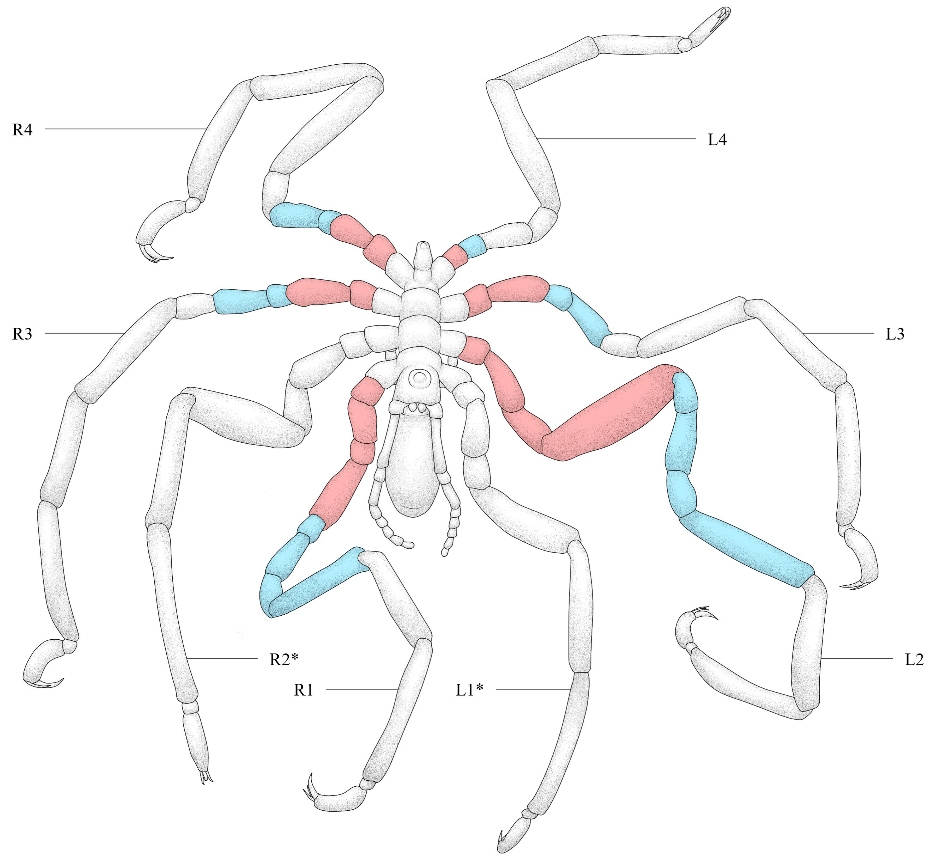
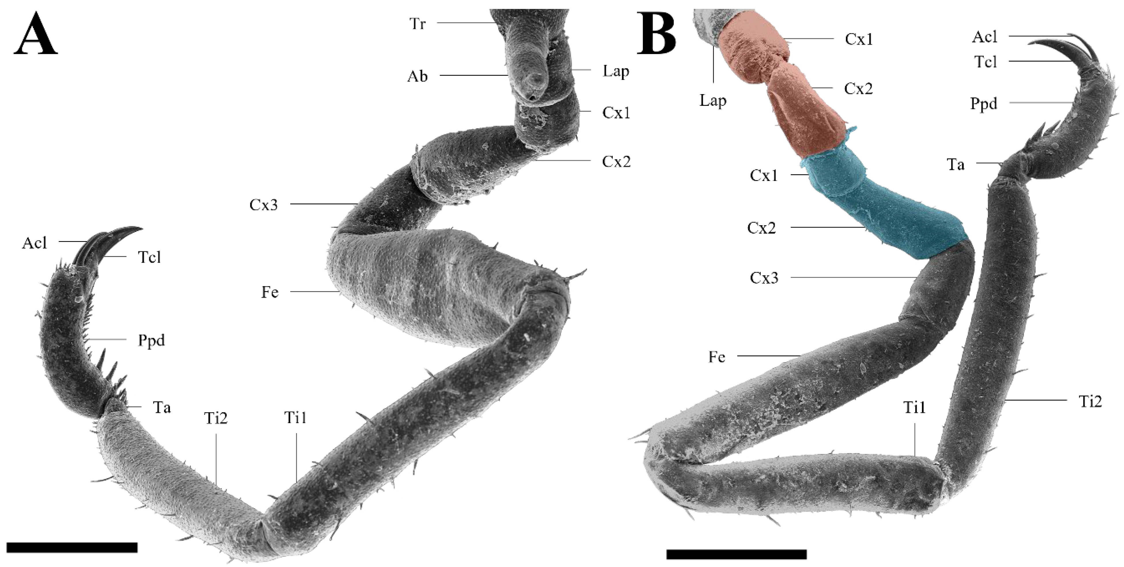
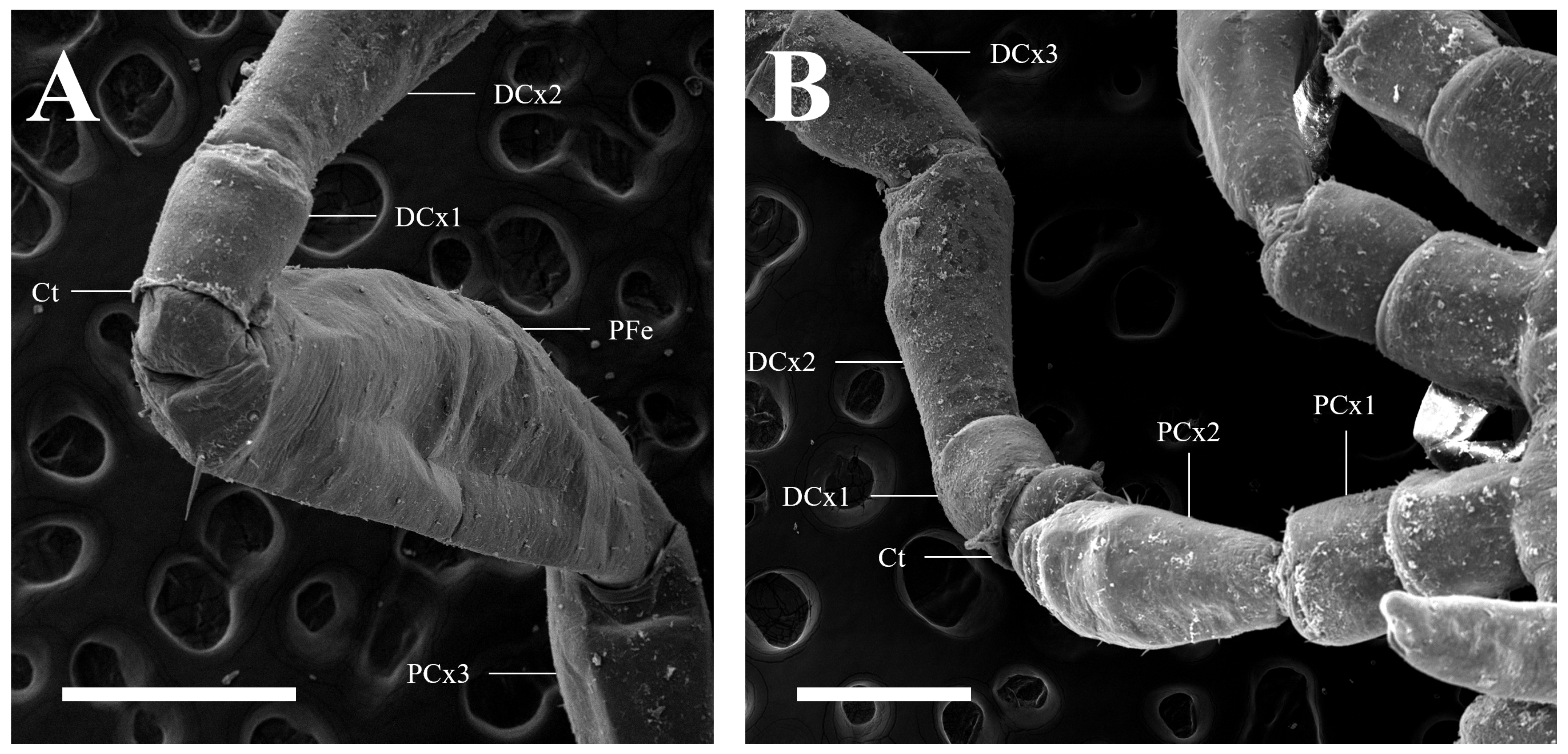
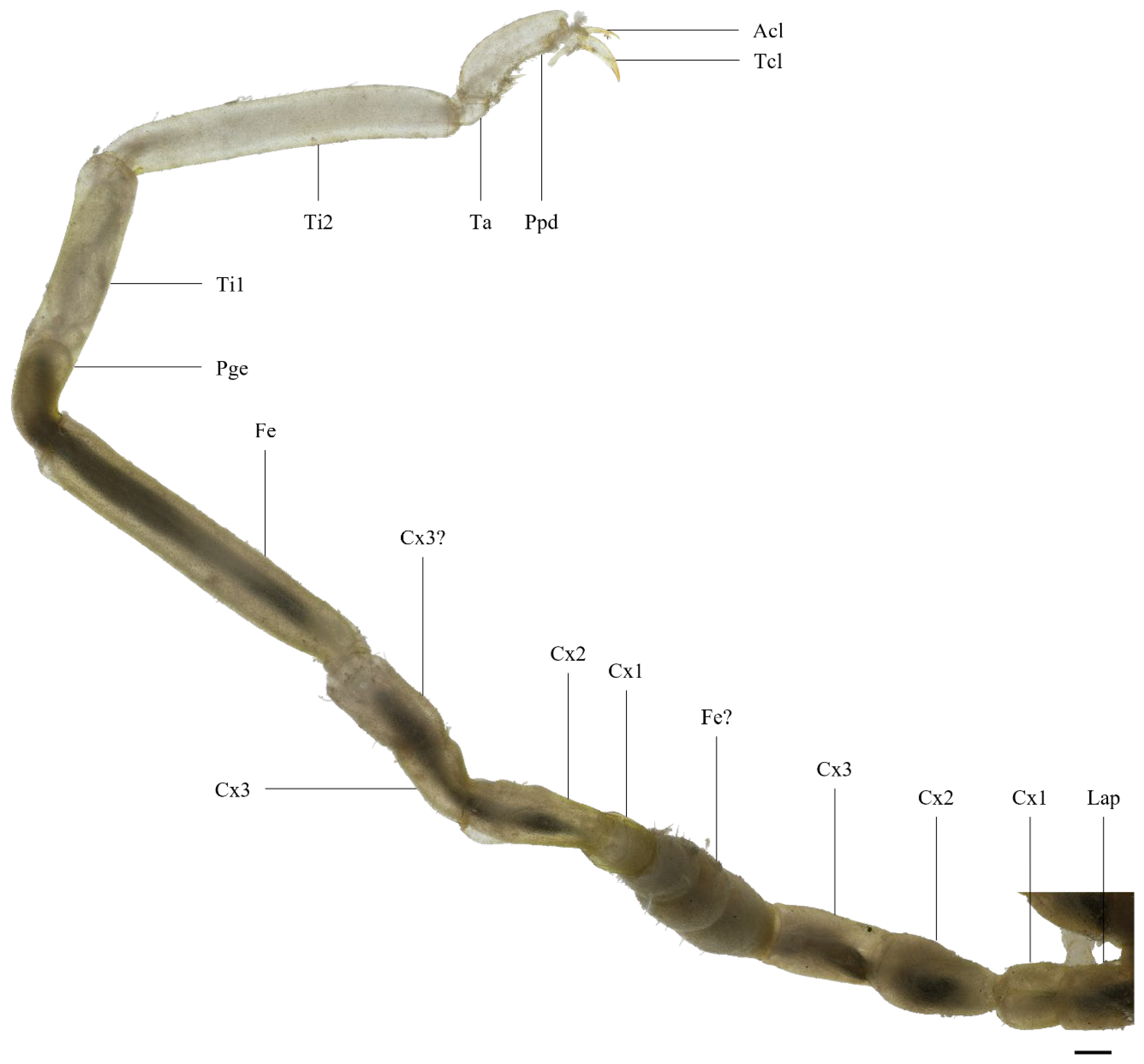
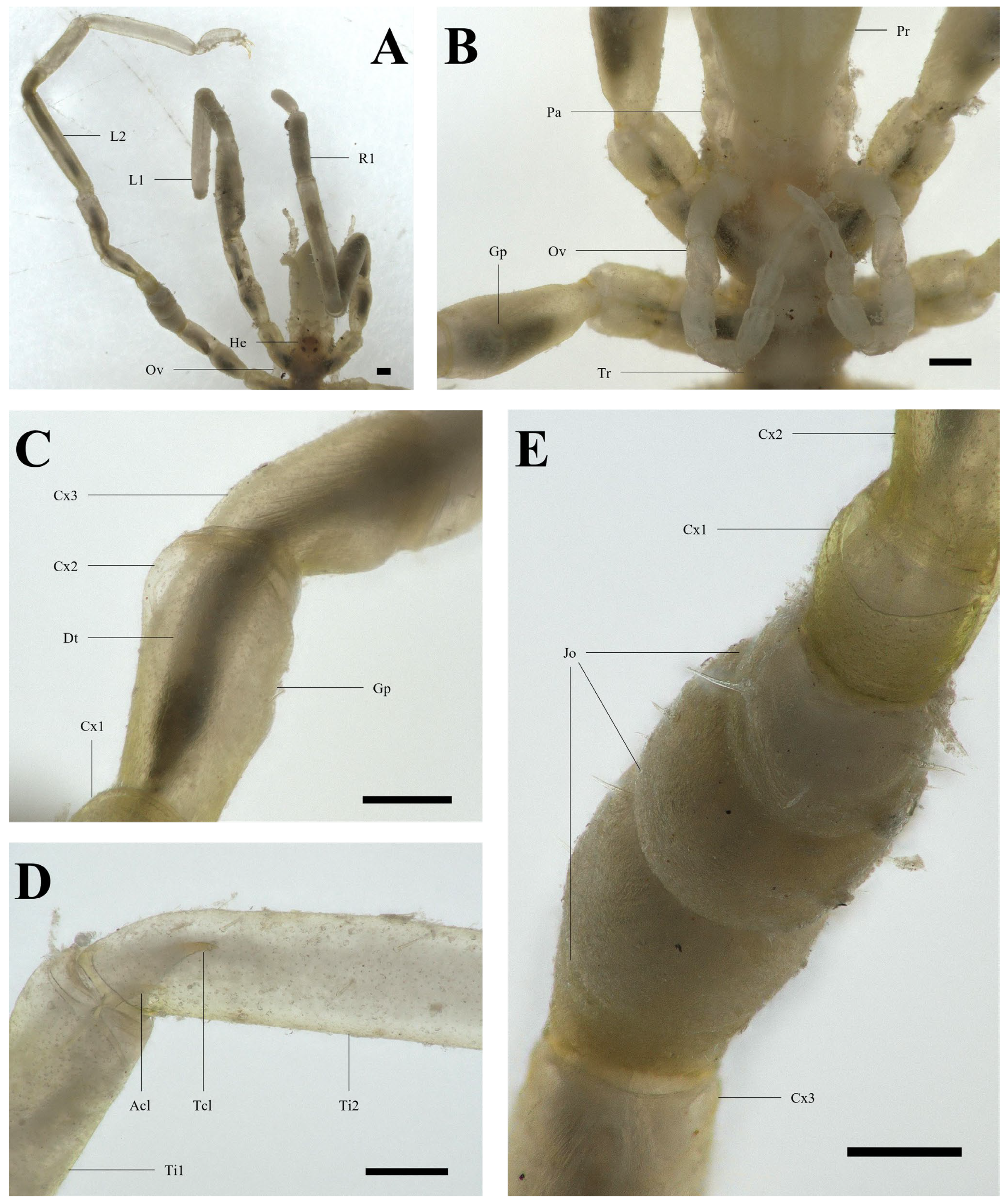
3.1. Malformed Individual 1 (MI1)

The malformation externally appeared as an extra set of segments in six of the eight legs (Figure 2), showing between 1 and 4 additional segments per leg (Table 1). An example of an abnormal leg can be seen in Figure 3. While no significance was found between the length of both segment copies (p = 0.49), the width of the distal copies was significantly smaller than the proximal ones (p = 0.0084).
The additional segments were observed to be duplicated as groups, rather than directly following their counterparts, as, for instance, in the third left leg where the order was [Cx1-Cx2]-[Cx1-Cx2] instead of [Cx1]-[Cx1]-[Cx2]-[Cx2] (i.e., LSTD). This was verified by coxae 2 being longer than coxae 1 in the normal and abnormal legs (Table 2). SEM photographs revealed cuticle tearing at the base of all distal copies of coxae 1 (Figure 4), reminding us of the proximal part of the leg exuviae being shed like a sleeve in P. litorale and N. gracile [17,19]. Gonopores, typically found ventrally on each of the second coxae in A. hilgendorfi [11], were, however, absent in the distal copies.
It can be seen on Video S1 that the distal ends of abnormal legs look white and empty. The video also shows that, despite being able to move its limbs independently, MI1 seemed unable to coordinate its movements. The terminal claws were disorientated: instead of grasping substrates below the body, these grasped substrates positioned above the animal.

3.2. Malformed Individual 2 (MI2)

MI2 showed only one aberrant limb (Figure 5 and Figure 6A), yet the anomaly was much deeper. At least all three coxae externally appeared as duplicated in tandem once, with the presence of two gonopores confirming the position of both coxae 2 (Figure 6B,C). However, four additional anomalies were noted on that single leg. First, a putative third coxa grew after yet another coxa 3 (see “Cx3?” in Figure 5). Its dimensions disprove the possibility of a coxa 1, which are much smaller, and it did not have a gonopore, unlike the other coxae 2 of that leg. Second, right after the proximal coxae was noted an unusual segment (see “Fe?” in Figure 5). Although its position hinted at a femur, it did not look like a femur or any normal segments whatsoever. It was, indeed, more opaque, the digestive tract being barely visible through it. In fact, its constricted appearance was similar to what was noted in [48] (see their Figure 4 and Figure 5), which is an elongated regenerate with joints between future segments (Figure 6E). Third, the yellowish cuticle pigmentation suddenly stopped at the beginning of tibia 1, with a stump-shaped structure inside the leg itself (see “Pge” in Figure 5). Beyond that, tibia 1 contained structures that looked like the regenerating legs observed in [48] (see their Figure 11). The following segments seemed empty, as if they only consisted of external cuticle. Fourth, a terminal and an auxiliary claw (Figure 6D) were visible between both tibias, inside them. The claws at the distal end of the leg, on the other hand, did not seem functional as they were almost falling off the propodus (Figure 5).
Video S1 shows that, unlike MI1, MI2 behaved normally, using all its legs to walk except for the abnormal one. It looked unable to move the abnormal limb actively, which remained immobile above the others. Still, that did not incapacitate the whole animal’s movements, and it could walk just as if the malformed leg was missing.
In [63], only two malformed A. hilgendorfi were detected among 1144 individuals, suggesting an anomaly prevalence rate of ~0.2% in the Belgian population of that species.

4. Discussion

An anomaly identical to the observed LSTD, or the four others described in MI2, could not be found in the literature regardless of the taxon considered, whether it be pycnogonids, chelicerates, or the entire arthropod phylum. In fact, many anomalies described in the literature concern entire legs and are rarely limited to a few segments (e.g., Heteroptera missing a femur and/or tibia [34]).
However, this phenomenon reminds us of the molting process of other sea spiders, in which the leg cuticle is cast like a sleeve [17,19]. The cuticle tearing noticed in Figure 4 would then be the proximal end of the exuviae being removed, while the transparent distal end in Video S1 would be due to them being moved beyond the internal organs. This is emphasized by MI1 being identified first as a normal juvenile and then as an abnormal adult, suggesting that the anomaly arises from a molt. The proximal copies of the duplicated podomeres in MI1 were found to be significantly wider, which makes sense considering that they are the newly formed podomeres and that pycnogonids become bigger after molting [19]. Plus, it was identified in several sea spider species that gonopores appear at the adult stage [66]. From the examination of several last-stage juveniles, this seems to be the case in A. hilgendorfi as well. This is consistent with the fact that the distal copies of coxae 2 did not feature gonopores, because they would have been contemporary to the last pre-adult stage.
Although the life history of MI2 was impossible to trace, the empty distal end of its malformed leg points to the same cause. The claws visible through the cuticle (Figure 6D) would be the newly formed ones, while the claws almost falling off at the top would be old cuticle, removed during a normal molt.
Even if the anomaly likely results from a molt, it is improbable that the observed phenomenon is usual and expected in every A. hilgendorfi. Unfortunately, the molting process of sea spiders has been poorly studied in the literature, making comparisons with normal individuals difficult. However, the specimens were observed for 24 h, and neither their anomaly nor ethology changed, despite their access to a proper substrate to lock their leg exuviae on. Since many empty leg cuticles were found at the bottom of aquariums in which other A. hilgendorfi not showing any anomalies were maintained, this species likely molts quickly. As the ovigerous legs of MI1 had already molted, and its two legs looked normal, the observed LSTD probably arose from the inability of the specimen to completely cast its old cuticle during the molting process. In fact, the state in which MI1 was found might resemble what happens during an expected regular pycnogonid molt. Therefore, while the malformation cannot be considered a “podomere duplication” sensu stricto, as it is only exuviae, it behaves effectively as if this was the case. Indeed, although the locomotion of MI1’s legs was not altered independently, the locomotion of the whole animal was compromised. Such a phenomenon occurring in the wild would likely result in a fitness close to zero (i.e., low survival). Regarding MI2, its only malformed leg did not seem to impact it, similarly to how autotomized specimens are viable in the wild [49].
LSTD was not the only anomaly in MI2. The abnormal putative femur reminds us of a regenerating and constricted segment [48], hinting at the incapacity of this leg to molt completely in the first place, that maybe even occurred during an anterior molt. The putative coxa 3, right after another coxa 3, might as well translate to an incompletely removed cuticle sleeve in a molt previous to the last one. Petrova and Bogomolova (2023) observed that regenerating legs fold inside amputation stumps [48], which might match the structures inside the first tibia of MI2, although external morphology analyses alone cannot assess them with certitude. The doubled gonopores also raise questions about both the anomaly origin and the ontogenetic development of A. hilgendorfi. As mentioned above, A. hilgendorfi seem to grow a gonopore during their juvenile-to-adult molt. While adult P. litorale stop molting (i.e., terminal anecdysis) [67], either adult A. hilgendorfi do continue molting or the anomaly arises from the last molt mistakenly triggering twice in that leg. Further research on their life cycle is necessary to address these hypotheses.
It must be mentioned that the context in which this study took place was not in trying to spot malformed individuals. Both cases here described were extreme and noticed “by chance”. Thus, there may lie in the Belgian population of A. hilgendorfi more subtle leg irregularities which would have been overlooked here. The counted ~0.2% prevalence of abnormal individuals should then be considered a minimum.
That prevalence, although likely underestimated, remains in the range of malformation ratios noted for different arthropod groups. For example, Hesse-Honegger and Wallimann (2008) collected 16,000+ Heteroptera in several locations and noted morphological anomalies at a ratio down to 1–3% in undisturbed habitats, and up to 22% close to a nuclear reprocessing plant [34]. In crustaceans, Rady (2022) found in the Suez Canal 28/269 (~10.4%) Portunus segnis (Forskål, 1775) [68] crabs with a malformation in any body part [21]. In chelicerates, Palmgren (1979) counted a ratio of 1/17,000 (~0.006%) gynandromorph spiders (several species included) [45]. In pycnogonids, Bouvier (1914) investigated 3268 P. litorale, but found only one abnormal specimen (~0.03%) [52]. On the other hand, Lotz and Bückmann (1968) observed 22/67 P. litorale correctly molting at least once in a period of ten months (~33%) [19]. That ratio is much greater than the 0.2% of the present study, which is another hint at the fact that MI1 and MI2 were abnormal.
The alien trait of A. hilgendorfi should not be neglected. Indeed, the introduction of a new species generally results in genetic bottlenecks in the colonized area, resulting in more homozygotes in the alien population [69]. This is associated with their lower fitness [70], and homozygosity has been linked many times to pathologic phenotypes (e.g., [71,72,73]). If that is so, the prevalence of leg anomalies—or any other malformations—would likely be higher in non-native populations of A. hilgendorfi. A study specifically looking for all malformations in several A. hilgendorfi populations (i.e., native vs. non-native) would be of interest to assess the consequences of its introduction for the species itself.
Finally, nothing is known about their molt dynamics (i.e., time required, different steps, etc.). Understanding how pycnogonids molt would not only help our understanding of how the abnormal patterns appeared but also put pycnogonids into perspective with other arthropods, a phylum in which they show a key taxonomic position.

Supplementary Materials

The following supporting information can be downloaded at https://www.mdpi.com/article/10.3390/arthropoda2020012/s1, Video S1: Behavior of the malformed specimens.

Author Contributions

A.F. and L.S. collected and analyzed the specimens. A.F., L.S. and G.C. wrote this paper. G.C. supervised the work. All authors have read and agreed to the published version of the manuscript.

Funding

This research was funded by the Fund for Scientific Research (F.R.S.–FNRS), FRIA grant number 40015401.

Institutional Review Board Statement

The animals used in our experiments were maintained and treated in compliance with the guidelines specified by the Belgian Ministry of Trade and Agriculture.

Data Availability Statement

The data used and/or analyzed during the current study are available from the corresponding author on reasonable request.

Acknowledgments

This work is part of A.F.’s doctoral thesis, supported by a FRIA grant (F.R.S-FNRS) and realized at the University of Mons (UMONS), Belgium. L.S. has a master in biology and is an active collaborator with the laboratory of Biology of Marine Organisms and Biomimetics (BOMB) from UMONS. G.C. is a research and teaching associate in UMONS. The authors deeply thank the two reviewers that helped them interpret the results of this study.

Conflicts of Interest

The authors declare that they have no conflicts of interest.

References

  1. Bamber, R.; El Nagar, A.; Arango, C. Pycnobase: World Pycnogonida Database. Available online: https://www.marinespecies.org/pycnobase (accessed on 28 November 2023).
  2. Arnaud, F.; Bamber, R.N. The Biology of Pycnogonida. In Advances in Marine Biology; Blaxter, J.H.S., Southward, A.J., Eds.; Academic Press: Cambridge, MA, USA, 1988; Volume 24, pp. 1–96. [Google Scholar]
  3. Calman, W.T.; Gordon, I. A dodecapodous pycnogonid. Proc. R. Soc. London. Ser. B Contain. Pap. A Biol. Character 1933, 113, 107–115. [Google Scholar] [CrossRef]
  4. Kühl, G.; Poschmann, M.; Rust, J. A ten-legged sea spider (Arthropoda: Pycnogonida) from the Lower Devonian Hunsrück Slate (Germany). Geol. Mag. 2013, 150, 556–564. [Google Scholar] [CrossRef]
  5. Ewing, H.E. The legs and leg-bearing segments of some primitive arthropod groups, with notes on leg-segmentation in the Arachnida. In Smithsonian Miscellaneous Collections; The Smithsonian Institution: Washington, DC, USA, 1928. [Google Scholar]
  6. Cole, L.J. Notes on the habits of pycnogonids. Biol. Bull. 1901, 2, 195–207. [Google Scholar] [CrossRef]
  7. Fahrenbach, W.H.; Arango, C.P. Microscopic anatomy of Pycnogonida: II. Digestive system. III. Excretory system. J. Morphol. 2007, 268, 917–935. [Google Scholar] [CrossRef] [PubMed]
  8. Miyazaki, K.; Makioka, T. Structure of the adult female reproductive system and oogenetic mode in the sea spider, Endeis nodosa (Pycnogonida; Endeidae). J. Morphol. 1991, 209, 257–263. [Google Scholar] [CrossRef] [PubMed]
  9. Miyazaki, K. Structure of the Adult Male Reproductive System in a Pycnogonid, Cilunculus armatus (Pycnogonida: Ammotheidae). Publ. Seto Mar. Biol. Lab. 1996, 37, 329–335. [Google Scholar] [CrossRef]
  10. Bain, B.A.; Govedich, F.R. Courtship and mating behavior in the Pycnogonida (Chelicerata: Class Pycnogonida): A summary. Invertebr. Reprod. Dev. 2004, 46, 63–79. [Google Scholar] [CrossRef]
  11. Arango, C.P.; Wheeler, W.C. Phylogeny of the sea spiders (Arthropoda, Pycnogonida) based on direct optimization of six loci and morphology. Cladistics 2007, 23, 255–293. [Google Scholar] [CrossRef] [PubMed]
  12. Tallamy, D.W. Sexual selection and the evolution of exclusive paternal care in arthropods. Anim. Behav. 2000, 60, 559–567. [Google Scholar] [CrossRef] [PubMed]
  13. Fahrenbach, W.H. Microscopic anatomy of Pycnogonida: I. Cuticle, epidermis, and muscle. J. Morphol. 1994, 222, 33–48. [Google Scholar] [CrossRef] [PubMed]
  14. Nakamura, K. Post embryonic development of a pycnogonid, Propallene longiceps. J. Nat. Hist. 1981, 15, 49–62. [Google Scholar] [CrossRef]
  15. Brenneis, G.; Bogomolova, E.V.; Arango, C.P.; Krapp, F. From egg to “no-body”: An overview and revision of developmental pathways in the ancient arthropod lineage Pycnogonida. Front. Zool. 2017, 14, 6. [Google Scholar] [CrossRef] [PubMed]
  16. Leach, W.E. The Zoological Miscellany: Being Descriptions of New or Interesting Animals; E. Nodder and Son: London, UK, 1814; Volume 1. [Google Scholar]
  17. King, P.E. Pycnogonids; Hutchinson: London, UK, 1973. [Google Scholar]
  18. Strøm, H. Physisk og Oeconomisk Beskrivelse over Fogderiet Sondmor, Beligende i Bergens Stift i Norge; Forste Part: Soroe, Denmark, 1762; pp. 1–570. [Google Scholar]
  19. Lotz, G.; Bückmann, D. Die Häutung und die Exuvie von Pycnogonum litorale (Ström)(Pantopoda). Zool. Jahrbücher Abt. Anat. Ontogonie Der Tiere 1968, 85, 529–536. [Google Scholar]
  20. Rudkin, D.M.; Cuggy, M.B.; Young, G.A.; Thompson, D.P. An Ordovician Pycnogonid (Sea Spider) with Serially Subdivided ‘Head’ Region. J. Paleontol. 2013, 87, 395–405. [Google Scholar] [CrossRef]
  21. Rady, A. First record of morphological malformations in the blue swimming crab, Portunus segnis (Portunidae: Brachyura) from the Timsah Lake, Suez Canal, Egypt. Egypt. J. Aquat. Biol. Fish. 2022, 26, 877–888. [Google Scholar] [CrossRef]
  22. Stojanović, D.Z.; Mitić, B.M.; Makarov, S.E. Bifurcation of an ultimate leg in Cryptops parisi Brolemann, 1920 (Chilopoda: Scolopendromorpha: Cryptopidae). Arthsel 2019, 28, 21–25. [Google Scholar] [CrossRef]
  23. Templin, J.; Jacuñski, L.; Napiórkowska, T. Metameric Malformations of Opisthosoma in Tegenaria atrica (Araneae, Agelenidae). Zool. Pol. 2009, 54–55, 33–42. [Google Scholar] [CrossRef]
  24. Schimkewitsch, W.; Dogiel, V. Ueber Regeneration bei Pantopoden. Bull. Acad. Imp. Sci. St. Petersbourg 6 Ser. 1913, 7, 1147–1156. [Google Scholar]
  25. Babcock, L.E. Trilobite malformations and the fossil record of behavioral asymmetry. J. Paleontol. 1993, 67, 217–229. [Google Scholar] [CrossRef]
  26. Bicknell, R.D.C.; Smith, P.M.; Bruthansová, J.; Holland, B. Malformed trilobites from the Ordovician and Devonian. PalZ 2022, 96, 1–10. [Google Scholar] [CrossRef]
  27. Maruzzo, D.; Bonato, L.; Brena, C.; Fusco, G.; Minelli, A. Appendage Loss and Regeneration in Arthropods: A Comparative View; CRC Press: Boca Raton, FL, USA, 2005. [Google Scholar]
  28. Bateson, W. Materials for the Study of Variation: Treated with Especial Regard to Discontinuity in the Origin of Species; Macmillan: London, UK, 1894. [Google Scholar]
  29. Gadeau de Kerville, H. Sur la furcation tératologique des pattes, des antennes et des palpes chez les Insectes. Bull. La. Société Entomol. Fr. 1898, 3, 93–95. [Google Scholar] [CrossRef]
  30. Vitali, F. Anomalies multiples chez un exemplaire tératologique d’Acanthinodera cumingii (Hope, 1833) (Coleoptera Cerambycidae). L’Entomologiste 2007, 63, 79–80. [Google Scholar]
  31. Román, E.V.; Marchant, M.; Sánchez, I. A new symphysomely case for Cryptops Leach, 1815 (Scolopendromorpha: Cryptopidae) in Chile. Rev. Colomb. Entomol. 2022, 48. [Google Scholar]
  32. Scholtz, G.; Ng, P.K.L.; Moore, S. A crab with three eyes, two rostra, and a dorsal antenna-like structure. Arthropod Struct. Dev. 2014, 43, 163–173. [Google Scholar] [CrossRef] [PubMed]
  33. Anger, K.; Harzsch, S.; Thiel, M. Developmental Biology and Larval Ecology: The Natural History of the Crustacea; Oxford University Press: Oxford, UK, 2020; Volume 7. [Google Scholar]
  34. Hesse-Honegger, C.; Wallimann, P. Malformation of True Bug (Heteroptera): A Phenotype Field Study on the Possible Influence of Artificial Low-Level Radioactivity. Chem. Biodivers. 2008, 5, 499–539. [Google Scholar]
  35. David, D. A seven-legged scorpion: The first teratological leg absence found in Scorpio maurus fuscus (Scorpiones: Scorpionidae). Euscorpius 2015, 2012, 1–4. [Google Scholar] [CrossRef]
  36. Napiórkowska, T.; Templin, J.; Napiórkowski, P. The Central Nervous System of Heterosymelic Individuals of the Spider Tegenaria atrica. Folia Biol. 2013, 61, 283–289. [Google Scholar] [CrossRef] [PubMed]
  37. Napiórkowska, T.; Napiórkowski, P.; Templin, J. Morphological and anatomical changes related to leg anomalies in Tegenaria atrica. Zoomorphology 2015, 134, 237–245. [Google Scholar] [CrossRef] [PubMed]
  38. Napiórkowska, T.; Templin, J.; Wołczuk, K. Morphology and the central nervous system of Eratigena atrica affected by a complex anomaly in the anterior part of the prosoma. Invert. Neurosci. 2017, 17, 11. [Google Scholar] [CrossRef] [PubMed]
  39. Napiórkowska, T.; Templin, J. Heterosymely and Accompanying Anomalies in the Spider Eratigena atrica (C. L. Koch, 1843) (Araneae: Agelenidae). Ann. Zool. 2018, 68, 909–914. [Google Scholar] [CrossRef]
  40. Juberthie, P.C. Tératologie expérimentale chez un Opilion (Arachnide). Development 1968, 19, 49–82. [Google Scholar] [CrossRef]
  41. Mikulska, I. Experimentally induced developmental monstrosities in the water spider Argyoneta aquatica (Clerck). Zool. Pol. 1973, 22, 127–134. [Google Scholar]
  42. Kaston, B.J. Abnormal Duplication of the Epigynum and Other Structural, Anomalies in Spiders. Trans. Am. Microsc. Soc. 1963, 82, 220–223. [Google Scholar] [CrossRef]
  43. Kaston, B.J. Deformities of External Genitalia in Spiders. J. N. Y. Entomol. Soc. 1963, 71, 30–39. [Google Scholar]
  44. Kaston, B.J. Spider Gynandromorphs and Intersexes. J. N. Y. Entomol. Soc. 1961, 69, 177–190. [Google Scholar]
  45. Palmgren, P. On the frequency of gynandromorphic spiders. Ann. Zool. Fenn. 1979, 16, 183–185. [Google Scholar]
  46. Baba, Y.G.; Suguro, T.; Naya, N.; Yamauchi, T. A gynandromorph of the funnel-web spider Allagelena Opulenta (Araneae: Agelenidae). Acta Arachnol. 2016, 65, 11–13. [Google Scholar] [CrossRef]
  47. Yaginuma, T. A colour anomaly in the spider Heteropoda venatoria (Linné) from Japan. Acta Arachnol. 1971, 23, 21–22. [Google Scholar] [CrossRef]
  48. Petrova, M.; Bogomolova, E. Walking leg regeneration in the sea spider Nymphon brevirostre Hodge, 1863 (Pycnogonida). Arthropod Struct. Dev. 2023, 77, 101310. [Google Scholar] [CrossRef]
  49. Fornshell, J.A. Walking leg regeneration observed in three families and four species of antarctic sea spiders. Arthropods 2019, 8, 110. [Google Scholar]
  50. Legakis, A. Chelicerata. European register of marine species: A check-list of the marine species in Europe and a bibliography of guides to their identification. In Collection Patrimoines Naturels; Costello, M.J., Emblow, C.S., White, R.J., Eds.; Muséum National d’Histoire Naturelle: Paris, France, 2001; Volume 50, p. 237. [Google Scholar]
  51. Ohshima, H. Six-Legged Pantopod, an Extraordinary Case of Hypomery in Arthropods. Proc. Imp. Acad. 1942, 18, 257–262. [Google Scholar] [CrossRef]
  52. Bouvier, M.E.-L. Quelques mots sur la variabilité du Pycnogonum litorale, Ström. J. Mar. Biol. Assoc. United Kingd. 1914, 10, 207–210. [Google Scholar] [CrossRef]
  53. Brenneis, G.; Frankowski, K.; Maaß, L.; Scholtz, G. The sea spider Pycnogonum litorale overturns the paradigm of the absence of axial regeneration in molting animals. Proc. Natl. Acad. Sci. USA 2023, 120, e2217272120. [Google Scholar] [CrossRef] [PubMed]
  54. Loeb, J. Studies in General Physiology; University of Chicago Press: Chicago, IL, USA, 1905; Volume 23, pp. 742–743. [Google Scholar]
  55. Scholtz, G.; Brenneis, G. The pattern of a specimen of Pycnogonum litorale (Arthropoda, Pycnogonida) with a supernumerary leg can be explained with the “boundary model” of appendage formation. Sci. Nat. 2016, 103, 13. [Google Scholar] [CrossRef] [PubMed]
  56. Ohshima, H. A Remarkable Case of Malformed Appendages in a Pantopod. Nymphonella tapetis. Proc. Imp. Acad. 1942, 18, 520–523. [Google Scholar] [CrossRef]
  57. Brenneis, G.; Scholtz, G. A postlarval instar of Phoxichilidium femoratum (Pycnogonida, Phoxichilidiidae) with an exceptional malformation. J. Morphol. 2021, 282, 278–290. [Google Scholar] [CrossRef] [PubMed]
  58. Fabricius, O. Fauna Groenlandica, Systematice Sistens Animalia Groenlandiae Occidentalis Hactenus Indagata, Quoad Nomen Specificium, Triviale, Vernaculumque, Synonyma Auctorum Plurimum, Descriptionem, Locum, Victum, Generationem, Mores, Usum Capturamque Singuli, Pro ut Detegendi Occasio Fuit, Maximaque Parte Secundum Proprias Bbservationes. Hafniae [= Copenhagen], Lipsiae [= Leipzig], Ioannis Gottlob Rothe. xvi + 452 pp., 1 pl (1780). Available online: https://www.biodiversitylibrary.org/page/13442285#page/7/ (accessed on 23 May 2024).
  59. Child, C.A.; Nakamura, K. A gynandromorph of the Japanese pycnogonid Anoplodactylus gestiens (Ortmann). Proc. Biol. Soc. Wash. 1982, 95, 292–296. [Google Scholar]
  60. Lucena, R.; Araújo, J.; Christoffersen, M. A new species of Anoplodactylus (Pycnogonida: Phoxichilidiidae) from Brazil, with a case of gynandromorphism in Anoplodactylus eroticus Stock, 1968. Zootaxa 2015, 4000, 428–444. [Google Scholar] [CrossRef] [PubMed]
  61. Le Roux, A.; Gélinaud, G.; Le Bail, Y.; Monnat, J.Y.; Morel, J.Y.; Paraire, O.; Ros, J. Occurrence of Ammothea hilgendorfi (Böhm, 1879) a pycnogonid from the north Pacific, in Étel river. Aod Les Cah. Nat. De L’observatoire Mar. 2020, 8, 21–32. [Google Scholar]
  62. Bamber, R.N. Anthropogenic spread of the immigrant sea-spider Ammothea hilgendorfi (Arthropoda: Pycnogonida: Ammotheidae) in UK waters. Mar. Biodivers. Rec. 2012, 5, e78. [Google Scholar] [CrossRef]
  63. Flandroit, A.; Caulier, G. Life Cycle, Phototaxis, Chemotaxis of the Invasive Pycnogonid Ammothea hilgendorfi; University of Mons: Mons, Belgium, 2021. [Google Scholar]
  64. Salvi, D.; Mariottini, P. Molecular taxonomy in 2D: A novel ITS2 rRNA sequence-structure approach guides the description of the oysters’ subfamily Saccostreinae and the genus Magallana (Bivalvia: Ostreidae). Zool. J. Linn. Soc. 2017, 179, 263–276. [Google Scholar] [CrossRef]
  65. Nakamura, K.; Fujita, T. Ammothea hilgendorfi (Pycnogonida: Ammotheidae) Associated with a Sea-Star, Coscinasterias acutispina (Echinodermata: Asteroidea), from Sagami Bay, Japan. Species Divers. 2004, 9, 251–258. [Google Scholar] [CrossRef]
  66. Bain, B.A. Larval types and a summary of postembryonic development within the pycnogonids. Invertebr. Reprod. Dev. 2003, 43, 193–222. [Google Scholar] [CrossRef]
  67. Tomaschko, K.H.; Wilhelm, E.; Bückmann, D. Growth and reproduction of Pycnogonum litorale (Pycnogonida) under laboratory conditions. Mar. Biol. 1997, 129, 595–600. [Google Scholar] [CrossRef]
  68. Lai, J.C.Y.; Ng, P.K.L.; Davie, P.J.F. A revision of the Portunus pelagicus (Linnaeus, 1758) species complex (Crustacea: Brachyura: Portunidae), with the recognition of four species. Raffles Bull. Zool. 2010, 58, 199–237. [Google Scholar]
  69. Puillandre, N.; Dupas, S.; Dangles, O.; Zeddam, J.-L.; Capdevielle-Dulac, C.; Barbin, K.; Torres-Leguizamon, M.; Silvain, J.-F. Genetic bottleneck in invasive species: The potato tuber moth adds to the list. Biol. Invasions 2008, 10, 319–333. [Google Scholar] [CrossRef]
  70. Reed, D.H.; Frankham, R. Correlation between Fitness and Genetic Diversity. Conserv. Biol. 2003, 17, 230–237. [Google Scholar] [CrossRef]
  71. Ingalls, A.M.; Dickie, M.M.; Shell, G.D. Obese, a new mutation in the house mouse. J. Hered. 1950, 41, 317–318. [Google Scholar] [CrossRef] [PubMed]
  72. Pauli, R.M. Dominance and homozygosity in man. Am. J. Med. Genet. 1983, 16, 455–458. [Google Scholar] [CrossRef] [PubMed]
  73. Coman, D.J.; Gardner, R.M. Deletions Revealing Recessive Genes: Deletions that reveal recessive genes. Eur. J. Hum. Genet. 2007, 15, 1103–2007. [Google Scholar] [CrossRef] [PubMed]
Figure 1. Normal adult female of Ammothea hilgendorfi. Ab: abdomen; Acl: auxiliary claw; Au: autotomized leg (between coxa 1 and coxa 2); Ch: chelifore (atrophied in that species); Cx1: coxa 1; Cx2: coxa 2; Cx3: coxa 3; Eyt: eye tubercle; Fe: femur; He: head; Lap: lateral process; Ov: oviger (inserted ventrally); Pa: palp; Pr: proboscis; Ppd: propodus; Ta: tarsus; Tcl: terminal claw; Ti1: tibia 1; Ti2: tibia 2; Tr: trunk.
Arthropoda 02 00012 g001
Figure 2. Abnormal adult female of Ammothea hilgendorfi (MI1). L1: first left leg; L2: second left leg; L3: third left leg; L4: fourth left leg; R1: first right leg; R2: second right leg; R3: third right leg; R4: fourth right leg. Red: proximal copies of duplicated segments; blue: distal copies. The * indicate the two normal legs.
Arthropoda 02 00012 g002
Figure 3. Fourth right leg of adult female A. hilgendorfi under Scanning Electron Microscopy (SEM). (A) normal leg; (B) example of malformed leg. Ab: abdomen; Acl: auxiliary claw; Cx1: coxa 1; Cx2: coxa 2; Cx3: coxa 3; Fe: femur; Lap: lateral process; Ppd: propodus; Ta: tarsus; Tcl: terminal claw; Ti1: tibia 1; Ti2: tibia 2; Tr: trunk. Red: proximal copies of the duplicated segments; blue: distal copies. Scale bars: 1 mm.
Arthropoda 02 00012 g003
Figure 4. Second left leg (A) and third left leg (B) of MI1 observed in SEM. Ct: cuticle tearing; DCx1: distal coxa 1; DCx2: distal coxa 2; DCx3: distal coxa 3; PCx1: proximal coxa 1; PCx2: proximal coxa 2; PCx3: proximal coxa 3; PFe: proximal femur. Scale bars: 500 µm.
Arthropoda 02 00012 g004
Figure 5. Dorsal view of the abnormal leg (second left) of MI2 photographed with a Keyence VHX-970F digital microscope. Acl: auxiliary claw; Cx1: coxa 1; Cx2: coxa 2; Cx3: coxa 3; Cx3?: putative coxa 3; Fe: femur; Fe?: putative femur; Lap: lateral process; Pge: pigmentation end; Ppd: propodus; Ta: tarsus; Tcl: terminal claw; Ti1: tibia 1; Ti2: tibia 2. Magnification: X200. Scale bar: 0.25 mm.
Arthropoda 02 00012 g005
Figure 6. Features of the abnormal leg of MI2, photographed with a Keyence VHX-970F digital microscope. (A) Anterior dorsal view of the specimen. He: head; L1: first left leg (autotomized after tarsus); L2: second left leg (malformed), Ov: oviger; R1: first right leg. (B) Ventral view of the specimen and proximal leg segments. Gp: gonopore (ventrally on the second coxa); Ov: oviger; Pa: palp; Pr: proboscis; Tr: trunk. (C) Lateral view of the distal copy of the second coxa. Cx1: distal coxa 1; Cx2: distal coxa 2; Cx3: distal coxa 3; Dt: digestive tract; Gp: gonopore. (D) Ventral view of claws inside empty distal segments. Acl: auxiliary claw; Tcl: terminal claw; Ti1: tibia 1; Ti2: tibia 2. (E) Dorsal view of the putative femur. Cx1: distal copy of coxa 1; Cx2: distal copy of coxa 2; Cx3: proximal copy of coxa 3; Jo: putative joints of future segments appearing as constrictions. Magnifications: X30 in (A); X100 in (B); X200 in (CE). Scale bars: 0.25 mm.
Arthropoda 02 00012 g006
Table 1. Number of segments counted on each leg of MI1 and their names, based on their positions and similarities to eight-segmented legs. Each last coxa 1 per leg is highlighted in bold. The cell background of normal legs is highlighted in light grey.
1st2nd3rd4th5th6th7th8th9th10th11th12th
1st leftCoxa 1Coxa 2Coxa 3FemurTibia 1Tibia 2TarsusPropodus
2nd leftCoxa 1Coxa 2Coxa 3FemurCoxa 1Coxa 2Coxa 3FemurTibia 1Tibia 1TarsusPropodus
3rd leftCoxa 1Coxa 2Coxa 1Coxa 2Coxa 3FemurTibia 1Tibia 2TarsusPropodus
4th leftCoxa 1Coxa 1Coxa 2Coxa 3FemurTibia 1Tibia 2TarsusPropodus
1st rightCoxa 1Coxa 2Coxa 3FemurCoxa 1Coxa 2Coxa 3FemurTibia 1Tibia 2TarsusPropodus
2nd rightCoxa 1Coxa 2Coxa 3FemurTibia 1Tibia 2TarsusPropodus
3rd rightCoxa 1Coxa 2Coxa 1Coxa 2Coxa 3FemurTibia 1Tibia 2TarsusPropodus
4th rightCoxa 1Coxa 2Coxa 1Coxa 2Coxa 3FemurTibia 1Tibia 2TarsusPropodus
Table 2. Mean ± standard deviation of the length at the center, and the width at the largest point, of each segment of each leg in MI1 (in µm).
Coxa 1Coxa 2Coxa 3FemurTibia 1Tibia 2TarsusPropodus
N141310108888
Length381 ± 83845 ± 131545 ± 771982 ± 4392013 ± 3722382 ± 540197 ± 31838 ± 77
Width381 ± 34417 ± 39395 ± 29488 ± 113390 ± 25358 ± 55208 ± 16287 ± 36
Disclaimer/Publisher’s Note: The statements, opinions and data contained in all publications are solely those of the individual author(s) and contributor(s) and not of MDPI and/or the editor(s). MDPI and/or the editor(s) disclaim responsibility for any injury to people or property resulting from any ideas, methods, instructions or products referred to in the content.

