Article

Description of the Early Larval Development in Freshwater Shrimp Atya lanipes Holthuis, 1963 (Decapoda: Caridea: Atyidae) from Puerto Rico

by
Stefani Cruz-Rosa
*,
Ángel S. Estruche-Santos
and
Omar Pérez-Reyes
Department of Environmental Sciences, College of Natural Sciences, University of Puerto Rico, Rio Piedras Campus, San Juan 00925, Puerto Rico
*
Author to whom correspondence should be addressed.
Arthropoda 2024, 2(2), 169-180; https://doi.org/10.3390/arthropoda2020013
Submission received: 11 March 2024 / Revised: 4 June 2024 / Accepted: 5 June 2024 / Published: 11 June 2024

Abstract

:
The family Atyidae is composed of species whose existence has been known since the seventeenth century. Widely found in the Caribbean, Atya lanipes is a freshwater scraper/filter feeder shrimp with an amphidromous complex life cycle. Hunte (1975) described the first larval (zoeal) stage of the species. However, no scientific study has described the early larval development of this species after the first stage. This study aimed to document the early larval development of Atya lanipes under laboratory conditions and compare its larval development with other previously described species of the Atyidae family. Larval development was recorded by taking daily photos and videos of larval (zoeal) growth using a stereo microscope. Larvae were also preserved in ethanol for further morphological analysis. The results revealed that the best conditions for Atya lanipes development were 30 ppm water salinity, constant gentle aeration, and 27 °C water temperature. Nine stages were identified for the description of the early larval development of Atya lanipes. Early larval stages differ primarily in interstage larval size, the appearance and development of the telson, appendage appearance, growth of antennae and antennules, and pigmentation. The present contribution represents the first study that describes the larval development of the Caribbean shrimp Atya lanipes.

1. Introduction

The family Atyidae is composed of species whose existence has been known since the seventeenth century. According to Hobbs (1982), in Brazil, the first species of the family Atyidae, named “Guaricuru”, was described in 1648 by Marcgrave [1]. Subsequently, different species of this family, such as Atya scabra Leach 1816, were described [2]. In Puerto Rico, three species have been reported: Atya scabra Leach 1816, Atya innocous Herbst 1792 [3], and Atya lanipes Holthuis 1963 [4]. Atya lanipes is morphologically considered the most primitive shrimp in comparison with other atyid species of the island because it is found in microhabitats with fast but not turbulent currents and has limited geographical distribution in the Caribbean [5]. Atya scabra is the most evolutionary advanced species and is usually found in waterfalls and habitats with strong water currents. Finally, among the three species, Atya innocous has an intermediate level of complexity, and its habitat preferences include deeper waters with slow current flow or microhabitats with strong water currents. The species co-occurs with A. lanipes and A. scabra in the area and shares similarities in morphology, with slight taxonomical differences [1].
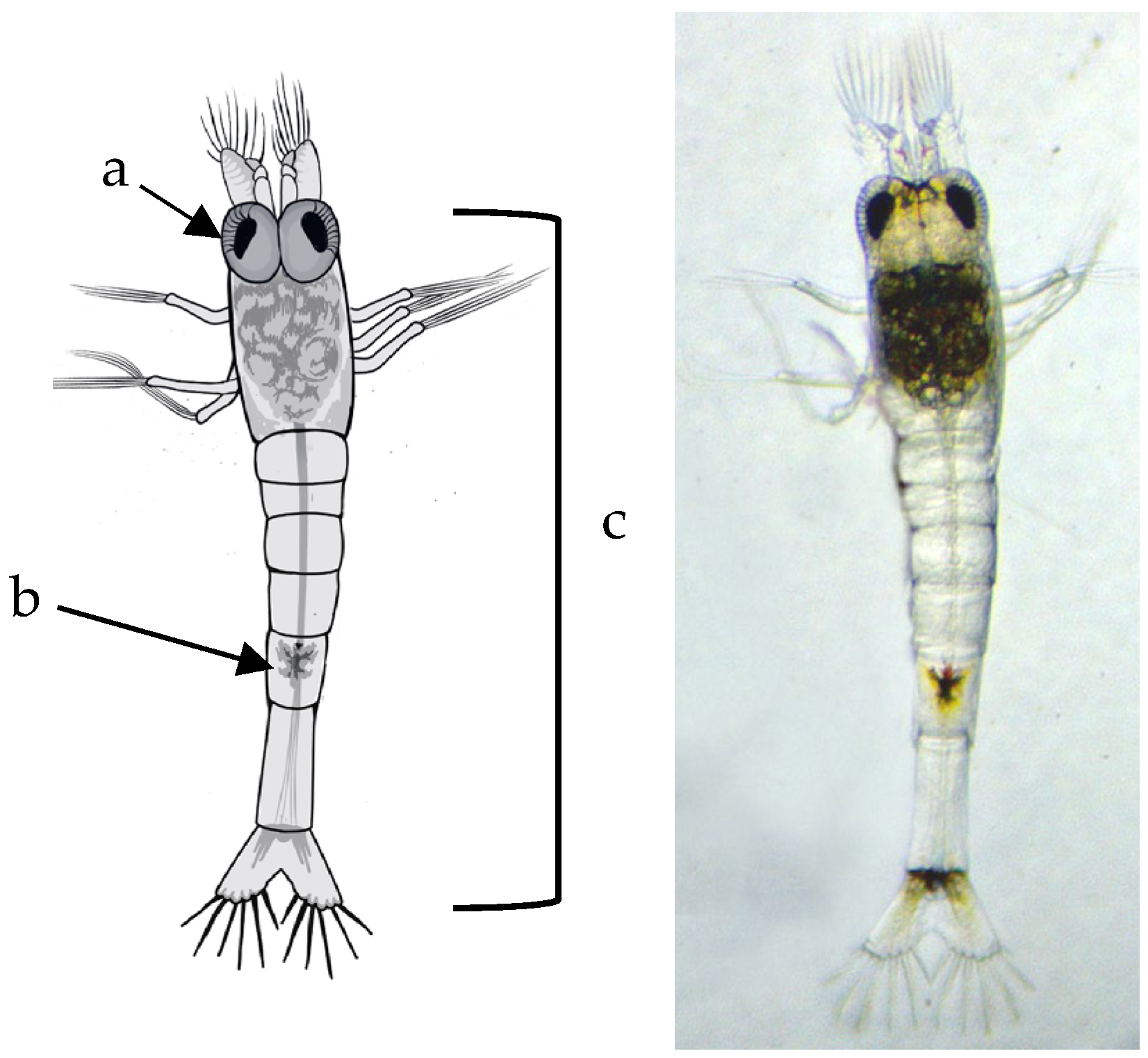
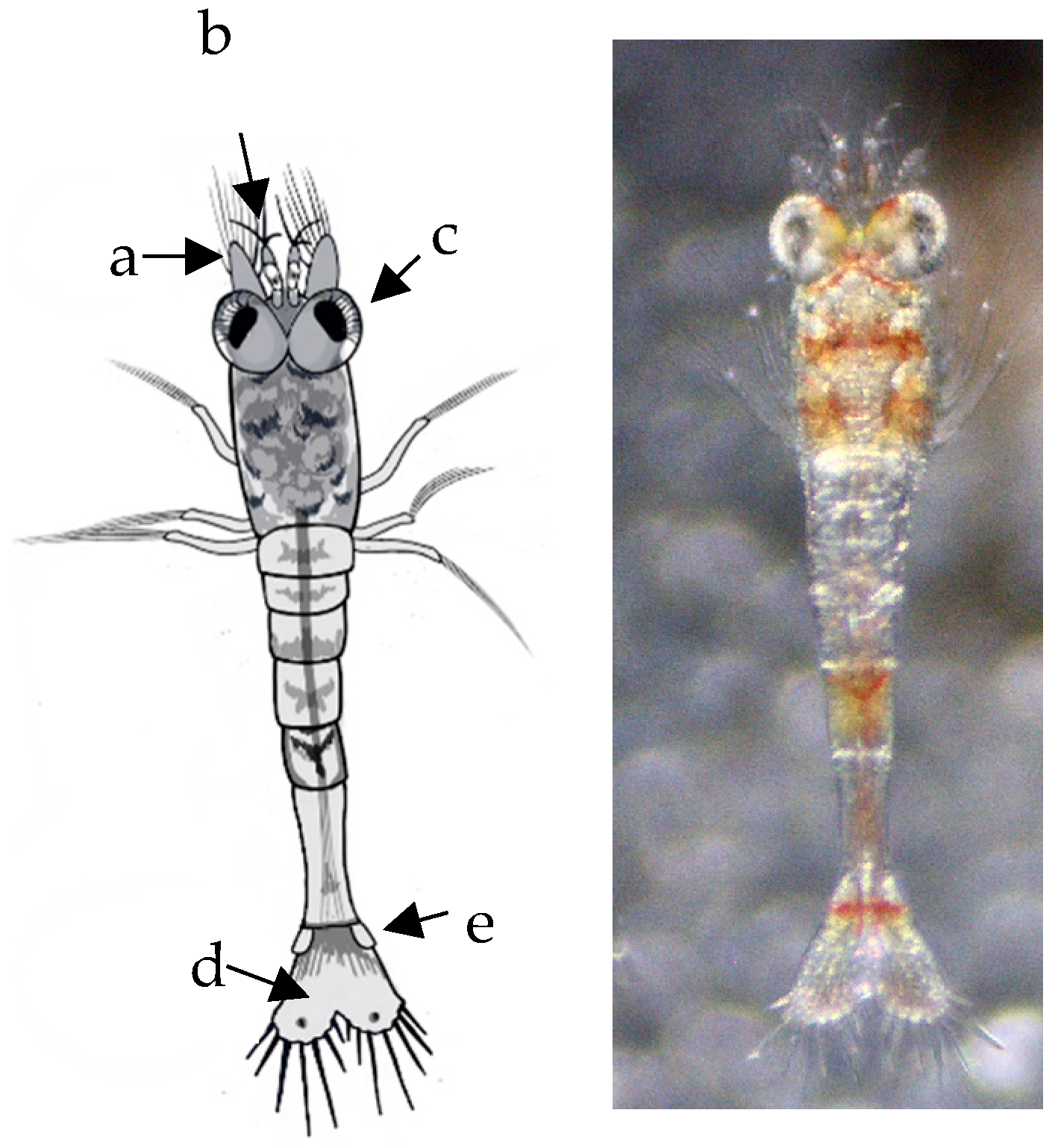
Taxonomically, the three species of Atya present in the Caribbean islands (A. scabra, A. innocous, and A. lanipes) are primarily (but not exclusively) distinguished by the shape and size of their pereiopods, the lateral lobules on the rostrum, and the denticles on the pleura of the abdomen. Pereiopods are the limbs that shrimp use to “walk”, differing from pleopods, whose function involves swimming and, in females, egg retention. In atyids, cheliped tips are equipped with tufts of denticulate setae like brushes (“long hair projections”) that allow the animal to filter the water column and scrape rock or organic-matter surfaces, capturing fine organic matter. Atya lanipes has slenderer pereiopods compared to Atya innocuous and A. scabra, which have stouter or more robust pereiopods. Morphologically, the merus of the third pereiopod of A. innocuous is broader than that of the fourth, and between 0.2 and 0.3 times as broad as it is long. In A. scabra, the merus, carpus, and propodus of the third pereiopod are significantly more inflated than the fourth; the merus is more than 0.3 times as broad as it is long. Taxonomically, the fourth pereiopods of Atya scabra are more robust or broader than those of A. innocuous and A. lanipes. However, other morphological characteristics differentiate Atya scabra from A. innocuous, such as the antennules’ peduncle, which is more prominent in A. innocuous than in A. scabra. The rostrum is also used to differentiate the three species of atyids mentioned above. Atya scabra presents subacute lateral lobules compared to A. innocuous with obtuse and Atya lanipes with no lateral lobules on the rostrum.
A functional morphology study conducted by Fryer (1977) in Dominica clearly described the characteristics of the chelae (second pair of pereiopods) [6]. Atyid species, such as Atya scabra and A. innocous, have modified cheliped fingers equipped with tufts of denticulate setae that allow them to filter the water column and capture fine organic matter suspended in the water or deposited on the rock surface or coarse organic matter in streams. Another mechanism in Atya lanipes and A. innocous involves scraping rocks or organic matter in streams to capture algae, bacteria, and detritus, their primary food source. This scraping behavior is also related to the capture and removal of insect larvae. All these modifications have been crucial for the colonization and survival of this species, making it a dominant species in the freshwater ecosystems of Puerto Rico and the Caribbean. For example, these species live mainly in microhabitats with fast and turbulent currents. Therefore, they need to feed and keep their eggs in the pleopods, among other life cycle activities, while coping with the physical force of currents and avoiding the effects of gravity on the slope. To accommodate these functions, chelae provide them with long hairbrushes, an evolutionary advantage allowing them to filter the water column and scrape the rock surface for effective feeding. The island of Puerto Rico has nine atyid species in rivers [7], which are vital for the removal and recycling of fine organic matter in freshwater ecosystems. In comparison to Atya lanipes and Atya innocous, Atya scabra primarily feeds by filtration. Their modified chelae facilitate efficient feeding on microscopic algae and fine organic matter. These species are significant for sediment recovery and play an important role in preventing stream eutrophication (accumulation of nutrients) [8].
The Atyidae family has an amphidromous life cycle, which highlights a significant relationship between river headwaters and estuarine/marine environments in freshwater shrimp [9,10,11]. First, gravid females release larvae in the upper reaches of the river (headwaters), after which larvae in their early stage of development (i.e., the zoeal stage) passively move to coastal/estuarine environments, where they undergo anamorphic growth. Subsequently, post-larvae migrate upstream as juveniles to complete their adult stage of development [11]. The larval stages of many of these atyids species have been described by different scientists under laboratory conditions (Table 1). The number of larval stages can vary among species of Atyidae, but it tends to be between at least seven and approximately twelve. An example is Atya innocuous, for which Hunte described twelve larval stages in 1979 (total development time: 76–119 days) [12]. Once these species complete their larval stages, they become juveniles (post-larva or megalopa), who undertake one of the most crucial migrations of their life cycle: to return to the river headwaters to complete their adulthood and reproduce. This juvenile stage, in turn, can also present several substages that vary among species. Subsequently, the adult Atyidae are in the upper parts of the river, where they remain until the end of their life cycle [9,10].
Atya lanipes is an endemic freshwater shrimp with a limited distribution in the Caribbean (Puerto Rico, Jamaica, St. Thomas, Cuba, and Hispaniola) [1,6] and a scraper/filter feeder with an amphidromous complex life cycle [11]. Migrations are ecologically significant throughout this life cycle because they temporarily provide variable components of different ecosystems [9] that affect habitats, productivity, and trophic relationships at different times of the year [9]. According to Crowl (2001), among others, Atya lanipes affect detrital processing and are vital for nutrient availability in the food web [8]. Atya lanipes are also significant for removing sediments and play an important role in the eutrophication of the aquatic environment (nutrient accumulation).
Atya lanipes is a slender shrimp exhibiting marked sexual dimorphism, with adult males reaching an average cephalothorax size of 29 ± 0.2 mm and females 19.7 ± 0.4 mm (personal observation). Dorsally, the cephalothorax and abdomen are dark green. A mustard-colored stripe runs centrally on the dorsal surface of the cephalothorax and abdomen, extending from the tip of the rostrum to the tip of the telson (personal observation). The orbital margin and rostrum are unarmed dorsally, and the ventral margins of the abdominal pleura are also unarmed. The pereiopods lack exopods. The fingers of the chelae on the first and second pereiopods bear tufts of long hair.
Previous studies on Atya lanipes in Puerto Rico [18,19] demonstrated that this shrimp species is reproductively active throughout the year. However, the highest number of ovigerous females is more commonly observed from March to October (the period of high rainfall), during which water temperatures reach a maximum of 28 °C. Additionally, a positive association was observed between the size of the ovigerous shrimp and the number of eggs, according to which a female with a body length of 73.2 mm can produce more than 11,000 eggs in a clutch.
Hunte (1975) described the first larval (zoeal) stage for Atya lanipes shrimp [12]. However, no scientific study has described early larval development in this species after the first stage. A comparison of larval morphological development in Atya lanipes with that of other Atyidae species (e.g., Atya innocuous and Atya scabra), Micratya, Jonga, Potimirim, and the seven species of Macrobrachium that inhabit the streams of Puerto Rico can provide a better understanding of the evolutionary process of Atyidae species in terms of their larval development. With a good description of the early larval development of this species, with a systematic comparison to that of other species in Puerto Rican and other Caribbean rivers, the relative composition of larvae sampled in the rivers could be known. This study could contribute to the understanding of the development of atyid larvae and also shed light on the relationship between temperature and salinity in larval development. All of this is focused on the perspective of global warming, climate change, and the amphidromous life cycle.
This study aimed to document the early larval development of the Atya lanipes shrimp species under laboratory conditions and compare it with other previously described Atyidae species.

2. Materials and Methods

Using Minnow traps (baited with dry cat food), the ovigerous specimens of Atya lanipes were collected from the Buruquena stream (18.321207, −65.819389) at El Verde Field Station, Río Grande, Puerto Rico. The collected shrimp were transported with constant aeration to the University of Puerto Rico at Río Piedras (Shrimp and Fish Ecology Laboratory). Gravid females were placed individually in an aquarium with one liter of dechlorinated water and heavy aeration until the eggs hatched.
Egg hatching usually occurred at night, and the free-swimming zoea larvae were collected early in the morning. Multiple environmental conditions were set to document the larval development of Atya lanipes, varying in salinity, temperature, and aeration of the aquariums. These conditions included salinity of 0.0–32 ppm, temperatures of 23–30 °C, the presence or absence of aeration, and a photoperiod of 14 h of light and 10 h of darkness. Pasteurized/filtered seawater and seawater diluted with dechlorinated water were used. Larvae were distributed in the aquariums at a rate of 100 larvae per liter of water. After 5–6 days, larvae were fed with spirulina powder, previous to these days no feeding behavior was observed (previous trial).
Larval development was observed using a stereo microscope (3.5X-90X LED Trinocular Zoom + 14MP USB 3.0 Digital Camera) by taking daily photos and videos of larvae development. For further morphological analysis and measurements, 5 larvae were preserved in ethanol per day. The body length measurements were taken from the tip of the distal margin of the eye until the distal margin of the telson; the Amscope software program (version v4.11.20131), was used for the body length measurements. Sketches were drawn using the photos of each larval development. Video recordings were used to obtain specific details of appendages and other parts of the larva not clearly identified in images of live and moving larvae.

Laboratory Rearing Conditions

Larval development was not observed with environmental conditions of 0.0 ppm salinity, temperatures below 23 °C, and the presence or absence of aeration. The larvae lived for 7–12 days without proceeding to the second larval stage without feeding. Alternatively, very little larval development was recorded with salinity concentrations varying from 5 to 30 ppm in the absence or presence of aeration and with a temperature below 23 °C. The larvae only managed to pass to the second larval stage, but they all died after 7–12 days. Environmental conditions with high salinity (25, 30, and 32 ppm), in the presence or absence of aeration, and temperatures between 24 and 27 °C resulted in rapid larval development.
Thus, the best conditions for Atya lanipes larval growth (although with very high mortality) were 30 ppm salinity, constant gentle aeration, and a temperature of 27 °C, with the most extensive development observed in the experiments. No food was provided until the 5th or 6th day.

3. Results

3.1. Larval Development

Larval morphological development was assessed until the last mortality was recorded (32 days after egg hatching). The mortality of the larvae was high, especially after 7, 12, and 25 days. Only one larva survived until Day 32. The larvae could not acclimate to freshwater to complete their metamorphosis into juvenile shrimp. Nine larval stages were identified for the description of the early development of Atya lanipes shrimp. The early larval stages differ primarily in interstage larval size, the appearance and development of the telson, appendage appearance, growth of antennae and antennules, and pigmentation.

3.2. First Larval Stage (1 to 3 Days)

The first larval stage of Atya lanipes is a free-swimming zoeae characterized by transparent coloration. They present some red chromatophores, usually with numerous dendrites, which are distributed in the telson, the fifth abdominal segment, and the antennular peduncle. The lipid content of the larvae is near the head, varying in green, yellow, and brown colors. The gastrointestinal tract has a transparent appearance. They have a bent body in the third abdominal segment. The rostrum is slender and reaches nearly to the end of the antennular peduncle. During the first stage, the larvae present large and sessile eyes, an antennular peduncle with a flagellum that also has three plumose setae, antennular structures with twelve plumose setae, antennas with a pair of twelve plumose setae, and three pairs of pleopods with four plumose setae. The telson is triangular, with a broad posterior margin strongly notched in the middle. The posterior margins have six pairs of plumose setae and one pair of spines. The innermost pair touches at the tips. Regarding behavior, planktonic movement with phototactic response was observed. During this stage, the larvae are lecithotrophic (Figure 1).

3.3. Second Larval Stage (3 to 8 Days)

The second stage is characterized or clearly identified by the presence of prominent large, stalked eyes. The larvae are still immobile, and greater pigmentation is observed in segment five, the telson, carapace, and the ventral and posterior parts of the eyes. The abdominal chromatophore disappears. Segments one to four remain transparent. The antennae are similar to those in the first stage, and the antennules are elongated with two segments and a flagellum with three plumose setae. The telson has eight pairs of plumose setae, maintaining the triangular shape. The larvae continue to be partially lecithotrophic with planktonic behavior and phototaxis (Figure 2).

3.4. Third Larval Stage (9 to 10 Days)

All characteristics observed in the second stage remain. However, greater pigmentation is observed throughout the larva, with the fifth abdominal segment becoming redder with shaded endings. In this stage, abdominal segments one to four that previously remained transparent present red lines, demonstrating the initial pigmentation of the area. The telson is elongated, and a pair of uropods emerge on each side (Figure 3). Two distal red chromatophores with shaded endings are observed on both sides of the telson. The third pereiopod presents two plumose setae. The antennule has three segments and a pair of chromatophores in the second segment. The larvae presented nutritive activity since food was observed in their gastrointestinal tract. This stage is critical because many larvae die from Day 7 to Day 8 (Figure 3).

3.5. Fourth Larval Stage (10 to 12 Days)

Larvae pigmentation remains similar to the third stage. Nevertheless, new chromatophores were observed: one in the initial ventral part of the telson, two more prominent in the distal part of the carapace, and one in the ventral part of the carapace. At this stage, the pair of distal chromatophores in the telson is more red-pigmented, with a perfect circular form without shades. The lines between the third and fourth abdominal segments have another chromatophore. Antennules remain with three segments but now with a pair of chromatophores in each line between segments. The presence of two telson exopods and a spine in each pleopod was recorded (Figure 4).

3.6. Fifth Larval Stage (11 to 14 Days)

During this stage, the pigmentation of the chromatophores increases in the previously mentioned regions. The most significant increased development of the uropods at the sixth abdominal segment. These uropods have six plumose setae on each side. The tip of the telson remains with a pair of eight plumose setae. The notch on the telson is less conspicuous than in stages one and two. Another remarkable development involves the pleopods, with four observed on each side of the larva. Finally, the antennule peduncles are long and narrow and maintain the three segments that characterized the previous stages. Although we had identified the presence of the antennal scale (scaphocerite), a short broad outer flagellum and a short plumose seta at its apex in previous stages, only at this stage did we distinctly observe them in the photos (Figure 5).

3.7. Sixth Larval Stage (14 to 18 Days)

At this stage, larvae have mobile eyes and benthic behavior with a significant amount of time in the corners of the aquariums. The rostrum is more prolonged and narrower than in previous stages. Pleopods develop with another spine. The uropods are longer than those of the previous stage, and the carapace is elongated (Figure 6).

3.8. Seventh Larval Stage (18 to 21 Days)

The second abdominal segment is shorter, and the third is larger. Larvae have a notable curve, as observed at the juvenile shrimp stage. The carapace is longer, and the eyes are more separated and mobile. The tip of the telson has a shorter plumose seta. The endopods of the uropods are present with a pair of four plumose setae (Figure 7).

3.9. Eight Larval Stage (21 to 25 Days)

Abdominal segments are shorter except for the third segment. Uropods are slender and larger in size. The telson tip is long and narrow with a rectangular form (with a slightly triangular form), and the plumose setae are shorter (Figure 8).

3.10. Ninth Larval Stage (25 to 32 Days)

Larvae are larger than in previous stages, and the uropods are similar in size. The telson tip is completely rectangular with very short plumose setae (Figure 9). Articulation between the telson and the sixth abdominal segment is visible. The telson shows articulation with sixth abdominal segment.

4. Discussion

Atya lanipes larvae hatched as free-swimming zoeae in 0 ppm salinity water. This is similar to other atyids like Atya innocous and Atya scabra [10,15]. The laboratory culture analysis of Atya lanipes larvae demonstrates that two parameters are essential in the development of the larvae: salinity and temperature. According to our results, a salinity of 30 ppm and a temperature of 27 °C yielded the highest larval development, although with a high mortality rate. Only five larvae completed the seventh stage, and only one developed to the ninth stage. These environmental variables are similar to those suggested by Cruz and Altson (1992) for the aquaculture of Atya lanipes and Atya scabra [15] where they reported that the best temperature for larvae culture and development was 28 °C and a salinity of 30 ppt. No survival of larvae was observed when the temperature exceeded 28 °C or the salinity was higher than 30 ppt. Previous studies have shown that the addition of food does not affect larval survival; therefore, food was introduced starting from the third larval stage [15]. Due to the last larva’s death, the acclimatization process to freshwater did not materialize, and the entire cycle could not be completed. Thus, we did not observe metamorphosis from larvae to juvenile shrimp. This study confirmed the lecithotrophy of this species, with evidence indicating that larvae start feeding at the third stage (8–11 days after hatching). Cruz and Altson (1992), Hunte (1977), Hernández-Vergara and Jimenez-Rojo (2008), and Abrunhosa and Moura (1988) reported life cycles with a duration between 27 and 119 days for Atya scabra and A. innocous, respectively [13,15,16,18]. In this study, the entire life cycle of Atya lanipes was not observed; it was only described as nine stages in 32 days.
The results regarding the early development of Atya lanipes larvae under laboratory conditions were similar to those associated with the early stages of their life cycle in the wild. Previous studies have demonstrated that Atya lanipes and A. scabra reach peak reproduction from March to July (the hottest months of the year) [18,19,20,21,22]. This period coincides with the hurricane season, which is characterized by high rainfall and higher discharge in streams, ensuring that the larvae reach the estuarine portion of the river. In the estuary, the larvae encounter optimum salinity levels ranging between 21 and 30 ppt. Additionally, during these months, the water temperature in the headwater streams reaches a maximum of 28 °C to 30 °C, the temperature necessary for the optimal development of the larvae [18,19,20,21,22].
The morphological development of Atya lanipes larvae is characterized by a progressive increase in pigmentation with each molt. The telson underwent significant changes, including an increase in the number of plumose setae. Initially, the telson is triangular-shaped in the first larval stages, but it gradually changes shape to become rectangular, coinciding with a reduction in the size of the plumose setae. The development of uropods on the sixth abdominal segment, with plumose setae present from the second stage, is maintained throughout the life cycle. The number of these setae remains consistent from their emergence through the ninth stage.
An increase in segmentation was observed in the antennular peduncles from the second larval stage, which was maintained until the ninth stage. However, they become more elongated at each stage. The antennae maintained the same structure and only became elongated and narrow.
Another significant morphological development involved the abdominal segments. In the early stages, these segments had similar size and shape. In more advanced stages, a change was already detected in segment three, which was larger than the others. This coincided with the change in the characteristic morphology of juvenile and adult shrimp and with the initiation of benthic behavior. Similar changes were observed by Cruz and Alston [15] in Atya lanipes and Atya scabra development.
Three pleopods were observed at the first stage on each larval side, whereas four pleopods were observed in the fifth stage. Additionally, during the first three stages, the pleopods had only four plumose setae, after which the pleopods had one spine in each seta. From the sixth stage onwards, two spines were identified.
This study demonstrated that the early larval development of Atya lanipes is similar to that previously described by Hunter [12] and Cruz and Altson (1992) [15] as well as to that of other atyid species, such as Atya innocuous [12] and A. scabra [16,17]. The differences remain in the days required for each molt and the time of the observed structures. In the description of Atya innocuous larval development, the ninth larval stage was accessed after 43–62 days, while that of Atya lanipes was accessed at 27–32 days. These data suggest that Atya lanipes undergo faster larval development than that recorded in previous studies on A. innocuous and A. scabra. Additionally, this study contributes to the description of the larval cycle of A. lanipes from Puerto Rico. Atya lanipes is an ecologically significant species in the topical streams of the Caribbean due to its role in the recycling and removal of fine organic matter in freshwater ecosystems.

Author Contributions

Conceptualization, S.C.-R. and O.P.-R.; methodology, S.C.-R.; software, S.C.-R.; validation, O.P.-R.; formal analysis S.C.-R., Á.S.E.-S. and O.P.-R.; investigation, S.C.-R., Á.S.E.-S. and O.P.-R.; resources, O.P.-R.; data curation, S.C.-R.; writing—original draft preparation, S.C.-R.; writing—review and editing, O.P.-R.; visualization, S.C.-R., Á.S.E.-S. and O.P.-R.; supervision, O.P.-R.; project administration, S.C.-R. and O.P.-R.; funding acquisition, S.C.-R. and O.P.-R. All authors have read and agreed to the published version of the manuscript.

Funding

This study was supported by the Puerto Rico Center for Environmental Neuroscience (PRCEN), grant # HRD-11736019 (PRCEN2), and the Shrimp and Fish Ecology Laboratory at the University of Puerto Rico—Río Piedras Campus.

Institutional Review Board Statement

Not applicable.

Informed Consent Statement

Not applicable.

Data Availability Statement

Data are contained within the article.

Acknowledgments

We thank the team of undergraduate and graduate students from the Shrimp and Fish Ecology Laboratory at the University of Puerto Rico—Río Piedras Campus.

Conflicts of Interest

The authors declare no conflicts of interest.

References

  1. Hobbs, H.H.; Hart, C.W. The shrimp genus Atya (Decapoda: Atyidae). Smit. Contri. Zool. 1982, 364, 1–143. [Google Scholar] [CrossRef]
  2. Leach, W.E. Sur une nouvelle distribution des classes des Crustacés. Bull. Sci. Soc. Philom. Paris 1816, 1816, 31–41. [Google Scholar]
  3. Herbst, J.F.W. Versuch einer Naturgeschichte der Krabben und Krebse Nebst Einer Systemalischen Beschreibung Ihrer Verschiedenen Arten; Gottlieb August Lange: Berlin, Germany, 1782; Volume 2, p. 266, (1791–1796). [Google Scholar]
  4. Holthuis, L.B. Two new species of freshwater shrimp (Crustacea Decapoda) from the West Indies. Proc. kon. Ned. Akad. Wetensch. Amst. (C) 1963, 66, 61–69. [Google Scholar]
  5. Chace, F.A., Jr.; Hobbs, H.H., Jr. Bredin-Archbold-Smithsonian Biological Survey of Dominica: The Freshwater and Terrestrial Decapod Crustaceans of the West Indies with Special Reference to Dominica; Smithsonian Institution Press: Washington, DC, USA, 1969. [Google Scholar]
  6. Fryer, G. Studies on the functional morphology and ecology of the atyid prawns of Dominica. Philos. Trans. R. Soc. Lond. B Biol. Sci. 1977, 277, 57–129. [Google Scholar] [PubMed]
  7. Perez-Reyes, O.; Crowl, T.A.; Hernandez-Garcia, P.J.; Ledesma-Fuste, R.; Villar-Fornes, F.A.; Covich, A.P. Freshwater decapods of Puerto Rico: A checklist and reports of new localities. Zootaxa 2013, 3717, 329–344. [Google Scholar] [CrossRef] [PubMed]
  8. Crowl, T.A.; McDowell, W.H.; Covich, A.P.; Johnson, S.L. Freshwater shrimp effects on detrital processing and nutrients in a tropical headwater stream. Ecology 2001, 82, 775–783. [Google Scholar] [CrossRef]
  9. Bauer, R.T. Amphidromy in shrimps: A life cycle between rivers and the sea. Lat. Am. J. Aquat. Res. 2013, 41, 633–650. [Google Scholar] [CrossRef]
  10. Bauer, R.T. Amphidromy and migrations of freshwater shrimps. I. Costs, benefits, evolutionary origins, and an unusual case of amphidromy. J. Crustac. Biol. 2011, 18021041, 145–156. [Google Scholar]
  11. Benstead, J.P.; March, J.G.; Pringle, C.M. Estuarine larval development and upstream post-larval migration of freshwater shrimps in two tropical rivers of Puerto Rico. Biotropica 2000, 32, 545–548. [Google Scholar] [CrossRef]
  12. Hunte, W. Atya Lanipes Holthuis, 1963, in Jamaica, Including Taxonomic Notes and Description of the First Larval Stage (Decapoda, Atyidae). Crustaceana 1975, 28, 66–72. [Google Scholar] [CrossRef]
  13. Hunte, W. Laboratory rearing of the atyid shrimps Atya innocous Herbst and Micratya poeyi Guerin-Meneville (Decapoda, Atyidae). Aquaculture 1977, 11, 373–378. [Google Scholar] [CrossRef]
  14. Hunte, W. The complete larval development of the freshwater shrimp Atya innocous (Herbst) reared in the laboratory (Decapoda, Atyidae). Crustaceana 1979, 5, 231–242. [Google Scholar]
  15. Cruz-Soltero, S.; Alston, D.E. Status Report on Research with Atya lanipes and A. scabra in Puerto Rico; Gulf and Caribbean Fisheries Institute: Marathon, FL, USA, 1992. [Google Scholar]
  16. Hernández-Vergara, M.P.; Jiménez-Rojo, S. Desarrollo larval y supervivencia de Atya scabra (Crustacea: Decapoda: Atyidae), a diferentes salinidades de cultivo. Cienc. Pesq. 2008, 16, 17–24. [Google Scholar]
  17. Abrunhosa, F.A.; Moura, M.G. O completo desenvolvimento larval do camarão Atya scabra (Leach) (Crustacea: Decapoda: Atyidae), cultivado em laboratório. Arq. Cienc. Mar 1988, 27, 127–146. [Google Scholar]
  18. Perez-Reyes, O. Abundances, Richness, and Life Histories of Freshwater Decapods of Puerto Rico. Master’s Thesis, University of Puerto Rico, Rio Piedras, Puerto Rico, 1999; p. 112. [Google Scholar]
  19. Pérez-Reyes, O.; Crowl, T.A.; Covich, A.P. Comparison of decapod communities across an urban-forest land use gradient in Puerto Rican streams. Urban Ecosyst. 2016, 19, 181–203. [Google Scholar] [CrossRef]
  20. Herrera-Correal, J.; Mossolin, E.C.; Wehrtmann, I.S.; Mantelatto, F.L. Reproductive aspects of the caridean shrimp Atya scabra (Leach, 1815) (Decapoda: Atyidae) in São Sebastião Island, southwestern Atlantic, Brazil. Lat. Am. J. Aquat. Res. 2013, 41, 676–684. [Google Scholar] [CrossRef]
  21. Galvão, R.; Bueno, S.L. Population structure and reproductive biology of the camacuto shrimp, Atya scabra (Decapoda, Caridea, Atyidae), from São Sebastião, Brazil. Crustac. Issues 2000, 12, 291–300. [Google Scholar]
  22. Almeida, A.O.; Mossolin, E.C.; Luz, J.R. Reproductive biology of the freshwater shrimp Atya scabra (Leach, 1815)(Crustacea: Atyidae) in Ilhéus, Bahia, Brazil. Zool. Stud. 2010, 49, 243–252. [Google Scholar]
Figure 1. Dorsal views of the first larval (zoeal) stage showing whole larvae of Atya lanipes. Average body length 1.7 ± 0.2 mm (N = 5). Chromatophores with different shades of red are present at the antennular peduncle and in the fifth abdominal segment. Eyes large and sessile. Telson is triangular with a notch in the middle. (a) Sessile eye, (b) abdominal chromatophore, and (c) body length.
Arthropoda 02 00013 g001
Figure 2. Dorsal views of the second larval (zoeal) stage showing whole larvae of Atya lanipes. Average body length 2.1 ± 0.5 mm (N = 5). Stalked eyes and large. Body parts: (a) antenna, (b) antennule, (c) stalked eyes, (d) telson, and (e) uropods.
Arthropoda 02 00013 g002
Figure 3. Dorsal views of the third larval (zoeal) stage showing whole larvae of Atya lanipes. Average body length 3.2 ± 0.3 mm (N = 5). Telson elongated with seven pairs of plumose setae and one pair of spines. The arrow shows the uropods. Two chromatophores are present on the telson.
Arthropoda 02 00013 g003
Figure 4. Dorsal views of the fourth larval (zoeal) stage showing whole larvae of Atya lanipes. Average body length 5.3 ± 1.4 mm (N = 5). The arrow shows the gastrointestinal tract. Telson with two chromatophores and uropods visible on each side.
Arthropoda 02 00013 g004
Figure 5. Dorsal views of the fifth larval (zoeal) stage showing whole larvae of Atya lanipes. Average body length 6.3 ± 0.5 mm (N = 5).
Arthropoda 02 00013 g005
Figure 6. Dorsal views of the sixth larval (zoeal) stage showing whole larvae of Atya lanipes. Average body length 6.5 ± 0.3 mm (N = 5).
Arthropoda 02 00013 g006
Figure 7. Dorsal views of the seventh larval (zoeal) stage showing whole larvae of Atya lanipes. Average body length 6.8 ± 0.4 mm (N = 5).
Arthropoda 02 00013 g007
Figure 8. Dorsal views of the eighth larval (zoeal) stage showing whole larvae of Atya lanipes. Average body length 7.2 ± 0.6 mm (N = 3).
Arthropoda 02 00013 g008
Figure 9. Dorsal views of the ninth larval (zoeal) stage showing whole larvae of Atya lanipes. Average body length 7.6 mm (N = 1). Articulation between the telson and the sixth abdominal segment is visible.
Arthropoda 02 00013 g009
Table 1. Summary of the larval atyid species stages described by different scientists under laboratory conditions.
LocalitySpeciesLarval StageDurationReference
Puerto RicoAtya lanipes932Present study
JamaicaAtya innocuous12 76–119Hunte 1975 [12], 1977 [13], 1979 [14]
Puerto RicoAtya lanipes
Atya scabra
12
12
62
74
Soltero 1991 [15]
MéxicoAtya scabra1027Hernández-Vergara and Jiménez-Rojo 2008 [16]
BrazilAtya scabra1153Abrunhosa and Moura, 1988 [17]
Disclaimer/Publisher’s Note: The statements, opinions and data contained in all publications are solely those of the individual author(s) and contributor(s) and not of MDPI and/or the editor(s). MDPI and/or the editor(s) disclaim responsibility for any injury to people or property resulting from any ideas, methods, instructions or products referred to in the content.

