Review

A Review of KSHV/HHV8-Associated Neoplasms and Related Lymphoproliferative Lesions

1
Department of Anatomical and Clinical Pathology Loma Linda, Loma Linda University Health, Loma Linda, CA 92354, USA
2
School of Medicine, Loma Linda University, Loma Linda, CA 92350, USA
*
Author to whom correspondence should be addressed.
Lymphatics 2025, 3(3), 20; https://doi.org/10.3390/lymphatics3030020
Submission received: 4 April 2025 / Revised: 9 June 2025 / Accepted: 8 July 2025 / Published: 15 July 2025

Abstract

There has been extensive research on the KSHV/HHV8 virus, which has led to a better understanding of viral transmission, pathogenesis, viral-driven lymphoid proliferation, neoplastic transformation, and how we might combat these processes clinically. On an extensive review of the literature, only two true KSHV/HHV8-positive lymphoid neoplasms are described: primary effusion lymphoma (PEL), which can also present as solid or extracavitary primary effusion lymphoma (EC-PEL) and diffuse large B-cell lymphoma (DLBCL). Two lymphoproliferative disorders have also been described, and while they are not true monotypic neoplasms, these lesions can transform into neoplasms: KSHV/HHV8-positive germinotropic lymphoproliferative disorder (GLPD) and multicentric Castleman disease (MCD). This review provides a somewhat concise overview of information related to KSHV/HHV8-positive lymphoid neoplasms and pertinent associated lymphoproliferative lesions.

1. Introduction

Human herpesvirus 8 or Kaposi sarcoma-associated herpesvirus (KSHV/HHV8) was initially identified in 1994 [1] after it was found in the tissues of Kaposi’s sarcoma lesions of AIDS patients. The newly discovered human herpesvirus was subsequently implicated in the pathogenesis of primary effusion lymphoma (PEL) and multicentric Castleman disease (MCD) [2,3,4]. KSHV/HHV8 was detected in infected B-cells in these lymphoproliferative and neoplastic lesions, although it was unclear how the virus was related.
Since then, the virus has also been detected in Germinotropic lymphoproliferative disorder (GLPD) and in cases of Diffuse large B-cell lymphoma (DLBCL) and was found to be a driving force in these lesions. There can be significant overlap between these lesions, from clinical picture to immunophenotype, making them difficult to distinguish from one another and confidently diagnose. This also poses a challenge clinically, as treatment of each disorder varies and can even develop concurrently, as we will discuss.

2. Biology of HHV8

2.1. Viral Structure

KSHV/HHV8 is a double-stranded DNA gammaherpesvirus virus with an icosahedral capsid and an outer lipid bilayer: the entire structure is between 120 and 150 nm [3]. Its genome consists of a 140.5-kb-long unique coding region flanked by multiple Guanine- and Cytosine-rich 801-bp terminal repeat sequences [3,5]. The coding region is comprised of 85–87 open reading frames (ORFs) [5,6] encoding for many pathogenically significant genes. The most notable genes code for homologs to our own immunologically active proteins, including interferon regulatory factors, complement-binding proteins, macrophage inflammatory proteins, bcl-2, interleukin-6, interleukin-8 receptor [5], and Fas-associated beta-convertase enzyme [7]. Many of these homologs inhibit cellular immune responses, the cytokine cascade, and apoptosis [3]. Other genes code for viral proteins like Kaposins or latency-associated nuclear antigens (LANAs), which play a role in cytokine function, maintaining the latent phase and, more importantly, cell transformation and oncogenesis [7].

2.2. Transmission

Studies show that the virus is shed in saliva more frequently than in genital or anorectal secretions of infected individuals [8,9,10]. There is enough evidence in this data to suggest that transmission of KSHV/HHV8 virus most commonly occurs by exposure to infected saliva [3,8,10,11]. Transmission, however, is highly dependent on prevalence. Geographic location is of utmost importance, as studies show that in endemic regions, infection is often acquired in childhood, demonstrating increasing seropositivity with age [10,11]. According to most recent data, prevalence of KSHV/HHV8 infection is high in certain regions of sub-Saharan Africa (30–90%), the Mediterranean (10–20%), and China (16%) [8]. Some sources also suggests region in South America may be high in prevalence [11], but these data sets are somewhat old, and new documentation is not available to support these numbers. In these endemic regions, the prevalence is increased in HIV-positive individuals [12], which supports the idea that a compromised immune system is a risk factor for KSHV/HHV8 infection [13].
In non-endemic regions, HIV status, immune status, and sexual practices are often closely related factors influencing viral transmission. While the literature often details that HIV-positive individuals are at increased risk of KSHV/HHV8 infection and related disease, this is closely related to the resulting immunodeficiency of HIV infection. It should be noted that not all HIV-positive individuals are severely immunocompromised, especially with the advent of anti-retroviral therapy (ART), and so immune status and HIV status become difficult to tease apart in some clinical pictures. It is also important to remember that immunosuppressed individuals are a subset of the immunocompromised population, and so post-transplant patients {10} or those with autoimmune disorders are at increased risk of KSHV/HHV8 infection and lesions.
Exposure to saliva and genital secretions increases risk of infection, so sexual practices are an important consideration in transmission. Sexual contact, especially in non-endemic regions, is a common mode of transmission. Many studies highlight men who have sex with men (MSM) as a population impacted by both HIV and KSHV/HHV8 infection; however, oroanal contact, orogenital contact, receptive condomless anal sex, and unprotected conventional sex all increase the risk of KSHV/HHV8 infection, and so commercial sex workers, and those with high-risk sexual practices are also impacted [8]. Other sources even suggest that injection drug use may also be a risk factor [13].
Studies of blood-borne transmission are mixed. Although rare cases of transfusion-related KSHV/HHV8 infection have been documented in endemic regions, including Uganda, studies in more developed regions including Western Europe and the United States have not found evidence to support this mode of transmission [8,12]. Transfusion-related KSHV/HHV8 infection remains rare, and risk of transmission via transfusion of blood products remains low.
Even more rare is vertical transmission, which is poorly studied. Rare cases of HHV8 DNA detected in newborns supports the theory of vertical transmission [14]; however, other studies show that amniotic fluid and cord blood are often negative in seropositive women with active KSHV/HHV8 infections, suggesting vertical transmission is unlikely, or, at the very least, rare [15]. Perinatal transmission may still occur, but when and how is unclear.

2.3. Latent Activity

Entry of KSHV/HHV8 into B cells occurs via DC-SIGN-mediated endocytosis [16]. DC-SIGN, or dendritic cell-specific ICAM-3-grabbing nonintegrin, is a receptor originally discovered on the surface of dendritic cells and macrophages that play a role in immune responses [17]. This same receptor was found to be on activated B-cells [16], which supports these lesions being derived from KSHV/HHV8 infected B-cells.
Following invasion into the cell, the viral genome circularizes and enters a latent phase, where only a few genes are expressed [2,3]. The most important of these is LANA1, or Latency-associated nuclear antigen 1, which has a number of activities. LANA1 binds to Kaposi’s sarcoma-associated herpesvirus terminal repeat (KSHV TR) DNA to mediate episome replication and persistence [18]. It maintains the latent state by altering cell proliferation and apoptosis by interacting with p53 regulatory protein [7]. LANA1 also binds to retinoblastoma (RB), leading to cell proliferation through the RB/E2F pathway [8,19]. This single viral product assists in viral genome replication and taking over cellular mechanisms to control the cell cycle.
Other important viral products made during latency include Kaposins, viral interferon regulatory factor 3 (vIRF3; also called LANA2), viral interleukin-6 (vIL-6), viral cyclin (vCYC), and viral FLICE-inhibitory protein (vFLIP) [2]. These products and their activities are not only responsible for establishing and maintaining infection of the virus but are also directly related to the development of KSHV/HHV8-associated lesions [19].
Kaposins are a group of polypeptides produced from a single ORF that can be translated in three different ways to express kaposin A, kaposin B, or kaposin C [20]. During the latent phase, kaposin B increases expression of cytokines (most importantly IL-6), by blocking degradation of their mRNAs. These mRNAs are usually unstable, but kaposin B’s binding activates a pathway preventing their decay, thus controlling cytokine turnover [21]. IL-6 and vIL-6, which is simply a homolog of IL-6 made by the virus, induce vascular endothelial growth factor (VEGF). This stimulates the growth of new vessels, increases vascular permeability, and plays a role in forming PEL-related effusions. The effects of VEGF can also be seen in MCD lesions, where lymph nodes have become highly vascularized. vIL-6 is multifunctional, and suppresses proapoptotic cathepsin D, preventing apoptosis [19]. Preventing apoptosis is a function that several viral products are involved in, including vFLIP, which is a homolog to cellular Fas-associated beta-convertase enzyme. This viral product inhibits apoptosis by blocking caspase activation mediated by FAS and tumor necrosis factor (TNF) [8]. vFLIP also plays a role in LANA activities, promoting cell transformation and tumor induction [7]. vCyclin and vFLIP contribute to tumor growth by constitutively activating cyclin-dependent kinase 6 and the transcription factor nuclear factor kappa B (NF-κB) pathway, respectively. The viral homolog of cyclin D (vCyclin) binds to cyclin-dependent kinase 6 (CDK6), which inactivates RB protein and leads to dysregulation of the cell cycle [22]. LANA2/viral interferon regulatory factor 3 (vIRF3) induces drug resistance by binding to polymerized microtubules, decreasing their stability [22]. The collective actions of just these few viral gene products leads to tumor proliferation and inhibition of apoptosis, while maintaining viral latency.

2.4. Lytic Activity

Transformation into the lytic phase, when the virus is actively replicating and creating viral progeny, requires both latent and lytic proteins [7]. Kaposins are an example of latent viral proteins that are selectively upregulated during the lytic phase [23]. The isoform Kaposin A regulates oncogenesis through cytohesin-1. Kaposin B, as previously discussed, stabilizes cytokine expressions such as interleukin 6 (IL6) and granulocyte-macrophage colony-stimulating factor (GM-CSF) by stabilizing cytokine mRNA containing adenylate uridylate-rich elements [22]. Modulation of immune and inflammatory response helps to promote tumorigenesis. Viral interferon regulatory factor-1 (vIRF1), produced mainly during the lytic phase, has an opposing function to human interfons, preventing apoptosis of infected cells. vIRF1 allows for viral control of apoptosis, regulating lysis of the cell and spread of the virus.
Viral genes that encode components of the lytic cycle are kept silent through epigenetic histone modification, which keeps chromatin in a quiescent state [24]. Under specific conditions, the latently infected cells can be induced to enter the lytic cycle through a not yet fully characterized process involving the Replication and Transcriptional Activator (RTA), serving as a major regulator that orchestrates the expression of viral lytic genes [25]. Certain KSHV/HHV8 genes that are typically expressed during lytic cycles, including vIL-6, vMIR1, and vMIR2, can be activated in the absence of full lytic activation, independent of viral RTA [26], suggesting that KSHV/HHV8 may not be confined to the concepts of latent and lytic cycles as we currently understand them.

2.5. Development of KSHV/HHV8 Lesions

The result of the lytic phase is ultimately lysis and cell death; therefore, it benefits the virus to remain in the latent phase to promote tumor growth [19]. Studies show that KSHV/HHV-8 maintains an undetectable latent infection in circulating B-lymphocytes and monocytes. When these circulating monocytes are exposed to certain inflammatory cytokines, they are induced to undergo lytic transformation. While they inevitably die, this allows for transmission of the virus to other cells [3,12,27]. As the virus infects other cells and viral-regulated cell proliferation occurs, lesions develop. This can be as simple and benign as reactive lymphoid hyperplasia (RLH), as seen in many viral infections. However, when there is polyclonal expansion of KSHV/HHV8 infected B-cells, we see lesions like MCD or GLPD. When one infected B-cell undergoes monoclonal changes and there is unregulated cellular proliferation, we see lesions like PEL and DLBCL. While simple in theory, in clinical practice these lesions can be difficult to diagnose and treat, as we will further discuss.

3. HHV8-Associated B-Cell Lymphoid Proliferations and Lymphomas

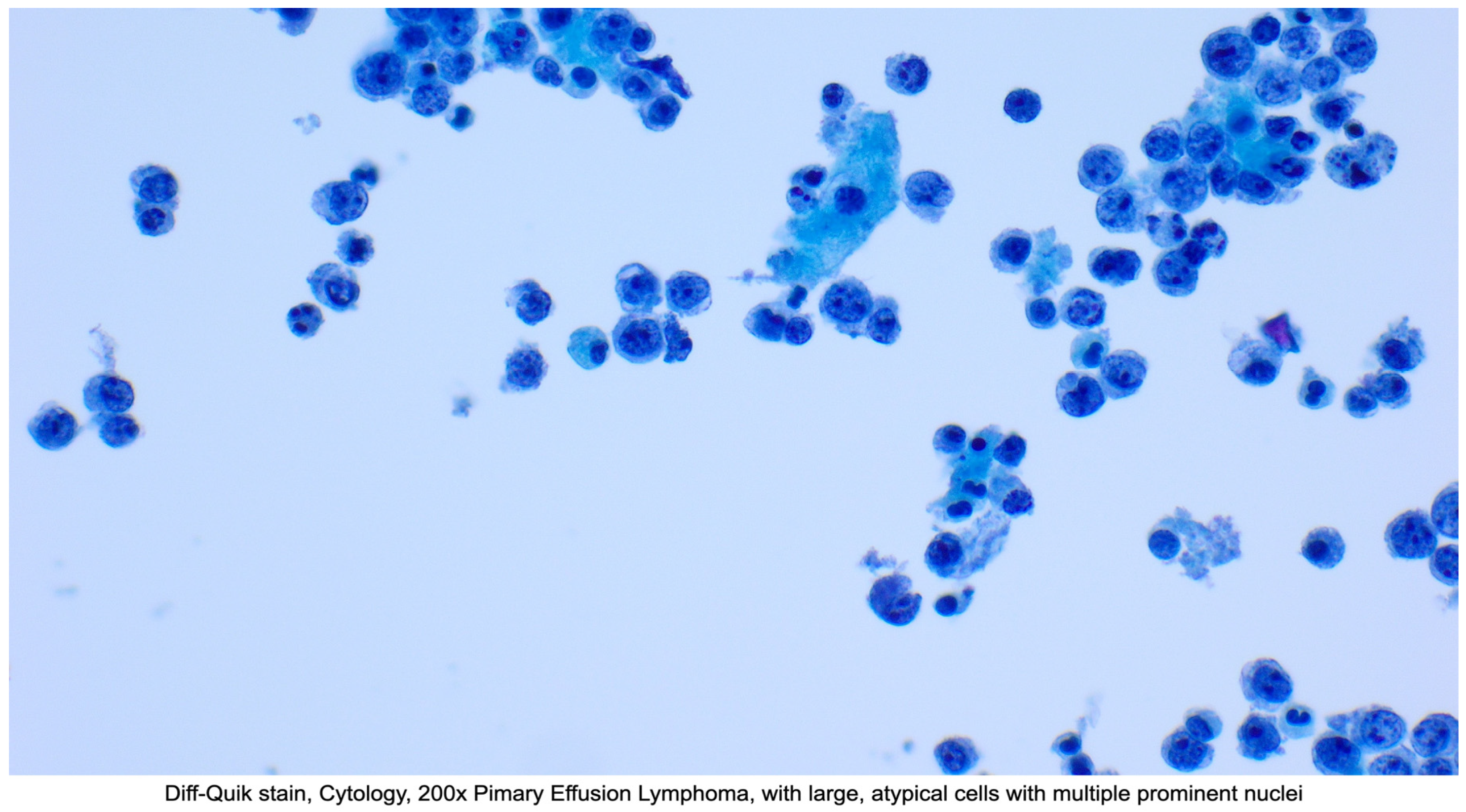
3.1. Primary Effusion Lymphoma (PEL) and Extracavitary Primary Effusion Lymphoma (EC-PEL)

3.1.1. Definition

Primary effusion lymphoma (PEL) is a clinically aggressive large B-cell lymphoma presenting as a serous effusion involving the pleural cavity, the peritoneal cavity, or pericardial cavity, without nodal involvement or tumor masses [28,29]. PEL typically involves a single serous cavity, but more than one cavity can be involved [30]. A subset of cases presents with nodal involvement or extranodal solid tumor masses. This variant is called extracavitary primary effusion lymphoma (EC-PEL) [31,32]. EC-PEL can occur with or without a lymphomatous effusion, but most frequently occurs after PEL has developed [30,32]. Extracavitary sites include the gastrointestinal tract, lung, skin, central nervous system, or bone marrow [28,29,33,34]. Lymphoma cells are coinfected by both KSHV/HHV8 and EBV [29].

3.1.2. Epidemiology

PEL occurs most often in HIV-positive adults with low CD4 + T-cell counts, but has been documented in other immunocompromised individuals, including transplant (immunosuppressed) patients or elderly (immunosenescence) individuals living in endemic regions [28,29,33,34]. Other manifestations of KSHV/HHV8 infection, including Kaposi sarcoma and multicentric Castleman disease, are present in approximately 50% of patients at presentation [29]. Approximately 80% of PEL cases are EBV-positive. The co-infection is seen predominantly in HIV-positive individuals, but can occur in HIV-negative individuals, most often living in endemic regions [29,35]. The age of diagnosis is dependent on location and HIV status; HIV-positive individuals were diagnosed at the median age of 42, while HIV-negative individuals were diagnosed at the median age of 73 [28,34]. PEL occurs almost exclusively in males [28,36].

3.1.3. Clinical Presentation

Clinical presentation depends on the cavity/cavities or extracavitary sites affected. Those with pericardial effusions often present with chest pain, shortness of breath, and hemodynamic instability in the event of cardiac tamponade [37,38]. Pleural effusion can also cause shortness of breath and chest pain, but may be accompanied by cough [39]. Effusion of the peritoneal cavity will present as ascites, which causes abdominal distension, sensation of fullness, abdominal discomfort or pain, and, occasionally, shortness of breath [40]. General symptoms can include weight loss, fever, and night sweats. Depending on the extent and severity of lymphoproliferation, some patients develop lymphadenopathy and/or splenomegaly [33].
Lab abnormalities range from hypoalbuminemia, thrombocytopenia, and anemia to elevated IL-6 and elevated KSHV/HHV8 viral load, which is relatively unique amongst HIV-associated lymphomas [41].

3.1.4. Diagnosis

Since these are effusions, fluid is generally collected by aspiration and assessed on smear (Papanicolaou stain and/or Diff-Quik) and cell block (H&E). The effusion specimen will likely be more cellular when compared to an individual without lymphomatous effusion. Lesional cells can range from plasmablastic to anaplastic in appearance. The plasmablasts in PEL are large neoplastic cells with round-to-irregular nuclei, prominent nucleoli, and basophilic cytoplasm, similarto the immature appearance of immunoblasts (see Figure 1). Vacuolation is variable. Anaplastic cells appear more poorly differentiated or undifferentiated and may exhibit variable size and pleomorphism. Anaplastic cells can occasionally resemble the Reed–Sternberg cells found in classical Hodgkin lymphoma. The proliferation rate is usually high, and numerous mitotic figures are typically seen [42].
For patients with EC-PEL, specimens are typically biopsies or resected, formalin-fixed, and evaluated by standard hematoxylin and eosin-stained sections. EC-PEL lesions can vary in size, depending on the location in which they arise. Lesional cells will appear similar to those seen on PEL cytology but may appear less pleomorphic on tissue sections [42]. The morphology of PEL and EC-PEL neoplastic cells are indistinguishable.
In PEL and EC-PEL, cell markers can be evaluated by flow cytometry or immunohistochemistry (IHC). Lymphoma cells stain positive for HHV8/LANA1 (see Figure 2), EBV/EBER, CD45, CD30, CD38, CD71, EMA, HLA-DR, and CD138. The cells are typically negative (or dim/patchy) for B-cell markers, including CD19, CD20, and CD79a. Cells are also negative for T-cell markers like CD3, CD4, and CD8. BCL-6 is also typically negative [42].
Lymphoma cells are post-GC B cells that have undergone an intense somatic mutation process on the immunoglobulin gene hypervariable region, which is demonstratable on molecular testing [30,42]. This becomes highly important when staining is poor or variable, and especially when there is mixed clinical presentation. For example, cases of DLBCL presenting with effusions have been documented. These individuals are severely immunodeficient and co-infected with KSHV/HHV8 and EBV, making PEL difficult to distinguish from DLBCL [43]. In these situations, a more definitive diagnosis can be provided with molecular analysis of Ig genes, as seen in Table 1. PEL cells are null phenotype while DLBCL will produce an IgM. EC-PEL and DLBCL are not always possible to distinguish, especially given their solid nature, but again, IgM favors DLBCL over PEL [44].
Unusual features for solid primary effusion lymphoma are the presence of multiple lymphadenopathies, negativity for CD138, and expression of IgM. Although primary effusion lymphoma frequently occurs in patients with HHV8-associated multicentric Castleman disease, these are considered separate entities, and there is currently no definitive evidence of evolution of multicentric Castleman disease to primary effusion lymphoma [45]. This is an important distinction between PEL and DLBCL, which will be discussed in the following section.

3.1.5. Treatment

Depending on HIV status, treatment of PEL may begin with antiretroviral therapy. In fact, complete remission of PEL has been documented in a few cases after the sole use of HAART [46]. Other treatments may include prophylaxis against opportunistic infections such as Pneumocystis jirovecii and cytomegalovirus (CMV), as well as granulocyte-macrophage colony-stimulating factor (GM-CSF), to prevent complications of chemotherapy-induced neutropenia [46,47,48]. These treatments highlight some of the difficulties that arise in treatment of PEL patients; they are severely immunocompromised, with co-infections of numerous viruses. More importantly, chemotherapy administration must be carefully managed in those who are immunocompromised.
NCCN (the National Comprehensive Cancer Network) recommends R-EPOCH (rituximab, etoposide phosphate, prednisone, vincristine sulfate (Oncovin), cyclophosphamide, and doxorubicin hydrochloride (hydroxydaunorubicin) or R-CHOP (rituximab, cyclophosphamide, and doxorubicin, vincristine sulfate (Oncovin), and prednisone) as first-line therapy, which is commonly used in the treatment of other non-Hodgkin lymphomas (NHLs).
Since PEL cells are typically CD20 negative, rituximab is not effective for killing lymphoma cells. However, treatment with rituximab clears B-cells infected with KSHV/HHV8, depleting the viral reservoir still producing the inflammatory cytokines driving these lesions [49]. Research is still ongoing in the exploration and development of novel treatments for PEL, including targeting activating pathways like NF-κB, JAK/STAT, and phosphatidylinositol 3-kinase (PI3K)/AKT. Bortezomib, for example, which plays a role in the NF-κB pathway, shows in vitro efficacy, which was not readily seen in clinical trials. Some immunomodulators, like thalidomide or lenalidomide, are being combined with other compounds, including RNAs (liposome-modified B-lymphocyte–induced maturation protein 1 small interfering RNA), which offer promising antiproliferative effects. Current trials of Lenalidomide combined with dose-adjusted EPOCH with rituximab (DA-EPOCH-R) are currently under way for the treatment of PEL, and have shown efficacy in treating other aggressive B-cell lymphomas [28,50].

3.1.6. Prognosis

Prognosis is influenced by viral status, extent of viral infection, cytokine production, protein expression, performance status, and body cavity involvement. The strongest predictor of improved survival is the use of HAART as an effective viral replication control, which is known to allow greater tolerance to therapies, including autologous stem cell transplantation; notably, though, the impact of antiretroviral therapy is lower in PEL than in other HIV-1 related lymphomas such as DLBCL [35,46,47,48]. EBV positivity in PEL patients is weakly associated with improved survivability, and has a more indolent course [30,33,36]. High levels of IL-6, CD47 positivity, and CD20 negativity are all associated with aggressive behavior, poor prognosis, and decreased overall survival [33,51]. Involvement of the pericardial cavity, in particular, as opposed to the pleural and peritoneal cavities, or involvement of multiple cavities, concurrently portends a poor prognosis [33]. Variants of PEL, such as EC-PEL (extracavitary PEL) and PEL-LL (primary effusion-like Lymphoma), may have a better prognosis, though drawing comparisons remains difficult [52,53,54]. Meanwhile, PT-PEL (post-transplant PEL) has a similarly poor prognosis, of less than a year [35].
PEL/EC PEL prognosis overall is poor, and median survival, even with treatment, remains < 24 months [22].

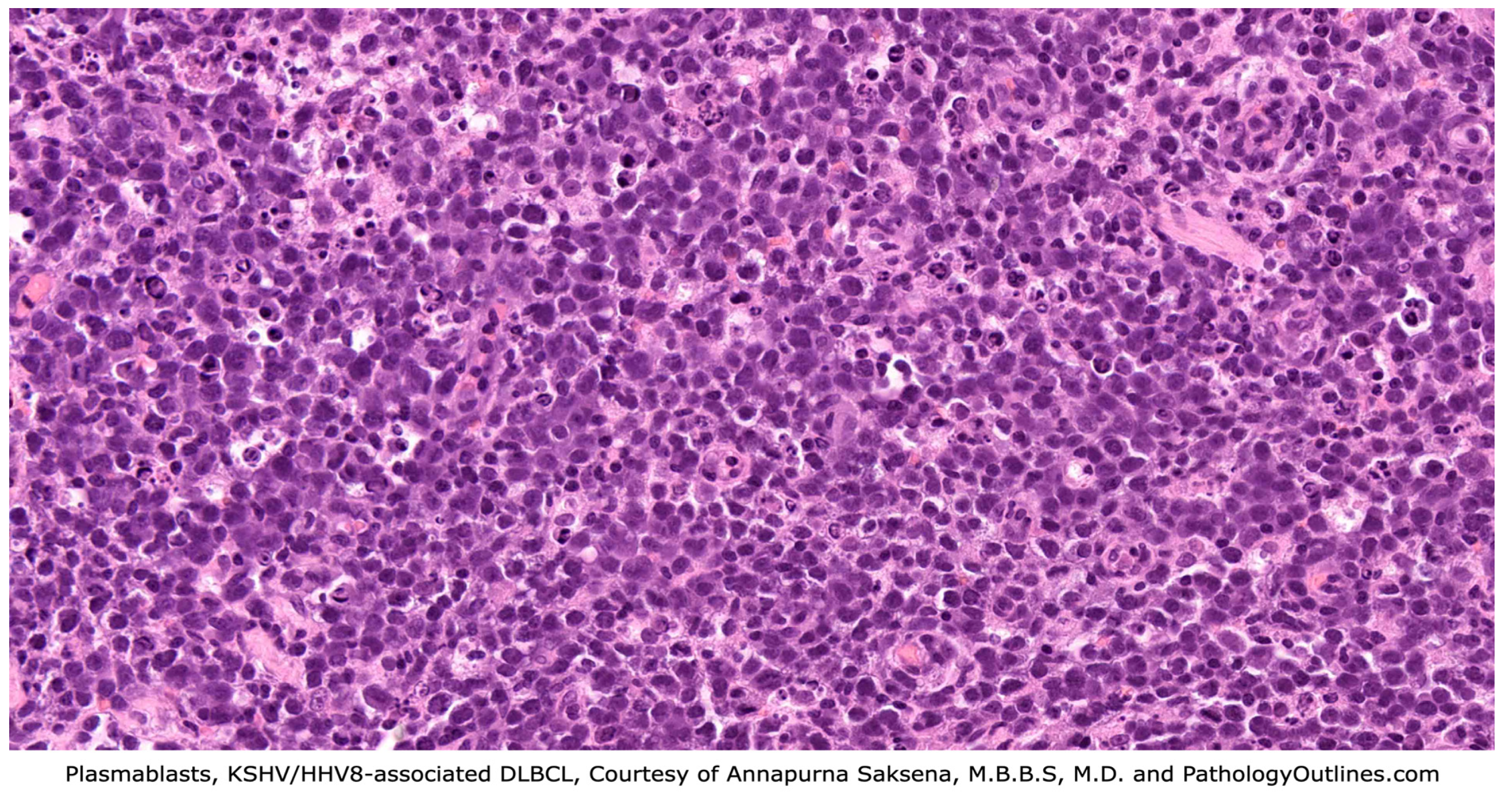
3.2. KSHV/HHV8-Positive Diffuse Large B-Cell Lymphoma (DLBCL)

3.2.1. Definition

KSHV/HHV8-positive DLBCL is a rare form of DLBCL that is associated with KSHV/HHV8 infection.

3.2.2. Epidemiology

This large B-cell lymphoma is often seen in individuals with severe immunodeficiency and who have been diagnosed with MCD [30,36]. Patients may have other KSHV/HHV-8 infection manifestations, including Kaposi sarcoma (KS). Most cases of KSHV/HHV8-positive DLBCL are seen in HIV-positive men aged 30–40 years of age [36]. Rarely does this lymphoma develop in the absence of MCD; however, these cases have been documented in immunosenescent individuals in KSHV/HHV-8 highly endemic regions. KSHV/HHV8-positive DLBCL occurs more than three times more in men than in women (3.3:1) [55].

3.2.3. Clinical Presentation

Patients usually present with generalized lymphadenopathy, which is often accompanied by splenomegaly [45,56]. These manifestations are often accompanied by B-type symptoms [24,57,58] and may involve peripheral blood and bone marrow. KSHV/HHV8-positive DLBCL can also disseminate to organs, including the lungs, liver, or gastrointestinal tract [25]. In a rare case, lymphoma involvement was confined to the spleen [45,59]. Differentials for plasmablastic lesions involving the spleen include PBL, which requires IHC and molecular testing to properly delineate. In another rare instance, one HIV-negative elderly male with a history of MCD developed massive ascites, which was immunophenotypically consistent with KSHV/HHV8-positive DLBCL [60]. Ruling out PEL in this case would have been vital for adequate diagnosis. These cases highlight the presentation overlap and difficulty of distinguishing KSHV/HHV8-positive DLBCL from other lesions. Labs may show cytopenias in one or all three cell hematopoietic lineages, especially when bone marrow is involved. LDH, in particular, is a useful prognostic factor in all DLBCLs, reflecting disease severity, response to treatment, and relapse. Hypercalcemia may also be a biomarker for more aggressive disease and/or high-risk features. Metabolic panels may show changes in renal or hepatic function, as well as changes in electrolytes. Some of these changes, however, reflect treatment effects and/or tumor lysis syndrome [61].

3.2.4. Diagnosis

Fine-needle aspiration and cytology can suggest a diagnosis of KSHV/HHV8-positive DLBCL; however, a definitive diagnosis should be made on excisional lymph node biopsy to evaluate background MCD and GLPD changes [56,62].
In gross appearance, lymph nodes are firm and fleshy in consistency and demonstrate homogeneous white cut surfaces. Hilar obliteration may be appreciated, and necrosis is variable. Extranodal sites may show a confluent appearance or discrete nodules. Histologically, lymph node architecture is effaced by diffuse or coalescing sheets of large, immature-appearing lymphoid cells [56]. Similarly seen in PEL, plasmablasts in DLBCL demonstrate classic morphology (see Figure 3) showing vesicular and eccentrically placed nuclei with one to two prominent nucleoli, and a moderate amount of amphophilic cytoplasm [25,56,63]. Occasionally, cells can appear scattered and may coalesce in small clusters once termed microlymphomas. Clonal studies, however, showed that these clusters are usually polyclonal and do not necessarily progress to lymphoma, so the term was eliminated. These KSHV/HHV8-positive blasts produce a cytoplastic IgM with lambda light chain and rarely kappa. While DLBCL and MCD share the feature of lesions with lambda-restricted plasmablasts infected by KSHV/HHV-8, the distinction is in their clonality. MCD lesions have polyclonal plasmablasts, while frank lymphoma is a monoclonal process [63]. Evidence suggests that DLBCL arises in the same plasmablasts of MCD lesions when DLBCL develops in the setting of MCD [57,64,65]. This is an important distinction between the two neoplasms as PEL can arise in the setting of MCD but does not develop directly from these lesions. Rare cases of KSHV/HHV8 DLBCL occur without MCD [62], suggesting these lesions can also arise de novo, but there is little documentation or research in this area.
Lymphomatous cells of KSHV/HHV8-positive DLBCL are naïve, non-mutated, pre-germinal center B cells [30]; they are positive for LANA, vIL-6, and IRF4/MUM1 [30,56] and negative for plasma cell markers, including CD38 and CD138. They show variable staining for B-cell markers like CD45 and CD20, but are negative for CD79a [45,56,57]. Molecular studies reveal monoclonal IGH rearrangement with a lack of somatic mutations of the IGH variable regions [45], supporting the fact that these are naïve and transformed B-cells. (See Table 1).
For hematopathologists making this diagnosis, it should be noted that the World Health Organization (WHO) describes this entity in Hematolymphoid Tumours (5th ed.) under the category of KSHV/HHV-associated B-cell lymphoid proliferations and lymphomas as “KSHV/HHV8-positive diffuse large B-cell lymphoma”. International consensus classification (ICC) categorizes this lesion similarly under HHV-8–associated lymphoproliferative disorders; however, the diagnosis “HHV-8–positive diffuse large B-cell lymphoma, NOS” is used.

3.2.5. Treatment

Similar to PEL/EC-PEL, NCCN recommends R-EPOCH or RCHOP as first-line therapy. Since neoplastic cells may not be CD20-positive, rituximab is not conventionally used to treat KSHV/HHV8-positive DLBCL; however, it should be noted that these lesions arise often in the setting of MCD, and patients are likely taking this medication, helping to deplete infected cells. Some guidelines recommend DA-EPOCH-R, similar to trials in PEL treatment [8]. DLBCL is particularly challenging to treat, due to poor response to conventional chemotherapy. A retrospective study on 67 KSHV/HHV8-positive DLBCL patients found no significant association between chemotherapy use and improved survival outcomes [58].

3.2.6. Prognosis

KSHV/HHV8-positive DLBCL is both rare and aggressive. HIV-positive individuals in which KSHV/HHV8-positive DLBCL arises in the setting of MCD have the poorest outcomes [56]. Research has suggested that age is the most important predictor of survival in DLBCL patients. A 2023 study found older age to be significantly associated with a lower overall survival rate. This same study also showed that survival was significantly better in nodal rather than extranodal KSHV/HHV8-positive DLBCL and that marital status of single had worse overall survival [58]. Most recent research in 2024 shows that survivors have an increased risk of malignancy within the first year of diagnosis, and the 1-year, 3-year, and 5-year overall survival was 63.6%, 59.7%, and 54.4%, respectively [66]. The overall prognosis of KSHV/HHV8-positive DLBCL is poor, with reported median survival of just a few months [56].

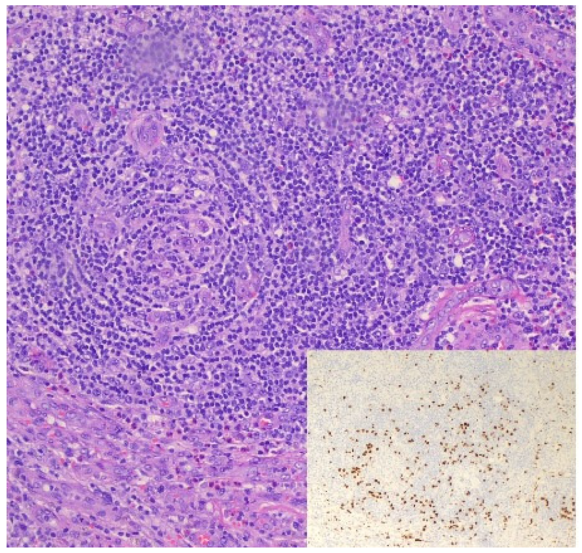
3.3. KSHV/HHV8-Associated Multicentric Castleman Disease (MCD)

3.3.1. Definition

Castleman disease was initially described in 1956, in asymptomatic patients with lymphoid proliferations arising in the mediastinum. Several morphological and clinical variants were subsequently identified [30,57]. Clinically, Castleman disease is divided into unicentric (involving a single lymph node) or multicentric (presenting in two or more lymph nodes). The multicentric form was described in 1980, and in its early stages, MCD was subcategorized into HIV-positive and HIV-negative cases. Later research showed that KSHV/HHV-8 status was more significant in classifying these lesions [57]. HIV status is still used in subclassifying KSHV/HHV8-associated MCD, which is a systemic, polyclonal lymphoproliferative disorder that is driven by KSHV/HHV8 infection [57,60,67]. MCD that arises in the absence of KSHV/HHV-8 infection is termed idiopathic multicentric Castleman disease (iMCD) and generally has a less aggressive clinical course than KSHV/HHV8-associated MCD [45].

3.3.2. Epidemiology

The majority of KSHV/HHV8 + MCD cases occur in HIV-positive patients with low CD4+ T-cell counts, predominantly in men (M:F; 8:1) and with a median age of 40–45 years. In HIV-negative individuals, patients tend to be older, with a median age of 65 years, and with a lower male-to-female ratio (2.4:1) [68].
Patients present with fever and other constitutional symptoms, including fatigue, weight loss, and myalgias. MCD is a relapsing–remitting disease, and symptoms often come and go in “flares.” The flares correlate with proinflammatory hypercytokinemia and viral load [68].

3.3.3. Clinical Presentation

The most common clinical manifestation in MCD is generalized lymphadenopathy, frequently involving the axillary, abdominal, pelvic, mediastinal, and cervical nodes, although any lymph nodes can be involved, and lymphadenopathy is often diffuse. Lymphadenopathy is often accompanied by hepatosplenomegaly, and rarely involves bone marrow [57,67,68,69]. Other symptoms documented include effusions, edema, pulmonary and/or gastrointestinal symptoms, autoimmune hemolytic anemia, or hemophagocytic lymphohistiocytosis (HLH) [67,68]. Lab findings are often significant for polyclonal hypergammaglobulinemia, hypoalbuminemia, elevated C-reactive protein, thrombocytopenia, and anemia [30,45,57,67,68]. The most notable lab finding is high serum levels of IL-6. This cytokine, as well as IL-10 and vIL-6, contributes to the systemic inflammatory symptoms experienced in this condition.
The majority of patients often have a concurrent diagnosis of Kaposi sarcoma (KS), and a smaller percentage of patients have concurrent or subsequent diagnosis of lymphoma [67,68].

3.3.4. Diagnosis

Core biopsies are inadequate for diagnosing MCD, and so an excisional lymph node biopsy is preferred. In gross appearance, lymph nodes are enlarged and show a range of features, from pinkish-gray and well-encapsulated [70], gray-white with ill-defined borders and cut surfaces with a multinodular appearance [71], to diffuse nodularity with prominent vasculature [72]. Calcifications are variable [73].
Microscopically, lymph nodes show lymphocyte-depleted germinal centers, vascular proliferation, and plasmacytosis [45]. The lesions are caused by the same elevated cytokines and growth factors that cause systemic inflammatory symptoms [57].
Castleman disease can present morphologically as hyaline-vascular, plasma cell, or mixed-pattern variants [74,75]; however, multicentric, rather than unicentric, tends to be a plasma cell/plasmacytic variant or mixed-pattern [11,45,68]. KSHV/HHV8-associated MCD, in particular, demonstrates plasmacytic histology with plasmablastic features (see Figure 4).
Follicles can be small with regressed germinal centers [45], and hypocellular in appearance [74]. There is marked vascular proliferation [57,59], usually with some degree of hyalinization [45,57]. Plasmacytosis in the interfollicular zones is marked [59], with some histology showing sheets of polytypic plasma cells [57]. These plasma cells are CD138-positive [68], and differ from lesional plasmablasts, which are seen primarily in mantle zones but can also be seen in perifollicular and interfollicular zones [57,68]. Plasmablasts are B-cells with plasmacytic differentiation [45], which range from medium to large [57,68] and show the same cell morphology consistently seen in KSHV/HHV8 lesions. Although these cells produce a monotypic IgM that is lambda-restricted [59,63], the cells are polyclonal, according to molecular studies [63,68]. Plasmablasts are HHV8-infected, as evidenced by the presence of LANA staining on immunohistochemistry. These cells are also positive for OCT2 [26], BLIMP1, IRF4 (MUM1) and negative for CD 20 and CD138 [26,68]. Some sources show that CD20 and CD79a staining is variable in up to 50% of cases [57,76]. Sources showed mixed reporting on PAX5 staining [30,45,57]. In rare cases, plasmablasts are co-infected with EBV, and are EBER-positive [4,45].
Interestingly, while ICC describes all HHV-8–associated lymphoproliferative disorders together, including “HHV-8–associated multicentric Castleman disease”, WHO categorizes “KSHV/HHV8-associated multicentric Castleman disease” under tumor-like lesions with B-cell predominance, although it has a subsection for KSHV/HHV8-associated lesions.

3.3.5. Treatment

In the most recent iteration of the National Comprehensive Cancer Network (NCCN) guidelines, there is guidance for the treatment of all forms of Castleman disease, including “HHV8-positive MCD.” Rituximab is first-line therapy of KSHV/HHV8-associated MCD and effectively depletes the reservoir of HHV8-infected B-cells, reducing cytokine levels and the systemic inflammatory response in MCD flares. Even in relapse, rituximab improves survival and makes KSHV/HHV8-associated MCD a more manageable lymphoproliferative disorder [77]. As per NCCN guidelines, rituximab may be combined with prednisone and/or liposomal doxorubicin, which causes reductions in lymphadenopathy and viral load [78]. Other treatments include antivirals, IL-6 inhibitors [79], and chemotherapy (RCHOP, RCVAD, and RCVP), depending on whether there is fulminant disease. Second-line therapy and subsequent therapy for relapsed/refractory or progressive disease are also available in the guidelines but are essentially the same treatments recommended in first-line therapy. While some institutions help to provide guidelines, no gold standard therapy has been established [78]. The Castleman Disease Collaborative Network (CDCN) offers an algorithm that takes into consideration KS status and includes recommendations for specific treatments amd numbers of cycles and timelines, as well as surveillance [80].
Difficulties in treatment of KSHV/HHV8-associated MCD arise similarly to those for other lesions discussed. Treatment can be difficult and may change when there is concurrent disease (e.g., concomitant KS is treated with the addition of etoposide or liposomal anthracyline). Depending on the degree of immunodeficiency in these individuals, chemotherapy must be carefully administered, to avoid serious side effects and possible infections. Recent developments of more targeted therapies, including IL-6 inhibitors (Tocilizumab), offer alternative treatments in those that cannot tolerate rituximab or when there is poor response [49].

3.3.6. Prognosis

KSHV/HHV8-associated MCD has an increased risk of developing lymphoma, as well as other KSHV/HHV8-related lesions [68]. In the past, the prognosis was poor, with an overall survival rate equal to, or less than, 2 years [68]. With the introduction of rituximab and antiretroviral therapy, the overall survival rate at 5 years has increased to 90–92% [68,81,82]. There is also an increased risk of relapse, with one study showing that patients experienced relapse with a mean time (first-time relapse) of 30 months, and a max of ten years [82]. A few cases also had a concomitant KSHV/HHV8-associated lymphoma at time of relapse, showing that although patients may achieve remission, there can still be progression of disease. The prognosis for KSHV/HHV8-associated MCD is improving, and, with the development of novel targeted therapies, will likely continue to improve.

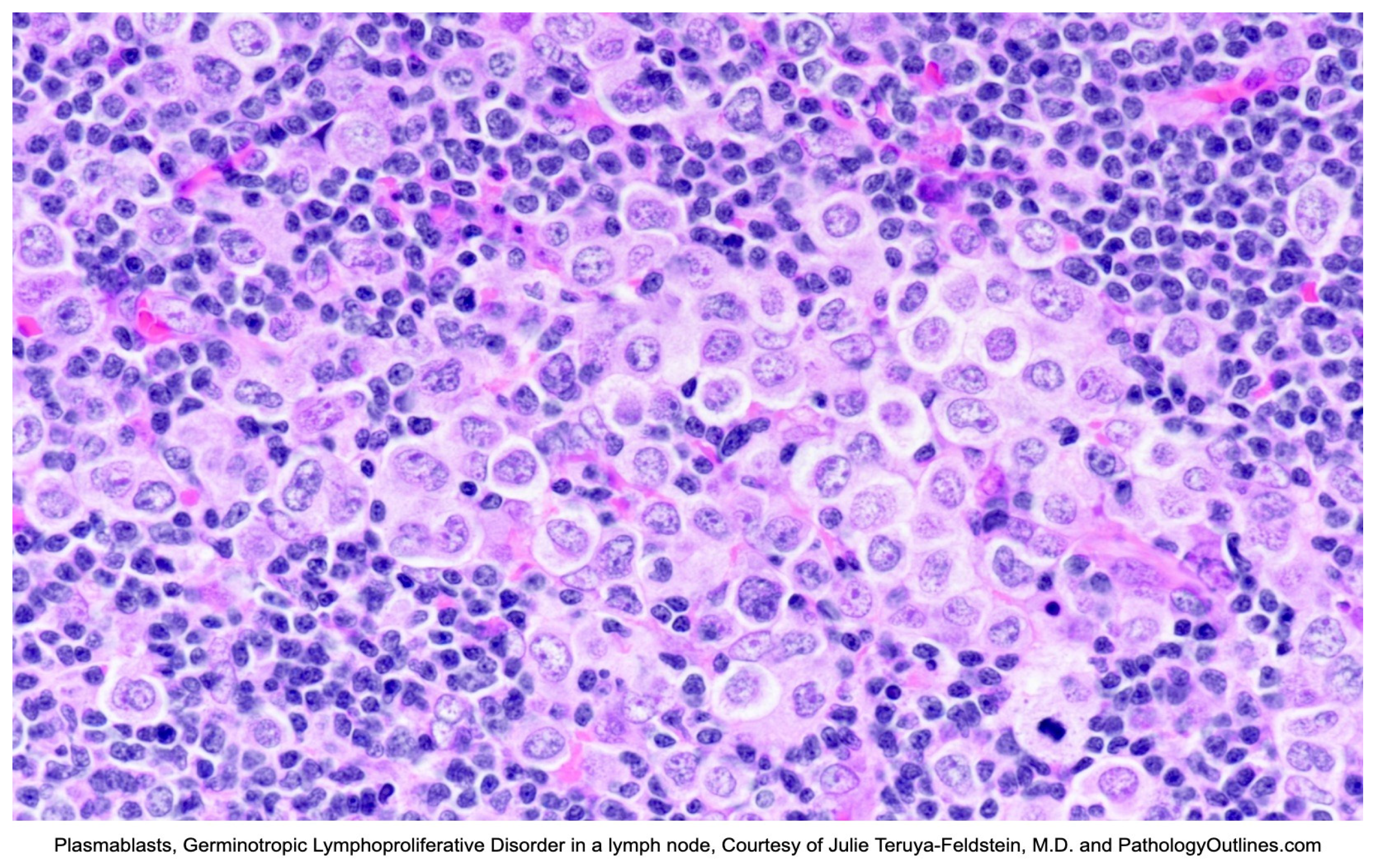
3.4. Germinotropic Lymphoproliferative Disorder (GLPD)

3.4.1. Definition

Germinotropic lymphoproliferative disorder, first described in 2002 [83], is one of two entities resulting from co-infection by KSHV/HHV8 and Epstein–Barr Virus (EBV). The role of EBV in this lesion is unclear, so it is suggested that KSHV/HHV8 and vIL-6 must be primary drivers [84].

3.4.2. Epidemiology

GLPD is rare and tends to affect the elderly (median age: 60 years, range: 20–86 years), although it can be seen at any age and has a slight male predominance [84]. HIV status is variable, although the majority of cases are in immunocompetent HIV-negative individuals [85]. It is unclear from the literature whether or not there is an increased incidence of GLPD in endemic regions of KSHV/HHV8; however, a number of cases from these areas have been described.

3.4.3. Clinical Presentation

GLPD is typically indolent, and patients may not be symptomatic. The most common clinical presentation is lymphadenopathy. This can involve one or multiple lymph nodes, often seen as enhancing nodes on computed tomography (CT) or as fluorodeoxyglucose (FDG) avid nodes on positron emission tomography (PET). Other reported findings included B-type symptoms (weight loss, night sweats, and fevers), effusions, leg swelling, paresthesia, abdominal pain, autoimmune hemolytic anemia, and splenomegaly. No lab abnormalities were specifically reported. The majority of patients, however, are healthy and asymptomatic [85].

3.4.4. Diagnosis

On excisional biopsy, lymph nodes are enlarged, ranging from 1.5 to 26 cm [84,85]. In gross appearance, the nodes are white, with cut surfaces showing focal nodularity [85]. Microscopically, there are often aggregates of plasmablasts replacing germinal centers, and in some cases, germinal centers are completely replaced by plasmablasts (see Figure 5). These large, atypical cells are polyclonal to oligoclonal and are coinfected with KSHV/HHV8 and EBV [84]. Occasionally, there can be scattered plasmablasts in mantle zones, sinuses, and interfollicular areas [84,85]. The background can share features with KSHV/HHV8-associated MCD, including marked plasmacytosis [45,84] with atrophic and hyalinized follicles and vascular proliferation [84,85]. Lesional cells are positive for EBV/EBER, HHV8/LANA, vIL-6, MUM1/IRF4, and PDL1, and show monotypic light-chain restriction [45,86,87,88]. Other plasma cell markers like CD38 and CD138 are variable [45]. T-cell markers may occasionally be positive. The cells are usually negative for B-cell markers, including CD20, CD79a, PAX5, BCL6, CD10, and CD30 [45]. The proliferation index is usually high [84]. Given the possible overlap with KSHV/HHV8 MCD, it should be noted that plasmablastic cells in MCD are not (or are rarely) EBV-positive, and are almost always lambda-restricted. Molecular studies will also show somatic hypermutation of Ig genes in GLPD, while MCD lacks somatic hypermutation (see Table 1).

3.4.5. Treatment

While NCCN offers guidance on the treatment of other KSHV/HHV8-associated lesions described in this review, in its current iteration, there is no guidance for the treatment of GLPD. In general, there is no standard care for these lesions.
Available data suggests that there are a variety of treatments to which GLPD often responds well, including chemotherapy, surgical excision, and radiotherapy [83]. Cases report that some patients on CHOP- or DA-EPOCH-based therapy achieved remission [2]. In fewer cases, patients underwent surgical excision, with some undergoing subsequent radiotherapy. The reported remission rate in these cases was 100% [89].
An interesting potential therapeutic role of PD1/PD-L1 immunotherapy has been suggested due to the PD-L1 expression found in some cases [90], but this still requires additional research [85].

3.4.6. Prognosis

Although GLPD is indolent in nature, there is risk of transformation into aggressive lymphoma [84], although the degree of this is unclear. GLPD is rare, and the prognosis is generally excellent. Most patients have little to no symptoms, and do not require treatment. Those with stable disease or disease progression respond well to radiation or chemotherapy, with or without rituximab, with reported survival times of up to 15 years [84,91].

4. Discussion and Conclusions

KSHV/HHV8 is a virus that can infect B-cells, resulting in lymphomas (PEL/EC-PEL and DLBCL) and lymphoproliferative disorders (MCD and GLPD), which can transform into aggressive lymphoma. Diagnosis can be complicated by complex clinical pictures and overlapping features. The plasmablastic morphology consistently seen throughout these lesions even overlaps with non-KSHV/HHV8-positive lesions like Plasmablastic Lymphoma (PBL) or ALCL [92], which further complicates the diagnostic process. IHC can be variable or atypical, which can make diagnosis with H&E and IHC alone difficult. Molecular testing can help to provide the necessary information to guide appropriate diagnosis and treatment, but the difficulty of treating more aggressive clinical disease and concurrent viral infection(s), commonly in the context of immunodeficiency, still remains. Rituximab and anti-viral therapy are vital in the treatment of KSHV/HHV8 lesions, as clearing the viral infection reduces symptoms, the risk of progression, and development of concurrent KSHV/HHV8 malignancies [77].
While infection can occur in immunocompetent patients and follow an indolent course, most patients with KSHV/HHV8 lesions are immunocompromised, and so these lesions often portend a poor prognosis. Although new and targeted treatments are improving overall survival times, with the exception of GLPD, life expectancy is still short, ranging from a few months to a few years.

Author Contributions

Conceptualization, Y.L. and J.R.; investigation, J.R. and K.Z.; writing—original draft preparation, J.R. and K.Z.; writing—review and editing, J.R., Y.L., and M.G. All authors have read and agreed to the published version of the manuscript.

Funding

This research received no external funding.

Conflicts of Interest

The authors declare no conflict of interest.

References

  1. Chang, Y.; Cesarman, E.; Pessin, M.S.; Lee, F.; Culpepper, J.; Knowles, D.M.; Moore, P.S. Identification of Herpesvirus-Like DNA Sequences in AIDS-Sssociated Kaposi’s Sarcoma. Science 1994, 266, 1865–1869. [Google Scholar] [CrossRef]
  2. Cesarman, E.; Chadburn, A.; Rubinstein, P.G. KSHV/HHV8-mediated hematologic diseases. Blood 2022, 139, 1013–1025. [Google Scholar] [CrossRef] [PubMed] [PubMed Central]
  3. De Paoli, P. Human herpesvirus 8: An update. Microbes Infect. 2004, 6, 328–335. [Google Scholar] [CrossRef] [PubMed]
  4. Guerrero, C.; Jain, T.; Kelemen, K. HHV-8-Associated Lymphoproliferative Disorders and Pathogenesis in an HIV-Positive Patient. Case Rep. Hematol. 2019, 2019, 4536157. [Google Scholar] [CrossRef] [PubMed] [PubMed Central]
  5. Russo, J.J.; Bohenzky, R.A.; Chien, M.C.; Chen, J.; Yan, M.; Maddalena, D.; Parry, J.P.; Peruzzi, D.; Edelman, I.S.; Chang, Y.; et al. Nucleotide sequence of the Kaposi sarcoma-associated herpesvirus (HHV8). Proc. Natl. Acad. Sci. USA 1996, 93, 14862–14867. [Google Scholar] [CrossRef] [PubMed] [PubMed Central]
  6. Dourmishev, L.A.; Dourmishev, A.L.; Palmeri, D.; Schwartz, R.A.; Lukac, D.M. Molecular genetics of Kaposi’s sarcoma-associated herpesvirus (human herpesvirus-8) epidemiology and pathogenesis. Microbiol. Mol. Biol. Rev. 2003, 67, 175–212. [Google Scholar] [CrossRef] [PubMed] [PubMed Central]
  7. Angius, F.; Ingianni, A.; Pompei, R. Human Herpesvirus 8 and Host-Cell Interaction: Long-Lasting Physiological Modifications, Inflammation and Related Chronic Diseases. Microorganisms 2020, 8, 388. [Google Scholar] [CrossRef] [PubMed] [PubMed Central]
  8. Human Herpesvirus-8 Disease: Adult and Adolescent OIS|NIH. Available online: https://clinicalinfo.hiv.gov/en/guidelines/hiv-clinical-guidelines-adult-and-adolescent-opportunistic-infections/human-herpesvirus (accessed on 23 April 2025).
  9. Taylor, M.M.; Chohan, B.; Lavreys, L.; Hassan, W.; Huang, M.L.; Corey, L.; Ashley Morrow, R.; Richardson, B.A.; Mandaliya, K.; Ndinya-Achola, J.; et al. Shedding of human herpesvirus 8 in oral and genital secretions from HIV-1-seropositive and -seronegative Kenyan women. J. Infect. Dis. 2004, 190, 484–488. [Google Scholar] [CrossRef] [PubMed] [PubMed Central]
  10. HHV-8 Found in Saliva, Suggests Spread by ‘Deep Kissing’ Cancer Network. Available online: https://www.cancernetwork.com/view/hhv-8-found-saliva-suggests-spread-deep-kissing (accessed on 11 November 2020).
  11. Souza, V.A.; Salzano, F.M.; Petzl-Erler, M.L.; Nascimento, M.C.; Mayaud, P.; Borges, J.D.; Pannuti, C.S. Variations in human herpesvirus type 8 seroprevalence in Native Americans, South America. Emerg. Infect. Dis. 2010, 16, 1003–1006. [Google Scholar] [CrossRef] [PubMed] [PubMed Central]
  12. Edelman, D.C. Human herpesvirus 8—A novel human pathogen. Virol. J. 2005, 2, 78. [Google Scholar] [CrossRef]
  13. Rewane, A.; Tadi, P. Herpes Virus Type 8. In StatPearls; StatPearls Publishing: Treasure Island, FL, USA, 2025. Available online: https://www.ncbi.nlm.nih.gov/books/NBK556023/ (accessed on 16 February 2025).
  14. Capan-Melser, M.; Mombo-Ngoma, G.; Akerey-Diop, D.; Basra, A.; Manego-Zoleko, R.; Würbel, H.; Lötsch, F.; Groger, M.; Skoll, M.; Schwing, J.; et al. Epidemiology of Human Herpes Virus 8 in Pregnant Women and their Newborns—A cross-sectional delivery survey in Central Gabon. Int. J. Infect. Dis. 2015, 39, 16–19. [Google Scholar] [CrossRef] [PubMed]
  15. Sarmati, L.; Carlo, T.; Rossella, S.; Montano, M.; Adalgisa, P.; Rezza, G.; Andreoni, M. Human herpesvirus-8 infection in pregnancy and labor: Lack of evidence of vertical transmission. J. Med. Virol. 2004, 72, 462–466. [Google Scholar] [CrossRef] [PubMed]
  16. Rappocciolo, G.; Hensler, H.R.; Jais, M.; Reinhart, T.A.; Pegu, A.; Jenkins, F.J.; Rinaldo, C.R. Human herpesvirus 8 infects and replicates in primary cultures of activated B lymphocytes through DC-SIGN. J. Virol. 2008, 82, 4793–4806. [Google Scholar] [CrossRef] [PubMed] [PubMed Central]
  17. McGreal, E.P.; Miller, J.L.; Gordon, S. Ligand recognition by antigen-presenting cell C-type lectin receptors. Curr. Opin. Immunol. 2005, 17, 18–24. [Google Scholar] [CrossRef] [PubMed] [PubMed Central]
  18. Komatsu, T.; Ballestas, M.E.; Barbera, A.J.; Kelley-Clarke, B.; Kaye, K.M. KSHV LANA1 binds DNA as an oligomer and residues N-terminal to the oligomerization domain are essential for DNA binding, replication, and episome persistence. Virology 2004, 319, 225–236. [Google Scholar] [CrossRef]
  19. Narkhede, M.; Arora, S.; Ujjani, C. Primary effusion lymphoma: Current perspectives. OncoTargets Ther. 2018, 11, 3747–3754. [Google Scholar] [CrossRef] [PubMed] [PubMed Central]
  20. Kleer, M.; MacNeil, G.; Adam, N.; Pringle, E.S.; Corcoran, J.A. A panel of Kaposi’s Sarcoma-Associated herpesvirus mutants in the polycistronic Kaposin locus for precise analysis of individual protein products. J. Virology. 2021, 96, e0156021. [Google Scholar] [CrossRef]
  21. McCormick, C.; Ganem, D. The kaposin B protein of KSHV activates the p38/MK2 pathway and stabilizes cytokine mRNAs. Science 2005, 307, 739–741. [Google Scholar] [CrossRef] [PubMed]
  22. Primary Effusion Lymphoma. Available online: https://www.pathologyoutlines.com/topic/lymphomaeffusion.html (accessed on 9 February 2025).
  23. Kleer, M.; MacNeil, G.; Pringle, E.S.; Corcoran, J.A. A panel of KSHV mutants in the polycistronic kaposin locus for precise analysis of individual protein products. J. Virol. 2022, 9, e0156021. [Google Scholar] [CrossRef]
  24. Dave, A.; Schwartz, M.; Van, J.; Owczarzak, L.; Miller, I.; Jain, S. A Challenging Diagnosis of HHV-8-Associated Diffuse Large B-Cell Lymphoma, Not Otherwise Specified, in a Young Man with Newly-Diagnosed HIV. Am. J. Case Rep. 2024, 25, e945162. [Google Scholar] [CrossRef] [PubMed] [PubMed Central]
  25. Fenu, E.M.; Beaty, M.W.; O’Neill, T.E.; O’Neill, S.S. Cardiac Involvement by Human Herpesvirus 8-Positive Diffuse Large B-Cell Lymphoma: An Unusual Presentation in a Patient with Human Immunodeficiency Virus. Case Rep. Pathol. 2022, 2022, 1298121. [Google Scholar] [CrossRef] [PubMed] [PubMed Central]
  26. Wang, H.W.; Pittaluga, S.; Jaffe, E.S. Multicentric Castleman disease: Where are we now? Semin. Diagn. Pathol. 2016, 33, 294–306. [Google Scholar] [CrossRef] [PubMed] [PubMed Central]
  27. Monini, P.; Colombini, S.; Stürzl, M.; Goletti, D.; Cafaro, A.; Sgadari, C.; Buttò, S.; Franco, M.; Leone, P.; Fais, S.; et al. Reactivation and Persistence of Human Herpesvirus-8 Infection in B Cells and Monocytes by Th-1 Cytokines Increased in Kaposi’s Sarcoma. Blood 1999, 93, 4044–4058. [Google Scholar] [PubMed]
  28. Shimada, K.; Hayakawa, F.; Kiyoi, H. Biology and management of primary effusion lymphoma. Blood 2018, 132, 1879–1888. [Google Scholar] [CrossRef] [PubMed]
  29. Said, J.; Cesarman, E. Primary effusion lymphoma. In WHO Classification of Tumours of Haematopoietic and Lymphoid Tissues; Swerdlow, S.H., Campo, E., Harris, N.L., Jaffe, E.S., Pileri, S.A., Stein, H., Thiele, J., Eds.; IARC Press: Lyon, France, 2017. [Google Scholar]
  30. Calabrò, M.L.; Sarid, R. Human herpesvirus 8 and lymphoproliferative disorders. Mediterr. J. Hematol. Infect. Dis. 2018, 10, e2018061. [Google Scholar] [CrossRef]
  31. Medeiros, L.J.; Miranda, R.N. Primary Effusion Lymphoma and Solid Variant of Primary Effusion Lymphoma. In Diagnostic Pathology: Lymph Nodes and Extranodal Lymphomas, 2nd ed.; Elsevier: Amsterdam, The Netherlands, 2018; pp. 510–517. ISBN 9780323477796. [Google Scholar] [CrossRef]
  32. Kim, Y.; Leventaki, V.; Bhaijee, F.; Jackson, C.C.; Medeiros, L.J.; Vega, F. Extracavitary/solid variant of primary effusion lymphoma. Ann. Diagn. Pathol. 2012, 16, 441–446. [Google Scholar] [CrossRef] [PubMed]
  33. Gathers, D.A.; Galloway, E.; Kelemen, K.; Rosenthal, A.; Gibson, S.E.; Munoz, J. Primary Effusion Lymphoma: A Clinicopathologic Perspective. Cancers 2022, 14, 722. [Google Scholar] [CrossRef] [PubMed] [PubMed Central]
  34. Zanelli, M.; Sanguedolce, F.; Zizzo, M.; Palicelli, A.; Bassi, M.C.; Santandrea, G.; Martino, G.; Soriano, A.; Caprera, C.; Corsi, M.; et al. Primary effusion lymphoma occurring in the setting of transplanted patients: A systematic review of a rare, life-threatening post-transplantation occurrence. BMC Cancer 2021, 21, 468. [Google Scholar] [CrossRef] [PubMed] [PubMed Central]
  35. Lurain, K.; Polizzotto, M.N.; Aleman, K.; Bhutani, M.; Wyvill, K.M.; Gonçalves, P.H.; Ramaswami, R.; Marshall, V.A.; Miley, W.; Steinberg, S.M.; et al. Viral, immunologic, and clinical features of primary effusion lymphoma. Blood 2019, 133, 1753–1761. [Google Scholar] [CrossRef] [PubMed] [PubMed Central]
  36. Hu, Z.; Pan, Z.; Chen, W.; Shi, Y.; Wang, W.; Yuan, J.; Wang, E.; Zhang, S.; Kurt, H.; Mai, B.; et al. Primary Effusion Lymphoma: A Clinicopathological Study of 70 Cases. Cancers 2021, 13, 878. [Google Scholar] [CrossRef] [PubMed] [PubMed Central]
  37. Albugami, S.; Al-Husayni, F.; AlMalki, A.; Dumyati, M.; Zakri, Y.; AlRahimi, J. Etiology of Pericardial Effusion and Outcomes Post Pericardiocentesis in the Western Region of Saudi Arabia: A Single-center Experience. Cureus 2020, 12, e6627. [Google Scholar] [CrossRef] [PubMed] [PubMed Central]
  38. Sagristà-Sauleda, J.; Mercé, A.S.; Soler-Soler, J. Diagnosis and management of pericardial effusion. World J. Cardiol. 2011, 3, 135–143. [Google Scholar] [CrossRef] [PubMed] [PubMed Central]
  39. Krishna, R.; Antoine, M.H.; Alahmadi, M.H.; Rudrappa, M. Pleural Effusion. In StatPearls; StatPearls Publishing: Treasure Island, FL, USA, 2025. Available online: https://www.ncbi.nlm.nih.gov/books/NBK448189/ (accessed on 22 January 2025).
  40. Chiejina, M.; Kudaravalli, P.; Samant, H. Ascites. In StatPearls; StatPearls Publishing: Treasure Island, FL, USA, 2025. Available online: https://www.ncbi.nlm.nih.gov/books/NBK470482/ (accessed on 22 January 2025).
  41. Gonçalves, P.H.; Uldrick, T.S.; Yarchoan, R. HIV-associated Kaposi sarcoma and related diseases. AIDS 2017, 31, 1903–1916. [Google Scholar] [CrossRef] [PubMed] [PubMed Central]
  42. Patel, S.; Xiao, P. Primary Effusion Lymphoma. Arch. Pathol. Lab. Med. 2013, 137, 1152–1154. [Google Scholar] [CrossRef] [PubMed]
  43. Wang, T.; Silva, O. HHV-8+ diffuse large B-cell lymphoma with EBV coinfection occurring posttransplant. Blood 2024, 144, 677. [Google Scholar] [CrossRef] [PubMed]
  44. Naresh, K. KSHV/HHV8-Associated B-Cell Lymphoid Proliferations and Lymphomas: Introduction; World Health Organization: Geneva, Switzerland, 2024; Available online: https://tumourclassification.iarc.who.int/chaptercontent/63/173 (accessed on 22 January 2025).
  45. Vega, F.; Miranda, R.N.; Medeiros, L.J. KSHV/HHV8-positive large B-cell lymphomas and associated diseases: A heterogeneous group of lymphoproliferative processes with significant clinicopathological overlap. Mod. Pathol. 2020, 33, 18–28. [Google Scholar] [CrossRef] [PubMed]
  46. Carbone, A.; Gloghini, A. PEL and HHV8-unrelated effusion lymphomas: Classification and diagnosis. Cancer 2008, 114, 225–227. [Google Scholar] [CrossRef] [PubMed]
  47. Chen, Y.B.; Rahemtullah, A.; Hochberg, E. Primary effusion lymphoma. Oncologist 2007, 12, 569–576. [Google Scholar] [CrossRef] [PubMed]
  48. Okada, S.; Goto, H.; Yotsumoto, M. Current status of treatment for primary effusion lymphoma. Intractable Rare Dis. Res. 2014, 3, 65–74. [Google Scholar] [CrossRef] [PubMed] [PubMed Central]
  49. Barlingay, G.; Findakly, D.; Hartmann, C.; Amar, S. The Potential Clinical Benefit of Tocilizumab Therapy for Patients with HHV-8-infected AIDS-related Multicentric Castleman Disease: A Case Report and Literature Review. Cureus 2020, 12, e7589. [Google Scholar] [CrossRef] [PubMed] [PubMed Central]
  50. Lurain, K.; Ramaswami, R.; Widell, A.; Ekwede, I.; George, J.; Stetler-Stevenson, M.; Raffeld, M.; Yuan, C.M.; Ziegelbauer, J.; Maldarelli, F.; et al. Phase I/II Study of Lenalidomide Combined with DA-EPOCH and Rituximab (DA-EPOCH-R2) in Primary Effusion Lymphoma in Patients with or without HIV. Blood 2019, 134 (Suppl. 1), 4096. [Google Scholar] [CrossRef]
  51. Panaampon, J.; Okada, S. Promising immunotherapeutic approaches for primary effusion lymphoma. Explor. Target. Antitumor Ther. 2024, 5, 699–713. [Google Scholar] [CrossRef] [PubMed] [PubMed Central]
  52. Guillet, S.; Gérard, L.; Meignin, V.; Agbalika, F.; Cuccini, W.; Denis, B.; Katlama, C.; Galicier, L.; Oksenhendler, E. Classic and extracavitary primary effusion lymphoma in 51 HIV-infected patients from a single institution. Am. J. Hematol. 2016, 91, 233–237. [Google Scholar] [CrossRef] [PubMed]
  53. Hayashino, K.; Meguri, Y.; Yukawa, R.; Komura, A.; Nakamura, M.; Yoshida, C.; Yamamoto, K.; Oda, W.; Imajo, K. Primary Effusion Lymphoma-like Lymphoma Mimicking Tuberculous Pleural Effusion: Three Case Reports and a Literature Review. Intern. Med. 2023, 62, 2531–2537. [Google Scholar] [CrossRef] [PubMed] [PubMed Central]
  54. De Goes, V.A.; Cortez, A.C.; Morbeck, D.L.; Costa, F.D.; Da Silveira, T.B. The role of autologous bone marrow transplantation in primary effusion lymphoma: A case report and literature review. Hematol. Transfus. Cell Ther. 2024, 46, S316–S321. [Google Scholar] [CrossRef]
  55. Sukswai, N.; Lyapichev, K.; Khoury, J.D.; Medeiros, L.J. Diffuse large B-cell lymphoma variants: An update. Pathology 2020, 52, 53–67. [Google Scholar] [CrossRef] [PubMed]
  56. Vega, F.; Chadburn, A. KSHV/HHV8-positive diffuse large B-cell lymphoma. In WHO Classification of Tumours of Haematopoietic and Lymphoid Tissues; Swerdlow, S.H., Campo, E., Harris, N.L., Jaffe, E.S., Pileri, S.A., Stein, H., Thiele, J., Eds.; IARC Press: Lyon, France, 2022. [Google Scholar]
  57. Chadburn, A.; Said, J.; Gratzinger, D.; Chan, J.K.C.; de Jong, D.; Jaffe, E.S.; Natkunam, Y.; Goodlad, J.R. HHV8/KSHV-Positive Lymphoproliferative Disorders and the Spectrum of Plasmablastic and Plasma Cell Neoplasms: 2015 SH/EAHP Workshop Report—Part 3. Am. J. Clin. Pathol. 2017, 147, 171–187. [Google Scholar] [CrossRef]
  58. Wang, F.; Du, Z. Demographic characteristics and prognosis of HHV8-positive diffuse large B-cell lymphoma, not otherwise specified: Insights from a population-based study with a 10-year follow-up. Medicine 2023, 102, e36464. [Google Scholar] [CrossRef] [PubMed] [PubMed Central]
  59. Gonzalez-Farre, B.; Martinez, D.; Lopez-Guerra, M.; Xipell, M.; Monclus, E.; Rovira, J.; Garcia, F.; Lopez-Guillermo, A.; Colomo, L.; Campo, E.; et al. HHV8-related lymphoid proliferations: A broad spectrum of lesions from reactive lymphoid hyperplasia to overt lymphoma. Mod. Pathol. 2017, 30, 745–760. [Google Scholar] [CrossRef] [PubMed]
  60. Chang, L.C.; Fu, P.A.; Wang, S.H.; Chang, K.C.; Hsu, Y.T. Kaposi sarcoma herpesvirus/human herpesvirus 8-positive diffuse large B-cell lymphoma characterized by malignant ascites: A case report. Pathol. Res. Pract. 2024, 255, 155185. [Google Scholar] [CrossRef] [PubMed]
  61. Dpt MALP. Diffuse Large B-Cell Lymphoma Diagnosis. Rare Disease Advisor. Available online: https://www.rarediseaseadvisor.com/disease-info-pages/diffuse-large-b-cell-lymphoma-diagnosis/ (accessed on 10 April 2025).
  62. HHV8 Positive DLBCL, NOS. Available online: https://www.pathologyoutlines.com/topic/lymphomaHHV8DLBCL.html (accessed on 9 February 2025).
  63. Carbone, A.; Cesarman, E.; Spina, M.; Gloghini, A.; Schulz, T.F. HIV-associated lymphomas and gamma-herpesviruses. Blood 2009, 113, 1213–1224. [Google Scholar] [CrossRef]
  64. Oksenhendler, E.; Boulanger, E.; Galicier, L.; Du, M.Q.; Dupin, N.; Diss, T.C.; Hamoudi, R.; Daniel, M.T.; Agbalika, F.; Boshoff, C.; et al. High incidence of Kaposi sarcoma-associated herpesvirus-related non-Hodgkin lymphoma in patients with HIV infection and multicentric Castleman disease. Blood 2002, 99, 2331–2336. [Google Scholar] [CrossRef] [PubMed]
  65. Dupin, N.; Diss, T.L.; Kellam, P.; Tulliez, M.; Du, M.-Q.; Sicard, D.; Weiss, R.A.; Isaacson, P.G.; Boshoff, C. HHV-8 is associated with a plasmablastic variant of Castleman disease that is linked to HHV-8–positive plasmablastic lymphoma. Blood 2000, 95, 1406–1412. [Google Scholar] [CrossRef]
  66. Bharathi, V.; Farooq, A. Epidemiological insights and survival patterns of HHV8-positive diffuse large B-cell lymphoma (HDN) associated with multicentric Castleman disease (CD): A comprehensive analysis using SEER database. JCO 2024, 42, e19080. [Google Scholar] [CrossRef]
  67. Bower, M.; Carbone, A. KSHV/HHV8-Associated Lymphoproliferative Disorders: Lessons Learnt from People Living with HIV. Hemato 2021, 2, 703–712. [Google Scholar] [CrossRef]
  68. Chadburn, A.; Cesarman, E. KSHV/HHV8-associated multicentric Castleman disease. In WHO Classification of Tumours of Haematopoietic and Lymphoid Tissues; Swerdlow, S.H., Campo, E., Harris, N.L., Jaffe, E.S., Pileri, S.A., Stein, H., Thiele, J., Eds.; IARC Press: Lyon, France, 2022. [Google Scholar]
  69. van Rhee, F.; Fajgenbaum, D. Insights into the etiology of Castleman disease. Blood 2024, 143, 1789–1790. [Google Scholar] [CrossRef] [PubMed]
  70. Abdul-Rahman, I.S.; Al-Amri, A.M.; Ghallab, K.Q. Castleman’s Disease: A Study of a Rare Lymphoproliferative Disorder in a University Hospital. Clin. Med. Blood Disord. 2009, 2, 25–36. [Google Scholar] [CrossRef] [PubMed]
  71. Fetica, B.; Pop, B.; Lisencu, C.; Rancea, A.C.; Coman, A.; Cucuianu, A.; Petrov, L. Castleman Disease. A Report of Six Cases. Clujul Med. 2014, 87, 192–197. [Google Scholar] [CrossRef] [PubMed] [PubMed Central]
  72. Madan, R.; Chen, J.-H.; Trotman-Dickenson, B.; Jacobson, F.; Hunsaker, A. The spectrum of Castleman’s disease: Mimics, radiologic pathologic correlation and role of imaging in patient management. Eur. J. Radiol. 2012, 81, 123–131. [Google Scholar] [CrossRef]
  73. Venkateswaran, J.M.D.; Balakrishna, J.M.D. Lymph Nodes & Spleen, Nonlymphoma. In Lymph Nodes-Inflammatory/Reactive Disorders: Castleman Disease; Courville, E.M.D., Miranda, R.N.M.D., Eds.; StatPearls Publishing: Treasure Island, FL, USA, 2023; Available online: https://www.pathologyoutlines.com/topic/lymphnodescastleman.html (accessed on 1 February 2025).
  74. Carbone, A.; Borok, M.; Damania, B.; Gloghini, A.; Polizzotto, M.N.; Jayanthan, R.K.; Fajgenbaum, D.C.; Bower, M. Castleman disease. Nat. Rev. Dis. Primers 2021, 7, 84. [Google Scholar] [CrossRef]
  75. Wu, D.; Lim, M.S.; Jaffe, E.S. Pathology of Castleman Disease. Hematol. Clin. N. Am. 2018, 32, 37–52. [Google Scholar] [CrossRef]
  76. Bacon, C.M.; Miller, R.F.; Noursadeghi, M.; McNamara, C.; Du, M.-Q.; Dogan, A. Pathology of bone marrow in human herpes virus-8 (HHV8)-associated multicentric Castleman disease. Br. J. Haematol. 2004, 127, 585–591. [Google Scholar] [CrossRef] [PubMed]
  77. Ramaswami, R.; Uldrick, T.S. Remission after rituximab for HHV8+ MCD: What next? Blood Adv. 2023, 7, 5661–5662. [Google Scholar] [CrossRef] [PubMed]
  78. Dunn, R.; Jariwal, R.; Venter, F.; Mishra, S.; Bhandohal, J.; Cobos, E.; Heidari, A. HHV-8-Associated Multicentric Castleman Disease, a Diagnostic Challenge in a Patient with Acquired Immunodeficiency Syndrome and Fever. J. Investig. Med. High Impact Case Rep. 2022, 10, 23247096221097526. [Google Scholar] [CrossRef] [PubMed] [PubMed Central]
  79. Bower, M. How I treat HIV-associated multicentric Castleman disease. Blood 2010, 116, 4415–4421. [Google Scholar] [CrossRef]
  80. Castleman Disease Collaborative Network. Treatment—CDCN. Available online: https://cdcn.org/treatment/ (accessed on 22 January 2025).
  81. Pria, A.D.; Pinato, D.; Roe, J.; Naresh, K.; Nelson, M.; Bower, M. Relapse of HHV8-positive multicentric Castleman disease following rituximab-based therapy in HIV-positive patients. Blood 2017, 129, 2143–2147. [Google Scholar] [CrossRef]
  82. Bower, M.; Newsom-Davis, T.; Naresh, K.; Merchant, S.; Lee, B.; Gazzard, B.; Stebbing, J.; Nelson, M. Clinical Features and Outcome in HIV-Associated Multicentric Castleman’s Disease. JCO 2011, 29, 2481–2486. [Google Scholar] [CrossRef]
  83. Du, M.-Q.; Diss, T.C.; Liu, H.; Ye, H.; Hamoudi, R.A.; Cabeçadas, J.; Dong, H.Y.; Harris, N.L.; Chan, J.K.C.; Rees, J.W.; et al. KSHV- and EBV-associated germinotropic lymphoproliferative disorder. Blood 2002, 100, 3415–3418. [Google Scholar] [CrossRef]
  84. Chadburn, A.; Said, J.; Du, M.; Vega, F. KSHV/HHV8-Positive Germinotropic Lymphoproliferative Disorder; World Health Organization: Geneva, Switzerland, 2024; Available online: https://tumourclassification.iarc.who.Int/chaptercontent/63/177 (accessed on 30 December 2024).
  85. Zanelli, M.; Zizzo, M.; Bisagni, A.; Froio, E.; De Marco, L.; Valli, R.; Filosa, A.; Luminari, S.; Martino, G.; Massaro, F.; et al. Germinotropic lymphoproliferative disorder: A systematic review. Ann. Hematol. 2020, 99, 2243–2253. [Google Scholar] [CrossRef] [PubMed]
  86. Bacha, D.; Chelly, B.; Kilani, H.; Charfi, L.; Douggaz, A.; Chatti, S.; Chelbi, E. HHV8/EBV Coinfection Lymphoproliferative Disorder: Rare Entity with a Favorable Outcome. Case Rep. Hematol. 2017, 2017, 1–8. [Google Scholar] [CrossRef]
  87. SEER. SEER Hematopoietic and Lymphoid Neoplasm Database. Available online: https://seer.cancer.gov/seertools/hemelymph/5a7d9f261ef55751804cbb20/ (accessed on 15 January 2025).
  88. HHV8 Positive Germinotropic Lymphoproliferative Disorder. Available online: https://www.pathologyoutlines.com/topic/lymphomaHHV8germinotropicLPD.html (accessed on 9 February 2025).
  89. Ghoneima, A.; Cooke, J.; Shaw, E.; Agrawal, A. Human herpes virus 8-positive germinotropic lymphoproliferative disorder: First case diagnosed in the UK, literature review and discussion of treatment options. BMJ Case Rep. 2020, 13, e231640. [Google Scholar] [CrossRef] [PubMed] [PubMed Central]
  90. Ronaghy, A.; Wang, H.Y.; Thorson, J.A.; Medeiros, L.J.; Xie, Y.; Zhang, X.; Sheikh-Fayyaz, S. PD-L1 and Notch1 expression in KSHV/HHV-8 and EBV associated germinotropic lymphoproliferative disorder: Case report and review of the literature. Pathology 2017, 49, 430–435. [Google Scholar] [CrossRef]
  91. Bhavsar, T.; Lee, J.C.; Perner, Y.; Raffeld, M.; Xi, L.; Pittaluga, S.; Jaffe, E.S. KSHV-associated and EBV-associated Germinotropic Lymphoproliferative Disorder: New Findings and Review of the Literature. Am. J. Surg. Pathol. 2017, 41, 795–800. [Google Scholar] [CrossRef] [PubMed] [PubMed Central]
  92. Zhou, J.; Nassiri, M. Lymphoproliferative Neoplasms with Plasmablastic Morphology: An Overview and Diagnostic Approach. Arch. Pathol. Lab. Med. 2022, 146, 407–414. [Google Scholar] [CrossRef]
Figure 1. Primary effusion lymphoma, Diff-Quik cytology, 200×, Large, atypical cells with multiple prominent nucleoli.
Lymphatics 03 00020 g001
Figure 2. Primary effusion lymphoma, Cell block preparation, HHV8 immunohistochemistry, 200×, showing positive staining in plasmablasts.
Lymphatics 03 00020 g002
Figure 3. KSHV/HHV8-associated DLBCL, 400×, diffuse sheet of plasmablasts with a background of apoptotic debris, courtesty of Annapurna Saksena, M.B.B.S, M.D. and PathologyOutlines.com, accessed on 9 February 2025.
Lymphatics 03 00020 g003
Figure 4. KSHV/HHV8-associated MCD, H&E, 200×, and HHV8 immunohistochemistry, 40×, showing positivity in plasmablasts.
Lymphatics 03 00020 g004
Figure 5. Germinotropic lymphoproliferative disorder, germinal center replaced by plasmablasts, courtesy of Julie Teruya-Feldstein, M.D. and PathologyOutlines.com, accessed on 9 February 2025.
Lymphatics 03 00020 g005
Table 1. Immunohistochemical, molecular, and clinical findings in GLPD, MCD, PEL, DLBCL, and PBL (a plasmablastic lesion for comparison).
GLPDMCDPELKSHV/HHV8 DLBCLPBL
IMMUNE STATUSCompetent to senescent Suppressed, associated with HIV-positivitySuppressed, associated with HIV-positivitySuppressed, associated with HIV-positivitySuppressed, associated with HIV-positivity
HHV8++++-
EBV/EBER+ >90%Usually −+ >80%Usually −+ >60%
CELL OF ORIGINGC B-CELLNAIVE, PRE-GC or IgM MEMORY B-CELLS GC to POST-GC B-CELLNAIVE PRE-GC B-CELLPOST-GC B-CELL
SOMATIC HYPERMUTATIONPRESENTLACKINGPRESENTPRESENTPRESENT
CLONALITY OF IGH REARRANGEMENTSPOLYCLONAL or OLIGOCLONALPOLYCLONAL or OLIGOCLONALMONOCLONALMONOCLONALMONOCLONAL; MYC::IGH, t(8;14)
CD45/LCA+++/−WEAK/−
CD19++/−
CD20+/−
CD79a−/+
PAX5WEAK/−
CD30+ −/+
EMA + +
CD38++++
CD138−/+++
MUM1/IRF4+++++
BLIMP-1+++++
IMMUNOGLOBULINVariableIgM (CYTOPLASMIC)NULLIgM (CYTOPLASMIC)IgG (CYTOPLASMIC)
Ig Light ChainKappa or Lambda, MONOTYPICKappa, MONOTYPICNULLKappa or Lambda, MONOTYPICKappa or Lambda, MONOTYPIC
Disclaimer/Publisher’s Note: The statements, opinions and data contained in all publications are solely those of the individual author(s) and contributor(s) and not of MDPI and/or the editor(s). MDPI and/or the editor(s) disclaim responsibility for any injury to people or property resulting from any ideas, methods, instructions or products referred to in the content.

