Abstract
Pharmacological relevance: Ethnic people residing in the Chittagong Hill Tracts (CHTs) of Bangladesh use Pouzolzia sanguinea to alleviate flatulence, for menstruation, inflammation, insomnia, and analgesia. However, there is no scientific evidence regarding the bioactivity of these plants. Aim: This study aimed to isolate bioactive fractional compounds from Pouzolzia sanguinea (IFCPS) crude extract to assess the anti-inflammatory, analgesic, and anxiolytic activities. Materials and Methods: Preparative TLC-bioautography and silica gel two-stage column chromatography were used to isolate bioactive fractional compounds from P. sanguinea methanol crude extracts. The anti-inflammatory, analgesic, and anxiolytic activities of extracts and IFCPS were studied by inhibiting protein denaturation, acetic acid-induced writhing, Eddy’s hot plate, field cross, and hole cross methods. Results: The dried crude extract’s chemical analysis revealed alkaloids, flavonoids, terpenoids, saponins, vitamin C, and tannins. Nine single isolated fractional compounds (IFC1PS to IFC9PS) were isolated through TLC. Among these, IFC2PS exhibited (p ˂ 0.01) the most potent anti-inflammatory activity in the inhibition of protein denaturation studies (70.51%), which was slightly lower than acetyl salicylic acid (82.29%), at160 µg/mL. This inhibitory effect occurred in a dose-dependent manner. IFC2PS exhibited the most potent peripheral analgesic and moderate central analgesic effects compared to the standard. In contrast, IFC1PS showed moderate effects in both areas. IFC8PS showed superior anxiolytic activities compared to crude extracts and other IFCPS. Conclusions: Out of the nine fractional compounds isolated, the IFC2PS reduced pain and inflammation, whilst IFC8PS exhibited anxiolytic activities. This is the first comprehensive study demonstrating the anti-inflammatory, analgesic, and anxiolytic effects of crude extracts and isolated fractional compounds from the whole plant of P. sanguinea, which may have immediate experimental and clinical applications.
1. Introduction
Pouzolzia sanguinea (P. sanguinea) is a medicinal plant belonging to the Urticaceae family and the genus Pouzolzia that may possess bioactive chemicals. Ethnic people of Chittagong Hill Tracts (CHTs) of southern Bangladesh use this plant to alleviate flatulence, inflammation, and irregular menstruation. They also apply it topically for skin diseases [1]. About 35 species of Pouzolzia are grown in CHTs in tropical and subtropical regions [2]. P. sanguinea (Blume) is an evergreen flowering shrub that grows well in warm climates, found across Asia, including Bangladesh, China, Thailand, Vietnam, and India [3].
The genus Pouzolzia has a long history in traditional medicine and is now known to have antibacterial [4], anti-inflammatory [5], antioxidant [6], and anti-snake venom [7] properties. Herbs such as P. hirta, P. indica, P. sanguinea, and P. zeylanica, which are deeply rooted in the culture and history of herbal medicine, have been utilized to treat ulcers, diarrhea, and inflammation [8]. P. bennettiana has been reported to possess phenolic, flavonoid, and antioxidant properties in methanolic extracts [9].
The extracts of P. sanguinea exhibited anti-inflammatory, analgesic, antipyretic, and antioxidant properties [10]. Three derivatives of dihydrostilbene (1–3) and five neolignans (4–8) were isolated from the ethyl acetate soluble fraction of P. sanguinea [11]. No research has been conducted to isolate and test bioactive fractional compounds from P. sanguinea on anti-inflammatory, analgesic, and anxiolytic activities.
This research seeks to isolate and identify fractional bioactive components from P. sanguinea. The bioactive components derived from this plant undergo thorough in vitro evaluations to assess their anti-inflammatory properties. Additionally, in vivo assessments evaluate their analgesic and anxiolytic effects. This study provides valuable insights into bioactive components that may have immediate experimental and clinical applications.
2. Materials and Methods
2.1. Chemicals and Solvents
The ethanol, methanol, chloroform, ammonium hydroxide, lead acetate, ethyl acetate, and n-hexane were purchased from Merck, Germany (analytical grade). Standard drugs were collected from various manufacturers: acetylsalicylic acid, 75 mg (Cardopin, BEXIMCO Bangladesh Ltd., Dhaka, Bangladesh) diclofenac sodium 100 mg, diazepam 5 mg (Clofenac; sedil; SQUARE Pharmaceuticals Ltd., Dhaka, Bangladesh) and morphine sulfate 10 mg (MSL tablet, UniHealth Pharmaceutical Ltd., Dhaka, Bangladesh). All standard marketed drugs were used as stock solutions for particular tests without further purification.
2.2. Collection and Preparation of Plant
The P. sanguinea whole plants were collected from the forested region of Sithakundo, Chattogram, between November 2019 and January 2020. They were identified by specialists at the Bangladesh Forest Research Institute Herbarium (BFRIH) in Chattogram, resulting in voucher number SA/611/20. Following comprehensive washing of the leaves with tap water to eliminate dust, sediment, and parasites, they were allowed to air dry for three weeks at room temperature in a shaded environment [12]. After the drying process of the leaves, a grinding machine was utilized (Xiangrong, China) to produce a coarse powder. The powder was maintained at a relative humidity of 70 ± 5% and a temperature of 30 °C ± 2 °C, and was stored in a dark environment until further analysis.
2.3. Preparation of Extract by Soxhlet Extraction Process
An analytical balance (AS 202.R2 Plus Radwag) was used to measure 200 g of P. sanguinea powder. The powder underwent hot extraction using a Soxhlet apparatus (SE-250~2000, Guangdong, China, Pyrex, 3.3 Glass) with 1000 mL of methanol. The solvent was evaporated through a process involving collection, filtering, and rotary evaporation (Wincom, 52A, Guangdong, China) at temperatures maintained below 50 °C. Following the evaporation of the solvent, a gummy concentrate consistency was generated [13]. It was replicated several times to obtain the desired quantities, and the extracts were combined.
2.4. Experimental Animals
For our investigation, we used healthy Swiss albino mice of either sex, weighing 20–25 g and aged 6–7 weeks, sourced from the Bangladesh Council of Scientific and Industrial Research (BCSIR), Chattogram and Rajshahi. The mice were housed in plastic cages in a controlled environment (temperature: 25 ± 1 °C; humidity: 55–65%; 12 h light/dark cycle) and were provided a BCSIR-formulated meal and water for two weeks before the trials. Measures were taken to alleviate their discomfort [14]. The Institutional Animal, Medical Ethics, Biosafety, and Biosecurity Committee (IAMEBBC) of the Institute of Biological Sciences, University of Rajshahi, Bangladesh gave approval for the research [Ref. No. 226/320/(69) IAMEBBC/IBSc, RU].
2.5. Phytochemical Characterization Analysis
2.5.1. Qualitative Phytochemical Screening
Qualitative analyses were performed to determine the chemical composition of crude extracts of P. sanguine, as described in the text book of Practical Pharmacognosy (Ghani, 2005). Phytochemical screening determined the intensity of color or the formation of precipitate. The qualitative outcome was evaluated using a positive ‘+’ sign to signify the presence of phytochemicals and a negative ‘−’ sign to indicate their absence [15].
2.5.2. Quantitative Phytochemical Analysis
Percentage of Alkaloid Content
Approximately 4.5 g of powdered P. sanguinea was placed into a 250 mL beaker, followed by the addition of 200 mL of ethanol (Merck, Darmstadt, Germany). The mixture was permitted to remain at room temperature for four hours. The sample mixture was filtered with Whatman No. 42 filter paper (125 mmcircle), and the extract was heated in a water bath (Memmert, Germany) until it reached one-quarter of the original volume. Then, 5 mL of concentrated ammonium hydroxide solution (Merck, Germany) was incrementally added to the reduced mixture of the sample until precipitation was complete. After filtration with Whatman No. 42 filter paper and drying in a Memmert oven at 40 °C, the precipitate was accurately weighed, indicating the total alkaloid content [16]. The percentage of alkaloids was calculated as follows: (weight of sample residue/weight of sample taken × 100).
Percentage of Glycoside Content
One gram of P. sanguinea powder was individually measured into 5 test tubes, to which 10 mL of 70% ethanol (Merck, Germany) was added. The test tubes were sealed with a screw cap and subjected to shaking in a shaker (Intertech, Taipei, Taiwan) at 350 rpm for five hours at ambient temperature. The solution was filtered with Whatman No. 42 filter paper. Subsequently, it was treated with 5 mL of distilled water and 1 mL of 12.5% lead acetate (Merck, Germany) to precipitate tannins, resins, and pigments. Then, 8 mL of distilled water was introduced and agitated in a shaker (Intertech, Taipei, Taiwan) at 350 revolutions per minute for ten minutes. Next, 2 mL of a 4.77% disodium hydrogen phosphate (Na2HPO4) solution (Merck, Germany) was added to the precipitate and subsequently filtered using Whatman No. 42 filter paper to obtain a clear filtrate. The filter was then dried in an oven (Memmert, Germany) at 40 °C and weighed [16]. The percentage of glycosides was then calculated (weight of sample residue/weight of sample taken × 100).
Percentage of Flavonoid Content
At room temperature, roughly 4.5 g of P. sanguinea powder was extracted with 100 mL of methanol (Merck, Germany). The extraction process was repeated three times. The solution was filtered using Whatman No. 42 filter paper and dried in a Memmert water bath at 50 °C until a consistent weight was attained [16]. The percentage of flavonoids was then calculated (weight of sample residue/weight of sample taken × 100).
2.6. Isolation of Bioactive Fractional Compounds from P. sanguinea by Column Chromatography
The core principle of column chromatography involves the separation of molecules based on their molecular weight and polarity. The active fraction was isolated by a reliable two-stage column chromatography. A 40/34 (large) glass column (HK081-108, Guangdong, China, Boro 3.3 glass) was used for the first-stage chromatography. The column generally comprised 270 g of silica gel-60 (Merck 70-230 Mesh @ 0.063-0.200 mm) combined with a suitable non-polar solvent to create slurry (Firdous et al., 2013 [12f]). The packed column was allowed to rest overnight after applying 30 g of whole plant methanol extracts (P. sanguinea) to the top of the column via the wet-packing method. Fractions were obtained using various combinations of n-hexane, acetone, methanol, and ethyl acetate to increase polarity [17]. Elution began with varied ratios of non-polar to polar solvents. The pump used 5 mL per minute of each solvent ratio to boost gravity recovery. The F1-F18 fractions were TLC-analyzed. Before weighing, the filtrates were concentrated and air-dried at an ambient temperature in a rotating vacuum evaporator. In the second stage of column chromatography, 8.5 g of the F14 fraction of P. sanguinea was placed in a smaller glass column (24/29) and packed overnight. The same wet-packing method and elution technique of the first-stage column chromatography were employed to isolate bioactive compounds for the second-stage column chromatography. TLC profiling confirmed isolated fractional compounds from P. sanguinea (IFCPS). The percentage of IFCPS was calculated as follows: weight of elution IFCPS residue/weight of sample taken × 100.
2.7. TLC Analysis
Thin-layer chromatography was used to confirm the isolation of bioactive fractional compounds from P. sanguinea (IFCPS). In a test tube, 1 mg of the sample was diluted with 1 mL of methanol. The test tube was placed in a sonicator for TLC profiling to provide a particle-free solution. The samples were applied to the TLC plates (AlugramXtra, Germany) using a fine capillary tube. The plates were positioned on the table to facilitate solvent evaporation. The sample-loaded plates were prepared in a chamber filled with a solvent combination of chloroform, methanol, and water in a ratio of 40:10:1. The plates were dried under a fume hood. The plates were examined in a UV chamber at 254 and 360 nm wavelengths, and Rf values were determined [18].
2.8. Acute Toxicity
The acute toxicity experiment used Swiss albino mice and the Lork technique, per OECD standards 423 [19]. For therapeutic purposes, animals were divided into six groups, where each group contained four mice. Groups one to five received 0.1, 0.5, 1.0, 2.0, and 5.0 g/kg of extracts, respectively, whilst Group 6, which was considered as thecontrol, received a vehicle (5% carboxy methyl cellulose sodium). Each group contained four mice. After oral dosing, toxicity indicators were evaluated at 10 min, 30 min, 1 h, 4 h, 8 h, 24 h, and 48 h, and mortality was tracked for seven days [20]. No mice died in the acute toxicity testing. The results led us to carefully select 500 mg/kg crude extract. The behavior of isolated fractional compounds, including sedation and respiratory movement, began to change at 100 mg/kg. Consequently, we selected 5 mg/kg doses and 15 mg/kg for the isolated fractional bioactive compounds from P. sanguinea (IFCPS) for further experiments, ensuring the safety of our subjects and the reliability of our results.
2.9. Quantitative Anti-Inflammatory Assay
The reaction mixture contained 3 mL of 5% egg albumin solution and 3 mL of IFCPS (IFC1PS to IFC9PS) to prepare final concentrations of 10, 20, 40, 80, and 160 µg/mL. A similar volume was used for crude extracts and the standard (acetylsalicylic acid). Double-distilled water was used as a control. To maintain consistency, the pH of all reaction mixtures (6.2 ± 6.5) was carefully adjusted with 0.1N HCl. The mixtures were incubated at 37 ± 2 °C in a BOD incubator (Labline Technologies, Ahmedabad, India) for 15 min before heating at 70 °C for 5 min. After cooling, their absorbance was measured at 660 nm [21]. The percentage of inhibition was calculated as follows: average absorbance [control − sample]/average absorbance control × 100.
2.10. In Vivo Analgesic Activity
2.10.1. Acetic Acid Writhing Test
The potential analgesic activities of crude extract and IFCPS (IFC1PS to IFC9PS) were examined using acetic acid-induced writhing [22]. Following a three-hour fast before the experiment, mice were randomly chosen and split into twelve groups of six mice each for the control, standard, and test groups (IFCPS and crude extracts). The dose of the crude extracts (500 mg/kg), IFCPS (15 mg/kg), standard (diclofenac-Na, 15 mg/kg), and control (1% Tween-80, 10 mL/kg) was adjusted according to the body weight of each mouse. A feeding needle gave test samples, control, and standard orally. A thirty-minute gap was allocated to facilitate adequate absorption of the administered substances. To induce writhing, a 7% acetic acid solution (10 mL/kg) was administered intraperitoneally to each group. After five minutes of acetic acid absorption, squirms were counted for 30 minutes. The percentage inhibition of writhing was calculated as follows: average writhes group [control − test]/average writhes control group × 100.
2.10.2. Eddy’s Hot Plate Test
The hot plate technique evaluated the analgesic effectiveness of crude extract and IFCPS (IFC1PS to IFC9PS). Prior to the experiment, all animals were fasted for three hours. For the control, standard, and IFCPS crude extracts, twelve groups of six mice were chosen randomly for each group. Body weight was taken into account while adjusting the IFCPS (5 mg/kg), crude extract (500 mg/kg), standard (Morphine, 5 mg/kg), and control (1% Tween-80, 10 mL/kg) dosages. A feeding needle provided the standard, control, and test samples orally. Every mouse was placed on a hot plate that was kept at 55 °C. Observations of jumping, paw licking, and paw withdrawal were recorded at intervals of 0, 30, 60, 90, and 120 min using a stopwatch. This in vivo model evaluates central pain [23].
2.11. Anxiolytic Activity
2.11.1. Open-Field Test
The open-field method used the square-cross test to evaluate the emotional behavior of mice. The box measured half a square meter and was partitioned into individual squares. The box was black and white, like a chessboard. The Swiss mice were divided into twelve separate groups, each consisting of six mice: IFCPS (IFC1PS to IFC9PS) group (5 mg/kg), the crude extract (500 mg/kg), the control (10 mL/kg, DW), and the standard (Diazepam) group (1 mg/kg). The mice in each group received their dosages orally in a systematic manner. After 30 min, the squares crossed by each mouse were recorded for 5 min. The samples and standard mean square field cross (MSFC) data were compared with the control group [24]. The percent inhibition of field cross was calculated as Control MSFC − Test MSFC)/Control MSFC × 100.
2.11.2. Hole Cross Test
The Hole Cross Test (HCT) was used to evaluate the emotional behavior of mice, a crucial step in our research. The test was conducted with minor alterations to the previously suggested procedure. The hole-board apparatus consisted of a gray wooden box of 40 cm × 40 cm × 25 cm, with 16 equidistant holes, with a diameter of 3 cm, uniformly made in the partition to move the mice freely from one chamber to another chamber. The Swiss mice were divided into twelve distinct groups, each including six mice: the IFCPS (IFC1PS to IFC9PS) dosage group (5 mg/kg), crude extract (500 mg/kg), control (10 mL/kg, DW), and standard (Diazepam) group (1 mg/kg). The mice in each group received their dosages orally. After 30 min, the holes crossed by each mouse were recorded for 5 min. The samples and standard data mean square hole cross (MSHC) were then compared to the control group [25]. The percent inhibition of hole cross was calculated (Control MSHC − test MSHC]/Control MSHC ×100), revealing significant insights into the emotional behavior of mice.
2.12. Statistical Analysis
The data are presented as mean ± standard error of the mean (SEM). Statistical analysis was conducted utilizing one-way ANOVA, followed by the Tukey post hoc test. Differences at p < 0.05 and p < 0.01 levels indicate statistical significance. Results were processed using the computer program Excel and GraphPad Prism 5 software.
3. Results
3.1. Phytochemical Characterization Analysis
3.1.1. Phytochemical Screening of Crude Extracts
The qualitative phytochemical investigations showed that the crude extracts contained alkaloids, glycosides, tannins, flavonoids, terpenoids, saponins, vitamin C, and amides (Table 1).

Table 1.
Qualitative investigation of phytochemicals of P. sanguinea whole plants.
3.1.2. Determination of Phytochemical Constituents
The quantitative analysis revealed that the percentage yield of alkaloids was 19.86%, with P. sanguinea exhibiting the highest percentage, followed by glycosides at 13.67% and flavonoids at 6.98%, as presented in Table 2.

Table 2.
Quantitative analysis of phytochemical constituents (%) of P. sanguinea.
3.2. Isolation of Fractional Compounds from Fraction F-14 of P. sanguinea
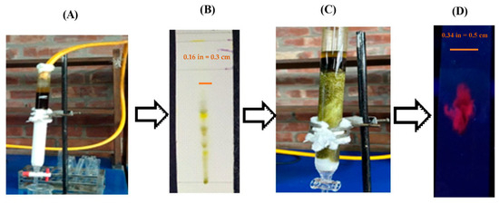
An 8.5 g (F14) methanol crude extract fraction from P. sanguinea was obtained from the first-stage column chromatography. This amount was used in the second-stage column chromatography, and the recovery rate was 92.18% (7.73g). Nine single isolated compounds (IFC1PS to IFC9PS) were isolated from fraction F14 of P. sanguinea (IFCF14PS). Each compound’s single color is clearly visible in the UV chamber at a short wavelength of 254 nm and a long wavelength of 366 nm (Table 3), providing a visual confirmation of their isolation. The TLC technique further confirms each of the single spots (single bioactive compounds) from IFC1PS to IFC9PS (Figure 1).

Table 3.
Results of mass and percentage of IFCPS eluent from the F14 fraction of P. sanguinea methanol extracts.

Figure 1.
Isolation of bioactive fractional compounds from the crude extract of P. sanguinea was conducted using two-stage column chromatography. (A) In the first stage, column chromatography was employed to isolate fractions F1–F18; (B) thin-layer chromatography (TLC) revealed multiple spots in fraction F14; (C) the second stage involved further isolation from fraction F14 to obtain IFC1PS to IFC9PS through additional column chromatography; (D) a single spot was identified by TLC, indicating the presence of only one compound in this fraction.
3.3. In Vitro Anti-Inflammatory Activity
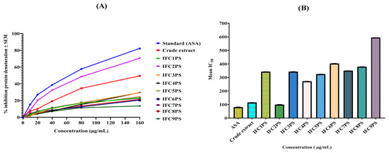
The percentage inhibition of protein denaturation was assessed to determine the efficacy of crude extract (CE) and IFCPS (IFC1PS to IFC9PS). The isolated fractional compounds from P. sanguinea (IFC2PS) demonstrated 70.51% potent inhibition of protein denaturation, with an IC50 value of 99.01 µg/mL. Conventional acetylsalicylic acid showed 82.29% inhibition and an IC50 value of 79.39µg/mL at 160 µg/mL (Table 4). In contrast, various isolated bioactive compounds exhibit mild effects. The IFC3PS and IFC4PS demonstrated mild activity; both were quantified at a similar rate of 29%. All test groups exhibited a dose-dependent activity of protein denaturation (Figure 2).

Table 4.
The effect of CEandIFCPS from P. sanuinea on protein denaturation.

Figure 2.
The anti-inflammatory effects of CE and IFCPS (IFC1PS to IFC9PS) were compared. (A) The IFC2PS showed potent activity, while CE exhibited moderate effects. The IFC3PS and IFC4PS showed mild effects. (B) The IC50 value of CE and IFCPS was also compared. The lowest IC50 value was identified in IFC2PS, followed by CE and other IFCPS. Each raw data point represents mean ± SEM (n = 3). The p-value p < 0.01), compared to control (one-way ANOVA followed by Tukey’s post hoc test).
3.4. Evaluation of Analgesic Activity
3.4.1. Evaluation of Peripheral Analgesic Activity (Acetic Acid-Induced Writhing Method)
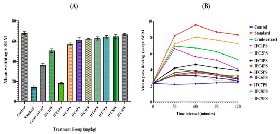
The writhing test evaluated the analgesic effects of crude extracts and IFCPS (IFC1PS to IFC9PS) from P. sanguinea. The IFC2PS at 15 mg/kg demonstrated the most significant (p ˂ 0.01) analgesic effect, with a 73.04% inhibition of writhing, surpassing the crude extract at 46.56%. In contrast, the standard drug diclofenac sodium exhibited a 78.92% inhibition (Figure 2). Most of the isolated fractional compounds from P. sanguinea (IFCPS) at 15 mg/kg doses exhibited mild analgesic effects compared to the standard. The IFC1PS fraction exhibited moderate analgesic effects (Table 5).

Table 5.
In vivo analgesic activities of CE and IFCPS (IFC1PS to IFC9PS) obtained from P. sanguinea.
3.4.2. Evaluation of Central Analgesic Activity (Eddy’s Hot Plate Method)
The central analgesic efficacy of crude extracts and IFCPS (IFC1PS to IFC9PS) was evaluated in mice using the hot plate method. By extending response time (30 min to 120 min) compared to the control at all observation periods, the use of IFC2PS at a dosage of 5 mg/kg and morphine (1 mg/kg) demonstrated substantial pain-relieving activity in the brain (p < 0.05) in the Figure 3. On the other hand, IFC1PS (5 mg/kg) and crude extract (500 mg/kg) showed modest central analgesic efficacy compared to the control (Table 6).

Figure 3.
The analgesic effects of CE and IFCPS (IFC1PS to IFC9PS) were compared. (A) The IFC2PS (15 mg/kg) showed significant analgesic activity, while CE (500 mg/kg) exhibited moderate effects. The IFC1PS exhibited mild effects, but others IFC3PS to IFC9PS did not show any effects by the acetic acid-induced writhing method; (B) the IFC2PS (5 mg/kg) showed a moderate central analgesic effect from 30 to 120 min compared to the morphine drug, while others did not show any effects by Eddy’s hot plate method. Each raw data point represents mean± SEM (n = 6). The p-value * p ˂ 0.01 and * p ˂ 0.05 aresignificant compared to the control.

Table 6.
The analgesic effects of CE and IFCPS (IFC1PS to IFC9PS) were tested in vivo using Eddy’s hot plate technique.
3.5. Determination of Anxiolytic Activities Using Open-Field and Hole Cross Tests
3.5.1. Open-Field Test
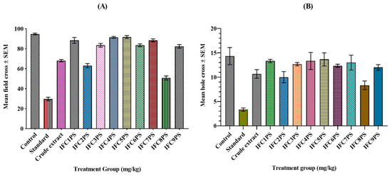
IFC8PS (46.48%) demonstrated significant (p ˂ 0.01) antidepressant activities, followed by IFC2PS (33.45%) at a dose of 5 mg/kg, compared to the standard (68.66%). The remaining IFCPS had a mild impact. In contrast, the crude extract of P. sanguinea showed a 28.17% inhibition of field cross (Table 7).

Table 7.
Effect of anxiolytic activities of CE and IFCPS (IFC1PS to IFC9PS) by the field cross model.
3.5.2. Hole Cross Test
The Hole Cross Test indicated that the IFC8PS exhibited a 41.86% hole cross, followed by IFC2PS at 30.23%, both at a 5 mg/kg dosage. This exceeded the methanol crude extract of P. sanguine, which showed 25.59% at 500 mg/kg. IFC8PS demonstrated moderate anxiolytic efficacy in comparison to the standard diazepam in the Figure 4. The remaining IFCPS had mild antidepressant activity (Table 8).

Figure 4.
The anxiolytic effects of CE and IFCPS were compared. (A) The IFC8PS exerted moderate anxiolytic effects compared with CE as determined by the field cross method; (B) IFC8PS showed moderate activity compared with CE and the standard drug diazepam. Both tests confirmed that IFCPS had moderate anxiolytic activities. The p-value (p ˂ 0.01) was significant compared to the control (one-way ANOVA followed by Tukey’s post hoc test).

Table 8.
Effects on anxiolytic activities of CE and IFCPS (IFC1PS to IFC9PS) by the hole cross model.
4. Discussion
Our initial study on P. sanguinea’s crude extracts revealed the presence of alkaloids, glycosides, flavonoids, terpenoids, saponins, vitamin C, and tannins. We, along with others, demonstrated that the bioactive compounds in alkaloids, terpenoids, flavonoids, and tannins produced analgesic, anti-inflammatory, and anxiolytic activities [26,27]. These findings suggest the potential applications of P. sanguinea’s bioactive compounds in various fields, thereby highlighting the implications of practice.
The polarity of solvents is a critical factor in the activity of phytochemical compounds, particularly in the isolation of bioactive molecules in column chromatography [28]. For example, ethyl acetate, a moderately polar solvent (index polarity of 4.4), is capable of dissolving terpenoids, flavonoids, sterols, and alkaloids. In contrast, methanol, a polar solvent with an index polarity of 5.1, can dissolve a variety of polar compounds, including amino acids, flavones [29], flavonoids, glycosides, polyphenols, saponins, sterol, terpenoids, and tannins [30,31]. N-hexane, a non-polar solvent, is effective in dissolving lignin, lipids, terpenoids, and sterols [32]. Taking into consideration solvent polarity, the isolation of bioactive fractional molecules (IFC1PS to IFC9PS) was carried out. They were subsequently tested in in vitro anti-inflammatory and in vivo analgesic and anxiolytic experiments.
The CE and IFC2PS significantly reduced heat-induced protein denaturation compared to diclofenac sodium, whereas the other IFCPS had little effect. The IFC2PS may inhibit protein denaturation and stabilize cell membranes against lysis [33,34]. Protein denaturation is a biological reaction during a prolonged inflammatory response, potentially leading to a loss of tissue function [35,36]. Consequently, IFC2PS, which stops proteins from breaking down and keeps cell membranes from lysing, could be a lead source of anti-inflammatory drug candidates.
Mouse selection is a rapid, easy, and accurate way to evaluate the peripheral form of analgesic activity. It may also examine centrally acting analgesic effects [37]. Prostacyclin, prostaglandin E, bradykinins, histamine, and biosynthetic prostaglandins are responsible for the pain experienced by mice after receiving an intraperitoneal injection of 7% acetic acid [38,39,40]. Additionally, some studies report that lipoxygenase products cause an analgesic response [41]. The percentage reduction in stomach squirms or cramps indicates the extent of analgesia, providing practical insights into the effectiveness of pain management strategies [42]. Non-steroidal anti-inflammatory drugs, such as diclofenac sodium, suppress the production of prostacyclin, serotonin, and prostaglandin, which reduce writhing in mice and other experimental animals [43].
The CE (46.56%), IFC1PS (25.98%), and IFC2PS (73.04%), which is remarkably similar to diclofenac sodium (78.92%), showed a significant reduction in writhing (p ˂ 0.01), whereas the other IFCPS had a mild effect. This suggests that the CE and IFCPS (Table 5) might reduce the synaptic transmission of painful impulses to the central nervous system, inhibiting acetic acid-induced writing. It also indicates that IFC2PS might inhibit mice writhing through a mechanism almost identical to diclofenac sodium’s, producing a potent peripheral analgesic effect. These findings hold promise for the development of new analgesic strategies.
The hot plate test, the second model utilized in this study, was selected for its practicality and applicability in the field of pharmacology and pain management due to its sensitivity to potent analgesics, minimal tissue damage with a 15s cutoff, high accuracy of results, and low time requirement [44], making it a reliable tool for our research. The hot plate technique yields two measurable behavioral responses: leaping and paw licking, achieved by maintaining a uniform temperature on the plate. Both are considered responses that are integrated at the supraspinal level. Opioids, specifically morphine, are the only analgesics that affect paw-licking behavior, distinguishing them within the field. IFC2PS at 5 mg/kg significantly (p ˂ 0.05) elevated the pain threshold by improving reaction time, starting at 30 min of observation, compared to the negative control. The maximum analgesic effects of the CE, IFC1PS, and IFC2PS were observed at 60 min, yielding 5.28 ± 0.02 s, 4.62 ± 0.21 s, and 7.22 ± 0.04 s, respectively. In contrast, morphine (5 mg/kg) demonstrated a value of 8.33 ± 0.01 s at the same time point. The observed value slightly decreased over 90 min, suggesting that IFC2PS possessed moderate central analgesic effects.
The proposed mechanism for the central analgesic actions of IFC2PS involves its unique ability to stimulate the periaqueductal grey matter (PAG) and the subsequent release of endogenous peptides, such as endorphins or enkephalins [45]. These peptides, crucial to analgesia, descend the spinal cord to inhibit the transmission of pain impulses at the dorsal horn synapse and decrease prostaglandins, leukotrienes, and other vital endogenous substances for central pain transmission [46]. This underscores the importance of understanding pain mechanisms, a vital aspect of our work. Consequently, IFC2PS may facilitate a potent central analgesic impact, demonstrating both central and peripheral analgesic effects.
We comprehensively and rigorously evaluated the anxiolytic activities of CE and IFCPS (IFC1PS to IFC9PS) from P. sanguinea using open-field cross and hole cross methods. The present study compared these compounds to benzodiazepines (diazepam) and utilized multiple methods to ensure robust and reliable results [47]. Benzodiazepines, as positive allosteric GABAA modulators, increase the total chloride ion conduction across the neuronal cell membrane, causing chloride ion influx and hyperpolarizing the neuron’s membrane potential. This effect reduces the difference between resting threshold potential and makes firing less likely, thereby reducing arousal of the cortical and limbic systems in the CNS, resulting in anxiolytic effects [48].
The CE, IFC2PS, and IFC8PSdemonstrated remarkable (p ˂ 0.01) anxiolytic activity compared to the control. In particular, CE (500 mg/kg), IFC2PS (5 mg/kg), and IFC8PS (5 mg/kg) induced calming effect on the Swiss mice, as confirmed by the reduction in the mice’s movements in the OFT and HCT. The plant-derived compound P. sanguinea, which contains alkaloids, glycosides, flavonoids, terpenes, triterpenes, and saponins, may have contributed to its anxiolytic effects [49]. A limitation of this research is that no pure compounds have been isolated. Isolation and characterization of compounds and determination of their activities may inspire future research aiming to identify effective treatment strategies for a wide range of human disorders.
5. Conclusions
This study is a comprehensive exploration of the anti-inflammatory, analgesic, and anxiolytic effects of crude extracts and isolated fractional compounds from the whole plants of P. sanguinea. Among the nine isolated fractional compounds (IFC1PS to IFC9PS), IFC2PS stood out with its unique properties, exhibiting the most significant anti-inflammatory and analgesic effects compared to the standard drug at the same dosage. The potential of IFC2PS to reduce pain and inflammation is an area for further exploration. IFC8PS, meanwhile, exhibited moderate effects on the standard drug and superior anxiolytic activities compared to crude extracts and other isolated fractions. IFC1PS has moderate analgesic and anti-inflammatory properties. Further investigation is required to determine the structure of IFC1PS, IFC2PS, and IFC8PS, and to elucidate the specific mechanisms that govern their pharmacological properties.
Author Contributions
Conceptualization, methodology, software, M.Q.A.; Validation and formal analysis, R.N.; Investigation, resources, S.M.S.I.; Data curation, writing original draft preparation, M.T.N. and M.Q.A.; writing—review and editing, visualization, supervision, M.T.N. and S.M.S.I.; project administration, S.M.S.I.; funding acquisition, M.T.N. All authors have read and agreed to the published version of the manuscript.
Funding
This research received no external funding.
Institutional Review Board Statement
The Institutional Animal, Medical Ethics, Biosafety, and Biosecurity Committee (IAMEBBC) of the Institute of Biological Sciences, University of Rajshahi, Bangladesh, authorized this work, [project identification code 226/320/(69) IAMEBBC/IBSc] on 10th December 2024.
Informed Consent Statement
Not applicable.
Data Availability Statement
The authors confirm that the data supporting the findings of this study are available within the article.
Acknowledgments
The authors express gratitude to the Department of Pharmacy at Southern University, Chattogram, Bangladesh, and the Plant Biotechnology and Genetic Engineering Lab, Institute of Biological Sciences, University of Rajshahi, Bangladesh, for their cooperation in providing laboratory assistance for the whole study. Works at MTN lab are being funded by the Royal Society, the Commonwealth Scholarship Commission, GrowMedtech, the Schlumberger Foundation, and the University of Bradford, UK.
Conflicts of Interest
The authors state that they have no conflicts of interest with this publication and that no major financial assistance might have impacted its conclusion. All writers reviewed and approved the manuscript.
References
- Abera, B.; Hailu, A.E. Evaluation of analgesic activities of 80% methanol leaf extract of Solanum incanum L. (Solanaceae) in mice. J. Drug Deliv. Ther. 2019, 9, 9–14. [Google Scholar] [CrossRef]
- Ahmed, A.; Rajendaran, K.; Jaiswal, D.; Singh, H.P.; Mishra, A.; Chandra, D.; Yadav, I.K.; Jain, D.A. Anti-snake venom activity of different extracts of Pouzolzia indica against Russel viper venom. Int. J. ChemTech Res. 2010, 2, 744–751. [Google Scholar]
- Ahsan, Q.; Alam, M.T.; Chowdhury, M.M.U.; Nasim, M.T.; Islam, S.M.S. In-vitro and in-vivo evaluation of pharmacological activities of Pouzolzia sanguinea. J. Bio-Sci. 2021, 29, 31–42. [Google Scholar] [CrossRef]
- Akindele, A.J.; Ibe, I.F.; Adeyemi, O.O. Analgesic and antipyretic activities of Drymaria cordata (Linn.) Wild (Caryophyllaceae) extract. Afr. J. Tradit. Complement. Altern. Med. 2012, 9, 25–35. [Google Scholar] [CrossRef][Green Version]
- Ayanwuyi, L.O.; Yaro, A.H.; Abodunde, O.M. Analgesic and anti-inflammatory effects of the methanol stem bark extract of Prosopis africana. Pharm. Biol. 2010, 48, 296–299. [Google Scholar] [CrossRef][Green Version]
- Battu, G.R.; Parimi, R.; Kottapalli, B.; Shekar, C. In vivo and in vitro pharmacological activity of Aristolochia tagala (syn: Aristolochia acuminata) root extracts. Pharm. Biol. 2011, 49, 1210–1214. [Google Scholar] [CrossRef]
- Chi, V.V. Dictionary of Vietnamese Medicinal Plants; Medicine Publishing House: Hanoi, Vietnam, 2012; pp. 210–215. [Google Scholar]
- Chung, K.T.; Wong, T.Y.; Wei, C.I.; Huang, Y.W.; Lin, Y. Tannins and human health: A review. Crit. Rev. Food Sci. Nutr. 1998, 38, 421–464. [Google Scholar] [CrossRef]
- Cowan, M.M. Plant product as antimicrobial agents. Clin. Microbiol. Rev. 1999, 12, 564–582. [Google Scholar] [CrossRef]
- Eddy, N.B.; Leimbach, D. Synthetic analgesics. II. Dithienylbutenyl and dithienylbutylamines. J. Pharmacol. Exp. Ther. 1953, 107, 385–393. [Google Scholar] [CrossRef] [PubMed]
- Fedotova, J.; Kubatka, P.; Büsselberg, D.; Shleikin, A.G.; Caprnda, M.; Dragasek, J.; Rodrigo, L.; Pohanka, M.; Gasparova, I.; Nosal, V.; et al. Therapeutical strategies for anxiety and anxiety-like disorders using plant-derived natural compounds and plant extracts. Biomed. Pharmacother. 2017, 95, 437–446. [Google Scholar] [CrossRef]
- Firdous, A.M.; Fayaz, A.L.; Mushtaq, A.S. Isolation of active components derived from rhizome of Euphorbia wallichii Hook. Int. J. Ayurvedic Herb. Med. 2013, 3, 1173–1183. [Google Scholar]
- García, M.D.; Fernandez, M.A.; Alvarez, A.; Saenz, M.T. Antinociceptive and anti-inflammatory effect of the aqueous extract from leaves of Pimenta racemosa var. Ozua (Mirtaceae). J. Ethnopharmacol. 2004, 91, 69–73. [Google Scholar] [CrossRef]
- Ghani, A. Practical Pharmacognosy Textbook, 3rd ed.; Parash Publishers: Dhaka, Bangladesh, 2005. [Google Scholar]
- Gupta, B.; Dandiya, P.; Gupta, M. A psycho-pharmacological analysis of behaviour in rats. Jpn. J. Pharmacol. 1971, 21, 293–298. [Google Scholar] [CrossRef] [PubMed]
- Hemavathy, A.; Shanthi, P.; Sowndharya, C.; Sundari, T.S.; Priyadharshni, K. Extraction and isolation of bioactive compounds from a therapeutic medicinal plant—Wrightia tinctoria (Roxb.) R. Br. Int. J. Pharmacogn. Phytochem. Res. 2019, 11, 199–204. [Google Scholar]
- Hossain, M.A.; Salehuddin, S.M. Simultaneous quantification of sinensetin and tetramethoxyflavone in misaikucing capsules using TLC-UV densitometric technique. J. Sci. Res. 2009, 1, 403–407. [Google Scholar] [CrossRef]
- Houghton, P.J.; Raman, A. Laboratory Handbook for the Fractionation of Natural Extracts, 3rd ed.; Chapman and Hall: New York, NY, USA, 1998. [Google Scholar]
- Ikwegbue, P.C.; Masamba, P.; Oyinloye, B.E.; Kappo, A.P. Roles of heat shock proteins in apoptosis, oxidative stress, human inflammatory diseases, and cancer. Pharmaceuticals 2018, 11, 2. [Google Scholar] [CrossRef]
- Islam, M.R.; Naima, J.; Proma, M.N.; Hussain, M.S.; Uddin, S.M.; Hossain, K.M. In-vivo and in-vitro evaluation of pharmacological activities of Ardisia solanaceae leaf extract. Clin. Phytosci. 2019, 5, 32. [Google Scholar] [CrossRef]
- Kong, H.S.; Musa, Z.; Kasim, M.; Sani, N.A. Qualitative and quantitative phytochemical analysis and antioxidant properties of leaves and stems of Clinacanthus nutans (Burm. f.) Lindau from two herbal farms of Negeri Sembilan, Malaysia. ASM Sci. J. 2019, 12, 1–3. [Google Scholar] [CrossRef]
- Kupchan, S.M.; Britton, R.W.; Ziegler, M.F.; Sigel, C.W. Bruceantin, a new potent antileukemic simaroubolide from Brucea antidysenterica. J. Org. Chem. 1973, 38, 178–179. [Google Scholar] [CrossRef]
- Larayetan, R.; Ololade, Z.S.; Ogunmola, O.O.; Ladokun, A. Phytochemical constituents, antioxidant, cytotoxicity, antimicrobial, antitrypanosomal, and antimalarial potentials of the crude extracts of Callistemon citrinus. eCAM 2019, 5410923. [Google Scholar] [CrossRef]
- Lee, K.H.; Choi, E.M. Analgesic and anti-inflammatory effects of Ligularia fischeri leaves in experimental animals. J. Ethnopharmacol. 2008, 120, 103–107. [Google Scholar] [CrossRef]
- Levini, J.D.; Lau, W.; Kwait, G.; Goetzl, E.J. Leukotnene B4 produces hyperalgesia that is dependent on the polymorphonuclear leucocytes. Sciences 1984, 225, 743–745. [Google Scholar] [CrossRef]
- Lira, S.M.; Canabrava, N.V.; Benjamin, S.R.; Silva, J.Y.G.; Viana, D.A.; Lima, C.L.S.; Paredes, P.F.M.; Marques, M.M.M.; Pereira, E.O.; Queiroz, E.A.M.; et al. Evaluation of the toxicity and hypoglycemic effect of the aqueous extracts of Cnidoscolus quercifolius Pohl. Braz. J. Med. Biol. Res. 2017, 50, e6361. [Google Scholar] [CrossRef] [PubMed]
- Maria, V.R. Determination of acute and sub-acute toxicity of six plant extracts. Medrech 2019, 6, 64–67. [Google Scholar]
- Nawaz, H.; Shad, M.A.; Rehman, N.; Andaleeb, H.; Ullah, N. Effect of solvent polarity on extraction yield and antioxidant properties of phytochemicals from bean (Phaseolus vulgaris) seeds. Braz. J. Pharm. Sci. 2020, 56, e17129. [Google Scholar] [CrossRef]
- Ngoua-Meye-Misso, R.L.; De, J.C.; Sima-Obiang, C.; Ondo, J.P.; Ndong-Atome, G.R.; Abessolo, F.O.; Obame-Engonga, L.C. Phytochemical studies, antiangiogenic, anti-inflammatory and antioxidant activities of Scyphocephalium ochocoa Warb. (Myristicaceae), medicinal plant from Gabon. Clin. Phytosci. 2018, 4, 15. [Google Scholar] [CrossRef]
- Nhung, L.T.H.; Huong, P.T.M.; Anh, N.T.; Tai, B.H.; Nhiem, N.X.; Doan, V.V. Two new norlignans from the aerial parts of Pouzolzia sanguinea (Blume) Merr. Nat. Prod. Res. 2020, 15, 157–164. [Google Scholar] [CrossRef]
- Payum, T.; Das, A.K.; Shankar, R.; Tamuly, C.; Hazarika, M. Antioxidant potential of Pouzolzia bennettiana—A nutritious traditional food plant used in Arunachal Pradesh, India. AJPLSR 2015, 3, 1–7. [Google Scholar]
- Rabe, T.; van Staden, J. Antibacterial activity of South African plants used for medicinal purposes. J. Ethnopharmacol. 1997, 56, 81–87. [Google Scholar] [CrossRef]
- Rahman, M.A.; Uddin, S.B.; Wilcock, C.C. Medicinal plants used by Chakma tribe in Hill Tracts districts of Bangladesh. IndianJ.Tradit. Knowl. 2007, 6, 508–517. [Google Scholar]
- Ranaweera, K.; Rupasinghe, H. In vitro anti-inflammatory properties of selected green leafy vegetables. Biomedicines 2018, 6, 107. [Google Scholar] [CrossRef] [PubMed]
- Ravtsova, T.I.; Friis, I.; Wilmot-Dear, C.M. Morphology and anatomy of fruits in Pouzolzia (Urticaceae) in relation to taxonomy. Kew Bull. 2003, 58, 297–327. [Google Scholar] [CrossRef]
- Ries, R.K. Principles of Addiction Medicine, 4th ed.; Wolters Kluwer: Philadelphia, PA, USA, 2009. [Google Scholar]
- Sangeetha, G.; Vidhya, R. In vitro anti-inflammatory activity of different parts of Pedalium murex (L.). Int. J. Herb. Med. 2016, 4, 31–36. [Google Scholar]
- Sasidharan, S.; Chen, Y.; Saravaran, D.; Sundram, K.M.; Latha, L.Y. Extraction, isolation and characterization of bioactive compounds from plants’ extracts. Afr. J. Tradit.Complement. Altern. Med. 2011, 8, 1–10. [Google Scholar] [CrossRef]
- Seibenhener, M.L.; Wooten, M.C. Use of the open field maze to measure locomotor and anxiety-like behavior in mice. J. Vis. Exp. 2015, 96, e52434. [Google Scholar] [CrossRef]
- Shinde, U.A.; Phadke, A.S.; Nair, A.M.; Mungantiwar, A.A.; Dikshit, V.J.; Saraf, M.N. Membrane stabilizing activity—A possible mechanism of action for the anti-inflammatory activity of Cedrus deodara wood oil. Fitoterapia 1999, 70, 251–257. [Google Scholar] [CrossRef]
- Siriwatanametanon, N.; Fiebich, B.L.; Efferth, T.; Prieto, J.M.; Heinrich, M. Traditionally used Thai medicinal plants: In Vitro anti-inflammatory, anticancer and antioxidant activities. J. Ethnopharmacol. 2010, 130, 196–207. [Google Scholar] [CrossRef] [PubMed]
- Takagi, K.; Watanabe, M.; Saito, H. Studies of the spontaneous movement of animals by the hole cross test; effect of 2-dimethyl-aminoethanol and its acyl esters on the central nervous system. Jpn. J. Pharmacol. 1971, 21, 797–810. [Google Scholar] [CrossRef]
- Trongsakul, S.A.; Panthong, D.; Kanjanpothi, D.; Taesotikul, T. The analgesic, antipyretic and anti-inflammatory activity of Diospyros variegata Kruz. J. Ethnopharmacol. 2003, 85, 221–225. [Google Scholar] [CrossRef]
- Turner, R.A. Screening Methods in Pharmacology, 4th ed.; Academic Press: New York, NY, USA, 1967. [Google Scholar]
- Uche, F.I.; Aprioku, J.S. The phytochemical constituents, analgesic and anti-inflammatory effects of methanol extract of Jatropha curcas leaves in Mice. J. Appl. Sci. 2008, 12, 99–102. [Google Scholar] [CrossRef]
- Widyawati, S.; Paini, T.D.; Budianta, W.; Kusuma, F.A.; Wijaya, E.L. Difference of solvent polarity to phytochemical content and antioxidant activity of Pluchea indica less leaves extracts. Int. J. Pharmacogn. Phytochem. Res. 2014, 6, 850–885. [Google Scholar]
- Wolde-Mariam, M.; Yarlagadda, R.; Asres, K. In vivo anti-inflammatory and antinociceptive activities of the aerial part extract of Dicliptera laxata. Int. J. Green Pharm. 2013, 7, 35–39. [Google Scholar] [CrossRef]
- Yadav, I.K.; Singh, H.P.; Jain, D.A.; Ahmed, A.; Indranil, D. In vitro antioxidant and radical scavenging activity of different extracts of Pouzolzia indica. Elixir Int. J. 2012, 47, 9143–9148. [Google Scholar]
- Yimer, T.; Birru, M.; Adugna, M.; Geta, M.; Yohannes, E. Evaluation of analgesic and anti-Inflammatory activities of 80% methanol root extract of Echinops kebericho M. (Asteraceae). J. Inflamm. Res. 2020, 30, 647–658. [Google Scholar] [CrossRef] [PubMed]
Disclaimer/Publisher’s Note: The statements, opinions and data contained in all publications are solely those of the individual author(s) and contributor(s) and not of MDPI and/or the editor(s). MDPI and/or the editor(s) disclaim responsibility for any injury to people or property resulting from any ideas, methods, instructions or products referred to in the content. |
© 2025 by the authors. Licensee MDPI, Basel, Switzerland. This article is an open access article distributed under the terms and conditions of the Creative Commons Attribution (CC BY) license (https://creativecommons.org/licenses/by/4.0/).