Review

Targeting Obesity in Cardiovascular Disease Management: Cardiac Adipose Tissue Is a Real Biomarker!

1
Cardiology Unit, Azienda Ospedaliera Universitaria Luigi Vanvitelli, 80138 Naples, Italy
2
Department of Advanced Medical and Surgical Sciences, University of Campania Luigi Vanvitelli, Piazza Luigi Miraglia, 2, 80138 Naples, Italy
3
Department of Translational Medical Sciences, Section of Cardiology, University of Campania Luigi Vanvitelli, 80131 Naples, Italy
4
Department of Precision Medicine, University of Campania Luigi Vanvitelli, 80131 Naples, Italy
5
Pharmacy Unit, Monaldi Hospital, 80131 Naples, Italy
6
Vanvitelli Cardiology and Intensive Care Unit, Monaldi Hospital, 80131 Naples, Italy
7
Department of Life Science, Health, and Health Professions, Link Campus University, 00165 Rome, Italy
*
Author to whom correspondence should be addressed.
Targets 2025, 3(2), 17; https://doi.org/10.3390/targets3020017
Submission received: 9 March 2025 / Revised: 17 May 2025 / Accepted: 19 May 2025 / Published: 23 May 2025

Abstract

:
Background: Obesity has been defined as a true worldwide “pandemic” by the World Health Organization and represents one of the major public health problems. It is associated with a reduction in life expectancy of about 7–8 years due to related cardiovascular diseases such as arterial hypertension, metabolic syndrome, insulin resistance, type 2 diabetes mellitus, and dyslipidemia. Adipose tissue is not merely a fat storage site but a true endocrine and immunologically active organ that secretes hormones and mediators (adipokines), influencing cardiovascular risk and host physiology. Objective: This review summarizes the current understanding of the role of epicardial adipose tissue (EAT) in cardiovascular disease pathophysiology and discusses its clinical diagnostic and therapeutic implications. Methods: A narrative non-systematic review was conducted focusing on recent literature concerning the biological and clinical aspects of cardiac adipose tissue, with particular emphasis on epicardial adipose tissue. The review examined its gene expression profile, secretory function, and interaction with cardiovascular structures and diseases. Findings: There are different types of adipose tissue, including cardiac adipose tissue, which comprises epicardial and pericardial (or paracardiac) fractions. Epicardial adipose tissue is unique due to its proximity to the heart and a distinct gene expression profile compared to other adipose depots such as visceral and subcutaneous fat. EAT plays a crucial role in the development and progression of cardiovascular diseases with high morbidity and mortality, acting both as a metabolic and inflammatory mediator. Conclusion: Cardiac adipose tissue, particularly EAT, is a key player in cardiometabolic disease. Understanding its pathophysiological role and incorporating imaging tools to evaluate EAT may enhance cardiovascular risk stratification and disease management.

1. Introduction

Obesity is increasing worldwide and is one of the most important risk factors for the development of type 2 diabetes (T2DM) and cardiovascular disease (CVD) [1]. In most obese individuals, the inability to form new cells capable of storing fat (adipocytes) causes fat accumulation in cells already present in adipose tissue [2,3]. These oversized (hypertrophied) cells send alarm signals to the immune system, in response of which some cells of the immune system (particularly macrophages and lymphocytes) are drawn back into the adipose tissue (AT) itself [3]. This results in the creation of an inflammatory environment that interferes with the maturation of new fat cells, making the AT insensitive to the action of insulin [4]. The creation of this vicious cycle of inflammatory character is associated with the onset of insulin resistance, which is one of the mechanisms underlying T2DM [5].
In the AT of patients with hypertrophic obesity, oversized adipocytes, engulfed by excess fat, and immune system cells called to the rescue from the AT itself by inflammatory molecules produce high levels of pro-inflammatory mediators, which further maintain this vicious cycle [2,3,4]. In recent years, among the various types of AT, cardiac AT is of particular interest due to its unique anatomical and physiological characteristics. It comprises three distinct layers: epicardial, pericardial, and paracardiac fat [6]. Of these, epicardial adipose tissue (EAT) has been matter of intense research because of its close anatomical proximity and functional relationship with the heart [7]. Located between the myocardium and the visceral layer of the pericardium, EAT is increasingly recognized as an active tissue involved in the regulation of metabolic processes, inflammation, and myocardial function. Additionally, unlike pericardial and paracardial fat, EAT is metabolically active, secreting pro-inflammatory cytokines that promote atherosclerosis and myocardial dysfunction [6]. The accumulation of excessive EAT has been linked to various cardiovascular diseases (CVDs), including atherosclerosis and heart failure. Consequently, advanced imaging techniques have become crucial for non-invasively evaluating EAT, allowing for detailed analysis of its volume, distribution, and potential impact on cardiovascular risk.
EAT is composed primarily of adipocytes, but also contains blood vessels, nerve fibers, and immune cells, contributing to its role in both metabolic and immune regulation [7]. CAT (cardiac adipose tissue) and EAT have different embryogenic origins [8].
EAT forms a protective layer around the heart due to its anatomical location. It is typically categorized based on its position relative to the heart and pericardium.
Epicardial fat is located between the myocardium and the visceral layer of the pericardium. This depot is highly metabolically active and contributes to the production of adipokines and inflammatory cytokines. It shares the coronary blood supply and is in direct contact with the myocardium.
Pericardial (or paracardial) fat is situated outside the parietal pericardium. While it does not have the same intimate relationship with the myocardium, it contributes to overall cardiac insulation and mechanical protection. Other advantageous functions include anti-inflammatory and anti-atherogenic properties through the expression of proteins as adiponectin and adrenomedullin [9,10]. EAT is also an important source of heat and it is involved in various biological processes: regulation of body fluid levels, wound healing, mesoderm development, and plasma membrane organization [11]. Physiologically, with aging, brown adipose tissue (BAT) tends to decrease, while white adipose tissue (WAT) may develop beige characteristics under external stimuli [9,10]. In the present article, we will review the current literature on obesity and CVD in light of the role of cardiac AT as a marker of cardiovascular risk/diseases, starting from the cellular characteristics of AT to its clinical relevance, to help the physicians better understand its role as a disease marker.

2. Literature Sources and Search Strategy

We performed a non-systematic review of the literature by applying the search strategy in different electronic databases (MEDLINE, EMBASE, Cochrane Register of Controlled Trials, and Web of Science). Original reports, meta-analyses, and review articles in peer-reviewed journals up to January 2025 regarding adipose tissue, cardiac adipose tissue, obesity, metabolic syndrome, and adipocytes molecular pathways were incorporated into the search strategy. The references of all identified articles were reviewed to look for additional papers of interest from which to extrapolate recently available data on epicardial, pericardial, and paracardiac AT, with a view to investigating possible biochemical pathways involved.

3. Physiological Role of Adipose Tissue: Is It for Good?

The AT is not only an energy depot but also plays a key role in metabolic regulation and homeostasis of the organism. It serves various physiological and pathological functions, as summarized in Table 1.
AT serves both beneficial and harmful functions depending on its regulation and interactions with other organs. Understanding these mechanisms is crucial for addressing metabolic diseases and cardiovascular health. AT is divided into white adipose tissue (WAT) and brown adipose tissue (BAT), with different cellular functions and characteristics [20]. At the cellular level, WAT is mainly composed of unilocular adipocytes. It is characterized by a large lipid droplet that occupies most of the cytoplasm, pushing the nucleus to the periphery. These adipocytes are responsible for storing lipids in the form of triglycerides and releasing them in the form of free fatty acids (FFAs), according to one’s energy needs [29]. BAT, on the other hand, is composed of multilocular adipocytes, characterized by the presence of numerous lipid droplets and a high density of mitochondria, containing the uncoupling protein UCP1. This allows for heat production through thermogenesis, dissipating energy as heat, rather than storing it as lipids, offering protective effects against obesity-related metabolic disorders [30,31]. Indeed, it is associated with healthier fat distribution, reduced visceral fat, and improved metabolic health, including lower blood glucose levels and reduced fat accumulation in the liver [13]. WAT also acts as an endocrine organ due to its secretion of hormones and cytokines, namely adipokines, which regulate energy balance, glucose homeostasis, and lipid metabolism [32]. Among these, leptin and adiponectin are particularly significant. Leptin, primarily produced by WAT, plays a crucial role in appetite regulation and energy expenditure by signaling satiety to the hypothalamus, thereby reducing food intake [14]. Additionally, leptin influences insulin sensitivity and has pro-inflammatory properties, contributing to immune responses [33]. Conversely, adiponectin enhances insulin sensitivity and exhibits anti-inflammatory effects. It promotes glucose uptake and fatty acid oxidation, thereby playing a protective role against metabolic disorders such as T2DM and CVD [15]. Notably, adiponectin levels are inversely correlated with body fat percentage; they decrease in obesity, which may contribute to the pathogenesis of insulin resistance [34]. The balance and function of these adipokines are essential for maintaining metabolic health, and their dysregulation is implicated in the development of various metabolic and inflammatory diseases [16]. The metabolic and immunological roles of adipose tissue, as reported in Table 1, have profound cardiovascular implications. Excessive secretion of pro-inflammatory adipocytokines by visceral adipose tissue, particularly TNF-α and IL-6, contributes to the development of systemic insulin resistance, a known risk factor for atherosclerosis. Furthermore, reduced production of adiponectin, an anti-inflammatory and vasoprotective molecule, impairs endothelial function, promoting oxidative stress, vascular dysfunction, and progression of atherosclerosis. Therefore, adipose tissue dysfunction appears to be a central mediator between obesity and cardiovascular disease [2].
In addition to WAT and BAT, there is also a third type of adipocyte, known as beige or brite (brown-in-white), that has been identified in recent years. These adipocytes are found within WAT and can acquire thermogenic characteristics like BAT in response to stimuli such as cold exposure or physical activity [20]. Cold exposure activates beige adipocytes through the sympathetic nervous system, primarily via β-adrenergic signaling, leading to the upregulation of uncoupling protein 1 (UCP1) and enhanced thermogenesis [26]. Recent studies also suggest that exercise induces the browning of WAT through the release of myokines such as irisin and catecholamines, which stimulate mitochondrial biogenesis and thermogenic gene expression in beige adipocytes [25]. This plasticity of beige fat highlights its potential role in energy homeostasis and metabolic health, particularly in counteracting obesity and insulin resistance [24].
AT is also composed of a complex population of stromal cells, including pre-adipocytes, endothelial cells, macrophages, and other leukocytes. They contribute to the regulation of adipose function. Adipose tissue macrophages (ATMs) play a crucial role in maintaining metabolic homeostasis through their phenotypic polarization into pro-inflammatory (M1) and anti-inflammatory (M2) states. In recent years, therapeutic interest in modulating macrophage polarization in adipose tissue has significantly increased. In particular, PPAR-γ receptor agonists, such as pioglitazone, have been shown to promote polarization toward the anti-inflammatory M2 phenotype, while reducing the number of pro-inflammatory M1 macrophages. Lifestyle interventions, such as regular physical activity and caloric restriction, have also shown beneficial effects on the immune composition of adipose tissue, favoring a less inflammatory microenvironment. These strategies represent promising approaches to reduce obesity-associated chronic inflammation and prevent cardiovascular complications [35]. Under lean conditions, M2 macrophages predominate, contributing to tissue remodeling and insulin sensitivity [17]. However, in obesity, there is a marked shift towards M1 polarization, characterized by increased infiltration of macrophages that secrete pro-inflammatory cytokines such as tumor necrosis factor-alpha (TNF-α) and interleukin-6 (IL-6), which contribute to systemic insulin resistance [18]. The recruitment of monocyte-derived macrophages into AT during obesity is mediated by chemokines like monocyte chemoattractant protein-1 (MCP-1), which exacerbates inflammatory responses and disrupts metabolic homeostasis [36]. Additionally, hypoxic conditions in hypertrophic AT activate hypoxia-inducible factor-1 alpha (HIF-1α), further promoting M1 polarization and inflammatory cytokine production [19]. Recent studies suggest that targeting macrophage polarization could serve as a potential therapeutic approach for obesity-related metabolic disorders. Specifically, promoting the M2 phenotype through pharmacological agents or lifestyle interventions such as exercise and dietary modifications could be effective in mitigating AT inflammation and improving insulin sensitivity [37].
Obesity is certainly a risk factor for CVD [2]. However, clinical research has revealed a paradoxically protective role of obesity in patients with chronic diseases, including CVD, suggesting that the biological “quality” of AT may be more important than total AT mass or body weight. Importantly, as a dynamic organ, it secretes a wide range of biologically active adipokines, microRNAs, gaseous messengers, and other metabolites that influence the cardiovascular system in both an endocrine and paracrine manner [13]. In spite of being able to mediate normal cardiovascular function under physiological conditions, AT undergoes a phenotypic shift characterized by the acquisition of pro-oxidant and pro-inflammatory properties in CVD.
Cardiac adipocytes (CAs) reside in three distinct regions: pericardial (PAT), epicardial (EAT), and intramyocardial AT (IAT). The PAT is situated between the visceral and parietal pericardium [38]. EAT plays a significant role in cardiovascular health and disease due to its proximity to the heart and coronary arteries. The IAT is observed within the myocardium in mice and humans [39]. The majority of intramyocardial adipocytes are in the inner myocardial wall within 100 µm from the endocardium in a normal adult mouse heart [40]. The IAT is infrequently observed in the normal adult human heart (as diagnosed by multisided computed tomography) but commonly detected in the subendocardium of patients with myocardial infarction (MI) [41]. Crucially, recent evidence suggests that AT depots such as perivascular AT and EAT can change their phenotype in response to local signals of vascular and myocardial origin, respectively. Recent clinical evidence supports the hypothesis that inflammatory EAT may induce myocardial fibrosis. A 2025 study by Zhu and colleagues, using cardiac magnetic resonance imaging, showed that in non-diabetic hypertensive patients, insulin resistance exacerbates myocardial fibrosis through functional alteration of the EAT. In particular, subjects with greater epicardial inflammation presented an increase in extracellular volume and a prolongation of T1 relaxation times, indirect indicators of fibrosis. These results suggest that EAT may act not only as a biomarker but also as an active mediator in the process of fibrotic remodeling of the myocardium [42]. Using this unique property of some AT depots to dynamically track cardiovascular biology could reveal new diagnostic and prognostic tools against CVD [8].
EAT has both protective and harmful roles depending on its state. Under normal conditions, it provides thermogenic and mechanical protection to the heart and supplies energy through FFAs. However, in obesity and metabolic disorders, EAT becomes dysfunctional, characterized by increased inflammation and fibrosis, contributing to myocardial damage and CVDs [43,44,45]. The secretion of pro-inflammatory and pro-fibrotic mediators by EAT can impair the cardiac structure and function, leading to conditions such as diastolic heart failure [45,46].

4. Mechanistic Insight: The Inflammatory-EAT Axis and Cardiovascular Risk

It has been shown that the activation of inflammatory and oxidative pathways is one of the major mechanisms by which EAT contributes to cardiovascular disease (CVD) [10,46]. In cases of obesity and insulin resistance, epicardial adipose tissue (EAT) cells begin to hypertrophy and lose their sensitivity to insulin. This results in an increase in lipolysis and the release of free fatty acids (FFAs) [12,47]. Consequently, a local hypoxic environment is created, leading to the recruitment of immune cells, particularly polarized M1 macrophages and activated T lymphocytes [18,19]. These immune cells release pro-inflammatory cytokines such as TNF-α, IL-6, and MCP-1, which in turn activate key signaling pathways, including nuclear factor kappa B (NF-κB) and c-Jun N-terminal kinase (JNK) [48,49]. The NF-κB pathway, upon activation by TNF-α and IL-6, induces the expression of pro-inflammatory cytokines and adhesion molecules, which amplify the inflammatory response. The JNK pathway, activated by oxidative stress and inflammatory signals, leads to the phosphorylation of key transcription factors and proteins involved in cell survival, apoptosis, and further production of ROS. Both NF-κB and JNK are involved in the promotion of endothelial dysfunction, a key step in the development of atherosclerosis. Additionally, dysfunctional EAT produces reactive oxygen species (ROS), which exacerbate oxidative stress and contribute to endothelial dysfunction, fibrosis, and the development of atherosclerosis [50,51]. This inflammatory–oxidative connection suggests that EAT is not merely a site of passive fat accumulation, but actively mediates myocardial damage and coronary artery disease (CAD), playing a pivotal role in cardiovascular health [23]. A schematic view is provided in Figure 1.

5. The Dark Side of Adipose Tissue

Chronic low-grade inflammation of AT is one of the mechanisms that lead to the progression of metabolic diseases such as T2DM and CVD [47]. WAT retains the ability to expand during adult life to accommodate chronic excess caloric intake [49]. AT expands through adipocyte hypertrophy, increases in fat-cell size and/or hyperplasia, and increases in fat-cell number [11]. Obesity can lead to adipocytes’ hypertrophy and hyperplasia [48]. AT expansion in obesity is accompanied by inflammatory changes within AT, contributing to chronic low-grade systemic inflammation that is characterized as mildly elevated levels of circulating cytokines, chemokines, and acute phase reactants [50]. Expansion of AT depots during weight gain is accompanied by an infiltration of new inflammatory cells, the major one initially being macrophages [51]. These pro-inflammatory cells are recruited in response to chemokines such as monocyte chemotactic protein-1 (MCP-1) produced by hypertrophic adipocytes [23].
Increased macrophage infiltration into AT forms a crown-like structure (CLS) around necrotic adipocytes. The number of CLSs is strongly correlated with the expression of inflammatory cytokines like TNF-α, indicating that infiltrating macrophages have a pro-inflammatory effect on AT in obesity [52]. Macrophage accumulation occurs largely in visceral rather than in subcutaneous adipose depots in both rodents and humans [53,54]. The pro-inflammatory AT macrophages are one of the key cell types responsible for producing pro-inflammatory cytokines such as TNF-α, IL-1β, and IL-6, which contribute to obesity-related adipose tissue inflammation [23].
In summary, EAT may lead to a systemic pro-inflammatory state (release of IL-6, TNF-α) and releases FFAs, which contribute to initiating and perpetuating a state of systemic insulin resistance (IR) [55]. In fact, the EAT volume correlates with fasting glucose, HbA1c, and HOMA-IR, acting both as a marker and contributor to systemic metabolic dysfunction [56].
In obesity, the upregulation of TNF-α stimulates the activation of the inhibitor of IκB kinase (IKK)-β and MAPKs (such as p38, c-Jun N-terminal kinase [JNK], and extracellular signal-regulated kinase [ERK]). These enzymes directly target serine residues on the insulin receptor substrate (IRS) protein, hindering its tyrosine phosphorylation in an NF-κB-dependent manner. This cascade ultimately culminates in insulin resistance (IR) within insulin-targeted tissues [57,58].
Excessive EAT accumulation in insulin-resistant (IR) patients has been demonstrated. In epicardial adipocytes, even under physiological conditions, insulin-dependent glucose uptake and the anti-lipolytic function of insulin are reduced compared to adipocytes of the subcutaneous fat depot, which is associated with the need to maintain high lipolysis activity [59]. IL-6 stands as another significant inflammatory mediator contributing to IR by activating the Janus kinase-signal transducer and activator of transcription (JAK–STAT) signaling pathway. This activation results in elevated expression of the suppressor of cytokine signaling 1 (SOCS1) and SOCS3 proteins, which, in turn, decrease the expression of glucose transporter-4 (GLUT4) and IRS-1, further exacerbating IR [60]. In addition to macrophages, T cells also are present in normal AT and demonstrate phenotypic change during weight gain. Both CD4+ and CD8+ T cells are found in AT and are increased in the obese state [50]. Interferon γ (IFNγ)-expressing Th1-polarized T cells appear to promote AT inflammation, and increased IFN-γ activity has been reported in AT in both mice and humans [61,62]. AT expansion and dysfunction contribute to the development of CVD through direct and indirect mechanisms. In obesity patients, systemic or local inflammation and IR can cause macrophages in AT to switch from anti-inflammatory and anti-atherosclerotic to pro-inflammatory and pro-atherosclerotic [27]. It is known that obesity may induce hypertension through visceral AT expansion, causing mechanical compression of the kidney, sympathetic nervous system activity, and activation of the renin–angiotensin–aldosterone system (RAAS) [2].
In obesity and metabolic syndrome, EAT expands and is more inflamed, secreting higher levels of pro-inflammatory adipokines and FFAs, which contribute to coronary artery disease (CAD), atrial fibrillation, and heart failure [63]. By regulating the release/uptake of FFAs, EAT plays an important role in CAD by supporting the efficiency of myocardial glucose utilization [64,65]. Recent studies have shown that EAT is associated with the occurrence and development of CAD [66]. In the meta-analysis by Wang et al. [66], a total of 21 studies encompassing 4975 subjects met the inclusion criteria, including 2377 diagnosed and assigned as the CAD group, while the other 2598 were assigned as the non-CAD group. Both the volume and thickness of EAT in the CAD group were larger compared to the non-CAD group (p < 0.00001). In a subgroup analysis within the CAD group, the severe stenosis group had a larger volume and thickness with respect to EAT when compared to the mild/moderate group (p < 0.001) thus indicating that the EAT thickness and volume are associated with CAD severity [66] and could be considered predictors of CAD itself [67]. A negative correlation between EAT thickness and physical activity was recently reported [68]. Moreover, cardiometabolic drugs (such as GLP-1RA and SGLT2i) have been shown to be powerful agents for EAT thickness reduction, more so than statins [69]. Furthermore, Zhihong et al. showed a correlation between EAT and acute coronary syndrome, with both EAT volume and EAT density identified as independent predictors [70].
Epicardial fat in subjects with CAD shows higher mRNA expression of genes involved in oxidative stress and higher levels of reactive oxygen species (ROS) products than subcutaneous fat, suggesting a higher oxidative stress in this tissue [44,71,72]. Since ROS induces chronic inflammation, the higher oxidative stress could activate inflammatory signals in epicardial fat and contribute to the development and progression of CAD [73,74].

6. Adipose Tissue Signaling Mediators: A Multiorgan Dialogue

AT communicates with various organs, including the brain, heart, liver, spleen, and skeletal muscles, by secreting related factors. Adipocytes participate in various physiological processes, such as growth and development, immune regulation, and inflammation, influencing the function of many organs by releasing hormones, cytokines, and fatty acids [75,76]. White adipocytes secrete adipokines, such as leptin and lipocalin, which have endocrine functions, regulating the central nervous system and the metabolic activity of peripheral organs to maintain the energy balance [77]. In contrast, BAT can secrete irisin, fibroblast growth factor 21 (FGF-21), IL-6, and neuregulin 4, which interact with other organs [78]. AT can also release proteins, lipids, and microRNAs into the circulation, exerting regulatory effects on target tissues or organs [79].
According to the literature, adipocyte function appears to be influenced by sympathetic nerves, which release norepinephrine, neuropeptide Y (NPY), and ATP. Specifically, norepinephrine stimulates lipolysis in both WAT and BAT, while NPY promotes adipocyte differentiation and lipid accumulation, favoring energy storage. Another example is the interaction between adipocytes and vascular cells, as adipocytes produce a large number of pro-angiogenic factors, including fibroblast growth factor 2 (FGF-2), VEGF, hepatocyte growth factor (HGF), and PDGF [80].
Additionally, endothelial cells regulate adipocyte function by secreting endothelin-1, which directly stimulates lipolysis in adipocytes. Long-term treatment of adipocytes with endothelin-1 in vitro leads to the desensitization of insulin signaling, resulting in reduced glucose transport. Plasma levels of endothelin-1 are elevated in patients with obesity and T2DM [28]. Moreover, endothelial cells regulate adipocyte function by releasing nitric oxide, which induces the relaxation of vascular smooth muscle through guanylate cyclase activation and cyclic GMP formation, promoting vasodilation and improving thermogenesis [81].
The dialogue between adipocytes and immune cells is also crucial, as the local infiltration of immune cells and the production of pro-inflammatory cytokines contribute to obesity and ITR [82]. In this regard, EAT dysfunction leads to the secretion of pro-inflammatory adipokines, contributing to atrial and ventricular fibrosis and the progression of coronary atherosclerosis. Additionally, through a process known as “vascular secretion,” dysfunctional EAT can release inflammatory mediators directly into the coronary artery wall, further impairing cardiac function [45]. In this regard, different AT depots exhibit distinct cytokine secretion profiles. EAT is highly metabolically active and secretes elevated levels of TNF-α, IL-6, IL-1β, and MCP-1, contributing to local inflammation, myocardial fibrosis, and coronary atherosclerosis [83]. EAT can also engage in “vascular secretion”, directly releasing inflammatory mediators into the coronary arteries, further impairing cardiac function. Visceral adipose tissue (VAT) also produces substantial amounts of TNF-α, IL-6, and resistin, promoting systemic inflammation, insulin resistance, and metabolic disorders. VAT is characterized by significant immune cell infiltration, which exacerbates chronic low-grade inflammation and contributes to cardiovascular diseases. In contrast, subcutaneous adipose tissue (SAT) has a more anti-inflammatory profile, secreting higher levels of adiponectin, which plays a protective role by enhancing insulin sensitivity and exerting cardioprotective effects. SAT exhibits lower immune cell infiltration compared to VAT and EAT, making it metabolically less harmful. Finally, AT and EAT may release a significant amount of pro-inflammatory adipokines, such as TNF-α and resistin, which induce IR, lead to metabolic disorders, and further impair heart function [4,5,84,85,86].

7. Imaging Diagnostics of EAT

The assessment of EAT is facilitated by advanced imaging techniques, which provide valuable insights into its quantity, composition, and distribution. The most commonly used imaging modalities include the following:
Computed Tomography (CT): CT imaging is considered the gold standard for quantifying the EAT volume. It provides precise measurements of the epicardial fat depots and allows for the assessment of its relationship with coronary artery disease and other cardiovascular conditions [7].
Cardiac Magnetic Resonance Imaging (cMRI): MRI is highly effective in evaluating the composition of EAT, including its fat content, and offers high-resolution imaging for detailed anatomical analysis. MRI can also differentiate between adipose tissue and adjacent structures such as the myocardium and pericardium. EAT quality (as assessed via CMR T1 times), but not EAT quantity, is independently associated with a composite endpoint of nonfatal myocardial infarction, heart failure hospitalization, and all-cause death [87].
Magnetic resonance imaging (MRI) is preferred over CT for qualitative assessments of EAT due to its superior soft tissue contrast, absence of ionizing radiation, and enhanced spatial resolution. MRI enables detailed characterization of the EAT composition, distribution, and tissue properties, making it particularly suitable for research settings focused on tissue functionality rather than solely volumetric analysis [88].
Echocardiography: Echocardiography is widely used in clinical settings to assess patients at risk of cardiovascular disease related to excess epicardial fat. Increased epicardial fat thickness has been associated with various conditions such as coronary artery disease, heart failure, atrial fibrillation, and diabetes. Echocardiographic evaluation of EAT can help identify individuals with a higher cardiovascular risk, particularly those with obesity, hypertension, or metabolic syndrome.
Although not capable of directly quantifying EAT, echocardiography can provide indirect evidence of increased epicardial fat, especially in the presence of associated cardiovascular abnormalities [72]. The parasternal long-axis view and parasternal short axis are the most commonly used echocardiographic view for assessing epicardial adipose tissue (EAT). This view provides an optimal visualization of the left ventricular wall, mitral valve, and left atrium and allows measurement of the thickness of the fat surrounding the heart. Epicardial fat thickness can be also appear as hyperechoic space if in large amounts (>15 mm). Maximum epicardial fat thickness is measured from 2D parasternal long-axis images at the point on the free wall of the right ventricle along the midline of the ultrasound beam, perpendicular to the aortic annulus, used as an anatomic landmark for this view [89]. Echocardiography, while widely used for assessing EAT, has several limitations in detecting subtle metabolic changes:
Limited Quantification: Echocardiography measures EAT thickness at a single point, providing a linear assessment rather than a volumetric one. This approach may not capture the full extent of fat distribution or regional variations in EAT, potentially overlooking subtle metabolic changes [90].
Low Sensitivity to Fat Composition: Echocardiography lacks the sensitivity to differentiate between different types of adipose tissue, such as distinguishing EAT from PAT. This limitation can hinder the detection of specific metabolic alterations within EAT [91].
Operator Dependency: The accuracy of echocardiographic measurements can be affected by the operator’s skill and experience, leading to variability in results. This variability can complicate the assessment of subtle metabolic changes in EAT [92].
Inability to Assess Fat Quality: Echocardiography cannot evaluate the biochemical composition of EAT, such as the lipid content or inflammatory markers. These factors are crucial for understanding the metabolic state of EAT and its role in cardiovascular risk.
In conclusion, although echocardiography is practical, studies show that it underestimates the EAT volume compared with CT/MRI [93,94,95,96].

8. EAT in the Cardiometabolic Diseases

As already stated above, EAT is involved in CVDs such as CAD, heart failure (HF), and atrial fibrillation (AF).
In CAD, its proximity to coronary arteries supports atherosclerosis through the already defined role in inflammation, oxidative stress, endothelial damage, lipid accumulation, and innate immune response [10,71]. Inflammation is sustained by the presence of pro-inflammatory cells such as M1 macrophages (that are related to coronary plaque instability and rupture), mast cells, CD8 lymphocytes, pro-inflammatory cytokines (IL-6, CCL2, and TNF-a), chemokine ligands, and receptors. Some new pro-inflammatory adipokines also play an important role in EAT inflammation, such as chemerin, serglycin, intelectin 1, and resistin (which is associated with increased endothelial cell permeability). EAT inflammation is a consequence both of adaptive and innate immune responses. In particular, the EAT innate immune response is characterized by the activation of nuclear factor-kB (NF-kB), JUN N-terminal kinase (JNK), and Toll-like receptors that are more prevalent in CAD patients and are responsible for the increasing production of inflammatory cytokines in EAT. Moreover, EAT has a unique transcriptome, which differs from that expressed by visceral fat. Indeed, the inflammatory response is more greatly represented in EAT than visceral fat. EAT becomes thicker, releasing fatty acids that progressively infiltrate coronary arteries, contributing to lipid accumulation [9].
Therefore, the atherosclerotic burden is mainly localized in areas where EAT is thicker [45,66]. Finally, it was observed that EAT was even more greatly increased in CAD patients with instable plaques [9].
EAT can also predict the prognosis of STEMI patients. The epicardial adipose mass index (EAMI) is a value calculated by dividing the EAT volume by the absolute value of the EAT attenuation index (measured using coronary computed tomography angiography). It was observed that the EAMI could be useful for predicting adverse events in patients 1 year after STEMI [97].
In experimental studies, surgical resection of EAT resulted in decreased coronary atherosclerosis [45].
EAT—and, in particular, peri-atrial EAT—is an independent risk factor for the development and recurrence of AF. Natriuretic peptides produced by cardiomyocytes in stress conditions stimulate peri-atrial EAT expansion. Peri-atrial EAT can also express some proteins with important arrhythmogenic properties (such as proteins involved in muscular contraction, oxidative phosphorylation, and calcium signaling). It can alter atrial conduction infiltrating the atrial myocardium and thus prolonging the P-wave and PR interval duration. EAT secretes pro-inflammatory and pro-fibrotic cytokines such as metalloproteinases and activin A, causing atrial fibrosis and creating the basis for atrial fibrillation. Furthermore, ganglionated cardiac plexi are localized in EAT. Their activation causes the shortening of the action potential duration and increased calcium entrance into the atrial myocardium [9].
Another possible explication of the connection between EAT and AF is the role of ion channels—in particular, KCa3.1. Indeed, it was observed that its inhibition through rapid atrial pacing reduced EAT macrophages’ migration, probably through the effect of this channel on CCL2 secretion via the p65/STAT3 pathway [9].
EAT has an important role in HF, especially in patients with HFpEF [98,99]. It can also affect ventricular function through inflammation, fibrosis, and autonomic dysregulation. EAT produces catecholamines, and their secretion dysfunction is involved in HFpEF and in the development of AF in this group of patients. Increased catecholamine levels result in a reduction in systolic function in HFrEF [100].
The relationship between EAT and metabolic syndrome has also been shown [101].
In particular, it was demonstrated in 60 healthy subjects that the EAT thickness positively correlates with the level of visceral adipose tissue deposition [102]. Furthermore, a recent study analyzed 72 subjects; those who presented clinical and metabolic parameters of metabolic syndrome and central visceral adipose tissue storage demonstrated greater EAT thickness compared with patients with prevalent peripheral fat, who had no alterations of metabolic syndrome parameters [102].
Patients with predominant visceral fat show a higher EAT thickness, which positively correlates with systolic and diastolic blood pressure, low-density lipoprotein cholesterol, glucose levels, and fasting insulin levels. Finally, it was observed that EAT was thicker in patients with metabolic syndrome than patients without it [71].
Patients affected by DM presented an increased thickness and volume (mainly in DM T2DM compared to T1DM) of EAT compared to non-DM patients [103]. It is also strongly associated with IR and, for this reason, it could be used as a predictor for the development of this condition [104]. Therefore, IR is responsible for high blood insulin levels, which cause AT and protein synthesis, determining an increased EAT volume [104]. IR also leads to increased levels of retinol binding protein 4 (RBP 4), an adipocytokine produced by adipose tissue, and lower levels of GLUT 4, a glucose transporter of cell membrane. This process leads to a reduction in insulin sensitivity, with a reduced glucose uptake by skeletal muscle cells and increased glucose production by liver cells [105]. Higher levels of RBP 4 and lower levels of GLUT 4 were described in EAT, demonstrating the connection between EAT and IR [104]. Another IR measurement method is the triglyceride-glucose index (TyG), which proved to be effective for the estimation of EAT thickness [106,107]. A confirmation of the relationship between EAT and T2DM was provided by the reduction in EAT thickness upon the administration of GLP1-RA, SGLT2i, metformin, subcutaneous insulin, and other antidiabetic drugs [108].
Obesity is associated with chronic systemic inflammation, which determines increased lipogenesis [84]. EAT is more sensitive to the lipogenesis process compared to other types of visceral adipose tissue, and for this reason, its thickness increases with body mass index (BMI) elevation. Leptin is secreted by white adipose cells and is increased in obesity. It determines vascular inflammation, oxidative stress, and vascular smooth muscle hypertrophy and it can contribute to the development of T2DM [109]. Furthermore, an increased BMI can determine hyperaldosteronism, which is responsible for the accumulation of EAT and its dysfunctional changes. Therefore, through the lack of anatomical barriers between EAT and myocardium or coronary arteries, EAT inflammation is also responsible for cardiovascular complications of obesity. Finally, the EAT thickness and EAT-related inflammation decrease with high-dose statin administration [45] but also with physical activity [68].
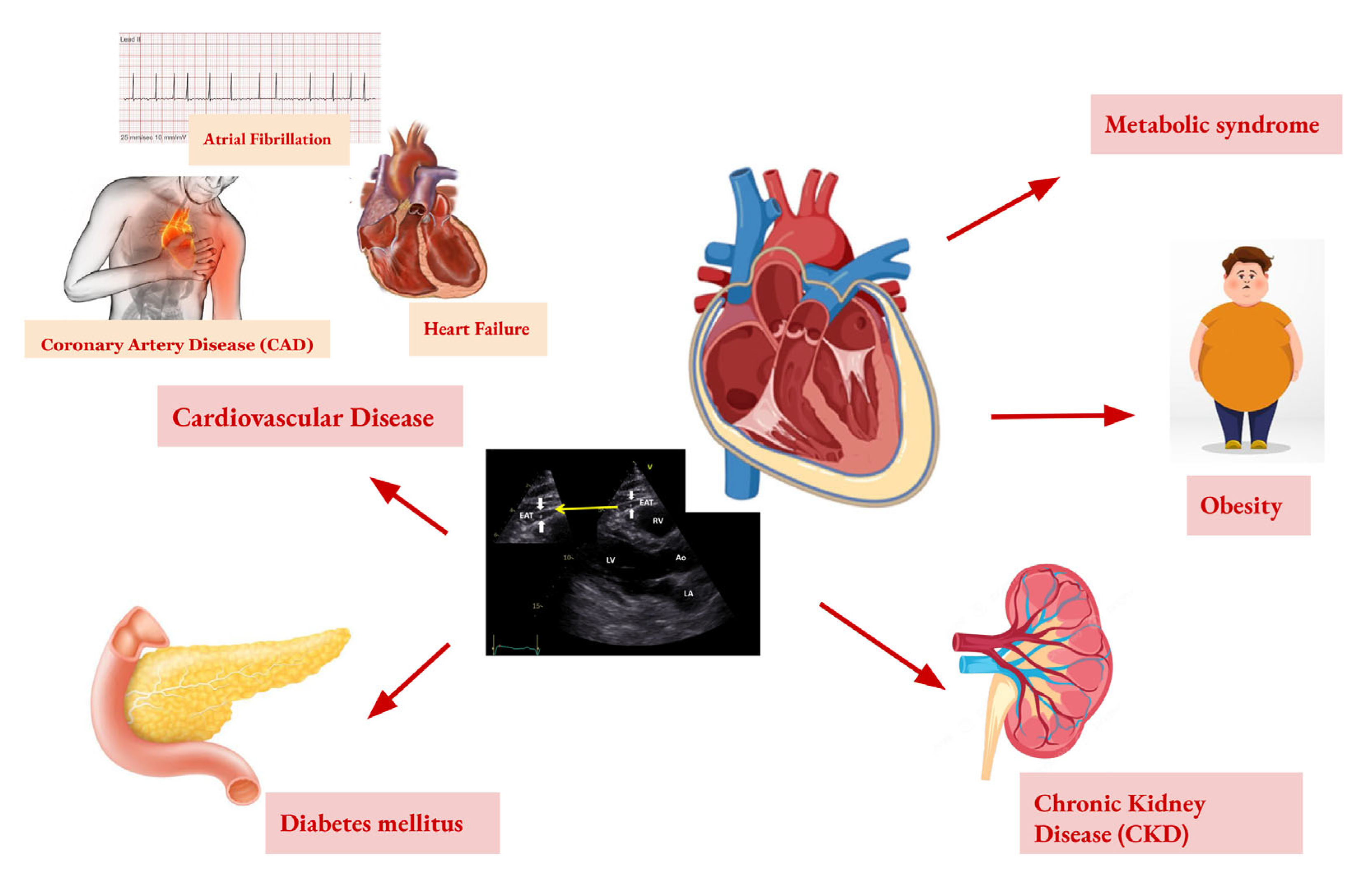
Chronic renal disease (CKD) is an important risk factor for CVD. Indeed, it was observed that patients receiving peritoneal dialysis or hemodialysis had more elevated levels of EAT. Consequently, this represents another risk factor for CKD patients. Therefore, CKD determines chronic inflammation, thus causing EAT dysfunction and inflammation, which affect the myocardium and coronary arteries. Finally, sevelamer, the non-calcium-based phosphate-binding agent, has been shown to reduce EAT thickness progression, along with a reduction in inflammation markers and serum cholesterol in CKD patients treated with hemodialysis [110]. Furthermore, patients affected by both DM and nephropathy presented an increased EAT thickness, which reflects an uncontrolled underlying disease, high body mass index, and raised cardiovascular risk markers [111]. A summary of the diagnostic view of EAT and the correlation with cardiometabolic diseases is provided in Figure 2.

9. Conclusions

Obesity is a major risk factor for metabolic dysfunction and CVD, primarily through the expansion of AT and the development of a chronic low-grade inflammatory state. Among the different fat depots, EAT has emerged as a key player in cardiovascular risk assessment. Unlike inert fat storage, EAT is a metabolically active tissue involved in the regulation of inflammatory responses, oxidative stress, and adipokine secretion.
EAT has been closely linked to the onset and progression of cardiovascular conditions such as atherosclerosis and heart failure. Its measurement is gaining recognition as a valuable prognostic tool, offering insights into disease progression and therapeutic efficacy. Lifestyle interventions, including physical activity and a balanced diet, have shown effectiveness in reducing EAT volume, which in turn may improve cardiac function and reduce inflammation [112].
From a diagnostic perspective, CT is currently the most accurate method for quantifying EAT. Echocardiography, although less precise, remains a practical non-invasive technique for initial assessment. Cardiac MRI provides the most detailed analysis, offering a high spatial resolution.
In conclusion, EAT is a significant biomarker and potential therapeutic target in the prevention and management of cardiovascular risk. Future research is needed to further elucidate the underlying molecular mechanisms and to develop targeted therapeutic approaches. Epicardial adipose tissue is increasingly recognized as a clinically relevant biomarker and potential therapeutic target in cardiovascular disease. Future directions include its integration into routine cardiovascular risk stratification, the development of targeted therapies to modulate its inflammatory and metabolic activity, and its use as a dynamic marker to monitor the treatment response. Advances in imaging technologies, combined with molecular research, will further elucidate the pathophysiological mechanisms of EAT, supporting its role in precision medicine and the development of personalized cardiovascular interventions. Artificial intelligence (AI) is revolutionizing the study of EAT by utilizing algorithms to analyze medical images such as CT and MRI, improving fat measurement and cardiovascular risk prediction.

Author Contributions

Conceptualization, G.C., F.N. and S.D.; methodology, P.G., G.C. and S.D.; resources, E.L., A.S., C.S., M.M., V.M.C., G.T. and F.S.L.; data curation, E.L., A.S., C.S., M.M., V.M.C., G.T. and F.S.L.; writing—original draft preparation, S.D., E.L., A.S., C.S., M.M., V.M.C., G.T. and F.S.L.; writing—review and editing, P.G., F.N. and G.C.; supervision, P.G. All authors have read and agreed to the published version of the manuscript.

Funding

This research received no external funding.

Institutional Review Board Statement

Not applicable.

Informed Consent Statement

Not applicable.

Data Availability Statement

The data from this manuscript are derived from publicly available published clinical trial and study results.

Conflicts of Interest

The authors declare no conflicts of interest.

References

  1. The Lancet Gastroenterology & Hepatology. Obesity: Another ongoing pandemic. Lancet Gastroenterol. Hepatol. 2021, 6, 411. [Google Scholar] [CrossRef] [PubMed]
  2. Oikonomou, E.K.; Antoniades, C. The role of adipose tissue in cardiovascular health and disease. Nat. Rev. Cardiol. 2019, 16, 83–99. [Google Scholar] [CrossRef]
  3. Polkinghorne, M.D.; West, H.W.; Antoniades, C. Adipose Tissue in Cardiovascular Disease: From Basic Science to Clinical Translation. Annu. Rev. Physiol. 2024, 86, 175–198. [Google Scholar] [CrossRef] [PubMed]
  4. Kawai, T.; Autieri, M.V.; Scalia, R. Adipose tissue inflammation and metabolic dysfunction in obesity. Am. J. Physiol. -Cell Physiol. 2021, 320, C375–C391. [Google Scholar] [CrossRef]
  5. Ahmed, B.; Sultana, R.; Greene, M.W. Adipose tissue and insulin resistance in obese. Biomed. Pharmacother. 2021, 137, 111315. [Google Scholar] [CrossRef]
  6. Mazurek, T.; Zhang, L.; Zalewski, A.; Mannion, J.D.; Diehl, J.T.; Arafat, H.; Sarov-Blat, L.; O’Brien, S.; Keiper, E.A.; Johnson, A.G.; et al. Human epicardial adipose tissue is a source of inflammatory mediators. Circulation 2003, 108, 2460–2466. [Google Scholar] [CrossRef]
  7. Iacobellis, G.; Corradi, D.; Sharma, A.M. Epicardial adipose tissue: Anatomic, biomolecular and clinical relationships with the heart. Nat. Clin. Pract. Cardiovasc. Med. 2005, 2, 536–543. [Google Scholar] [CrossRef]
  8. Lu, Z.; Jiang, Z.; Tang, J.; Lin, C.P.; Zhang, H. Functions and origins of cardiac fat. FEBS J. 2022, 290, 1705–1718. [Google Scholar] [CrossRef]
  9. Iacobellis, G. Epicardial adipose tissue in contemporary cardiology. Nat. Rev. Cardiol. 2022, 19, 593–606. [Google Scholar] [CrossRef]
  10. Iacobellis, G.; Bianco, A.C. Epicardial adipose tissue: Emerging physiological, pathophysiological and clinical features. Trends Endocrinol. Metab. 2011, 22, 450–457. [Google Scholar] [CrossRef]
  11. Sakers, A.; De Siqueira, M.K.; Seale, P.; Villanueva, C.J. Adipose-tissue plasticity in health and disease. Cell 2022, 185, 419–446. [Google Scholar] [CrossRef] [PubMed]
  12. Trayhurn, P.; Beattie, J.H. Physiological role of adipose tissue: White adipose tissue as an endocrine and secretory organ. Proc. Nutr. Soc. 2001, 60, 329–339. [Google Scholar] [CrossRef]
  13. Romacho, T.; Elsen, M.; Rohrborn, D.; Eckel, J. Adipose tissue and its role in organ crosstalk. Acta Physiol. 2014, 210, 733–753. [Google Scholar] [CrossRef]
  14. Pico, C.; Palou, M.; Pomar, C.A.; Rodriguez, A.M.; Palou, A. Leptin as a key regulator of the adipose organ. Rev. Endocr. Metab. Disord. 2022, 23, 13–30. [Google Scholar] [CrossRef] [PubMed]
  15. Chandran, M.; Phillips, S.A.; Ciaraldi, T.; Henry, R.R. Adiponectin: More than just another fat cell hormone? Diabetes Care 2003, 26, 2442–2450. [Google Scholar] [CrossRef]
  16. Ouchi, N.; Parker, J.L.; Lugus, J.J.; Walsh, K. Adipokines in inflammation and metabolic disease. Nat. Rev. Immunol. 2011, 11, 85–97. [Google Scholar] [CrossRef]
  17. Odegaard, J.I.; Chawla, A. Alternative macrophage activation and metabolism. Annu. Rev. Pathol. 2011, 6, 275–297. [Google Scholar] [CrossRef]
  18. Lumeng, C.N.; Saltiel, A.R. Inflammatory links between obesity and metabolic disease. J. Clin. Investig. 2011, 121, 2111–2117. [Google Scholar] [CrossRef]
  19. Boutens, L.; Stienstra, R. Adipose tissue macrophages: Going off track during obesity. Diabetologia 2016, 59, 879–894. [Google Scholar] [CrossRef]
  20. Rosen, E.D.; Spiegelman, B.M. What we talk about when we talk about fat. Cell 2014, 156, 20–44. [Google Scholar] [CrossRef]
  21. Vianello, E.; Dozio, E.; Arnaboldi, F.; Marazzi, M.G.; Martinelli, C.; Lamont, J.; Tacchini, L.; Sigruner, A.; Schmitz, G.; Corsi Romanelli, M.M. Epicardial adipocyte hypertrophy: Association with M1-polarization and toll-like receptor pathways in coronary artery disease patients. Nutr. Metab. Cardiovasc. Dis. 2016, 26, 246–253. [Google Scholar] [CrossRef]
  22. Kanneganti, T.D.; Dixit, V.D. Immunological complications of obesity. Nat. Immunol. 2012, 13, 707–712. [Google Scholar] [CrossRef]
  23. Han, C.Y.; Subramanian, S.; Chan, C.K.; Omer, M.; Chiba, T.; Wight, T.N.; Chait, A. Adipocyte-Derived Serum Amyloid A3 and Hyaluronan Play a Role in Monocyte Recruitment and Adhesion. Diabetes 2007, 56, 2260–2273. [Google Scholar] [CrossRef]
  24. Sidossis, L.; Kajimura, S. Brown and beige fat in humans: Thermogenic adipocytes that control energy and glucose homeostasis. J. Clin. Investig. 2015, 125, 478–486. [Google Scholar] [CrossRef]
  25. Leitner, D.R.; Fruhbeck, G.; Yumuk, V.; Schindler, K.; Micic, D.; Woodward, E.; Toplak, H. Obesity and Type 2 Diabetes: Two Diseases with a Need for Combined Treatment Strategies—EASO Can Lead the Way. Obes. Facts 2017, 10, 483–492. [Google Scholar] [CrossRef]
  26. Villarroya, J.; Cereijo, R.; Gavalda-Navarro, A.; Peyrou, M.; Giralt, M.; Villarroya, F. New insights into the secretory functions of brown adipose tissue. J. Endocrinol. 2019, 243, R19–R27. [Google Scholar] [CrossRef]
  27. Wang, S.; Liu, Y.; Chen, J.; He, Y.; Ma, W.; Liu, X.; Sun, X. Effects of multi-organ crosstalk on the physiology and pathology of adipose tissue. Front. Endocrinol. 2023, 14, 1198984. [Google Scholar] [CrossRef]
  28. Marcelin, G.; Silveira, A.L.M.; Martins, L.B.; Ferreira, A.V.; Clement, K. Deciphering the cellular interplays underlying obesity-induced adipose tissue fibrosis. J. Clin. Investig. 2019, 129, 4032–4040. [Google Scholar] [CrossRef]
  29. Cristancho, A.G.; Lazar, M.A. Forming functional fat: A growing understanding of adipocyte differentiation. Nat. Rev. Mol. Cell Biol. 2011, 12, 722–734. [Google Scholar] [CrossRef]
  30. Cannon, B.; Nedergaard, J.A.N. Brown Adipose Tissue: Function and Physiological Significance. Physiol. Rev. 2004, 84, 277–359. [Google Scholar] [CrossRef]
  31. Luo, L.; Liu, M. Adipose tissue in control of metabolism. J. Endocrinol. 2016, 231, R77–R99. [Google Scholar] [CrossRef] [PubMed]
  32. Booth, A.; Magnuson, A.; Fouts, J.; Foster, M.T. Adipose tissue: An endocrine organ playing a role in metabolic regulation. Horm. Mol. Biol. Clin. Investig. 2016, 26, 25–42. [Google Scholar] [CrossRef]
  33. Newman, G.; Gonzalez-Perez, R.R. Leptin-cytokine crosstalk in breast cancer. Mol. Cell. Endocrinol. 2014, 382, 570–582. [Google Scholar] [CrossRef]
  34. Hara, K.; Horikoshi, M.; Yamauchi, T.; Yago, H.; Miyazaki, O.; Ebinuma, H.; Imai, Y.; Nagai, R.; Kadowaki, T. Measurement of the high-molecular weight form of adiponectin in plasma is useful for the prediction of insulin resistance and metabolic syndrome. Diabetes Care 2006, 29, 1357–1362. [Google Scholar] [CrossRef]
  35. Cipolletta, D.; Feuerer, M.; Li, A.; Kamei, N.; Lee, J.; Shoelson, S.E.; Benoist, C.; Mathis, D. PPAR-gamma is a major driver of the accumulation and phenotype of adipose tissue Treg cells. Nature 2012, 486, 549–553. [Google Scholar] [CrossRef]
  36. Hill, D.A.; Lim, H.W.; Kim, Y.H.; Ho, W.Y.; Foong, Y.H.; Nelson, V.L.; Nguyen, H.C.B.; Chegireddy, K.; Kim, J.; Habertheuer, A.; et al. Distinct macrophage populations direct inflammatory versus physiological changes in adipose tissue. Proc. Natl. Acad. Sci. USA 2018, 115, E5096–E5105. [Google Scholar] [CrossRef]
  37. Orliaguet, L.; Dalmas, E.; Drareni, K.; Venteclef, N.; Alzaid, F. Mechanisms of Macrophage Polarization in Insulin Signaling and Sensitivity. Front. Endocrinol. 2020, 11, 62. [Google Scholar] [CrossRef]
  38. Pouliopoulos, J.; Chik, W.W.B.; Kanthan, A.; Sivagangabalan, G.; Barry, M.A.; Fahmy, P.N.A.; Midekin, C.; Lu, J.; Kizana, E.; Thomas, S.P.; et al. Intramyocardial Adiposity After Myocardial Infarction. Circulation 2013, 128, 2296–2308. [Google Scholar] [CrossRef]
  39. Zhang, H.; Pu, W.; Liu, Q.; He, L.; Huang, X.; Tian, X.; Zhang, L.; Nie, Y.; Hu, S.; Lui, K.O.; et al. Endocardium Contributes to Cardiac Fat. Circ. Res. 2016, 118, 254–265. [Google Scholar] [CrossRef]
  40. Ichikawa, Y.; Kitagawa, K.; Chino, S.; Ishida, M.; Matsuoka, K.; Tanigawa, T.; Nakamura, T.; Hirano, T.; Takeda, K.; Sakuma, H. Adipose Tissue Detected by Multislice Computed Tomography in Patients After Myocardial Infarction. JACC Cardiovasc. Imaging 2009, 2, 548–555. [Google Scholar] [CrossRef]
  41. Akoumianakis, I.; Antoniades, C. The interplay between adipose tissue and the cardiovascular system: Is fat always bad? Cardiovasc. Res. 2017, 113, 999–1008. [Google Scholar] [CrossRef] [PubMed]
  42. Zhu, R.; Wang, W.; Gao, Y.; Wang, J.; Li, B.; Cheng, Z.; Ji, C.; Gu, H.; Yuan, X.; Yang, S.; et al. Insulin resistance aggravates myocardial fibrosis in non-diabetic hypertensive patients by altering the function of epicardial adipose tissue: A cardiac magnetic resonance study. Diabetol. Metab. Syndr. 2025, 17, 133. [Google Scholar] [CrossRef] [PubMed]
  43. Iacobellis, G.; Barbaro, G. Epicardial adipose tissue feeding and overfeeding the heart. Nutrition 2019, 59, 1–6. [Google Scholar] [CrossRef] [PubMed]
  44. Gaborit, B.; Sengenes, C.; Ancel, P.; Jacquier, A.; Dutour, A. Role of Epicardial Adipose Tissue in Health and Disease: A Matter of Fat? Compr. Physiol. 2017, 7, 1051–1082. [Google Scholar] [CrossRef]
  45. Packer, M. Epicardial Adipose Tissue May Mediate Deleterious Effects of Obesity and Inflammation on the Myocardium. J. Am. Coll. Cardiol. 2018, 71, 2360–2372. [Google Scholar] [CrossRef]
  46. Ansaldo, A.M.; Montecucco, F.; Sahebkar, A.; Dallegri, F.; Carbone, F. Epicardial adipose tissue and cardiovascular diseases. Int. J. Cardiol. 2019, 278, 254–260. [Google Scholar] [CrossRef]
  47. O’Rourke, R.W. Adipose tissue and the physiologic underpinnings of metabolic disease. Surg. Obes. Relat. Dis. 2018, 14, 1755–1763. [Google Scholar] [CrossRef]
  48. Wang, Q.A.; Tao, C.; Gupta, R.K.; Scherer, P.E. Tracking adipogenesis during white adipose tissue development, expansion and regeneration. Nat. Med. 2013, 19, 1338–1344. [Google Scholar] [CrossRef]
  49. Harvey, I.; Boudreau, A.; Stephens, J.M. Adipose tissue in health and disease. Open Biol. 2020, 10. [Google Scholar] [CrossRef]
  50. Chait, A.; den Hartigh, L.J. Adipose Tissue Distribution, Inflammation and Its Metabolic Consequences, Including Diabetes and Cardiovascular Disease. Front. Cardiovasc. Med. 2020, 7, 522637. [Google Scholar] [CrossRef]
  51. Weisberg, S.P.; McCann, D.; Desai, M.; Rosenbaum, M.; Leibel, R.L.; Ferrante, A.W. Obesity is associated with macrophage accumulation in adipose tissue. J. Clin. Investig. 2003, 112, 1796–1808. [Google Scholar] [CrossRef] [PubMed]
  52. Li, X.; Ren, Y.; Chang, K.; Wu, W.; Griffiths, H.R.; Lu, S.; Gao, D. Adipose tissue macrophages as potential targets for obesity and metabolic diseases. Front. Immunol. 2023, 14, 1153915. [Google Scholar] [CrossRef]
  53. Cinti, S.; Mitchell, G.; Barbatelli, G.; Murano, I.; Ceresi, E.; Faloia, E.; Wang, S.; Fortier, M.; Greenberg, A.S.; Obin, M.S. Adipocyte death defines macrophage localization and function in adipose tissue of obese mice and humans. J. Lipid Res. 2005, 46, 2347–2355. [Google Scholar] [CrossRef] [PubMed]
  54. Subramanian, S.; Han, C.Y.; Chiba, T.; McMillen, T.S.; Wang, S.A.; Haw, A.; Kirk, E.A.; O’Brien, K.D.; Chait, A. Dietary Cholesterol Worsens Adipose Tissue Macrophage Accumulation and Atherosclerosis in Obese LDL Receptor–Deficient Mice. Arterioscler. Thromb. Vasc. Biol. 2008, 28, 685–691. [Google Scholar] [CrossRef] [PubMed]
  55. Iacobellis, G. Local and systemic effects of the multifaceted epicardial adipose tissue depot. Nat. Rev. Endocrinol. 2015, 11, 363–371. [Google Scholar] [CrossRef]
  56. Gaborit, B.; Julla, J.B.; Fournel, J.; Ancel, P.; Soghomonian, A.; Deprade, C.; Lasbleiz, A.; Houssays, M.; Ghattas, B.; Gascon, P.; et al. Fully automated epicardial adipose tissue volume quantification with deep learning and relationship with CAC score and micro/macrovascular complications in people living with type 2 diabetes: The multicenter EPIDIAB study. Cardiovasc. Diabetol. 2024, 23, 328. [Google Scholar] [CrossRef]
  57. Gao, Z.; Hwang, D.; Bataille, F.; Lefevre, M.; York, D.; Quon, M.J.; Ye, J. Serine Phosphorylation of Insulin Receptor Substrate 1 by Inhibitor κB Kinase Complex. J. Biol. Chem. 2002, 277, 48115–48121. [Google Scholar] [CrossRef]
  58. Zabolotny, J.M.; Kim, Y.-B.; Welsh, L.A.; Kershaw, E.E.; Neel, B.G.; Kahn, B.B. Protein-tyrosine Phosphatase 1B Expression Is Induced by Inflammation in Vivo. J. Biol. Chem. 2008, 283, 14230–14241. [Google Scholar] [CrossRef]
  59. Naryzhnaya, N.V.; Koshelskaya, O.A.; Kologrivova, I.V.; Kharitonova, O.A.; Evtushenko, V.V.; Boshchenko, A.A. Hypertrophy and Insulin Resistance of Epicardial Adipose Tissue Adipocytes: Association with the Coronary Artery Disease Severity. Biomedicines 2021, 9, 64. [Google Scholar] [CrossRef]
  60. Amano, S.U.; Cohen, J.L.; Vangala, P.; Tencerova, M.; Nicoloro, S.M.; Yawe, J.C.; Shen, Y.; Czech, M.P.; Aouadi, M. Local Proliferation of Macrophages Contributes to Obesity-Associated Adipose Tissue Inflammation. Cell Metab. 2014, 19, 162–171. [Google Scholar] [CrossRef]
  61. O’Rourke, R.W.; Metcalf, M.D.; White, A.E.; Madala, A.; Winters, B.R.; Maizlin, I.I.; Jobe, B.A.; Roberts, C.T.; Slifka, M.K.; Marks, D.L. Depot-specific differences in inflammatory mediators and a role for NK cells and IFN-γ in inflammation in human adipose tissue. Int. J. Obes. 2009, 33, 978–990. [Google Scholar] [CrossRef] [PubMed]
  62. Rocha, V.Z.; Folco, E.J.; Sukhova, G.; Shimizu, K.; Gotsman, I.; Vernon, A.H.; Libby, P. Interferon-γ, a Th1 Cytokine, Regulates Fat Inflammation. Circ. Res. 2008, 103, 467–476. [Google Scholar] [CrossRef]
  63. La, V.; Nair, V.; Sunny, S.; Benharash, P.; Thankam, F.G. Epicardial Adipocytes in Cardiac Pathology and Healing. Cardiovasc. Drugs Ther. 2024, 1–13. [Google Scholar] [CrossRef]
  64. Konwerski, M.; Gasecka, A.; Opolski, G.; Grabowski, M.; Mazurek, T. Role of Epicardial Adipose Tissue in Cardiovascular Diseases: A Review. Biology 2022, 11, 355. [Google Scholar] [CrossRef]
  65. Ayton, S.L.; Gulsin, G.S.; McCann, G.P.; Moss, A.J. Epicardial adipose tissue in obesity-related cardiac dysfunction. Heart 2022, 108, 339–344. [Google Scholar] [CrossRef]
  66. Wang, Q.; Chi, J.; Wang, C.; Yang, Y.; Tian, R.; Chen, X. Epicardial Adipose Tissue in Patients with Coronary Artery Disease: A Meta-Analysis. J. Cardiovasc. Dev. Dis. 2022, 9, 253. [Google Scholar] [CrossRef]
  67. Hwang, I.-C.; Park, H.E.; Choi, S.-Y. Epicardial Adipose Tissue Contributes to the Development of Non-Calcified Coronary Plaque: A 5-Year Computed Tomography Follow-up Study. J. Atheroscler. Thromb. 2017, 24, 262–274. [Google Scholar] [CrossRef]
  68. Tunc Suygun, E.; Vardar Yagli, N.; Suygun, H. Relationship between epicardial adipose tissue thickness and sedentary time, physical activity level, and physical performance in patients with hypertension. J. Hum. Hypertens. 2025, 39, 274–278. [Google Scholar] [CrossRef]
  69. Myasoedova, V.A.; Parisi, V.; Moschetta, D.; Valerio, V.; Conte, M.; Massaiu, I.; Bozzi, M.; Celeste, F.; Leosco, D.; Iaccarino, G.; et al. Efficacy of cardiometabolic drugs in reduction of epicardial adipose tissue: A systematic review and meta-analysis. Cardiovasc. Diabetol. 2023, 22, 23. [Google Scholar] [CrossRef]
  70. Gao, Z.; Zuo, Y.; Jia, L.; Yin, Y.; Yang, X.; Xu, L.; Hao, Z. Correlation analysis between epicardial adipose tissue and acute coronary syndrome. Sci. Rep. 2025, 15, 3015. [Google Scholar] [CrossRef]
  71. Villasante Fricke, A.C.; Iacobellis, G. Epicardial Adipose Tissue: Clinical Biomarker of Cardio-Metabolic Risk. Int. J. Mol. Sci. 2019, 20, 5989. [Google Scholar] [CrossRef] [PubMed]
  72. Mahabadi, A.A.; Berg, M.H.; Lehmann, N.; Kalsch, H.; Bauer, M.; Kara, K.; Dragano, N.; Moebus, S.; Jockel, K.H.; Erbel, R.; et al. Association of epicardial fat with cardiovascular risk factors and incident myocardial infarction in the general population: The Heinz Nixdorf Recall Study. J. Am. Coll. Cardiol. 2013, 61, 1388–1395. [Google Scholar] [CrossRef]
  73. Salgado-Somoza, A.; Teijeira-Fernández, E.; Fernández, Á.L.; González-Juanatey, J.R.; Eiras, S. Proteomic analysis of epicardial and subcutaneous adipose tissue reveals differences in proteins involved in oxidative stress. Am. J. Physiol. Heart Circ. Physiol. 2010, 299, H202–H209. [Google Scholar] [CrossRef]
  74. Wu, Y.; Zhang, A.; Hamilton, D.J.; Deng, T. Epicardial Fat in the Maintenance of Cardiovascular Health. Methodist DeBakey Cardiovasc. J. 2017, 13, 20. [Google Scholar] [CrossRef]
  75. Ohashi, K.; Shibata, R.; Murohara, T.; Ouchi, N. Role of anti-inflammatory adipokines in obesity-related diseases. Trends Endocrinol. Metab. 2014, 25, 348–355. [Google Scholar] [CrossRef]
  76. Wang, Z.V.; Scherer, P.E. Adiponectin, the past two decades. J. Mol. Cell Biol. 2016, 8, 93–100. [Google Scholar] [CrossRef]
  77. Liu, R.; Nikolajczyk, B.S. Tissue Immune Cells Fuel Obesity-Associated Inflammation in Adipose Tissue and Beyond. Front. Immunol. 2019, 10, 1587. [Google Scholar] [CrossRef]
  78. Cao, Y. Angiogenesis and Vascular Functions in Modulation of Obesity, Adipose Metabolism, and Insulin Sensitivity. Cell Metab. 2013, 18, 478–489. [Google Scholar] [CrossRef]
  79. van Harmelen, V.; Eriksson, A.; Astrom, G.; Wahlen, K.; Naslund, E.; Karpe, F.; Frayn, K.; Olsson, T.; Andersson, J.; Rydeén, M.; et al. Vascular Peptide Endothelin-1 Links Fat Accumulation With Alterations of Visceral Adipocyte Lipolysis. Diabetes 2008, 57, 378–386. [Google Scholar] [CrossRef]
  80. Rapoport, R.M.; Draznin, M.B.; Murad, F. Endothelium-dependent relaxation in rat aorta may be mediated through cyclic GMP-dependent protein phosphorylation. Nature 1983, 306, 174–176. [Google Scholar] [CrossRef]
  81. Villarroya, F.; Cereijo, R.; Villarroya, J.; Giralt, M. Brown adipose tissue as a secretory organ. Nat. Rev. Endocrinol. 2016, 13, 26–35. [Google Scholar] [CrossRef] [PubMed]
  82. Shamsi, F.; Wang, C.-H.; Tseng, Y.-H. The evolving view of thermogenic adipocytes—Ontogeny, niche and function. Nat. Rev. Endocrinol. 2021, 17, 726–744. [Google Scholar] [CrossRef]
  83. Song, Y.; Tan, Y.; Deng, M.; Shan, W.; Zheng, W.; Zhang, B.; Cui, J.; Feng, L.; Shi, L.; Zhang, M.; et al. Epicardial adipose tissue, metabolic disorders, and cardiovascular diseases: Recent advances classified by research methodologies. MedComm 2023, 4, e413. [Google Scholar] [CrossRef] [PubMed]
  84. Bodenstab, M.L.; Varghese, R.T.; Iacobellis, G. Cardio-Lipotoxicity of Epicardial Adipose Tissue. Biomolecules 2024, 14, 1465. [Google Scholar] [CrossRef] [PubMed]
  85. Kitagawa, T.; Yamamoto, H.; Hattori, T.; Sentani, K.; Takahashi, S.; Senoo, A.; Kubo, Y.; Yasui, W.; Sueda, T.; Kihara, Y. Tumor Necrosis Factor-α Gene Expression in Epicardial Adipose Tissue is Related to Coronary Atherosclerosis Assessed by Computed Tomography. J. Atheroscler. Thromb. 2018, 25, 269–280. [Google Scholar] [CrossRef]
  86. Klein, S.; Gastaldelli, A.; Yki-Jarvinen, H.; Scherer, P.E. Why does obesity cause diabetes? Cell Metab. 2022, 34, 11–20. [Google Scholar] [CrossRef]
  87. Duca, F.; Mascherbauer, K.; Dona, C.; Koschutnik, M.; Binder, C.; Nitsche, C.; Halavina, K.; Beitzke, D.; Loewe, C.; Bartko, P.; et al. Association of epicardial adipose tissue on magnetic resonance imaging with cardiovascular outcomes: Quality over quantity? Obesity 2024, 32, 1670–1679. [Google Scholar] [CrossRef]
  88. Kotha, S.; Plein, S.; Greenwood, J.P.; Levelt, E. Role of epicardial adipose tissue in diabetic cardiomyopathy through the lens of cardiovascular magnetic resonance imaging—A narrative review. Ther. Adv. Endocrinol. Metab. 2024, 15, 20420188241229540. [Google Scholar] [CrossRef]
  89. Iacobellis, G.; Willens, H.J. Echocardiographic Epicardial Fat: A Review of Research and Clinical Applications. J. Am. Soc. Echocardiogr. 2009, 22, 1311–1319. [Google Scholar] [CrossRef]
  90. Talman, A.H.; Psaltis, P.J.; Cameron, J.D.; Meredith, I.T.; Seneviratne, S.K.; Wong, D.T. Epicardial adipose tissue: Far more than a fat depot. Cardiovasc. Diagn. Ther. 2014, 4, 416–429. [Google Scholar] [CrossRef]
  91. Christensen, R.H.; von Scholten, B.J.; Lehrskov, L.L.; Rossing, P.; Jorgensen, P.G. Epicardial adipose tissue: An emerging biomarker of cardiovascular complications in type 2 diabetes? Ther. Adv. Endocrinol. Metab. 2020, 11, 2042018820928824. [Google Scholar] [CrossRef] [PubMed]
  92. Muzurovic, E.M.; Vujosevic, S.; Mikhailidis, D.P. Can We Decrease Epicardial and Pericardial Fat in Patients With Diabetes? J. Cardiovasc. Pharmacol. Ther. 2021, 26, 415–436. [Google Scholar] [CrossRef] [PubMed]
  93. Chong, B.; Jayabaskaran, J.; Ruban, J.; Goh, R.; Chin, Y.H.; Kong, G.; Ng, C.H.; Lin, C.; Loong, S.; Muthiah, M.D.; et al. Epicardial Adipose Tissue Assessed by Computed Tomography and Echocardiography Are Associated With Adverse Cardiovascular Outcomes: A Systematic Review and Meta-Analysis. Circ. Cardiovasc. Imaging 2023, 16, e015159. [Google Scholar] [CrossRef]
  94. van Woerden, G.; van Veldhuisen, D.J.; Gorter, T.M.; Ophuis, B.; Saucedo-Orozco, H.; van Empel, V.P.M.; Willems, T.P.; Geelhoed, B.; Rienstra, M.; Westenbrink, B.D. The value of echocardiographic measurement of epicardial adipose tissue in heart failure patients. ESC Heart Fail. 2022, 9, 953–957. [Google Scholar] [CrossRef]
  95. Banerjee, A.; Cowley, A.C.; Ayton, S.L.; Dattani, A.; Yeo, J.L.; Brady, E.M.; Deshpande, A.; Dey, D.; McCann, G.P.; Gulsin, G.S. 2 A comparison of epicardial adipose tissue quantification between cardiac magnetic resonance and computed tomography in people with and without type 2 diabetes. Heart 2024, 110, A1. [Google Scholar] [CrossRef]
  96. Nerlekar, N.; Baey, Y.-W.; Brown, A.J.; Muthalaly, R.G.; Dey, D.; Tamarappoo, B.; Cameron, J.D.; Marwick, T.H.; Wong, D.T. Poor Correlation, Reproducibility, and Agreement Between Volumetric Versus Linear Epicardial Adipose Tissue Measurement. JACC Cardiovasc. Imaging 2018, 11, 1035–1036. [Google Scholar] [CrossRef]
  97. Liu, Z.; Wang, J.; Yang, Y.; Cheng, J.; Yang, M.; Zhang, Y. Prediction of major adverse cardiovascular events following ST-segment elevation myocardial infarction using cardiac obesity marker-epicardial adipose tissue mass index: A prospective cohort study. Front. Cardiovasc. Med. 2025, 12, 1539500. [Google Scholar] [CrossRef]
  98. van Woerden, G.; van Veldhuisen, D.J.; Manintveld, O.C.; van Empel, V.P.M.; Willems, T.P.; de Boer, R.A.; Rienstra, M.; Westenbrink, B.D.; Gorter, T.M. Epicardial Adipose Tissue and Outcome in Heart Failure With Mid-Range and Preserved Ejection Fraction. Circ. Heart Fail. 2022, 15, e009238. [Google Scholar] [CrossRef]
  99. Timóteo, A.T.; Barbas Albuquerque, F.; Lacerda Teixeira, B. Pericardium, epicardial adipose tissue, and heart failure with preserved ejection fraction: Pathophysiology, quantification and treatment target. Int. J. Cardiol. 2024, 412, 132303. [Google Scholar] [CrossRef]
  100. Hillege, H.L.; Girbes, A.R.; de Kam, P.J.; Boomsma, F.; de Zeeuw, D.; Charlesworth, A.; Hampton, J.R.; van Veldhuisen, D.J. Renal function, neurohormonal activation, and survival in patients with chronic heart failure. Circulation 2000, 102, 203–210. [Google Scholar] [CrossRef]
  101. Mustafina, I.; Dolganov, A.; Zagidullin, N.; Pavlov, V. The relationship of epicardial adipose tissue and metabolic syndrome. Atherosclerosis 2023, 379, S110. [Google Scholar] [CrossRef]
  102. Iacobellis, G.; Assael, F.; Ribaudo, M.C.; Zappaterreno, A.; Alessi, G.; Di Mario, U.; Leonetti, F. Epicardial fat from echocardiography: A new method for visceral adipose tissue prediction. Obes. Res. 2003, 11, 304–310. [Google Scholar] [CrossRef]
  103. Li, Y.; Liu, B.; Li, Y.; Jing, X.; Deng, S.; Yan, Y.; She, Q. Epicardial fat tissue in patients with diabetes mellitus: A systematic review and meta-analysis. Cardiovasc. Diabetol. 2019, 18, 3. [Google Scholar] [CrossRef]
  104. Iacobellis, G.; Leonetti, F. Epicardial Adipose Tissue and Insulin Resistance in Obese Subjects. J. Clin. Endocrinol. Metab. 2005, 90, 6300–6302. [Google Scholar] [CrossRef]
  105. Nono Nankam, P.A.; Blüher, M. Retinol-binding protein 4 in obesity and metabolic dysfunctions. Mol. Cell. Endocrinol. 2021, 531, 111312. [Google Scholar] [CrossRef]
  106. Karaaslan, H.; Inan, H.; Elmas, A.N. The Association Between Epicardial Adipose Tissue Thickness and the Triglyceride-glucose Index in Prediabetic Obese Patients. Angiology 2025, 33197251320147. [Google Scholar] [CrossRef]
  107. Pop, A.; Danila, M.; Giuchici, S.; Buriman, D.; Lolescu, B.; Sturza, A.; Muntean, D.; Lascu, A. Epicardial adipose tissue as target of the incretin-based therapies in cardio-metabolic pathologies: A narrative review. Can. J. Physiol. Pharmacol. 2025. [Google Scholar] [CrossRef]
  108. Yang, X.; Feng, C.; Feng, J. Epicardial Adipose Tissue and Diabetic Cardiomyopathy. J. Cardiovasc. Pharmacol. Ther. 2023, 28, 10742484231151820. [Google Scholar] [CrossRef]
  109. Koh, K.K.; Park, S.M.; Quon, M.J. Leptin and cardiovascular disease: Response to therapeutic interventions. Circulation 2008, 117, 3238–3249. [Google Scholar] [CrossRef]
  110. D’Marco, L.; Puchades, M.J.; Gorriz, J.L.; Romero-Parra, M.; Lima-Martinez, M.; Soto, C.; Bermudez, V.; Raggi, P. Epicardial Adipose Tissue, Adiponectin and Leptin: A Potential Source of Cardiovascular Risk in Chronic Kidney Disease. Int. J. Mol. Sci. 2020, 21, 978. [Google Scholar] [CrossRef]
  111. Kleinaki, Z.; Agouridis, A.P.; Zafeiri, M.; Xanthos, T.; Tsioutis, C. Epicardial adipose tissue deposition in patients with diabetes and renal impairment: Analysis of the literature. World J. Diabetes 2020, 11, 33–41. [Google Scholar] [CrossRef] [PubMed]
  112. Iacobellis, G.; Singh, N.; Wharton, S.; Sharma, A.M. Substantial changes in epicardial fat thickness after weight loss in severely obese subjects. Obesity 2008, 16, 1693–1697. [Google Scholar] [CrossRef]
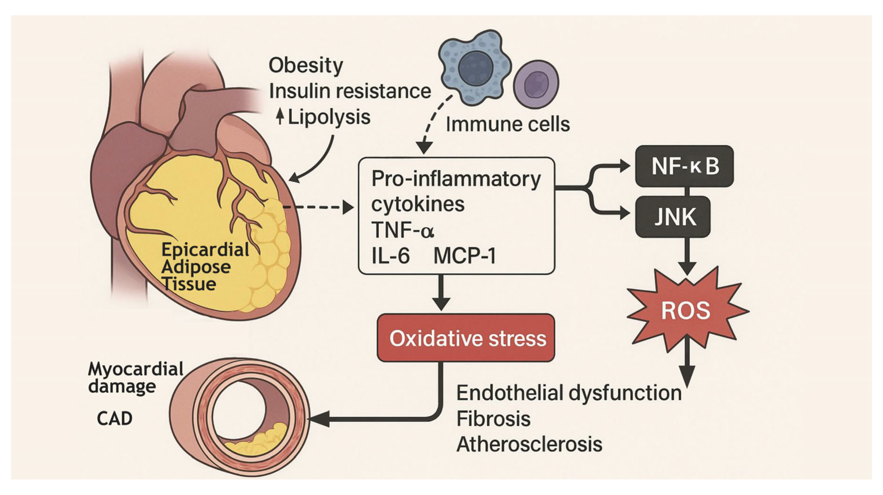
Figure 1. Schematic representation of inflammatory and oxidative mechanisms induced by epicardial adipose tissue (EAT).
Targets 03 00017 g001
Figure 2. Epicardial adipose tissue: echocardiographic view and its correlation with cardiometabolic diseases. At the center, the parasternal long-axis view, commonly used for assessing epicardial adipose tissue (EAT), shows EAT as an echo-free space between the outer wall of the myocardium and the visceral layer of the pericardium. Secondly, the figure highlights the correlation between increased EAT and several cardiometabolic diseases. Specifically, thicker EAT is associated with coronary artery disease (CAD) through atherosclerosis, inflammation, and oxidative stress. Peri-atrial EAT is identified as an independent risk factor for atrial fibrillation (AF). EAT is also suggested to play a role in both subtypes of heart failure (HF), HFpEF and HFrEF. In metabolic syndrome, EAT thickness shows a positive correlation with visceral fat and is linked to elevated blood pressure, lipid levels, glucose, and insulin resistance. Patients with diabetes mellitus (DM), particularly type 2, typically present with increased EAT thickness and volume, associated with insulin resistance. Finally, the figure indicates that elevated EAT levels are observed in chronic kidney disease (CKD), potentially contributing to cardiovascular risk in this population.
Targets 03 00017 g002
Table 1. Physiological and pathological functions of adipose tissue.
AspectPhysiological Role
(For Good)
Pathological Role (For Bad)Signaling Mediators and
Multiorgan Crosstalk
Energy StorageStores excess energy as triglycerides, providing a reserve during fasting [12]Excessive accumulation leads to obesity, increasing metabolic stress [2] Free fatty acids released into circulation can cause lipotoxicity in liver and muscle [13]
Insulation and Protection Provides thermal insulation and cushions vital organs [13]Excess adiposity can impair organ function (e.g., fatty liver, cardiac adiposity) [2]Physical expansion affects mechanical and endocrine functions in adjacent tissues [11]
Endocrine FunctionSecretes hormones like leptin (regulates appetite) and adiponectin (enhances insulin sensitivity) [14]Dysregulation leads to leptin resistance and decreased adiponectin, contributing to insulin resistance and metabolic syndrome [15]Adipokines influence the hypothalamus, pancreas, and other endocrine organs [16]
Immune FunctionModulates immune responses through anti-inflammatory cytokines [17]Chronic low-grade inflammation via increased pro-inflammatory cytokines (e.g., TNF-α, IL-6) promotes insulin resistance and atherosclerosis [18]Cytokine release affects systemic inflammation and interacts with immune cells in the liver, heart, and vascular system [19]
Lipid MetabolismRegulates lipid storage and mobilization, maintaining lipid homeostasis [20]Dysregulation leads to ectopic fat deposition (e.g., liver steatosis), altering lipid profiles and increasing cardiovascular risk [4]Altered lipid metabolism affects the liver (NAFLD), muscle (insulin resistance), and cardiovascular system (atherosclerosis) [21]
Glucose MetabolismEnhances insulin sensitivity and glucose uptake, maintaining glucose homeostasis [15]Impaired function leads to insulin resistance and type 2 diabetes [5]Adipose-derived factors influence glucose metabolism in liver, muscle, and pancreas [16]
Vascular FunctionProduces factors that regulate vascular tone and endothelial function [8]Dysfunction contributes to endothelial damage, hypertension, and atherosclerosis [22]Adipokines and cytokines affect vascular smooth muscle cells and endothelial health [23]
Brown Adipose Tissue (BAT)Facilitates thermogenesis through uncoupling protein 1 (UCP1), contributing to energy expenditure and weight control [24]Reduced BAT activity may impair thermogenesis, leading to weight gain and metabolic dysfunction [25]BAT-derived signals influence metabolic rate and energy homeostasis systemically [26]
Multiorgan CrosstalkMaintains systemic metabolic balance through coordinated signaling with liver, muscle, brain, and cardiovascular system [13]Disrupted signaling leads to multiorgan dysfunction, contributing to metabolic syndrome, cardiovascular diseases, and neurodegenerative conditions [27]Adipokines, cytokines, and lipid mediators establish a complex network influencing systemic homeostasis and disease development [28]
Disclaimer/Publisher’s Note: The statements, opinions and data contained in all publications are solely those of the individual author(s) and contributor(s) and not of MDPI and/or the editor(s). MDPI and/or the editor(s) disclaim responsibility for any injury to people or property resulting from any ideas, methods, instructions or products referred to in the content.