Article

SK-03-92 Treatment Causes Release of a Lethal Factor Protein That Kills Staphylococcus aureus Cells

1
Department of Microbiology, University of Wisconsin-La Crosse, La Crosse, WI 54601, USA
2
Department of Chemistry and Biochemistry, University of Wisconsin-La Crosse, La Crosse, WI 54601, USA
*
Author to whom correspondence should be addressed.
Targets 2024, 2(2), 80-92; https://doi.org/10.3390/targets2020005
Submission received: 29 March 2024 / Revised: 15 May 2024 / Accepted: 21 May 2024 / Published: 22 May 2024

Abstract

:
Background: Staphylococcus aureus is a leading cause of skin and bloodstream infections in humans. Antibiotic resistant strains of S. aureus continue to be a problem in treating patients that are infected, so treatment options are needed. A drug discovery project identified SK-03-92 as a novel anti-staphylococcal drug, but the SK-03-92 mechanism of action is unknown. We hypothesized that a lethal factor was being released by the bacteria that killed siblings. Methods: In this study, filtration through molecular weight cut-off filters as well as boiling, trypsin treatment, and proteinase K treatment were used to ascertain what the lethal factor was released by SK-03-92 treated S. aureus cells. Results: Filtration through molecular weight cut-off filters demonstrated the lethal factor released by SK-03-92 treated S. aureus cells had a molecular cut-off between 10,000 Da and 30,000 Da that killed fresh S. aureus cells but was not released by untreated cells. Through proteinase K digestion, trypsin digestion, and boiling experiments, the lethal factor was shown to be a protein. Further experiments are needed to identify what proteins released following SK-03-92 treatment cause the death of S. aureus cells. Conclusions: The data show that SK-03-92 treatment causes S. aureus to release a lethal factor protein that kills S. aureus cells, suggesting a new mechanism of action for an antibacterial drug.

1. Introduction

Staphylococcus aureus is the leading cause of human skin and soft tissue infections with approximately 700,000 of these infections per year in the United States [1]. Moreover, S. aureus is responsible for 120,000 bloodstream infections in the United States and is a frequent cause of endocarditis in humans [2,3]. Treatment of S. aureus infections is challenging because of prevailing drug resistance among the strains [4,5] and the production of biofilms within the human body [6]. New antibiotics are needed to keep up with these challenges.
Since new drugs are needed to treat S. aureus infections, our research group undertook a drug discovery project that screened methylene chloride extractions of native plants and fungi. A stilbene-based natural product was isolated from Comptonia peregrina (L.) Coulter with activity against S. aureus [7], which was followed by a structure-function analysis of hundreds of analogs that led to the discovery of the lead compound SK-03-92, an (E)-3-(2-(benzo[b]thiophen-2-yl)vinyl)-(5)-methoxyphenol. The SK-03-92 drug killed a number of S aureus strains including multidrug-resistant and methicillin-resistant S. aureus in less than 30 min [8]. Moreover, SK-03-92 kills intracellular Mycobacterium tuberculosis cells [9].
To try to uncover the mechanism of action of the lead compound SK-03-92, an mRNA microarray was conducted comparing the untreated S. aureus cells to those treated with the SK-03-92 drug [10]. A total of 52 genes displayed significant differences in transcription when comparing the untreated versus SK-03-92 treated cells. The identified genes had differences in transcript abundance with functions in biofilm formation, protein metabolism, transcriptional regulation, transport, and purine synthesis. Three of the genes that were identified could be involved in bacterial cell death: MW2284, MW2285, and lrgA [11,12,13].
Transcript abundance differences were the greatest for the MW2284 and MW2285 genes (14.1- and 26.3-fold less, respectively) following treatment with SK-03-92 compared to the untreated cells [10]. Transcription of the lrgA gene also fell 3.2-fold in the drug treated cells versus untreated cells. The lrgA gene encodes an antiholin that complexes with holin to prevent cell lysis [13,14].
Preliminary bioinformatic analyses suggested that MW2284 and MW2285 comprised a two-component system with homologies to LytTR superfamily proteins [10]. Several Gram-positive bacterial species express LytTR superfamily proteins that regulate biofilm formation, bacteriocin production, competence, or autolysis of the bacteria [15,16,17,18,19,20,21,22,23]. Further work has demonstrated that MW2284 and MW2285 may regulate biofilm formation and affect transcription of the cidA and lrgA genes in S. aureus [11]. Because of the ties to biofilm production, MW2284 was labeled BrpR (biofilm regulating protein regulator) and MW2285 was named BrpS (biofilm regulating protein sensor). More recent bioinformatic analyses suggest that BrpR/BrpS comprise a LytTR regulatory system (LRS) [24]. The LRSs regulate the expression of bacteriocins, biofilms, competence, and bacterial cell death through the utilization of an LytTR family transcriptional regulator and a corresponding transmembrane protein inhibitor [25,26,27].
Our hypothesis was that the SK-03-92 drug targets the BrpR/BrpS LRS, leading to less transcription of both genes that encode this LRS. Less BrpR/BrpS protein in turn caused the regulation of some other gene that led to the release of a lethal factor that could kill fresh S. aureus cells. This study confirms that SK-03-92 treatment releases a lethal factor protein that kills other S. aureus cells.

2. Results

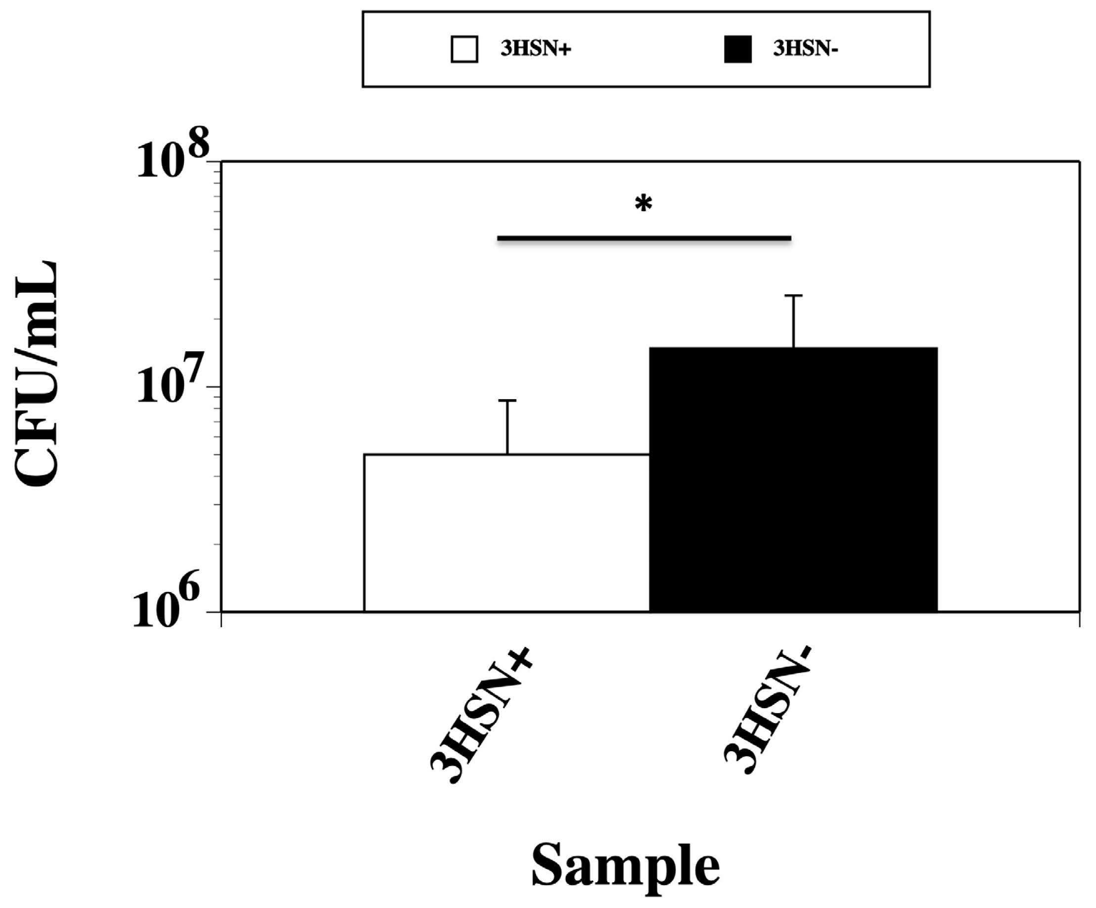
2.1. Supernatant from SK-03-92 Treated Cells Kills Fresh S. aureus Cells

The SK-03-92 drug kills 90% of S. aureus cells within 30 min [8]. One possibility for this rapid killing would be the release of a lethal factor by the treated cells. Supernatants collected from SK-03-92 treated cells (3HSN+) that were added to fresh S. aureus cells led to a threefold significantly lower viable count (5.0 × 10⁶ CFU/mL) than the supernatants from the untreated cells (1.5 × 10⁷; p < 0.05; Figure 1).

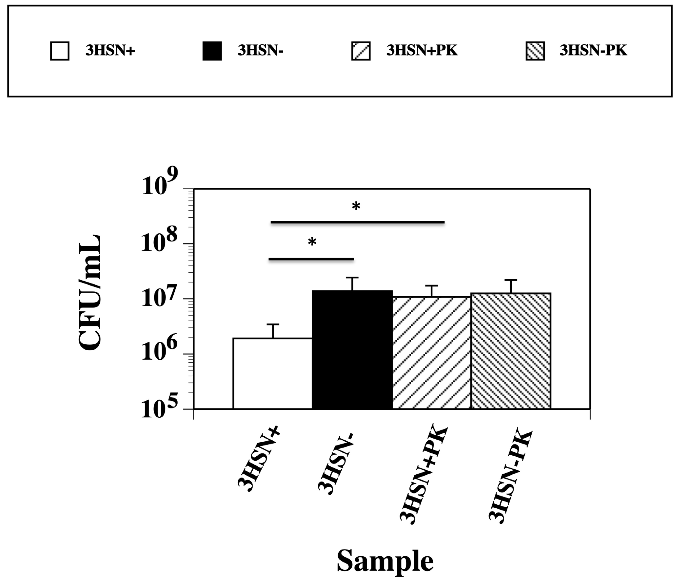
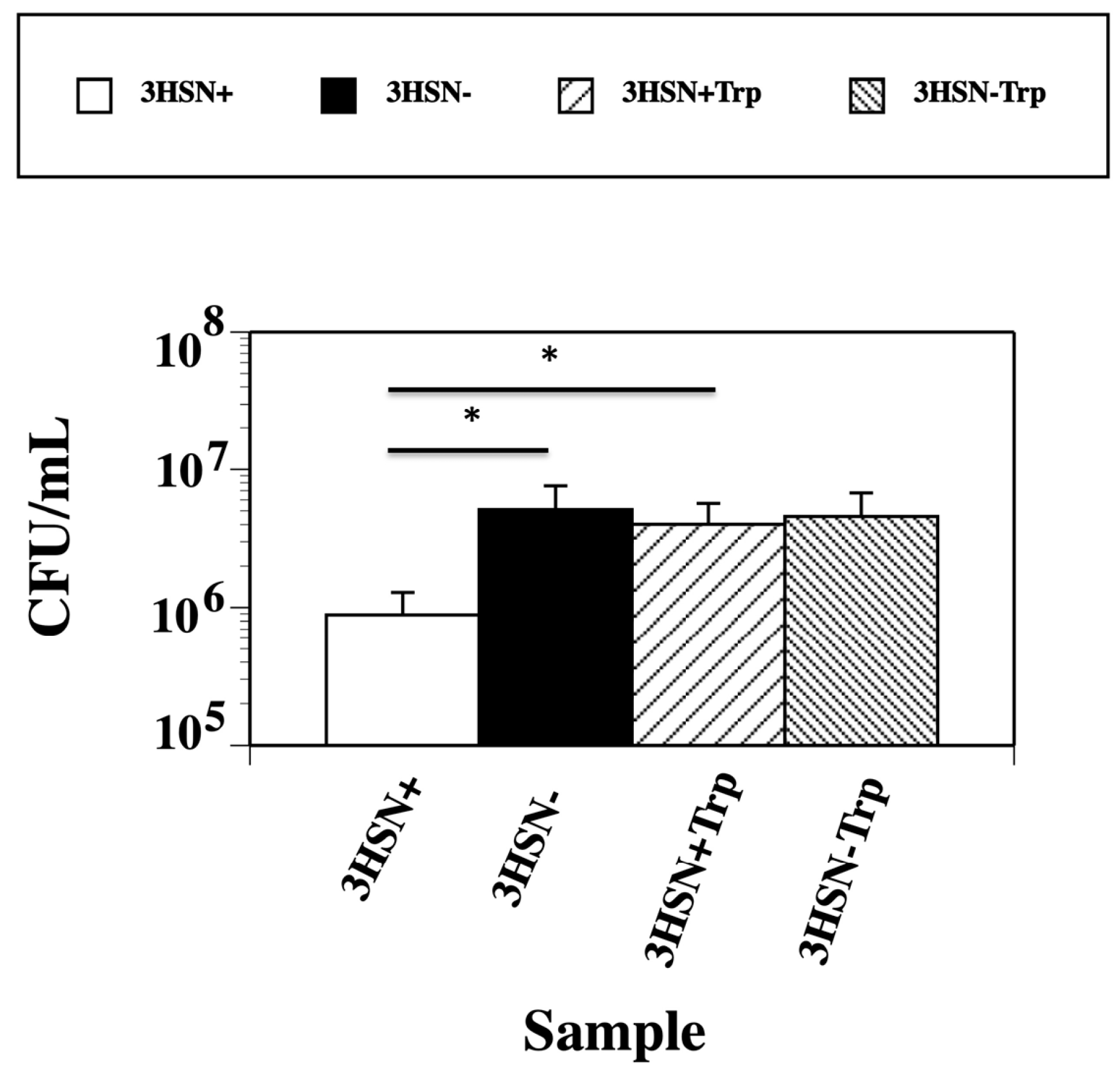
2.2. Boiling, Trypsin, and Proteinase K Treatment of 3HSN+ Supernatant Inactivates Killing Ability

The lethal factor was hypothesized to be a protein. To determine whether the lethal factor was a protein, a boiling experiment that denatures proteins and a proteinase K treatment that digests proteins were performed on the 3HSN+ supernatants. Boiled 3HSN+ preparations (4.2 × 10⁶ CFU/mL) displayed viable counts similar to the 3HSN− preparations (6.2 × 10⁶ CFU/mL), which were significantly higher than the 3HSN+ preparations that were not boiled (6.7 × 10⁴ CFU/mL; p < 0.01; Figure 2).
Furthermore, the proteinase K digested 3HSN+ samples (3HSN+PK) exhibited viable counts (1.09 × 10⁷ CFU/mL) similar to the untreated samples (3HSN−, 1.38 × 10⁷ CFU/mL) and proteinase K digested untreated samples (1.26 × 10⁷ CFU/mL), which were significantly higher than the SK-03-92 treated samples without proteinase K digestion (1.91 × 10⁶ CFU/mL; p < 0.01; Figure 3).
Finally, trypsin digested 3HSN+ samples (3HSN+Trp) showed viable counts (4.02 × 10⁶ CFU/mL) that were comparable to the trypsin digested untreated samples (3HSN−Trp, 4.58 × 10⁶ CFU/mL; Figure 4). However, the undigested 3HSN+ samples were significantly lower than the trypsin digested 3HSN+ samples (8.84 × 10⁵ CFU/mL, p < 0.05) as well as the undigested 3HSN− samples (5.1 × 10⁶ CFU/mL, p < 0.05). Through the boiling, trypsin digestion, and proteinase K digestion experiments, we were able to conclude that the lethal factor was a protein.

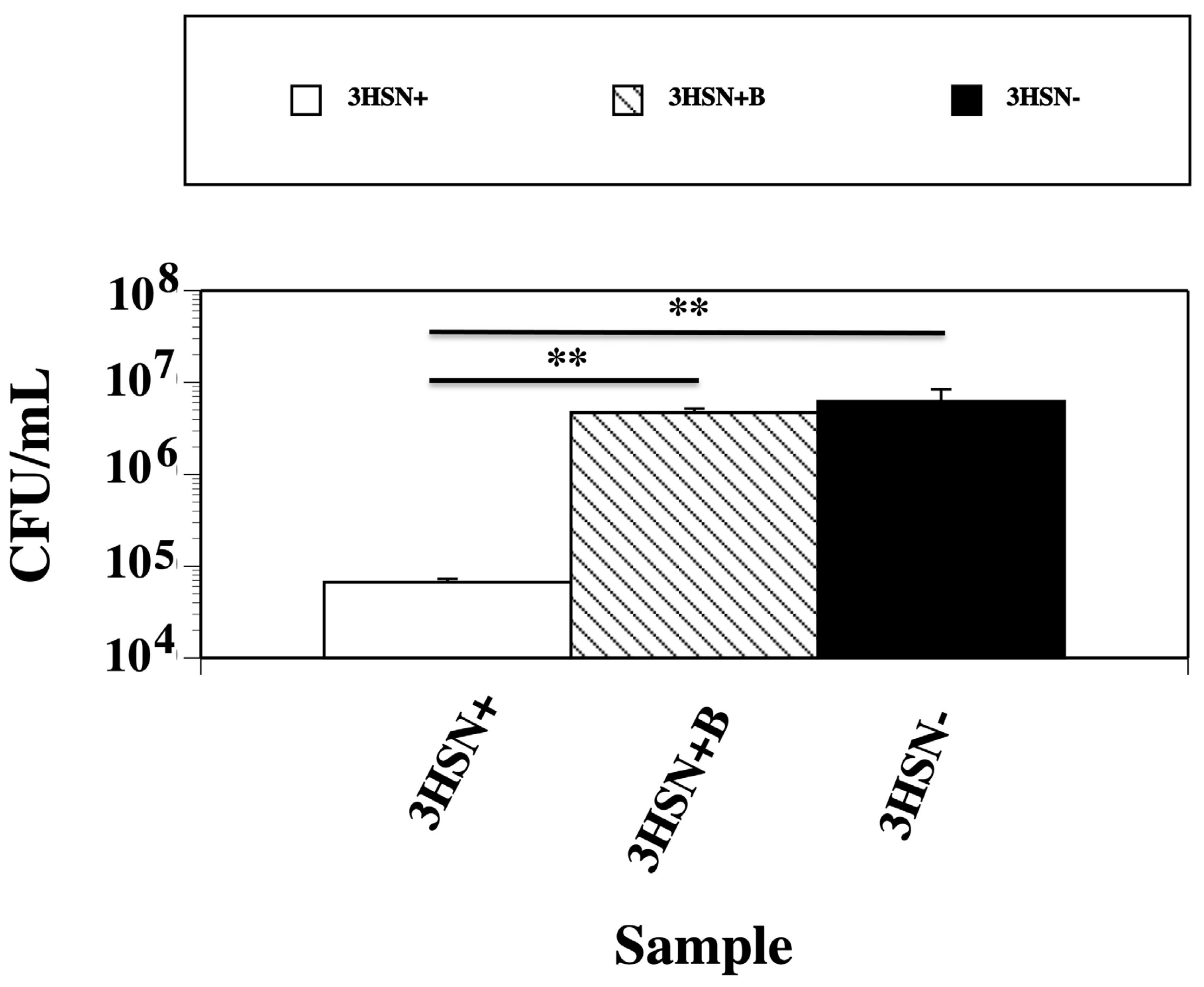
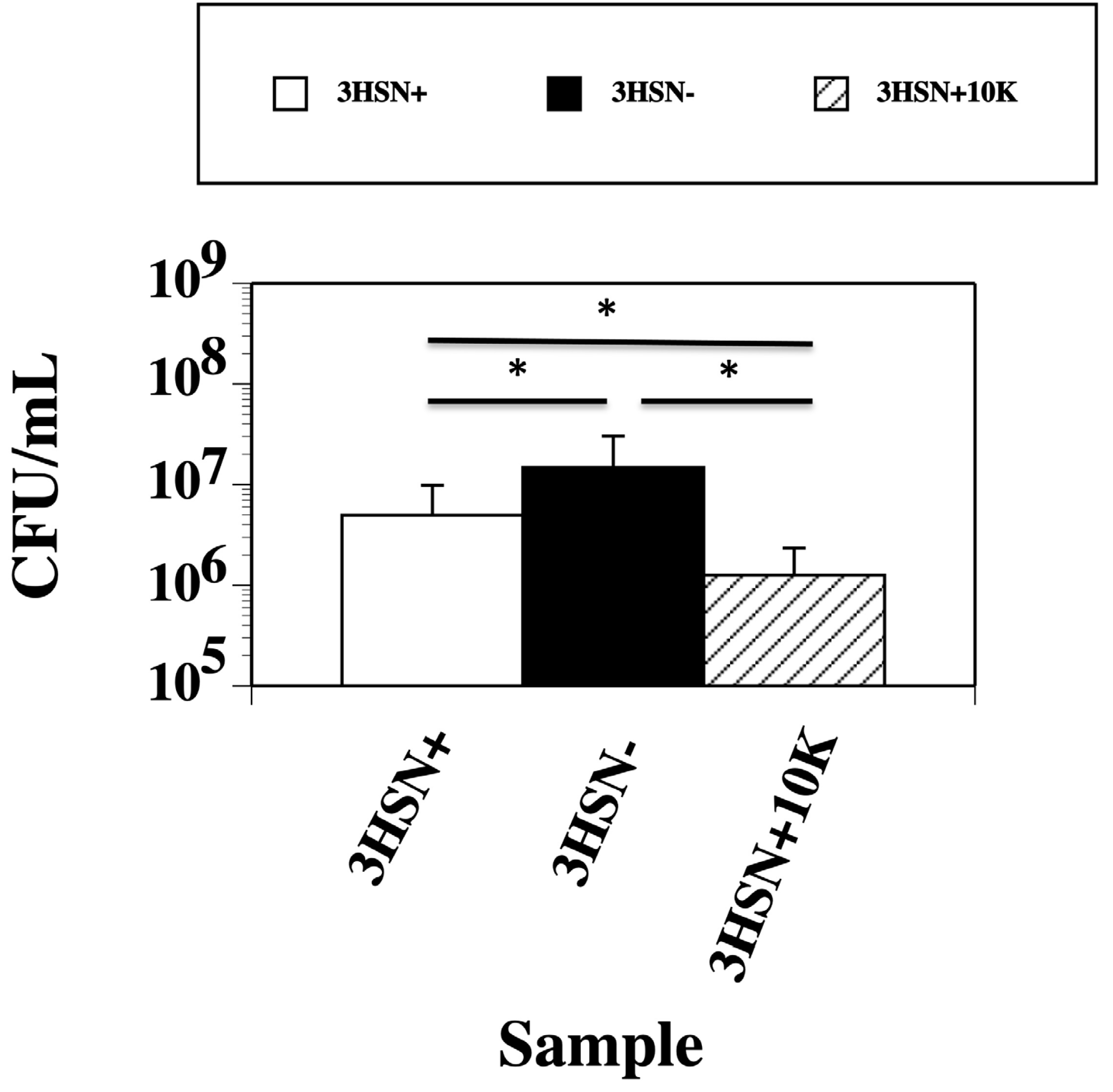
2.3. Concentrating the 3HSN+ Samples Caused an Increase in the Killing Ability

The lethal factor protein may be in low abundance. To concentrate the protein, the 3HSN supernatants were passed through Amicon 10K cut-off filters that will retain any proteins with a molecular weight higher than 10,000 Da. These concentrated preparations were then run through the kill assay and compared to the unconcentrated samples. The 3HSN+ samples had threefold lower viable counts (5.0 × 10⁶) CFU/mL) compared to the untreated 3HSN− samples (91.5 × 10⁷ CFU/mL; p < 0.05; Figure 5). The killing ability of the 3HSN+ samples significantly increased on average 3.9-fold compared to the unconcentrated 3HSN+ preparations (p < 0.05; Figure 5).

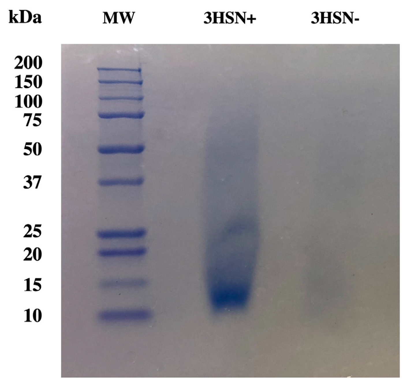
2.4. Gel Electrophoresis Shows an Approximately 12.5 kDa Protein Had the Highest Abundance

To better understand the relative size of the lethal factor protein, concentrated 3HSN+ and 3HSN− samples were separated on a polyacrylamide gel. The most abundant protein band observed had an approximate molecular weight of 12.5 kDa in the 3HSN+ lane (Figure 6). Other protein bands that were seen in the 3HSN+ lanes had approximate molecular weights of 15 kDa and 21 kDa in size. None of these protein bands were seen in the 3HSN− lane.

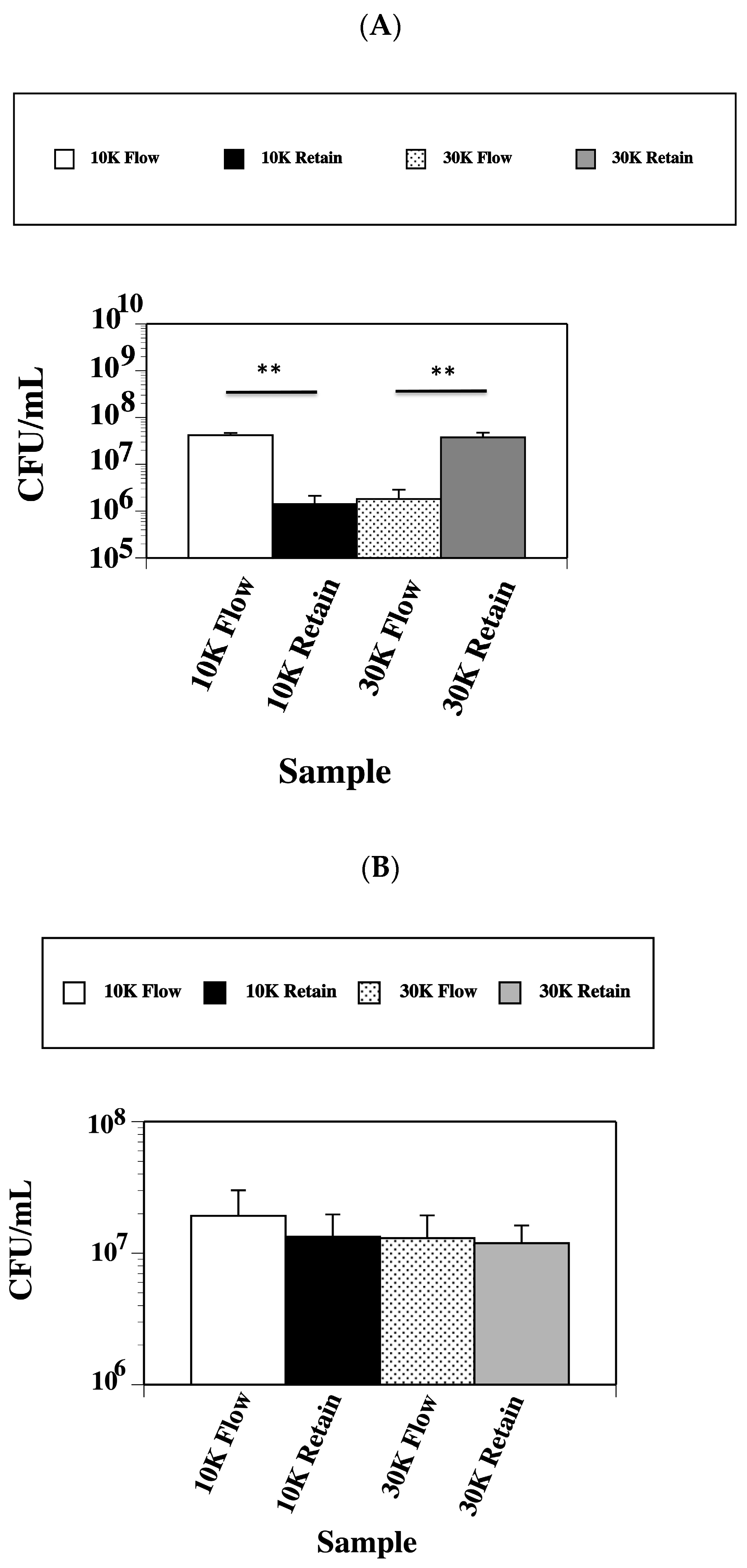
2.5. Lethal Factor Protein Has a Molecular Weight between a 10 kDa and 30 kDa Size

Concentrating the proteins through Amicon 10K filters showed that the lethal factor had a molecular weight higher than 10,000 Da. To further refine the approximate molecular weight of the lethal factor, 3HSN+ and 3HSN− samples were first passed sequentially through Amicon 10K filters. Flow through material (10K Flow) displayed viable counts of 4.2 × 10⁷ CFU/mL and the material retained on the 10K filter (10K Retained) had significantly lower viable counts of 1.41 × 10⁶ CFU/mL, p < 0.01) when used in the kill assay (Figure 7A). Next, the proteins retained on the 10K filter were in turn filtered through Amicon 30 K filters. Flow material from the 30K filter (30K Flow) showed a viable count of 1.83 × 10⁶ CFU/mL in the kill assay, which was significantly lower (p < 0.01) than the viable count (3.78 × 10⁷ CFU/mL) from the proteins retained on the 30K filter (30K Retain). Samples of 3HSN− that were processed the same way as described above displayed viable counts of 1.92 × 10⁷ CFU/mL (10K Flow), 1.33 × 10⁷ CFU/mL (10K Retain), 1.30 × 10⁷ CFU/mL (30K Flow), and 1.19 × 10⁷ CFU/mL (30K Retain), which were not significantly different between the conditions tested (Figure 7B). These results suggest that the lethal factor protein has a molecular weight between 10,000 and 30,000 Da in size.

2.6. Mass Spectrometry Detected Multiple Proteins

Protein bands sent out for in-gel digestion followed by mass spectrometry identified multiple proteins in each sample. The mass spectrometry analysis showed a tentative identification of the 13 kDa protein as SCIN (complement inhibitor protein) and the 17 kDa protein as CHIP (chemotaxis inhibitory protein).

3. Discussion

Antibiotic resistance continues to be a problem for S. aureus, so new treatment strategies are needed [4]. Our previous work with the lead compound SK-03-92 showed that the drug killed 90% of S. aureus cells in less than 30 min [8], however, a mechanism of action was not determined. Our hypothesis was that a lethal factor was released by S. aureus cells treated with the SK-03-92 drug that culminated in S. aureus cell death. In this study, we confirmed that the lethal factor is a protein with a molecular weight between 10 kDa and 30 kDa.
The 3-h supernatants (3HSNs) collected from S. aureus cells treated with the SK-03-92 drug were shown to kill fresh S. aureus cells, indicating that a lethal factor was released by the drug treated population. Boiling, trypsin, and proteinase K treatments inactivated the killing ability of the preparations, proving that the lethal factor was a protein. However, the identity of the protein is still unknown.
Based on prior work, we hypothesized that the SK-03-92 treatment affected a putative regulatory system labeled BrpR/S, which led to the release of a lethal factor such as an autolysin, mutacin, holin, or bacteriocin that rapidly killed the S. aureus cells. A previous microarray study of SK-03-92 treated versus untreated S. aureus cells demonstrated that three genes could be tied to a potential mechanism of action of the SK-03-92 drug: brpR, brpS, and lrgA [10]. The brpR and brpS genes had the highest differences in transcript abundance when comparing treated versus untreated cells. Initially, the BrpR/S system was thought to be a 2-component system [11]. However, additional bioinformatic analyses suggest that BrpR/BrpS acts as an LRS to regulate the transcription of other genes [24]. The LRS regulatory systems have a transmembrane-bound protein and a cytosolic LytTR domain-containing protein (LTR) [25,26,27]. BrpS has predicted multi-transmembrane domains, and BrpR appears to be a cytosolic protein with a LytTR-domain [11,24].
The BrpR/S system in S. aureus shares extensive homology to the ComC/D/E system in S. pneumoniae [28,29,30,31,32] as well as the ComC/D/E 2-component system and BrsR/M LRS in S. mutans [15,16,17,18,22,23]. Overall, the BrpR/S system shares the highest homology with the BrsR/M system in S. mutans [22]. The S. aureus BrpR protein shares a 25% identity with the S. mutans BrsR protein, whereas the S. aureus BrpS protein has a 30% homology with the S. mutans BrsM protein [11,24]. Both S. mutans proteins comprise a LRS responsible for sensing a competence-stimulating peptide (CSP), regulating the production of a bacteriocin and inducing late-stage competence [22]. ComC/D/E is also tied to the generation of cell death in S. mutans through the secretion of a lethal factor protein [29,32].
BrpR contains a LytTR DNA binding-motif [11,24] as do some of the streptococcal proteins noted above [17,28,32]. Both BrsR and ComE are multifunctional regulatory proteins that share homology with the BrpR protein.
Our previous data suggest that BrpS could be a receptor for a CSP-like pheromone released by S. aureus in response to resource competition. The (E)-3-(2-(benzo[b]thiophen-2-yl)vinyl)-(5)-methoxyphenol SK-03-92 lead drug is an analog of a stilbene structure that could have developed by convergent evolution to protect the sweet fern plant from bacterial colonization to force colonizing Gram-positive bacteria to enter late-stage competence through the production of CSP-like pheromones [33,34]. Part of this event would be induced cell lysis of the bacteria or competence-induced fratricide [29,32,34].
Several prior studies have examined the effect of CSP-like pheromones on bacterial cell viability and the production of biofilms. Zhang et al. observed that the addition of CSP to growth media induced an average 76.3% reduction in cell survival and an 89.3% increase in biofilm biomass in S. mutans [18]. Other studies have shown that recaptured supernatants or exposure to increased levels of exogenous CSP caused both an increase in cell death and biofilm formation [19,20,35,36].
Consistent with the late-competence model, cell suicide and increased biofilm formation in the staphylococcal response to SK-03-92 exposure may be tied to holin/antiholin action. LrgA is an antiholin that functions to prevent cell autolysis by binding CidA holins [13]. Our prior work showed a 3.2-fold reduction in lrgA transcription in cells exposed to SK-03-92 when compared to non-exposed cells [10]. In addition to cell death, a non-functional lrgA gene has also been correlated to the increased production of bacterial biofilms [37]. Cell lysis causes a rapid release of extracellular DNA (eDNA). The eDNA that is released acts as a scaffold for newly forming biofilms [6].
Many recent prior studies have examined the antibacterial properties of stilbene compounds [12,38,39,40,41,42,43]. Genome-wide RNA sequencing analysis of resveratrol treated S. aureus cells has shown no significant changes in brpR, brpS, or lrgA transcription [44,45]. Other stilbene compounds that have been identified do not have hydroxy at the one position and a methoxy at the number five position on the benzene ring, so we do not believe that these other compounds share the same mechanism of action as SK-03-92.
Our fractionation data from this study showed that the molecular weight of the lethal factor protein is between 10,000 and 30,000 Da. The CidA holin protein is around 12,000 Da [13], which would correlate with the Amicon size cut-off filtration and gel electrophoresis results. The mass spectrometry analysis was inconclusive, but the data suggested at least one of the proteins around 13 kDa in size may be the SCIN protein, and the 17 kDa size protein could be CHIP. Both proteins are tied to immune evasion, but neither of these staphylococcal proteins are associated with cell lysis of the S. aureus cell [46]. However, both proteins are encoded by genes that are part of the staphylococcal immune evasion cluster of genes that is part of the b-hemolysin converting bacteriophage inserted into the chromosome [47]. Within the gene cluster is a hol gene encoding a holin protein, so it possible that the holin protein is expressed and released after treatment with the SK-03-92 drug, which could lead to S. aureus cell lysis. A recent paper has shown that a brpS mutation affected the susceptibility of S. aureus to a novel compound labeled MEL-B [48]. They hypothesized that the BrpS protein could be tied to the staphylococcal cell wall. More work is needed to elucidate the identity of the lethal factor protein and determine whether the release of a CSP-like pheromone by SK-03-92 treatment may trigger these events.

4. Materials and Methods

4.1. Bacterial Strains and Growth Media

In this study, S. aureus strain Newman was used [49]. All media used in the study were purchased from Thermo Fisher Scientific, Pittsburgh, PA, USA. The bacteria were grown in brain heart infusion (BHI) broth shaken at 250 rpm at 37 °C or on trypticase soy agar (TSA) plates at 37 °C.

4.2. Collection of SK-03-92 Treated and Untreated Culture Supernatants

S. aureus strain Newman was grown on a TSA plate for less than 24 h at 37 °C. Bacterial cells from the TSA plate were suspended in 5 mL of phosphate buffered saline (PBS) and set to a concentration of 1 × 10⁸ CFU/mL by matching the turbidity to a 0.5 McFarland standard (Thermo Fisher Scientific). The bacteria were diluted 10-fold in PBS to 1 × 10⁷ CFU/mL. Next, aliquots of the bacterial suspension were dispensed into two 14 mL plastic tubes. To each tube, an equal volume of BHI broth was added to make a 1:1 ratio. One tube received BHI broth alone (negative) and the other tube received BHI broth plus 4 μg/mL of the SK-03-92 drug (treated). Both tubes were vortexed and then statically incubated for 3 h at 37 °C. The samples were then individually filter-sterilized through a 0.2 μm filter (Millipore, Burlington, MA, USA) and the supernatants collected. Samples treated with SK-03-92 were labeled as 3-h supernatants treated (3HSN+) and untreated samples were marked as 3HSN−. All samples were either immediately used or incubated at 4 °C for less than 4 days before use.

4.3. Bacterial Kill Assay

The 3HSN+ and 3HSN− supernatants were diluted 1:4 in PBS to reduce the concentration of SK-03-92 to below the MIC of 2 μg/mL for S. aureus strain Newman. Fresh S. aureus cells from a TSA plate grown at 37 °C were suspended to a concentration of 1 × 10⁶ CFU/mL in PBS. One milliliter aliquots of S. aureus cells in PBS were added to two tubes. One tube received an equal volume of the 3HSN+ supernatant, and the other tube had an equal volume of 3HSN− added. Both tubes were incubated for 24 h at 37 °C. After incubation, aliquots from each tube were 10-fold serially diluted in PBS, 100 mL from each dilution plated on BHI agar plates in duplicate, and incubated for 24 h at 37 °C. Colony counts were taken for both the 3HSN+ and 3HSN− populations. Kill assays were carried out three to ten times.

4.4. Boiling 3HSN Samples

To determine the effect of boiling on the killing capacity of the 3HSN+ supernatant, a 500 μL aliquot of the 3HSN+ supernatant was boiled for 30 min at 95 °C. The bacterial kill assays described above were run to compare the boiled 3HSN+ preparation to the non-boiled 3HSN+ sample as well as the 3HSN− supernatant. All samples were incubated for 24 h at 37 °C. Following incubation, the supernatants were 10-fold serially diluted in PBS, 100 μL aliquots of each dilution plated onto two BHI agar plates, the plates incubated for 24 h at 37 °C, and viable colony counts taken for each condition tested. A total of three separate runs were conducted to compare the boiled to non-boiled samples.

4.5. Digesting 3HSN Samples with Proteinase K

Two hundred microliter aliquots of either the 3HSN+ or 3HSN− supernatants were digested with 2 μL (1 unit) of thermolabile proteinase K (New England Biolabs, Ipswich, MA, USA) at 37 °C for 15 min. Samples were then heated for 10 min at 55 °C to inactivate the proteinase K. Bacterial kill assays with fresh S. aureus cells were set up using the proteinase K digested 3HSN+ and 3HSN− preparations as well as the undigested 3HSN+ and 3HSN− preparations. All samples were incubated for 24 h at 37 °C, 10-fold serially diluted in PBS, and 100 mL aliquots of each dilution from each condition tested were plated in duplicate onto BHI agar plates, incubated for 24 h at 37 °C, and viable colony counts undertaken. The proteinase K digestion was repeated four more times for a total of five runs.

4.6. Digesting 3HSN Samples with Trypsin

Two hundred microliter aliquots of either the 3HSN+ or 3HSN− supernatants were digested with trypsin (Sigma Aldrich, St. Louis, MO, USA) at 37 °C for 15 min. To each sample was added 50 mL of heat inactivated fetal calf serum (Thermo Fisher Scientific) for 10 min at room temperature. Bacterial kill assays with fresh S. aureus cells were set up using the trypsin digested 3HSN+ and 3HSN− preparations as well as the undigested 3HSN+ and 3HSN− preparations. All samples were incubated for 24 h at 37 °C, 10-fold serially diluted in PBS, and 100 m aliquots of each dilution from each condition tested were plated in duplicate onto BHI agar plates, incubated for 24 h at 37 °C, and viable colony counts taken. The trypsin digestion was repeated two more times for a total of three runs.

4.7. Size Cut-Off Filtration

To obtain a better understanding of the size of the lethal factor protein as well as concentrate it, the 3HSN preparations were filtered through two different protein size cut-off filters, a 10K Amicon filter (Millipore, Burlington, MA, USA) that retains proteins with a size greater than 10,000 Da, and a 30K Amicon filter (Millipore) that retains proteins with a size greater than 30,000 Da. Briefly, 15 mL of either the 3HSN+ or 3HSN− supernatant prepared as described above was filtered through 10K Amicon filters at 4 °C, 4000× g for 20 min. Retained proteins were collected from the 10 K filters by inverting the filter and placing the filters in a sterile 1.5 mL microfuge tube under 4000× g centrifugal force for 10 min at 4 °C. The bacterial kill assays noted above were performed on both the proteins retained on the filters as well as the flow through supernatants. Next, the proteins retained by the 10K filter were filtered through a 30K filter at 4 °C, 4000× g for 20 min. Flow through supernatants were used for the bacterial kill assays. The proteins retained on the 30K filter were stripped away by inverting the filters into 1.5 mL microfuge tubes under 4000× g centrifugal force for 10 min at 4 °C. Bacterial kill assays were performed on the proteins retained from passage through the 30K filters. This procedure was repeated two more times for a total of three runs.

4.8. Gel Electrophoresis

Preparations of 3HSN passed through a 10K Amicon filter had their protein concentrations measured using a Nanodrop A280 spectrophotometer (Thermo Fisher Scientific). Forty microliters of the 10K 3HSN samples and 10 μL of 5X sodium dodecyl sulfate loading dye (BioRad, Hercules, AC, USA) were mixed and heated to 95 °C for 10 min. Any kDa acrylamide gels (BioRad) were loaded with an equivalent 5 mg/mL of protein from the 10K 3HSN+ and 10K 3HSN− preparations. A 4 μL aliquot of Precision Plus ProteinTM All Blue Standard (BioRad) was run as a molecular weight marker. Electrophoresis was conducted for 40 min at 180 volts, 90 mAMPs, and 40 Watts. Gels were stained with Coomassie Blue R250 dye (BioRad; 0.5 g Brilliant Blue R250, 50 mL isopropyl alcohol, 20 mL glacial acetic acid, 200 mL ddH2O) for at least 1 h at room temperature. Each gel was rinsed with ddH2O and destained (250 mL isopropyl alcohol, 100 mL glacial acetic acid, and 650 mL ddH2O) for at least 1 h at room temperature.

4.9. Mass Spectrometry

Buffer exchanges were performed on 10K 3HSN+ samples by adding 10 mL of 20 mM Tris pH 8.0 solution (Buffer A) to the proteins retained on a 10K Amicon filter and pelleting by centrifugation at 4000× g for 10 min at 4 °C. The buffer exchanges were repeated six times. Proteins retained on the 10K filter were then resuspended in 10 mL of Buffer A. The resuspended solution was then loaded onto a Bio-Scale Mini Macro-Prep High Q cartridge (BioRad) for ion exchange. Buffer A was run through the column first, followed by Buffer B (20 mM Tris pH 8.0, 1 M NaCl), and finally Buffer C (20 mM Tris pH 8.0, 4 M NaCl). Fractions were collected and pooled into four groups based on adsorption readings at 280 nM. All pools were then tested for the presence of the lethal factor protein by conducting the kill assays as described above. Aliquots from each pool were separated by gel electrophoresis using Any kDa gels and silver stained with a kit (Thermo Fisher Scientific), followed by destaining as described above. Two bands of 13 kDa and 17 kDa in size from a pool with kill activity were excised from the polyacrylamide gel and sent to the Iowa State University Protein Facility (Ames, IA, USA) for in-gel digestion followed by liquid chromatography on an EASY-nLC1200 (Thermo Fisher Scientific) coupled to a Q Exactive tandem mass spectrometry analysis using a Q ExactiveTM Hybrid Quadrupole-Orbitrap mass spectrometer (Thermo Fisher Scientific). Data were searched against publicly available databases using Mascot and Sequest HT.

4.10. Statistics

A Student’s t test was used for the statistical analyses. p values of ≤0.05 were considered significant.

5. Conclusions

SK-03-92 treatment of S. aureus cells releases a lethal factor protein with an approximate molecular weight between 10 kDa and 30 kDa that can kill other S. aureus cells.

Author Contributions

Funding acquisition was carried out by W.R.S., J.F.M., A.Z., M.M. and S.C.; Conceptualization was conducted by W.R.S., J.F.M., A.Z. and M.M.; Methodology and analysis were undertaken by W.R.S., J.F.M., A.Z., M.M., S.C. and K.G.; and writing was conducted by W.R.S. and M.M. All authors have read and agreed to the published version of the manuscript.

Funding

This study was funded by a UW-System-WiSys grant to W.R.S. and J.M., UWL RSEL grants to A.Z. and M.M., and a UWL Undergraduate Research grant to S.C.

Data Availability Statement

The data from this study are readily available from the author.

Acknowledgments

We wish to thank Jean Lee for the S. aureus Newman strain and Joel Nott at the Protein Facility of the Iowa State University Office of Biotechnology for the analysis of samples by in-gel digestion and LC-MS/MS.

Conflicts of Interest

The authors declare no conflicts of interest.

References

  1. Suaya, J.A.; Mera, R.M.; Cassidy, A.; O’Hara, P.; Amrine-Madsen, H.; Burstin, S.; Miller, L.G. Incidence and cost of hospitalization associated with Staphylococcus aureus skin and soft tissue infections in the United States from 2001 through 2009. BMC Infect. Dis. 2014, 14, 296. [Google Scholar] [CrossRef] [PubMed]
  2. Klein, E.Y.; Sun, D.L.; Laxminarayan, R. The changing epidemiology of methicillin-resistant Staphylococcus aureus hospitalizations in the United States: A national observational study. Am. J. Epidemiol. 2013, 177, 666–674. [Google Scholar] [CrossRef] [PubMed]
  3. Klein, E.Y.; Mojica, N.; Jiang, W.; Cosgrove, S.E.; Septimus, E.E.; Morgan, D.J.; Laxminarayan, R. Trends in methicillin-resistant Staphylococcus aureus hospitalizations in the United States, 2010–2014. Clin. Infect. Dis. 2017, 65, 1921–1923. [Google Scholar] [CrossRef]
  4. WHO (World Health Organization). Global Antimicrobial Resistance and Use Surveillance System (GLASS) Report. 2021. Available online: https://www.who.int/publications/i/item/9789240027336 (accessed on 22 February 2024).
  5. Lee, A.S.; de Lencastre, H.; Garau, J.; Kluytmans, J.; Malhotra-Kumar, S.; Peschel, A.; Harbath, S. Methicillin-resistant Staphylococcus aureus. Nat. Rev. Dis. Primers 2018, 4, 18033. [Google Scholar] [CrossRef] [PubMed]
  6. Schilcher, K.; Horswill, A.R. Staphylococcal biofilm development: Structure, regulation, and treatment strategies. Microbiol. Mol. Biol. Rev. 2020, 84, e00026-19. [Google Scholar] [CrossRef] [PubMed]
  7. Kabir, M.S.; Englebrecht, K.; Polanowski, R.; Krueger, S.M.; Ignasiak, R.; Rott, M.; Schwan, W.R.; Stemper, M.E.; Reed, K.D.; Sherman, D.; et al. New classes of Gram-positive selective antibacterials: Inhibitors of MRSA and surrogates of the causative agents of anthrax and tuberculosis. Bioorg. Med. Chem. Lett. 2010, 18, 5745–5749. [Google Scholar] [CrossRef] [PubMed]
  8. Schwan, W.R.; Kabir, M.S.; Kallaus, M.; Krueger, S.; Monte, A.; Cook, J.M. Synthesis and minimum inhibitory concentrations of SK-03-92 against Staphylococcus aureus and other gram-positive bacteria. J. Infect. Chemother. 2012, 18, 124–126. [Google Scholar] [CrossRef] [PubMed]
  9. Schwan, W.R. SK-03-92 drug kills intracellular Mycobacterium tuberculosis. Antibiotics 2023, 12, 1385. [Google Scholar] [CrossRef] [PubMed]
  10. Schwan, W.R.; Polanowski, R.; Dunman, P.M.; Medinia-Bielski, S.; Lane, M.; Rott, M.; Lipker, L.; Wescott, A.; Monte, A.; Cook, J.M.; et al. Identification of Staphylococcus aureus cellular pathways affected by the stilbenoid lead drug SK-03-92 using a microarray. Antibiotics 2017, 6, 17. [Google Scholar] [CrossRef]
  11. Zank, A.; Schulte, L.; Brandon, X.; Carstensen, L.; Wescott, A.; Schwan, W.R. Mutations of the brpR and brpS genes affect biofilm formation in Staphylococcus aureus. World J. Clin. Infect. Dis. 2022, 12, 20–32. [Google Scholar] [CrossRef]
  12. Sheng, J.Y.; Chen, T.T.; Tan, X.J.; Chen, T.; Jia, A.Q. The quorum-sensing effects of stilbenoids and their potential structure-activity relationship. Bioorg. Med. Chem. Lett. 2015, 25, 5217–5220. [Google Scholar] [CrossRef] [PubMed]
  13. Ranjit, D.K.; Endres, J.L.; Bayles, K.W. Staphylococcus aureus CidA and LrgA proteins exhibit holin-like properties. J. Bacteriol. 2011, 193, 2468–2476. [Google Scholar] [CrossRef] [PubMed]
  14. Bayles, K. The biological role of death and lysis in biofilm development. Nat. Rev. Microbiol. 2007, 5, 721–726. [Google Scholar] [CrossRef] [PubMed]
  15. Li, Y.H.; Tang, N.; Aspiras, M.B.; Lau, P.C.; Lee, J.H.; Ellen, R.P.; Cvitkovitch, D.G. A quorum-sensing signaling system essential for genetic competence in Streptococcus mutans is involved in biofilm formation. J. Bacteriol. 2002, 10, 2699–2708. [Google Scholar] [CrossRef] [PubMed]
  16. Shanker, R.; Federle, M.J. Quorum sensing regulation of competence and bacteriocins in Streptococcus pneumoniae and mutans. Genes 2017, 1, 15. [Google Scholar] [CrossRef] [PubMed]
  17. Kreth, J.; Hung, D.C.I.; Merritt, J.; Perry, J.; Zhu, L.; Goodman, S.D.; Cvitkovich, D.G.; Shi, W.; Qi, F. The response regulator ComE in Streptococcus mutans functions both as a transcription activator of mutacin production and repressor of CSP biosynthesis. Microbiology 2007, 153, 1799–1807. [Google Scholar] [CrossRef] [PubMed]
  18. Zhang, K.; Ou, M.; Wang, W.; Ling, J. Effects of quorum sensing on cell viability in Streptococcus mutans biofilm formation. Biochem. Biophys. Res. Commun. 2009, 379, 933–938. [Google Scholar] [CrossRef]
  19. Qi, F.; Kreth, J.; Levesque, C.M.; Kay, O.; Mair, R.W.; Shi, W.; Cvitkovitch, D.G.; Goodman, S.D. Peptide pheromone induced cell death of Streptococcus mutans. FEMS Microbiol. Lett. 2005, 251, 321–326. [Google Scholar] [CrossRef] [PubMed]
  20. Perry, J.A.; Cvitkovitch, D.G.; Levesque, C.M. Cell death in Streptococcus mutans biofilms: A link between CSP and extracellular DNA. FEMS Microbiol. Lett. 2009, 299, 261–266. [Google Scholar] [CrossRef] [PubMed]
  21. Merritt, J.; Qi, F. The mutacins of Streptococcus mutans: Regulation and ecology. Mol. Oral Microbiol. 2012, 2, 57–69. [Google Scholar] [CrossRef] [PubMed]
  22. Xie, Z.; Okinaga, T.; Niu, G.; Qi, F.; Merritt, J. Identification of a novel bacteriocin regulatory system in Streptococcus mutans. Mol. Microbiol. 2010, 6, 1431–1447. [Google Scholar] [CrossRef] [PubMed]
  23. Perry, J.A.; Jones, M.B.; Peterson, S.N.; Civtkovitch, D.G.; Levesque, C.M. Peptide alarmone signalling triggers an auto-active bacteriocin necessary for bacterial competence. Mol. Microbiol. 2009, 72, 905–917. [Google Scholar] [CrossRef] [PubMed]
  24. Moore, M. Identifying the Lethal Factor Protein Secreted by Staphylococcus aureus Treatment with SK-03-92 Drug. Master’s Thesis, University of Wisconsin-La Crosse, La Crosse, WI, USA, 2023. [Google Scholar]
  25. Ulrich, L.E.; Koonin, E.V.; Zhulin, I.B. One-component systems dominate signal transduction in prokaryotes. Trends Microbiol. 2005, 13, 52–56. [Google Scholar] [CrossRef]
  26. Zou, Z.; Qin, H.; Brenner, A.E.; Raghavan, R.; Milar, J.A.; Gu, Q.; Xie, Z.; Kreth, J.; Merritt, J. LytTR regulatory systems: A potential new class of prokaryotic sensory system. PLoS Genet. 2018, 10, e1007709. [Google Scholar] [CrossRef] [PubMed]
  27. Williams, R.H.; Whitworth, D.E. The genetic organization of prokaryotic two-component system signaling pathways. BMC Genom. 2010, 11, 720. [Google Scholar] [CrossRef] [PubMed]
  28. Pestova, E.V.; Havarstein, L.S.; Morrison, D.A. Regulation of competence for genetic transformation in Streptococcus pneumoniae by an auto-induced peptide pheromone and a two-component regulatory system. Mol. Microbiol. 1996, 21, 853–862. [Google Scholar] [CrossRef] [PubMed]
  29. Claverys, J.P.; Martin, B.; Havarstein, L.S. Competence-induced fratricide in streptococci. Mol. Microbiol. 2007, 6, 1423–1433. [Google Scholar] [CrossRef] [PubMed]
  30. Galperin, M. Telling bacteria: Do not LYtTR. Structure 2008, 16, 657–659. [Google Scholar] [CrossRef] [PubMed]
  31. Carrolo, M.; Pinto, F.R.; Melo-Cristino, J.; Ramirez, M. Pherotype influences biofilm growth and recombination in Streptococcus pneumoniae. PLoS ONE 2014, 9, e92138. [Google Scholar] [CrossRef] [PubMed]
  32. Martin, B.; Granadel, C.; Campo, N.; Hénard, V.; Prudhomme, M.; Claverys, J.P. Expression and maintenance of ComD-ComE, the two-component signal-transduction system that controls competence of Streptococcus pneumoniae. Mol. Microbiol. 2010, 75, 1513–1528. [Google Scholar] [CrossRef] [PubMed]
  33. Havarstein, L.S.; Gaustad, P.; Nes, I.F.; Morrison, D.A. Identification of the streptococcal competence-pheromone receptor. Mol. Microbiol. 1996, 21, 863–869. [Google Scholar] [CrossRef] [PubMed]
  34. Li, Y.H.; Lau, P.C.; Lee, J.H.; Ellen, R.P.; Cvitkovitch, D.G. Natural genetic transformation of Streptococcus mutans growing in biofilms. J. Bacteriol. 2001, 183, 897–908. [Google Scholar] [CrossRef] [PubMed]
  35. Dufour, D.; Levesque, C.M. Cell death of Streptococcus mutans induced by a quorum-sensing peptide occurs via a conserved streptococcal autolysin. J. Bacteriol. 2013, 195, 105–114. [Google Scholar] [CrossRef] [PubMed]
  36. Ween, O.; Gaustad, P.; Havarstein, L.S. Identification of DNA binding sites for ComE, a key regulator of natural competence in Streptococcus pneumoniae. Mol. Microbiol. 1999, 33, 817–827. [Google Scholar] [CrossRef] [PubMed]
  37. Brunskill, E.W.; Bayles, K.W. Identification and molecular characterization of a putative regulatory locus that affects autolysis in Staphylococcus aureus. J. Bacteriol. 1996, 178, 611–618. [Google Scholar] [CrossRef] [PubMed]
  38. Chen, J.-Y.; Lian, X.; Fan, Y.W.; Ao, Z.Y.; Zhang, W.J.; Pan, Y.C.; Chen, L.P.; Yuan, J.; Wu, J.W. Four new stilbenes and one new flavonoid with potential antibacterial and anti-SARS-CoV-2 activity from Cajanus cajan. J. Nat. Med. 2023, 77, 858–866. [Google Scholar] [CrossRef]
  39. El-Mahdy, A.M.; Alqahtani, M.; Almukainzi, M.; Alghoribi, M.F.; Abdel-Rhman, S.H. Effect of resveratrol and curcumin on gene expression of methicillin-resistant Staphylococcus aureus (MRSA) toxins. J. Microbiol. Biotechnol. 2024, 34, 141–148. [Google Scholar] [CrossRef] [PubMed]
  40. Kasparova, P.; Vankova, E.; Brazdova, L.; Lokocova, K.; Matatkova, O.; Masak, L. Antibiofilm agent pterostilbene is able to enhance antibiotics action against Staphylococcus epidermidis. Microb. Pathog. 2021, 152, 104632. [Google Scholar] [CrossRef] [PubMed]
  41. Lewis, A.J.; Richards, A.C.; Mendez, A.A.; Dhakal, B.K.; Jones, T.A.; Sundsbak, J.L.; Eto, D.S.; Rousek, A.A.; Mulvey, M.A. Plant phenolics inhibit focal adhesion kinase and suppress host cell invasion by uropathogenic Escherichia coli. Infect. Immun. 2024, 92, e0008024. [Google Scholar] [CrossRef] [PubMed]
  42. Li, B.L.; Chen, J.Y.; Hu, J.J.; Fan, Y.W.; Ao, Z.Y.; Zhang, W.J.; Lian, X.; Liang, H.J.; Li, Q.; Guan, X.X.; et al. Three stilbenes from pigeon pea with promising anti-methicillin-resistant Staphylococcus aureus biofilm formation activity. Int. Microbiol. 2024, 27, 535–544. [Google Scholar] [CrossRef] [PubMed]
  43. Yu, Q.; Sun, L.; Peng, F.; Sun, C.; Xiong, F.; Sun, M.; Liu, J.; Peng, C.; Zhou, Q. Antimicrobial activity of stilbenes from Bletilla striata against Cutibacterium acnes and its effect on cell membrane. Microorganisms 2023, 11, 2958. [Google Scholar] [CrossRef] [PubMed]
  44. Qin, N.; Tan, X.; Jiao, Y.; Liu, L.; Zhao, W.; Yang, S.; Jia, A. RNA-Seq-based transcriptome analysis of methicillin-resistant Staphylococcus aureus biofilm inhibition by ursolic acid and resveratrol. Sci. Rep. 2014, 4, 5467. [Google Scholar] [CrossRef] [PubMed]
  45. Tan, X.; Qin, N.; Wu, C.; Sheng, J.; Yang, R.; Zheng, B.; Ma, Z.; Liu, L.; Peng, X.; Jia, A. Transcriptome analysis of the biofilm formed by methicillin-susceptible Staphylococcus aureus. Sci. Rep. 2015, 5, 11997. [Google Scholar] [CrossRef] [PubMed]
  46. Verkaik, N.J.; Bernard, M.; Boelens, H.A.; de Vogel, C.P.; Nouwen, J.L.; Verbrugh, H.A.; Melles, D.C.; van Belkum, A.; van Wamel, W.J. Immune evasion cluster-positive bacteriophages are highly prevalent among human Staphylococcus aureus strains, but they are not essential in the first stages of nasal colonization. Clin. Microbiol. Infect. 2011, 17, 343–348. [Google Scholar] [CrossRef] [PubMed]
  47. van Wamel, W.J.; Rooijakkers, S.H.; Ruyken, M.; van Kessel, K.P.; van Strijp, J.A. The innate immune modulators staphylococcal complement inhibitor and chemotaxis inhibitory protein of Staphylococcus aureus are located on beta-hemolysin-converting bacteriophages. J. Bacteriol. 2006, 188, 1310–1315. [Google Scholar] [CrossRef] [PubMed]
  48. Yamauchi, S.; Shimoda, S.; Kawahara, A.; Sugahara, T.; Yamamoto, S.; Kitabayashi, M.; Sogabe, A.; Jansen, C.A.; Tobe, R.; Hirakawa, R.; et al. Identification of four genes responsible for antimicrobial resistance of MEL-B against S. aureus. Biochem. Biophys. Res. Commun. 2024, 699, 149566. [Google Scholar] [CrossRef] [PubMed]
  49. Duthie, E.S.; Lorenz, L.L. Staphylococcal coagulase: Mode of action and antigenicity. J. Gen. Microbiol. 1952, 6, 95–107. [Google Scholar] [CrossRef]
Figure 1. Killing ability of 3-h supernatants from the SK-03-92 treated S. aureus Newman cells (3HSN+, white column) compared to the untreated cells (3HSN−, black column). All supernatants were added to fresh S. aureus cells and incubated overnight onto BHI agar plates at 37 °C. The data represent the mean ± standard deviation from 10 separate runs represented as colony forming units (CFUs) per mL. * equals a p value < 0.05.
Targets 02 00005 g001
Figure 2. Boiling 3-h supernatants (3HSNs) negates the killing ability. S. aureus cells were treated with SK-03-92 for three hours (3HSN+, white column) or were untreated (3HSN−, black column). An aliquot of 3HSN+ was boiled for 30 min (3HSN+B, lined column). All preparations were added to fresh S. aureus cells and incubated overnight at 37 °C. The data represent the mean ± standard deviation from three separate runs represented as colony forming units (CFUs)/mL. ** equals a p value < 0.01.
Targets 02 00005 g002
Figure 3. Proteinase K treatment of 3-h supernatants (3HSNs) negated the killing ability. S. aureus cells were treated with SK-03-92 for three hours (3HSN+, white column) or were untreated (3HSN−, black column). Aliquots of 3HSN+ and 3HSN− were digested with proteinase K for 15 min at 37 °C and the enzymes inactivated in these samples are represented as 3HSN+PK (right lined column) and 3HSN−PK (left lined column). All preparations were added to fresh S. aureus cells and incubated overnight at 37 °C. The data represent the mean ± standard deviation from five separate runs represented as colony forming units (CFUs)/mL. * equals a p value < 0.05.
Targets 02 00005 g003
Figure 4. Trypsin treatment of 3-h supernatants (3HSNs) negated the killing ability. S. aureus cells were treated with SK-03-92 for three hours (3HSN+, white column) or were untreated (3HSN−, black column). Aliquots of 3HSN+ and 3HSN− were digested with trypsin for 15 min at 37 °C and the enzymes inactivated in these samples are represented as 3HSN+Trp (right lined column) and 3HSN−Trp (left lined column). All preparations were added to fresh S. aureus cells and incubated overnight at 37 °C. The data represent the mean ± standard deviation from five separate runs represented as colony forming units (CFUs)/mL. * equals a p value < 0.05.
Targets 02 00005 g004
Figure 5. Concentrating the 3-h supernatant proteins (3HSN) led to a higher S. aureus killing ability versus the unconcentrated samples. The 3-h supernatants were collected from S. aureus cells treated with the SK-03-92 drug (3HSN+, white column) and untreated cells (3HSN−, black column). Aliquots of 3HSN+ samples were then filtered through Amicon 10K cut-off filters and the proteins retained on the filter (3HSN+10K, lined column) as well as the unfiltered 3HSN+ and 3HSN− samples were added to fresh S. aureus cells and incubated overnight at 37 °C. The data represent the mean ± standard deviation from 10 separate runs represented as colony forming units (CFUs)/mL. * equals a p value < 0.05.
Targets 02 00005 g005
Figure 6. Sodium dodecyl sulfate polyacrylamide gel separation of SK-03-92 treated 3-h supernatant (3HSN+) and untreated cell supernatant (3HSN−) concentrated through 10,000 Da cut-off 10K Amicon filters. A precision blue protein standard (MW) was run to assess the molecular weight of the proteins on the gel.
Targets 02 00005 g006
Figure 7. Amicon filtration to obtain the size the molecular weight of the lethal factor. The 3-h supernatants were collected from S. aureus cells that were either (A) treated with the SK-03-92 drug (3HSN+) or (B) untreated (3HSN−). Each sample was first passed through an Amicon10K filter, and flow through proteins (10K Flow, white column) and proteins retained on the filter (10K Retain, black column) were tested for their killing ability against fresh S. aureus cells. Next, the 10K retained proteins were passed through an Amicon 30K filter, and flow through proteins (30K Flow, spotted column) and retained proteins (30K Retain, gray column) were tested for their killing ability against fresh S. aureus cells. The data represent the mean ± standard deviation from three separate runs represented as colony forming units (CFUs)/mL. ** equals a p value < 0.01.
Targets 02 00005 g007
Disclaimer/Publisher’s Note: The statements, opinions and data contained in all publications are solely those of the individual author(s) and contributor(s) and not of MDPI and/or the editor(s). MDPI and/or the editor(s) disclaim responsibility for any injury to people or property resulting from any ideas, methods, instructions or products referred to in the content.

