Fluorescent Imaging Agents for Brain Diseases


Abstract
:1. Introduction
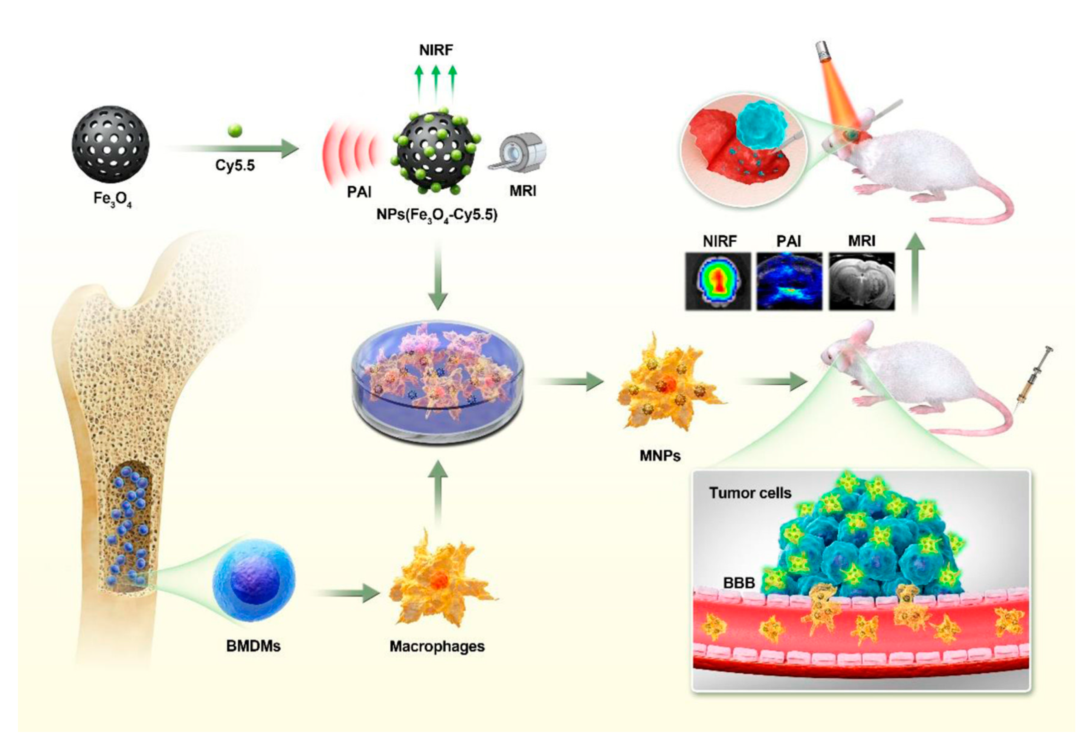
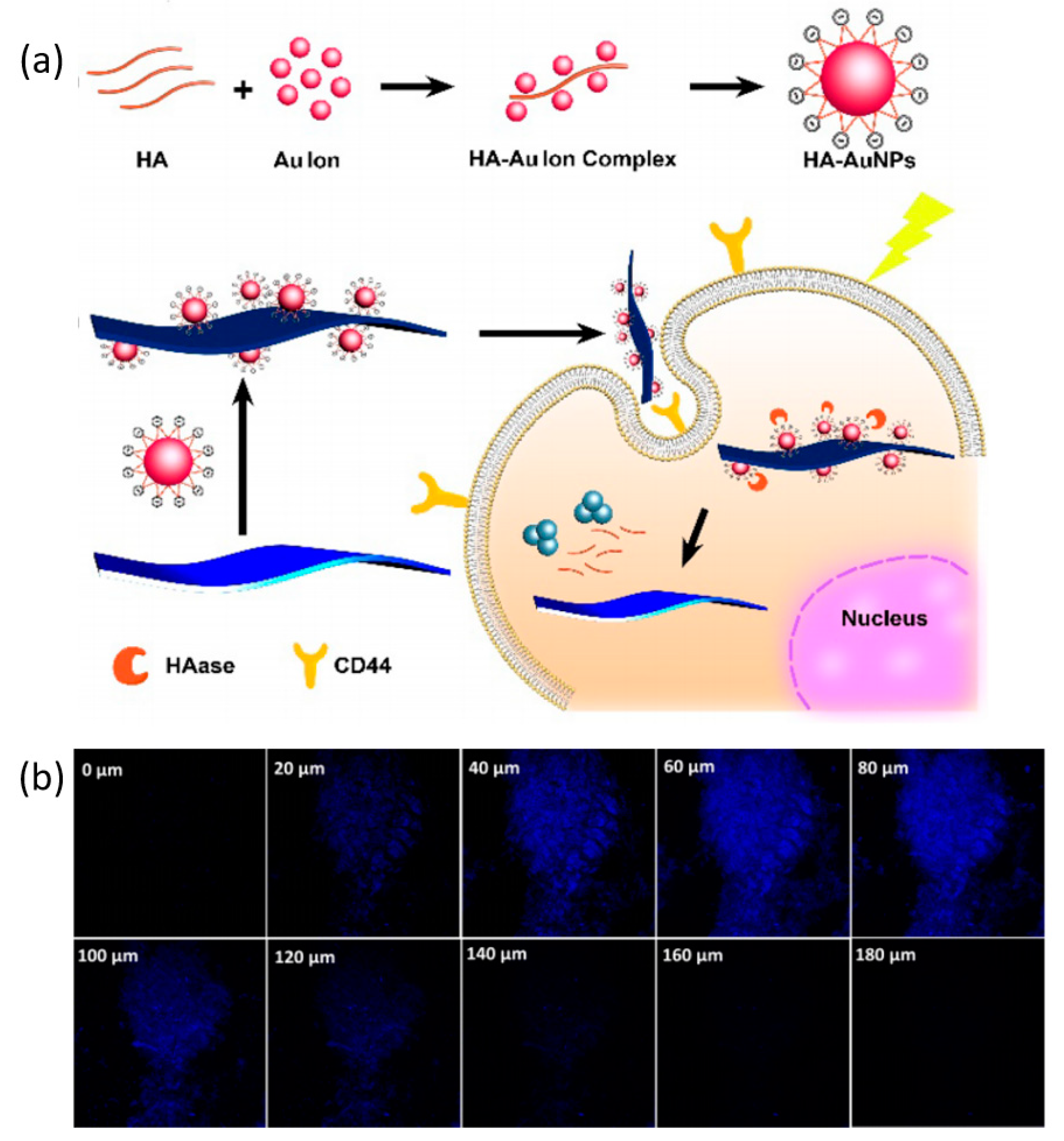
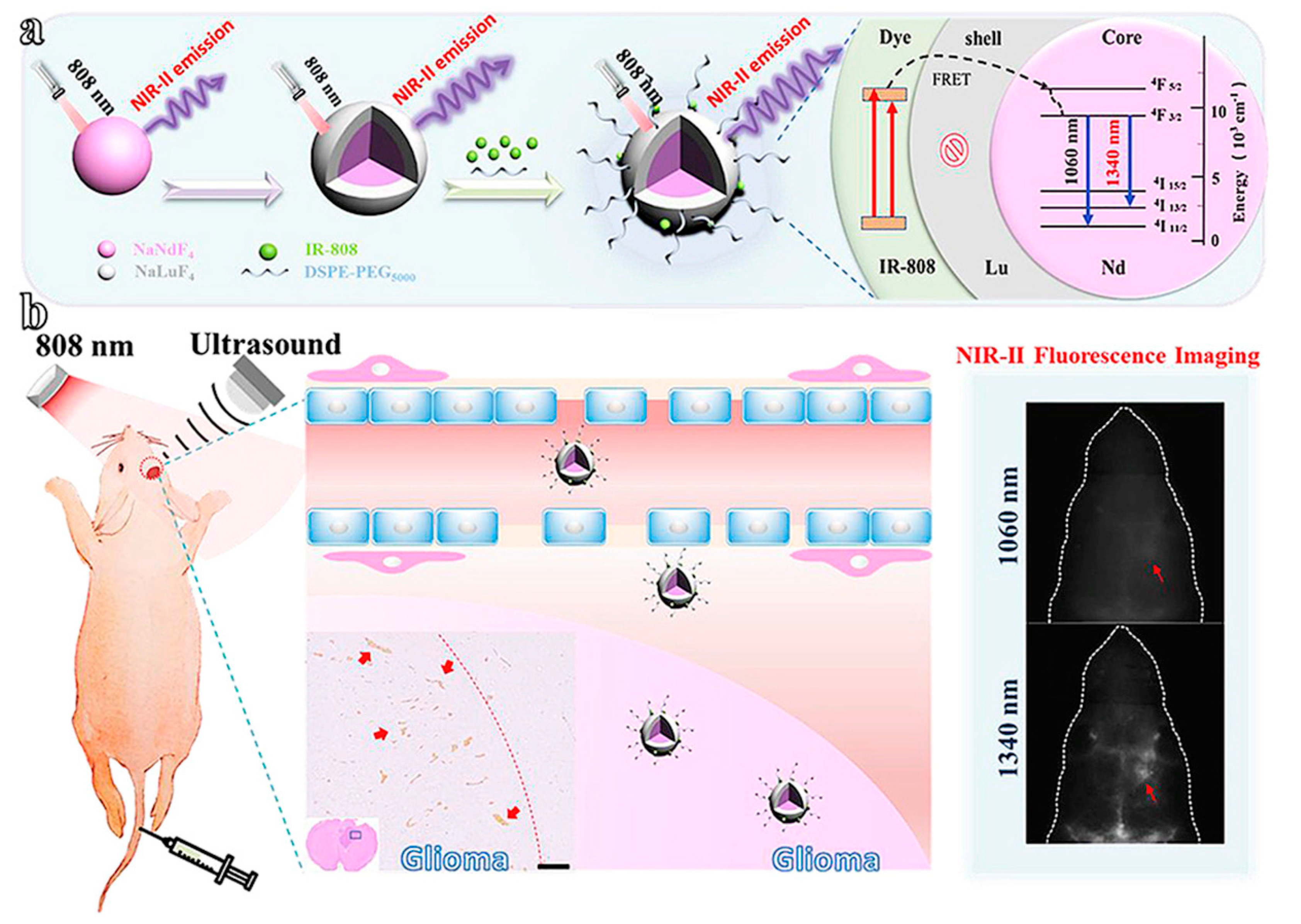
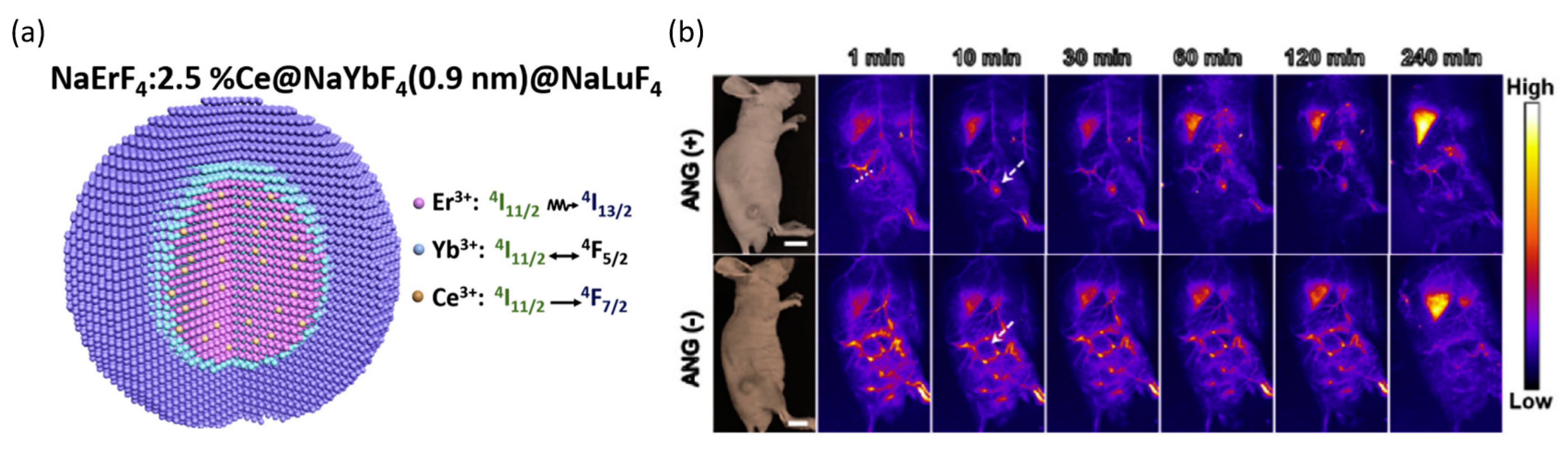
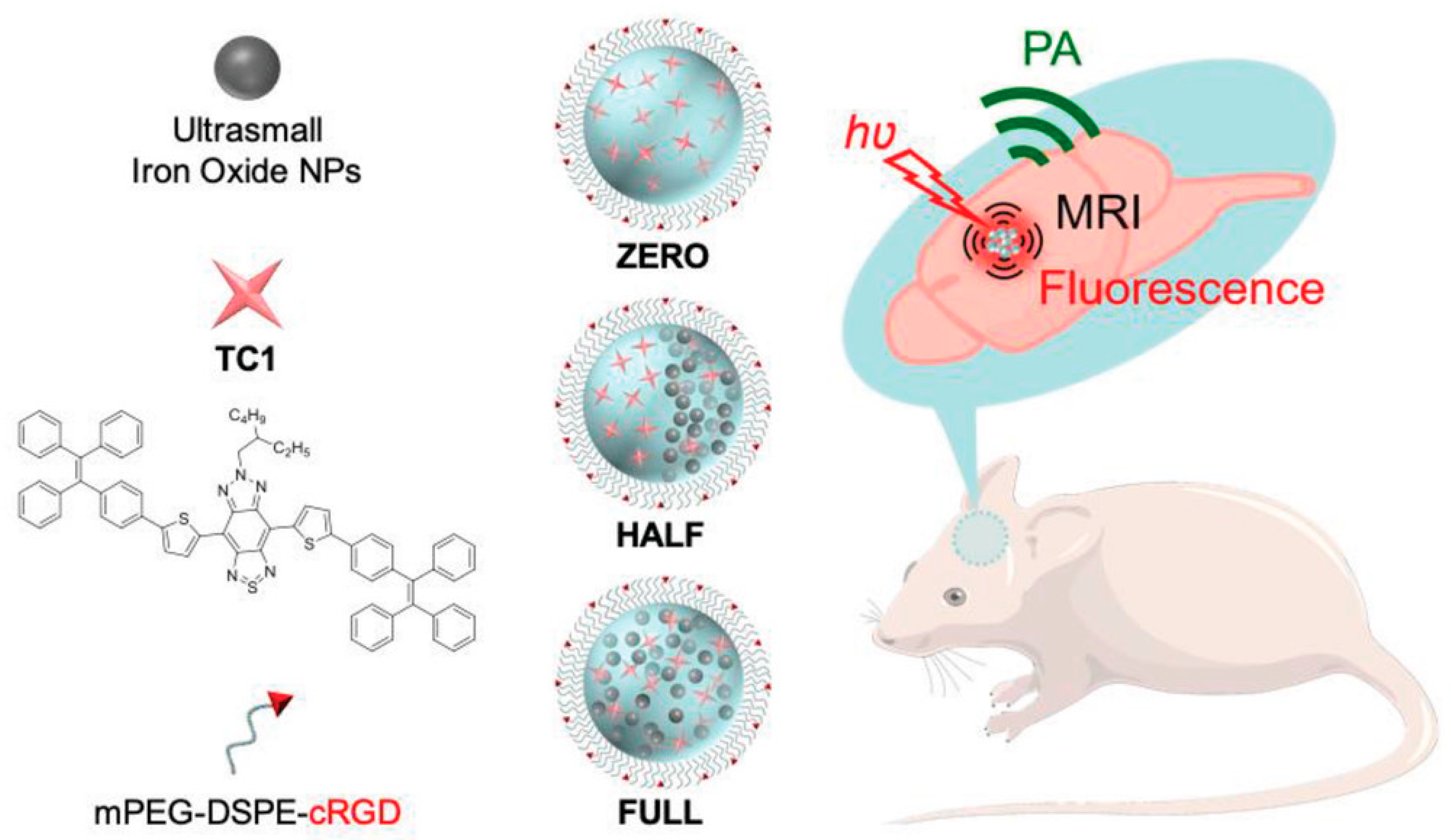
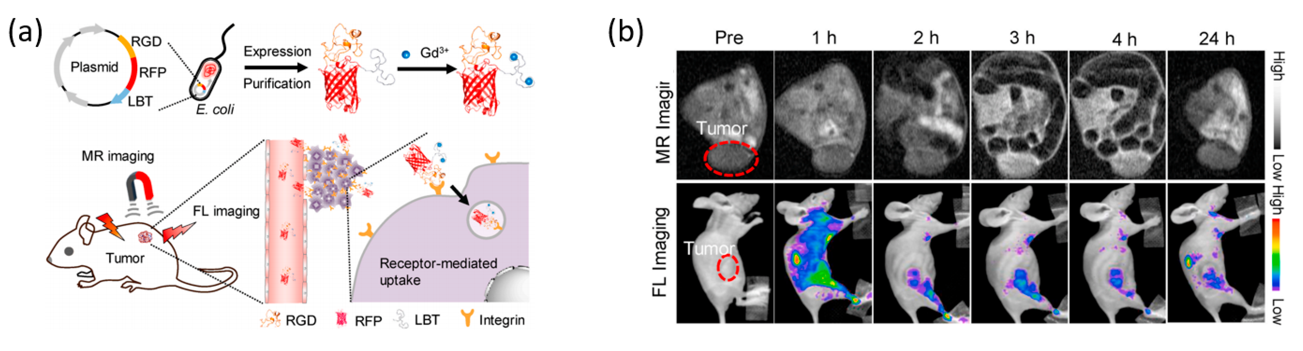
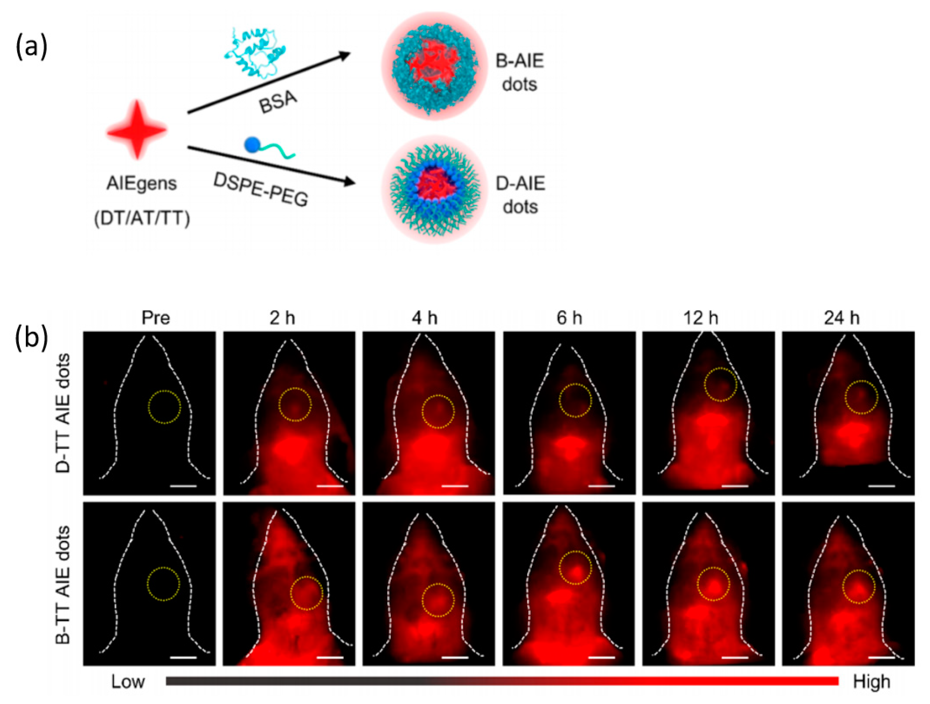
2. Brain Tumors
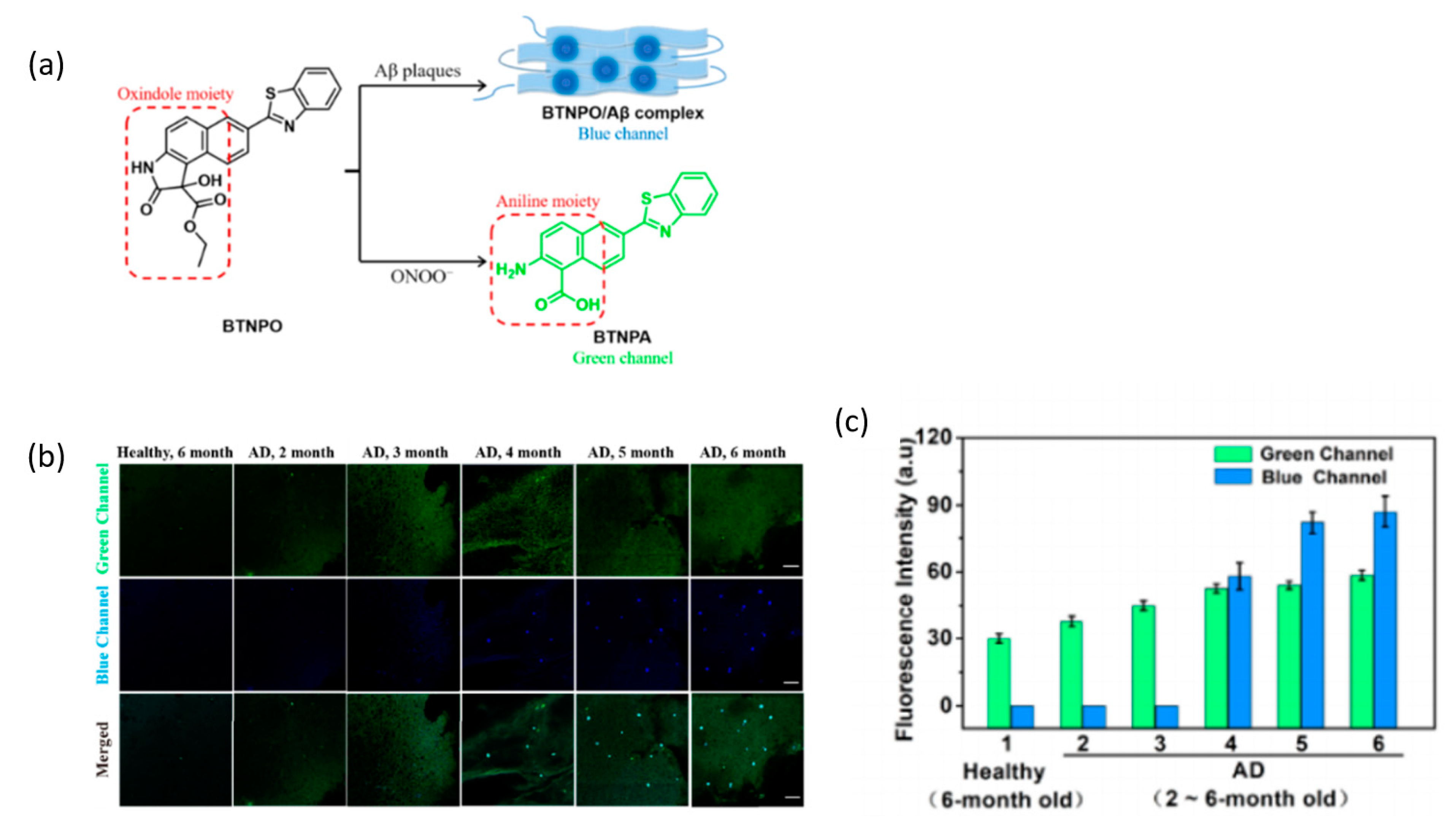
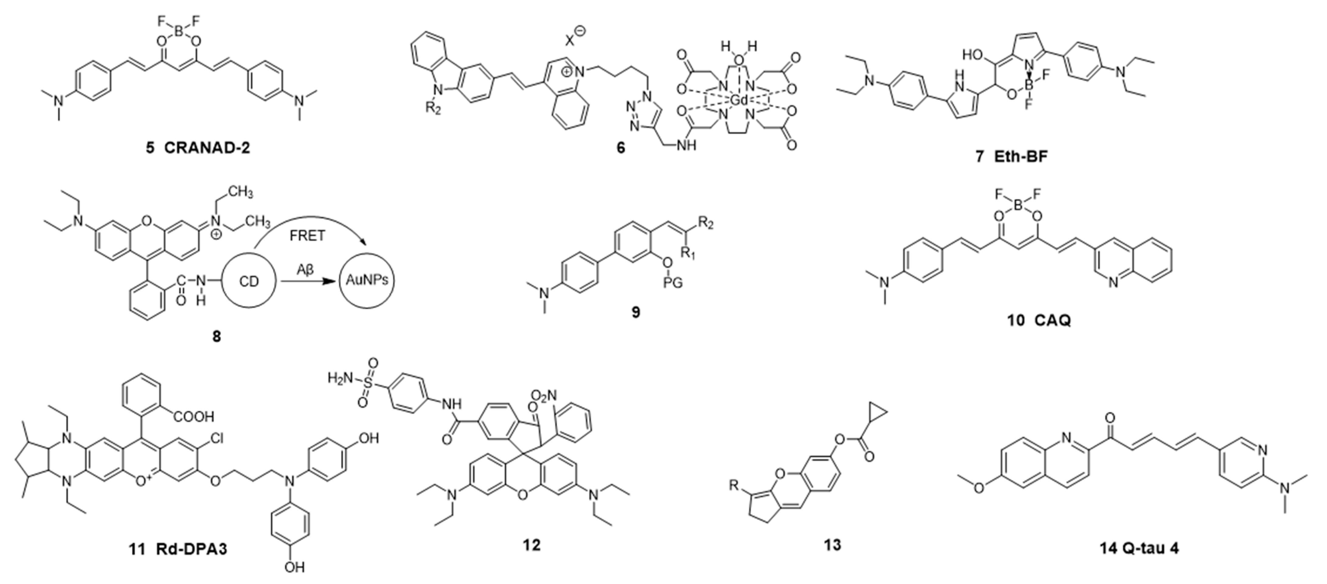
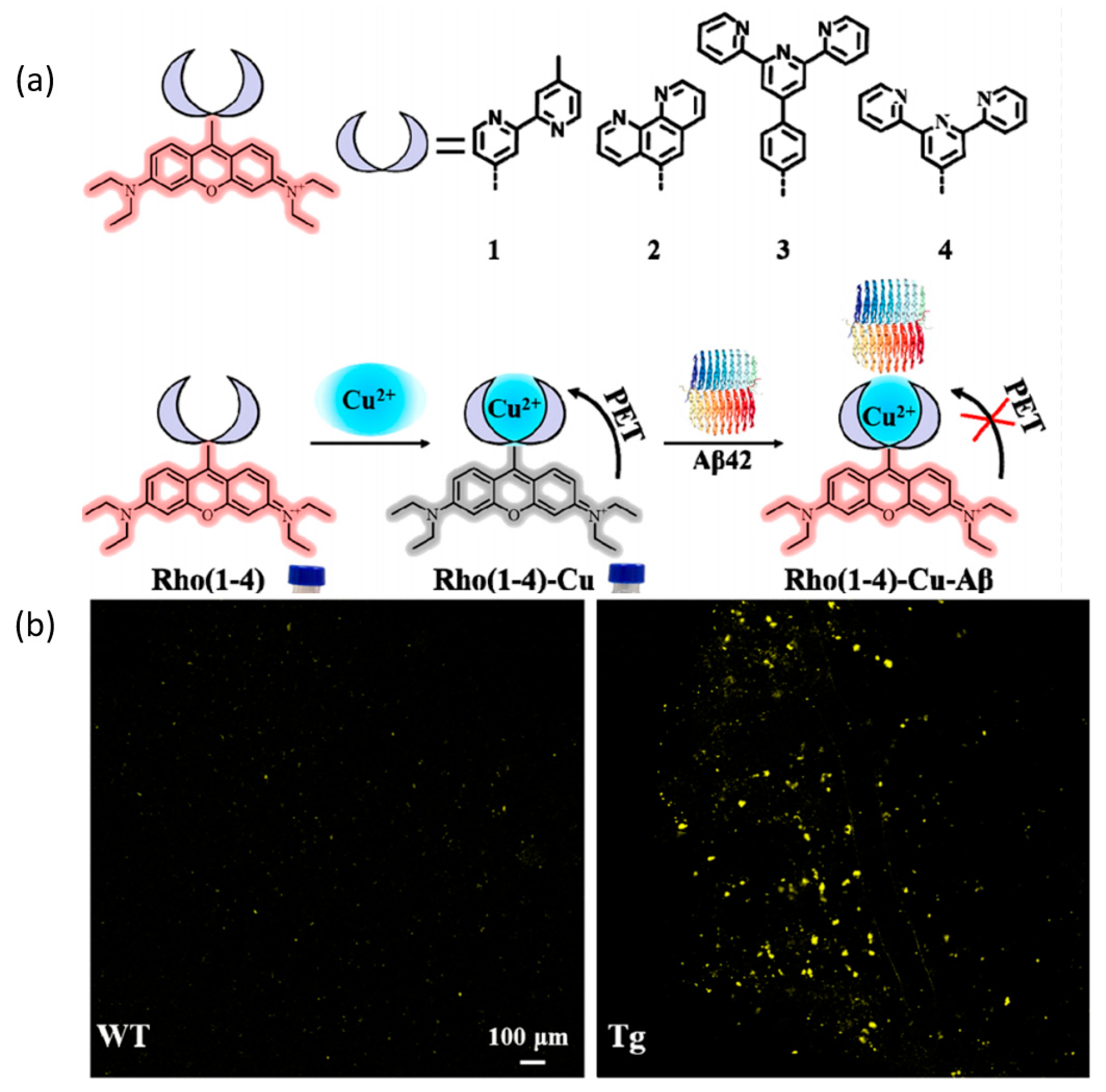
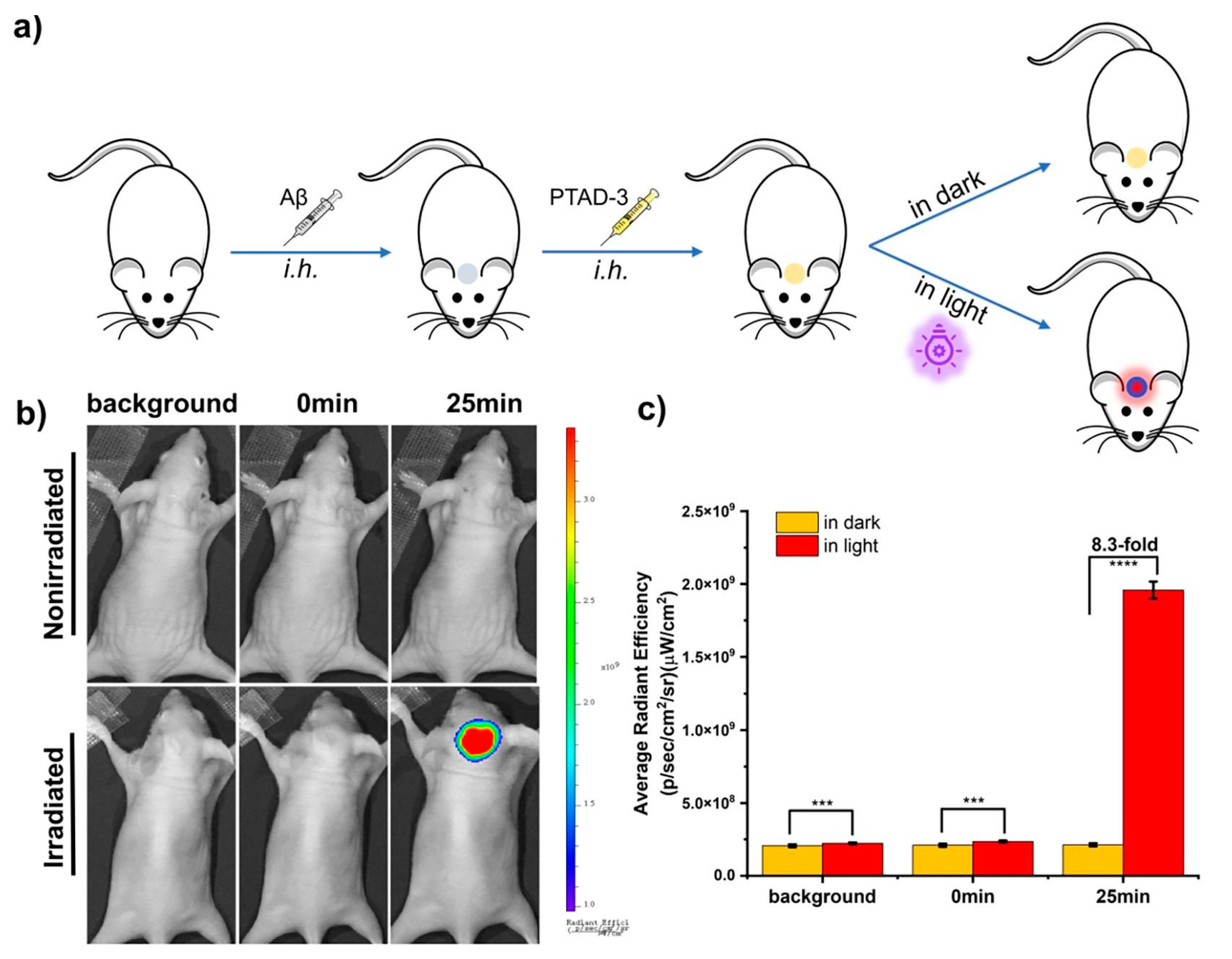
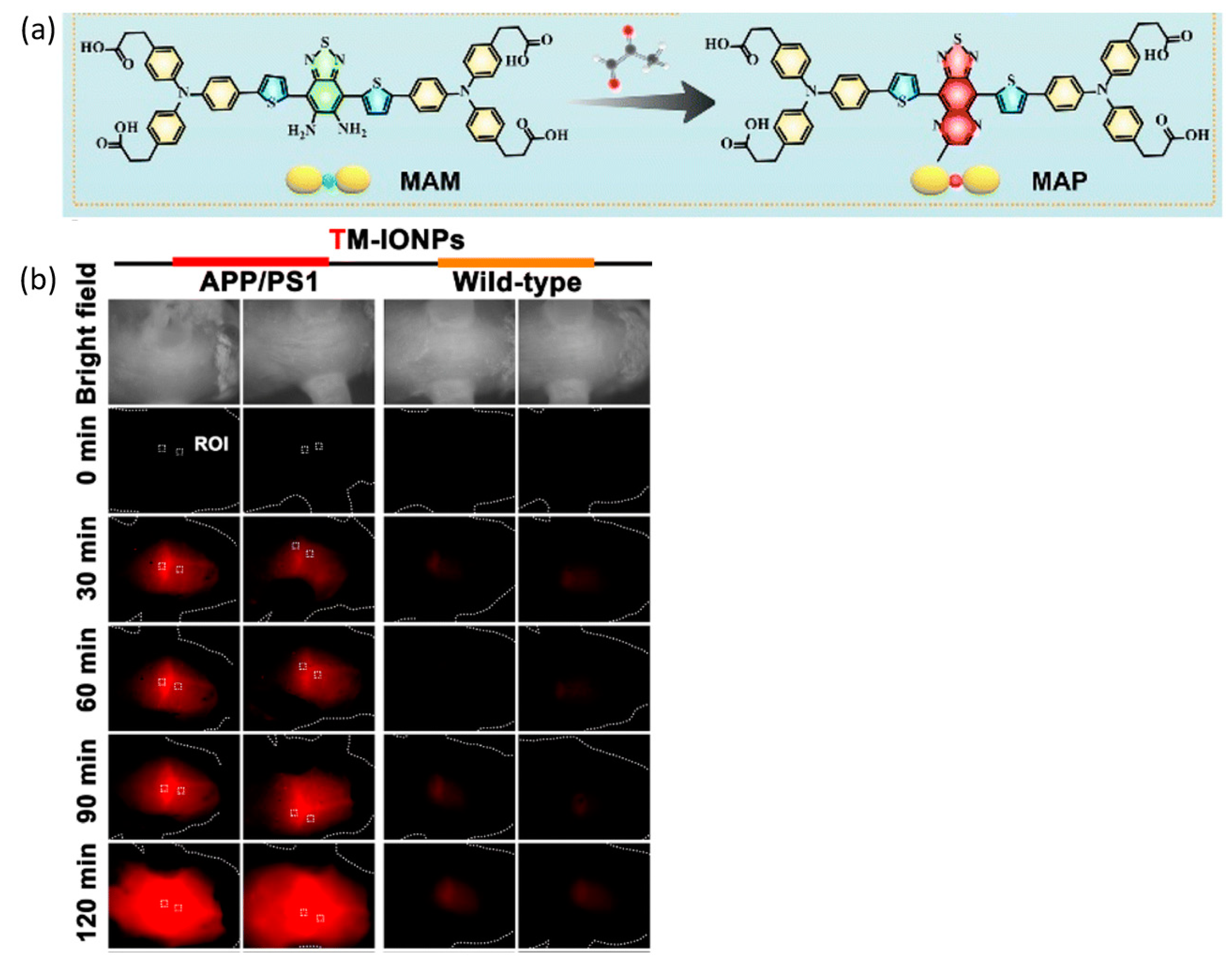
3. Alzheimer’s Disease
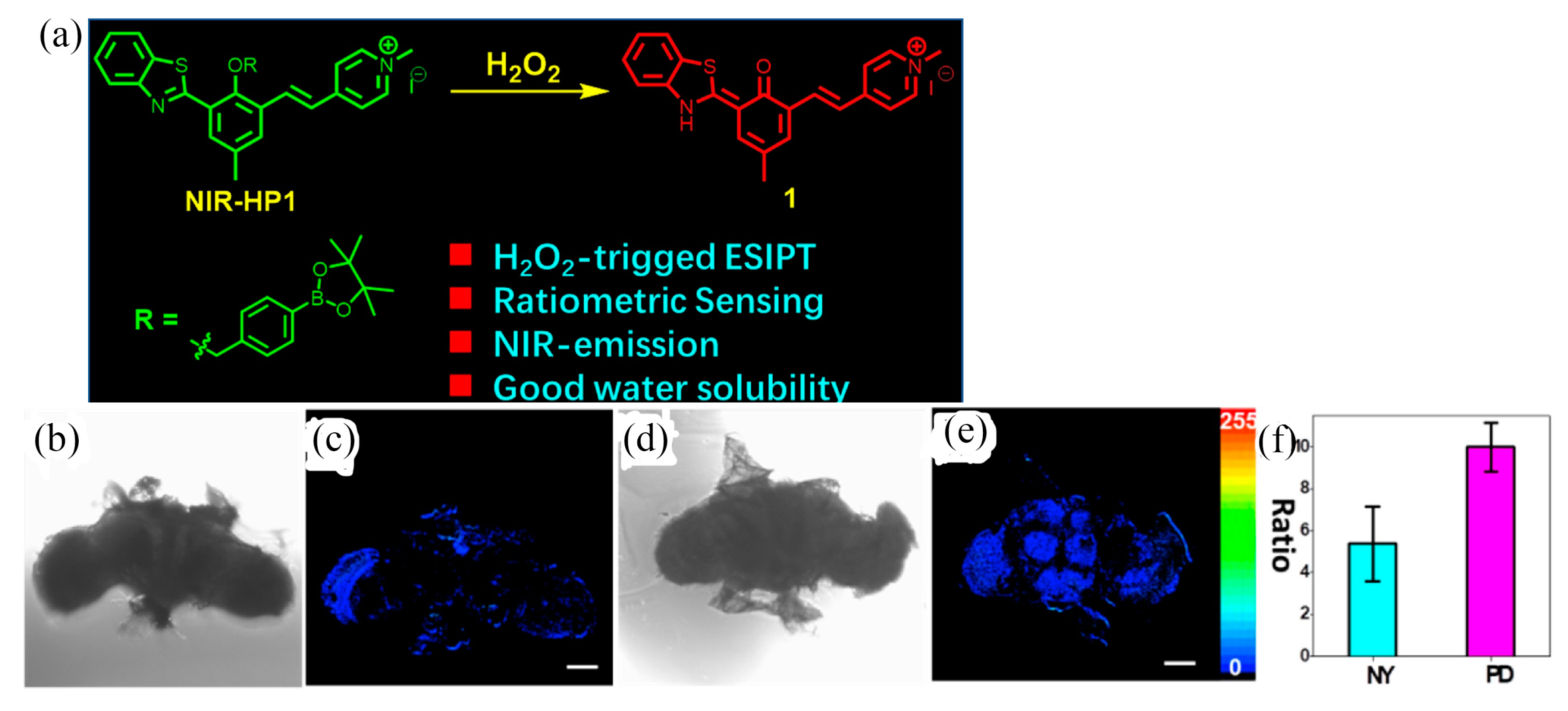
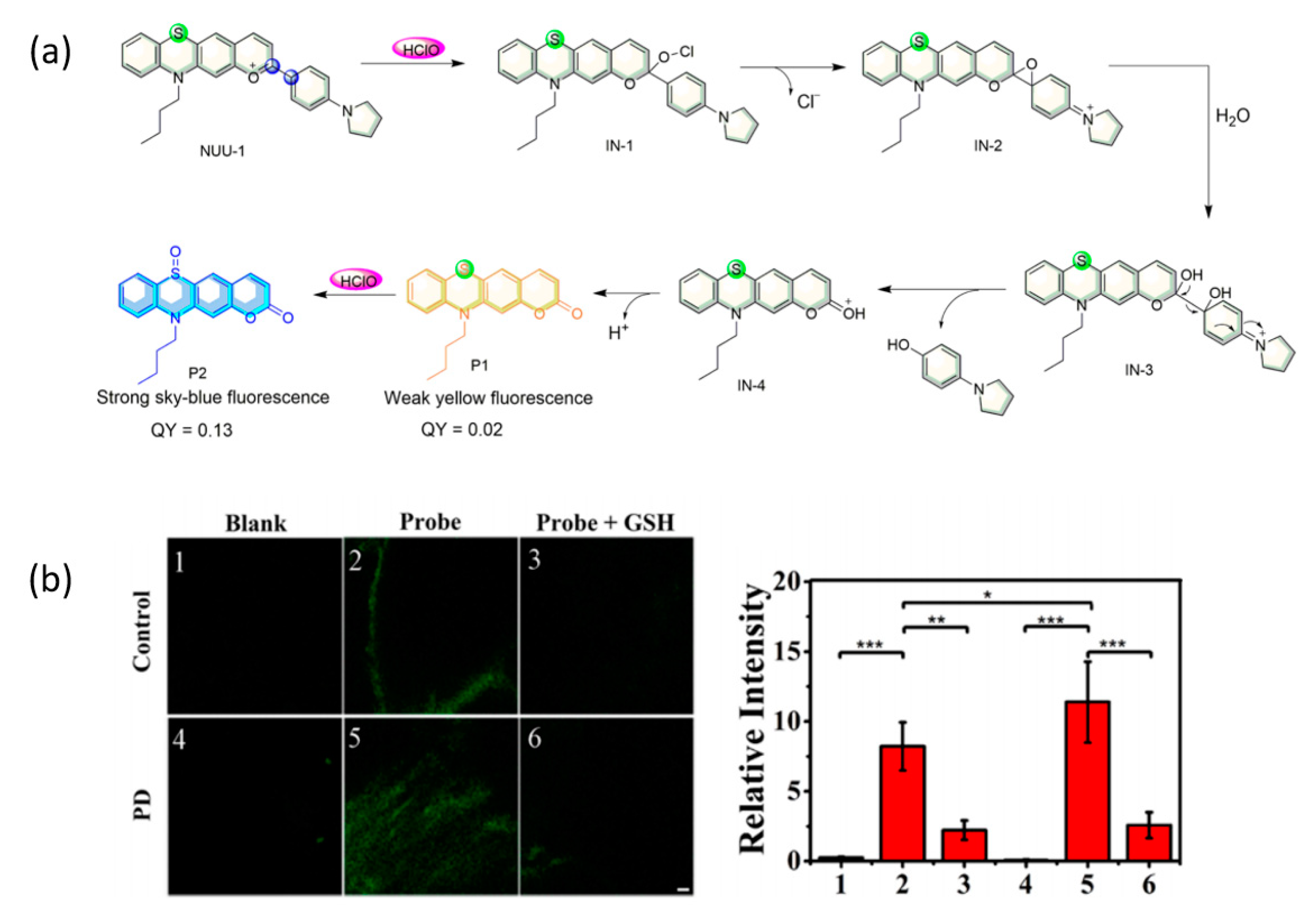
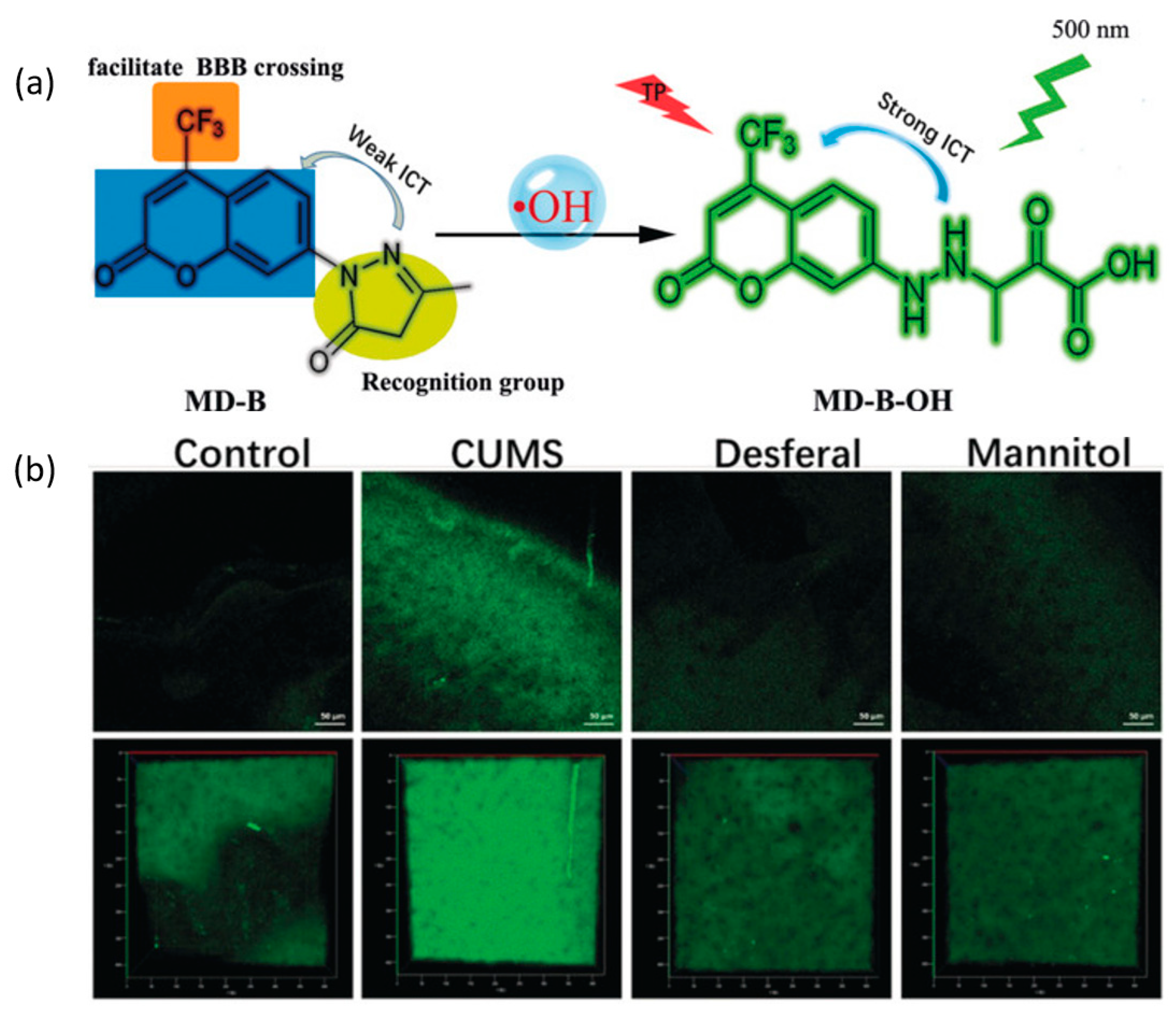
4. Parkinson’s Disease
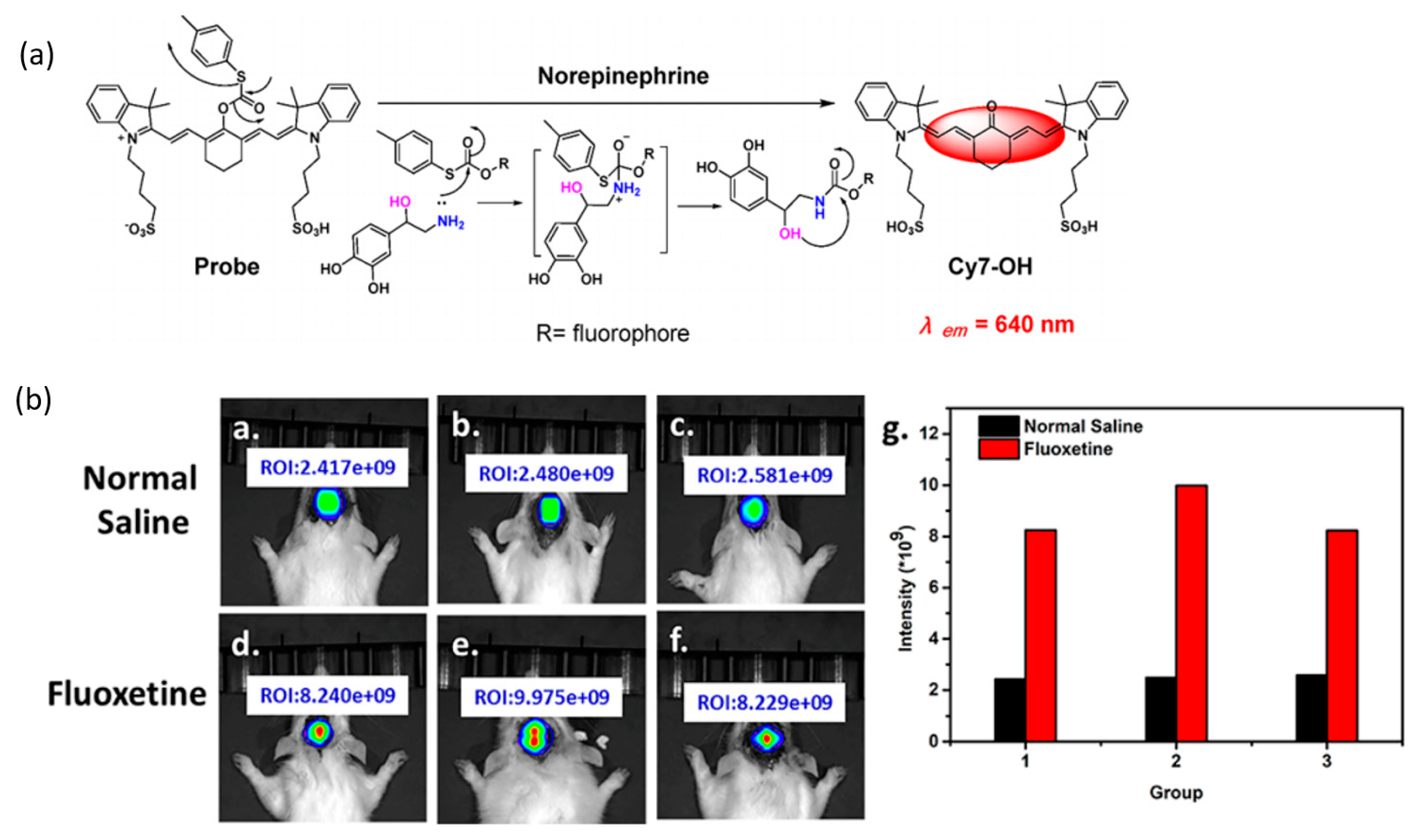
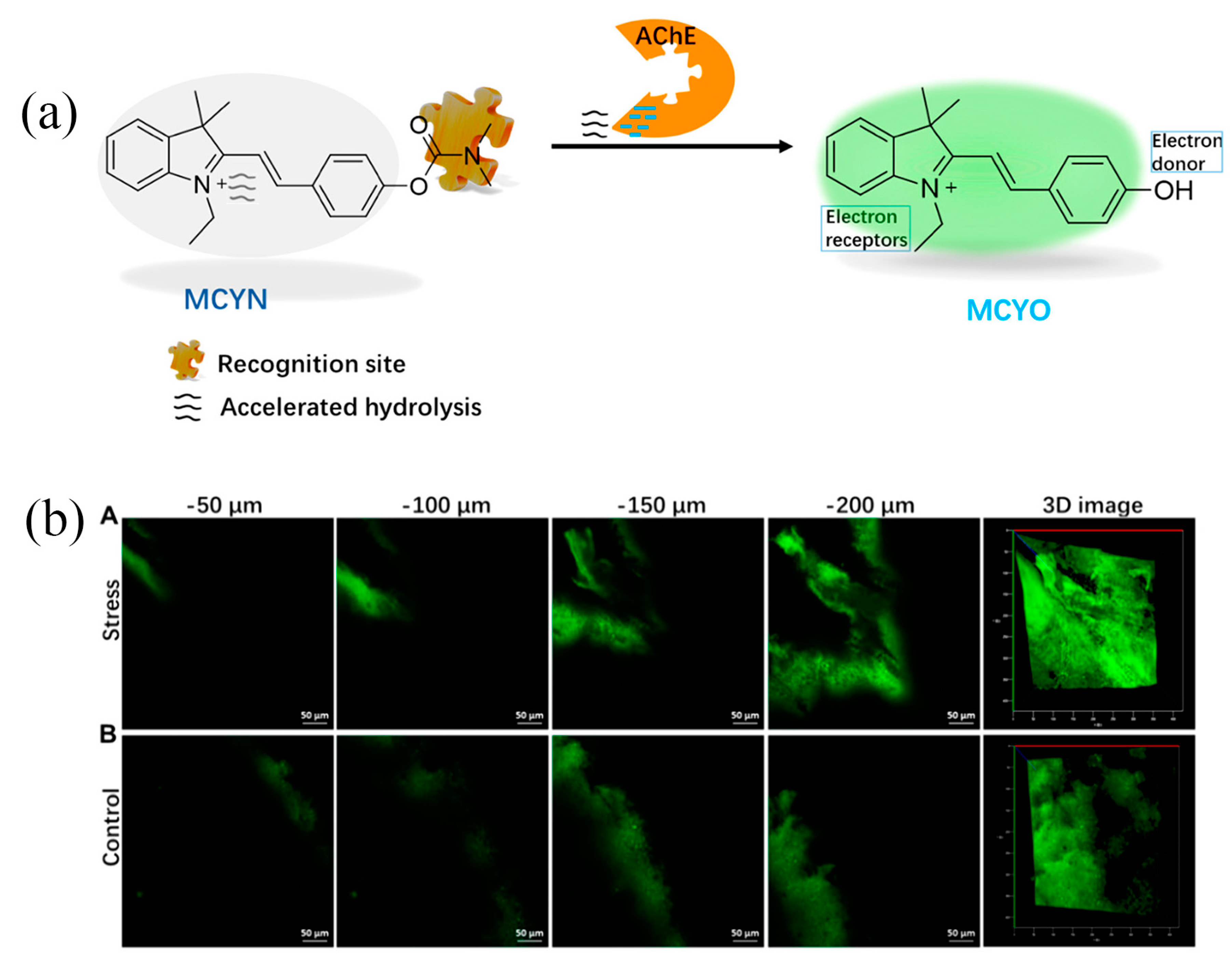
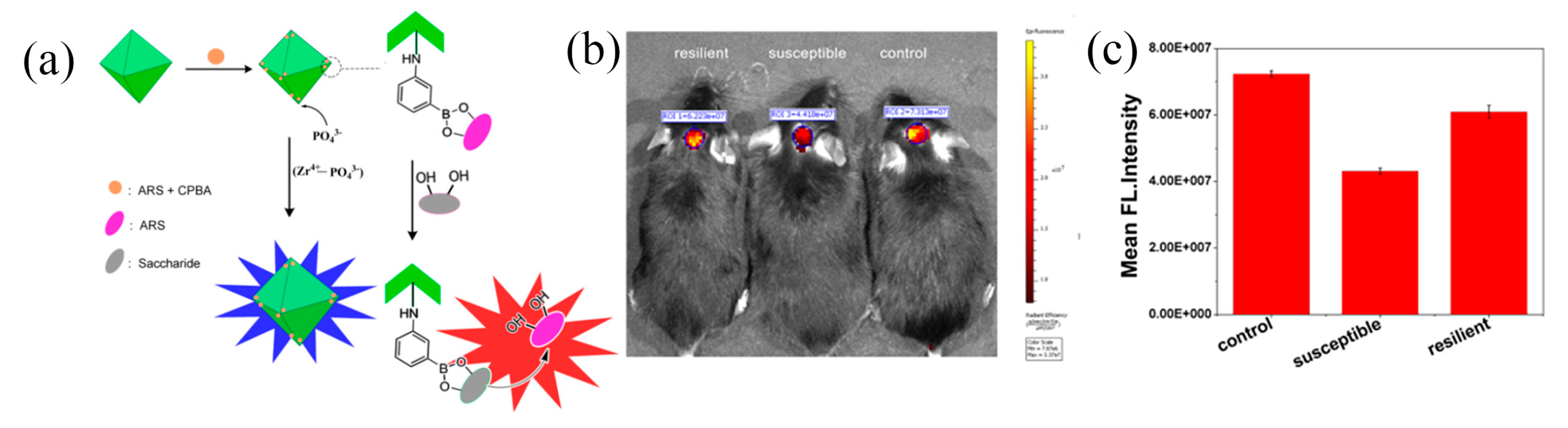
5. Depression
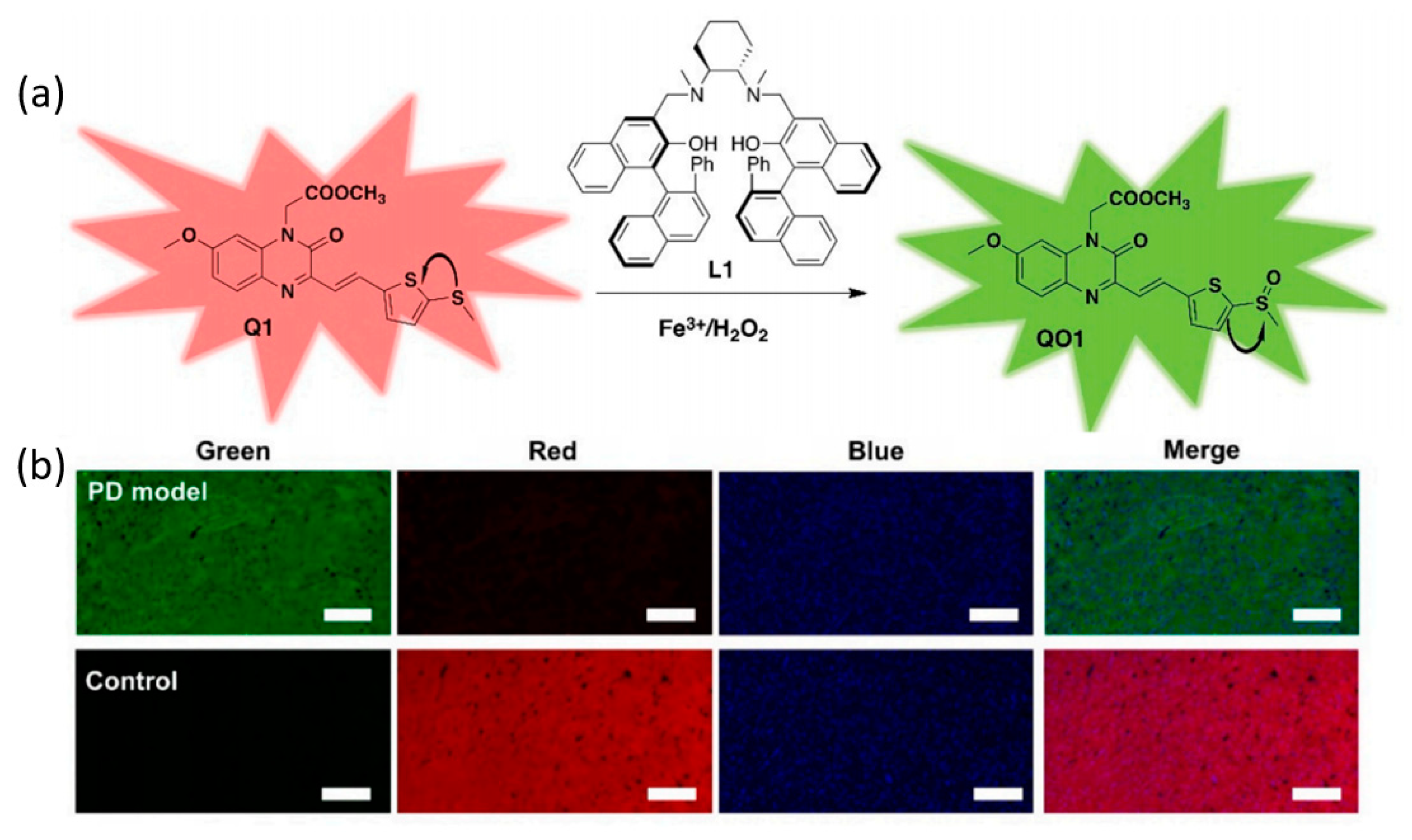
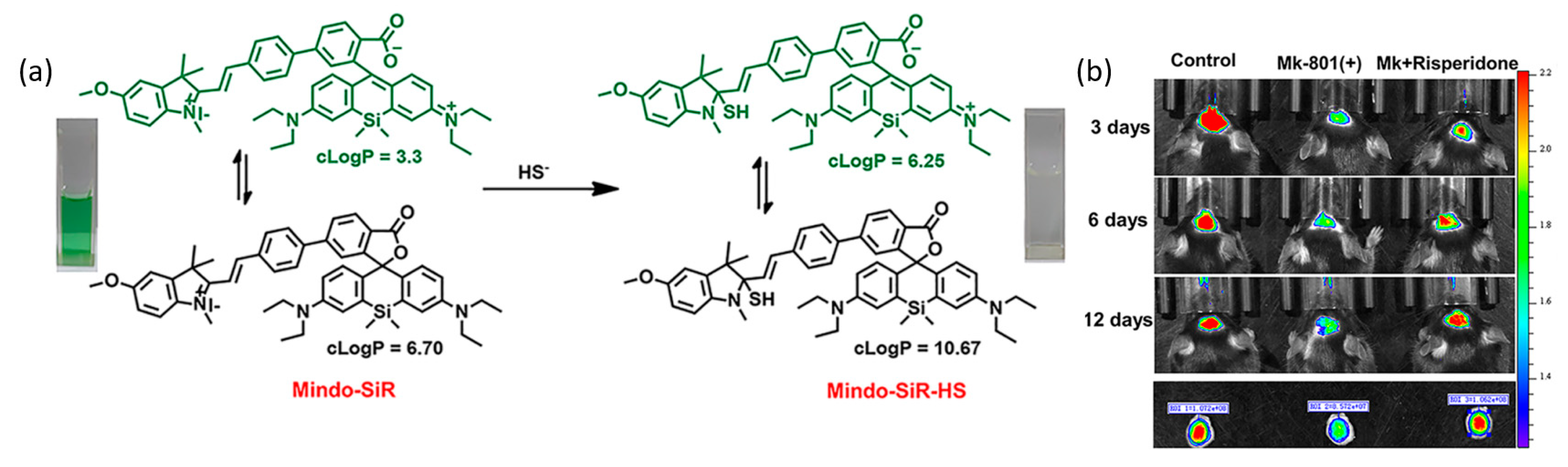
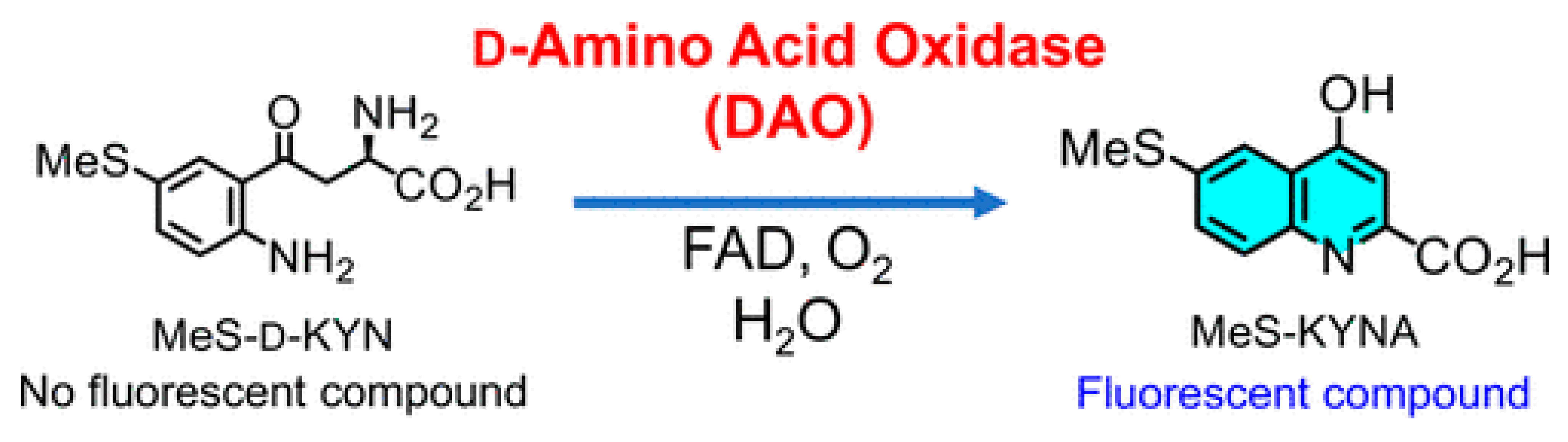
6. Schizophrenia
7. Conclusions and Prospects
Author Contributions
Funding
Institutional Review Board Statement
Informed Consent Statement
Data Availability Statement
Conflicts of Interest
References
- McEwen, B.S. The Brain on Stress: Toward an Integrative Approach to Brain, Body, and Behavior. Perspect. Psychol. Sci. 2013, 8, 673–675. [Google Scholar] [CrossRef] [PubMed]
- Fornito, A.; Bullmore, E.T. Connectomics: A new paradigm for understanding brain disease. Eur. Neuropsychopharmaco. 2015, 25, 733–748. [Google Scholar] [CrossRef] [PubMed]
- Butterfield, D.A.; Perluigi, M.; Sultana, R. Oxidative stress in Alzheimer’s disease brain: New insights from redox proteomics. Eur. J. Pharmacol. 2006, 545, 39–50. [Google Scholar] [CrossRef] [PubMed]
- Butterfield, D.A. Proteomics: A new approach to investigate oxidative stress in Alzheimer’s disease brain. Brain Res. Brain Res. Rev. 2004, 1000, 1–7. [Google Scholar] [CrossRef]
- Harris, A.N.; Beatty, S.S.; Estrada, A.H.; Winter, B.; Bohannon, M.; Sosa, I.; Hanscom, J.; Mainville, C.A.; Gallagher, A.E. Investigation of an N-Terminal Prohormone of Brain Natriuretic Peptide Point-of-Care ELISA in Clinically Normal Cats and Cats With Cardiac Disease. J. Vet. Intern. Med. 2017, 31, 994–999. [Google Scholar] [CrossRef]
- Fauss, D.; Motter, R.; Dofiles, L.; Rodrigues, M.A.V.; You, M.; Diep, L.; Yang, Y.; Seto, P.; Tanaka, K.; Baker, J.; et al. Development of an enzyme-linked immunosorbent assay (ELISA) to measure the level of tyrosine hydroxylase protein in brain tissue from Parkinson’s disease models. J. Neurosci. Methods 2013, 215, 245–257. [Google Scholar] [CrossRef]
- Sizova, D.; Charbaut, E.; Delalande, F.; Poirier, F.; High, A.A.; Parker, F.; Van Dorsselaer, A.; Duchesne, M.; Diu-Hercend, A. Proteomic analysis of brain tissue from an Alzheimer’s disease mouse model by two-dimensional difference gel electrophoresis. Neurobiol. Aging 2007, 28, 357–370. [Google Scholar] [CrossRef]
- Yu, C.H.; Song, G.S.; Yhee, J.Y.; Kim, J.H.; Im, K.S.; Nho, W.G.; Lee, J.H.; Sur, J.H. Histopathological and Immunohistochemical Comparison of the Brain of Human Patients with Alzheimer’s Disease and the Brain of Aged Dogs with Cognitive Dysfunction. J. Comp. Pathol. 2011, 145, 45–58. [Google Scholar] [CrossRef]
- Ibanez, F.G.; Picard, K.; Bordeleau, M.; Sharma, K.; Bisht, K.; Tremblay, M.È. Immunofluorescence Staining Using IBA1 and TMEM119 for Microglial Density, Morphology and Peripheral Myeloid Cell Infiltration Analysis in Mouse Brain. J. Visualized Exp. 2019, 152, e60510. [Google Scholar] [CrossRef]
- Wang, G.; Achim, C.L.; Hamilton, R.L.; Wiley, C.A.; Soontornniyomkij, V. Tyramide Signal Amplification Method in Multiple-Label Immunofluorescence Confocal Microscopy. Methods 1999, 18, 459–464. [Google Scholar] [CrossRef]
- Iwaki, T.; Kume-Iwaki, A.; Liem, R.K.H.; Goldman, J.E. αB-crystallin is expressed in non-lenticular tissues and accumulates in Alexander’s disease brain. Cell 1989, 57, 71–78. [Google Scholar] [CrossRef]
- Ferrer, I.; Santpere, G.; Arzberger, T.; Bell, J.; Blanco, R.; Boluda, S.; Budka, H.; Carmona, M.; Giaccone, G.; Krebs, B.; et al. Brain Protein Preservation Largely Depends on the Postmortem Storage Temperature: Implications for Study of Proteins in Human Neurologic Diseases and Management of Brain Banks: A BrainNet Europe Study. J. Neuropathol. Exp. Neurol. 2007, 66, 35–46. [Google Scholar] [CrossRef] [PubMed]
- Bles, M.; Haynes, J.-D. Detecting concealed information using brain-imaging technology. Neurocase 2008, 14, 82–92. [Google Scholar] [CrossRef] [PubMed]
- Garcia-Alloza, M.; Bacskai, B.J. Techniques for brain imaging in vivo. Neuro Mol. Med. 2004, 6, 65–78. [Google Scholar] [CrossRef] [PubMed]
- Bowman, F.D. Brain Imaging Analysis. Annu. Rev. Stat. Appl. 2014, 1, 61–85. [Google Scholar] [CrossRef] [PubMed]
- Li, W.; Yin, S.; Shen, Y.; Li, H.; Yuan, L.; Zhang, X.B. Molecular Engineering of pH-Responsive NIR Oxazine Assemblies for Evoking Tumor Ferroptosis via Triggering Lysosomal Dysfunction. J. Am. Chem. Soc. 2023, 145, 3736–3747. [Google Scholar] [CrossRef]
- Rao, J.; Dragulescu-Andrasi, A.; Yao, H. Fluorescence imaging in vivo: Recent advances. Curr. Opin. Biotechnol. 2007, 18, 17–25. [Google Scholar] [CrossRef]
- Laissue, P.P.; Alghamdi, R.A.; Tomancak, P.; Reynaud, E.G.; Shroff, H. Assessing phototoxicity in live fluorescence imaging. Nat. Methods 2017, 14, 657–661. [Google Scholar] [CrossRef]
- Liu, J.; Zhang, W.; Zhou, C.; Li, M.; Wang, X.; Zhang, W.; Liu, Z.; Wu, L.; James, T.D.; Li, P.; et al. Precision Navigation of Hepatic Ischemia–Reperfusion Injury Guided by Lysosomal Viscosity-Activatable NIR-II Fluorescence. J. Am. Chem. Soc. 2022, 144, 13586–13599. [Google Scholar] [CrossRef]
- Ren, T.B.; Wang, Z.Y.; Xiang, Z.; Lu, P.; Lai, H.H.; Yuan, L.; Zhang, X.B.; Tan, W. A General Strategy for Development of Activatable NIR-II Fluorescent Probes for In Vivo High-Contrast Bioimaging. Angew. Chem. Int. Ed. 2021, 60, 800–805. [Google Scholar] [CrossRef]
- Mao, L.; Han, Y.; Zhang, Q.W.; Tian, Y. Two-photon fluorescence imaging and specifically biosensing of norepinephrine on a 100-ms timescale. Nat. Commun. 2023, 14, 1419. [Google Scholar] [CrossRef] [PubMed]
- Zhao, Y.; Kim, H.S.; Zou, X.; Huang, L.; Liang, X.; Li, Z.; Kim, J.S.; Lin, W. Harnessing Dual-Fluorescence Lifetime Probes to Validate Regulatory Mechanisms of Organelle Interactions. J. Am. Chem. Soc. 2022, 144, 20854–20865. [Google Scholar] [CrossRef]
- Wu, X.; Wang, R.; Qi, S.; Kwon, N.; Han, J.; Kim, H.; Li, H.; Yu, F.; Yoon, J. Rational Design of a Highly Selective Near-Infrared Two-Photon Fluorogenic Probe for Imaging Orthotopic Hepatocellular Carcinoma Chemotherapy. Angew. Chem. Int. Ed. 2021, 60, 15418–15425. [Google Scholar] [CrossRef]
- Weller, M.; Wick, W.; Aldape, K.; Brada, M.; Berger, M.; Pfister, S.M.; Nishikawa, R.; Rosenthal, M.; Wen, P.Y.; Stupp, R.; et al. Glioma. Nat. Rev. Dis. Prim. 2015, 1, 15017. [Google Scholar] [CrossRef]
- Hong, G.; Diao, S.; Chang, J.; Antaris, A.L.; Chen, C.; Zhang, B.; Zhao, S.; Atochin, D.N.; Huang, P.L.; Andreasson, K.I.; et al. Through-skull fluorescence imaging of the brain in a new near-infrared window. Nat. Photonics 2014, 8, 723–730. [Google Scholar] [CrossRef]
- Frangioni, J.V. In vivo near-infrared fluorescence imaging. Curr. Opin. Chem. Biol. 2003, 7, 626–634. [Google Scholar] [CrossRef]
- Liu, Y.; Liu, J.; Chen, D.; Wang, X.; Zhang, Z.; Yang, Y.; Jiang, L.; Qi, W.; Ye, Z.; He, S.; et al. Fluorination Enhances NIR-II Fluorescence of Polymer Dots for Quantitative Brain Tumor Imaging. Angew. Chem. Int. Ed. 2020, 59, 21049–21057. [Google Scholar] [CrossRef]
- Chen, S.; Miao, H.; Jiang, X.; Sun, P.; Fan, Q.; Huang, W. Starlike polymer brush-based ultrasmall nanoparticles with simultaneously improved NIR-II fluorescence and blood circulation for efficient orthotopic glioblastoma imaging. Biomaterials 2021, 275, 120916. [Google Scholar] [CrossRef]
- Liu, Z.; Ren, F.; Zhang, H.; Yuan, Q.; Jiang, Z.; Liu, H.; Sun, Q.; Li, Z. Boosting often overlooked long wavelength emissions of rare-earth nanoparticles for NIR-II fluorescence imaging of orthotopic glioblastoma. Biomaterials 2019, 219, 119364. [Google Scholar] [CrossRef] [PubMed]
- Ren, F.; Liu, H.; Zhang, H.; Jiang, Z.; Xia, B.; Genevois, C.; He, T.; Allix, M.; Sun, Q.; Li, Z.; et al. Engineering NIR-IIb fluorescence of Er-based lanthanide nanoparticles for through-skull targeted imaging and imaging-guided surgery of orthotopic glioma. Nano Today 2020, 34, 100905. [Google Scholar] [CrossRef]
- Wang, S.; Shen, H.; Mao, Q.; Tao, Q.; Yuan, G.; Zeng, L.; Chen, Z.; Zhang, Y.; Cheng, L.; Zhang, J.; et al. Macrophage-Mediated Porous Magnetic Nanoparticles for Multimodal Imaging and Postoperative Photothermal Therapy of Gliomas. ACS Appl. Mater. Interfaces 2021, 13, 56825–56837. [Google Scholar] [CrossRef]
- Guo, B.; Feng, Z.; Hu, D.; Xu, S.; Middha, E.; Pan, Y.; Liu, C.; Zheng, H.; Qian, J.; Sheng, Z.; et al. Precise Deciphering of Brain Vasculatures and Microscopic Tumors with Dual NIR-II Fluorescence and Photoacoustic Imaging. Adv. Mater. 2019, 31, e1902504. [Google Scholar] [CrossRef]
- Duan, Y.; Hu, D.; Guo, B.; Shi, Q.; Wu, M.; Xu, S.; Kenry; Liu, X.; Jiang, J.; Sheng, Z.; et al. Nanostructural Control Enables Optimized Photoacoustic–Fluorescence–Magnetic Resonance Multimodal Imaging and Photothermal Therapy of Brain Tumor. Adv. Funct. Mater. 2019, 30, 1907077. [Google Scholar] [CrossRef]
- Zhao, H.; Zhao, H.; Jiao, Y.; Zhu, Y.; Liu, C.; Li, F.; Wang, Y.; Gu, Z.; Yang, D. Biosynthetic molecular imaging probe for tumor-targeted dual-modal fluorescence/magnetic resonance imaging. Biomaterials 2020, 256, 120220. [Google Scholar] [CrossRef] [PubMed]
- Sheng, Z.; Guo, B.; Hu, D.; Xu, S.; Wu, W.; Liew, W.H.; Yao, K.; Jiang, J.; Liu, C.; Zheng, H.; et al. Bright Aggregation-Induced-Emission Dots for Targeted Synergetic NIR-II Fluorescence and NIR-I Photoacoustic Imaging of Orthotopic Brain Tumors. Adv. Mater. 2018, 30, e1800766. [Google Scholar] [CrossRef] [PubMed]
- Gao, D.; Li, Y.; Wu, Y.; Liu, Y.; Hu, D.; Liang, S.; Liao, J.; Pan, M.; Zhang, P.; Li, K.; et al. Albumin-Consolidated AIEgens for Boosting Glioma and Cerebrovascular NIR-II Fluorescence Imaging. ACS Appl. Mater. Interfaces 2023, 15, 3–13. [Google Scholar] [CrossRef]
- Liu, J.W.; Wang, Y.M.; Zhang, C.H.; Duan, L.Y.; Li, Z.; Yu, R.Q.; Jiang, J.H. Tumor-Targeted Graphitic Carbon Nitride Nanoassembly for Activatable Two-Photon Fluorescence Imaging. Anal. Chem. 2018, 90, 4649–4656. [Google Scholar] [CrossRef]
- Scheltens, P.; Blennow, K.; Breteler, M.M.B.; de Strooper, B.; Frisoni, G.B.; Salloway, S.; Van der Flier, W.M. Alzheimer’s disease. Lancet 2016, 388, 505–517. [Google Scholar] [CrossRef]
- Chasseigneaux, S.; Allinquant, B. Functions of Aβ, sAPPα and sAPPβ: Similarities and differences. J. Neurochem. 2012, 120, 99–108. [Google Scholar] [CrossRef]
- Cavallucci, V.; D’Amelio, M.; Cecconi, F. Aβ Toxicity in Alzheimer’s Disease. Mol. Neurobiol. 2012, 45, 366–378. [Google Scholar] [CrossRef]
- Tanzi, R.E.; Moir, R.D.; Wagner, S.L. Clearance of Alzheimer’s Aβ Peptide: The Many Roads to Perdition. Neuron 2004, 43, 605–608. [Google Scholar] [CrossRef] [PubMed]
- Ni, R.; Villois, A.; Dean-Ben, X.L.; Chen, Z.; Vaas, M.; Stavrakis, S.; Shi, G.; de Mello, A.; Ran, C.; Razansky, D.; et al. In-vitro and in-vivo characterization of CRANAD-2 for multi-spectral optoacoustic tomography and fluorescence imaging of amyloid-beta deposits in Alzheimer mice. Photoacoustics 2021, 23, 100285. [Google Scholar] [CrossRef]
- Wang, X.; Chan, H.N.; Desbois, N.; Gros, C.P.; Bolze, F.; Li, Y.; Li, H.W.; Wong, M.S. Multimodal Theranostic Cyanine-Conjugated Gadolinium(III) Complex for In Vivo Imaging of Amyloid-beta in an Alzheimer’s Disease Mouse Model. ACS Appl. Mater. Interfaces 2021, 13, 18525–18532. [Google Scholar] [CrossRef] [PubMed]
- Xiang, J.; Xiang, C.; Zhou, L.; Sun, M.; Feng, L.; Liu, C.; Cai, L.; Gong, P. Rational Design, Synthesis of Fluorescence Probes for Quantitative Detection of Amyloid-beta in Alzheimer’s Disease Based on Rhodamine-Metal Complex. Anal. Chem. 2022, 94, 11791–11797. [Google Scholar] [CrossRef] [PubMed]
- Li, H.; Wang, J.; Li, Y.; Chen, X.; Zhang, W.; Zhao, Y.; Liu, G.; Pan, J. Detection of Aβ oligomers in early Alzheimer’s disease diagnose by in vivo NIR-II fluorescence imaging. Sens. Actuators B Chem. 2022, 358, 131481. [Google Scholar] [CrossRef]
- Hamd-Ghadareh, S.; Salimi, A.; Parsa, S.; Mowla, S.J. Development of three-dimensional semi-solid hydrogel matrices for ratiometric fluorescence sensing of Amyloid beta peptide and imaging in SH-SY5 cells: Improvement of point of care diagnosis of Alzheimer’s disease biomarker. Biosens Bioelectron. 2022, 199, 113895. [Google Scholar] [CrossRef] [PubMed]
- Gu, Y.; Ding, Z.; Zheng, C.; Xu, Y.; Liu, T.; Mao, C.; Ran, C.; Yang, J.; Wang, P. Light-controlled fluorescent probes for precisely monitoring brain amyloid-β aggregates in Alzheimer’s disease. Chem. Eng. J. 2022, 446, 137385. [Google Scholar] [CrossRef]
- Wu, J.; Shao, C.; Ye, X.; Di, X.; Li, D.; Zhao, H.; Zhang, B.; Chen, G.; Liu, H.K.; Qian, Y. InVivo Brain Imaging of Amyloid-beta Aggregates in Alzheimer’s Disease with a Near-Infrared Fluorescent Probe. ACS Sens. 2021, 6, 863–870. [Google Scholar] [CrossRef]
- Koppal, T.; Drake, J.; Yatin, S.; Jordan, B.; Varadarajan, S.; Bettenhausen, L.; Butterfield, D.A. Peroxynitrite-Induced Alterations in Synaptosomal Membrane Proteins. J. Neurochem. 1999, 72, 310–317. [Google Scholar] [CrossRef]
- Xie, X.; Liu, G.; Niu, Y.; Xu, C.; Li, Y.; Zhang, J.; Jiao, X.; Wang, X.; Tang, B. Dual-Channel Imaging of Amyloid-beta Plaques and Peroxynitrite To Illuminate Their Correlations in Alzheimer’s Disease Using a Unimolecular Two-Photon Fluorescent Probe. Anal. Chem. 2021, 93, 15088–15095. [Google Scholar] [CrossRef]
- Wang, P.; Yu, L.; Gong, J.; Xiong, J.; Zi, S.; Xie, H.; Zhang, F.; Mao, Z.; Liu, Z.; Kim, J.S. An Activity-Based Fluorescent Probe for Imaging Fluctuations of Peroxynitrite (ONOO(-)) in the Alzheimer’s Disease Brain. Angew. Chem. Int. Ed. Engl. 2022, 61, e202206894. [Google Scholar] [CrossRef] [PubMed]
- He, Z.; Liu, D.; Liu, Y.; Li, X.; Shi, W.; Ma, H. Golgi-Targeted Fluorescent Probe for Imaging NO in Alzheimer’s Disease. Anal. Chem. 2022, 94, 10256–10262. [Google Scholar] [CrossRef] [PubMed]
- Lai, Y.; Dang, Y.; Sun, Q.; Pan, J.; Yu, H.; Zhang, W.; Xu, Z. Design of an activatable NIR-II nanoprobe for the in vivo elucidation of Alzheimer’s disease-related variations in methylglyoxal concentrations. Chem. Sci. 2022, 13, 12511–12518. [Google Scholar] [CrossRef] [PubMed]
- Yang, Y.; Zhang, L.; Wang, J.; Cao, Y.; Li, S.; Qin, W.; Liu, Y. Diagnosis of Alzheimer’s Disease and In Situ Biological Imaging via an Activatable Near-Infrared Fluorescence Probe. Anal. Chem. 2022, 94, 13498–13506. [Google Scholar] [CrossRef] [PubMed]
- Elbatrawy, A.A.; Hyeon, S.J.; Yue, N.; Osman, E.E.A.; Choi, S.H.; Lim, S.; Kim, Y.K.; Ryu, H.; Cui, M.; Nam, G. “Turn-On” Quinoline-Based Fluorescent Probe for Selective Imaging of Tau Aggregates in Alzheimer’s Disease: Rational Design, Synthesis, and Molecular Docking. ACS Sens. 2021, 6, 2281–2289. [Google Scholar] [CrossRef]
- Bloem, B.R.; Okun, M.S.; Klein, C. Parkinson’s disease. Lancet 2021, 397, 2284–2303. [Google Scholar] [CrossRef]
- Jenner, P. Oxidative stress in Parkinson’s disease. Ann. Neurol. 2003, 53, S26–S38. [Google Scholar] [CrossRef]
- Sun, Q.; Xu, J.; Ji, C.; Shaibani, M.S.S.; Li, Z.; Lim, K.; Zhang, C.; Li, L.; Liu, Z. Ultrafast Detection of Peroxynitrite in Parkinson’s Disease Models Using a Near-Infrared Fluorescent Probe. Anal. Chem. 2020, 92, 4038–4045. [Google Scholar] [CrossRef] [PubMed]
- Kang, H.; Shu, W.; Yu, J.; Gao, M.; Han, R.; Jing, J.; Zhang, R.; Zhang, X. A near-infrared fluorescent probe for ratiometric imaging peroxynitrite in Parkinson’s disease model. Sens. Actuators B Chem. 2022, 359, 131393. [Google Scholar] [CrossRef]
- Wang, H.Q.; Takahashi, R. Expanding Insights on the Involvement of Endoplasmic Reticulum Stress in Parkinson’s Disease. Antioxid. Redox Signal. 2007, 9, 553–561. [Google Scholar] [CrossRef]
- Yan, M.; Fang, H.; Wang, X.; Xu, J.; Zhang, C.; Xu, L.; Li, L. A two-photon fluorescent probe for visualizing endoplasmic reticulum peroxynitrite in Parkinson’s disease models. Sens. Actuators B Chem. 2021, 328, 129003. [Google Scholar] [CrossRef]
- Rao, G.N.; Berk, B.C. Active oxygen species stimulate vascular smooth muscle cell growth and proto-oncogene expression. Circ. Res. 1992, 70, 593–599. [Google Scholar] [CrossRef]
- Liu, Y.; Bai, L.; Li, Y.; Ni, Y.; Xin, C.; Zhang, C.; Liu, J.; Liu, Z.; Li, L.; Huang, W. Visualizing hydrogen peroxide in Parkinson’s disease models via a ratiometric NIR fluorogenic probe. Sens. Actuators B Chem. 2019, 279, 38–43. [Google Scholar] [CrossRef]
- Schapira, A.H.V. Mitochondria in the aetiology and pathogenesis of Parkinson’s disease. Lancet Neurol. 2008, 7, 97–109. [Google Scholar] [CrossRef]
- Li, H.; Xin, C.; Zhang, G.; Han, X.; Qin, W.; Zhang, C.-W.; Yu, C.; Jing, S.; Li, L.; Huang, W. A mitochondria-targeted two-photon fluorogenic probe for the dual-imaging of viscosity and H2O2 levels in Parkinson’s disease models. J. Mater. Chem. B 2019, 7, 4243–4251. [Google Scholar] [CrossRef]
- Yang, J.; Wang, L.; Su, Y.; Shen, L.; Gao, X.; Shi, L.; Zhu, X. Color-convertible fluorescent nanoprobe for Parkinson’s disease diagnosis. Chem. Eng. J. 2022, 429, 132368. [Google Scholar] [CrossRef]
- Hou, J.-T.; Kwon, N.; Wang, S.; Wang, B.; He, X.; Yoon, J.; Shen, J. Sulfur-based fluorescent probes for HOCl: Mechanisms, design, and applications. Coord Chem. Rev. 2022, 450, 214232. [Google Scholar] [CrossRef]
- Chen, J.; Lu, Y.; Wu, Y.; Chen, Z.; Liu, X.; Zhang, C.; Sheng, J.; Li, L.; Chen, W.; Song, X. De Novo Design of a Robust Fluorescent Probe for Basal HClO Imaging in a Mouse Parkinson’s Disease Model. ACS Chem. Neurosci. 2021, 12, 4058–4064. [Google Scholar] [CrossRef] [PubMed]
- Li, S.; Huo, F.; Yin, C. NIR fluorescent probe for dual-response viscosity and hydrogen sulfide and its application in Parkinson’s disease model. Dyes Pigm. 2022, 197, 109825. [Google Scholar] [CrossRef]
- Disease, G.B.D.; Injury, I.; Prevalence, C. Global, regional, and national incidence, prevalence, and years lived with disability for 354 diseases and injuries for 195 countries and territories, 1990–2017: A systematic analysis for the Global Burden of Disease Study 2017. Lancet 2018, 392, 1789–1858. [Google Scholar] [CrossRef]
- Bhatt, S.; Nagappa, A.N.; Patil, C.R. Role of oxidative stress in depression. Drug Discov. Today 2020, 25, 1270–1276. [Google Scholar] [CrossRef]
- Berk, M.; Copolov, D.L.; Dean, O.; Lu, K.; Jeavons, S.; Schapkaitz, I.; Anderson-Hunt, M.; Bush, A.I. N-Acetyl Cysteine for Depressive Symptoms in Bipolar Disorder—A Double-Blind Randomized Placebo-Controlled Trial. Biol. Psychiatry 2008, 64, 468–475. [Google Scholar] [CrossRef]
- Zhang, Y.; Wang, X.; Bai, X.; Li, P.; Su, D.; Zhang, W.; Zhang, W.; Tang, B. Highly Specific Cys Fluorescence Probe for Living Mouse Brain Imaging via Evading Reaction with Other Biothiols. Anal. Chem. 2019, 91, 8591–8594. [Google Scholar] [CrossRef]
- Ding, Q.; Tian, Y.; Wang, X.; Li, P.; Su, D.; Wu, C.; Zhang, W.; Tang, B. Oxidative Damage of Tryptophan Hydroxylase-2 Mediated by Peroxisomal Superoxide Anion Radical in Brains of Mouse with Depression. J. Am. Chem. Soc. 2020, 142, 20735–20743. [Google Scholar] [CrossRef]
- Zhang, W.; Wang, R.; Liu, W.; Wang, X.; Li, P.; Zhang, W.; Wang, H.; Tang, B. Te-containing carbon dots for fluorescence imaging of superoxide anion in mice during acute strenuous exercise or emotional changes. Chem. Sci. 2018, 9, 721–727. [Google Scholar] [CrossRef]
- Pell, E.J.; Schlagnhaufer, C.D.; Arteca, R.N. Ozone-induced oxidative stress: Mechanisms of action and reaction. Physiol. Plant 1997, 100, 264–273. [Google Scholar] [CrossRef]
- Li, P.; Wang, J.; Wang, X.; Ding, Q.; Bai, X.; Zhang, Y.; Su, D.; Zhang, W.; Zhang, W.; Tang, B. In situ visualization of ozone in the brains of mice with depression phenotypes by using a new near-infrared fluorescence probe. Chem. Sci. 2019, 10, 2805–2810. [Google Scholar] [CrossRef]
- Lipinski, B. Hydroxyl Radical and Its Scavengers in Health and Disease. Oxid. Med. Cell Longev. 2011, 2011, 809696. [Google Scholar] [CrossRef] [PubMed]
- Wang, X.; Li, P.; Ding, Q.; Wu, C.; Zhang, W.; Tang, B. Illuminating the Function of the Hydroxyl Radical in the Brains of Mice with Depression Phenotypes by Two-Photon Fluorescence Imaging. Angew. Chem. Int. Ed. 2019, 58, 4674–4678. [Google Scholar] [CrossRef] [PubMed]
- Martinowich, K.; Manji, H.; Lu, B. New insights into BDNF function in depression and anxiety. Nat. Neurosci. 2007, 10, 1089–1093. [Google Scholar] [CrossRef]
- Li, P.; Guo, X.; Bai, X.; Wang, X.; Ding, Q.; Zhang, W.; Zhang, W.; Tang, B. Golgi Apparatus Polarity Indicates Depression-Like Behaviors of Mice Using in Vivo Fluorescence Imaging. Anal. Chem. 2019, 91, 3382–3388. [Google Scholar] [CrossRef] [PubMed]
- Carroll, R.C.; Zukin, R.S. NMDA-receptor trafficking and targeting: Implications for synaptic transmission and plasticity. Trends Neurosci. 2002, 25, 571–577. [Google Scholar] [CrossRef] [PubMed]
- Frederickson, C.J.; Koh, J.-Y.; Bush, A.I. The neurobiology of zinc in health and disease. Nat. Rev. Neurosci. 2005, 6, 449–462. [Google Scholar] [CrossRef] [PubMed]
- Traynelis, S.F.; Cull-Candy, S.G. Pharmacological properties and H+ sensitivity of excitatory amino acid receptor channels in rat cerebellar granule neurones. J. Physiol. 1991, 433, 727–763. [Google Scholar] [CrossRef]
- Wang, X.; Bai, X.; Su, D.; Zhang, Y.; Li, P.; Lu, S.; Gong, Y.; Zhang, W.; Tang, B. Simultaneous Fluorescence Imaging Reveals N-Methyl-d-aspartic Acid Receptor Dependent Zn(2+)/H(+) Flux in the Brains of Mice with Depression. Anal. Chem. 2020, 92, 4101–4107. [Google Scholar] [CrossRef]
- Zhu, H.; Jia, P.; Wang, X.; Tian, Y.; Liu, C.; Li, X.; Wang, K.; Li, P.; Zhu, B.; Tang, B. In Situ Observation of Lysosomal Hypobromous Acid Fluctuations in the Brain of Mice with Depression Phenotypes by Two-Photon Fluorescence Imaging. Anal. Chem. 2022, 94, 11783–11790. [Google Scholar] [CrossRef]
- Moret, C.; Briley, M. The importance of norepinephrine in depression. Neuropsychiatr. Dis. Treat. T 2011, 7, 9–13. [Google Scholar] [CrossRef]
- Zhou, N.; Huo, F.; Yue, Y.; Yin, C. Specific Fluorescent Probe Based on “Protect-Deprotect” To Visualize the Norepinephrine Signaling Pathway and Drug Intervention Tracers. J. Am. Chem. Soc. 2020, 142, 17751–17755. [Google Scholar] [CrossRef]
- Walczak-Nowicka, Ł.J.; Herbet, M. Acetylcholinesterase Inhibitors in the Treatment of Neurodegenerative Diseases and the Role of Acetylcholinesterase in their Pathogenesis. Int. J. Mol. Sci. 2021, 22, 9290. [Google Scholar] [CrossRef]
- Wang, X.; Li, P.; Ding, Q.; Wu, C.; Zhang, W.; Tang, B. Observation of Acetylcholinesterase in Stress-Induced Depression Phenotypes by Two-Photon Fluorescence Imaging in the Mouse Brain. J. Am. Chem. Soc. 2019, 141, 2061–2068. [Google Scholar] [CrossRef]
- Zhang, W.; Liu, X.; Li, P.; Zhang, W.; Wang, H.; Tang, B. In Situ Fluorescence Imaging of the Levels of Glycosylation and Phosphorylation by a MOF-Based Nanoprobe in Depressed Mice. Anal. Chem. 2020, 92, 3716–3721. [Google Scholar] [CrossRef] [PubMed]
- van Os, J.; Kenis, G.; Rutten, B.P.F. The environment and schizophrenia. Nature 2010, 468, 203–212. [Google Scholar] [CrossRef] [PubMed]
- Ide, M.; Ohnishi, T.; Toyoshima, M.; Balan, S.; Maekawa, M.; Shimamoto-Mitsuyama, C.; Iwayama, Y.; Ohba, H.; Watanabe, A.; Ishii, T.; et al. Excess hydrogen sulfide and polysulfides production underlies a schizophrenia pathophysiology. EMBO Mol. Med. 2019, 11, e10695. [Google Scholar] [CrossRef]
- Geng, Y.; Zhang, G.; Chen, Y.; Peng, Y.; Wang, X.; Wang, Z. Si-Rhodamine Derivatives for Brain Fluorescence Imaging and Monitoring of H(2)S in the Brain of Schizophrenic Mice before and after Treatment. Anal. Chem. 2022, 94, 1813–1822. [Google Scholar] [CrossRef] [PubMed]
- Pernot, P.; Mothet, J.-P.; Schuvailo, O.; Soldatkin, A.; Pollegioni, L.; Pilone, M.; Adeline, M.-T.; Cespuglio, R.; Marinesco, S. Characterization of a Yeast d-Amino Acid Oxidase Microbiosensor for d-Serine Detection in the Central Nervous System. Anal. Chem. 2008, 80, 1589–1597. [Google Scholar] [CrossRef]
- Sakamoto, T.; Odera, K.; Onozato, M.; Sugasawa, H.; Takahashi, R.; Fujimaki, Y.; Fukushima, T. Direct Fluorescence Evaluation of d-Amino Acid Oxidase Activity Using a Synthetic d-Kynurenine Derivative. Anal. Chem. 2022, 94, 14530–14536. [Google Scholar] [CrossRef]
- Herculano-Houzel, S.; Avelino-de-Souza, K.; Neves, K.; Porfirio, J.; Messeder, D.; Mattos Feijo, L.; Maldonado, J.; Manger, P.R. The elephant brain in numbers. Front. Neuroanat. 2014, 8, 46. [Google Scholar] [CrossRef]
- Zhu, S.; Tian, R.; Antaris, A.L.; Chen, X.; Dai, H. Near-Infrared-II Molecular Dyes for Cancer Imaging and Surgery. Adv. Mater. 2019, 31, 1900321. [Google Scholar] [CrossRef]


| Brain Disease | Probe Name | λ ex/λ em (nm) | LOD | Bioactive Molecule | Biological Model | References |
|---|---|---|---|---|---|---|
| Brain tumors | m-PBTQ | 900/950 | N/A | C57BL/6 mice, ND2:SmoA1 mice | [27] | |
| TQFP-10 NPs | 723/1020 | N/A | subcutaneous U87 malignant (MG) tumor-bearing mice | [28] | ||
| NaNdF4@NaLuF4/IR-808@DSPE-PEG5000 NP | 808/1060,1340 | N/A | U87-Luc cells, the tumor-bearing mice | [29] | ||
| DCNPs | 808/1525 | N/A | U87 cells, orthotopic glioma bearing mice | [30] | ||
| MFe3O4-Cy5.5 | 680/710 | N/A | macrophages, C6 glioma cells | [31] | ||
| CP NPs | 980/1156 | 12.5 µg/mL | N/A | Nude mice | [32] | |
| UIONPs | 690~800/750~950 | N/A | brain tumor-bearing mice, C6 cells | [33] | ||
| RGD-RFP-LBT-Gd | 584/607 | N/A | tumor-bearing mice | [34] | ||
| TB1 | 652/929 | N/A | BALB/c nude mice | [35] | ||
| B-AIE | 280/340 | N/A | C6-Luc cells, BALB/c nude mice | [36] | ||
| HA-AuNPs | 355/438 | hyaluronidase | HEK-293 cells | [37] | ||
| Alzheimer’s disease | CRANAD-2 | 640/820 | Aβ fibrils | arcAβ mice | [42] | |
| Gd(DOTA)-cyanine | 465/600~760 | Aβ | SH-SY5Y cells, 5XFAD-Tg mice, double transgenic (APP/PS1) model mice | [43] | ||
| Rho4-Cu | 540/594 | Aβ42 | APP/PS1 mice | [44] | ||
| Eth-BF | 808/1015 | Aβ oligomers | HaCat, HeLa and bEnd.3 cells, APP/PS1 Tg mice | [45] | ||
| RB-CDs | 430/582~675 | 0.5 pM | Aβ peptide | SH-SY5 cells | [46] | |
| PTAD-3 | 453/580 | Aβ aggregates | HeLa and U87 cells, APP/PS1 transgenic mice | [47] | ||
| CAQ | 565/635 | Aβ aggregates | Nematode AD model, 5× FAD-transgenic mice | [48] | ||
| BTNPO | 340/418 | peroxynitrite | PC12 cells, APP/PS1 double transgenic mice | [49] | ||
| Rd-DPA3 | 602/702 | peroxynitrite | PC12 cells, AD mouse | [51] | ||
| Golgi-NO | 560/589 | nitric oxide | HepG2 and SH-SY5Y cells, SH-SH5Y cells | [52] | ||
| TM-IONP | 808/1000~1700 | 210 nM | methylglyoxal | AD mouse | [53] | |
| Chy-1 | 670/715 | 0.12 ng/mL | butyrylcholinesterase | SH-SY5Y cells, HepG2 and 293T cells, AD model transgenic mice | [54] | |
| Q-tau 4 | 424/637 | tau | Human Neuron Cell Line (SHSY-5Y) | [55] | ||
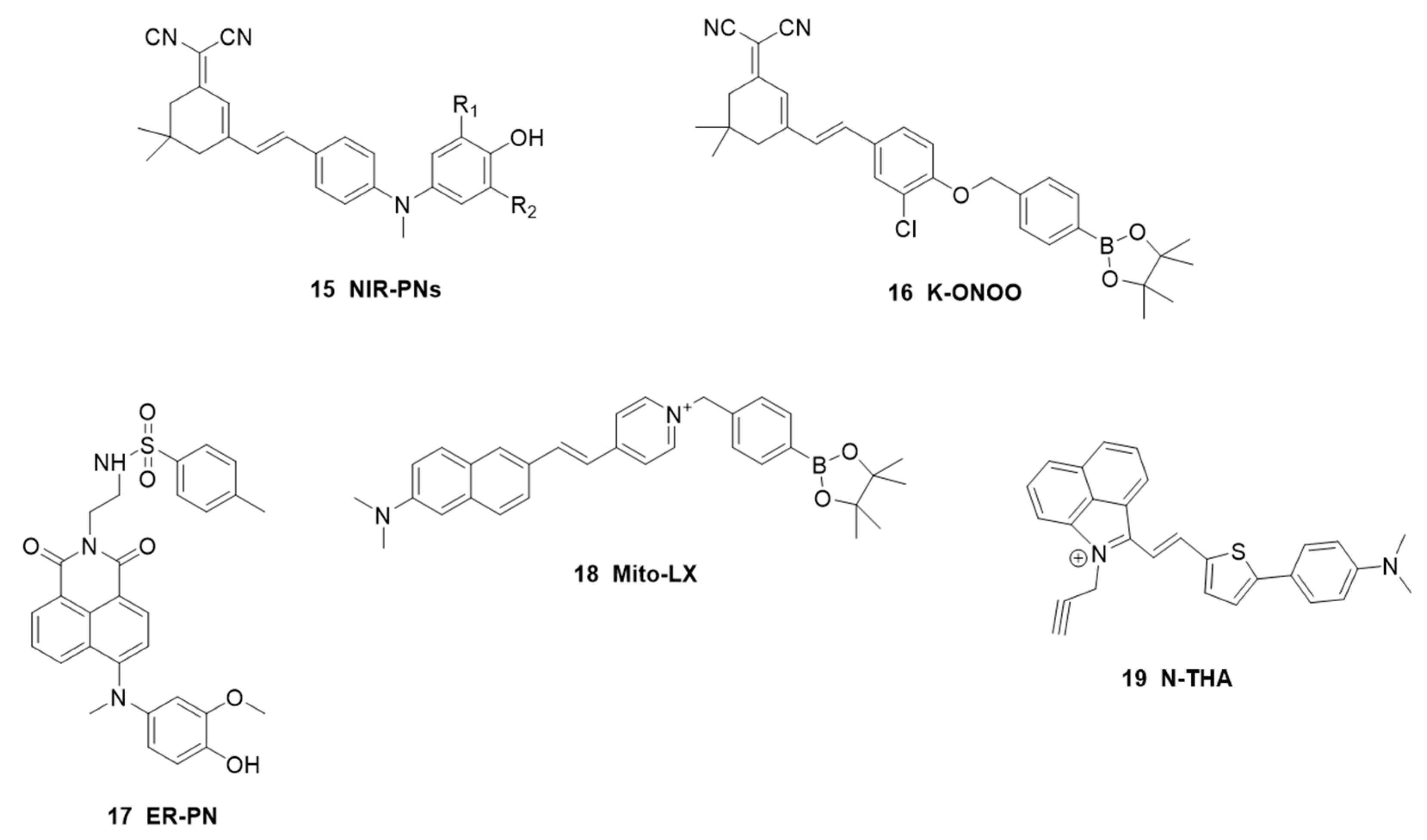
| Parkinson’s disease | NIR-PN1 | 510 /670 | 4.59 nM | peroxynitrite | PC12 and SH-SY5Y cells, parkin null Drosophila, WLZ3 C. elegans | [58] |
| K-ONOO | 405/570 | 212 nM | peroxynitrite | HeLa cells, zebrafish | [59] | |
| ER-PN | 488/540 | 8.3 nM | peroxynitrite | PC12 cells, WLZ3 elegans | [61] | |
| NIR-HP1 | 395/500,650 | 0.27 μM | hydrogen peroxide | living cells, zebrafish and drosophila | [63] | |
| Mito-LX | 380/585 | hydrogen peroxide/viscosity | HepG2 cells, zebrafish and drosophila | [65] | ||
| TAT-Polyp-QL | 458/600 | 0.3832 μM | iron and reactive oxygen | PC12 cells, SD rats | [66] | |
| NUU-1 | 438/503 | 25.8 nM | hypochlorous acid | SH-SY5Y cells, drosophila, PD mouse | [68] | |
| N-THA | 385/516 | viscosity and hydrogen sulfide | HeLa cells, PD mouse | [69] | ||
| Depression | TCS | 340/443 | 0.16 μM | cysteine | PC12 cells, C57BL/6J mice | [73] |
| TCP | 370/495 | 21.5 nM | superoxide anion radical | PC12 cells, C57BL/6J mice | [74] | |
| Te-CDs | 380/440 | 8.0 pM | superoxide anion radical | HepG2 cells, macrophages, Hela cells and lung cancer cells, BalB/C mice | [75] | |
| ACy7 | 570/690 | 10 nM | ozone | RAW 264.7 Macrophages, CUMS mouse model | [77] | |
| TCE | 370/500 | 2.4 μΜ | hydroxyl radical | PC12 cells, PC12 cells, C57BL/6J mice | [79] | |
| Golgi-P | 700/780 | brain-derived neurotrophic factor | PC12 cells, PC12 cells, C57BL/6J mice | [81] | ||
| DNP | 390/460 | 0.32 μM | Zn2+ and H+ | PC12 cells, C57 mouse | [85] | |
| NH–HOBr | 400/505 | 15 nM | hypobromic acid | PC12 cells, RAW 264.7 Macrophages, zebrafish, mice | [86] | |
| Cy7 -OH | 550/640 | norepinephrine | PC12 cells, HepG2 cells, SD rats | [88] | ||
| MCYN | 520/560 | 0.36 μM | acetylcholinesterase | PC12 cells, mice | [90] | |
| UIO-66-NH2 | 425/610 | phosphorylation | HL-7702 cells, mice | [91] | ||
| Schizophrenia | Mindo-SiR | 650/680 | 0.45 μM | hydrogen sulfide | bEnd.3 cells, schizophrenia mouse | [94] |
| MeS-D-KYN | 364/450 | 18.2 mU/mL | D-amino acid oxidase | LLC-PK1 cells | [96] |
Disclaimer/Publisher’s Note: The statements, opinions and data contained in all publications are solely those of the individual author(s) and contributor(s) and not of MDPI and/or the editor(s). MDPI and/or the editor(s) disclaim responsibility for any injury to people or property resulting from any ideas, methods, instructions or products referred to in the content. |
© 2023 by the authors. Licensee MDPI, Basel, Switzerland. This article is an open access article distributed under the terms and conditions of the Creative Commons Attribution (CC BY) license (https://creativecommons.org/licenses/by/4.0/).
Share and Cite
Che, F.; Zhao, X.; Wang, X.; Li, P.; Tang, B. Fluorescent Imaging Agents for Brain Diseases. Targets 2023, 1, 5-33. https://doi.org/10.3390/targets1010003
Che F, Zhao X, Wang X, Li P, Tang B. Fluorescent Imaging Agents for Brain Diseases. Targets. 2023; 1(1):5-33. https://doi.org/10.3390/targets1010003
Chicago/Turabian StyleChe, Feida, Xiaoming Zhao, Xin Wang, Ping Li, and Bo Tang. 2023. "Fluorescent Imaging Agents for Brain Diseases" Targets 1, no. 1: 5-33. https://doi.org/10.3390/targets1010003
APA StyleChe, F., Zhao, X., Wang, X., Li, P., & Tang, B. (2023). Fluorescent Imaging Agents for Brain Diseases. Targets, 1(1), 5-33. https://doi.org/10.3390/targets1010003