
Review

Nailfold Capillaroscopy: An Essential Tool in the Assessment of Systemic Sclerosis

by
Rossella De Angelis
Rheumatology Unit, Microcirculation Imaging Service, Department of Clinical and Molecular Sciences, Polytechnic University of Marche, 60126 Ancona, Italy
Sclerosis 2026, 4(2), 10; https://doi.org/10.3390/sclerosis4020010
Submission received: 10 March 2026 / Revised: 1 May 2026 / Accepted: 4 May 2026 / Published: 7 May 2026
(This article belongs to the Special Issue Recent Advances in Understanding Systemic Sclerosis, 2nd Edition)

Abstract

Nailfold capillaroscopy has earned its place as a cornerstone of clinical assessment in systemic sclerosis (SSc). Its ability to detect early microvascular changes, distinguish primary from secondary Raynaud’s phenomenon, and contribute to disease classification has fundamentally reshaped the clinical approach to early diagnosis and disease stratification. The recognition of distinct capillaroscopic patterns offers a structured framework for tracking disease evolution and identifying patients who warrant closer surveillance or proactive therapeutic intervention. The inclusion of capillaroscopic abnormalities in the ACR/EULAR 2013 classification criteria validates its diagnostic importance and facilitates identification of patients with early or limited cutaneous disease. Beyond diagnosis, emerging evidence supports prognostic applications, particularly for predicting digital ulcers, though the predictive value for other organ complications requires further validation. As a non-invasive, safe, and reproducible technique, capillaroscopy is particularly well-suited to long-term disease monitoring. Quantitative scoring systems allow for rigorous, objective tracking of microangiopathic progression and hold considerable promise as outcome measures in clinical trials targeting vasculopathy. Ongoing technological advances, particularly in automated image analysis and integration with functional assessment tools, promise to enhance the clinical utility of capillaroscopy while reducing operator dependency. Standardization efforts and validation of capillaroscopic parameters as clinical trial endpoints will be crucial for realizing the full potential of this technique.

1. Introduction

Systemic sclerosis (SSc) is a chronic connective tissue disease characterized by three main pathological hallmarks: vasculopathy, autoimmunity, and fibrosis. The disease has complex pathogenesis and variable clinical presentation, ranging from limited to diffuse cutaneous involvement with potential multi-organ complications [1].
Microangiopathy represents arguably the earliest and most consistent hallmark of SSc, frequently antedating the onset of skin fibrosis by months or even years. Raynaud’s phenomenon (RP), typically the first clinical manifestation, reflects the underlying microvascular dysfunction [2,3]. The ability to directly visualize peripheral microvascular architecture in real time has therefore emerged as a decisive advantage in securing early diagnosis and guiding disease monitoring.
Nailfold videocapillaroscopy (NVC) is currently considered the gold standard technique for non-invasive assessment of peripheral microvasculature. From the seminal observations by Brown and O’Leary in 1925 [4] to the landmark validation studies by Maricq and colleagues in the 1980s [5,6,7], the technique has undergone a remarkable transformation—from a niche research instrument to an indispensable pillar of comprehensive clinical evaluation in SSc. The technique allows direct visualization of capillary morphology, enabling distinction between primary and secondary RP, early diagnosis of SSc, and monitoring of disease progression [7,8,9,10].
The inclusion of abnormal nailfold capillaries in the 2013 ACR/EULAR classification criteria for SSc (with a weight of 2 points) underscores the diagnostic importance of this examination [11,12]. Studies have shown that the addition of capillaroscopic abnormalities increases the sensitivity of SSc classification criteria from 67% to 99%, particularly for patients with limited cutaneous disease [13].
This review provides a comprehensive and critical appraisal of NVC in SSc, encompassing technical principles, pattern recognition, diagnostic and prognostic applications, its role in treatment monitoring, and emerging future directions.

2. Technical Principles of Nailfold Capillaroscopy

Modern nailfold videocapillaroscopy employs optical magnification systems (typically 200–300×) equipped with digital imaging capabilities. The examination focuses on the nailfold region where capillary loops are generally oriented parallel to the skin surface, allowing optimal visualization of capillary architecture. The standard procedure involves (a) patient acclimatization at room temperature (20–25 °C) for at least 15–20 min; (b) application of immersion oil to the nailfold to improve optical contact; (c) systematic examination of digits 2–5 of both hands (thumbs are typically excluded); (d) image acquisition and documentation [14,15].
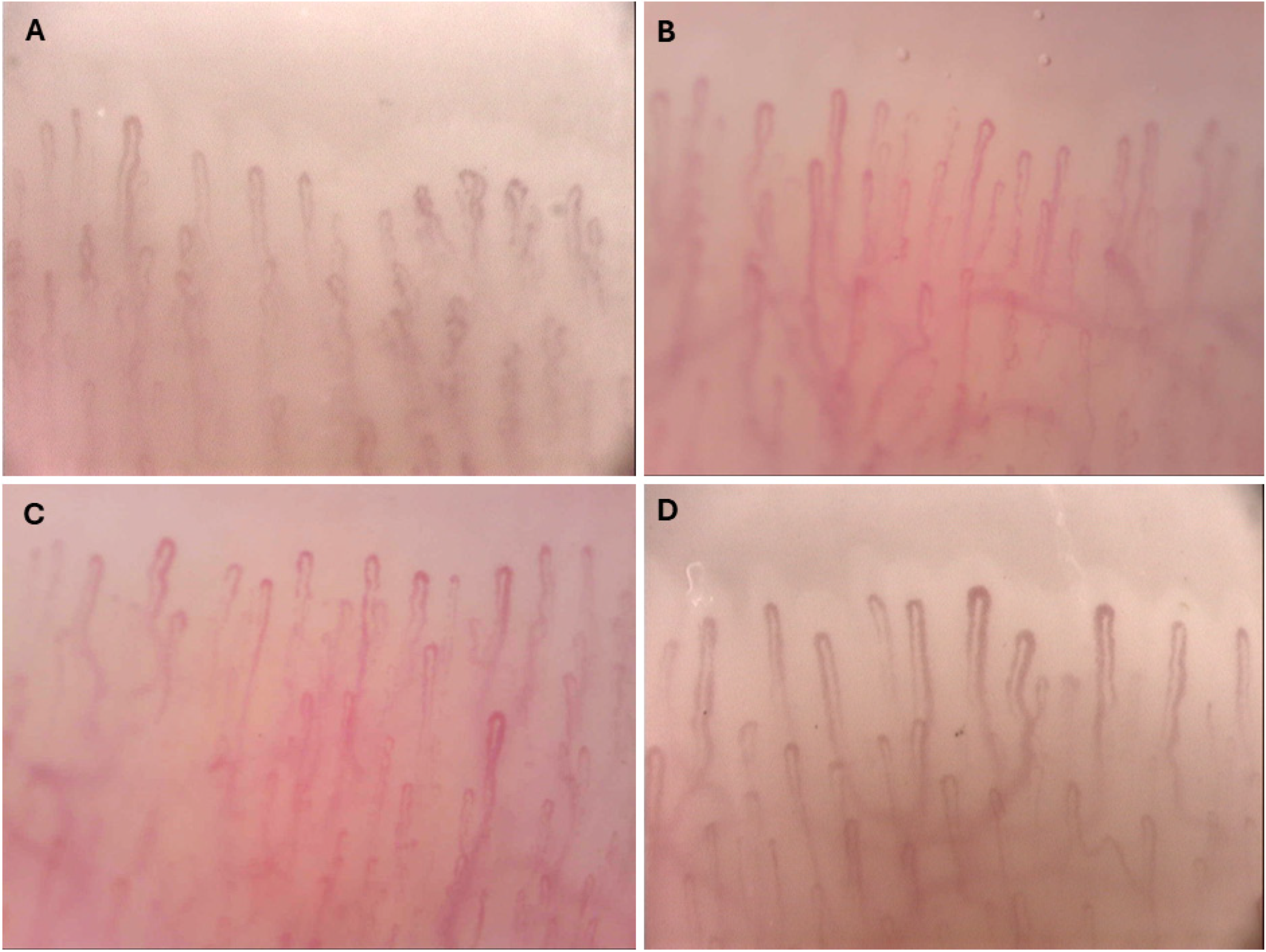
In healthy individuals, nailfold capillaries appear as regularly distributed hairpin-shaped loops arranged parallel to each other. The normal capillary density ranges from 9 to 12 capillaries per millimeter in the distal nailfold row. Each loop consists of an arterial (afferent) limb, an apex, and a venous (efferent) limb, with the apex typically being slightly wider. The capillary diameter normally does not exceed 20 micrometers [14,15] (Figure 1). Moreover, there is an inter-individual and intra-individual variability in capillary patterns among healthy subjects, demonstrating that certain “unusual” findings (like multiple crossed capillaries, meandering loops or isolated microhemorrhages) can occur in healthy individuals, in the presence of confounding factors such as age, reduced or increased skin transparency, smoking habits, manicure/manual trauma and high blood pressure [14,16]. This helps clinicians better distinguish truly pathological patterns from normal variants, which is particularly important for early detection of connective tissue diseases like systemic sclerosis.
The fundamental technical proficiency required for videocapillaroscopy can be acquired relatively rapidly through structured self-directed learning under expert supervision, with clinically acceptable image quality attainable within approximately five hours of supervised practice [17]; however, proficiency in image recognition and interpretive accuracy necessitates more extensive experience [18].
Capillaroscopic evaluation can be performed using qualitative (pattern recognition) and/or quantitative approaches. Qualitative assessment focuses on overall morphological patterns and is particularly useful for diagnosis, while quantitative analysis involves measurement of specific parameters (capillary density, dimensions, number of giant capillaries, hemorrhages) and is valuable for longitudinal monitoring and research applications [10,14,15,19].

3. Capillaroscopic Patterns in Systemic Sclerosis

The hallmark capillaroscopic features of SSc, collectively termed the “scleroderma pattern”, include (Figure 2):
  • Giant capillaries (megacapillaries): capillary loops with diameter > 50 micrometers.
  • Microhemorrhages: hemosiderin deposits resulting from capillary breakup.
  • Loss of capillaries: reduction in capillary density with avascular areas.
  • Disorganization of the normal capillary architecture.
  • Neoangiogenesis: extremely tortuous and dilated capillaries (meandering) indicating a chronic and active microangiopathy that initiates a decrease in the tissue blood flow, and a subsequent neoformation of ramified or bushy capillaries representing a compensatory process.
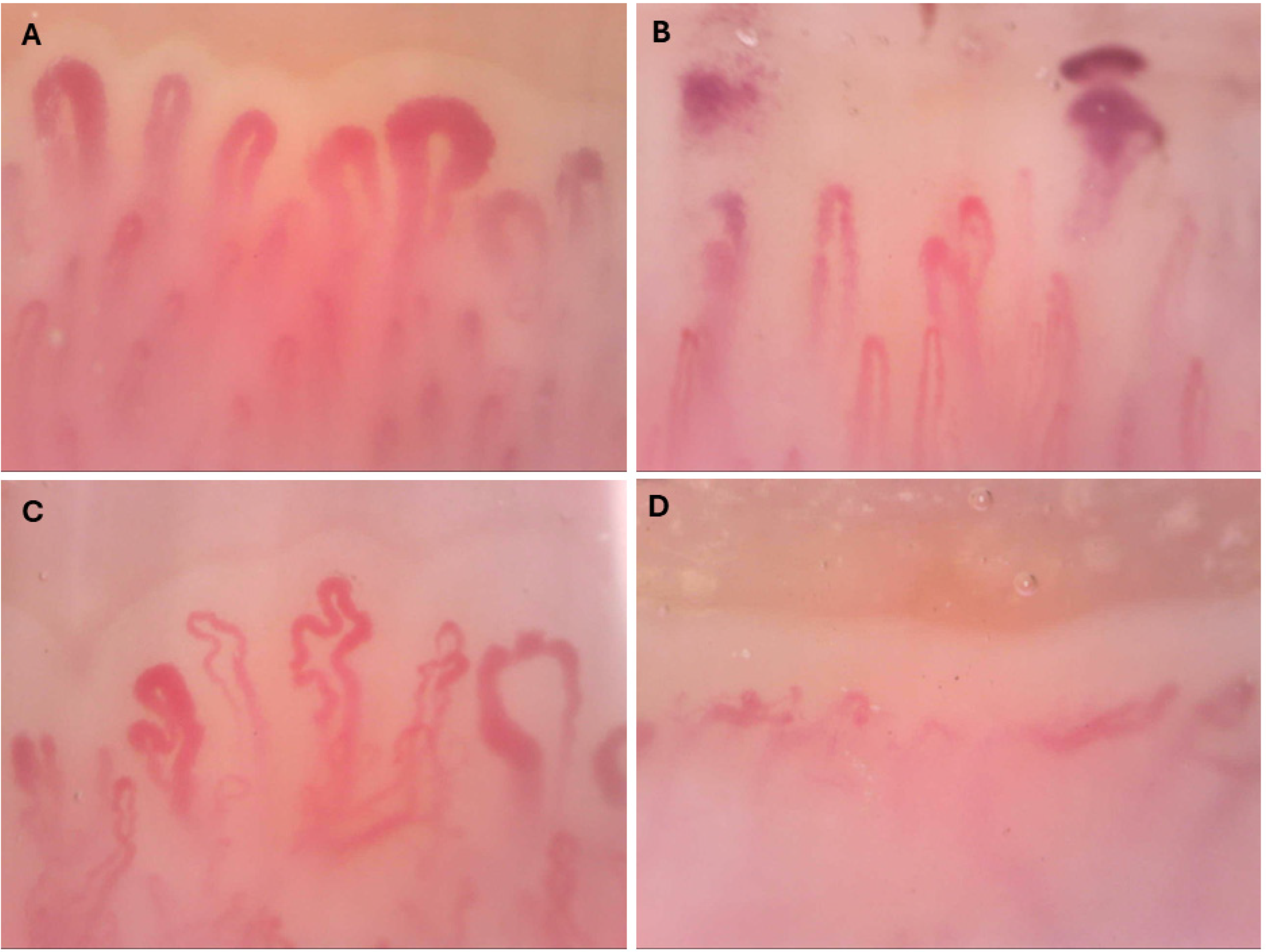
Figure 2. Nailfold videocapillaroscopy 200×. Scleroderma pattern. (A) Giant capillaries. (B) Microhemorrhages with hemosiderin deposits. (C) Disorganization of the normal capillary architecture with angiogenetic-branching loops. (D) Loss of capillaries with avascular areas.
Sclerosis 04 00010 g002
Cutolo and colleagues introduced a landmark classification of SSc capillaroscopic patterns into three progressive stages [20]: “early”, “active” and “late” pattern. The early pattern is characterized by few giant capillaries and microhemorrhages, with relatively well-preserved capillary distribution and no evident loss of capillaries. This pattern may be observed in patients with recent-onset RP and may represent the earliest recognizable stage of SSc microangiopathy [19,20]. The presence of even a single giant capillary combined with microhemorrhages is sufficient to identify the scleroderma pattern and distinguish secondary from primary RP. The active pattern shows frequent giant capillaries, frequent microhemorrhages, moderate loss of capillaries, and mild disorganization of capillary architecture [19,20]. Absence of extensive avascular areas distinguishes this from the late pattern.
The late pattern is dominated by severe capillary loss with extensive avascular areas (“capillary desertification”), marked disorganization of the remaining capillary architecture, and prominent neoangiogenesis (ramified/bushy capillaries). Giant capillaries and microhemorrhages may be less evident or absent in this stage. This pattern reflects advanced microvascular damage with attempted compensatory mechanisms through neovascularization [19,20].
The active and late patterns, both characterized by progressive capillary loss—one of the most important microangiopathic features of SSc—may correlate with more aggressive disease and increased risk of complications [21].
Some studies have considered dynamic progression between capillaroscopic patterns [9,21,22,23]. Approximately 50% of SSc patients show transition to more advanced patterns during follow-up. The estimated progression time from early to active pattern is approximately 28 ± 20 months, while progression from early to late pattern occurs over 36 ± 29 months. Notably, clinical symptoms often progress in parallel with morphological microvascular changes, and approximately 60% of patients show concordance between capillaroscopic and clinical progression [22].
Recently, analysis from a large real-world cohort of SSc patients [23] demonstrated that the spectrum of NVC abnormalities classified does not mirror the disease duration. Indeed, even during the initial disease stages, a substantial number of patients exhibit active or even late patterns. For instance, within the first two years of SSc, a late pattern which is traditionally considered indicative of advanced stages, is observed in 11% of patients even displaying severe complications, Conversely, 14.6% of patients with disease duration exceeding 10 years still exhibited a well-preserved early pattern, associated with significantly lower prevalence of disease complications. These observations challenge the assumption that capillaroscopic patterns reflect a linear temporal progression and underscore the imperative to reconcile existing classification frameworks with the most recent developments in the literature [9,21,23].

4. Diagnostic Value of Nailfold Capillaroscopy

Among the foremost clinical applications of NVC is its capacity to discriminate between primary (benign) and secondary RP in the setting of connective tissue disease. Patients with primary RP may show normal or non-specific capillaroscopic patterns (subtle/minor morphologic changes) [10], while those with secondary RP exhibit definite, well-detailed morphological abnormalities [9,10,11,12,13,14].
A 20-year prospective study of 586 patients with RP demonstrated that SSc-specific autoantibodies and microvascular damage as assessed by NVC are independent predictive factors for progression to SSc, enabling closer monitoring and potentially earlier therapeutic intervention [24]. Specifically, those patients at baseline were 60 times more likely to develop definite SSc, and 79.5% of patients with one of these autoantibodies (anti-topoisomerase or anti-centromere) and abnormal capillary microscopy findings developed definite SSc during follow-up.
NVC operates at three distinct but complementary levels of the diagnostic pathway, which are worth distinguishing explicitly. First, in the context of Raynaud’s phenomenon triage, the presence of even a single, typical giant capillary and microhemorrhages may be sufficient to flag secondary RP and prompt further investigation. Second, within the VEDOSS (Very Early Diagnosis of Systemic Sclerosis) framework, NVC abnormalities represent one of the core risk-stratifying features in patients with RP: the five-year EUSTAR-VEDOSS registry study demonstrated that combinations of puffy fingers, SSc-specific autoantibodies, and abnormal NVC markedly increase the probability of progression to definite SSc, while the absence of ANA identifies a very low-risk group in whom watchful waiting may be appropriate [25]. Importantly, “early SSc” and “very early SSc” are not interchangeable concepts: the former may already satisfy ACR/EULAR 2013 classification criteria, whereas the latter describes a pre-classification stage where NVC plays a pivotal screening role [25,26]. Third, in established SSc classification, NVC contributes 2 points to the 2013 ACR/EULAR scoring system, underpinning its role in formal disease categorization [12]. Maintaining this three-tier conceptual framework helps clinicians apply capillaroscopy with the appropriate level of clinical application at each stage of the disease.
It also should be emphasized that the inclusion of NVC abnormalities (enlarged and/or giant capillaries, microhemorrhages, and/or capillary loss) in the 2013 ACR/EULAR classification criteria for SSc represents a significant improvement over the 1980 ACR criteria, with sensitivity increasing from 75% to 91% and specificity from 72% to 92% [12].
The value of capillaroscopy in early diagnosis of SSc has been validated in cohort studies using algorithms [27,28]. The PRINCE study developed a prognostic index based on nailfold capillaroscopy to predict the 5-year transition from isolated Raynaud’s phenomenon to scleroderma spectrum disorders. The study examined 104 consecutive adult patients with isolated RP and externally validated the index in another cohort of 100 patients, with follow-up ranging from 1 to 8 years. The research identified that giant loops and microhemorrhages, along with the number of capillaries, were the only factors that played a significant prognostic role. The prognostic capillaroscopic index is a weighted combination of different capillaroscopy parameters that allow physicians to stratify RP patients easily, helping identify those at high risk of developing scleroderma spectrum disorders [27]. An advanced algorithm combines autoantibodies and capillaroscopic findings as a powerful diagnostic tool for detecting emerging connective tissue diseases in patients with RP. Different parameters such as antinuclear antibodies, number of capillaries, and the presence of giant capillaries have been combined in this algorithm to stratify RP based on SSc incident risk [28].

5. Prognostic Value of Nailfold Capillaroscopy

5.1. Vascular Complications

Digital ulcers (DUs) represent one of the most prevalent and clinically significant vascular complications encountered in SSc. The most significant predictors for DU development are low capillary density, presence of avascular areas, and a high megacapillary-to-capillary ratio [10,12,14,29].
The Capillaroscopy Skin Ulcer Risk Index (CSURI) was developed specifically to predict digital ulcer risk [29]. The CSURI is calculated using the formula: (N × D2)/M, where N is the number of capillaries per millimeter in the most preserved area, D is the maximum diameter of the largest giant capillary, and M is the number of capillaries in one millimeter at the center of the most affected area. A CSURI score > 2.96 predicts digital ulcer development within 3 months with 92.9% sensitivity, 81.4% specificity, and 97.2% negative predictive value. In patients with a history of DUs, the positive predictive value exceeds 81%. Additionally, reduced capillary density (mean score > 1.67) has been identified as an independent predictor of DU occurrence within 6–12 months [29].
Several studies have explored correlations between capillaroscopic findings and pulmonary arterial hypertension (PAH) [30,31,32,33]. Lower capillary density and increased avascular areas, as well as a predominance of active/late patterns, have been associated with the presence and severity of PAH in SSc patients; indeed, patients with SSc-associated PAH tend to show more severe microvascular damage on NVC compared to those without PAH [30,31,32]. While NVC alone is not sufficient to diagnose PAH, which requires right heart catheterization (RHC) confirmation, the evidence supports its integration into PAH screening algorithms to improve their performance [33].

5.2. Internal Organ Involvement

Interstitial lung disease (ILD) is one of the most frequent and life-threatening complications of SSc. The late scleroderma pattern, as well as reduction in capillary density are the most common abnormality identified in ILD-SSc patients [34,35]. More accurately, as in ILD, pulmonary function abnormalities usually reflect a restrictive lung defect with reduced lung compliance and volumes; patients showing moderate-to-severe restrictive patterns have major NVC abnormalities [36]. A sub-study of the SENSCIS trial explored whether NVC could serve as a biomarker of disease progression. In patients with risk factors for rapid FVC decline, there was a numerical reduction in mean capillary density over 52 weeks with placebo, while it remained stable with nintedanib [37]. Nevertheless, the predictive value for ILD development and progression requires further validation, by inserting NVC abnormalities in multi-itemed algorithms including, besides NVC, PFTs, an evaluation of respiratory symptoms and esophageal involvement, especially in those awaiting the planned annual CT scan [35].
Emerging evidence also suggests potential correlations with cardiac, renal, and gastrointestinal involvement, though these require additional investigation.
Cardiac involvement in SSc—including myocardial fibrosis, diastolic dysfunction, arrhythmias, and coronary microvascular dysfunction (CMD)—is a leading cause of SSc-related mortality. The peripheral microvascular damage documented by NVC appears to mirror myocardial microvascular pathology, providing a rationale for its predictive role. The most compelling evidence comes from a notable investigation which investigated the association between NVC abnormalities and coronary flow reserve (CFR), a measure of CMD assessed by transthoracic echocardiography in 39 SSc patients without overt cardiovascular disease. The NVC avascular score was significantly associated with CMD, suggesting that capillary rarefaction in the nailfold reflects structural microvascular remodeling occurring simultaneously in the coronary circulation [38]. At a broader level, a landmark study [39], conducted within the Leiden SSc Cohort (N = 287), demonstrated that the combination of NVC patterns and SSc-specific autoantibodies significantly improved the identification of patients at high risk of cardiopulmonary involvement, compared to autoantibodies alone [39].
The predictive value of NVC for future cardiac involvement was assessed prospectively in a two-cohort study using the Medsger Disease Severity Scale (DSS) over 18–24 months of follow-up [40]. More severe NVC patterns (active and late) were associated with higher odds ratios for novel severe organ involvement, including cardiac involvement, validated across both cohorts [40]. A similar pilot study also suggested that worsening capillaroscopic patterns over time predicted future severe organ involvement, including cardiac disease [41].
A large multicenter study [42] using data from 334 SSc patients across seven EUSTAR tertiary centers evaluated NVC quantitatively and qualitatively. While statistically underpowered for organ-specific outcomes, it found that normal capillary density was associated with lower risk of overall organ progression, and that severe NVC patterns (active and late) correlated with novel cardiopulmonary involvement [42].
Gastrointestinal (GI) involvement is the most prevalent internal organ manifestation in SSc, affecting up to 90% of patients, with dysmotility, malabsorption, gastroesophageal reflux, and pseudo-obstruction representing the main clinical expressions. Pathogenesis is driven by a combination of neurovascular dysfunction, smooth muscle atrophy, and fibrosis [1,3]. Evidence directly linking NVC patterns to GI involvement is more limited and methodologically heterogeneous than for cardiac or pulmonary disease. Nonetheless, some studies have documented associations [40,43,44,45]. A two-cohort study [40] found that more severe NVC patterns at baseline were associated with higher odds of novel severe GI involvement at 18–24 months using the Medsger DSS. Similarly, among 113 SSc patients followed for 24 months, 10% developed novel GI involvement, with NVC parameters contributing to risk stratification in multivariate analysis [44]. A systematic review further noted associations between scleroderma NVC patterns and esophageal involvement/malabsorption, while acknowledging the limited evidence compared to the cardiopulmonary domain [45]. Overall, despite sound pathophysiological plausibility (shared microangiopathic mechanism), GI-specific NVC prediction lacks the methodological rigor and sample sizes of the cardiopulmonary literature, and results should be interpreted with caution.
Scleroderma renal crisis (SRC) is the most severe renal complication of SSc, characterized by acute-onset malignant hypertension and rapidly progressive renal failure due to thrombotic microangiopathy. Its prevalence ranges from 1% to 14% across different geographical cohorts [1,2,3,23]. Direct evidence for NVC as a predictor of renal involvement is the weakest among the organ systems reviewed here. In the prospective two-cohort study by Smith et al. [40], renal involvement was among the nine organ domains evaluated according to the Medsger DSS; the study noted trends toward association with more severe NVC patterns, but specific analysis of renal outcomes was limited by the low prevalence of SRC during the relatively short 18–24-month follow-up. Further data [44] reported that 6% of patients developed SRC during a 24-month observation period when NVC parameters were included in the multivariate analyses of overall organ involvement. A systematic review [45] acknowledged that scleroderma NVC patterns had been associated with SRC in several series but emphasized that this association is confounded by disease subtype (dcSSc vs. lcSSc), autoantibody profile, and treatment with corticosteroids. A recent paper reports on a notable association between the late NVC pattern and SRC. Specifically, in the subgroup of patients with disease duration ≤ 2 years, renal crisis was significantly more frequent in those with the late pattern compared to the early pattern (7.1% vs. 0%, p ≤ 0.01). Furthermore, in the multivariable analysis restricted to patients with disease duration ≤ 5 years, the presence of the late pattern was significantly associated with SRC (p = 0.012). The authors suggest that severe microvascular damage may play a role in predicting SRC development, likely through ischemic injury to renal tissue caused by damage to kidney arterioles even in early disease phases [23].
In summary, current suggestions endorse NVC as a tool for stratifying overall SSc severity and vasculopathy progression [11,42,45], but do not yet support its use as an autonomous organ-specific predictor beyond digital ulcers and PAH screening. Evidence for GI involvement is suggestive but methodologically weak. Evidence for renal involvement remains largely confounded by other disease factors, especially by disease duration. Across all domains, the literature shares important limitations: most studies are cross-sectional; cohort sizes are small; NVC scoring methods vary across centers, limiting comparability; and organ-specific outcomes are often underpowered (Table 1). Ongoing and future multicenter prospective studies with standardized quantitative NVC assessment—including the EULAR Study Group on Microcirculation framework—will be essential to definitively establish the organ-predictive value of NVC across the full spectrum of SSc visceral disease [10,40,41,42,46].

6. Associations with Cutaneous Disease Subsets and Autoantibodies

Capillaroscopic patterns show noteworthy associations with SSc subsets and autoantibody profiles. Patients with early and active patterns more frequently present with limited cutaneous SSc (lcSSc), while the late pattern is more commonly observed in diffuse cutaneous disease (dcSSc) [8,47,48]. In particular, the proportion of early patterns were more frequent in patients with SSc sine scleroderma (i.e., lack of skin involvement) (12.1% and 36.2%) compared with both lcSSc (7.6% and 21.8%) and dcSSc (1.8% and 14.9%) (p = 0.003 and 0.001, respectively), whereas the late pattern was uncommon (8.6%) in ssSSc, with an increasing prevalence from lcSSc (21.2%) to dcSSc (47.6%, p < 0.001) [49]. Moreover, advanced microangiopathy by NVC, i.e., avascular areas, identifies dcSSc patients at risk of reduced survival due to SSc-related causes [48]. This relationship reflects the parallelism between advanced microvascular changes and the severity of skin involvement [48,49,50].
Several clinical studies reported a significant association between SSc-related autoantibodies and NVC patterns: more specifically, anti-centromere autoantibodies (ACAs) were associated more often with an early NVC pattern, whereas anti-topoisomerase I autoantibodies more frequently showed an active/late NVC pattern [47,51]. The presence of anti-topoisomerase appears to be related to an earlier expression of the active and late NVC patterns, whereas the presence of anti-centromere seems to be related to a delayed expression of the late NVC pattern [47,51]. Detailed data from 2754 SSc patients from the EULAR scleroderma trials (EUSTAR) reported that the late pattern was present in 47% of anti-topoisomerase positive patients vs. 28% of anti-centromere patients (p < 0.05), whereas the early and active patterns were more frequent in anti-centromere patients than in anti-topoisomerase patients (44% vs. 28%, p < 0.05) [52]. Recent findings suggest that the mode and ages of clinical SSc onset are associated with specific SSc-related autoantibodies. When categories were defined based on mode of onset (RP group—RP onset at least 1 year before NRP; Simultaneous group—RP onset within the same year of NRP; NRP group—RP onset after at least 1 year after NRP) a higher prevalence of anti-centromere antibodies was found in the RP group, while the simultaneous group had more dcSSc, anti-topoisomerase-I antibodies, and higher mRSS [53].
SSc-related autoantibodies exert distinct pathogenic mechanisms targeting endothelial cells, fibroblasts, and vascular smooth muscle cells [51], suggesting their active participation in SSc pathophysiology through interactions at the endothelial–fibroblastic interface—thereby providing a cellular and molecular underpinning for the morphological alterations observed on NVC [51].

7. Monitoring and Treatment Response Assessment

With respect to treatment response, both vasoactive and immunomodulating therapies have been shown to influence microvascular alterations as assessed by NVC. Among vasoactive drugs, long-term combination therapy with bosentan and iloprost consistently demonstrated improvement in capillary density compared to iloprost alone [54,55]. Iloprost monotherapy showed variable effects depending on the administration schedule and follow-up duration [55]. Among immunomodulating treatments, autologous hematopoietic stem cell transplantation (HSCT) produced the most significant and sustained improvement in NVC patterns, already detectable at 3 months and maintained at 2 years [56], while rituximab was associated with stabilization of microvascular abnormalities [57].
A recent prospective cohort study [58] investigated the evolution of nailfold capillary density in 80 newly diagnosed SSc patients over a median follow-up of 27 months, analyzing the impact of mycophenolate mofetil-MMF treatment and SSc-associated autoantibodies on microvascular changes assessed by NVC. The principal finding was that nailfold capillary density improved over time in a substantial subgroup of patients (28 out of 80), with a significant increase in mean capillary density already detectable at 12 months of follow-up. Treatment with MMF was significantly associated with fewer fingers showing worsened capillary density and with a less steep decline in mean capillary density over time in a generalized estimating equation model. Notably, increasing MMF dosage showed a positive dose-dependent association with capillary density, suggesting that stronger immunosuppression correlates with more favorable microvascular outcomes. These findings are consistent with earlier evidence that cyclophosphamide (CYC) may improve the scleroderma pattern [59]. Taken together, these results suggest that vasoactive and immunosuppressive treatments may contribute to nailfold capillary remodeling in SSc, calling for randomized controlled trials to confirm these findings and establish whether NVC-detected microvascular changes translate into meaningful clinical benefit for patients.
The role of NVC as an outcome measure in clinical trials is increasingly recognized, but important caveats remain. The available treatment response literature is largely based on small observational or open-label datasets, and even the SENSCIS sub-study—the most rigorous dataset to date—demonstrated a numerical signal for capillary density stabilization with nintedanib rather than definitive validation of NVC as a treatment response biomarker [37]. Accordingly, NVC change should currently be regarded as a promising exploratory or pharmacodynamic biomarker, rather than a near-ready surrogate endpoint. Full qualification as a surrogate endpoint would require prospective demonstration of responsiveness to change, inter- and intra-reader reproducibility at the level required for trials, definition of a minimal clinically important difference (MCID) for key parameter such as shapes, sizes and capillary density, number of hemorrhages, and presence/extent of avascular areas [46], and—critically—evidence of linkage between capillaroscopic improvement and patient-important outcomes such as digital ulcer burden, functional capacity, or organ-specific progression. These remain active and high-priority research questions for the field [60,61,62].

8. Limitations and Future Perspectives

Despite its well-established diagnostic and prognostic value, nailfold capillaroscopy is subject to several inherent limitations [14,18,55]:
  • Operator-dependent assessment requiring specific training and experience.
  • Inter- and intra-observer variability in qualitative pattern recognition.
  • Time-consuming procedure when performed comprehensively.
  • Technical challenges in patients with finger deformities, dystrophic cuticles, or advanced disease.
  • Limited standardization of equipment and image acquisition protocols across centers.
  • Incomplete understanding of the relationship between capillaroscopic changes and specific organ complications.
The routine applicability of certain indices has important practical consequences that deserve acknowledgment. For example, a longitudinal multicenter study found that CSURI could not be calculated in ~40% of patients due to absent megacapillaries, while predictive performance and inter-assessor agreement were more modest than in the original validation, questioning its generalizability across centers [29,63]. These findings underscore the need for complementary quantitative indices applicable across the full spectrum of SSc microangiopathy, including late-pattern patients where giant capillaries may no longer be present.
In this context, a practical advance worth highlighting is the EULAR Fast Track algorithm, developed to help operators of varying expertise reliably distinguish scleroderma from non-scleroderma patterns. The algorithm was specifically designed to improve inter-reader agreement and to support wider uptake of capillaroscopy beyond highly specialized expert centers [60].
Moreover, several technological advances promise to enhance the clinical utility of capillaroscopy, such as automated image analysis using machine learning algorithms that could reduce inter-observer variability, accelerate assessment, and improve reproducibility [64]. Deep learning approaches are being developed for automated pattern recognition, capillary counting, and quantification of morphological abnormalities. Moreover, optical coherence tomography (OCT) offers three-dimensional visualization of cutaneous microcirculation with higher resolution than conventional capillaroscopy and can potentially provide information about capillary depth, blood flow dynamics, and deeper vascular structures [65]. Integration of morphological capillaroscopy with functional assessment techniques such as laser speckle contrast analysis or thermography could provide comprehensive evaluation of both structural and functional aspects of peripheral vasculopathy [66,67].

9. Research Priorities

Future investigative efforts should be directed towards: (i) worldwide standardization of image acquisition protocols and reporting standards; (ii) validation of quantitative parameters as outcomes in clinical trials; (iii) better understanding of capillaroscopic–clinical correlations for specific organ involvement; (iv) development and validation of predictive models incorporating capillaroscopy with other biomarkers; (v) investigation of capillaroscopy’s role in guiding therapeutic decisions and monitoring treatment response.
Consensus-based guidelines for equipment specifications, acquisition procedures, and environmental conditions have been established, and structured reporting templates that define minimum required parameters (e.g., capillary density, morphology, hemorrhages) and standardized terminology have been proposed [14,15,16,17,46,68,69].
Validated training programs to reduce inter-operator variability and facilitate adoption in non-specialist centers are currently underway (EULAR Study Group on Capillaroscopy & Microcirculation in Rheumatic Diseases). While qualitative capillaroscopic patterns are well-established, quantitative parameters have yet to be rigorously validated as surrogate endpoints or outcome measures in therapeutic trials. In this regard, longitudinal studies determining the responsiveness to change in key metrics such as capillary density, avascular areas, and giant capillary frequency are more expected [42,70], as well as validation of semi-automated and automated image analysis software against expert readings to ensure reproducibility at scale. Finally, and of utmost significance, a definition of MCID for quantitative capillaroscopic parameters would be welcome, especially addressing future, specific organ damage (i.e., PAH, DUs, ILD) with emphasis on early predictors [71].
Capillaroscopy yields its greatest clinical value when integrated into multiparametric models, so future work should be aimed at building and validating machine learning models that combine capillaroscopic data with serological biomarkers and clinical variables. Perhaps the most clinically impactful frontier lies in using capillaroscopy as a dynamic tool for treatment monitoring; key research questions should include defining threshold changes that should prompt treatment escalation or de-escalation in daily clinical practice.
Addressing these research priorities in a coordinated, international effort will elevate capillaroscopy from a predominantly descriptive modality into a robust, quantitative, and therapeutically decisive instrument. Progress in standardization and digital analysis will underpin advances in all other domains, while integrative biomarker models promise to deliver genuinely personalized, precision medicine approaches to patients living with SSc.

10. Conclusions

In conclusion, NVC offers a unique and irreplaceable window into the peripheral microcirculation, delivering diagnostic precision, prognostic insight, and dynamic monitoring capabilities that few other tools can rival in SSc. Its deeper integration into routine clinical practice and translational research holds the promise of earlier diagnosis, more refined risk stratification, and—most critically—meaningfully improved outcomes for patients navigating this complex and burdensome disease.

Funding

This research received no external funding.

Data Availability Statement

No new data were created or analyzed in this study. Data sharing is not applicable to this article.

Conflicts of Interest

The author declares no conflicts of interest.

References

  1. Al-Gburi, S.; Moinzadeh, P.; Krieg, T. Pathophysiology in systemic sclerosis: Current insights and future perspectives. Sclerosis 2025, 3, 17. [Google Scholar] [CrossRef] [Scilit]
  2. Matucci-Cerinic, M.; Kahaleh, B.; Wigley, F.M. Evidence that systemic sclerosis is a vascular disease. Arthritis Rheum. 2013, 65, 1953–1962. [Google Scholar] [CrossRef] [Scilit] [PubMed]
  3. Pauling, J.D.; Allanore, Y.; Buch, M.; Cutolo, M.; Del Galdo, F.; Denton, C.P.; Di Donato, S.; Domsic, R.T.; Frech, T.; Herrick, A.L.; et al. Exploiting a unified vascular framework to predict organ-specific complications and accomplish disease modification in systemic sclerosis. Lancet Rheumatol. 2025, 7, e895–e906. [Google Scholar] [CrossRef] [Scilit] [PubMed]
  4. Brown, G.E.; O’Leary, P.A. Skin capillaries in scleroderma. Arch. Intern. Med. 1925, 36, 73–88. [Google Scholar] [CrossRef] [Scilit]
  5. Maricq, H.R.; LeRoy, E.C.; D’Angelo, W.A.; Medsger, T.A.; Rodnan, G.P.; Sharp, G.C.; Wolfe, J.F. Diagnostic potential of in vivo capillary microscopy in scleroderma and related disorders. Arthritis Rheum. 1980, 23, 183–189. [Google Scholar] [CrossRef] [Scilit]
  6. Maricq, H.R.; Weinberger, A.B.; LeRoy, E.C. Early detection of scleroderma-spectrum disorders by in vivo capillary microscopy: A prospective study of patients with Raynaud’s phenomenon. J. Rheumatol. 1982, 9, 289–291. [Google Scholar]
  7. Maricq, H.R.; Harper, F.E.; Khan, M.M.; Tan, E.M.; LeRoy, E.C. Microvascular abnormalities as possible predictors of disease subsets in Raynaud phenomenon and early connective tissue disease. Clin. Exp. Rheumatol. 1983, 1, 195–205. [Google Scholar]
  8. Caramaschi, P.; Canestrini, S.; Martinelli, N.; Volpe, A.; Pieropan, S.; Ferrari, M.; Bambara, L.M.; Carletto, A.; Biasi, D. Scleroderma patients nailfold videocapillaroscopic patterns are associated with disease subset and disease severity. Rheumatology 2007, 46, 1566–1569. [Google Scholar] [CrossRef] [Scilit]
  9. Ghizzoni, C.; Sebastiani, M.; Manfredi, A.; Campomori, F.; Colaci, M.; Giuggioli, D.; Ferri, C. Prevalence and evolution of scleroderma pattern at nailfold videocapillaroscopy in systemic sclerosis: Clinical and prognostic implications. Microvasc. Res. 2015, 99, 92–95. [Google Scholar] [CrossRef] [Scilit]
  10. Herrick, A.L.; Cutolo, M. Clinical implications from capillaroscopic analysis in patients with Raynaud’s phenomenon and systemic sclerosis. Arthritis Rheum. 2010, 62, 2595–2604. [Google Scholar] [CrossRef] [Scilit] [PubMed]
  11. Avouac, J.; Fransen, J.; Walker, U.A.; Riccieri, V.; Smith, V.; Muller, C.; Miniati, I.; Tarner, I.H.; Bellando-Randone, S.; Cutolo, M.; et al. Preliminary criteria for the very early diagnosis of systemic sclerosis: Results of a Delphi Consensus Study from EULAR Scleroderma Trials and Research Group. Ann. Rheum. Dis. 2011, 70, 476–481. [Google Scholar] [CrossRef] [Scilit] [PubMed]
  12. van den Hoogen, F.; Khanna, D.; Fransen, J.; Johnson, S.R.; Baron, M.; Tyndall, A.; Matucci-Cerinic, M.; Naden, R.P.; Medsger, T.A., Jr.; Carreira, P.E.; et al. Classification criteria for systemic sclerosis: An American college of rheumatology/European league against rheumatism collaborative initiative. Ann. Rheum. Dis. 2013, 72, 1747–1755. [Google Scholar] [CrossRef] [Scilit]
  13. Minier, T.; Guiducci, S.; Bellando-Randone, S.; Bruni, C.; Lepri, G.; Czrjack, L.; Distler, O.; Walker, U.A.; Fransen, J.; Allanore, Y.; et al. Preliminary analysis of the very early diagnosis of systemic sclerosis (VEDOSS) EUSTAR multicentre study: Evidence for puffy fingers as a pivotal sign for suspicion of systemic sclerosis. Ann. Rheum. Dis. 2014, 73, 2887–2893. [Google Scholar] [CrossRef] [Scilit]
  14. Etehad Tavakol, M.; Fatemi, A.; Karbalaie, A.; Emrani, Z.; Erlandsson, B.E. Nailfold capillaroscopy in rheumatic diseases: Which parameters should be evaluated? BioMed Res. Intern. 2015, 2015, 974530. [Google Scholar] [CrossRef] [Scilit]
  15. Ingegnoli, F.; Herrick, A.L.; Schioppo, T.; Bartoli, F.; Ughi, N.; Pauling, J.D.; Sulli, A.; Cutolo, M.; Smith, V.; European League Against Rheumatism (EULAR) study group on microcirculation in rheumatic diseases and the Scleroderma Trial consortium. Reporting items for capillaroscopy in clinical research on musculoskeletal diseases: A systematic review and international Delphi consensus. Rheumatology 2021, 60, 1410–1418. [Google Scholar] [CrossRef] [Scilit]
  16. Ingegnoli, F.; Gualtierotti, R.; Lubatti, C.; Bertolazzi, C.; Gutierrez, M.; Boracchi, P.; Fornili, M.; De Angelis, R. Nailfold capillary patterns in healthy subjects: A real issue in capillaroscopy. Microvasc. Res. 2013, 90, 90–95. [Google Scholar] [CrossRef] [Scilit]
  17. De Angelis, R.; Cutolo, M.; Salaffi, F.; Restrepo, J.P.; Grassi, W. Quantitative and qualitative assessment of one rheumatology trainee’s experience with a self-teaching programme in videocapillaroscopy. Clin. Exp. Rheumatol. 2009, 27, 651–653. [Google Scholar]
  18. Gutierrez, M.; Bertolazzi, C.; Tardella, M.; Becciolini, A.; Di Carlo, M.; Dottori, M.; Grassi, W.; De Angelis, R. Interreader reliability in assessment of nailfold capillary abnormalities by beginners: Pilot study of an intensive videocapillaroscopy training program. J. Rheumatol. 2012, 39, 1248–1255. [Google Scholar] [CrossRef] [Scilit]
  19. Cutolo, M.; Pizzorni, C.; Secchi, M.E.; Sulli, A. Capillaroscopy. Best Pract. Res. Clin. Rheumatol. 2008, 22, 1093–1108. [Google Scholar] [CrossRef] [Scilit] [PubMed]
  20. Cutolo, M.; Sulli, A.; Pizzorni, C.; Accardo, S. Nailfold videocapillaroscopy assessment of microvascular damage in systemic sclerosis. J. Rheumatol. 2000, 27, 155–160. [Google Scholar] [PubMed]
  21. Boulon, C.; Aiouaz, S.; Blaise, S.; Mangin, M.; Decamps-Le Chevoir, J.; Senet, P.; Lazareth, I.; Baudot, N.; Tribout, L.; Imbert, I.; et al. Correlation between capillaroscopic classifications and severity in systemic sclerosis: Results from SCLEROCAP study at inclusion. Clin. Exp. Rheumatol. 2019, 37, 63–68. [Google Scholar] [PubMed]
  22. Sulli, A.; Pizzorni, C.; Smith, V.; Zampogna, G.; Ravera, F.; Cutolo, M. Timing of transition between capillaroscopic patterns in systemic sclerosis. Arthritis Rheum. 2012, 64, 821–825. [Google Scholar] [CrossRef] [Scilit]
  23. De Angelis, R.; Ferri, C.; Cipolletta, E.; Riccieri, V.; Di Battista, M.; Bajocchi, G.; Bellando-Randone, S.; Bruni, C.; Orlandi, M.; Zanframundo, G.; et al. Prevalence, distribution and associations of the scleroderma capillaroscopic patterns: New insights from the Italian SPRING-SIR registry. Rheumatology 2026, 65, keaf672. [Google Scholar] [CrossRef] [Scilit]
  24. Koenig, M.; Joyal, F.; Fritzler, M.J.; Roussin, A.; Abrahamowics, M.; Boire, G.; Goulet, J.-R.; Rich, E.; Grodzicky, T.; Raymond, Y.; et al. Autoantibodies and microvascular damage are independent predictive factors for the progression of Raynaud’s phenomenon to systemic sclerosis: A twenty-year prospective study of 586 patients, with validation of proposed criteria for early systemic sclerosis. Arthritis Rheum. 2008, 58, 3902–3912. [Google Scholar] [CrossRef] [Scilit]
  25. Bellando-Randone, S.; Del Galdo, F.; Lepri, G.; Minier, T.; Huscher, D.; Furst, D.E.; Allanore, Y.; Distler, O.; Czrjack, L.; Bruni, C.; et al. Progression of patients with Raynaud’s phenomenon to systemic sclerosis: A five-year analysis of the European Scleroderma Trial and Research group multicentre, longitudinal registry study for Very Early Diagnosis of Systemic Sclerosis (VEDOSS). Lancet Rheumatol. 2021, 3, e834–e843. [Google Scholar] [CrossRef] [Scilit]
  26. LeRoy, E.C.; Medsger, T.A. Criteria for classification of systemic sclerosis. J. Rheumatol. 2001, 2, 1573–1576. [Google Scholar]
  27. Ingegnoli, F.; Boracchi, P.; Gualtierotti, R.; Lubatti, C.; Meani, L.; Zahalkova, L.; Zeni, S.; Fantini, F. Prognostic model based on nailfold capillaroscopy for identifying Raynaud’s phenomenon patients at high risk for the development of a scleroderma spectrum disorder: PRINCE (Prognostic Index for Nailfold Capillaroscopy Examination). Arthritis Rheum. 2008, 58, 2174–2182. [Google Scholar] [CrossRef] [Scilit] [PubMed]
  28. Ingegnoli, F.; Boracchi, P.; Gualtierotti, R.; Biganzoli, E.M.; Zeni, S.; Lubatti, C.; Fantini, F. Improving outcome prediction of systemic sclerosis from isolated Raynaud’s phenomenon: Role of autoantibodies and nail-fold capillaroscopy. Rheumatology 2010, 49, 797–805. [Google Scholar] [CrossRef] [Scilit]
  29. Sebastiani, M.; Manfredi, A.; Vukatana, G.; Moscatelli, S.; Riato, L.; Bocci, M.; Iudici, M.; Principato, A.; Mazzucca, S.; Del Medico, P.; et al. Predictive role of capillaroscopy skin ulcer risk index in systemic sclerosis: A multicentre validation study. Ann. Rheum. Dis. 2012, 71, 67–70. [Google Scholar] [CrossRef] [Scilit]
  30. Giuggioli, D.; Riccieri, V.; Cipolletta, E.; Del Papa, N.; Ingegnoli, F.; Spinella, A.; Pellegrino, G.; Risa, A.M.; de Pinto, M.; Papa, S.; et al. Peripheral microangiopathy changes in pulmonary arterial hypertension related to systemic sclerosis: Data from a multicenter observational study. Front. Cardiovasc. Med. 2022, 9, 924899. [Google Scholar] [CrossRef] [Scilit]
  31. De Angelis, R.; Riccieri, V.; Cipolletta, E.; Del Papa, N.; Ingegnoli, F.; Bosello, S.; Spinella, A.; Pellegrino, G.; de Pinto, M.; Papa, S.; et al. Significant nailfold capillary loss and late capillaroscopic pattern are associated with pulmonary arterial hypertension in systemic sclerosis. Rheumatology 2024, 3, 1616–1623. [Google Scholar] [CrossRef] [Scilit] [PubMed]
  32. Riccieri, V.; Vasile, M.; Iannace, N.; Stefanantoni, K.; Sciarra, I.; Vizza, C.D.; Badagliacca, R.; Poscia, R.; Papa, S.; Mezzapesa, M.; et al. Systemic sclerosis patients with and without pulmonary arterial hypertension: A nailfold capillaroscopy study. Rheumatology 2013, 52, 1525–1528. [Google Scholar] [CrossRef] [Scilit] [PubMed]
  33. Smith, V.; Vanhaecke, A.; Vandecasteele, E.; Guerra, M.; Paolino, S.; Melsens, K.; Cutolo, M. Nailfold videocapillaroscopy in systemic sclerosis-related pulmonary arterial hypertension: A systematic literature review. J. Rheumatol. 2020, 47, 888–895. [Google Scholar] [CrossRef] [Scilit]
  34. Anghel, D.; Prioteasa, O.G.; Nicolau, I.N.; Bucurica, S.; Belinski, D.O.; Popescu, G.G.; Ghinescu, M.C.; Bobirca, A.; Groseanu, M.L.; Bojinca, V.C. The role of nailfold videocapillaroscopy in diagnosis and monitoring of interstitial lung disease associated with rheumatic autoimmune diseases. Diagnostics 2025, 15, 362. [Google Scholar] [CrossRef] [Scilit]
  35. De Angelis, R.; Cipolletta, E.; Francioso, F.; Carotti, M.; Farah, S.; Giovagnoni, A.; Salaffi, F. Low-carbon monoxide diffusing capacity, patient reported measures, and reduced nailfold capillary density are associated with interstitial lung disease in systemic sclerosis. J. Pers. Med. 2024, 14, 635. [Google Scholar] [CrossRef] [Scilit]
  36. Rajendran, A.; Maikap, D.; Padhan, P.; Misra, R.; Singh, P. A comparison of nailfold videocapillaroscopy in mixed connective tissue disease interstitial lung disease vs systemic sclerosis interstitial lung disease: A single-center study. Mediterr. J. Rheumatol. 2023, 35, 263–271. [Google Scholar] [CrossRef] [Scilit] [PubMed]
  37. Smith, V.; Denton, C.P.; Herrick, A.L.; Ittrich, C.; Alves, M.; Cutolo, M. Nailfold capillaroscopy in patients with systemic sclerosis-associated interstitial lung disease: A substudy of the SENSCIS trial. RMD Open 2025, 11, e005704. [Google Scholar] [CrossRef] [Scilit]
  38. Zanatta, E.; Famoso, G.; Boscain, F.; Montisci, R.; Pigatto, E.; Polito, P.; Schiavon, F.; Iliceto, S.; Cozzi, F.; Doria, A.; et al. Nailfold avascular score and coronary microvascular dysfunction in systemic sclerosis: A newsworthy association. Autoimmun. Rev. 2019, 18, 177–183. [Google Scholar] [CrossRef] [Scilit]
  39. Markusse, I.M.; Meijs, J.; de Boer, B.; Bakker, J.A.; Schippers, H.P.C.; Schouffoer, A.A.; Marsan, N.A.; Kroft, L.J.M.; Ninaber, M.K.; Huizing, T.W.J.; et al. Predicting cardiopulmonary involvement in patients with systemic sclerosis: Complementary value of nailfold videocapillaroscopy patterns and disease-specific autoantibodies. Rheumatology 2017, 56, 1081–1088. [Google Scholar] [CrossRef] [Scilit]
  40. Smith, V.; Riccieri, V.; Pizzorni, C.; Decuman, S.; Deschepper, E.; Bonroy, C.; Sulli, A.; Piette, Y.; de Keyser, F.; Cutolo, M. Nailfold capillaroscopy for prediction of novel future severe organ involvement in systemic sclerosis. J. Rheumatol. 2013, 40, 2023–2028. [Google Scholar] [CrossRef] [Scilit]
  41. Smith, V.; Decuman, S.; Sulli, A.; Bonroy, C.; Piette, Y.; Deschepper, E.; de Keyser, F.; Cutolo, M. Do worsening scleroderma capillaroscopic patterns predict future severe organ involvement? A pilot study. Ann. Rheum. Dis. 2012, 71, 1636–1639. [Google Scholar] [CrossRef] [Scilit]
  42. Vanhaecke, A.; Cutolo, M.; Distler, O.; Riccieri, V.; Allanore, Y.; Denton, C.P.; Hachulla, E.; Ingegnoli, F.; Deschepper, E.; Avouac, J.; et al. Nailfold capillaroscopy in SSc: Innocent bystander or promising biomarker for novel severe organ involvement/progression? Rheumatology 2022, 61, 4384–4396. [Google Scholar] [CrossRef] [Scilit]
  43. McFarlane, I.M.; Bahmra, M.S.; Kreps, A.; Iqbal, S.; Al-Ani, F.; Saladini-Aponte, C.; Grant, C.; Singh, S.; Awwal, K.; Koci, K.; et al. Gastrointestinal manifestations of systemic sclerosis. Rheumatology 2018, 8, 235. [Google Scholar] [CrossRef]
  44. Sieiro Santos, C.; Rego Salgueiro, R.; Moriano Morales, C.; Álvarez Castro, C.; Díez Álvarez, E. Nailfold Capillaroscopy for Prediction of Novel Severe Organ Involvement in Systemic Sclerosis [abstract]. Arthritis Rheumatol. 2023, 75. [Google Scholar]
  45. Londoño-Mora, J.L.; Leal-Vargasa, D.D.; Quintana-Lopes, G. Prognostic value of capillaroscopy in organ involvement and identification of subtypes in systemic sclerosis (SS): A systematic literature review. Rev. Colomb. Rheumatol. 2020, 27, 10–15. [Google Scholar] [CrossRef] [Scilit]
  46. Smith, V.; Herrick, A.L.; Ingegnoli, F.; Damjanov, N.; De Angelis, R.; Denton, C.P.; Distler, O.; Espejo, K.; Foeldvari, I.; Frech, T.; et al. Standardisation of nailfold capillaroscopy for the assessment of patients with Raynaud’s phenomenon and systemic sclerosis. Autoimmun. Rev. 2020, 19, 102458. [Google Scholar] [CrossRef] [Scilit]
  47. Cutolo, M.; Pizzorni, C.; Tuccio, M.; Burroni, A.; Craviotto, C.; Basso, M.; Seriolo, B.; Sulli, A. Nailfold videocapillaroscopic patterns and serum autoantibodies in systemic sclerosis. Rheumatology 2004, 43, 719–726. [Google Scholar] [CrossRef] [Scilit] [PubMed]
  48. Tolosa-Vilella, C.; Del Mar Rodero-Roldán, M.; Guillen-Del-Castillo, A.; Marín-Ballvé, A.; Boldova-Aguar, R.; Marí-Alfonso, B.; Feijoo-Massó, C.; Colunga-Argüelles, D.; Rubio-Rivas, M.; Trapiella-Martínez, L.; et al. Nailfold videocapillaroscopy patterns in systemic sclerosis: Implications for cutaneous subsets disease features and prognostic value for survival. Clin. Exp. Rheumatol. 2023, 41, 1695–1703. [Google Scholar] [CrossRef] [Scilit] [PubMed]
  49. De Angelis, R.; Ferri, C.; Giuggioli, D.; Bajocchi, G.; Dagna, L.; Bellando-Randone, S.; Zanframundo, G.; Foti, R.; Cacciapaglia, F.; Cuomo, G.; et al. Systemic sclerosis sine scleroderma: Clinical and serological features and relationships with other cutaneous subsets in a large series of patients from the national registry ‘SPRING’ of the Italian Society for Rheumatology. RMD Open 2023, 9, e002890. [Google Scholar] [CrossRef] [Scilit]
  50. Pizzorni, C.; Sulli, A.; Paolino, S.; Ruaro, B.; Smith, V.; Trombetta, A.C.; Cutolo, M. Progression of organ involvement in systemic sclerosis patients with persistent “late” nailfold capillaroscopic pattern of microangiopathy: A prospective study. J. Rheumatol. 2017, 44, 1941–1942. [Google Scholar] [CrossRef] [Scilit] [PubMed]
  51. Hysa, E.; Campitiello, R.; Sammori, S.; Gotelli, E.; Cere, A.; Pesce, G.; Pizzorni, C.; Sulli, A.; Smith, V.; Cutolo, M. Specific autoantibodies and microvascular damage progression assessed by nailfold videocapillaroscopy in systemic sclerosis: Are there peculiar associations? An update. Antibodies 2023, 12, 3. [Google Scholar] [CrossRef] [Scilit] [PubMed]
  52. Ingegnoli, F.; Ardoino, I.; Boracchi, P.; Cutolo, M.; Airò, P.; Ananieva, L.P.; Ancuta, C.; Andrade, L.E.; Becvar, R.; Benenati, A.; et al. Nailfold capillaroscopy in systemic sclerosis: Data from the EULAR scleroderma trials and research (EUSTAR) database. Microvasc. Res. 2013, 89, 122–128. [Google Scholar] [CrossRef] [Scilit] [PubMed]
  53. Peretti, S.; Bruni, C.; Bonomi, F.; De Angelis, R.; Bajocchi, G.; Giuggioli, D.; Orlandi, M.; Zanframundo, G.; Foti, R.; Visalli, E.; et al. Age and onset timing of Raynaud’s phenomenon and first non-Raynaud symptom as prognostic factors in systemic sclerosis: A retrospective analysis from the Italian national multicenter Systemic Sclerosis Progression INvestiGation registry of the Italian Society for Rheumatology (SPRING-SIR). Ther. Adv. Musculoskelet. Dis. 2026, 1, 1759720X251410243. [Google Scholar]
  54. Cutolo, M.; Zampogna, G.; Vremis, L.; Smith, V.; Pizzorni, C.; Sulli, A. Longterm effects of endothelin receptor antagonism on microvascular damage evaluated by nailfold capillaroscopic analysis in systemic sclerosis. J. Rheumatol. 2013, 40, 40–45. [Google Scholar] [CrossRef] [Scilit]
  55. Lemmers, J.M.J.; Velauthapillai, A.; van Herwaarden, N.; Vonk, M.C. Change of the microvascularization in systemic sclerosis, a matter of air. Best Pract. Res. Clin. Rheumatol. 2021, 35, 101683. [Google Scholar] [CrossRef] [Scilit]
  56. Miniati, I.; Guiducci, S.; Conforti, M.L.; Rogai, V.; Fiori, G.; Cinelli, M.; Saccardi, R.; Guidi, S.; Bosi, A.; Tyndall, A.; et al. Autologous stem cell transplantation improves microcirculation in systemic sclerosis. Ann. Rheum. Dis. 2009, 68, 94–98. [Google Scholar] [CrossRef] [Scilit]
  57. Vilela, V.S.; da Silva, B.R.A.; da Costa, C.H.; Lopes, A.J.; Levy, R.A.; Rufino, R. Effects of treatment with rituximab on microcirculation in patients with long-term systemic sclerosis. BMC Res. Notes 2018, 11, 874. [Google Scholar] [CrossRef] [Scilit]
  58. Wildt, M.; Andréasson, K.; Hamberg, V.; Hesselstrand, R.; Wuttge, D.M. Treatment with mycophenolate mofetil is associated with improved nailfold vasculature in systemic sclerosis. Rheumatology 2024, 63, 385–391. [Google Scholar] [CrossRef] [Scilit]
  59. Caramaschi, P.; Volpe, A.; Pieropan, S.; Tinazzi, I.; Mahamid, H.; Bambara, L.M.; Biasi, D. Cyclophosphamide treatments improve microvessel damage in systemic sclerosis. Clin. Rheumatol. 2009, 28, 391–395. [Google Scholar] [CrossRef] [Scilit]
  60. Smith, V.; Vanhaecke, A.; Herrick, A.L.; Distler, O.; Guerra, M.G.; Denton, C.P.; Deschepper, E.; Foeldvari, I.; Gutierrez, M.; Hachulla, E.; et al. Fast track algorithm: How to differentiate a “scleroderma pattern” from a “non-scleroderma pattern”. Autoimmun. Rev. 2019, 18, 102394. [Google Scholar] [CrossRef] [Scilit]
  61. Pauling, J.D. Could nailfold videocapillaroscopy usher in a new era of preventative disease-modifying therapeutic intervention in systemic sclerosis? Rheumatology 2017, 56, 1053–1055. [Google Scholar] [CrossRef] [Scilit]
  62. Valenzuela, A.; Concha, S.; Kayser, C. The power of quantitative algorithms in nailfold videocapillaroscopy. Rheumatology 2024, 63, 3203–3204. [Google Scholar] [CrossRef] [Scilit]
  63. Walker, U.A.; Jaeger, V.K.; Bruppacher, K.M.; Dobrota, R.; Arlettaz, L.; Banyai, M.; Beron, J.; Chizzolini, C.; Grochenig, E.; Mueller, R.B.; et al. Prospective evaluation of the capillaroscopic skin ulcer risk index in systemic sclerosis patients in clinical practice: A longitudinal, multicentre study. Arthritis Res. Ther. 2018, 20, 239. [Google Scholar] [CrossRef] [Scilit] [PubMed]
  64. Lledó-Ibáñez, G.M.; Sáez-Comet, L.; Freire-Dapena, M.; Mesa-Navas, M.; Martín-Cascón, M.; Guillén Del Castillo, A.; Simeon, C.P.; Martinez-Robles, E.; Todolí-Parra, J.; Varela, D.C.; et al. CAPI-Detect: Machine learning in capillaroscopy reveals new variable influencing diagnosis. Rheumatology 2025, 64, 3667–3675. [Google Scholar] [CrossRef] [Scilit]
  65. Abignano, G.; Green, L.; Eng, S.; Emery, P.; Del Galdo, F. Nailfold microvascular imaging by dynamic optical coherence tomography in systemic sclerosis: A case-controlled pilot study. J. Investig. Dermatol. 2022, 142, 1050–1057. [Google Scholar] [CrossRef] [Scilit]
  66. Della Rossa, A.; Cazzato, M.; d’Ascanio, A.; Tavoni, A.; Bencivelli, W.; Pepe, P.; Mosca, M.; Baldini, C.; Rossi, M.; Bombardieri, S. Alteration of microcirculation is a hallmark of very early systemic sclerosis patients: A laser speckle contrast analysis. Clin. Exp. Rheumatol. 2013, 31, 109–114. [Google Scholar]
  67. Cutolo, M.; Vanhaecke, A.; Ruaro, B.; Deschepper, E.; Ickinger, C.; Melsens, K.; Piette, Y.; Trombetta, A.C.; De Keyser, F.; Smith, V.; et al. Is laser speckle contrast analysis (LASCA) the new kid on the block in systemic sclerosis? A systematic literature review and pilot study to evaluate reliability of LASCA to measure peripheral blood perfusion in scleroderma patients. Autoimmun. Rev. 2018, 17, 775–780. [Google Scholar] [PubMed]
  68. Ingegnoli, F.; Pireddu, D.; Platania, E.; De Angelis, R.; Alunno, A.; Ariani, A.; Barsotti, S.; Batani, V.; Belloli, L.; Bezzi, A.; et al. Clinical practice guidelines for reporting nailfold videocapillaroscopy: A Delphi consensus on behalf of the Italian Society of Rheumatology study group on capillaroscopy. Clin. Exp. Rheumatol. 2026. Online ahead of print. [Google Scholar] [CrossRef] [Scilit]
  69. Emrani, Z.; Karbalaie, A.; Fatemi, A.; Etahadtavakol, M.; Erlandsson, B.E. Capillary density: An important parameter in nailfold capillaroscopy. Microvasc. Res. 2017, 109, 7–18. [Google Scholar] [CrossRef] [Scilit]
  70. Tello, B.G.; Ramos Ibañez, E.; Fanlo Mateo, P.; Sáez Cómet, L.; Martínez Robles, E.; Ríos Blanco, J.J.; Marí Alfonso, B.; Espinosa Garriga, G.; Todolí Parra, J.; Ortego-Centeno, N.; et al. The challenge of comprehensive nailfold videocapillaroscopy practice: A further contribution. Clin. Exp. Rheumatol. 2022, 40, 1926–1932. [Google Scholar]
  71. Paxton, D.; Pauling, J.D. Does nailfold capillaroscopy help predict future outcomes in systemic sclerosis? A systematic literature review. Semin. Arthritis Rheum. 2018, 48, 482–494. [Google Scholar] [CrossRef] [Scilit] [PubMed]
Figure 1. Nailfold videocapillaroscopy 200×. Images of capillary architecture of normal subjects, with typical intra- and inter-individual variability (AD). Hairpin-shaped loops and some tortuous loops appear as regularly distributed, arranged parallel to each other.
Sclerosis 04 00010 g001
Table 1. Summary of the current level of evidence supporting nailfold videocapillaroscopy, NVC, as a prognostic tool across the main organ domains in systemic sclerosis, graded as established, promising, or investigational based on study design, sample size, reproducibility of findings, and integration into clinical practice.
Organ Domain/OutcomeEvidence LevelMain Limitations
Digital ulcers (DU) predictionEstablishedModest inter-observer agreement outside original validation cohorts.
Pulmonary arterial hypertension (PAH)PromisingEstablished screening algorithms do not currently incorporate NVC; most studies are cross-sectional and single-center.
Interstitial lung disease (ILD)PromisingPredictive value for ILD development and progression requires further validation.
Cardiac involvement/coronary microvascular dysfunctionInvestigationalSmall cohort sizes; mostly cross-sectional design; lacks prospective multicenter validation.
Gastrointestinal (GI) involvementInvestigationalLimited and methodologically heterogeneous evidence; associations based on composite organ-involvement scores rather than GI-specific endpoints.
Renal involvement/scleroderma renal crisis (SRC)InvestigationalLow SRC prevalence limits statistical power; associations confounded by disease subtype, autoantibody profile, and corticosteroid use.
Legend: Established: consistent evidence from multiple studies, integrated or endorsed in clinical guidelines or practice recommendations. Promising: replicated associations with biological plausibility but requiring prospective multicenter validation before clinical implementation. Investigational: preliminary or heterogeneous evidence; findings should be interpreted with caution and confirmed in adequately powered prospective studies.
Disclaimer/Publisher’s Note: The statements, opinions and data contained in all publications are solely those of the individual author(s) and contributor(s) and not of MDPI and/or the editor(s). MDPI and/or the editor(s) disclaim responsibility for any injury to people or property resulting from any ideas, methods, instructions or products referred to in the content.