Review

Targeted but Troubling: CYP450 Inhibition by Kinase and PARP Inhibitors and Its Clinical Implications

1
Faculty of Pharmacy, University of Mostar, Matice Hrvatske bb, 88000 Mostar, Bosnia and Herzegovina
2
School of Medicine, University of Mostar, Kralja Petra Krešimira IV bb, 88000 Mostar, Bosnia and Herzegovina
3
Faculty of Food and Technology, Josip Juraj Strossmayer University of Osijek, Franje Kuhača 18, 31000 Osijek, Croatia
4
Department of Pharmacy, School of Medicine, University of Split, 21000 Split, Croatia
5
University Clinical Hospital Mostar, Kralja Tvrtka bb, 88000 Mostar, Bosnia and Herzegovina
6
Agency for Medicinal Products and Medical Devices of Bosnia and Herzegovina, Veljka Mlađenovića bb, 78000 Banja Luka, Bosnia and Herzegovina
7
Department of Pharmacy, Faculty of Medicine, University of Banja Luka, Save Mrkalja 14, 78000 Banja Luka, Bosnia and Herzegovina
*
Author to whom correspondence should be addressed.
Drugs Drug Candidates 2025, 4(2), 24; https://doi.org/10.3390/ddc4020024
Submission received: 28 April 2025 / Revised: 20 May 2025 / Accepted: 23 May 2025 / Published: 26 May 2025
(This article belongs to the Section Marketed Drugs)

Abstract

:
Cytochrome P450 (CYP450) enzymes are pivotal in the metabolism of numerous anticancer agents, with CYP3A4 being the predominant isoform involved. Inhibition of CYP450 enzymes is a major mechanism underlying clinically significant drug-drug interactions (DDIs), particularly in oncology, where polypharmacy is frequent. This review aims to provide a comprehensive and critical overview of CYP450 enzyme inhibition, focusing specifically on the impact of kinase inhibitors (KIs) and poly adenosine diphosphate-ribose polymerase (PARP) inhibitors. A systematic review of the current literature was conducted, focusing on the molecular mechanisms of CYP450 inhibition, including reversible, time-dependent, mechanism-based, and pseudo-irreversible inhibition. Specific attention was given to the inhibitory profiles of clinically relevant KIs and PARP inhibitors, with analysis of pharmacokinetic consequences and regulatory considerations. Many KIs, such as abemaciclib and ibrutinib, demonstrate time-dependent or quasi-irreversible inhibition of CYP3A4, while PARP inhibitors like olaparib and rucaparib exhibit moderate reversible and time-dependent CYP3A4 inhibition. These inhibitory activities can significantly alter the pharmacokinetics of co-administered drugs, leading to increased risk of toxicity or therapeutic failure. Regulatory guidelines now recommend early identification of time-dependent and mechanism-based inhibition using physiologically based pharmacokinetic) (PBPK) modeling. CYP450 inhibition by KIs and PARP inhibitors represents a critical but often underappreciated challenge in oncology pharmacotherapy. Understanding the mechanistic basis of these interactions is essential for optimizing treatment regimens, improving patient safety, and supporting personalized oncology care. Greater clinical vigilance and the integration of predictive modeling tools are necessary to mitigate the risks associated with CYP-mediated DDIs.

1. Introduction

The cytochrome P450 (CYP450) enzyme system represents the cornerstone of phase I drug metabolism, responsible for the oxidative biotransformation of over 75% of clinically used pharmaceuticals [1]. Among the various isoforms, CYP3A4 stands out as the most abundantly expressed and catalytically versatile enzyme in both hepatic and intestinal tissues [2]. Its broad substrate specificity makes it particularly susceptible to pharmacokinetic interactions when co-administered with drugs capable of inhibiting or inducing its activity [3]. Inhibition of CYP450 enzymes, especially CYP3A4, is one of the most clinically relevant mechanisms of drug-drug interactions (DDIs), often resulting in increased systemic exposure of co-administered drugs and heightened risk of adverse drug reactions or toxicity [4,5].
DDIs mediated by CYP450 inhibition pose significant challenges in pharmacotherapy, particularly when involving drugs with narrow therapeutic indices [6]. These interactions can be reversible or irreversible and may not always be predicted solely based on chemical structure or dose. With the increasing prevalence of polypharmacy, especially in complex therapeutic areas such as oncology, the clinical consequences of CYP450 inhibition are of growing concern [7]. Despite extensive in vitro and in vivo screening strategies, many CYP-mediated DDIs are still only identified during post-marketing surveillance or real-world clinical practice [8]. Oncology represents a particularly high-risk domain for DDIs due to the frequent use of combination regimens, supportive care medications, and the metabolic complexity of many targeted therapies [9]. Kinase inhibitors (KIs) and poly-adenosine diphosphate ribose polymerase (PARP) inhibitors, two prominent classes of targeted anticancer agents, are increasingly recognized not only as substrates but also as perpetrators of CYP450 inhibition [10,11]. These interactions can influence the pharmacokinetics of co-administered drugs, compromise therapeutic efficacy, and increase the risk of toxicity, especially in heavily pre-treated or comorbid patients [12]. Furthermore, the clinical translation of in vitro findings related to CYP inhibition by these agents is often complicated by interindividual variability, pharmacogenomic factors, and organ dysfunction in oncology patients [13].
The aim of this review is to provide a comprehensive and critical overview of CYP450 enzyme inhibition as a mechanistic basis for DDIs, with a particular focus on KIs and PARP inhibitors.

2. Cytochrome P450 Enzymes and Mechanisms of Inhibition

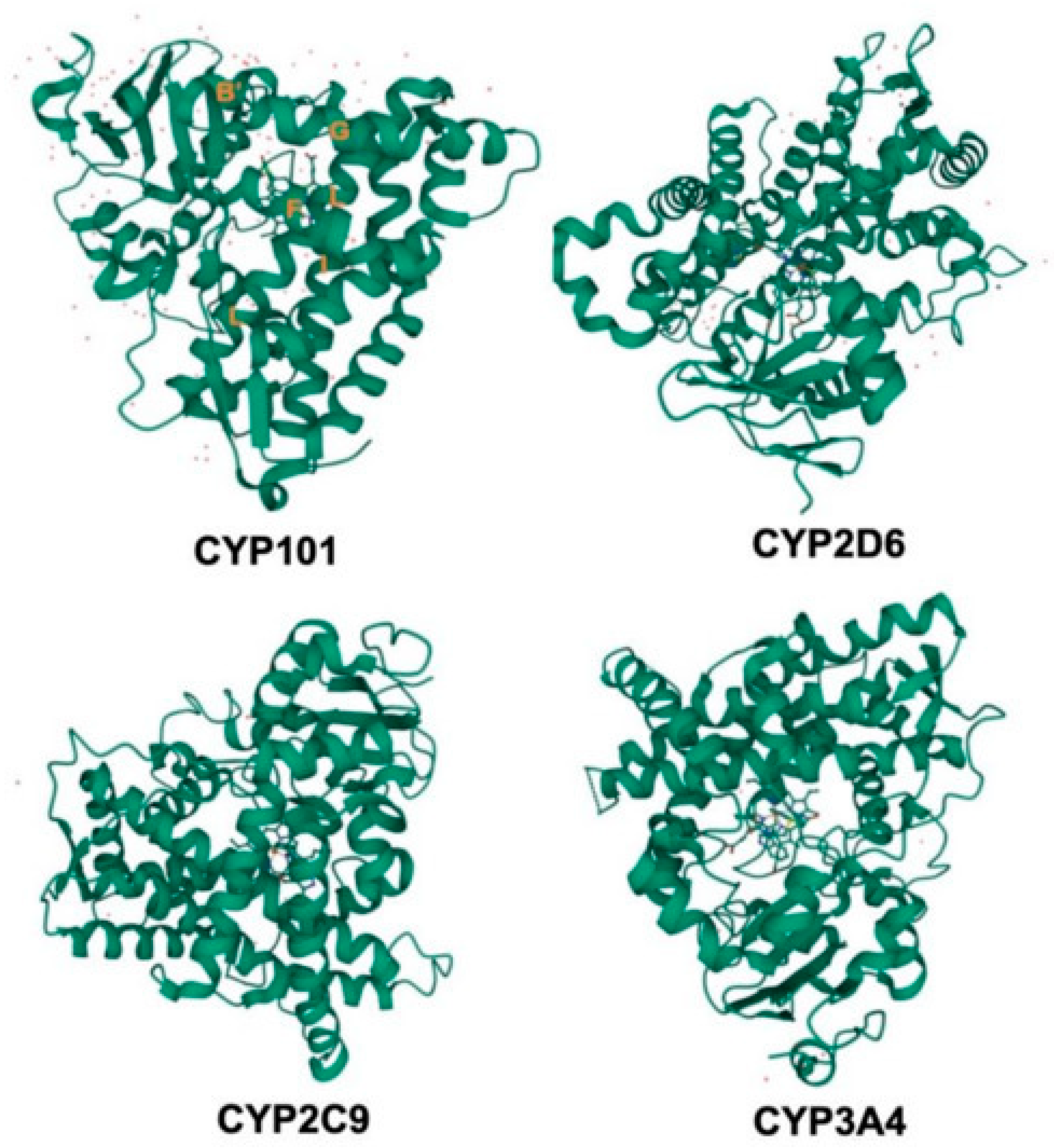
2.1. Structure and Function of CYP450 Enzymes

CYP450 enzymes (Figure 1) constitute a vast superfamily of membrane-bound hemoproteins that play a central role in the metabolism of endogenous compounds and the biotransformation of xenobiotics, including the majority of clinically used drugs [14]. These enzymes catalyze phase I metabolic reactions, predominantly oxidative processes such as hydroxylation, epoxidation, and dealkylation, which often precede phase II conjugation and facilitate elimination [15]. CYP enzymes are most abundantly expressed in the liver, particularly in the smooth endoplasmic reticulum, but are also present in extrahepatic tissues such as the intestine, lung, brain, and kidneys, where they contribute to tissue-specific metabolism and homeostasis [16].
Structurally, CYP enzymes are composed of approximately 400–500 amino acid residues and contain a conserved heme prosthetic group within the active site. The heme iron, in its ferric (Fe3+) state, is coordinated by a cysteine thiolate as the axial ligand and participates in a highly specialized catalytic cycle involving electron transfer from nicotinamide adenine dinucleotide phosphate via cytochrome P450 reductase [17,18]. In the absence of a substrate, a water molecule occupies the sixth coordination site of the heme iron, maintaining a low-spin state. Upon substrate binding, this water molecule is displaced, leading to a shift to a high-spin state that facilitates the reduction of Fe3+ to Fe2+ and the subsequent binding of molecular oxygen. This dynamic coordination change is essential for catalytic activation and is a hallmark of CYP enzymology [1].
The catalytic cycle of CYP enzymes involves a monooxygenase reaction in which one atom of molecular oxygen is incorporated into the substrate while the other is reduced to water. This process generates highly reactive intermediates such as Compound I, a ferryl–oxo porphyrin π-cation radical species that abstracts hydrogen from the substrate, leading to hydroxylated or otherwise oxidized products. Such transformations are critical for drug detoxification but can also result in the formation of reactive metabolites with potential toxicological consequences [17]. The diversity of CYP isoforms underlies their broad substrate specificity. In humans, over 57 functional CYP genes have been identified, with the majority of drug metabolism mediated by just a few key isoforms, notably CYP3A4, CYP2D6, CYP2C9, CYP2C19, and CYP1A2 [19]. These enzymes are responsible for the metabolism of approximately 90–95% of all marketed drugs [19]. CYP3A4 alone accounts for the oxidative clearance of nearly 50% of pharmaceuticals and exhibits significant interindividual variability due to genetic polymorphisms, environmental inducers, inhibitors, and disease states [20].
Importantly, CYP enzymes not only mediate metabolism but also act as potential sources of drug-drug interactions [21]. Inhibition of CYP activity—whether reversible, time-dependent, or mechanism-based—can result in elevated plasma concentrations of co-administered drugs, leading to toxicity. Conversely, induction can lower drug levels and compromise therapeutic efficacy. The functional integrity of these enzymes, therefore, is a critical determinant of pharmacokinetic behavior and clinical outcome, particularly in therapeutic areas such as oncology where polypharmacy is the norm and the therapeutic window is narrow [22]. CYP450 enzymes are anchored to cellular membranes, primarily the smooth endoplasmic reticulum in hepatocytes, through a hydrophobic N-terminal domain. This membrane association not only stabilizes the enzyme structure but also facilitates interactions with partner proteins such as cytochrome P450 reductase and cytochrome b₅, which are necessary for electron transfer during catalysis [23]. In eukaryotic cells, CYP enzymes rely on electrons donated by NADPH via cytochrome P450 reductase, a flavoprotein containing flavin adenine dinucleotide (FAD) and flavin mononucleotide (FMN) prosthetic groups, to complete the catalytic cycle [19]. In certain cases, such as the biosynthesis of steroid hormones in mitochondria, electron transfer instead proceeds through adrenodoxin and adrenodoxin reductase, highlighting the compartmentalized nature of CYP function [19].
The catalytic versatility of CYP enzymes is largely attributed to the flexible architecture of their active sites, which can accommodate a wide range of substrates, from small molecules to bulky lipophilic drugs. The heme prosthetic group at the core of the active site facilitates the activation of molecular oxygen and the formation of high-energy intermediates that enable the oxidation of otherwise inert C–H bonds [24]. Structural studies, including X-ray crystallography, have demonstrated that substrate binding induces conformational changes that align the target atom for optimal oxidation while simultaneously displacing the axial water ligand from the heme iron, thereby triggering the catalytic cycle [25]. Classification of CYP enzymes reflects their evolutionary divergence and functional specialization. The enzymes are grouped into families and subfamilies based on amino acid sequence identity, with the designation “CYP” followed by a number (family), a letter (subfamily), and another number (individual enzyme), e.g., CYP3A4. Families 1 through 4 are primarily involved in xenobiotic metabolism, while other families, such as CYP7, CYP11, and CYP17, are engaged in the biosynthesis and catabolism of endogenous substrates, including steroids, bile acids, and fat-soluble vitamins [26]. Notably, some CYP enzymes are considered “moonlighting” proteins, performing multiple distinct physiological functions in different tissues or catalyzing different reactions depending on the context—CYP7B1, for example, functions in steroid metabolism in the brain and in bile acid synthesis in the liver [27].
CYP450 enzymes are extremely well conserved, membrane-bound hemoproteins whose structural features expressly define their functional abilities in drug metabolism. The typical CYP450 protein consists of approximately 400–500 amino acid residues folded into a predominantly α-helical conformation, with some β-sheet areas contributing to the stability of the global fold [26]. One of the characteristics of the enzyme is the deeply buried active site centered around a heme prosthetic group that is absolutely necessary for catalytic activity. The iron atom of the heme group is coordinated by a conserved cysteine residue, which acts as the axial ligand and facilitates the complex catalytic cycle of substrate oxidation [25].
Structurally, CYP450 enzymes exhibit a conserved fold with a series of α-helices (designated A–L) and flexible loop regions (e.g., the FG-loop) that are critical for substrate access, binding, and product release. The helix packing forms a hydrophobic substrate-binding cavity whose shape and volume may vary significantly among isoforms, thereby explaining the broad substrate specificity of the enzyme family. The FG-loop, in particular, plays a starring role in conformational changes that regulate the size and exposure of the substrate-binding pocket to manage the kinetics and specificity of the enzyme reaction. Upon substrate entry, binding tends to displace a water ligand coordinated to the ferric iron of the heme in favor of a low-spin to high-spin transition, which facilitates electron transfer and molecular oxygen activation. This conformational change is essential to start the monooxygenase reaction cycle. The process terminates with the formation of a highly reactive ferryl-oxo species (Compound I), which abstracts hydrogen from the substrate, resulting in hydroxylation or other oxidative reactions [18].
Structural flexibility also contributes to the liability of CYP enzymes towards drug inhibition. Many inhibitors are similar to the spatial and electronic properties of the substrate to occupy the active site or allosteric pockets, thereby inhibiting substrate metabolism. Time-dependent or mechanism-based inhibitors tend to interact covalently or pseudo-covalently with the heme group or proximal residues, exploiting these structural dynamics for enzyme inactivation. The structure of the CYP450 enzyme has been extensively studied by X-ray crystallography, and a conserved general structure across species with important isoform-specific differences that dictate their metabolic profiles was disclosed [28]. Figure 1 shows a superposition of representative CYP450 enzyme structures highlighting the conserved helices, the heme group, and flexible loop areas that collectively establish substrate recognition and catalytic activity.
Functionally, CYP enzymes influence numerous physiological processes beyond drug metabolism, including the regulation of hormone synthesis, cholesterol homeostasis, arachidonic acid metabolism, and the degradation of neuroactive steroids. They also contribute to the formation of reactive oxygen species (ROS) and reactive metabolites, which, if not adequately neutralized, may lead to cellular damage, carcinogenesis, or idiosyncratic drug toxicity [26]. Given this dual role in detoxification and bioactivation, CYP enzymes are central not only to pharmacology and toxicology but also to endogenous metabolic regulation and pathophysiology.
The CYP450 enzyme system represents a highly dynamic and adaptable network of oxidative catalysts with profound implications for drug metabolism, pharmacokinetics, and clinical pharmacology. Understanding their structural determinants, catalytic mechanisms, and interaction with xenobiotics is critical for anticipating drug–drug interactions, optimizing therapeutic regimens, and guiding rational drug design. These considerations are especially relevant in the context of oncology, where many targeted therapies are metabolized or influenced by CYP enzymes—either as substrates, inhibitors, or inducers—adding complexity to polypharmacy and combination treatment strategies.

2.2. Types of CYP Inhibition

Inhibition of cytochrome P450 enzymes represents a critical mechanism by which DDIs alter the pharmacokinetics and safety profiles of therapeutics. Inhibitory interactions can lead to elevated plasma levels of drugs that are substrates for the same enzyme, potentially causing toxicity or adverse effects. The inhibition of CYP enzymes can occur through multiple mechanistic pathways, each differing in reversibility, binding dynamics, dependence on metabolic activation, and duration of effect [29]. Understanding these types of inhibition is fundamental in evaluating and predicting DDIs during drug development and clinical use.

2.2.1. Reversible Inhibition

Reversible inhibition refers to the temporary and non-covalent binding of an inhibitor to the CYP enzyme, resulting in inhibition that can be overcome by dilution or removal of the inhibitor. These interactions are characterized by rapid association and dissociation rates and typically fall into three major kinetic categories: competitive, non-competitive, and uncompetitive inhibition [15].
Competitive inhibition occurs when the inhibitor binds to the active site of the enzyme, directly competing with the substrate for access. This type of inhibition is characterized by an increase in the apparent Michaelis constant (Km) without affecting the maximal velocity (Vmax), and its extent depends on the relative concentrations and affinities of the substrate and inhibitor [30]. Many clinically relevant DDIs, such as the inhibition of CYP3A4 by ketoconazole (Table 1), fall under this mechanism [31].
Non-competitive inhibition involves binding of the inhibitor to an allosteric site distinct from the substrate-binding site. This reduces the overall enzymatic activity regardless of substrate concentration, decreasing the Vmax while Km remains unchanged. Non-competitive inhibitors are less common but can significantly alter enzyme function, particularly in systems with multiple substrates or conformational states [40]. Uncompetitive inhibition arises when the inhibitor binds only to the enzyme-substrate complex, further stabilizing it and preventing turnover. This rare type of inhibition reduces both Km and Vmax and is typically observed in specific enzymatic contexts or artificial systems rather than physiologically [41]. These types of inhibition are often detected and quantified in vitro using kinetic assays, enabling calculation of inhibition constants (Ki) and establishing concentration-response relationships for predictive DDI modeling.

2.2.2. Irreversible Inhibition

Alternatively to reversible mechanisms, irreversible inhibition involves the formation of a stable covalent bond between the inhibitor and the enzyme, resulting in permanent loss of enzymatic activity. Recovery of function requires de novo synthesis of the enzyme, which can take several days, depending on the enzyme’s turnover rate and tissue expression. Irreversible inhibitors often act by covalently modifying the heme group or amino acid residues within the active site [42]. Classic examples include certain alkylating agents or electrophilic metabolites formed from prodrugs or environmental toxins.

2.2.3. Pseudo-Irreversible Inhibition

Pseudo-irreversible or quasi-irreversible inhibition is a distinct subtype in which the inhibitor forms a quasi-stable, tightly bound complex with the enzyme, often through coordination with the heme iron, without forming a covalent bond. Although technically reversible, dissociation is so slow that the inhibition appears irreversible under physiological conditions [43]. For example, some macrolide antibiotics (e.g., erythromycin) form such complexes with CYP3A4, leading to prolonged inhibition even after the drug has been cleared from plasma [44].

2.2.4. Mechanism-Based Inhibition

One of the most clinically significant and pharmacologically insidious forms of CYP inhibition is mechanism-based inhibition (MBI), also referred to as suicide inhibition [45]. In this process, the inhibitor is metabolized by the CYP enzyme to generate a reactive intermediate that covalently modifies and inactivates the enzyme. The inactivation is both time-dependent and metabolism-dependent, meaning that the inhibitory effect intensifies over time and requires active catalysis to occur [46]. MBIs are irreversible and require new enzyme synthesis for recovery. This type of inhibition is particularly dangerous in clinical settings, as it may not be evident in early-phase pharmacokinetic testing. Drugs such as bergamottin (from grapefruit juice) [47] and ritonavir [38] exhibit MBI against CYP3A4, leading to long-lasting interactions with substrates like simvastatin or midazolam.

2.2.5. Time-Dependent Inhibition

Time-dependent inhibition (TDI) refers to any inhibitory process where the extent of enzyme inhibition increases with incubation time, regardless of whether the interaction is reversible or irreversible [48]. TDI is a broader category that includes MBI but also encompasses reversible inhibitors that bind slowly or transition to tighter-binding states over time. Identification of TDI during drug development is critical, as it often signals potential for clinically relevant DDIs even when plasma concentrations of the inhibitors are low. Regulatory agencies such as the Food and Drug Administration (FDA) and the European Medicines Agency (EMA) require TDI testing as part of the in vitro DDI evaluation process [49,50].

2.2.6. Direct (Non-Metabolic) Inhibition

Direct inhibition refers to cases where the inhibitor binds to the enzyme without undergoing metabolic activation. This is most often observed with competitive or non-competitive reversible inhibitors but may also include pseudo-irreversible types. Direct inhibitors exert their effects immediately upon administration and typically show predictable concentration-response behavior [51]. Many commonly used drugs fall into this category.

2.3. CYP3A4 as a Major Target in Oncology

The broad substrate specificity and high catalytic efficiency make CYP3A4 particularly important in the pharmacokinetics of anticancer agents. In oncology, where treatment regimens frequently involve the co-administration of multiple systemic therapies, supportive medications, and adjuvant drugs, CYP3A4 emerges as a key determinant of drug exposure, therapeutic efficacy, and the risk of toxicity. Many of the novel targeted anticancer therapies, especially oral kinase inhibitors and PARP inhibitors, are metabolized by CYP3A4, and some also act as inhibitors or inducers of this enzyme. This dual role—as both a substrate and a perpetrator—creates a complex network of potential DDIs that must be carefully managed. The risk is further amplified in patients with compromised hepatic function or polypharmacy, where altered enzyme capacity can exacerbate the pharmacokinetic variability of narrow-therapeutic-index drugs. Table 2 shows some of the oncology medications and their relationship with CYP enzymes.
CYP3A4 is localized not only in hepatocytes but also in enterocytes lining the small intestine, contributing significantly to first-pass metabolism. This dual localization introduces another layer of complexity, as inhibition of intestinal CYP3A4 by co-administered drugs or dietary components (such as furanocoumarins in grapefruit juice) can dramatically increase systemic drug exposure, even without changes in hepatic clearance. Such effects have been documented with drugs like midazolam, simvastatin, and, increasingly, oral tyrosine kinase inhibitors such as lapatinib and osimertinib [62]. In oncology, CYP3A4 inhibition can have both therapeutic and adverse consequences. On the one hand, inhibitors such as ritonavir or cobicistat have been deliberately used to boost plasma concentrations of co-administered drugs (a strategy well established in antiviral therapy and now explored in oncology). Contrariwise, unintentional inhibition by co-medications can lead to life-threatening toxicities. For example, co-administration of CYP3A4 inhibitors with drugs like vincristine, which has a narrow therapeutic window and is extensively metabolized by CYP3A4, has been associated with neurotoxicity, gastrointestinal toxicity, and increased myelosuppression [63].
MBI of CYP3A4 is of particular concern in the oncology setting. Several kinase inhibitors, including ibrutinib (Figure 2) and neratinib, and certain PARP inhibitors, such as olaparib, have been implicated as time-dependent or mechanism-based inhibitors of CYP3A4. These interactions are not always predictable from static in vitro assays and may only manifest after repeated dosing, making real-world pharmacovigilance and physiologically based pharmacokinetic (PBPK) modeling crucial tools for anticipating such effects [64]. Interindividual variability in CYP3A4 activity—driven by genetic polymorphisms, epigenetic regulation, and environmental factors—adds to the unpredictability of drug response in cancer patients. Although CYP3A4 polymorphisms are less functionally impactful than those of CYP2D6 or CYP2C19, variability in expression and inducibility can still meaningfully influence treatment outcomes [65].
CYP3A4 is a central node in the pharmacokinetic landscape of oncology therapeutics. Its role as a major metabolic pathway for many anticancer agents, combined with its susceptibility to inhibition and induction, underscores the need for careful assessment of DDIs in clinical oncology. Therapeutic drug monitoring, clinical decision-support tools, and increased awareness of CYP3A4-mediated interactions are essential to ensure safe and effective use of both established and emerging oncologic treatments.

3. KIs as Perpetrators of CYP Inhibition

KIs have revolutionized cancer therapy by enabling targeted modulation of aberrant signaling pathways involved in tumor growth and survival [66]. These small-molecule drugs are designed to selectively bind to the adenosine triphosphate-binding sites of protein kinases, thereby inhibiting phosphorylation cascades that drive oncogenic processes [67]. However, despite their therapeutic specificity, many kinase inhibitors interact with drug-metabolizing enzymes, particularly cytochrome P450 isoforms, most notably CYP3A4. As a result, kinase inhibitors are giving rise to clinically significant DDIs [68]. A defining pharmacokinetic feature of many KIs is their high lipophilicity and metabolic instability, which predispose them to extensive hepatic metabolism [69,70]. CYP3A4 is the predominant enzyme involved in their biotransformation, but paradoxically, several KIs also exhibit inhibitory activity toward the same enzyme [62]. This dual role creates complex interaction profiles, especially in patients receiving polypharmacy or combination therapies where overlapping metabolic pathways exist. Inhibition can occur via multiple mechanisms, including reversible competitive inhibition, TDI, and, in some cases, MBI (Table 3).
For example, abemaciclib (Figure 3), a CDK4/6 inhibitor used in hormone receptor-positive breast cancer, is both a major CYP3A4 substrate and a time-dependent inhibitor of the enzyme [71]. Its primary metabolite, formed via CYP3A4-mediated oxidation, may contribute to its inhibitory activity. Clinical studies have shown that co-administration of abemaciclib with strong CYP3A4 inhibitors like clarithromycin or ketoconazole can significantly increase plasma concentrations of the drug, necessitating dose reductions to mitigate toxicity [57]. Similarly, ibrutinib, a Bruton’s tyrosine kinase (BTK) inhibitor used in chronic lymphocytic leukemia and mantle cell lymphoma, is a substrate of CYP3A4 but also exerts inhibitory effects that may be quasi-irreversible [72]. Co-administration with CYP3A4 inhibitors such as voriconazole or posaconazole has been shown to increase ibrutinib exposure by several-fold, raising the risk of bleeding and cardiac arrhythmias [78]. Furthermore, ibrutinib’s own potential to inhibit CYP3A4 raises concern when it is combined with other substrates of the enzyme, particularly those with narrow therapeutic windows. Another example is osimertinib, an EGFR inhibitor indicated for non-small cell lung cancer with T790M mutations. While primarily a substrate of CYP3A4, in vitro data suggest it also has weak to moderate inhibitory activity on the enzyme. Although not a strong inhibitor clinically, this dual interaction profile becomes relevant when osimertinib is used in combination regimens with other metabolically vulnerable agents [73].
In certain cases, kinase inhibitors may exhibit MBI, where the drug is metabolically activated to a reactive intermediate that covalently binds and inactivates the enzyme. This form of inhibition is particularly concerning because it leads to prolonged enzyme suppression even after the inhibitor is cleared from systemic circulation. Although not all KIs undergo MBI, the possibility must be evaluated early in drug development, particularly when time-dependent inhibition is observed in vitro. Importantly, CYP inhibition by kinase inhibitors does not only affect co-administered drugs. Inhibitory effects can also alter the metabolic clearance of the inhibitor itself, leading to nonlinear pharmacokinetics and drug accumulation. This self-inhibition phenomenon is observed with drugs like lapatinib, which not only inhibits but is also extensively metabolized by CYP3A4 [74]. Such behavior complicates dose-response relationships and increases interpatient variability in drug exposure. When significant inhibition is detected, especially with TDI or MBI characteristics, the FDA’s and EMA’s labeling recommendations often include specific guidance on avoiding strong CYP3A4 inhibitors or inducers, as well as potential dose adjustments.

4. PARP Inhibitors as Perpetrators of CYP Inhibition

PARP inhibitors represent a novel class of targeted anticancer therapies that exploit defects in DNA damage repair pathways, particularly in BRCA1/2-mutated cancers [79]. By inhibiting PARP enzymes involved in base excision repair, these agents induce synthetic lethality in tumor cells with homologous recombination deficiency [80,81]. While their therapeutic impact is substantial, particularly in ovarian, breast, pancreatic, and prostate cancers, emerging evidence suggests that PARP inhibitors can also modulate drug metabolism, particularly via interactions with cytochrome P450 enzymes such as CYP3A4. This introduces the potential for clinically significant DDIs, especially in oncology patients receiving multiple concomitant therapies.
Among the currently approved PARP inhibitors, olaparib, niraparib, rucaparib, and talazoparib are the most widely used. These drugs differ not only in their PARP isoform selectivity and pharmacokinetics but also in their potential to inhibit metabolic enzymes and transporters. Olaparib, the first PARP inhibitor to gain regulatory approval, is primarily metabolized by CYP3A4 and has been shown to act as a weak to moderate inhibitor of the same enzyme in vitro [75]. Clinical studies have confirmed that co-administration of olaparib with strong CYP3A4 inhibitors (e.g., itraconazole) increases olaparib exposure (area under the curve—AUC) significantly, necessitating dose adjustments [82]. Moreover, olaparib has demonstrated TDI properties, suggesting a potential for delayed or sustained inhibition of CYP3A4 in vivo, particularly after repeated dosing [76].
Niraparib, in contrast, is primarily metabolized via non-CYP enzymatic pathways (e.g., carboxylesterases), and its interaction with CYP450 enzymes is minimal [77]. However, in vitro data have revealed that niraparib can inhibit CYP3A4 and CYP2C8 at higher concentrations, though the clinical relevance of these findings remains uncertain [82]. Due to its unique metabolic profile, niraparib is generally considered to pose a lower risk for CYP-mediated DDIs compared to other PARP inhibitors. Rucaparib presents a more complex interaction profile. While it undergoes metabolism via multiple pathways, including CYP1A2 and CYP3A4, it has been shown to act as a moderate inhibitor of CYP1A2, CYP2C9, and CYP3A4 [76]. Notably, rucaparib inhibits CYP3A4 in a direct and time-dependent manner and also interacts with multiple transporter proteins such as BCRP and P-gp [83]. These combined effects can significantly influence the pharmacokinetics of co-administered drugs, particularly in combination regimens used in advanced cancers.
Talazoparib is distinct from other PARP inhibitors in that it undergoes minimal hepatic metabolism and is primarily eliminated unchanged via renal excretion. Accordingly, its potential to inhibit CYP enzymes is considered low [57]. However, data on talazoparib’s interaction with CYP3A4 are limited, and ongoing clinical experience may uncover additional inhibitory mechanisms not yet fully characterized.
From a mechanistic perspective, CYP inhibition by PARP inhibitors may involve direct reversible binding to the active site or time-dependent inhibition through reactive intermediates, particularly in the case of olaparib and rucaparib. However, unlike many kinase inhibitors, mechanism-based (irreversible) inactivation of CYP enzymes by PARP inhibitors has not been conclusively demonstrated. Nonetheless, given the increasing use of these agents in combination with chemotherapeutics, antiemetics, antifungals, and other supportive medications—all of which may be CYP3A4 substrates or inhibitors—the potential for metabolic DDIs remains clinically relevant.
Furthermore, many patients treated with PARP inhibitors are elderly, comorbid, and exposed to fluctuating renal and hepatic function, all of which can amplify the impact of even mild CYP inhibition [84,85]. Given that CYP3A4 plays a central role in the metabolism of immunosuppressants, statins, anticoagulants, and corticosteroids, co-administration with PARP inhibitors may necessitate additional therapeutic drug monitoring (TDM) and dose adjustments. While PARP inhibitors are not as potent CYP inhibitors as some kinase inhibitors, several members of this class, particularly olaparib and rucaparib, exhibit moderate and clinically relevant CYP3A4 inhibitory activity. The risk of drug–drug interactions should be evaluated during treatment planning, especially in polypharmacy settings. As the use of PARP inhibitors expands across tumor types and into earlier lines of therapy, greater attention to their metabolic interaction profiles is essential to optimize treatment safety and efficacy.

5. Conclusions

CYP450 enzyme inhibition remains a central mechanism through which clinically relevant drug–drug interactions arise, especially in complex therapeutic areas such as oncology. This review has outlined the structural and functional basis of CYP450 activity, with particular emphasis on the diverse types of enzymatic inhibition—including reversible, time-dependent, mechanism-based, and pseudo-irreversible inhibition—and their clinical consequences. KIs and PARP inhibitors, two classes of targeted anticancer agents, have been shown to not only undergo metabolism via CYP450 enzymes, particularly CYP3A4, but also act as perpetrators of inhibition. While kinase inhibitors frequently exert time-dependent or mechanism-based inhibition, PARP inhibitors such as olaparib and rucaparib demonstrate moderate reversible and time-dependent effects, posing a variable but notable risk for pharmacokinetic interactions.
Given the widespread expression of CYP3A4, its role in first-pass and systemic metabolism, and the increasing use of polypharmacy in oncology patients, careful assessment of CYP-mediated interactions is essential. PPBPK modeling, TDM, and regulatory guidance must be integrated into the clinical decision-making process to ensure optimal treatment outcomes and minimize toxicity. As targeted therapies continue to expand across tumor types and treatment settings, understanding their inhibitory potential on CYP450 enzymes will remain vital for personalized oncology care and safe multidrug regimens.

Author Contributions

Conceptualization, B.T.; methodology, M.K.; validation, J.B.; formal analysis, J.B.; investigation, M.K.; data curation, I.Ć. and B.T.; writing—original draft preparation, M.K. and B.T.; writing—review and editing, J.B.; supervision, I.Ć. All authors have read and agreed to the published version of the manuscript.

Funding

This research received no external funding.

Data Availability Statement

Additional data can be obtained upon request from authors.

Conflicts of Interest

The authors declare no conflicts of interest.

Abbreviations

The following abbreviations are used in this manuscript:
AUCArea Under the Curve
BTKBruton’s Tyrosine Kinase
CYP450Cytochrome P450
DDI Drug–Drug Interaction
EMA European Medicines Agency
EGFREpidermal Growth Factor Receptor
FDAFood and Drug Administration
FMN Flavin Mononucleotide
FADFlavin Adenine Dinucleotide
HR+Hormone Receptor Positive
KIKinase Inhibitor
MBIMechanism-Based Inhibition
NSCLCNon-Small Cell Lung Cancer
PARPPoly-Adenosine Diphosphate Ribose Polymerase
PBPKPhysiologically Based Pharmacokinetic (modeling)
ROSReactive Oxygen Species
TDITime-Dependent Inhibition
TDMTherapeutic Drug Monitoring

References

  1. Zhao, M.; Ma, J.; Li, M.; Zhang, Y.; Jiang, B.; Zhao, X.; Huai, C.; Shen, L.; Zhang, N.; He, L.; et al. Cytochrome P450 Enzymes and Drug Metabolism in Humans. Int. J. Mol. Sci. 2021, 22, 12808. [Google Scholar] [CrossRef] [PubMed]
  2. Kato, M. Intestinal first-pass metabolism of CYP3A4 substrates. Drug Metab. Pharmacokinet. 2008, 23, 87–94. [Google Scholar] [CrossRef] [PubMed]
  3. Gougis, P.; Hilmi, M.; Geraud, A.; Mir, O.; Funck-Brentano, C. Potential cytochrome P450-mediated pharmacokinetic interactions between herbs, food, and dietary supplements and cancer treatments. Crit. Rev. Oncol. Hematol. 2021, 166, 103342. [Google Scholar] [CrossRef] [PubMed]
  4. Zurth, C.; Koskinen, M.; Fricke, R.; Prien, O.; Korjamo, T.; Graudenz, K.; Denner, K.; Bairlein, M.; von Bühler, C.J.; Wilkinson, G.; et al. Drug-Drug Interaction Potential of Darolutamide: In Vitro and Clinical Studies. Eur. J. Drug Metab. Pharmacokinet. 2019, 44, 747–759. [Google Scholar] [CrossRef]
  5. Shirasaka, Y.; Sager, J.E.; Lutz, J.D.; Davis, C.; Isoherranen, N. Inhibition of CYP2C19 and CYP3A4 by omeprazole metabolites and their contribution to drug-drug interactions. Drug Metab. Dispos. 2013, 41, 1414–1424. [Google Scholar] [CrossRef]
  6. Wang, X.; Dowty, M.E.; Tripathy, S.; Le, V.H.; Huh, Y.; Curto, M.; Winton, J.A.; O’Gorman, M.T.; Chan, G.; Malhotra, B.K. Assessment of the Effects of Abrocitinib on the Pharmacokinetics of Probe Substrates of Cytochrome P450 1A2, 2B6 and 2C19 Enzymes and Hormonal Oral Contraceptives in Healthy Individuals. Eur. J. Drug Metab. Pharmacokinet. 2024, 49, 367–381. [Google Scholar] [CrossRef]
  7. Sharma, M.; Loh, K.P.; Nightingale, G.; Mohile, S.G.; Holmes, H.M. Polypharmacy and potentially inappropriate medication use in geriatric oncology. J. Geriatr. Oncol. 2016, 7, 346–353. [Google Scholar] [CrossRef]
  8. Sevrioukova, I.F.; Poulos, T.L. Current Approaches for Investigating and Predicting Cytochrome P450 3A4-Ligand Interactions. Adv. Exp. Med. Biol. 2015, 851, 83–105. [Google Scholar]
  9. Ye, F.; Dewanjee, S.; Li, Y.; Jha, N.K.; Chen, Z.S.; Kumar, A.; Vishakha; Behl, T.; Jha, S.K.; Tang, H. Advancements in clinical aspects of targeted therapy and immunotherapy in breast cancer. Mol. Cancer 2023, 22, 105. [Google Scholar] [CrossRef]
  10. Wendl, T.; Frechen, S.; Gerisch, M.; Heinig, R.; Eissing, T. Physiologically-based pharmacokinetic modeling to predict CYP3A4-mediated drug-drug interactions of finerenone. CPT Pharmacomet. Syst. Pharmacol. 2022, 11, 199–211. [Google Scholar] [CrossRef]
  11. Stodtmann, S.; Nuthalapati, S.; Eckert, D.; Kasichayanula, S.; Joshi, R.; Bach, B.A.; Mensing, S.; Menon, R.; Xiong, H. A Population Pharmacokinetic Meta-Analysis of Veliparib, a PARP Inhibitor, Across Phase 1/2/3 Trials in Cancer Patients. J. Clin. Pharmacol. 2021, 61, 1195–1205. [Google Scholar] [CrossRef] [PubMed]
  12. Stanisławiak-Rudowicz, J.; Karbownik, A.; Szkutnik-Fiedler, D.; Otto, F.; Grabowski, T.; Wolc, A.; Grześkowiak, E.; Szałek, E. Bidirectional pharmacokinetic drug interactions between olaparib and metformin. Cancer Chemother. Pharmacol. 2024, 93, 79–88. [Google Scholar] [CrossRef] [PubMed]
  13. Beunk, L.; Nijenhuis, M.; Soree, B.; de Boer-Veger, N.J.; Buunk, A.M.; Guchelaar, H.J.; Houwink, E.J.F.; Risselada, A.; Rongen, G.A.P.J.M.; van Schaik, R.H.N.; et al. Dutch Pharmacogenetics Working Group (DPWG) guideline for the gene-drug interaction between CYP2D6, CYP3A4 and CYP1A2 and antipsychotics. Eur. J. Hum. Genet. 2024, 32, 278–285. [Google Scholar] [CrossRef] [PubMed]
  14. Taylor, C.; Crosby, I.; Yip, V.; Maguire, P.; Pirmohamed, M.; Turner, R.M. A Review of the Important Role of CYP2D6 in Pharmacogenomics. Genes 2020, 11, 1295. [Google Scholar] [CrossRef]
  15. Bojić, M. Predklinička ispitivanja inhibicijskog i interakcijskog potencijala novih lijekova na razini citokroma P450. Farm. Glas. 2015, 71, 229–242. [Google Scholar]
  16. Stading, R.; Couroucli, X.; Lingappan, K.; Moorthy, B. The role of cytochrome P450 (CYP) enzymes in hyperoxic lung injury. Expert Opin. Drug Metab. Toxicol. 2021, 17, 171–178. [Google Scholar] [CrossRef]
  17. Rendić, S. Summary of information on human CYP enzymes: Human P450 metabolism data. Drug Metab. Rev. 2002, 34, 83–448. [Google Scholar]
  18. Laursen, T.; Jensen, K.; Møller, B.L. Conformational changes of the NADPH-dependent cytochrome P450 reductase in the course of electron transfer to cytochromes P450. Biochim. Biophys. Acta Proteins Proteom. 2011, 1814, 132–138. [Google Scholar] [CrossRef]
  19. Williams, P.A.; Cosme, J.; Vinkovic, D.M.; Ward, A.; Angove, H.C.; Day, P.J.; Vonrhein, C.; Tickle, I.J.; Jhoti, H. Crystal structures of human cytochrome P450 3A4 bound to metyrapone and progesterone. Science 2004, 305, 683–686. [Google Scholar] [CrossRef]
  20. Drug Metabolism—The Importance of Cytochrome P450 3A4. Available online: https://www.medsafe.govt.nz/profs/puarticles/march2014drugmetabolismcytochromep4503a4.htm (accessed on 29 March 2025).
  21. Mar, P.L.; Gopinathannair, R.; Gengler, B.E.; Chung, M.K.; Perez, A.; Dukes, J.; Ezekowitz, M.D.; Lakkireddy, D.; Lip, G.Y.H.; Miletello, M.; et al. Drug Interactions Affecting Oral Anticoagulant Use. Circ. Arrhythm. Electrophysiol. 2022, 15, e007956. [Google Scholar] [CrossRef]
  22. Coates, S.; Lazarus, P. Hydrocodone, Oxycodone, and Morphine Metabolism and Drug—Drug Interactions. J. Pharmacol. Exp. Ther. 2023, 387, 150–169. [Google Scholar] [CrossRef] [PubMed]
  23. Koppula, P.; Zhuang, L.; Gan, B. Cytochrome P450 reductase (POR) as a ferroptosis fuel. Protein Cell 2021, 12, 675–679. [Google Scholar] [CrossRef] [PubMed]
  24. Wójcikowski, J.; Danek, P.J.; Basińska-Ziobroń, A.; Pukło, R.; Daniel, W.A. In vitro inhibition of human cytochrome P450 enzymes by the novel atypical antipsychotic drug asenapine: A prediction of possible drug-drug interactions. Pharmacol. Rep. 2020, 72, 612–621. [Google Scholar] [CrossRef] [PubMed]
  25. Kondža, M. Mehanizam Inhibicijskoga Učinka na Citokrom P450 3A4. Ph.D. Thesis, Faculty of Pharmacy and Biochemistry, University of Zagreb, Zagreb, Croatia, 2022. [Google Scholar]
  26. Rendić, S.; Medić-Šarić, M. Metabolizam Lijekova i Odabranih Ksenobiotika, 1st ed.; Medicinska Naklada: Zagreb, Croatia, 2013. [Google Scholar]
  27. Zhao, B.; Waterman, M.R. Moonlighting cytochrome P450 monooxygenases. IUBMB Life 2011, 63, 473–477. [Google Scholar] [CrossRef]
  28. Veith, A.; Moorthy, B. Role of cytochrome P450s in the generation and metabolism of reactive oxygen species. Curr. Opin. Toxicol. 2018, 7, 44–51. [Google Scholar] [CrossRef]
  29. Kondža, M.; Brizić, I.; Jokić, S. Flavonoids as CYP3A4 inhibitors in vitro. Biomedicines 2024, 12, 644. [Google Scholar] [CrossRef]
  30. Bjelaković, G.; Stojanović, I.; Bjelaković, G.B.; Pavlović, D.; Kocić, G.; Daković-Milić, A. Competitive inhibitors of enzymes and their therapeutic application. Med. Biol. 2002, 9, 201–206. [Google Scholar]
  31. Usmani, K.A.; Tang, J. Human cytochrome P450: Metabolism of testosterone by CYP3A4 and inhibition by ketoconazole. Curr. Protoc. Toxicol. 2004, 20, 4–13. [Google Scholar] [CrossRef]
  32. Deodhar, M.; Rihani, S.B.A.; Darakjian, L.; Turgeon, J.; Michaud, V. Assessing the Mechanism of Fluoxetine-Mediated CYP2D6 Inhibition. Pharmaceutics 2021, 13, 148. [Google Scholar] [CrossRef]
  33. Williams, J. Enzyme inhibition and induction. Anaesth. Intensive Care Med. 2008, 9, 165–166. [Google Scholar] [CrossRef]
  34. Wang, X.; Dowty, M.E.; Wouters, A.; Tatulych, S.; Connell, C.A.; Le, V.H.; Tripathy, S.; O’Gorman, M.T.; Winton, J.A.; Yin, N.; et al. Assessment of the Effects of Inhibition or Induction of CYP2C19 and CYP2C9 Enzymes, or Inhibition of OAT3, on the Pharmacokinetics of Abrocitinib and Its Metabolites in Healthy Individuals. Eur. J. Drug Metab. Pharmacokinet. 2021, 47, 419–429. [Google Scholar] [CrossRef] [PubMed]
  35. Pai, M.P.; Graci, D.M.; Amsden, G.W. Macrolide drug interactions: An update. Ann. Pharmacother. 2000, 34, 495–513. [Google Scholar] [CrossRef] [PubMed]
  36. Westphal, J.F. Macrolide-induced clinically relevant drug interactions with cytochrome P-450 (CYP) 3A4: An update focused on clarithromycin, azithromycin and dirithromycin. Br. J. Clin. Pharmacol. 2000, 50, 285–295. [Google Scholar] [CrossRef] [PubMed]
  37. Fairman, D.A.; Collins, C.; Chapple, S. Progress curve analysis of CYP1A2 inhibition: A more informative approach to the assessment of mechanism-based inactivation? Drug Metab. Dispos. 2007, 35, 2159–2165. [Google Scholar] [CrossRef]
  38. Loos, N.H.C.; Beijnen, J.H.; Schinkel, A.H. The Mechanism-Based Inactivation of CYP3A4 by Ritonavir: What Mechanism? Int. J. Mol. Sci. 2022, 23, 9866. [Google Scholar] [CrossRef]
  39. Wang, Y.H.; Jones, D.R.; Hall, S.D. Prediction of cytochrome P450 3A inhibition by verapamil enantiomers and their metabolites. Drug Metab. Dispos. 2004, 32, 259–266. [Google Scholar] [CrossRef]
  40. Blat, Y. Non-competitive inhibition by active site binders. Chem. Biol. Drug Des. 2010, 75, 535–540. [Google Scholar] [CrossRef]
  41. Boulton, S.; Selvaratnam, R.; Blondeau, J.P.; Lezoualc’h, F.; Melacini, G. Mechanism of selective enzyme inhibition through uncompetitive regulation of an allosteric agonist. J. Am. Chem. Soc. 2018, 140, 9624–9637. [Google Scholar] [CrossRef]
  42. Johnson, D.S.; Weerapana, E.; Cravatt, B.F. Strategies for discovering and derisking covalent, irreversible enzyme inhibitors. Future Med. Chem. 2010, 2, 949–964. [Google Scholar] [CrossRef]
  43. Kim, H.G.; Lee, H.S.; Jeon, J.S.; Choi, Y.J.; Choi, Y.J.; Yoo, S.Y.; Kim, S.K. Quasi-irreversible inhibition of CYP2D6 by berberine. Pharmaceutics 2020, 12, 916. [Google Scholar] [CrossRef]
  44. Orlando, R.; De Martin, S.; Pegoraro, P.; Quintieri, L.; Palatini, P. Irreversible CYP3A inhibition accompanied by plasma protein–binding displacement: A comparative analysis in subjects with normal and impaired liver function. Clin. Pharmacol. Ther. 2009, 85, 319–326. [Google Scholar] [CrossRef] [PubMed]
  45. Zhou, S.; Chan, S.Y.; Goh, B.C.; Chan, E.; Duan, W.; Huang, M.; McLeod, H.L. Mechanism-based inhibition of cytochrome P450 3A4 by therapeutic drugs. Clin. Pharmacokinet. 2005, 44, 279–304. [Google Scholar] [CrossRef] [PubMed]
  46. Bojić, M.; Kondža, M.; Rimac, H.; Benković, G.; Maleš, Ž. The effect of flavonoid aglycones on the CYP1A2, CYP2A6, CYP2C8 and CYP2D6 enzymes activity. Molecules 2019, 24, 3174. [Google Scholar] [CrossRef] [PubMed]
  47. Bailey, D.G.; Dresser, G.K.; Bend, J.R. Bergamottin, lime juice, and red wine as inhibitors of cytochrome P450 3A4 activity: Comparison with grapefruit juice. Clin. Pharmacol. Ther. 2003, 73, 529–537. [Google Scholar] [CrossRef]
  48. Kondža, M.; Mandić, M.; Ivančić, I.; Vladimir-Knežević, S.; Brizić, I. Artemisia annua L. Extracts Irreversibly Inhibit the Activity of CYP2B6 and CYP3A4 Enzymes. Biomedicines 2023, 11, 232. [Google Scholar] [CrossRef]
  49. Food and Drug Administration. ICH M12 Drug Interaction Final Guidance—In Vitro DDI Assessments. Available online: https://www.fda.gov/media/184437/download (accessed on 26 April 2025).
  50. European Medicines Agency. Guideline on the investigation of drug interactions. Available online: https://www.ema.europa.eu/en/documents/scientific-guideline/guideline-investigation-drug-interactions-revision-1_en.pdf (accessed on 26 April 2025).
  51. Ochs, R.S. Understanding enzyme inhibition. J. Chem. Educ. 2000, 77, 1453. [Google Scholar] [CrossRef]
  52. Van Erp, N.P.; Gelderblom, H.; Karlsson, M.O.; Li, J.; Zhao, M.; Ouwerkerk, J.; Sparreboom, A. Influence of CYP3A4 inhibition on the steady-state pharmacokinetics of imatinib. Clin. Cancer Res. 2007, 13, 7394–7400. [Google Scholar] [CrossRef]
  53. Hamilton, M.; Wolf, J.L.; Drolet, D.W.; Fettner, S.H.; Rakhit, A.K.; Witt, K.; Lum, B.L. The effect of rifampicin, a prototypical CYP3A4 inducer, on erlotinib pharmacokinetics in healthy subjects. Cancer Chemother. Pharmacol. 2014, 73, 613–621. [Google Scholar] [CrossRef]
  54. Gao, N.; Zhang, X.; Hu, X.; Kong, Q.; Cai, J.; Hu, G.; Qian, J. The influence of CYP3A4 genetic polymorphism and proton pump inhibitors on osimertinib metabolism. Front. Pharmacol. 2022, 13, 794931. [Google Scholar] [CrossRef]
  55. Teo, Y.L.; Saetaew, M.; Chanthawong, S.; Yap, Y.S.; Chan, E.C.Y.; Ho, H.K.; Chan, A. Effect of CYP3A4 inducer dexamethasone on hepatotoxicity of lapatinib: Clinical and in vitro evidence. Breast Cancer Res. Treat. 2012, 133, 703–711. [Google Scholar] [CrossRef]
  56. De Zwart, L.; Snoeys, J.; De Jong, J.; Sukbuntherng, J.; Mannaert, E.; Monshouwer, M. Ibrutinib dosing strategies based on interaction potential of CYP3A4 perpetrators using physiologically based pharmacokinetic modeling. Clin. Pharmacol. Ther. 2016, 100, 548–557. [Google Scholar] [CrossRef] [PubMed]
  57. Posada, M.M.; Morse, B.L.; Turner, P.K.; Kulanthaivel, P.; Hall, S.D.; Dickinson, G.L. Predicting clinical effects of CYP3A4 modulators on abemaciclib and active metabolites exposure using physiologically based pharmacokinetic modeling. J. Clin. Pharmacol. 2020, 60, 915–930. [Google Scholar] [CrossRef] [PubMed]
  58. Otsuki, A.; Kumondai, M.; Kobayashi, D.; Kikuchi, M.; Ueki, Y.; Sato, Y.; Mano, N. Plasma venetoclax concentrations in patients with acute myeloid leukemia treated with CYP3A4 inhibitors. Yakugaku Zasshi 2024, 144, 775–779. [Google Scholar] [CrossRef] [PubMed]
  59. Lin, Q.M.; Li, Y.H.; Lu, X.R.; Wang, R.; Pang, N.H.; Xu, R.A.; Hu, G.X. Characterization of genetic variation in CYP3A4 on the metabolism of cabozantinib in vitro. Chem. Res. Toxicol. 2019, 32, 1583–1590. [Google Scholar] [CrossRef]
  60. Dutreix, C.; Munarini, F.; Lorenzo, S.; Roesel, J.; Wang, Y. Investigation into CYP3A4-mediated drug–drug interactions on midostaurin in healthy volunteers. Cancer Chemother. Pharmacol. 2013, 72, 1223–1234. [Google Scholar] [CrossRef]
  61. Gibbons, J.A.; de Vries, M.; Krauwinkel, W.; Ohtsu, Y.; Noukens, J.; van der Walt, J.S.; Ouatas, T. Pharmacokinetic drug interaction studies with enzalutamide. Clin. Pharmacokinet. 2015, 54, 1057–1069. [Google Scholar] [CrossRef]
  62. Mikus, G.; Foerster, K.I. Role of CYP3A4 in kinase inhibitor metabolism and assessment of CYP3A4 activity. Transl. Cancer Res. 2017, 6 (Suppl. S10), S1592–S1599. [Google Scholar] [CrossRef]
  63. Guilhaumou, R.; Solas, C.; Bourgarel-Rey, V.; Quaranta, S.; Rome, A.; Simon, N.; Andre, N. Impact of plasma and intracellular exposure and CYP3A4, CYP3A5, and ABCB1 genetic polymorphisms on vincristine-induced neurotoxicity. Cancer Chemother. Pharmacol. 2011, 68, 1633–1638. [Google Scholar] [CrossRef]
  64. Gertz, M.; Cartwright, C.M.; Hobbs, M.J.; Kenworthy, K.E.; Rowland, M.; Houston, J.B.; Galetin, A. Cyclosporine inhibition of hepatic and intestinal CYP3A4, uptake and efflux transporters: Application of PBPK modeling in the assessment of drug—Drug interaction potential. Pharm. Res. 2013, 30, 761–780. [Google Scholar] [CrossRef]
  65. Kato, M.; Chiba, K.; Ito, T.; Koue, T.; Sugiyama, Y. Prediction of interindividual variability in pharmacokinetics for CYP3A4 substrates in humans. Drug Metab. Pharmacokinet. 2010, 25, 367–378. [Google Scholar] [CrossRef]
  66. Ferguson, F.M.; Gray, N.S. Kinase inhibitors: The road ahead. Nat. Rev. Drug Discov. 2018, 17, 353–377. [Google Scholar] [CrossRef] [PubMed]
  67. Kannaiyan, R.; Mahadevan, D. A comprehensive review of protein kinase inhibitors for cancer therapy. Expert Rev. Anticancer Ther. 2018, 18, 1249–1270. [Google Scholar] [CrossRef] [PubMed]
  68. van Leeuwen, R.W.; van Gelder, T.; Mathijssen, R.H.; Jansman, F.G. Drug–drug interactions with tyrosine-kinase inhibitors: A clinical perspective. Lancet Oncol. 2014, 15, e315–e326. [Google Scholar] [CrossRef] [PubMed]
  69. Hartmann, J.T.; Haap, M.; Kopp, H.G.; Lipp, H.P. Tyrosine kinase inhibitors—A review on pharmacology, metabolism and side effects. Curr. Drug Metab. 2009, 10, 470–481. [Google Scholar] [CrossRef]
  70. Gabora, K.; Piciu, A.; Badulescu, I.C.; Larg, M.I.; Stoian, A.I.; Piciu, D. Current evidence on thyroid related adverse events in patients treated with protein tyrosine kinase inhibitors. Drug Metab. Rev. 2019, 51, 562–569. [Google Scholar] [CrossRef]
  71. Xu, X.Y.; Chen, J.; Chen, Z.X.; Zhang, Z.Y.; Jin, L.H.; Luo, J.C.; Qian, J.C. CYP3A4 activity variations can lead to stratified metabolism of abemaciclib. Int. J. Biol. Macromol. 2025, 304, 140836. [Google Scholar] [CrossRef]
  72. Eisenmann, E.D.; Fu, Q.; Muhowski, E.M.; Jin, Y.; Uddin, M.E.; Garrison, D.A.; Baker, S.D. Intentional modulation of ibrutinib pharmacokinetics through CYP3A inhibition. Cancer Res. Commun. 2021, 1, 79–89. [Google Scholar] [CrossRef]
  73. MacLeod, A.K.; Lin, D.; Huang, J.T.J.; McLaughlin, L.A.; Henderson, C.J.; Wolf, C.R. Identification of novel pathways of osimertinib disposition and potential implications for the outcome of lung cancer therapy. Clin. Cancer Res. 2018, 24, 2138–2147. [Google Scholar] [CrossRef]
  74. Teng, W.C.; Oh, J.W.; New, L.S.; Wahlin, M.D.; Nelson, S.D.; Ho, H.K.; Chan, E.C.Y. Mechanism-based inactivation of cytochrome P450 3A4 by lapatinib. Mol. Pharmacol. 2010, 78, 693–703. [Google Scholar] [CrossRef]
  75. McCormick, A.; Swaisland, H.; Reddy, V.P.; Learoyd, M.; Scarfe, G. In vitro evaluation of the inhibition and induction potential of olaparib, a potent poly (ADP-ribose) polymerase inhibitor, on cytochrome P450. Xenobiotica 2018, 48, 555–564. [Google Scholar] [CrossRef]
  76. Liao, M.; Beltman, J.; Giordano, H.; Harding, T.C.; Maloney, L.; Simmons, A.D.; Xiao, J.J. Clinical pharmacokinetics and pharmacodynamics of rucaparib. Clin. Pharmacokinet. 2022, 61, 1477–1493. [Google Scholar] [CrossRef] [PubMed]
  77. Zhao, D.; Long, X.; Wang, J. Metabolism-related pharmacokinetic drug–drug interactions with poly (ADP-ribose) polymerase inhibitors. Oncol. Rep. 2022, 47, 1–10. [Google Scholar] [CrossRef] [PubMed]
  78. Brüggemann, R.J.; Verheggen, R.; Boerrigter, E.; Stanzani, M.; Verweij, P.E.; Blijlevens, N.M.; Lewis, R.E. Management of drug—Drug interactions of targeted therapies for haematological malignancies and triazole antifungal drugs. Lancet Haematol. 2022, 9, e58–e72. [Google Scholar] [CrossRef] [PubMed]
  79. Dziadkowiec, K.N.; Gąsiorowska, E.; Nowak-Markwitz, E.; Jankowska, A. PARP inhibitors: Review of mechanisms of action and BRCA1/2 mutation targeting. Menopause Rev. 2016, 15, 215–219. [Google Scholar] [CrossRef]
  80. Mateo, J.; Lord, C.J.; Serra, V.; Tutt, A.; Balmaña, J.; Castroviejo-Bermejo, M.; De Bono, J.S. A decade of clinical development of PARP inhibitors in perspective. Ann. Oncol. 2019, 30, 1437–1447. [Google Scholar] [CrossRef]
  81. Dirix, L.; Swaisland, H.; Verheul, H.M.; Rottey, S.; Leunen, K.; Jerusalem, G.; Plummer, R. Effect of itraconazole and rifampin on the pharmacokinetics of olaparib in patients with advanced solid tumors: Results of two phase I open-label studies. Clin. Ther. 2016, 38, 2286–2299. [Google Scholar] [CrossRef]
  82. Bruin, M.; Sonke, G.S.; Beijnen, J.H.; Huitema, A.D.R. Pharmacokinetics and pharmacodynamics of PARP inhibitors in oncology. Clin. Pharmacokinet. 2022, 61, 13. [Google Scholar] [CrossRef]
  83. Liao, M.; Jaw-Tsai, S.; Beltman, J.; Simmons, A.D.; Harding, T.C.; Xiao, J.J. Evaluation of in vitro absorption, distribution, metabolism, and excretion and assessment of drug-drug interaction of rucaparib, an orally potent poly (ADP-ribose) polymerase inhibitor. Xenobiotica 2020, 50, 1032–1042. [Google Scholar] [CrossRef]
  84. Deshpande, P.P.; Perazella, M.A.; Jhaveri, K.D. PARP inhibitors and the kidney. J. Onco-Nephrol. 2021, 5, 42–47. [Google Scholar] [CrossRef]
  85. Graziani, G.; Szabó, C. Clinical perspectives of PARP inhibitors. Pharmacol. Res. 2005, 52, 109–118. [Google Scholar] [CrossRef]
Figure 1. Model of tertiary structure of human CYP enzymes [1].
Ddc 04 00024 g001
Figure 2. Chemical structure of ibrutinib.
Ddc 04 00024 g002
Figure 3. Chemical structure of abemaciclib.
Ddc 04 00024 g003
Table 1. Types of CYP inhibitions and their properties.
Type of InhibitionEffect on Km/VmaxClinical ExamplesRelevance in OncologyReferences
competitiveKm
Vmax unchanged
ketoconazole (CYP3A4)
fluoxetine (CYP2D6)
high (e.g., oral anticancer agents)[31,32]
non-competitiveVmax
Km unchanged
ketoconazole (CYP3A4)rare in clinical practice[33]
uncompetitiveKm
Vmax
theoretical; rare in vivono practical relevance[32]
directdepends on subtypefluconazole (CYP2C9)relevant in supportive care[34]
pseudo-irreversiblesimilar to irreversibleerythromycin (CYP3A4)may prolong cytotoxic exposure[35,36]
irreversiblepermanent enzyme inactivationfurafylline (CYP1A2)may increase toxicity of anticancer drugs[37]
mechanism-based inhibitionenzyme inactivationritonavir (CYP3A4)highly relevant with kinase inhibitors[38]
time-dependent inhibition inhibition increases over timeverapamil (CYP3A4)clinically important due to progressive effect[39]
↑—increase; ↓—decrease.
Table 2. Chosen oncology medications and CYP3A4 relationship.
Drug NameDrug Class/TargetMain Clinical UsesNotes on CYP3A4 MetabolismReferences
imatinibtyrosine kinase inhibitor (BCR-ABL)chronic myeloid leukemiaprimarily metabolized by CYP3A4; exposure can be increased by CYP3A4 inhibitors[52]
erlotinibEGFR inhibitorNSCLCextensively metabolized by CYP3A4 and CYP1A2[53]
osimertinibEGFR inhibitor (T790M mutant)NSCLC (EGFR-mutant)CYP3A4 is the main enzyme responsible for its metabolism[54]
lapatinibHER2/EGFR inhibitorHER2-positive breast cancermetabolized by CYP3A4; caution with inhibitors/inducers[55]
ibrutinibBTK inhibitorchronic lymphocytic leukemiaprimarily metabolized by CYP3A4; strong inhibitors increase AUC significantly[56]
abemaciclibCDK4/6 inhibitorHR+/HER2- breast cancermajor substrate of CYP3A4; time-dependent inhibition potential[57]
venetoclaxBCL-2 inhibitorCLL, AMLMetabolized by CYP3A4; risk of tumor lysis syndrome increased with strong inhibitors[58]
cabozantinibmulti-kinase inhibitorrenal cell carcinoma, thyroid cancerCYP3A4 is the primary metabolic pathway[59]
midostaurinFLT3 inhibitoracute myeloid leukemiaCYP3A4 extensively involved in metabolism; dose adjustment recommended with inhibitors[60]
enzalutamideandrogen receptor inhibitorprostate cancerCYP3A4 involved in metabolism; also induces CYP3A4[61]
Table 3. KIs and PARP inhibitors as CYP450 inhibitors.
Drug NameClassType of InhibitionMechanismClinical ImplicationsReferences
abemaciclibCDK4/6 inhibitorTDITDI observed in vitro; major substrate and weak inhibitorincreased exposure with CYP3A4 inhibitors; requires dose adjustment[71]
ibrutinibBTK inhibitorpseudo-irreversibleforms a strong complex with CYP3A4; also self-inhibitshigh risk of toxicity with strong CYP3A4 inhibitors (e.g., azoles)[72]
osimertinibEGFR inhibitorweak
reversible
minimal inhibition in vitro; mainly a substratemoderate interaction potential in combinations[73]
lapatinibHER2/EGFR inhibitorreversible
competitive
substrate and reversible inhibitor; may self-inhibit metabolismnonlinear PK; requires monitoring in combinations[74]
olaparibPARP inhibitorTDImoderate TDI; metabolism via CYP3A4; inhibition increases with exposurerequires dose adjustment with CYP3A4 inhibitors/inducers[75]
rucaparibPARP inhibitordirect
time-dependent
moderate inhibitor of multiple CYPs; affects metabolism and transportersincreases exposure to CYP3A4 substrates[76]
niraparibPARP inhibitorweakprimarily metabolized by non-CYP enzymeslow DDI risk via CYP450[77]
talazoparibPARP inhibitorweakminimal CYP involvement; primarily renally excretedlow potential for CYP-mediated DDIs[78]
Disclaimer/Publisher’s Note: The statements, opinions and data contained in all publications are solely those of the individual author(s) and contributor(s) and not of MDPI and/or the editor(s). MDPI and/or the editor(s) disclaim responsibility for any injury to people or property resulting from any ideas, methods, instructions or products referred to in the content.

