Article

Neuropharmacological Assessment of Sulfonamide Derivatives of Para-Aminobenzoic Acid through In Vivo and In Silico Approaches

1
Pharmaceutical Chemistry Research Laboratory I, Department of Pharmaceutical Engineering & Technology, Indian Institute of Technology, Banaras Hindu University, Varanasi 221005, India
2
Department of Drug Pharmaceutical Chemistry, Poona College of Pharmacy, Bharati Vidyapeeth (Deemed to Be University), Pune 411038, India
3
Neurotherapeutics Laboratory, Department of Pharmaceutical Engineering and Technology, Indian Institute of Technology, Banaras Hindu University, Varanasi 221005, India
4
Shobhaben Pratapbhai Patel School of Pharmacy & Technology Management, Shri Vile Parle Kelavani Mandal’s Narsee Monjee Institute of Management Studies, Mumbai 400056, India
*
Author to whom correspondence should be addressed.
Drugs Drug Candidates 2024, 3(4), 674-693; https://doi.org/10.3390/ddc3040038
Submission received: 2 August 2024 / Revised: 27 September 2024 / Accepted: 2 October 2024 / Published: 7 October 2024
(This article belongs to the Section Preclinical Research)

Abstract

:
Background/Objectives: Alzheimer’s disease (AD), a complex neurogenerative disorder, manifests as dementia and concomitant neuropsychiatric symptoms, including apathy, depression, and circadian disruption. The pathology involves a profound degeneration of the hippocampus and cerebral cortex, leading to the impairment of both short-term and long-term memory. The cholinergic hypothesis is among the various theories proposed, that assume the loss of the cholinergic tract contributes to the onset of AD and proves clinically effective in managing mild to moderate stages of the disease. This study explores the potential therapeutic efficacy of sulfonamide-based butyrylcholinesterase inhibitors in mitigating scopolamine-induced amnesia in rats. Methods: Behavioral assessments utilizing Y-maze, Barnes maze, and neurochemical assays were conducted to evaluate the effectiveness of the test compounds. Results: Results demonstrated a significant reduction in the impact of scopolamine administration on behavioral tasks at a dose of 20 mg/kg for both compounds. Correspondingly, neurochemical assays corroborated these findings. In silico docking analysis on rat butyrylcholinesterase (BChE) was performed to elucidate the binding mode of the compounds. Subsequent molecular dynamics studies unveiled the formation of stable complexes between the test compounds and rat BChE. Conclusions: These findings contribute valuable insights into the potential therapeutic role of sulfonamide-based butyrylcholinesterase inhibitors in addressing memory deficits associated with AD, emphasizing their in silico molecular interactions and stability.

1. Introduction

Alzheimer’s disease (AD) is characterized by intricate pathophysiology, notably marked by the presence of β-amyloid plaques and neurofibrillary tangles (NFT) of tau protein in the brain. The typical manifestation initiates with short-term memory loss, evolving into long-term memory impairment, disturbances in the sleep–wake cycle, mood fluctuations, confusion, language deficits, and ultimately culminating in the deterioration of bodily functions [1]. The amyloid β deposits present in interstitial spaces impede synaptic communication. Simultaneously, NFT interferes with the transportation of nutrients and other substances within neuronal cells [2]. It is anticipated that the prevalence of AD is expected to increase. The projection indicates that by the year 2050, approximately one out of every 85 individuals worldwide will be afflicted by this neurodegenerative condition [3].
The primary hypotheses regarding the disease generation and progression include amyloid-β cascades, the tau hypothesis, neuroinflammation, oxidative stress, cholinergic disruption and loss, glucose hypermetabolism, etc. In light of recent evidence, gut microbiome, bacteria-derived metabolites, and some immune and endocrinal-related pathways have shown a certain degree of correlation with disease origin and progression [4]. The emerging therapeutic targets of the disease are ‘monoamine oxidase B, glycogen synthase kinase 3β, N-methyl D-aspartate (NMDA) receptors, β secretase-1, tau kinase, matrix metalloproteases’ [5]. But, the two classes of drugs, i.e., acetylcholinesterase (AChE) inhibitors and N-methyl-D-aspartic acid (NMDA) receptor antagonists are currently being clinically used [6]. However, donepezil acts as a dual inhibitor for both AChE and BChE with IC50 values of 22 nM and 4.15 µM, respectively [7].
Davies and Maloney proposed a cholinergic hypothesis for AD in 1976. The study reported a reduction in the activity of choline acetyltransferase (ChAT) in the amygdala, cortex, and hippocampus regions in AD patients as compared to the control. ChAT is a critical enzyme for the production of acetylcholine (ACh) [8,9]. The impairment of cholinergic signaling in the CA3 neurons of the hippocampus adversely affects both information processing and memory functions [10]. Cholinergic disruption in AD results in memory loss through multiple pathways. Reduced ACh levels impair hippocampal function, synaptic plasticity, and interactions with other neurotransmitter systems. The interruption in cholinergic signaling in the cerebral cortex caused impaired ‘decision-making and attention’ [11]. The treatment of the patient with AChE inhibitors causes a slow synaptic ACh metabolism and prolongs its action. Its administration results in the improvement in ‘activities of daily living’ (ADL) such as self-care and complex higher-order thinking skills. The use of centrally acting cholinesterase (ChE) inhibitors such as donepezil, galantamine, and rivastigmine shows fewer side effects and improvement in the cognitive process in mild to moderate AD patients.
The synaptic catabolism of ACh results in the termination of its action and is governed by the ChE, i.e., ‘AChE and butyrylcholinesterase’ (BChE). The process is usually controlled by AChE in a healthy brain, with a minor role of BChE [12]. Interestingly, BChE is generally present in the ‘amygdala, hippocampus, and neocortex’ in association with glial, vascular, and neuronal cells. An AChE knockout genetic study on mice suggested that the cholinergic activity remained normal and inferred that its function was performed by BChE in the brain [13]. Another study indicated a reduction in amyloid-β fibrillar structures in the ‘cerebral cortex’ of BChE-knockout mice viz. 5XFAD/BChE-KO mice [14]. An increase in the ratio of BChE: AChE to 11:1 from 1:5 was observed in AD patients due to cholinergic neuronal loss [15,16]. Withdrawal of ethopropazine, a selective BChE inhibitor in AD patients, led to cognitive impairment. Cognitive improvement was observed after its re-administration [17].
Sulfonamide is an interesting class of compounds with a wide range of activities viz., antibacterial, antithyroid, antidiabetics, diuretics, and carbonic anhydrase inhibitors. In our earlier studies, we developed sulfonamide-based cholinesterase inhibitors (I and II). Compound I, a multifunctional compound viz. 3,6-diphenyl-1,4-bis(phenylsulfonyl) piperazine-2,5-dione derivative, displayed good AChE and BChE inhibitions. Further, a significant improvement was observed in working memory, when tested against ‘scopolamine-induced amnesia’ in mice. Compound II, a β-alanine-based sulfonamide derivative, also showed improvement in hippocampal-dependent spatial working memory in rats, when injected with amyloid-β1–42 [18]. We applied machine learning techniques for the model development to predict blood–brain barrier permeable BChE inhibitors i.e., compounds 34 and 37, as potent BChE inhibitors with IC50 below 100 nM [19]. In the present study, we evaluated these compounds against scopolamine-induced amnesia in rats to assess their pharmacodynamic effect on memory and learning (Figure 1).

2. Results

2.1. Y-Maze

2.1.1. Effects of Scopolamine (SCO) and Treatments on Spontaneous Alteration in Rats

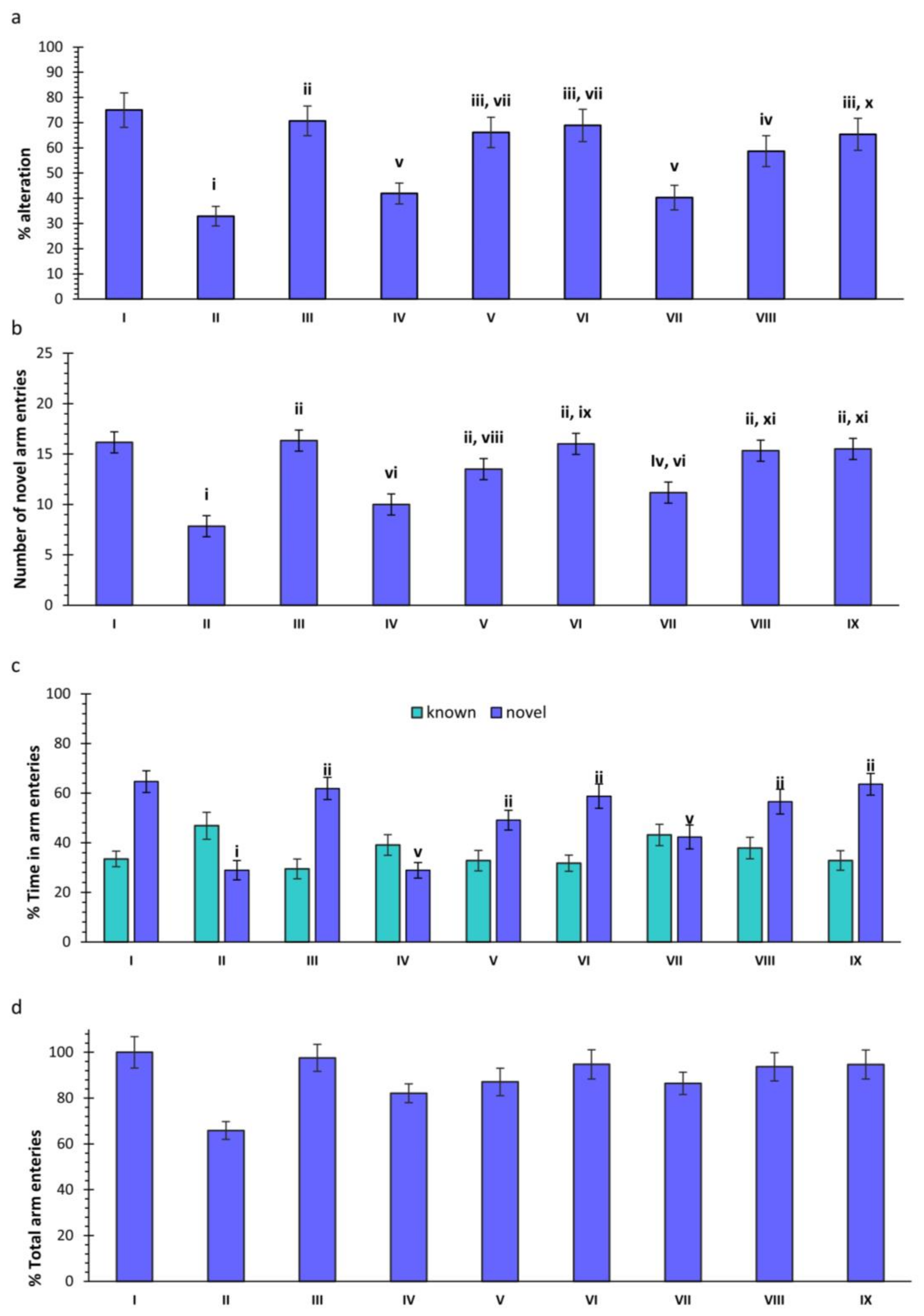
SCO treatment significantly reduced spontaneous alteration (p < 0.001, group II vs. group I). Donepezil (DNP) treatment improved spontaneous alteration significantly (p < 0.001, group III vs. group II). Compound 34 demonstrated significant improvement at 10 mg/kg dose (p < 0.01, group V vs. group II) and 20 mg/kg (p < 0.01, group VI vs. group II). Compound 37 improved spontaneous alteration at 10 mg/kg (p < 0.05, group: VII vs. group II) and 20 mg/kg (p < 0.01, group IX vs. group II). There was no significant improvement at 5 mg/kg for both compounds (Figure 2a).

2.1.2. Effect of SCO and Treatments on Novel Arm Entries in Rats

SCO treatment significantly reduced novel arm entries (p < 0.001, group II vs. group I). DNP treatment markedly improved entries (p < 0.001, group III vs. group II). Compound 34 demonstrated significant improvement at 10 and 20 mg/kg (p < 0.001, group V vs. group II and group VI vs. group II). However, at 5 mg/kg, there was no significant improvement compared to SCO, but a notable difference with the DNP treated group (p < 0.001, group IV vs. group III) was observed. Compound 37 showed a substantial increase in entries at 5 (p < 0.05, group VII vs. group II), 10 (p < 0.001, group VIII vs. group II), and 20 mg/kg (p < 0.001, group IX vs. group II) (Figure 2b).

2.1.3. Effect of SCO and Treatments on Novel and Known Arm Entries in Rats

SCO treatment showed no overall significant change in known arm entries among groups. However, there was a slight increase in time spent in the known arm in the SCO-treated group. Moreover, a significant decrease in the % of time spent in the novel arm was observed in the SCO group (p < 0.001, group II vs. group I) compared to control. DNP treatment significantly increased the % of time spent in the novel arm (p < 0.001, group III vs. group II). Compound 34, at 5 mg/kg, did not improve the % of time in the novel arm compared to the SCO group. However, higher doses significantly improved time (p < 0.001, groups V vs. group II and group VI vs. group II). Compound 37 significantly improved the % of time spent in the novel arm at 10 and 20 mg/kg (p < 0.001, group VIII vs. group II and group IX vs. group II) compared to the SCO treated group. At 5 mg/kg, the % of time in the novel arm improved, but no significant difference was observed compared to the SCO group (Figure 2c).

2.1.4. Effect of SCO and Treatments on Total Arm Entries in Rats

There was no significant difference in the total % of arm entries among the various groups (Figure 2d).

2.2. Barnes Maze

2.2.1. Effect of SCO and Treatments on Primary Errors in Rats

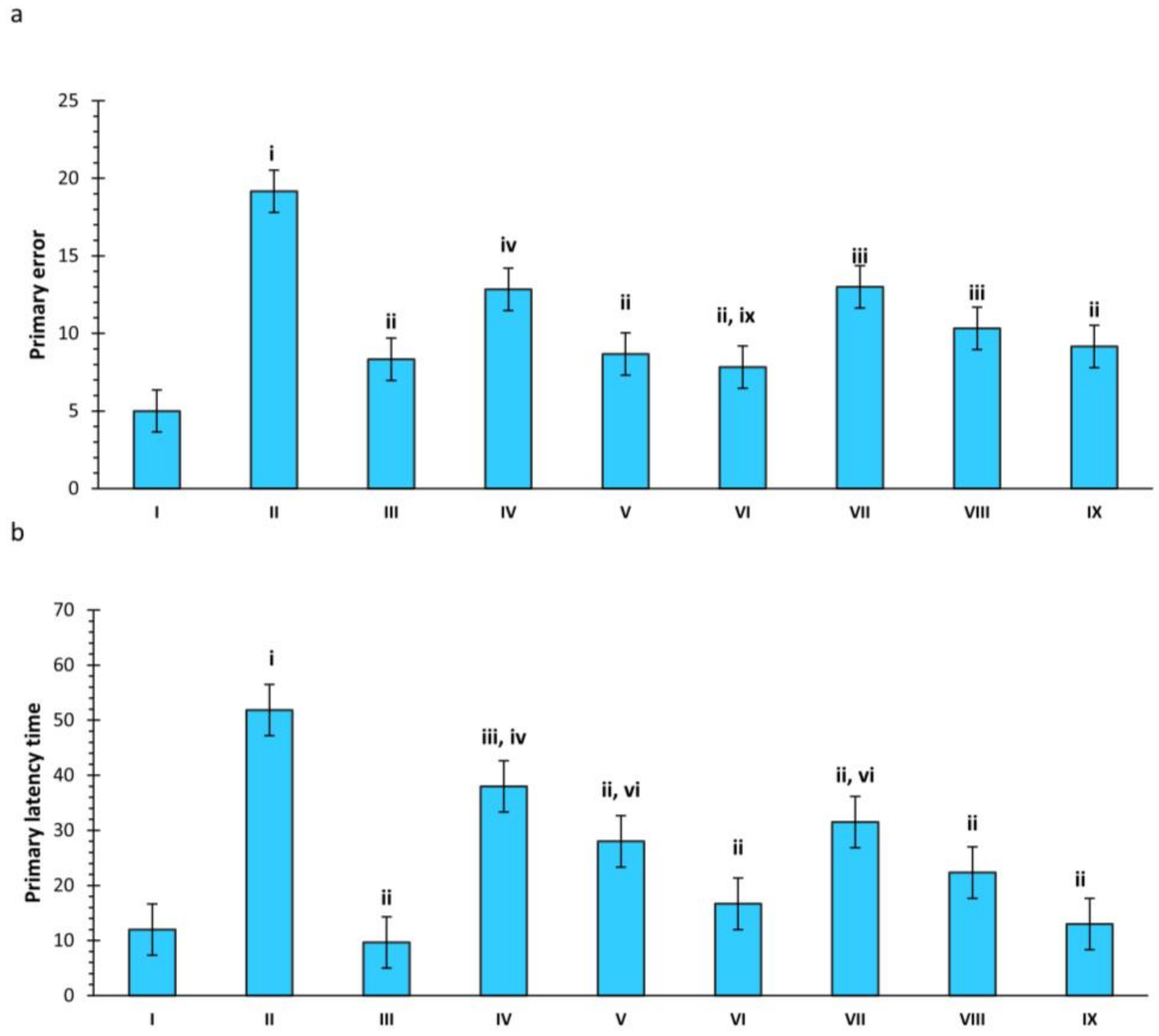
SCO administration increased primary errors significantly (p < 0.001, group II vs. group I). DNP showed marked improvement compared to SCO (p < 0.001, group III vs. group II). Compound 34 significantly reduced primary errors at 5 mg/kg (p < 0.05, group IV vs. group II) and demonstrated improvement at doses 10 (p < 0.001 vs. group SCO) and 20 (p < 0.001 vs. group SCO) mg/kg. Compound 37 exhibited significant improvement across all treatment groups compared to the control (Figure 3a).

2.2.2. Effect of SCO and Treatments on Primary Latency Time in Rats

SCO treatment increased primary latency significantly (p < 0.001, group II vs. group I) compared to the control group. Compounds 34 and 37 both demonstrated a significant reduction in primary latency. The 20 mg/kg dose exhibited the maximum reduction in primary latency (p < 0.001, group VI vs. group II and group IX vs. group II) for both compounds compared to SCO (Figure 3b).

2.3. Neurochemical Analysis

2.3.1. Effect of SCO and Treatments on Total Cholinesterase Activity

SCO treatment significantly increased total ChE activity with acetylthiocholine iodide (ATCI) as a substrate in the hippocampus and prefrontal cortex (PFC) (p < 0.001, group II vs. group I). DNP showed a significant decrease in activity (p < 0.001, group III vs. group II), but other treatment groups did not exhibit a significant decrease compared to the SCO group. Compounds 34 and 37 at a dose of 20 mg/kg significantly inhibited total ChE activity in PFC with ATCI as a substrate (p < 0.05, groups: VI vs. II and IX vs. II). DNP also significantly reduced ChE activity in PFC (p < 0.001, group: III vs. II).
Total ChE activity using butyrylthiocholine iodide (BTCI) as a substrate demonstrated a significant increase in the SCO-treated groups within the hippocampus and prefrontal cortex (p < 0.001, group II vs. group I).
Compounds 34 and 37 at doses of 10 mg/kg (p < 0.05, group V vs. group II for 34 and group VIII vs. group II for 37) and 20 mg/kg (p < 0.001, group VI vs. group II for 34 and p < 0.01, group IX vs. group II for 37) significantly decreased enzyme activity in the hippocampus. Similarly, significant inhibition was observed in PFC for compounds 34 and 37 at a dose of 10 (p < 0.05, group V vs. group II for 34 and group VIII vs. group II for 37) and 20 mg/kg (p < 0.001, group VI vs. group II and p < 0.01, group IX vs. group II). Notably, DNP treatment did not show significant cholinesterase inhibition in both the hippocampus and PFC with BTCI as a substrate (Figure 4).

2.3.2. Effect of Scopolamine and Various Treatments on Catalase (CAT) Activity

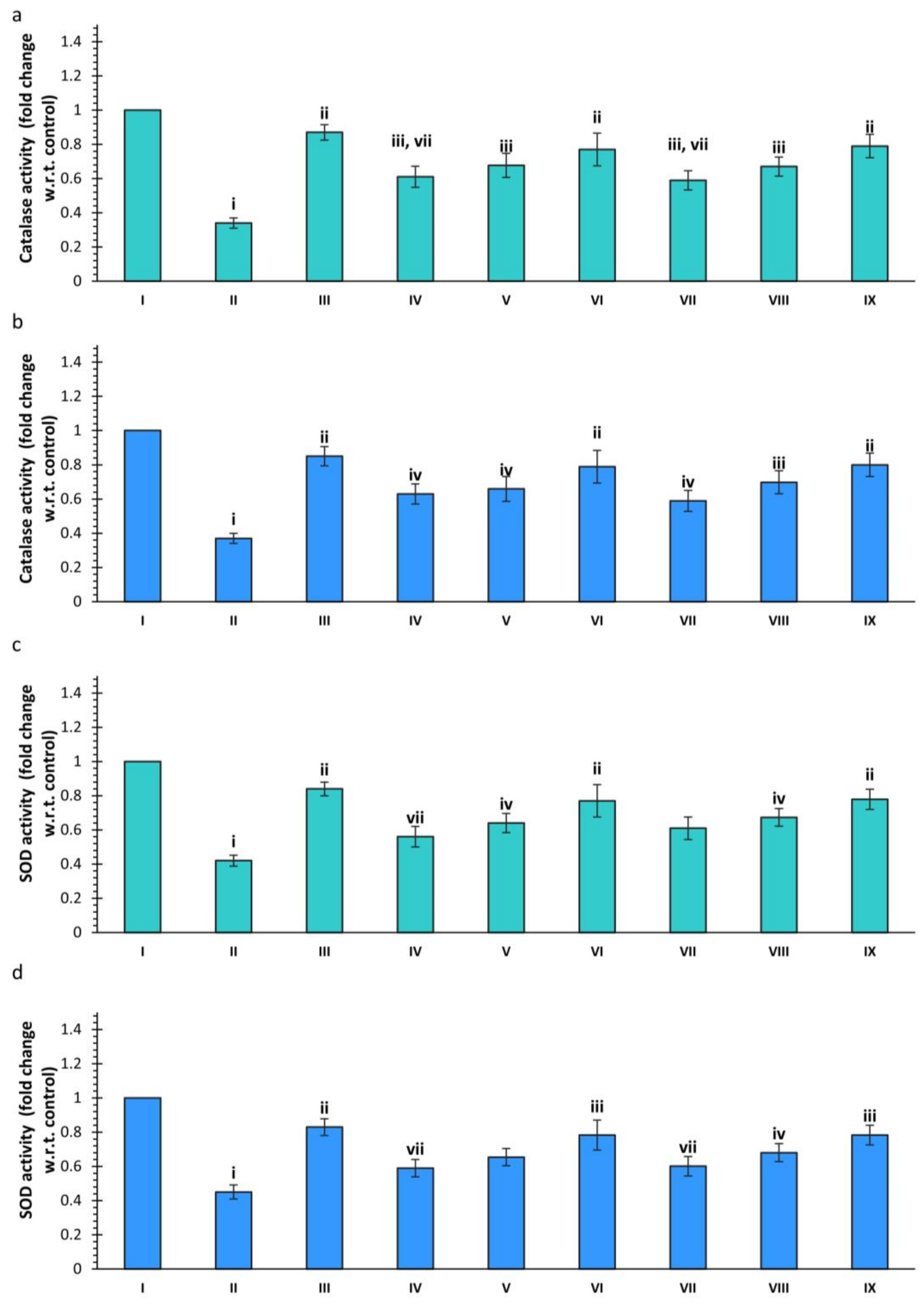
The SCO treatment also caused a reduction in the CAT activity compared to the control (p < 0.001, group II vs. group I). However, DNP and the selected compounds displayed significantly higher CAT activity than SCO treated group in both the hippocampus and PFC. Both the compounds showed maximum hippocampal CAT activity for 20 mg/kg, with no significant difference observed when compared with DNP. Further, CAT activity of PFC was significantly higher for 20 mg/kg (p < 0.001, group: IX vs. II) as compared to the disease group but no significant difference was observed when compared to the DNP (Figure 5).

2.3.3. Effect of Scopolamine and Various Treatments on SOD Activity

SCO treatment also caused a significant reduction in the SOD activity compared to control (p < 0.001, group II vs. group I) in both hippocampus and PFC. In contrast, DNP showed significantly higher SOD activity in both regions (p < 0.001, group III vs. group II). The dose of 20 mg/kg of compounds 34 and 37 showed significantly higher SOD activity in the hippocampus (p < 0.001, group VI vs. group II and group IX vs. group II). The activity of SOD was significantly higher at 20 mg/kg for compound 37 in PFC (p < 0.01, group IX vs. group II) (Figure 5).

2.4. Biochemical Analysis

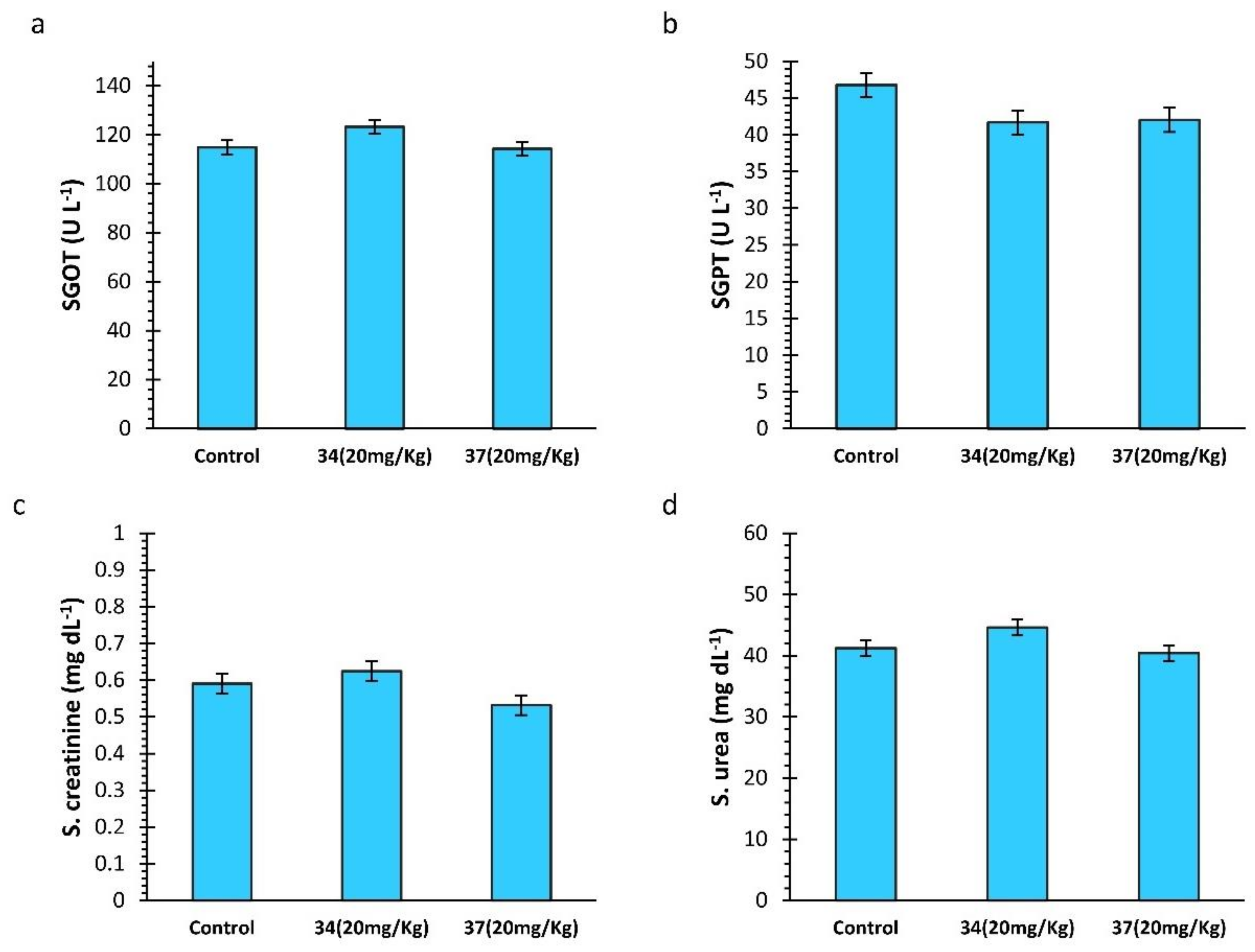
The biochemical analysis of SGPT and SGOT enzymes did not indicate any significant difference between the control and the treatments. Similarly, no significant difference was observed between the control and the treatment groups for serum creatinine and urea levels (Figure 6).

2.5. Homology Modelling

PDB id 4TPK was used as a template to build a homology model of rat BChE. The template was a protein model of the human BChE enzyme that shared a sequence similarity of 80.10% with the human sequence and had a good X-ray resolution of 2.7 Å. The obtained structure had a Global Model Quality Estimate (GMQE) score of 0.88. A homology model may have GMQE in the range of 0–1, with a value of 1 representing the ideal structure. GMQE is a perceptron-based scoring function that utilizes template structure and template target sequence alignment. It evaluates the model based on a comparison with already existing PDBs. QMEAN scores are a linear combination of various structural aspects of protein such as Cβ, all-atoms, solvation, and torsion potentials and its expected range lies between −4–1. QMEAN score of the developed model was about 0.87 ± 0.05, which was close to 1 and indicated that the model represented good quality. The model showed that 98.7% of residues were in the allowed region of the Ramachandran plot. A multistage energy minimization resulted in an improvement in the score to 99.6% residues and was in the allowed region of the plot (Figure 7).

2.6. Molecular Docking

The protein refinement resulted in the improvement in homology model quality, which was used for molecular docking. The results showed the order of the binding energies as:
37 < DNP < 34
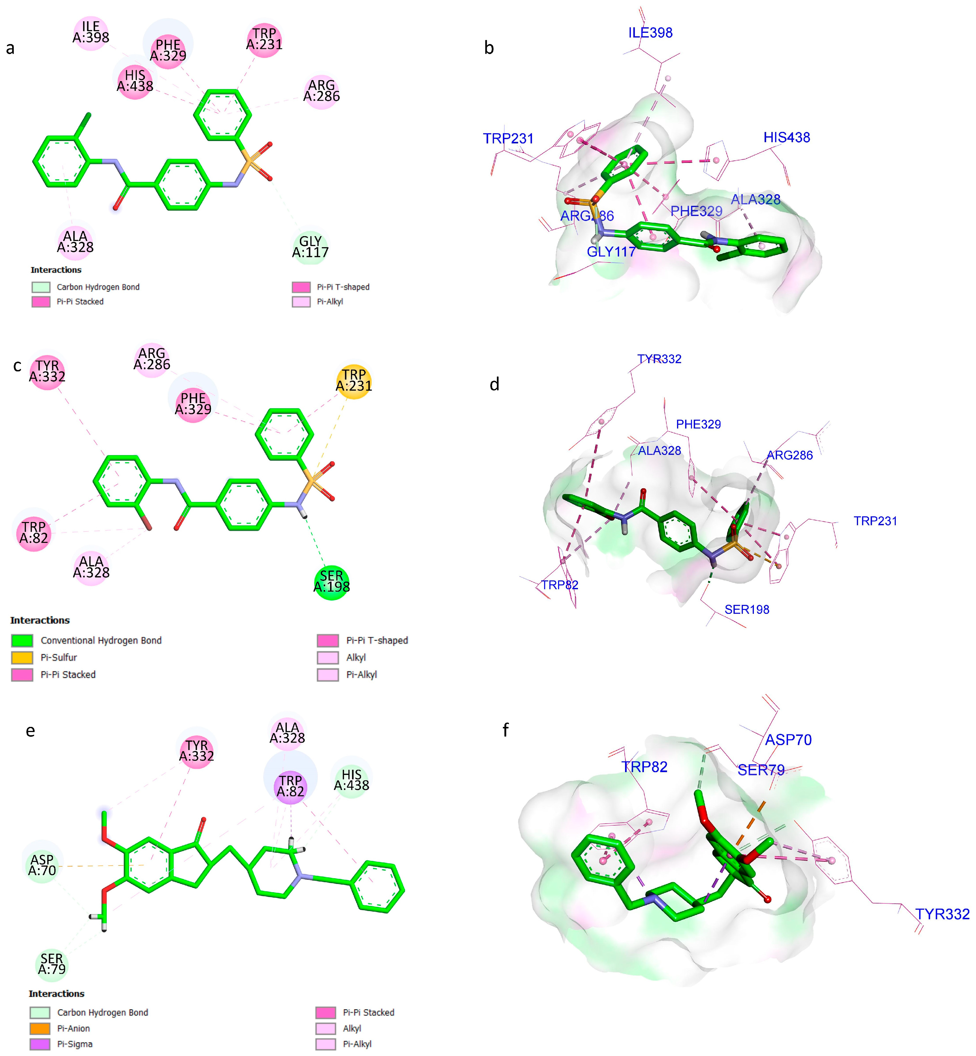
Compound 34 showed hydrogen bonding with Gly117, π-π interactions with ‘Trp231, Phe329 and His438’ and π-alkyl interactions with ‘Arg286, Ala328 and Ile398’. Whereas, compound 37 interacted with ‘Ser198’ (hydrogen bond). The π-π interactions were observed with ‘Trp82, Phe329, and Tyr332’ and π-alkyl interactions with ’Arg286 and Ala328’. DNP showed hydrogen bonding with ‘Ser79 and Tyr332’, π-anion interaction with ‘Asp70’, π-alkyl interaction with ‘Tyr332 and Trp82’ showed π-π and π-σ interactions (Figure 8). Ligand efficiency (LE) determines the binding energy per atom. Compound 37 displayed the highest LE, followed by compound 34 and DNP showed the lowest LE among all (Table 1).

2.7. Molecular Dynamics

The MD simulations of compounds 34, 37, and DNP complexed with rat BChE were carried out in order to ascertain the binding stability. The pre-MD phase involves the preparation of complexes for simulation through energy minimization, temperature equilibration, and density equilibration. The data are reported in Table S1 and Figures S1–S4 in Supplementary Materials.
Root mean square deviation (RMSD) is a measurement of complex stability throughout the course of an MD run. A little structural deviation is normal due to the dynamic nature of the system. A globular protein is expected to exhibit an RMSD value within the range of 1 to 3 Å. The mean RMSD values were 1.123 ± 0.176 Å for BChE, and 1.177 ± 0.157 Å, 1.104 ± 0.163 Å, and 1.160 ± 0.143 Å for BChE in complex with compounds 34, 37, and DNP, respectively. These complexes were stable and not much deviation was observed throughout the run. Ligand RMSD indicated a different aspect. RMSD deviation of compound 34 indicated that after 35 ns the ligand acquired a stable trajectory in the pocket for the rest of the time. On the other hand, compound 37 displayed the most stable trajectory for the complete run. DNP also acquired a stable trajectory after 30 ns. The average RMSD values were 1.664 ± 0.350 Å, 2.299 ± 0.172 Å, and 0.775 ± 0.226 Å for compounds 34, 37, and DNP, respectively. The root mean square fluctuation (RMSF) is akin to RMSD but signifies the average deviation in each residue (for proteins) or atom (for ligands) throughout the simulation. In general, there was no significant distinction in RMSF among the different complexes. Compound 34 displayed stabilization of ‘Gly116, Ser198, Ala199, Leu286, and Leu288’ as compared to the uninhibited enzyme. Compound 37 displayed stabilisation of ‘Asp70, Trp82, Gly117, Tyr332, and His438’ and DNP showed stabilisation for ‘Asp70, Trp82, Gly117, Ser198, Ala199, Leu288, Glu325, Tyr332, and His438’. The chloro-phenyl ring of compound 34 showed much fluctuation as compared to the bromo-phenyl ring of compound 37 (Figure 9).
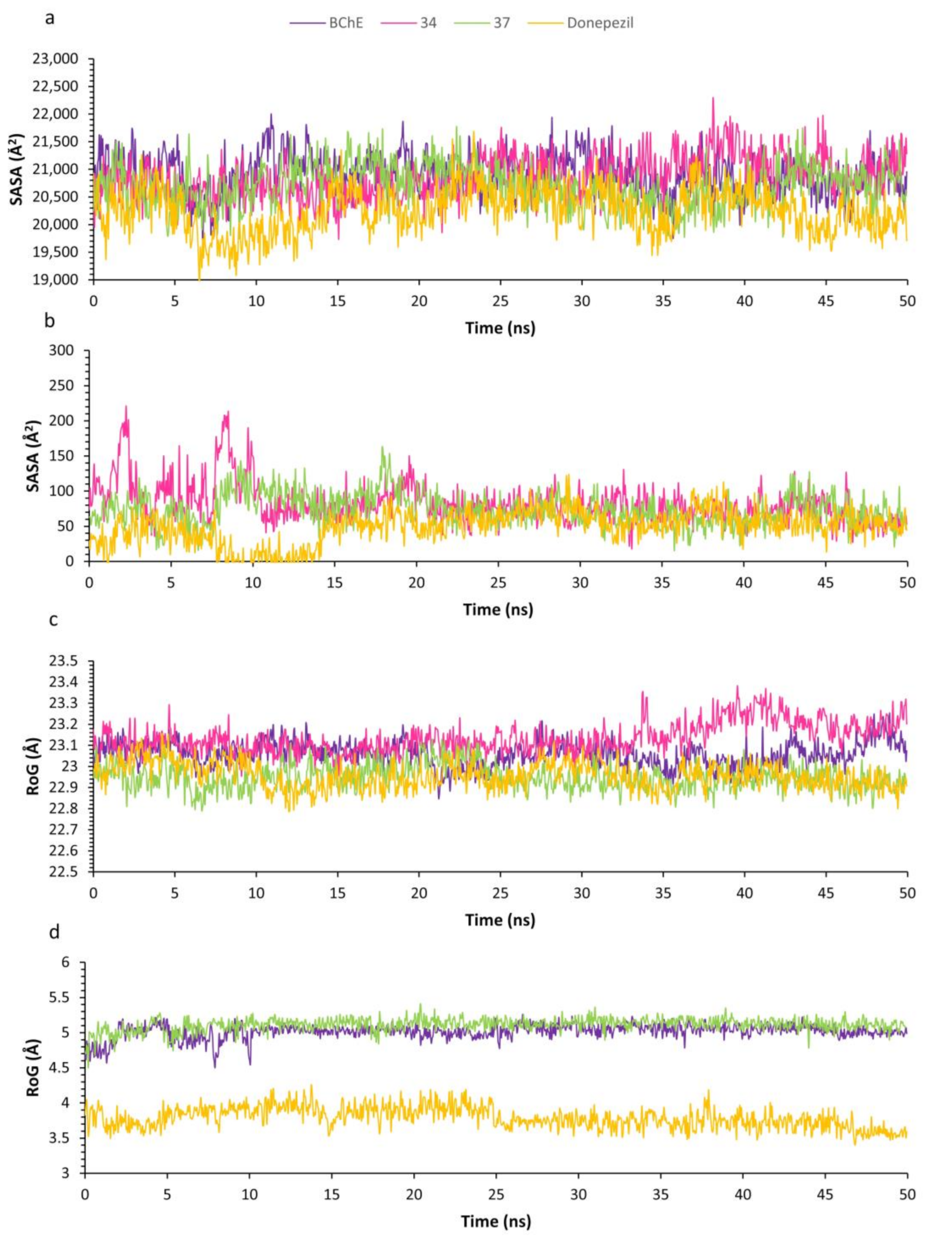
Solvent accessible surface area (SASA) refers to the exposure of solvent molecules, specifically water. The mean SASA values were 20,902.252 ± 370.595 Å2 for BChE, and 20,870.432 ± 377.164 Å2, 20,705.088 ± 373.456 Å2, and 20,260.389 ± 403.405 Å2 for BChE complexed with compounds 34, 37, and DNP, respectively. A marginal reduction in SASA occurred upon ligand binding, with the DNP complex exhibiting the highest decrease. The average SASA values were 82.3818 ± 28.871 Å2 for compound 34, 74.424 ± 22.494 Å2 for compound 37, and 49.425 ± 24.186 Å2 for DNP. The SASA for DNP was the minimum among all with significant fluctuation indicating that the ligand was solvent-exposed. The radius of gyration (RoG) serves as an indicator of protein stability, with an increase observed during protein unfolding. The mean RoG values were 23.058 ± 0.06 Å for BChE, and 23.135 ± 0.073 Å, 22.950 ± 0.06 Å, and 22.957 ± 0.063 Å for BChE in complex with compounds 34, 37, and DNP, respectively. No significant difference in RoG(s) was noted among the various protein-ligand complexes. The average RoG values for compounds 34, 37, and DNP were 5.017 ± 0.103 Å, 5.112 ± 0.093 Å, and 3.790 ± 0.151 Å, respectively. No significant hydrogen bonding was detected in any of the ligands, suggesting that π-interactions were the primary contributors to ligand binding (Figure 10).

3. Discussion

AD, a form of dementia, may contribute to over 60% of the cases and have an impact on families and society at large, affecting life expectancy. It is also a progressive neurodegenerative disease resulting in the fall in ADL, which culminates in a vegetative state and death. The current treatment avenues involve the use of selective AChE inhibitors, viz. DNP, rivastigmine for the mitigation of memory loss through cholinergic activation. Cholinergic activation promotes neurogenesis in the hippocampal region, contributing to memory and cognitive enhancement. The administration of SCO results in impairment in learning and memory processing, causing a temporary amnestic effect [20,21].
In this study, we assessed the impact of BChE inhibitors on scopolamine-induced amnesia in rats. Inhibitors of AChE and BChE have demonstrated enhancements in memory and cognitive function. Compounds 34 and 37, obtained from our previous study, were selective sulfonamide-based BChE inhibitors with potent in vitro inhibition activities. The spontaneous alternation in the Y-maze is the indicator of the working memory [22]. The reduction in % spontaneous alterations was substantially improved by administering compounds 34 and 37 at 10 and 20 mg/kg, similar to the DNP. There was no significant difference between the treatment groups and DNP in % alteration.
Further, the novel arm entries in Y-maze were significantly improved for test compounds and DNP as compared to the disease group. The locomotion and mobility of the patient are significantly reduced with the severity of AD [23]. The treatment groups and DNP did not show significant differences in the total arm entries in comparison to SCO treatment. Barnes maze is another crucial parameter for testing hippocampal function and an indicator for spatial learning and memory. The primary latency was improved in the case of the treatment groups as well as DNP as compared to SCO. The primary errors were reduced, with no significant difference observed between the treatment groups and the standard DNP group.
To assess the efficacy of the BChE inhibitors further, a series of biochemical analyses were carried out on the hippocampus and PFC tissues. Both these regions in humans and rodents are involved in the processes related to memory [24]. Cholinergic pathways and ACh are essential for memory, learning, and cognitive processes. In AD, it is evident that cholinergic neuronal loss and increased cholinesterase activity are responsible for the deterioration in brain functioning. Hence, the total cholinesterase activity was evaluated with the help of ATCI and BTCI as substrates. BTCI is selectively hydrolysed by BChE present in the brain. The study indicated that the administration of SCO led to a substantial increase in total cholinesterase activity in both the hippocampus and PFC for both substrates. The treatment with compounds 34 and 37, at 20 mg/kg, caused a significant reduction in the enzyme activity in both regions compared to SCO treatment. A significant decrease in the enzyme activity was observed in the case of BTCI as substrate in comparison to DNP, as the test compounds showed selectivity towards BChE. The overproduction of the reactive oxidative species (ROS) contributes to AD which leads to neuronal damage. SOD and CAT are the protective antioxidant enzymes against ROS. The treatment groups showed a significant restoration in SOD and CAT activities, especially at a dose of 20 mg/kg as compared to SCO treatment. Similar results were also obtained in the case of the DNP treatment. The alteration in SGPT and SGOT levels is an indicator of hepatic dysfunction. The drugs, including sulfonamides, salicylates, and sulfonylureas, cause a moderate increase in SGPT and SGOT levels [25]. There was no significant change in the enzyme levels at a dose of 20 mg/kg in the case of test compounds. Further, no significant change in serum urea and creatinine levels was observed. Therefore, sulfonamide derivatives may not have any adverse effects on hepatic dysfunction. The in silico docking study indicated that compounds 34 and 37 bound firmly with BChE. Further, binding with the enzyme was due to π-interactions rather than the hydrogen bonding, as revealed in MD studies. The enzyme-ligand complexes were stable for the complete simulation run.

4. Materials and Methods

4.1. Materials

Scopolamine hydrobromide (SCO) and Donepezil (DNP) were procured from Sigma-Aldrich (St. Louis, MO, USA). All other chemicals of analytical grade were used in the study.

4.2. Experimental Animals

In this study, male Wistar rats weighing between 150–200 g were utilized. The rats were accommodated in polyacrylic cages (6 animals per cage) at 25 ± 2 °C and 50 ± 10% relative humidity, following a 12-h light/dark cycle.
The animals underwent a one-week acclimatization period with unrestricted access to food and water ad libitum. Food access was restricted one hour prior to the commencement of the behavioral study. The experimental procedures were approved by the Institutional Animal Ethics Committee of Banaras Hindu University, Varanasi, India, under protocol number Dean/2021/IAEC/2565.

4.3. Experimental Designs

4.3.1. Drugs and Treatments

The animals were segregated into nine groups, with six animals each. The treatments used in the study are reported in Table 2.
All compounds, except SCO, were administered for seven consecutive days, with SCO being administered on the seventh day to induce amnesia. Behavioral experiments were conducted for 30 min following SCO administration (5 mg/kg) in groups III to IX [26].

4.3.2. LD50 Determination

LD50 values of compounds 34 and 37 were determined using OECD guideline 423—acute toxicity class method. The compounds were tested for 500 and 2000 mg/kg doses with three female Wistar rats in each group. After administration of doses, the rats were observed for 72 h (Tables S2–S13 in Supplementary Materials). LD50 of the compounds was calculated according to the guideline [27].

4.3.3. Y-Maze Test

It was used to evaluate intermediate ‘working and spatial memory’. The test compounds 34, 37, and DNP were evaluated on the seventh treatment day. Initially, a training session was conducted with one arm closed using a partition, and the rat was allowed to maze exploration for 15 min. The animal entered the maze with its head facing toward the center. The training was performed after dosing and four hours before the test session. The test session was carried out after a half-hour of administration of SCO.
In this session, the novel arm was unblocked, allowing the rodent to move freely in the maze for five minutes. The occurrences of arm entries were documented, with repeated entries in the same arm suggesting memory impairment. Memory improvement was assessed based on ‘novel arm entries and spontaneous alterations’ in three consecutive components (ABC, BCA, CAB, not ABA) [28,29,30].
The maze was wiped with 70% ethanol after each session to eliminate any olfactory cues. The ‘% spontaneous alteration’ was calculated as:
%   S p o n t a n e o u s   a l t e r a t i o n = N u m b e r   o f   a l t e r a t i o n ( t o t a l   a r m   e n t r i e s 2 ) × 100

4.3.4. Barnes Maze

The Barnes maze consists of a circular platform with a diameter of 122 cm, elevated 100 cm above the ground, and containing 20 evenly spaced holes, each measuring 10 cm in diameter. Illumination is provided by white light exceeding 600 lux, and sound, surpassing 80 dB, is introduced using a siren.

4.3.5. Habituation

Habituation to the platform and escape box was conducted a day prior to the acquisition phase to minimize anxiety. The animal was exposed to a three-minute habituation period under light conditions with no noise.

4.3.6. Acquisition Phase

This phase lasted five days, with one session per day. Each session included two 180-s trials separated by a 15-min interval. Training began by placing the animal in a black-covered box at the center of the platform. After activating light and sound, the animal was released following a 10-s delay to explore the maze and find the escape box using visual cues. Upon entering the escape box, the entrance was closed, stimuli were deactivated, and the animal stayed for 30 s before returning to its cage. If unsuccessful, the animal was gently guided to the box and given 30 s to explore. The maze was wiped with 70% ethanol after each trial to remove olfactory cues. Over the five days, primary latency (time to find the escape box) and primary errors (holes checked before finding the escape hole) were recorded.

4.3.7. Probe Trial

The probe trial was conducted on the seventh day after treatment, 30 min post i.p. administration of SCO. Following a similar approach with the escape hole closed, the rat had a 90-s window for maze exploration, during which primary latency and errors were documented [31,32].

4.3.8. Neurochemical Analysis

Following the experiments, the animals were euthanized, and their brains were collected. The hippocampus and PFC)were utilized for neurochemical analysis. Tissue homogenization in 10 mM phosphate buffer saline (PBS; pH 7.4) and subsequent centrifugation at 15,000 rpm for 15 min at 4 °C yielded the supernatant for further analysis.
ChE activity was assessed using ATCI and BTCI via the Ellman method. Initially, 10 µL of the supernatant was diluted with 100 µL of PBS, followed by the addition of freshly prepared 50 µL substrate solutions (5 mM), incubated for 5 min. Subsequently, 1.5 mM DTNB solution was added, and absorbance was measured at 415 nm against blank.
CAT enzyme, responsible for converting H2O2 into water and oxygen, was measured in tissue homogenate. Mixing 10 µL of supernatant with 150 µL of PBS, followed by the addition of 250 µL of H2O2 (160 mM) and incubation for 1 min at 37 °C, was performed. Further, 1.5 mL of a stopping solution of 5% K2Cr2O7/glacial CH3COOH (1:3 v/v) was added. The resulting green color, indicative of dichromate oxidation to chromic (III) sulphate, was compared with a control mixture, excluding the enzyme. Absorbance was measured at 570 nm using a Synergy HTX multimode reader (BioTek, Santa Clara, CA, USA) with a blank as the reference [33].
SOD activity was assessed using Markland’s method, relying on the pyrogallol autoxidation process. To 10 µL of tissue homogenate, 200 µL of 0.1 M Tris-HCl containing 1 mM EDTA (pH 8.2) was added, followed by the addition of 50 µL of 4.5 mM pyrogallol solution prepared in 1 µM HCl. Absorbance was measured after 1 min at 325 nm. A control sample lacking tissue supernatant served to determine enzyme activity. Experiments were conducted in triplicates and enzyme activities were normalized relative to the control group.

4.3.9. Biochemical Analysis

Serum levels of alanine aminotransferase (ALT/SGPT), aspartate aminotransferase (AST/SGOT), urea, and creatinine were measured in animals treated with compounds 34, 37, and the control group using a commercially available kit from Tara Clinical System (Dombivli, MH, India).

4.3.10. Homology Modelling

The protein model of the rat BChE (UniProt accession code—Q9JKC1) was prepared from SWISS-MODEL, a web server that is accessible via the ExPASy (https://swissmodel.expasy.org/ accessed on 22 June 2023) and validated using previously reported protocol [34].

4.3.11. Molecular Docking

The homology model obtained was refined and subjected to energy minimization using the previously reported protocol. The ligand structures are available in SMILES format were converted into Tripos MOL2 format using RDkit (ver. 2022.9.4), subjected to energy minimization through MMFF94s forcefield, and were subsequently converted into PDBQT format using AutodcokTools-1.5.6 [35].
Additionally, grid maps were generated using a grid box size of 84 × 66 × 72, with the grid center positioned at coordinates 51.05, 28.382, and 54.297 (X, Y, and Z, respectively) and a grid point spacing of 0.375 Å [36]. Discovery Studio Visualiser 2020 was used for post-docking analysis and visualization.

4.3.12. Molecular Dynamics

The molecular dynamics simulations of compounds 34, 37, and DNP were carried out using Amber 2022 [34,37]. Further, the post-MD processing was performed through cpptraj [38].

5. Conclusions

It is evident from the study that sulphonamide derivatives (compounds 34 and 37) improved memory and learning when challenged against scopolamine-induced amnesia in rats. Further, both compounds were potent with no significant changes in SGOT, SGPT, serum urea, and serum creatinine levels. The response was mediated by inhibition of the brain cholinesterase enzyme. The compounds also showed considerable neuroprotection against scopolamine-induced stress as reflected in the levels of SOD and CAT enzyme, especially at a dose of 20 mg/kg. Thus, compounds 34 and 37 serve as potential drug candidates for further exploration for the treatment of AD.

Supplementary Materials

The following supporting information can be downloaded at: https://www.mdpi.com/article/10.3390/ddc3040038/s1, Figure S1: (a) Potential energy vs. Time, (b) Temperature vs. Time, (c) Density vs. Time and (d) RMSD of protein backbone of BChE; Figure S2: (a) Potential energy vs. Time, (b) Temperature vs. Time, (c) Density vs. Time and (d) RMSD of protein backbone of BChE-compound 34 complex; Figure S3: (a) Potential energy vs. Time, (b) Temperature vs. Time, (c) Density vs. Time and (d) RMSD of protein backbone of BChE-compound 37 complex; Figure S4: (a) Potential energy vs. Time, (b) Temperature vs. Time, (c) Density vs. Time and (d) RMSD of protein backbone of BChE-DNP complex; Table S1: Protocol for energy minimization carried out before molecular dynamic simulation in AMBER20; Table S2. LD50 determination protocol for compound 34; Table S3. Effect of compound 34 on the body wt. of the animals at the dose of 300 mg/kg; Table S4. The onset of toxicity with compound 34 in the period of 72 h; Table S5. LD50 determination protocol for the 34; Table S6. Effect of compound 34 on the body wt. of the animals at the dose of 2000 mg/kg; Table S7. The onset of toxicity with compound 34 in the period of 72 h; Table S8. LD50 determination protocol for the compound 37; Table S9. Effect of compound 37 on the body wt. of the animals at the dose of 300 mg/kg; Table S10. The onset of toxicity with compound 37 in the period of 72 h; Table S11. LD50 determination protocol for the 37; Table S12. Effect of compound 37 on the body wt. of the animals at the dose of 2000 mg/kg; Table S13. The onset of toxicity with compound 37 in the period of 72 h.

Author Contributions

Conceptualization, A.G. and Q.A.; methodology, A.G. and P.T.; software, A.G.; formal analysis, A.G., Q.A., and R.S; writing—original draft preparation, A.G. and P.T.; writing—review and editing, R.S. and Q.A. supervision, A.K., S.K., and S.K.S.; project administration, S.K. and S.K.; funding acquisition, S.K.S. All authors have read and agreed to the published version of the manuscript.

Funding

The authors would like to acknowledge the financial support from the Ministry of Education (MoE), New Delhi, India in the form of teaching assistantships to A.G., P.T., R.S., and Q.A.

Institutional Review Board Statement

This study was approved by the Institutional Animal Ethics Committee of the Banaras Hindu University, Varanasi, India (Reference no: Dean/2021/IAEC/2565). All methods were performed in accordance with relevant guidelines and regulations.

Informed Consent Statement

Not applicable.

Data Availability Statement

The additional data are available in Supplementary Materials and can also be made available by the corresponding author on reasonable request.

Conflicts of Interest

The authors declare no conflicts of interest.

References

  1. Talita, F.-V.H.; Isabella, G.M.; Flavia, S.R.; Fabiola, R.M. Alzheimer’s disease: Targeting the cholinergic system. Curr. Neuropharmacol. 2016, 14, 101–115. [Google Scholar]
  2. Hanseeuw, B.J.; Betensky, R.A.; Jacobs, H.I.; Schultz, A.P.; Sepulcre, J.; Becker, J.A.; Cosio, D.M.O.; Farrell, M.; Quiroz, Y.T.; Mormino, E.C. Association of amyloid and tau with cognition in preclinical Alzheimer disease: A longitudinal study. JAMA Neurol. 2019, 76, 915–924. [Google Scholar] [CrossRef] [PubMed]
  3. Brookmeyer, R.; Johnson, E.; Ziegler-Graham, K.; Arrighi, H.M. Forecasting the global burden of Alzheimer’s disease. Alzheimer’s Dement. 2007, 3, 186–191. [Google Scholar] [CrossRef] [PubMed]
  4. Du, X.; Wang, X.; Geng, M. Alzheimer’s disease hypothesis and related therapies. Transl. Neurodegener. 2018, 7, 2. [Google Scholar] [CrossRef] [PubMed]
  5. Ganeshpurkar, A.; Swetha, R.; Kumar, D.; Gangaram, G.P.; Singh, R.; Gutti, G.; Jana, S.; Kumar, D.; Kumar, A.; Singh, S.K. Protein-protein interactions and aggregation inhibitors in Alzheimer’s disease. Curr. Top. Med. Chem. 2019, 19, 501–533. [Google Scholar] [CrossRef]
  6. Firdaus, Z.; Singh, T.D. An Insight in Pathophysiological Mechanism of Alzheimer’s Disease and its Management Using Plant Natural Products. Mini Rev. Med. Chem. 2021, 21, 35–57. [Google Scholar] [CrossRef]
  7. Zemek, F.; Drtinova, L.; Nepovimova, E.; Sepsova, V.; Korabecny, J.; Klimes, J.; Kuca, K. Outcomes of Alzheimer’s disease therapy with acetylcholinesterase inhibitors and memantine. Expert Opin. Drug Saf. 2014, 13, 759–774. [Google Scholar]
  8. Liu, P.-P.; Xie, Y.; Meng, X.-Y.; Kang, J.-S. History and progress of hypotheses and clinical trials for Alzheimer’s disease. Signal Transduct. Target. Ther. 2019, 4, 29. [Google Scholar] [CrossRef]
  9. Davies, P.; Maloney, A.J.R. Selective loss of central cholinergic neurons in Alzheimer’s disease. Lancet 1976, 2, 1403. [Google Scholar] [CrossRef]
  10. Rogers, J.L.; Kesner, R.P. Cholinergic modulation of the hippocampus during encoding and retrieval. Neurobiol. Learn. Mem. 2003, 80, 332–342. [Google Scholar] [CrossRef]
  11. Muir, J.; Dunnett, S.; Robbins, T.; Everitt, B. Attentional functions of the forebrain cholinergic systems: Effects of intraventricular hemicholinium, physostigmine, basal forebrain lesions and intracortical grafts on a multiple-choice serial reaction time task. Exp. Brain Res. 1992, 89, 611–622. [Google Scholar] [CrossRef] [PubMed]
  12. Giacobini, E. Cholinomimetic Replacement of Cholinergic Function in Alzheimer Disease. In Treatment of Dementias; Springer: Berlin/Heidelberg, Germany, 1992; pp. 19–34. [Google Scholar]
  13. Mesulam, M.-M.; Guillozet, A.; Shaw, P.; Levey, A.; Duysen, E.; Lockridge, O. Acetylcholinesterase knockouts establish central cholinergic pathways and can use butyrylcholinesterase to hydrolyze acetylcholine. Neuroscience 2002, 110, 627–639. [Google Scholar] [CrossRef] [PubMed]
  14. Darvesh, S.; Reid, G. Reduced fibrillar β-amyloid in subcortical structures in a butyrylcholinesterase-knockout Alzheimer disease mouse model. Chem.-Biol. Interact. 2016, 259, 307–312. [Google Scholar] [CrossRef]
  15. Greig, N.H.; Utsuki, T.; Ingram, D.K.; Wang, Y.; Pepeu, G.; Scali, C.; Yu, Q.-S.; Mamczarz, J.; Holloway, H.W.; Giordano, T. Selective butyrylcholinesterase inhibition elevates brain acetylcholine, augments learning and lowers Alzheimer β-amyloid peptide in rodent. Proc. Natl. Acad. Sci. USA 2005, 102, 17213–17218. [Google Scholar] [CrossRef] [PubMed]
  16. Giacobini, E. Cholinergic function and Alzheimer’s disease. Int. J. Geriatr. Psychiatry 2003, 18 (Suppl. S1), S1–S5. [Google Scholar] [CrossRef]
  17. Darvesh, S.; MacKnight, C.; Rockwood, K. Butyrylcholinesterase and cognitive function. Int. Psychogeriatr. 2001, 13, 461–464. [Google Scholar] [CrossRef]
  18. Swetha, R.; Kumar, D.; Gupta, S.K.; Ganeshpurkar, A.; Singh, R.; Gutti, G.; Kumar, D.; Jana, S.; Krishnamurthy, S.; Singh, S.K. Multifunctional hybrid sulfonamides as novel therapeutic agents for Alzheimer’s disease. Future Med. Chem. 2019, 11, 3161–3178. [Google Scholar] [CrossRef]
  19. Ganeshpurkar, A.; Singh, R.; Kumar, D.; Gutti, G.; Gore, P.; Sahu, B.; Kumar, A.; Singh, S.K. Identification of sulfonamide-based butyrylcholinesterase inhibitors using machine learning. Future Med. Chem. 2022, 14, 1049–1070. [Google Scholar] [CrossRef]
  20. Flood, J.F.; Cherkin, A. Scopolamine effects on memory retention in mice: A model of dementia? Behav. Neural Biol. 1986, 45, 169–184. [Google Scholar] [CrossRef]
  21. Skalicka-Wozniak, K.; Budzynska, B.; Biala, G.; Boguszewska-Czubara, A. Scopolamine-Induced Memory Impairment Is Alleviated by Xanthotoxin: Role of Acetylcholinesterase and Oxidative Stress Processes. ACS Chem. Neurosci. 2018, 9, 1184–1194. [Google Scholar] [CrossRef]
  22. Lee, G.-Y.; Lee, C.; Park, G.H.; Jang, J.-H. Amelioration of Scopolamine-Induced Learning and Memory Impairment by α-Pinene in C57BL/6 Mice. Evid.-Based Complement. Altern. Med. 2017, 2017, 4926815. [Google Scholar] [CrossRef] [PubMed]
  23. Mali, K.K.; Sutar, G.V.; Dias, R.J.; Devade, O.A. Evaluation of Nootropic Activity of Limonia acidissima Against Scopolamine-induced Amnesia in Rats. Turk. J. Pharm. Sci. 2021, 18, 3–9. [Google Scholar] [CrossRef]
  24. Jin, J.; Maren, S. Prefrontal-hippocampal interactions in memory and emotion. Front. Syst. Neurosci. 2015, 9, 170. [Google Scholar] [CrossRef] [PubMed]
  25. Sulfasalazine. In LiverTox: Clinical and Research Information on Drug-Induced Liver Injury; National Institute of Diabetes and Digestive and Kidney Diseases: Bethesda, MD, USA, 2012.
  26. Ngoupaye, G.T.; Pahaye, D.B.; Ngondi, J.; Moto, F.C.O.; Bum, E.N. Gladiolus dalenii lyophilisate reverses scopolamine-induced amnesia and reduces oxidative stress in rat brain. Biomed. Pharmacother. 2017, 91, 350–357. [Google Scholar] [CrossRef]
  27. Schlede, E. Oral acute toxic class method: OECD Test Guideline 423. Rapp. Istisan 2002, 41, 32–36. [Google Scholar]
  28. Omotoso, G.O.; Gbadamosi, I.T.; Afolabi, T.T.; Abdulwahab, A.B.; Akinlolu, A.A. Ameliorative effects of Moringa on cuprizone-induced memory decline in rat model of multiple sclerosis. ACB 2018, 51, 119–127. [Google Scholar] [CrossRef] [PubMed]
  29. Hritcu, L.; Ionita, R.; Motei, D.E.; Babii, C.; Stefan, M.; Mihasan, M. Nicotine versus 6-hydroxy-l-nicotine against chlorisondamine induced memory impairment and oxidative stress in the rat hippocampus. Biomed. Pharmacother. 2017, 86, 102–108. [Google Scholar] [CrossRef]
  30. Miedel, C.J.; Patton, J.M.; Miedel, A.N.; Miedel, E.S.; Levenson, J.M. Assessment of Spontaneous Alternation, Novel Object Recognition and Limb Clasping in Transgenic Mouse Models of Amyloid-β and Tau Neuropathology. J. Vis. Exp. JoVE 2017, 28, 55523. [Google Scholar]
  31. Gawel, K.; Gibula, E.; Marszalek-Grabska, M.; Filarowska, J.; Kotlinska, J.H. Assessment of spatial learning and memory in the Barnes maze task in rodents—Methodological consideration. Naunyn-Schmiedeberg’s Arch. Pharmacol. 2019, 392, 1–18. [Google Scholar] [CrossRef]
  32. Gibula-Tarlowska, E.; Kotlinska, J.H. Kissorphin improves spatial memory and cognitive flexibility impairment induced by ethanol treatment in the Barnes maze task in rats. Behav. Pharmacol. 2020, 31, 272–282. [Google Scholar] [CrossRef]
  33. Kumar, D.; Gupta, S.K.; Ganeshpurkar, A.; Gutti, G.; Krishnamurthy, S.; Modi, G.; Singh, S.K. Development of Piperazinediones as dual inhibitor for treatment of Alzheimer’s disease. Eur. J. Med. Chem. 2018, 150, 87–101. [Google Scholar] [CrossRef] [PubMed]
  34. Ganeshpurkar, A.; Singh, R.; Gore, P.G.; Kumar, D.; Gutti, G.; Kumar, A.; Singh, S.K. Structure-based screening and molecular dynamics simulation studies for the identification of potential acetylcholinesterase inhibitors. Mol. Simul. 2020, 46, 169–185. [Google Scholar] [CrossRef]
  35. Tosco, P.; Stiefl, N.; Landrum, G. Bringing the MMFF force field to the RDKit: Implementation and validation. J. Cheminform. 2014, 6, 37. [Google Scholar] [CrossRef]
  36. Morris, G.M.; Huey, R.; Lindstrom, W.; Sanner, M.F.; Belew, R.K.; Goodsell, D.S.; Olson, A.J. AutoDock4 and AutoDockTools4: Automated docking with selective receptor flexibility. J. Comput. Chem. 2009, 30, 2785–2791. [Google Scholar] [CrossRef]
  37. Ganeshpurkar, A.; Singh, R.; Kumar, D.; Divya; Shivhare, S.; Kumar, A.; Singh, S.K. Computational binding study with α7 nicotinic acetylcholine receptor of Anvylic-3288: An allosteric modulator. Mol. Simul. 2020, 46, 975–986. [Google Scholar] [CrossRef]
  38. Roe, D.R.; Cheatham, T.E., III. PTRAJ and CPPTRAJ: Software for processing and analysis of molecular dynamics trajectory data. J. Chem. Theory Comput. 2013, 9, 3084–3095. [Google Scholar] [CrossRef]
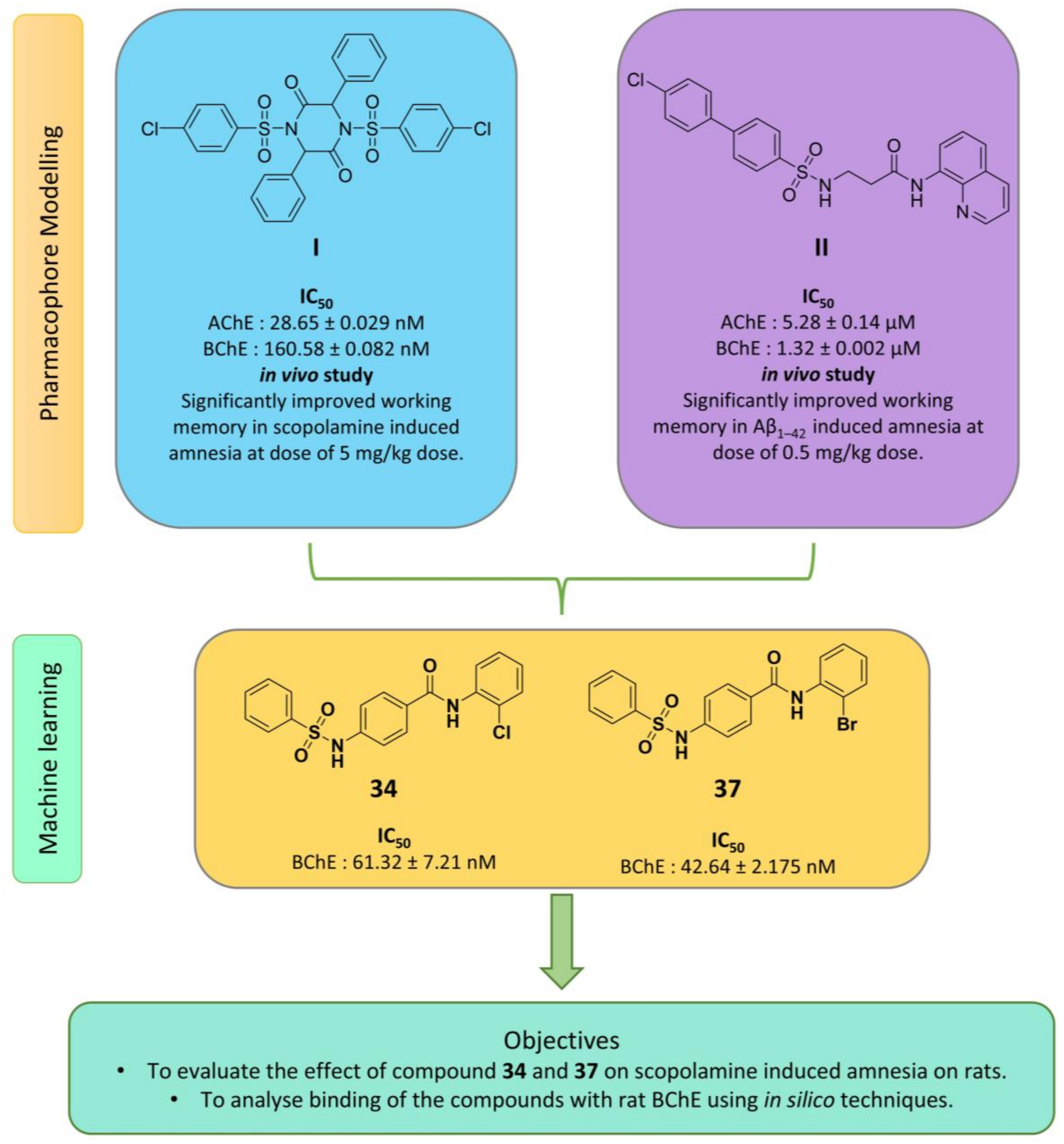
Figure 1. Schematic representation of rationale and plan of study.
Ddc 03 00038 g001
Figure 2. The impact of scopolamine-induced impairment of compounds 34, 37, and DNP on (a) % spontaneous alteration, (b) novel arm entries, (c) % time in arm entries, and (d) % total arm entries is presented. The data are expressed as Mean ± SEM. (i p < 0.001 when compared to control group, ii p < 0.001 when compared to SCO group, iii p < 0.01 when compared to SCO group, iv p < 0.05 when compared to SCO group, v p < 0.05 when compared to DNP group, vi p < 0.001 when compared to DNP group, vii p < 0.05 when compared to when compound 34 (5 mg/kg) group, viii p < 0.01 when compared to compound 34 (10 mg/kg) group, ix p < 0.001 when compared to compound 34 (20 mg/kg) group, x p < 0.05 when compared to compound 37 (5 mg/kg) group, xi p < 0.01 when compared to compound 37 (10 mg/kg) group).
Ddc 03 00038 g002
Figure 3. The influence of compounds 34, 37, and DNP on (a) primary error and (b) primary latency time is observed. Data are presented as Mean ± SEM. (i p < 0.001 compared to control group, ii p < 0.001 compared to SCO group, iii p < 0.01 compared to SCO group, iv p < 0.05 compared to SCO group, vi p < 0.01 compared to DNP group, ix p < 0.01 compared to compound 34 (10 mg/kg) group).
Ddc 03 00038 g003
Figure 4. The effects of compounds 34, 37, and DNP on total ChE activity, utilizing ATCI as a substrate in (a) the hippocampus and (b) the PFC, as well as BTCI as a substrate in (c) the hippocampus and (d) the PFC, are evaluated. The data are presented as Mean ± SEM. (i p < 0.001 compared to control group, ii p < 0.001 compared to SCO group, iii p < 0.01 compared to SCO group, iv p < 0.05 compared to SCO group, vi p < 0.01 compared to DNP group, vii p < 0.05 compared to DNP group).
Ddc 03 00038 g004
Figure 5. The effects of compounds 34, 37, and DNP on CAT activity in (a) the hippocampus and (b) the PFC, along with SOD activity in (c) the hippocampus and (d) the PFC, are analyzed. Data are presented as Mean ± SEM. (i p < 0.001 compared to control group, ii p < 0.001 compared to SCO group, iii p < 0.01 compared to SCO group, iv p < 0.05 compared to SCO group, vii p < 0.05 compared to DNP group).
Ddc 03 00038 g005
Figure 6. The effects of compounds 34 and 37 on (a) SGOT, (b) SGPT, (c) serum creatinine, and (d) serum urea levels are presented. Data are expressed as Mean ± SEM.
Ddc 03 00038 g006
Figure 7. Ramachandran plot of homology model (a) before and (b) after energy minimization. The residues indicated in purple circles are outlier while in black circles are in expected stereochemistry.
Ddc 03 00038 g007
Figure 8. The 2D and 3D interaction maps of (a,b) 34, (c,d) 37, and (e,f) DNP.
Ddc 03 00038 g008
Figure 9. (a) RMSD of protein-ligand complexes, (b) RMSD of the ligands, (c) RMSF of protein-ligand complexes, and (d) RMSF of the ligands.
Ddc 03 00038 g009
Figure 10. (a) SASA of protein-ligand complexes, (b) SASA of the ligands, (c) RoG of protein-ligand complexes, and (d) RoG of the ligands.
Ddc 03 00038 g010
Table 1. Binding energies and ligand efficiencies of various ligands against rat BChE.
Compound CodeBinding Energy (kcal/mol)Ligand Efficiency
34−9.93−0.38
37−10.38−0.40
DNP−10.36−0.37
Table 2. Plan of study describing the various treatments.
GroupTreatmentDose (mg/kg)Number of Animals
I (Control)--6
II (Disease Control)--6
IIIDNP56
IVCompound 3456
VCompound 34106
VICompound 34206
VIICompound 3756
VIIICompound 37106
IXCompound 37206
DNP and SCO were dissolved in distilled water, while the investigational compounds were suspended in 0.5% sodium carboxymethyl cellulose (SCMC) prior to administration. SCO was administered via intraperitoneal injection (i.p), whereas the other compounds were delivered orally (p.o.) using an oral gavage.
Disclaimer/Publisher’s Note: The statements, opinions and data contained in all publications are solely those of the individual author(s) and contributor(s) and not of MDPI and/or the editor(s). MDPI and/or the editor(s) disclaim responsibility for any injury to people or property resulting from any ideas, methods, instructions or products referred to in the content.