Article

Indazole–Quinolone Hybrids as Anti-Virulence Agents against Pseudomonas aeruginosa

1
AGIR (Agents Infectieux, Résistance et Chimiothérapie), UR4294, UFR de Pharmacie, Université de Picardie Jules Verne, 1 Rue des Louvels, 80037 Amiens, France
2
Unité de Développements Analytiques et Bioanalyse, IRBA (Institut de Recherche Biomédicale des Armées), 91220 Brétigny-sur-Orge, France
*
Authors to whom correspondence should be addressed.
These authors contributed equally to this work.
Drugs Drug Candidates 2024, 3(3), 512-536; https://doi.org/10.3390/ddc3030030
Submission received: 17 June 2024 / Revised: 11 July 2024 / Accepted: 17 July 2024 / Published: 19 July 2024
(This article belongs to the Section Medicinal Chemistry and Preliminary Screening)

Abstract

Antibiotic resistance is a critical public health issue. Among the multi-drug resistant microorganisms in question, Pseudomonas aeruginosa has been designated by the WHO as a priority threat. Its virulence is orchestrated through quorum sensing (QS). This sophisticated communication network relies on the release and perception of autoinducers acting as population density indicators. Therefore, the interest of a quorum silencing pharmacological approach has unfolded to quench bacterial pathogenicity without impairing growth. In this article, we reported the development of a family of indazole–quinolone hybrids as anti-virulence agents. These new biaromatic compounds were designed as potential specific QS quenchers against P. aeruginosa. Our transdisciplinary research methodology included their synthesis using palladocatalyzed cross-coupling reactions, as well as their in silico physicochemical and in vitro biological evaluation. The hit 7-chloro-2-indazolyl-4-quinolone Ie shows a promising anti-biofilm and anti-pyocyanin efficiency (35% inhibition at 25 µM and 35% inhibition at 100 µM, respectively) without an anti-pseudomonal bacteriostatic effect. It also demonstrated a moderate eukaryotic cytotoxicity. Its anti-QS properties have been investigated using metabolomic and molecular modelling studies.

Graphical Abstract

1. Introduction

Antibiotic (ATB) resistance constitutes a critical public health issue [1]. Among the relevant multi-drug-resistant microorganisms, Pseudomonas aeruginosa has been pointed out by the WHO as a priority threat [2]. This Gram-negative bacterium especially displays its pathogenicity through the development of respiratory biofilms, favouring an antibiotic tolerance and it is usually lethal for patients suffering from cystic fibrosis [3,4]. Its virulence is orchestrated through quorum sensing (QS) [5]. This sophisticated communication network relies on the release and perception of autoinducers acting as population density indicators. The biomass growth leads to an accumulation of these signalling molecules, which are responsible for the stimulation of gene expression via the activation of QS-associated transcription factors. This induces the biosynthesis of proteins coordinating the adaptation of bacterial colonies regarding their environment, especially those implicated in the virulence pathways. Therefore, the interest of a quorum silencing pharmacological approach has unfolded to quench bacterial pathogenicity without impairing growth [6]. Such anti-virulence agents (AVAs) could potentiate the host immune system response in monotherapy and restore the efficacy of current ATBs in bitherapy by reducing the setting-up of the protective barrier provided by biofilms. These could circumvent the selection pressure issues over sensitive strains driven by ATB misuse.
Three main interconnected QS molecular systems regulate pseudomonal pathogenicity [7]. Considering the widespread occurrence of N-acyl-homoserine lactone-mediated communication in las and rhl circuits in Gram-negative bacteria (GNB), the third species-specific pqs network emerges as a pool of promising therapeutic targets for the design of inhibitors [8,9,10]. In this QS pathway, 2-heptyl-3-hydroxy-4(1H)-quinolone—known as the Pseudomonas quinolone signal (PQS)—or its precursor, 2-heptyl-4(1H)-quinolone (HHQ), induces the secretion of virulence factors such as pyocyanin via the activation of the PqsR (Pseudomonas quinolone signal Receptor) transcriptional regulator.
In the last decade, a ligand-based approach pointed out different alkylquinolone autoinducer analogues possessing a quinolone (QSI-1), quinazolone (QSI-2), pyranone (QSI-3) or pyridinone (QSI-4) core as PqsR inhibitors, but none of them have currently been clinically assessed (Table 1) [11,12,13,14]. The most promising AVAs that have reached the preclinical stage of development are the benzamide–benzimidazole hybrid M64 (QSI-5), its N, N-biaryl-malonamide derivative D88 (QSI-6) and the indole-naphthalene compound SPR00305 (QSI-7) [15,16,17,18,19]. In full accordance with the new anti-infective concept, none of these QS inhibitors (QSIs) affects pseudomonal growth at anti-virulence tested concentrations (n.b., minimum inhibitory concentration (MIC) not specified for QSI-1,7). Furthermore, these AVAs exhibited no or weak eukaryotic cytotoxicity at 50 µM on different murine or human cell lines (n.b., not described for QSI-1,2,7). In particular, no significant impact on lung carcinoma epithelial A549 cell viability was observed after treatment with M64 or its analogue D88 [18,20]. As shown in Table 1, those and SPR00305 revealed the best in vitro anti-QS and anti-virulence properties that have been reported in the literature (e.g., an anti-PQS IC50 of 0.05–0.25 µM for SPR00305; 50% biofilm formation inhibition at 10 µM for M64 and D88; anti-pyocyanin IC50 in the submicromolar range for these three QSIs). This QS silencing efficacy was confirmed in vivo in PA14-infected mouse models (40, 50 and 100% decrease in HHQ levels at 12, 14 and 12 h post-infection from thigh, lung and burn injuries after per os administration of SPR00305, and intravenous and subcutaneous injection of M64 and D88, respectively) [17,18,19]. To date, M64 is the only QSI whose anti-infective potential has been demonstrated in vivo both as a monotherapy (resulting in a significant reduction in bacterial loads in a murine acute lung infection model, suggesting a facilitated host clearance) and in combination with a sub-therapeutic dose of ciprofloxacin (resulting in an enhanced survival of burned and PA14-infected mice compared to ciprofloxacin alone) [17]. Of note, the ADMET (absorption, distribution, metabolism, excretion and toxicity) profile of M64 needs to be improved [17,19]. This has begun with the development of D88, which is characterized by an interesting aqueous solubility (490 µM in PBS) [18]. Despite its poor druglikeness, M64 remains an interesting pharmacological tool for anti-virulence drug design studies. Moreover, the heterogeneity of the various anti-virulence tests, described in the literature and summarised in Table 1, limits the comparison of activity between the compounds of interest.
Since the most promising PqsR inhibitors such as M64 and SPR00305 possess a recurrent biaromatic scaffold associated with different spacers, we developed a family of indazole–quinolone hybrids as AVAs against P. aeruginosa that potentially act as specific QS quenchers. The design of this new building block was based on a hybridization between (i) the quinolone core of PqsR autoinducers or of their analogue QSI-1 and (ii) an indazole fragment chosen as a bioisostere of the benzimidazole and indole moieties of M64 and SPR00305, respectively (Figure 1). In this paper, we report the synthesis of two series that differ in position 2 of the 4-quinolone by a C-C bond (series I), or by a piperazine linker (series II) between the two heteroaromatic cores, respectively. Pharmacomodulations focused on the influence of (i) a cross-coupling in the different 4′, 5′ and 6′ positions of the indazole moiety and (ii) the substitution of the 4-quinolone by various relevant electron withdrawing groups (e.g., chloro and cyano) that are usually found in position 6 or 7 of PqsR inhibitors [11,12]. In addition, physicochemical and biological prerequisites that the newly designed compounds have to fulfil as AVAs were investigated in terms of in silico-predicted druggability, but also anti-pseudomonal bacteriostatic effects and eukaryotic cytotoxicity. We also describe their in vitro anti-virulence evaluation including anti-biofilm and anti-pyocyanin assays, as well as an anti-QS study performed using metabolomics and molecular modelling.

2. Results and Discussion
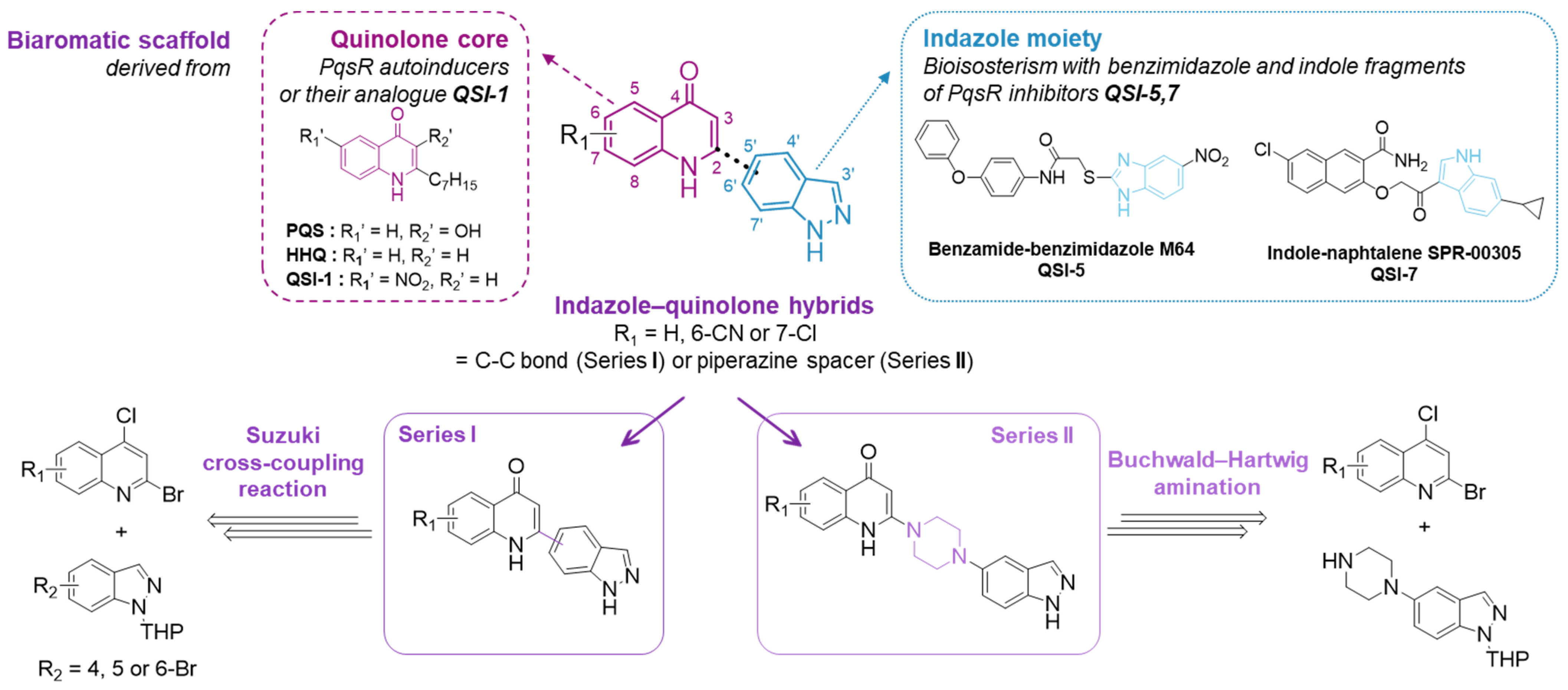
2.1. Chemistry

The design of new indazole–quinolone hybrids relies on palladocatalyzed cross-coupling reactions between various 2-bromo-4-chloroquinoline precursors and indazole ligands such as (i) 4-, 5- or 6-indazolylboronic esters for series I or (ii) the 5-piperazinyl-indazole derivative for series II.

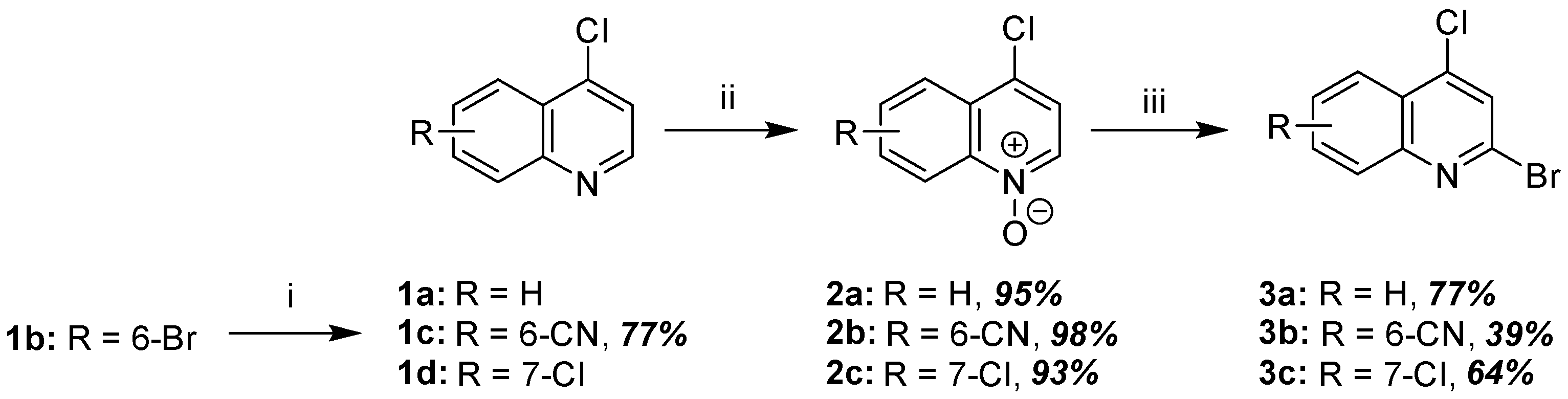
2.1.1. Synthesis of 2-Bromo-4-Chloroquinoline Precursors

Different 2-bromo-4-chloroquinolines 3a–c were synthesized in two steps, starting from the corresponding 4-chloroquinolines 1a and 1c,d (Scheme 1). The N-oxide intermediates 2a–c were first prepared using m-CPBA with excellent yields. Their bromination was performed in the presence of POBr3 and a catalytic amount of DMF to improve the selectivity of the reaction in position 2 compared to position 3 [21]. Of note, a palladocatalyzed cyanation reaction successfully afforded the 6-cyano-4-chloroquinoline 1c from the 6-bromo precursor 1b that was commercially available like 1a and 1d.

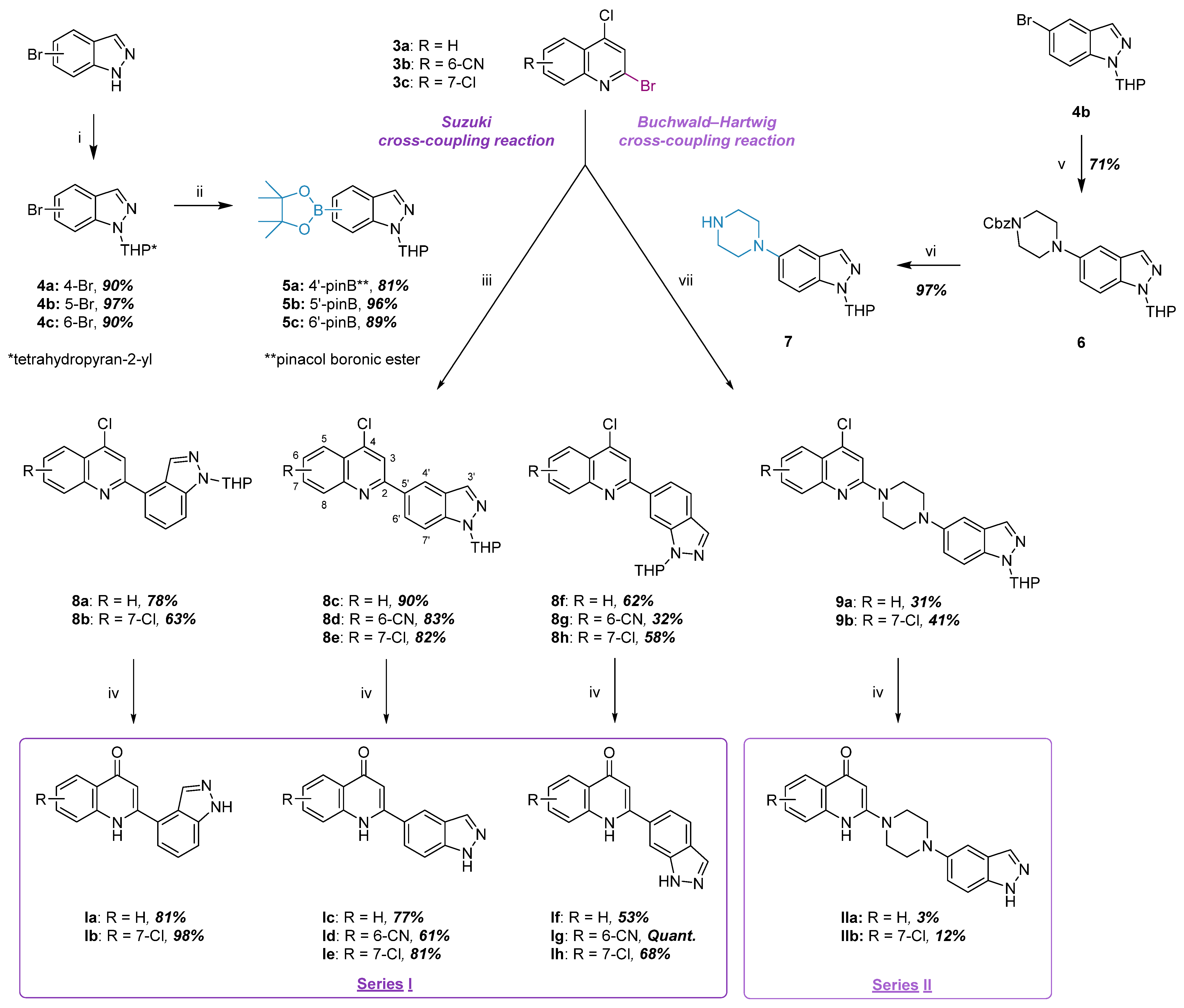
2.1.2. Synthesis of Indazole–Quinolone Hybrids

An indazole moiety was introduced to complete our biaromatic scaffold. As shown in Scheme 2, two series were developed presenting a C-C bond (series I) or a piperazine linker (series II) between the two heteroaromatic cores, respectively. In series I, a key Suzuki cross-coupling reaction was carried out between the 2-bromo-4-chloroquinolines 3a–c and different 4′-, 5′- or 6′-indazolylboronic acid pinacol esters 5a–c. These indazole derivatives and their N1-THP-protected bromo precursors 4a–c were prepared according to the literature procedure [22]. The attempted indazole–quinoline hybrids 8a–h were synthesized using Pd(PPh3)4 as a catalyst in the presence of Cs2CO3 as a weak base in 1,4-dioxane under inert and reflux conditions, producing a 32–90% yield (Table S1) [22]. The indazol-5-yl boronic ester 5b appeared more reactive than its analogues 5a and 5c due to the higher electrophilicity of position 5′ compared to the 4′ and 6′ positions of the ring (natural atomic charge densities of −0.16, −0.22 and −0.18 for carbons C4, C5 and C6 of the indazole nucleus, respectively [23]). To develop series II, a Buchwald–Hartwig amination of 2-bromo-4-chloroquinoline precursors was performed following conditions described by Lohou et al. [24]. Herein, the combination of 5′-(piperazin-1-yl)-1-THP-indazole 7 with 3a and 3c using Pd(OAc)2 and XantPhos as a catalyst/ligand system afforded the hybrids 9a,b in a 31 and 41% yield, respectively. Of note, a good selectivity towards electrophilic position 2 compared to position 4 of the 2-bromo-4-chloroquinolines was observed for both C-C and C-N couplings. The N-indazolylpiperazine derivative 7 was previously synthesized in two steps. The palladocatalyzed hybridization of 5-bromo-1-THP-indazole 4b with N-Cbz-piperazine successfully provided its N-protected precursor 6. In this protocol, the replacement of XantPhos by BINAP as a ligand allowed a yield optimization from 24 to 71% [25]. N-Cbz removal was subsequently achieved via catalytic hydrogenation to give the compound 7 in excellent yield.
A final treatment of key indazole–quinoline hybrids 8a–h and 9a,b with a mixture of AcOH/H2O 4:1 under reflux simultaneously allowed the hydroxydehalogenation of the quinoline core and the cleavage of the THP-protecting group on the indazole moiety to afford the attempted final products Ia–h and IIa,b in good-to-modest yields, respectively [26].
Various new indazole–quinolone hybrids were synthesized in 4–5 steps. The two designed series differ in the nature of the spacer between the two aromatic rings, as follows: (i) series I with eight molecules has a C-C bond between the two nuclei, and (ii) series II with two derivatives has a piperazine spacer. The compounds of series I and II were obtained in 9–51% and 1–4% global yields, respectively (Table S1). Their physicochemical and biological properties as pseudomonal AVAs were investigated.

2.2. Physicochemical and Biological Prerequisite Evaluation

The druggability of the newly designed indazole–quinolone hybrids as AVAs was studied in relation to their physicochemical ability to cross bacterial membranes, but also their innocuousness toward pseudomonal cell growth and host–cell viability.

2.2.1. In Silico-Predicted Physicochemical Properties

Drugs have to fulfil specific physicochemical prerequisites in order to cross the lipopolysaccharidic diderm barrier of GNB. Indeed, the major entry pathway through the outer membrane (OM) is constituted by hydrophilic porin channels, while intracytoplasmic passive diffusion is kept to sufficiently lipophilic molecules [27]. Of note, the biogenesis of OM vesicles (OMVs) would be involved in the internalization of the signalling molecule PQS [28]. With this in mind, a prediction of the physicochemical and ADME profile of indazole–quinolone hybrids was performed using both the Epik and QikProp Schrödinger software [29,30], as well as Entryway, a free web application [31]. The interest of these calculations appears significant since experimental measurements could not technically be carried out for indazole–quinolone hybrids.
As shown in Figure S1A,B, the indazole–quinolone hybrids make no infraction to the Lipinski’s rule of five. This result appears in favour of a passive diffusion through cytoplasmic membranes, yet insufficient to predict a suitable OM permeability in GNB. New guidelines called “eNTRy rules” were recently developed for this purpose [32,33]. These stipulate that the accumulation of small and highly polar molecules in GNB (MW ≤ 600 Da; average clogPo/w = −0.1 and clogD7.4 = −2.8 compared to 2.7 and 1.6 for other non-ATB drugs, respectively) should be facilitated if they possess (i) at least one non-sterically hindered ionizable nitrogen (like a primary amine), (ii) a low three-dimensionality (globularity ≤ 0.25) and (iii) a relatively strong rigidity (flexibility ≤ 5 rotatable bonds) [27,34]. These requirements are mainly driven to cross the hydrophilic porin channels. Indeed, the outer leaflet of OM is composed of lipopolysaccharides (LPSs) that are anchored thanks to a tightly stacking lipid A moiety. This is made up of acyl chains coupled with a phosphorylated disaccharide. In LPSs, a long negatively charged, structurally variable polysaccharide is linked to lipid A via a conserved core oligosaccharide. This protective layer prevents therapeutic molecules from passively diffusing. As shown in Figure S1B, zwitterionic ciprofloxacin fits the eNTRy rules and is used as a reference for porin permeability. In the OmpF (outer membrane porin F) representative channel protein, a narrow constriction site is shaped inside the barrel with both negatively and positively charged amino acids allowing the permeation of only a few sterically convenient ionized drugs such as fluoroquinolones. Furthermore, the pKa values of ciprofloxacin ensure the co-existence at neutral pH of both charged and non-charged species (Figure S1C) [35]. This physicochemical profile makes this class of ATBs the best model for crossing the OM via porins in their ionized form, but also the cell phospholipidic bilayer in their higher-lipophilic non-ionized form. The indazole–quinolone hybrids possess no ionizable functions at physiological pH, as shown in Figure S1D (e.g., compound Ie and its quinolinol tautomer). This lack of compliance with the first eNTRy rule would exclude them from transport across porin channels (Figure S1A,B). However, considering the structural analogy of these newly synthesized biaromatic quinolone-based compounds with PQS, a similar internalization from OMVs could be hypothesized [28,36].
In 2019, Li et al. studied the biophysical interactions between extracellular PQS and the P. aeruginosa OM using molecular dynamics [37]. Their computer simulations demonstrated that PQS’ association with the membrane induces its curvature thanks to a particular docking–folding–insertion sequence. The vesiculation is initiated by the binding of PQS with lipid A phosphates notably via its heterocyclic amine as a hydrogen bond donor. Concomitantly, PQS assumes a specific closed conformation which is favoured by its high heptyl chain flexibility. This facilitates its engagement into the hydrophilic portion of the OM. Compared to PQS, the indazole–quinolone hybrids exhibit a lower globularity, enabling their internalization via the same quinolone-induced biogenesis of OMVs (Figure S1A,B). Their subsequent release in the periplasm would be followed by a passive diffusion through the cytoplasmic membrane. In particular, clogPo/w values reveal a stronger lipophilicity of compounds IIa,b bearing a piperazine spacer compared to Ia–h presenting a C-C hybridization. In series I, the 7-chloroquinolone derivatives Ie,h exhibit optimal clogPo/w values to facilitate passive diffusion, whereas their 6-cyano analogues Id,g possess a significantly increased polarity (e.g., 2.82 for Ie vs. 1.65 for Id). These data are correlated with PCaco results predicting a better gut–blood barrier permeability for hybrids Ie,h compared to Id,g (Figure S1A).

2.2.2. Pseudomonal Minimum Inhibitory Concentrations (MICs)

The MICs of indazole–quinolone hybrids and QSI-3 were determined in the ciprofloxacine-sensitive P. aeruginosa DSM 1117 (ATCC 27853) strain, a reference used as a quality control in antimicrobial susceptibility testing [38]. These revealed no effect on bacterial growth up to a concentration of 128 µg/mL (MIC of ciprofloxacine = 0.06 µg/mL, Table 2). These results constitute a favourable prerequisite for the development of AVAs.

2.2.3. Eukaryotic Cytotoxicity

The cytotoxicity of newly synthesized products was evaluated in vitro on the human hepatoma cell line HepG2. The number of living cells was estimated after incubation with different concentrations of tested compounds for 48 h according to a CellTiter-Glo® Luminescent Cell Viability Assay. No cytotoxicity was observed for 2-indazolyl-4-quinolones Id,f,h at 10 µM or at 100 µM. However, the derivatives Ia,c,e moderately reduced eukaryotic cell viability at 100 µM (Table 2). In comparison, QSI-3 revealed a 30% inhibition of viability in the murine macrophage cell line RAW264.7 at 100 µM [13].
Once the physicochemical and biological druggability prerequisites of the new indazole–quinolone hybrids had been validated, their anti-virulence evaluation was carried out.

2.3. In Vitro Anti-Virulence Evaluation

In order to evaluate the ability of indazole–quinolone hybrids to quench P. aeruginosa virulence, two types of analyses were performed—an anti-biofilm screening followed by an anti-pyocyanin assay. In this primary efficacy study, the common laboratory reference PAO1 strain was chosen for its moderately virulent phenotype and its ability to quickly produce irreversible biofilms [39].

2.3.1. Anti-Biofilm Activity

Biofilm formation was quantitatively assessed on the P. aeruginosa PAO1 strain using crystal violet (CV) staining, after treatment for 24 h with the new indazole–quinolone hybrids at different concentrations (25–100 µM) or with QSI-3 reported as an anti-biofilm reference (IC50 = 20.31 µM, [13,14]). As shown in Figure 2, compounds Ib, Id,e, Ig,h and IIb displayed an interesting biofilm inhibitory activity, while their unsubstituted analogues Ia, Ic and If, as well as QSI-3, were revealed to be inactive. Dose-independent results were noticed in an unexplained manner since no precipitate was notably observed during the experiments. The lack of concordance between these experimental results and those reported in the literature for QSI-3 could be explained by the different bacterial culture conditions (PAO1 biofilm growth monitored in CaCl2/MgSO4-supplemented M9 compared to ABTGC medium [13], respectively). Indeed, the medium composition would significantly affect the adhesion, consistency and architecture of the biofilm developed, but also its detectability via the CV dyeing assay [40]. Nevertheless, using the same normalized method, the newly synthesized molecules bearing an electron-withdrawing substituent in position 6 or 7 of the quinolone core exhibited a stronger inhibitory activity than the selected pyranone-based reference QSI-3. In particular, the 7-chloro derivatives Ib, Ie and Ih were revealed to be the most potent hybrids, with anti-biofilm efficiencies of 32, 35 and 29% at 25 µM, respectively (Table 2). Of note, the compound IIb with a piperazine spacer between the two heteroaromatic moieties appeared less active than its analogue Ie without a spacer. According to this preliminary result, the introduction of a large and rigid linker seems to have a negative impact on anti-biofilm properties.
To extend the anti-virulence screening, the ability of three hybrids from series I to reduce pyocyanin secretion was also investigated. The anti-biofilm hit compound Ie and the less potent but not cytotoxic 6-cyano inhibitor Id, as well as their inactive non-substituted analogue Ic, were chosen for this further evaluation.

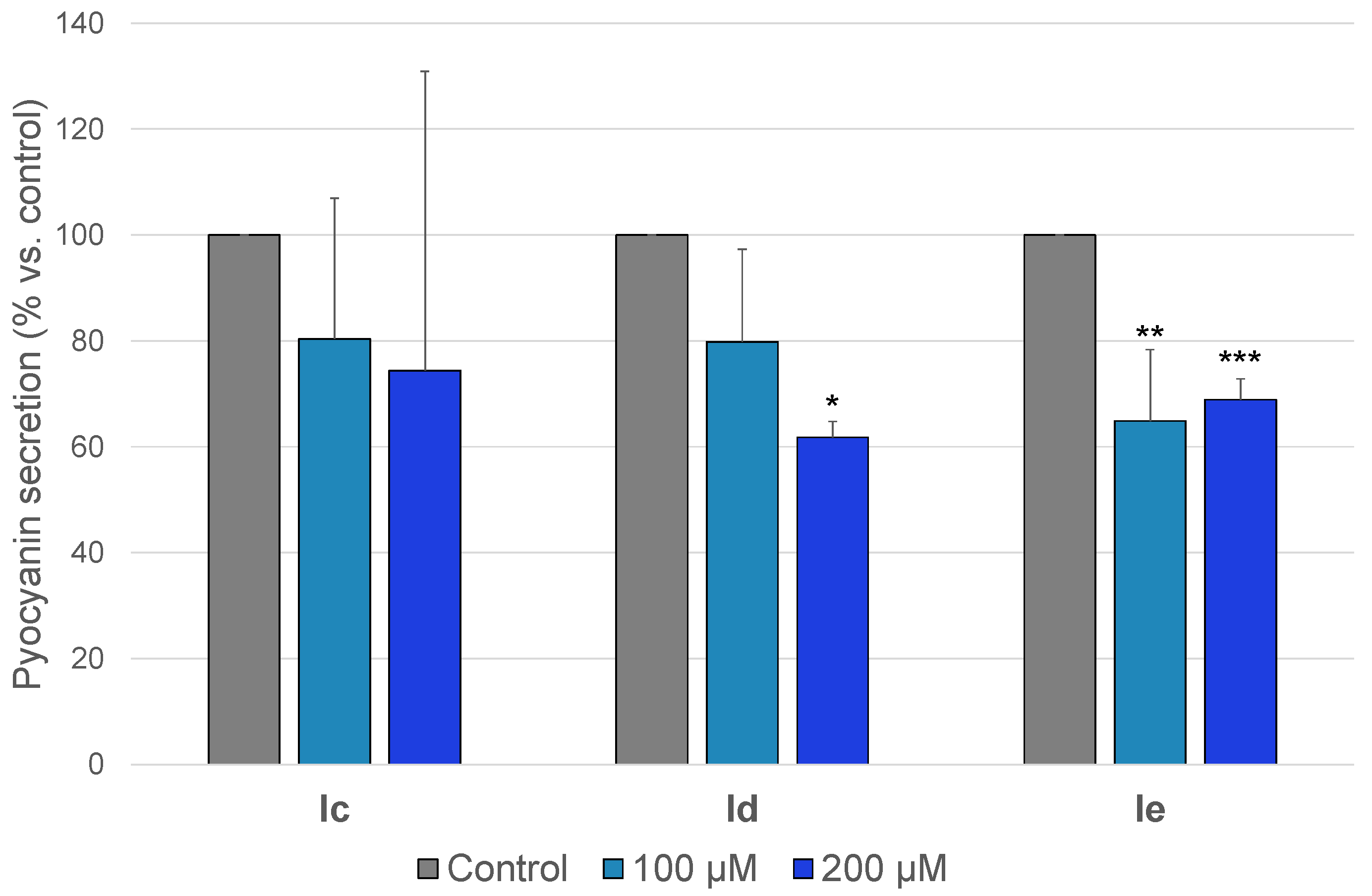
2.3.2. Anti-Pyocyanin Activity

Anti-pyocyanin assays were carried out on the P. aeruginosa PAO1 strain by measuring the redox pigment secretion using UV/Vis spectrometry after treatment for 48 h with the selected products Ic–e at different concentrations. The anti-biofilm agents Id,e displayed an inhibition of pyocyanin production by 38 and 31% at 200 µM, respectively, whereas the hybrid Ic was inactive (Figure 3, Table 2). In comparison, QSI-3 was described as being capable of inducing a 60% decrease in pyocyanin secretion at 40 µM [13]. The 7-chloro derivative Ie also exhibited an anti-pyocyanin efficiency of 35% at 100 µM. This provides novel evidence of the importance of quinolone core substitution with an electron-withdrawing group at position 7 for anti-virulence activity.
Finally, the results obtained from the anti-biofilm and anti-pyocyanin evaluation highlighted the 7-chloro indazole–quinolone hybrid Ie as being a novel anti-virulence hit compound.

2.4. Anti-QS Studies
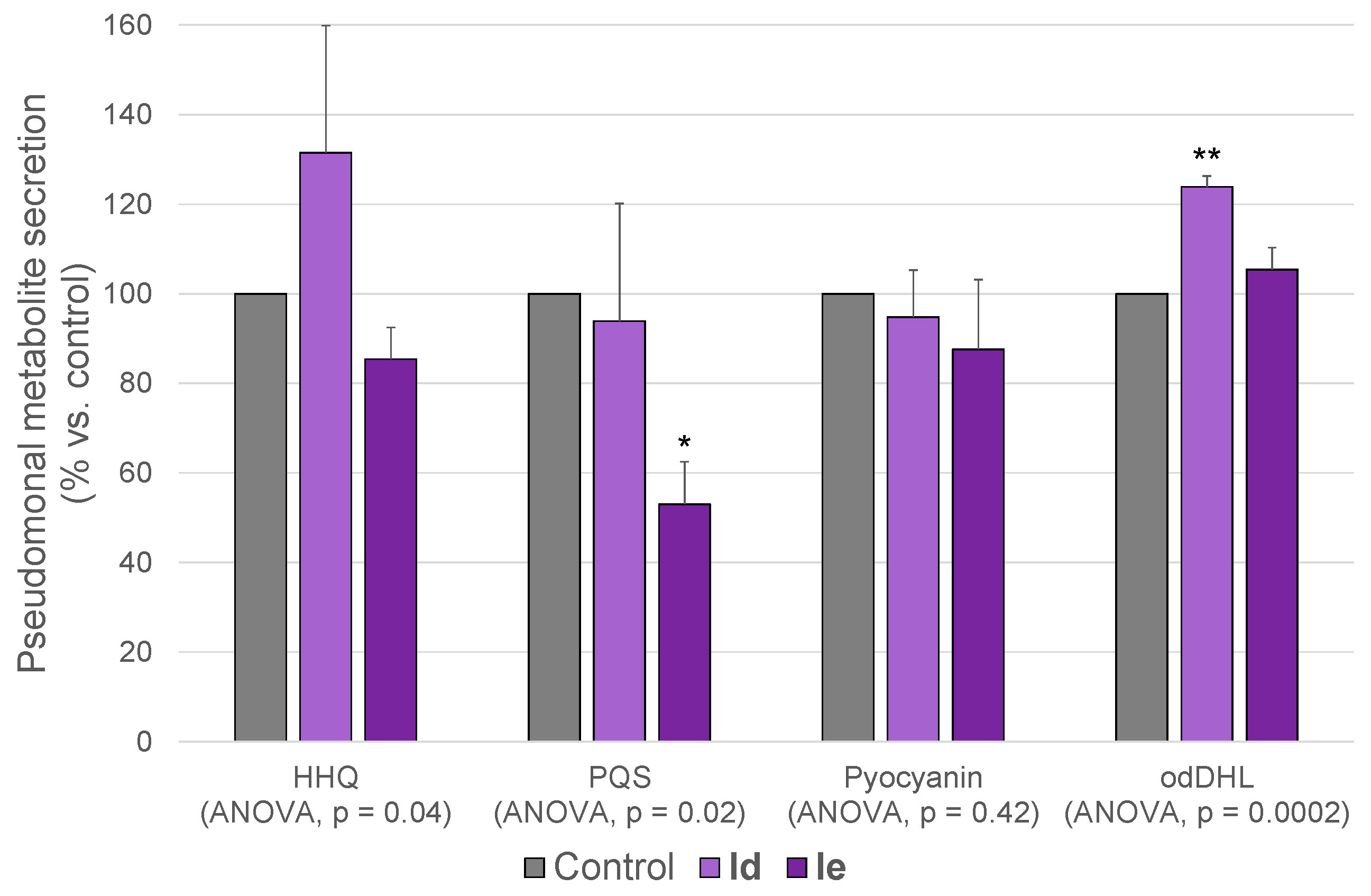
The mechanism of the anti-virulence action of indazole–quinolone hybrids was investigated using metabolomics and molecular modelling, with the aim of analyzing their potential as QS quenchers. In particular, the use of metabolomics to analyze the relevant QS-related metabolites impacted by the tested compounds allowed us to indirectly identify the P. aeruginosa communication pathways that were affected. In this anti-QS study, the hypervirulent reference PA14 strain was used with the aim of ensuring the detectability of the signals in the secretome [39]. Molecular modelling investigations provided a better understanding of the indazole–quinolone hybrid binding on their putative target PqsR.

2.4.1. Metabolomics

An in vitro metabolomic profiling was performed on the P. aeruginosa PA14 strain, using UPLC/HRMS, after treatment for 24 h with either AVA Id or Ie at 200 µM. A subsequent statistical targeted analysis allowed us to evaluate the pseudomonal secretion of two signalling molecules and one virulence factor associated with the pqs system (HHQ, PQS and pyocyanin), as well as of an autoinducer implicated in the las circuit (odDHL) (Table 3). Of note, Savitskii et al. recently reported a similar study about QS metabolites [41].
The indazole–quinolone hydrid Ie displayed a 47% PQS production inhibition (Dunnett’s test vs. control, p value = 0.02), whereas the 6-cyano derivative Id revealed no significant effect on the QS pathways (Figure 4). However, no impact of Ie on HHQ and pyocyanin rates in the bacterial culture supernatant was shown. This could be explained by the low basal excretion of HHQ and the delayed secretion of pyocyanin compared to PQS. Indeed, the quantitative analysis was carried out in the extracellular medium. Therefore, specific anti-pqs system properties were evidenced for the anti-virulence hit compound Ie via this innovative assay, since no influence on the level of odDHL was detected.

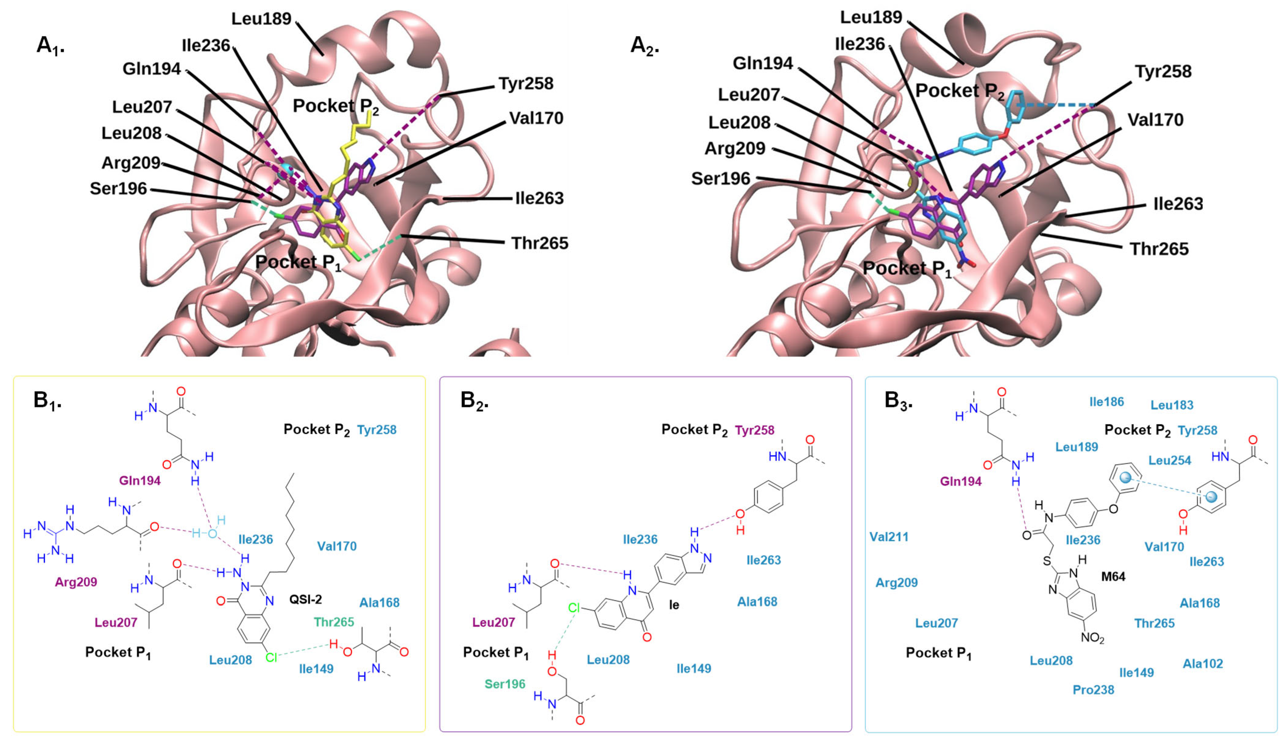
2.4.2. Molecular Modelling

Considering their structural design analogy with alkylquinolone signalling molecules PQS and HHQ, as well as with the PqsR inhibitors QSI-1,2 possessing a quinolone or quinazolone core, the anti-virulence activity of indazole–quinolone hybrids could be supported by anti-QS properties. Concerning the AVA Ie, its anti-QS activity was evidenced by metabolomic studies with a 47% PQS production inhibition (Figure 4). To better understand its mechanism of action, a molecular docking study of Ie in the autoinduction site of PqsR was applied using a co-crystallized structure with the bound ligand QSI-2 as a protein model (PDB code 4JVI).
The low docking enthalpy calculated for Ie and QSI-2 appeared to be in favour of a good affinity for the hydrophobic autoinduction domain of PqsR (−8.5 and −7.8 kcal/mol for Ie and QSI-2, respectively). In compliance with the results described by Ilangovan et al. [12], the molecular docking of QSI-2 in this protein model revealed a hydrophobic binding mode that was also brought out for Ie (Figure 5A1,B1,2). As shown in Figure 5A1, the quinolone core of Ie fitted in the pocket P1 of the receptor occupied by the quinazolone of QSI-2 and its indazole nucleus in P2 as the alkyl chain of the autoinducer analogue. Given its relatively high lipophilicity (clogPo/w = 2.82), the biaromatic compound Ie interacted with numerous hydrophobic residues constituting the pockets P1 and P2 such as Leu208, Ile236 and Ile 263. Additionally, Ie displayed (i) a hydrogen bond between the HBD (hydrogen bond donor) amine function of its quinolone ring and Leu207 (CO), (ii) a hydrogen bond between the HBD amine function of its indazole ring and Tyr258 (OH) and (iii) a halogen interaction between the chloro substituent in position 7 of its quinolone and Ser196 (OH) (Figure 5B2, length of these three polar bindings: 3.74, 2.99 and 3.80 Å, respectively). As previously reported in the literature [12], the polar binding mode of QSI-2 to PqsR involved (i) a hydrogen bond network between the HBD amino substituent in position 3 of its quinazoline and Leu207 (CO), as well as Gln194 (NH2) and Arg209 (CO), via a water molecule and (ii) a halogen interaction between the chloro substituent in position 7 of its quinazoline and Thr265 (OH) (Figure 5B1).
Furthermore, the superimposition of the co-crystallized structure of PqsR with the bound ligand M64 (PDB code 6B8A) and the PqsR crystal model 4JVI with the docked molecule Ie exhibited important similarities between both protein binding modes of these two biaromatic AVAs. Indeed, Ie mimicked the position of M64 in the PqsR autoinduction site. As shown in Figure 5A2, its quinolone core occupied P1 and its indazole nucleus fitted in P2 like the benzimidazole and benzamide moieties of the reference inhibitor, respectively. According to this study and as described in the literature [42,43], the amide linker of M64 displayed a key quenching hydrogen bond with Gln194 (NH2) and a π-stacking in P2 with Tyr258 that also interacted with Ie (Figure 5B3).
Figure 5. Molecular docking study in the PqsR autoinduction site. (A). Topology diagrams of the PQS-binding hydrophobic domain of PqsR (pockets P1 and P2) interacting with QSI-2 (in yellow) or QSI-5 (M64 in cyan) compared to Ie (in purple). (A1) The docking pose of Ie is superimposed on the co-crystallized ligand QSI-2 bound to PqsR. This protein model used for the molecular docking procedure was collected from the RCSB Protein Data Bank (PDB of Research Collaboratory for Structural Bioinformatics) under the access code 4JVI. (A2) The co-crystallized structure of PqsR with the bound ligand M64 (PDB code 6B8A) is overlaid by the PqsR crystal model 4JVI with the docked molecule Ie. The protein structure 6B8A is shown in Figure 5A2 in the same orientation as 4JVI in Figure 5A1. The protein appears as a pink cartoon representation. A relevant water molecule is depicted as a cyan sphere. Ie, QSI-2 and M64 are in stick representations. Nitrogen, oxygen and chlorine atoms are coloured in blue, red and green, respectively. The location of the main residues involved in the interactions with ligands are depicted in black. Dark red, light green and light blue dashed lines indicate hydrogen bonds, halogens and π-stacking interactions, respectively. (B). Schemes generated in accordance with Ligplot schematic diagrams present the significant interactions between the residues lining the autoinduction site of PqsR and (B1) QSI-2, (B2) Ie or (B3) M64 [44,45]. Residues shown in light blue are involved in hydrophobic interactions that are considered relevant within a distance of 3.90 Å. π-stacking interactions are depicted as light blue dashed lines associated with spheres. Hydrogen and halogen interactions with distances from 2.86 to 3.98 are represented by dark red and light green dashed lines connected with the corresponding residues, respectively.
Ddc 03 00030 g005
A supplementary docking study of the 4′-indazolyl derivative Ib bearing a 7-chloroquinolone core that revealed an interesting anti-biofilm efficacy was performed. It exhibited the same key interactions as Ie in the PqsR autoinduction site, contrary to the inactive unsubstituted 5′-indazolyl analogue Ic (Figure S2). This strengthens the analysis of the structure–activity relationships of this new family, demonstrating the importance of the 7-chloro substituent of the quinolone for binding with the receptor.
Considering these results, Ie could compete with the alkylquinolone autoinducer analogue QSI-2 and the benzamide–benzimidazole hybrid M64 with the native ligand PQS for binding to the PqsR autoinduction site. As suggested by Shandil et al., the signalling molecule would fit into its receptor, establishing hydrophobic interactions in pockets P1 and P2, and a hydrogen bond between the HBA (hydrogen bond acceptor) carbonyl group of its quinolone core and Leu197 (NH) [43]. In 2017, Kamal et al. hypothesized that the withdrawal of a water molecule forming a hydrogen bond network with Gln194 (NH), Leu208 (CO) and Arg209 (CO) in the basal unbound protein state could lead to the activation of the transcriptional factor by PQS [11]. Indeed, the HBA hydroxyl group in position 3 of the native autoinducer quinolone ring engaged a competitive hydrogen bond with this water molecule in their docking model, impeding its interaction with the protein backbone and unlocking the receptor. Furthermore, the mechanism of action of QSI-1 was refined in their study [11]. This PqsR inverse agonist displayed a key quenching hydrogen bond between the HBD amide function in position 3 of its quinolone core and Leu207 (CO) and Arg209 (CO). This not only provoked a withdrawal of the previously cited water molecule, but also led to an inactivating conformational change in the transcriptional factor. Considering its similar exclusive HBD bond to Leu 207, the indazole–quinolone hybrid Ie could likewise achieve an inverse agonistic activity.
Hence, these molecular docking investigations highlighted several relevant interactions between the indazole–quinolone hybrid Ie and the PqsR autoinduction domain matching with the binding modes of the two reference inhibitors QSI-2 and M64. Taking the metabolomic and molecular modelling studies into account, PqsR appeared to be a plausible pharmacological target for the newly developed AVA Ie that could inhibit the receptor competing with the signalling molecule PQS.

3. Materials and Methods

3.1. Chemistry

The synthetic procedures and analytical descriptions of the new indazole–quinolone hybrids Ia–h and IIa,b are presented in this section. All their precursors were also characterized. Data concerning compounds 1c, 2a–c, 3a–c, 4a–c, 5a–c, 6 and 7 are reported as supporting information. Of note, NMR spectra were in full accordance with those reported in the literature for compounds 4a–b and 5b [46,47]. The reference anti-biofilm agent QSI-3 was prepared following a procedure described by Li et al. [13].
All commercial reagents were purchased from commercial suppliers and were used without further purification unless otherwise specified. Anhydrous solvents were purchased in sealed containers from Sigma-Aldrich or were dried on the PureSolv MD5 solvent purification system from Serlabo Technologies. Merck Silica gel 60 (40–63 μm) or a Reveleris Grace flash system was used for column chromatography (normal or reverse phase). Melting points (mps) were determined on a Stuart SMP3 apparatus and IR measurements were performed on a Jasco FT/IR-4600 instrument fitted with an ATR-golden gate. 1H and 13C NMR spectra were recorded on a Bruker AC400 spectrometer. Chemical shifts (δ) are expressed in parts per million (ppm) downfield from tetramethylsilane as an internal standard, and the signals are quoted as s (singlet), br s (broad singlet), d (doublet), dd (doublet of doublet), t (triplet), td (triplet of doublet), q (quartet), quint (quintet) or m (multiplet). Coupling constant values are given in Hertz. Mass spectra (MS) and high-resolution mass spectra (HRMS) (electrospray in positive mode, ESI+) were recorded on a Shimadzu LCMS-2020 system and a Micromass-Waters ACQUITY UPLC H-Class system coupled with a SYNAPT G2-Si Q-TOF instrument, respectively. High-performance liquid chromatography (HPLC) was performed to evaluate purity on a Shimadzu LC-20AD apparatus using UV detection, after gradient elution on a Kinetex 5 μm C18 100 Å column. An injection volume of 20 µL and a mobile phase composed of water/acetonitrile with 0.1% trifluoroacetic acid (TFA) from 95:5 to 5:95 with a flow of 1 mL/min were configured.

3.1.1. Synthesis of Indazole–Quinoline Hybrids 8a-h and 9a,b

  • General procedure for the synthesis of compounds 8a–h
After adding them to a Schlenk flask, the 2-bromo-4-chloroquinoline precursor 3a, 3b or 3c (0.5–1.8 mmol) was dissolved with the 4-, 5- or 6-indazolylboronic ester 5a–c (1.1 equiv) and Cs2CO3 (2 equiv) in 1,4-dioxane (7–15 mL) under Ar. The solution was stirred at rt for 10 min. Pd(PPh3)4 (0.05 equiv) was added, and the mixture was heated under reflux for 19–44 h. After the evaporation of 1,4-dioxane, the residue was taken up in CH2Cl2 (30 mL) and the organic phase was washed with brine (2 × 30 mL), dried over Na2SO4 and was concentrated under reduced pressure. The crude material was purified using column chromatography on silica gel (Cyclohexane/EtOAc 90:10 (8a–c,e–f,h), Cyclohexane/EtOAc 90:10 to 85:15 (8g) or CH2Cl2/MeOH 99.5:0.5 (8d)) to give compounds 8a–h in a 32–90% yield.
  • 4-chloro-2-(1-(tetrahydro-2H-pyran-2-yl)-1H-indazol-4-yl)quinoline (8a)
Yellow solid was obtained from compounds 3a (1.32 mmol) and 5a in dry 1,4-dioxane (10 mL) in 78% yield. mp 110 °C. IR ν (cm−1): 2940, 2846, 1583, 1491, 1396, 1313, 1254, 1162, 1075, 1031, 974, 912, 864, 788, 756, 693. 1H NMR (400 MHz, chloroform-d) δ (ppm): 1.67–1.83 (m, 3H), 2.09–2.22 (m, 2H), 2.59–2.68 (m, 1H), 3.75–3.81 (m, 1H), 4.02–4.07 (m, 1H), 5.82 (dd, 1H, 3J = 9.1 Hz, 4J = 2.8 Hz), 7.53 (dd, 1H, 3J = 8.3 Hz, 3J = 7.3 Hz), 7.65 (ddd, 1H, 3J = 8.2 Hz, 3J = 6.9 Hz, 4J = 1.2 Hz), 7.72–7.75 (m, 2H), 7.81 (ddd, 1H, 3J = 8.4 Hz, 3J = 6.9 Hz, 4J = 1.4 Hz), 8.07 (s, 1H), 8.23–8.27 (m, 2H), 8.9 (s, 1H). 13C NMR (100 MHz, chloroform-d) δ (ppm): 22.6, 25.3, 29.6, 67.4, 85.6, 112.0, 120.2, 121.4, 123.1, 124.1, 125.5, 126.6, 127.6, 130.3, 130.8, 132.5, 135.2, 140.5, 143.1, 149.2, 157.2. MS-ESI m/z: [M35Cl + H]+ 364.1, [M35Cl + MeCN + Na]+ 427.1, [2M35Cl + Na]+ 749.2,. HRMS-ESI: m/z calculated for C21H19ClN3O, [M35Cl + H]+ 364.1217, found 364.1221.
  • 4,7-dichloro-2-(1-(tetrahydro-2H-pyran-2-yl)-1H-indazol-4-yl)quinoline (8b)
Orange solid was obtained from compounds 3c (1.38 mmol) and 5a in dry 1,4-dioxane (10 mL) in 63% yield. mp 166 °C. IR ν (cm−1): 2947, 2922, 2844, 1604, 1580, 1538, 1487, 1467, 1442, 1414, 1326, 1261, 1207, 1168, 1119, 1073, 1031, 999, 975, 910, 861, 807, 789, 732, 700, 620. 1H NMR (400 MHz, chloroform-d) δ (ppm): 1.66–1.86 (m, 3H), 2.09–2.22 (m, 2H), 2.58–2.68 (m, 1H), 3.75–3.81 (m, 1H), 4.03–4.07 (m, 1H), 5.81 (dd, 1H, 3J = 9.1 Hz, 4J = 2.7 Hz), 7.51 (dd, 1H, 3J = 8.3 Hz, 3J = 7.4 Hz), 7.58 (dd, 1H, 3J = 8.9 Hz, 4J = 2.0 Hz), 7.71 (d, 1H, 3J = 7.1 Hz), 7.75 (d, 1H, 3J = 8.4 Hz), 8.04 (s, 1H), 8.17 (d, 1H, 3J = 8.9 Hz), 8.22 (d, 1H, 4J = 2.0 Hz), 8.85 (s, 1H). 13C NMR (100 MHz, chloroform-d) δ (ppm): 22.6, 25.3, 29.6, 67.5, 85.6, 112.4, 120.3, 121.6, 123.0, 123.9, 125.5, 126.5, 128.5, 129.1, 132.0, 135.1, 136.9, 140.5, 143.1, 149.5, 158.3. MS-ESI m/z: [M35Cl + H]+ 398.0, [2M35Cl + Na]+ 819.1. HRMS-ESI: m/z calculated for C21H18Cl2N3O, [M35Cl + H]+ 398.0827, found 398.0832.
  • 4-chloro-2-(1-(tetrahydro-2H-pyran-2-yl)-1H-indazol-5-yl)quinoline (8c)
White solid was obtained from compounds 3a (1.65 mmol) and 5b in dry 1,4-dioxane (15 mL) in 90% yield. mp 156 °C. IR ν (cm−1): 2944, 2859, 2361, 1576, 1487, 1402, 1206, 1077, 1038, 844, 795, 764, 663. 1H NMR (400 MHz, d6-DMSO) δ (ppm): 1.57–1.64 (m, 2H), 1.75–1.80 (m, 1H), 1.98–2.08 (m, 2H), 2.39–2.46 (m, 1H), 3.75–3.81 (m, 1H), 3.90–3.93 (m, 1H), 5.92 (dd, 1H, 3J = 9.7 Hz, 4J = 2.4 Hz), 7.73 (ddd, 1H, 3J = 8.3 Hz, 3J = 6.9 Hz, 4J = 1.2 Hz), 7.87–7.89 (m, 2H), 8.15 (d, 1H, 3J = 8.1 Hz), 8.20 (dd, 1H, 3J = 8.4 Hz, 4J = 1.3 Hz), 8.27 (s, 1H), 8.43 (dd, 1H, 3J = 8.9 Hz, 4J = 1.7 Hz), 8.48 (s, 1H), 8.76 (d, 1H, 4J = 0.9 Hz). DEPT-Q NMR (100 MHz, d6-DMSO) δ (ppm): 22.2, 24.8, 29.0, 66.6, 84.1, 110.78, 118.9, 120.7, 123.6, 124.4, 124.6, 125.8, 127.7, 129.6, 130.9, 131.1, 134.7, 140.1, 142.2, 148.3, 156.6. MS-ESI m/z: [M35Cl + H]+ 364.1. HRMS-ESI: m/z calculated for C21H19N3OCl, [M35Cl + H]+ 364.1217, found 364.1231.
  • 4-chloro-2-(1-(tetrahydro-2H-pyran-2-yl)-1H-indazol-5-yl)quinoline-6-carbonitrile (8d)
White solid was obtained from compounds 3b (0.45 mmol) and 5b in dry 1,4-dioxane (7 mL) in 83% yield. mp 202 °C. IR ν (cm−1): 2944, 2862, 2226, 1611, 1582, 1482, 1444, 1364, 1271, 1181, 1081, 1039, 989, 931, 909, 871, 803, 664, 621. 1H NMR (400 MHz, chloroform-d) δ (ppm): 1.66–1.85 (m, 3H), 2.11–2.20 (m, 2H), 2.55–2.63 (m, 1H), 3.77–3.82 (m, 1H), 4.04–4.07 (m, 1H), 5.58 (dd, 1H, 3J = 9.2 Hz, 4J2 = 2.6 Hz), 7.74 (d, 1H, 3J = 8.9 Hz), 7.88 (dd, 1H, 3J = 8.7 Hz, 4J = 1.8 Hz), 8.12 (s, 1H), 8.15 (s, 1H), 8.21 (d, 1H, 3J = 8.7 Hz), 8.28 (dd, 1H, 3J = 8.9 Hz, 4J = 1.7 Hz), 8.54 (d, 1H, 4J = 1.7 Hz), 8.58 (d, 1H, 4J = 1.5 Hz). DEPT-Q NMR (100 MHz, chloroform-d) δ (ppm): 22.6, 25.2, 29.6, 67.7, 85.7, 110.5, 111.0, 118.6, 120.4, 121.4, 124.9, 125.4, 126.2, 130.5, 131.1, 131.4 (2C), 135.2, 140.6, 143.5, 150.1, 160.1. MS-ESI m/z: [M35Cl + H]+ 389.1, [2M35Cl + Na]+ 799.2, [2M35Cl + MeCN + Na]+ 840.2. HRMS-ESI: m/z calculated for C22H18ClN4O, [M35Cl + H]+ 389.1169, found 389.1164.
  • 4,7-dichloro-2-(1-(tetrahydro-2H-pyran-2-yl)-1H-indazol-5-yl)quinoline (8e)
Yellow solid was obtained from compounds 3c (1.8 mmol) and 5b in dry 1,4-dioxane (15 mL) in 82% yield. mp 143 °C. IR ν (cm−1): 2942, 2857, 1578, 1482, 1205, 1076, 1037, 871, 808, 670, 640, 617. 1H NMR (400 MHz, chloroform-d) δ (ppm): 1.68–1.82 (m, 3H), 2.10–2.20 (m, 2H), 2.55–2.65 (m, 1H), 3.76–3.82 (m, 1H), 4.04–4.08 (m, 1H), 5.78 (dd, 1H, 3J = 9.3 Hz, 4J = 2.6 Hz), 7.53 (dd, 1H, 3J = 8.9 Hz, 4J = 2.1 Hz), 7.72 (d, 1H, 3J = 8.9 Hz), 7.99 (s, 1H), 8.11–8.16 (m, 3H), 8.25 (dd, 1H, 3J = 8.9 Hz, 4J = 1.6 Hz), 8.48 (s, 1H). 13C NMR (100 MHz, chloroform-d) δ (ppm): 22.7, 25.3, 29.6, 67.6, 85.6, 110.8, 119.2, 120.9, 123.7, 125.4, 125.5, 126.3, 128.0, 128.9, 131.8, 135.1, 136.7, 140.4, 143.1, 149.6, 158.4. MS-ESI m/z: [M35Cl + H]+ 398.1. HRMS-ESI: m/z calculated for C21H18N3OCl2, [M35Cl + H]+ 398.0827, found 398.0831.
  • 4-chloro-2-(1-(tetrahydro-2H-pyran-2-yl)-1H-indazol-6-yl)quinoline (8f)
White solid was obtained from compounds 3a (1.3 mmol) and 5c in dry 1,4-dioxane (10 mL) in 62% yield. mp 128 °C. IR ν (cm−1): 3060, 2937, 2847, 1588, 1544, 1492, 1413, 1347, 1216, 1075, 1038, 990, 936, 837, 752, 694. 1H NMR (400 MHz, chloroform-d) δ (ppm): 1.67–1.85 (m, 3H), 2.10–2.23 (m, 2H), 2.62–2.71 (m, 1H), 3.80–3.86 (m, 1H), 4.05–4.09 (m, 1H), 5.90 (dd, 1H, 3J = 9.3 Hz, 4J = 2.7 Hz), 7.63 (ddd, 1H, 3J = 8.3 Hz, 3J = 6.9 Hz, 4J = 1.3 Hz), 7.80 (ddd, 1H, 3J = 8.4 Hz, 3J = 6.9 Hz, 4J = 1.5 Hz), 7.85 (dd, 1H, 3J = 8.4 Hz, 4J = 0.8 Hz), 7.97 (dd, 1H, 3J = 8.5 Hz, 4J = 1.4 Hz), 8.06 (s, 1H), 8.10 (d, 1H, 4J = 0.9 Hz), 8.23 (m, 2H), 8.36 (d, 1H, 4J = 1.5 Hz). 13C NMR (100 MHz, chloroform-d) δ (ppm): 22.8, 25.3, 29.6, 67.6, 85.1, 109.3, 119.6, 121.1, 121.6, 124.1, 125.5, 125.6, 127.5, 130.2, 130.8, 134.1, 137.3, 140.2, 143.3, 149.2, 157.5. MS-ESI m/z: [M35Cl + H]+ 364.1, [M35Cl + Na]+ 386.1, [M35Cl + MeCN + Na]+ 427.1, [2M35Cl + Na]+ 749.2.
  • 4-chloro-2-(1-(tetrahydro-2H-pyran-2-yl)-1H-indazol-6-yl)quinoline-6-carbonitrile (8g)
White solid was obtained from compounds 3b (0.8 mmol) and 5c in dry 1,4-dioxane (7 mL) in 32% yield. mp 205 °C. IR ν (cm−1): 2944, 2854, 2226, 1589, 1489, 1449, 1420, 1348, 1278, 1233, 1079, 1040, 992, 946, 919, 888, 834, 691, 639. 1H NMR (400 MHz, chloroform-d) δ (ppm): 1.69–1.89 (m, 3H), 2.11–2.24 (m, 2H), 2.61–2.71 (m, 1H), 3.81–3.87 (m, 1H), 4.04–4.08 (m, 1H), 5.91 (dd, 1H, 3J = 9.1 Hz, 4J = 2.7 Hz), 7.88 (dd, 1H, 3J = 8.5 Hz, 4J = 0.6 Hz), 7.93 (dd, 1H, 3J = 8.7 Hz, 4J = 1.8 Hz), 7.98 (dd, 1H, 3J = 8.5 Hz, 4J = 1.4 Hz), 8.11 (d, 1H, 4J = 0.6 Hz), 8.18 (s, 1H), 8.29 (d, 1H, 3J = 8.8 Hz), 8.40 (d, 1H, 4J = 1.2 Hz), 8.63 (d, 1H, 4J = 1.5 Hz). 13C NMR (100 MHz, chloroform-d) δ (ppm): 22.6, 25.3, 29.6, 67.6, 85.2, 109.9, 111.0, 118.6, 121.0, 121.1, 121.9, 125.2, 126.2, 130.6, 131.5, 131.6, 134.1, 136.1, 140.1, 143.7, 150.1, 160.3. MS-ESI m/z: [M35Cl + H]+ 389.1, [M35Cl + MeCN + H]+ 430.1. HRMS-ESI: m/z calculated for C22H18ClN4O, [M35Cl + H]+ 389.1169, found 389.1182.
  • 4,7-dichloro-2-(1-(tetrahydro-2H-pyran-2-yl)-1H-indazol-6-yl)quinoline (8h)
White solid was obtained from compounds 3c (1.3 mmol) and 5c in dry 1,4-dioxane (10 mL) in 58% yield. mp 158 °C. IR ν (cm−1): 2952, 2846, 1590, 1540, 1470, 1421, 1344, 1314, 1210, 1076, 1039, 994, 943, 873, 842, 814, 696, 627. 1H NMR (400 MHz, chloroform-d) δ (ppm): 1.69–1.89 (m, 3H), 2.10–2.23 (m, 2H), 2.61–2.71 (m, 1H), 3.81–3.87 (m, 1H), 4.05–4.09 (m, 1H), 5.89 (dd, 1H, 3J = 9.3 Hz, 4J = 2.8 Hz), 7.56 (dd, 1H, 3J = 8.9 Hz, 4J = 2.1 Hz), 7.84 (dd, 1H, 3J = 8.5 Hz, 4J = 0.7 Hz), 7.93 (dd, 1H, 3J = 8.5 Hz, 4J = 1.4 Hz), 8.03 (s, 1H), 8.10 (d, 1H, 4J = 0.6 Hz), 8.15 (d, 1H, 3J = 8.9 Hz), 8.21 (d, 1H, 3J = 1.9 Hz), 8.35 (d, 1H, 4J = 1.2 Hz). 13C NMR (100 MHz, chloroform-d) δ (ppm): 22.7, 25.3, 29.6, 67.7, 85.1, 109.4, 119.7, 121.0, 121.7, 124.0, 125.5, 125.8, 128.4, 129.0, 134.1, 136.7, 136.9, 140.2, 143.3, 149.5, 158.5. MS-ESI m/z: [M35Cl + H]+ 398.1, [M35Cl + MeCN + Na]+ 461.1. HRMS-ESI: m/z calculated for C21H18Cl2N3O, [M35Cl + H]+ 398.0827, found 398.0837.
  • General procedure for synthesis of compounds 9a,b
After adding them to a Schlenk flask, XantPhos (0.12 equiv), Pd(OAc)2 (0.1 equiv) and the 5-(piperazin-1-yl)-1-THP-indazole 7 (1.2 equiv) were dissolved in dry 1,4-dioxane (6–13 mL) under Ar. The solution was pre-heated at 100 °C for a duration of 5 min. The 2-bromo-4-chloroquinoline precursor 3a or 3b (0.7–1.8 mmol) and Cs2CO3 (2.8 equiv) were added, and the mixture was heated under reflux for 28 h. After evaporation of 1,4-dioxane, the residue was taken up in EtOAc (30 mL) and the organic phase was washed with brine (2 × 30 mL), dried over Na2SO4 and was concentrated under reduced pressure. The crude material was purified using column chromatography on silica gel (Cyclohexane/EtOAc 80:20 (9a) or Cyclohexane/EtOAc 100:0 to 90:10 (9b)) to afford compounds 9a–b in a 31–41% yield.
  • 4-chloro-2-(4-(1-(tetrahydro-2H-pyran-2-yl)-1H-indazol-5-yl)piperazin-1-yl)quinoline (9a)
Orange solid was obtained from compounds 3a (0.7 mmol) and 7 in dry 1,4-dioxane (6 mL) in 31% yield. mp 130 °C. IR ν (cm−1): 2948, 2844, 1593, 1496, 1416, 1225, 1153, 1077, 1040, 959, 846, 842, 789, 756, 607. 1H NMR (400 MHz, chloroform-d) δ (ppm): 1.64–1.80 (m, 3H), 2.05–2.17 (m, 2H), 2.52–2.61 (m, 1H), 3.25–3.27 (m, 4H), 3.71–3.77 (m, 1H), 3.91–3.93 (m, 4H), 4.01–4.05 (m, 1H), 5.69 (dd, 1H, 3J = 9.3 Hz, 4J = 2.7 Hz), 7.15 (s, 1H), 7.17 (d, 1H, 4J = 1.9 Hz), 7.26 (dd, 1H, 3J = 9.1 Hz, 4J = 2.2 Hz), 7.32 (ddd, 1H, 3J = 8.1 Hz, 3J = 6.9 Hz, 4J = 1.1 Hz), 7.54 (d, 1H, 3J = 9.1 Hz), 7.60 (ddd, 1H, 3J = 8.4 Hz, 3J = 6.9 Hz, 4J = 1.4 Hz), 7.74 (dd, 1H, 3J = 8.4 Hz, 4J = 1.2 Hz), 7.94 (s, 1H), 8.01 (dd, 1H, 3J = 8.3 Hz, 4J = 1.0 Hz). 13C NMR (100 MHz, chloroform-d) δ (ppm): 22.8, 25.3, 29.6, 45.5 (2C), 51.6 (2C), 67.6, 85.6, 106.6, 109.6, 111.0, 121.5, 121.6, 123.4, 124.0, 125.4, 127.1, 130.7, 133.6, 136.0, 143.6, 146.9, 148.6, 157.0. MS-ESI m/z: [M35Cl + H]+ 448.2, [M35Cl + Na]+ 470.2. HRMS-ESI: m/z calculated for C25H27N5OCl, [M35Cl + H]+ 448.1904, found 448.1918.
  • 4,7-dichloro-2-(4-(1-(tetrahydro-2H-pyran-2-yl)-1H-indazol-5-yl)piperazin-1-yl)quinoline (9b)
Yellow solid was obtained from compounds 3c (1.77 mmol) and 7 in dry 1,4-dioxane (13 mL) in 41% yield. mp 130 °C. IR ν (cm−1): 2930, 2809, 1664, 1591, 1496, 1411, 1329, 1270, 1223, 1152, 1074, 1039, 995, 960, 903, 834, 799. 1H NMR (400 MHz, chloroform-d) δ (ppm): 1.64–1.80 (m, 3H), 2.05–2.17 (m, 2H), 2.51–2.61 (m, 1H), 3.23–3.26 (m, 4H), 3.71–3.77 (m, 1H), 3.90–3.93 (m, 4H), 4.01–4.05 (m, 1H), 5.69 (dd, 1H, 3J = 9.3 Hz, 4J = 2.6 Hz), 7.10 (s, 1H), 7.16 (d, 1H, 4J = 1.9 Hz), 7.23–7.26 (m, 2H), 7.54 (d, 1H, 3J = 9.1 Hz), 7.72 (d, 1H, 4J = 2.0 Hz), 7.90 (d, 1H, 3J = 8.8 Hz), 7.94 (s, 1H). 13C NMR (100 MHz, chloroform-d) δ (ppm): 22.8, 25.3, 29.6, 45.3 (2C), 51.5 (2C), 67.6, 85.6, 106.7, 109.5, 111.0, 115.8, 119.9, 121.6, 124.0, 125.4, 126.0, 133.6, 136.0, 136.7, 143.4, 146.8, 149.2, 157.4. MS-ESI m/z: [M35Cl + H]+ 482.2, [M35Cl + Na]+ 504.2, [M35Cl + MeCN + Na]+ 545.2. HRMS-ESI: m/z calculated for C25H26N5OCl2, [M35Cl + H]+ 482.1514, found 482.1508.

3.1.2. Synthesis of Indazole–Quinolone Hybrids Ia-h and IIa,b

The indazole–quinoline hybrid 8a, 8b, 8c, 8d, 8e, 8f, 8g, 8h, 9a or 9b (0.1–1 mmol) was dissolved in AcOH/H2O 4:1 (5–19 mL). The reaction mixture was heated under reflux for 18–47 h and was then neutralized with 28% NH4OH solution. The precipitate was filtered, washed with water and either (i) directly dried under reduced pressure (Ia,b,f,g,h and IIa) or (ii) dissolved in MeOH (Ic,e), CH2Cl2/MeOH 96:4 (Id) or MeCN with 1% TFA (IIb), dried over Na2SO4 and was concentrated under reduced pressure. A further purification was performed except for Ib,g,h using various methods: (i) recrystallization in MeOH (Ie), (ii) column chromatography on silica gel (CH2Cl2/MeOH 96:4 to 90:10 (Ia), CH2Cl2/MeOH 96:4 to 92:8 (Ic,f), CH2Cl2/MeOH 99:1 to 90:10 (Id), or (iii) flash reverse-phase chromatography on a C18 column (H2O/MeCN with 0.1% TFA 95:5 to 20:80, dissolution of crude product in DMSO, injection volume of 500 µL, MeCN addition of 5% each 5 min, flow of 30 mL/min, UV detection at 254 and 248 nm) (IIa–b) to give compounds Ia–h and IIa–b in 8%-Quant at a 3–12% yield, respectively.
  • 2-(1H-indazol-4-yl)-4-quinolone (Ia)
Yellow solid was obtained from compound 8a (1 mmol) in water/AcOH (19 mL) in 81% yield. mp 275 °C. IR ν (cm−1): 3055, 2923, 1595, 1554, 1505, 1472, 1353, 1139, 1082, 951, 862, 782, 750, 664. 1H NMR (400 MHz, d6-DMSO) δ (ppm): 6.41 (s, 1H, H3), 7.37 (ddd, 1H, 3J = 9.0 Hz, 3J = 7.0 Hz, 4J = 1.0 Hz, H6 or H7), 7.49–7.56 (m, 2H), 7.69 (ddd, 1H, 3J = 8.4 Hz, 3J = 7.0 Hz, 4J = 1.5 Hz, H6 or H7), 7.75–7.79 (m, 2H), 8.15 (dd, 1H, 3J = 8.1 Hz, 4J = 1.2 Hz, H5′ or H7′), 8.24 (s, 1H, H3′), 11.87 (br s, 1H, NH), 13.49 (br s, 1H, NH). 13C NMR (100 MHz, d6-DMSO) δ (ppm): 108.5, 112.4, 119.2, 120.4, 120.5, 123.4, 124.7, 125.0, 126.0, 127.5, 131.8, 132.5, 140.4, 140.9, 149.3, 176.4. MS-ESI m/z: [M + H]+ 262.1, [M + MeCN + Na]+ 325.1, [2M + H]+ 523.3, [2M + Na]+ 545.3, [2M + MeCN + Na]+ 586.3. HRMS-ESI: m/z calculated for C16H12N3O, [M + H]+ 262.0980, found 262.0984. Purity: 94%.
  • 7-chloro-2-(1H-indazol-4-yl)-4-quinolone (Ib)
Brown solid was obtained from compound 8b (0.4 mmol) in AcOH/H2O 4:1 (7.5 mL) in 98% yield. mp 180 °C. IR ν (cm−1): 3061, 2957, 1631, 1590, 1554, 1504, 1453, 1406, 1351, 1241, 1076, 952, 927, 872, 821, 783, 742, 692. 1H NMR (400 MHz, d6-DMSO) δ (ppm): 6.39 (s, 1H, H3), 7.38 (d, 1H, 3J = 8.6 Hz, H5), 7.49 (d, 1H, 3J = 7.0 Hz, H5′ or H7′), 7.56 (t, 1H, 3J = 7.6 Hz, H6′), 7.77–7.79 (m, 2H, H8 and H5′ or H7′), 8.13 (d, 1H, 3J = 8.6 Hz, H6), 8.25 (s, 1H, H3′), 11.95 (br s, 1H, NH), 13.48 (br s, 1H, NH). 13C NMR (100 MHz, d6-DMSO) δ (ppm): 109.2, 112.5, 118.0, 120.3, 120.5, 123.7 (2C), 126.1, 126.9, 127.1, 132.5, 136.4, 140.3, 141.3, 149.4, 176.3. MS-ESI m/z: [M35Cl + H]+ 296.0, [M + Na]+ 318.0, [M35Cl + MeCN + H]+ 337.1. HRMS-ESI: m/z calculated for C16H11N3OCl, [M35Cl + H]+ 296.0591, found 296.0602. Purity: 82%.
  • 2-(1H-indazol-5-yl)-4-quinolone (Ic)
Brown solid was obtained from compound 8c (0.5 mmol) in AcOH/H2O 4:1 (10 mL) in 77% yield. mp 270 °C. IR ν (cm−1): 3237, 2944, 1630, 1595, 1552, 1506, 1434, 840, 791, 749, 658. 1H NMR (400 MHz, CD3OD) δ (ppm): 6.62 (s, 1H, H3), 7.42 (ddd, 1H, 3J = 8.1 Hz, 3J = 6.8 Hz, 4J = 1.2 Hz, H6 or H7), 7.69–7.74 (m, 2H), 7.76–7.80 (m, 2H), 8.20 (d, 1H, 4J = 0.8 Hz, H3′), 8.24–8.27 (m, 2H). 13C NMR (100 MHz, CD3OD) δ (ppm): 108.5, 112.1, 119.7, 122.0, 124.6, 125.3, 125.7, 126.0, 127.0, 128.2, 133.6, 136.0, 142.0, 142.3, 154.1, 180.5. MS-ESI m/z: [M + H]+ 262.1, [M + MeCN + Na]+ 325.0, [2M + Na]+ 545.2. HRMS-ESI: m/z calculated for C16H12N3O, [M + H]+ 262.0980, found 262.0988. Purity: 91%.
  • 2-(1H-indazol-5-yl)-4-oxo-1,4-dihydroquinoline-6-carbonitrile (Id)
Yellow solid was obtained from compound 8d (0.1 mmol) in AcOH/H2O 4:1 (5 mL) in 61% yield. mp 250 °C. IR ν (cm−1): 3222, 3138, 2954, 2226, 1675, 1634, 1583, 1554, 1481, 1392, 1172, 1129, 1024, 945, 828, 799, 717. 1H NMR (400 MHz, d6-DMSO) δ (ppm): 6.50 (s, 1H, H3), 7.73 (d, 1H, 3J = 8.7 Hz, H7′), 7.80 (d, 1H, 3J = 8.6 Hz, H6′), 7.91 (d, 1H, 3J = 8.7 Hz, H8), 8.02 (dd, 1H, 3J = 8.7 Hz, 4J = 1.7 Hz, H7), 8.27 (s, 1H, H3′), 8.33 (s, 1H, H4′), 8.44 (d, 1H, 4J = 1.2 Hz, H5), 12.12 (br s, 1H, NH), 13.41 (br s, 1H, NH). 13C NMR (100 MHz, d6-DMSO) δ (ppm): 105.4, 108.6, 111.0, 118.9, 120.3, 120.7, 122.9, 124.4, 125.4, 125.8, 130.6, 133.7, 134.8, 140.6, 143.0, 151.7, 175.7. MS-ESI m/z: [M + H]+ 287.1, [M + MeCN + H]+ 328.1, [2M + H]+ 573.2. HRMS-ESI: m/z calculated for C17H11N4O, [M + H]+ 287.0933, found 287.0929. Purity: 96%.
  • 7-chloro-2-(1H-indazol-5-yl)-4-quinolone (Ie)
Yellow solid was obtained from compound 8e (0.3 mmol) in AcOH/H2O 4:1 (5 mL) in 81% yield. mp 290 °C. IR ν (cm−1): 3242, 3065, 2950, 2360, 1629, 1592, 1549, 1501, 1406, 1249, 1172, 1076, 954, 924, 801, 724, 639. 1H NMR (400 MHz, d6-DMSO) δ (ppm): 6.45 (s, 1H, H3), 7.38 (d, 1H, 3J = 8.7 Hz, H5), 7.73–7.88 (m, 2H, H6′ and H7′), 7.84 (s, 1H), 8.11 (d, 1H, 3J = 8.6 Hz, H6), 8.27 (s, 1H), 8.31 (s, 1H), 11.87 (br s, 1H, NH), 13.38 (br s, 1H, NH). 13C NMR (100 MHz, d6-DMSO) δ (ppm): 107.2, 111.0, 118.2, 120.7, 122.7, 122.9, 124.0, 125.4, 125.9, 126.8, 134.6, 136.5, 140.6, 141.4, 151.9, 175.2. MS-ESI m/z: [M35Cl + H]+ 296.0, [M35Cl + MeCN + Na]+ 359.1, [2M35Cl + Na]+ 613.1. HRMS-ESI: m/z calculated for C16H11N3OCl, [M35Cl + H]+ 296.0591, found 296.0591. Purity: 95%.
  • 2-(1H-indazol-6-yl)-4-quinolone (If)
Brown solid was obtained from compound 8f (0.6 mmol) in AcOH/H2O 4:1 (12.5 mL) in 53% yield. mp 346 °C. IR ν (cm−1): 3128, 2939, 2864, 1632, 1598, 1502, 1470, 1440, 1350, 1251, 1145, 947, 834, 788, 746, 697. 1H NMR (400 MHz, d6-DMSO) δ (ppm): 6.43 (s, 1H, H3), 7.35 (ddd, 1H, 3J = 8.6 Hz, 3J = 7.0 Hz, 4J = 0.8 Hz, H7), 7.54 (d, 1H, 3J = 8.3 Hz, H5′), 7.68 (ddd, 1H, 3J = 8.4 Hz, 3J = 7.0 Hz, 4J = 1.5 Hz, H6), 7.81 (d, 1H, 3J = 8.3 Hz, H8), 7.96 (d, 1H, 3J = 8.4 Hz, H4′), 8.02 (s, 1H, H7′), 8.13 (d, 1H, 3J = 8.1 Hz, H5), 8.21 (s, 1H, H3′), 11.86 (br s, 1H, NH), 13.47 (br s, 1H, NH). 13C NMR (100 MHz, d6-DMSO) δ (ppm): 107.8, 109.4, 118.8, 119.8, 121.4, 123.3, 123.8, 124.8, 124.9, 131.8, 132.0, 133.7, 139.7, 140.6, 150.6, 176.9. MS-ESI m/z: [M + H]+ 262.1, [M + MeCN + Na]+ 325.1, [2M + H]+ 523.2, [2M + Na]+ 545.2. HRMS-ESI: m/z calculated for C16H12N3O, [M + H]+ 262.0980, found 262.0990. Purity: 98%.
  • 2-(1H-indazol-6-yl)-4-oxo-1,4-dihydroquinoline-6-carbonitrile (Ig)
Brown solid was obtained from compound 8g (0.2 mmol) in AcOH/H2O 4:1 (5 mL) in 100% yield. mp 180 °C. IR ν (cm−1): 3303, 3241, 3131, 3072, 2948, 2227, 1632, 1577, 1555, 1484, 1361, 1238, 1188, 1079, 943, 840, 830, 677. 1H NMR (400 MHz, d6-DMSO) δ (ppm): 6.57 (s, 1H, H3), 7.54–8.45 (m, 7H), 12.30 (br s, 1H, NH), 13.46 (br s, 1H, NH). 13C NMR (100 MHz, d6-DMSO) δ (ppm): 105.7, 109.1, 109.8, 118.8, 119.7, 120.6, 121.5, 124.0, 124.2, 130.6, 131.2, 133.6, 133.7, 139.7, 143.1, 151.9, 175.3. MS-ESI m/z: [M + H]+ 287.1. HRMS-ESI: m/z calculated for C17H11N4O, [M + H]+ 287.0933, found 287.0939. Purity: 82%.
  • 7-chloro-2-(1H-indazol-6-yl)-4-quinolone (Ih)
Brown solid was obtained from compound 8h (0.6 mmol) in AcOH/H2O 4:1 (12.5 mL) in 68% yield. mp 180 °C. IR ν (cm−1): 3063, 2948, 1628, 1598, 1578, 1547, 1498, 1451, 1406, 1358, 1243, 1140, 1077, 954, 924, 876, 823, 792, 752, 661. 1H NMR (400 MHz, d6-DMSO) δ (ppm): 6.46 (s, 1H, H3), 7.34–8.21 (m, 7H), 11.81 (br s, 1H, NH), 13.48 (br s, 1H, NH). 13C NMR (100 MHz, d6-DMSO) δ (ppm): 108.2, 109.4, 118.1, 119.7, 121.5, 123.7, 123.8, 123.9, 127.0, 131.8, 133.8, 136.4, 139.8, 141.5, 151.2, 176.3. MS-ESI m/z: [M35Cl + H]+ 296.1, [M35Cl + MeCN + H]+ 337.1, [2M35Cl + H]+ 591.2, [2M35Cl + Na]+ 613.2. HRMS-ESI: m/z calculated for C16H11N3OCl, [M35Cl + H]+ 296.0591, found 296.0591. Purity: 87%.
  • 2-(4-(1H-indazol-5-yl)piperazin-1-yl)-4-quinolone (IIa)
Brown oil was obtained from compound 9a (0.4 mmol) in AcOH/H2O 4:1 (8 mL) in 3% yield. 1H NMR (400 MHz, CD3OD) δ (ppm): 3.38–3.40 (m, 4H, 2 CH2), 3.98–4.01 (m, 4H, 2 CH2), 7.31 (s, 1H), 7.34 (d, 1H, 3J = 7.3 Hz), 7.50–7.55 (m, 2H), 7.80–7.86 (m, 2H), 7.97 (br s, 1H, NH), 8.05 (t, 1H, 3J = 6.9 Hz, H6 or H7), 8.16 (d, 1H, 3J = 8.1 Hz), 8.58 (t, 1H, 3J = 7.9 Hz, H6 or H7), 8.86 (br s, 1H, NH). 13C NMR (100 MHz, CD3OD) δ (ppm): 48.0 (2C), 52.0 (2C), 107.5, 112.1, 112.2, 117.7, 118.7, 123.2, 124.6, 126.3, 134.7 (2C), 139.6, 143.8, 146.8, 147.1, 155.4, 168.4. HRMS-ESI: m/z calculated for C20H20N5O, [M + H]+ 346.1668, found 346.1659. Purity: 91%.
  • 7-chloro-2-(4-(1H-indazol-5-yl)piperazin-1-yl)-4-quinolone (IIb)
Brown oil was obtained from compound 9b (0.6 mmol) in AcOH/H2O 4:1 (12 mL) in 12% yield. IR ν (cm−1): 3180, 3055, 2919, 2849, 1660, 1433, 1182, 1127, 1009, 954, 839, 799, 722. 1H NMR (400 MHz, d6-DMSO) δ (ppm): 3.30–3.32 (m, 4H, 2 CH2), 3.87–3.90 (m, 4H, 2 CH2), 6.64 (s, 1H, H3), 7.20 (d, 1H, 4J = 1.7 Hz, H4′), 7.28 (d, 1H, 3J = 9.1 Hz, H7′), 7.45–7.48 (m, 2H, H6 and H6′), 7.90 (d, 1H, 4J = 1.8 Hz, H8), 7.95 (s, 1H, H3′), 8.02 (d, 1H, 3J = 8.7 Hz, H5). 13C NMR (100 MHz, d6-DMSO) δ (ppm): 46.3 (2 C), 49.9 (2C), 91.5, 105.1, 110.9, 115.7, 118.0, 120.5, 124.4, 125.1, 132.5, 136.2 (2C), 137.0, 140.2, 145.0, 154.5, 166.2. MS-ESI m/z: [M35Cl + H]+ 380.1, [M35Cl + MeCN + Na]+ 443.1. HRMS-ESI: m/z calculated for C20H19N5OCl, [M35Cl + H]+ 380.1278, found 380.1277. Purity: 93%.

3.2. Biological Assays

3.2.1. MIC Determination

A broth-microdilution method was employed to determine the MICs of the evaluated products according to the guidelines provided by the Clinical and Laboratory Standards Institute (CLSI) [48]. Briefly, P. aeruginosa DSM 1117 was previously grown in an aerobic atmosphere for 18–24 h at 37 °C on tryptone soy agar (TSA) in a Petri dish. Two-fold serial dilutions of a stock solution of tested compounds at 6.4 mg/mL in DMSO were prepared in cation-adjusted Mueller–Hinton (MH) broth, at a volume of 230 µL per well in 96-well microtiter plates (VWR International, Radnor, PA, USA). These reach final concentrations ranging from 0.06 to 128 µg/mL. The vehicle (DMSO) was used as a positive culture control (non-treated cells), and ciprofloxacin was used as antibiotic reference. In total, 20 µL of a standardized inoculum were added to each well (except for the negative culture control, which was prepared with saline solution). This was obtained from a 10-fold dilution of a 0.5 McFarland bacterial suspension in saline solution (9 g/L of NaCl in osmosis water), leading to a final bacterial concentration of approximately 8 × 10⁵ CFU/mL per well. Plates were incubated for 18–24 h at 37 °C. The MIC was first visually determined as the lowest concentration at which the medium was clear. Then, the result was verified via measurement of the optical density (OD) at 600 nm (MIC was assessed as the lowest concentration for which OD600 < 0.1) using a Thermo Scientific Multiskan FC microplate photometer (Life Technologies Holdings Pte. Ltd. (a part of Thermo Fischer Scientific Inc.), Singapore).

3.2.2. Cytotoxicity Assay

The cytotoxicity of newly synthesized compounds was investigated on a human hepatoma cell line (HepG2 from ECACC, Merck, Darmstadt, Germany). Briefly, cells were cultured in Dulbecco’s Modified Eagle’s Medium (DMEM) supplemented with 10% fetal bovine serum in a humidified atmosphere of 5% carbon dioxide at 37 °C in 75 cm2 sterile flasks. Serial dilutions of a stock solution of tested compounds at 10 mM in DMSO were prepared in modified DMEM, at a volume of 75 µL per well in 96-well microtiter plates. These reach final concentrations ranging from 0.0003 to 100 μM. The vehicle (DMSO) was used as a positive culture control (non-treated cells). In total, 75 µL of cells were added to each well. After a 48 h incubation period, cell viability was determined thanks to the CellTiter-Glo® Luminescent Cell Viability Assay (Promega, Madison, WI, USA), according to the manufacturer’s protocol. Cell viability was expressed as percentage of control.

3.2.3. Biofilm Formation Assay

The quantitative analysis of biofilm formation was performed using the crystal violet (CV) staining assay according to the originally described O’Toole method [49]. Briefly, P. aeruginosa PAO1 (or PA14) was previously grown in an aerobic atmosphere for 24 h at 37 °C on TSA in a Petri dish. Serial dilutions of a stock solution of tested compounds at 5 mM in DMSO were prepared in M9 minimal medium supplemented with CaCl2 and Mg2SO4 [50], at a volume of 230 µL per well in 96-well microtiter plates. These reach final concentrations ranging from 0 to 250 µM (0, 25, 50, 75, 100, 125, 150, 200, 250 µM for newly synthesized products and 0, 0.1, 0.25, 0.5, 1, 1.5, 10, 25, 50, 100 µM for QSI-3). In total, 20 µL of a standardized inoculum were added to each well (except for the negative culture control, which was prepared with saline solution). This was obtained from a 10-fold dilution of a 0.5 McFarland bacterial suspension in saline solution (9 g/L of NaCl in osmosis water), leading to a final bacterial concentration of approximately 8 × 10⁵ CFU/mL per well. Plates were incubated for 24 h at 37 °C under static conditions. The following day, bacterial growth was estimated by recording the OD600 in each well.
Subsequently, the planktonic cell-containing medium was removed and the wells were washed three times with sterile phosphate-buffered saline (PBS). A total of 280 µL of 0.1% (m/v) CV solution in osmosis water was added to each well. After 15 min of impregnation, the unbound CV dye was discarded. The adhered stained biomass was washed three times with PBS and was dried at room temperature for 24 h. Finally, biofilm was dissolved in 280 µL of 33% glacial acetic acid. Its quantification was carried out by measuring the OD600 in each well and by subtracting the absorbance values obtained in medium sterility conditions (defined as 0% biofilm blank). The biofilm inhibition rate was normalized relative to the amount of biofilm that was formed in the absence of treatment (defined as 100% biofilm control).

3.2.4. Pyocyanin Quantification Assay

According to a method adapted from the literature [51], an overnight TSA culture of P. aeruginosa PAO1 was grown in Luria Broth (LB) in an aerobic atmosphere at 37 °C for 24 h. The inoculum was prepared by diluting bacterial colonies in fresh LB until OD600 = 0.05. A 60 µL aliquot of 20 mM stock solution of tested compounds in DMSO was added to 6 mL of this PAO1 suspension into Falcon™ 15 mL conical centrifuge tubes to reach final concentrations of 100 µM and 200 µM. Positive and negative controls were prepared using 60 µL of DMSO in 6 mL of inoculum or fresh medium, respectively. The samples were incubated in a shaking water bath under aerobic conditions at 37 °C and 200 rpm for 48 h. After a first centrifugation (3000 rpm, 20 °C, 10 min), 3 mL of each supernatant was collected and mixed two times with 2 mL of chloroform by vortexing for a duration of 30 s and by centrifuging (3000 rpm, 20 °C, 2 min) to extract the neutral–basic blue–green form of pyocyanin. Then, its acid red form was extracted by adding 2 mL of 0.2 M HCl to the combined organic phases, proceeding in the same technical conditions. After the transfer of a 250 µL aliquot to a microplate, the absorbance of the separated upper aqueous phase was measured at 492 nm. The pyocyanin inhibition rate was normalized relative to the amount of virulence factor that was secreted in the absence of treatment (defined as 100% pyocyanin production control).

3.2.5. Metabolomics

During a biofilm formation assay on the P. aeruginosa PA14 strain, 200 µL of the planktonic cell-containing medium was collected in the wells containing 75, 100, 150 and 200 µM of tested compounds after 24 h of incubation at 37 °C. These samples were centrifugated (3000 rpm, 20 °C, 10 min) and 100 µL of the bacterial culture supernatant was stored in cryotubes at −80 °C to be studied using metabolomics.
Briefly, 400 µL of cold methanol was added to 100 µL of supernatant, vortexed for a duration of 30 s and was centrifuged (14,000 rpm, 4 °C, 10 min) to precipitate proteins. Then, 450 µL of the second supernatant were filtrated through a cartridge PhreeTM (Phenomenex) to remove phospholipids. The extracts were evaporated to dryness under N2 for a duration of 90 min and were re-suspended in 75 µL of H2O/MeOH + 0.1% formic acid (90:10, v/v). Metabolomic profiles were acquired using an ultra-high-pressure liquid chromatography (UPLC) coupled to a high-resolution mass spectrometer (HRMS, LC1290-QToF 6550A, Agilent), equipped with an electrospray source (ESI) in negative mode. Chromatographic separation was performed on a Phenomenex Kinetex Biphenyl column (150 mm × 2.1 mm, 1.7 µm), equipped with a guard Phenomenex Kinetex Biphenyl (4 mm × 2.1 mm), at 0.4 mL/min, 40 °C and using an injection volume of 5 µL. Mobile phases A and B were H2O + 0.1% formic acid and MeOH + 0.1% formic acid, respectively. The gradient elution was 100% solvent A for a duration of 4 min, followed by a linear gradient from 100 to 0% of phase A from 4 to 18 min. The chromatographic system returned to 100% solvent A in 0.1 min and the run ended with an equilibration step of 11 min. ESI source conditions were set as follows: dry gas temperature at 290 °C and flow at 14 L/min, fragmentor voltage at 400 V, sheath gas temperature at 350 °C and flow at 12 L/min, nozzle voltage at 450 V and capillary voltage at −3600 V. A mass spectrometer calibration occurred during the analytical sequence by following two reference ions: m/z 112.9855 (TFA anion) and m/z 1033.9881 (HP-0921 TFA adduct) (G1969-85001, Agilent). The instrument was set to acquire over the full m/z range of 75–1200, with the MS acquisition rate of 2 spectra/s.
From an untargeted metabolome study, a targeted method, using MassHunter Quantitative Analysis (for Q-ToF) (version 10.1, Agilent), was applied to extract the signals of four metabolites linked to the QS. The analysis of standards permitted the confirmation of their retention time and the m/z of the corresponding detected ions in the mass spectra. Data were normalized with the means of signals for each metabolite in the control group (i.e., non-treated bacterial culture) and were reported as a percentage.

3.3. Molecular Modelling

The structure of the PqsR autoinduction domain co-crystalized with QSI-2 was collected from the RCSB Protein Data Bank (PDB) under the access code 4JVI [12], and was used for the molecular docking procedure. The protein receptor and ligands were prepared for molecular docking with UCSF Chimera and AutoDockTools [52,53]. The water molecule participating in a hydrogen bond network with ligands QSI-2, Gln194 and Arg209 was left in place. Docking computations were performed with AutoDock Vina [54]. Exhaustiveness was set to 100, while all other docking parameters kept their default values. The coordinates of the centre of the docking box were center_x = 32.00, center_y = 57.00, center_z = 10.00, and its sizes were size_x = 18.00, size_y = 15.0, size_z = 10.00. The suitability of AutoDock Vina for this study was assessed by positioning QSI-2 in the protein binding domain. The low RMSD (Root Mean Squared Deviation) value of 1.5 Å showed the weak deviation of the best generated pose compared to the crystallographic reference. Docking poses were visualized with VMD software (version 1.9.3, 30 November 2016) [55].
The structure of the PqsR autoinduction domain co-crystalized with M64 was collected from the RCSB PDB under the access code 6B8A [43]. The PqsR crystal model 4JVI with the docked molecule Ie was superimposed on the protein structure 6B8A with the bound ligand M64 using ChimeraX [56]. A good RMSD value on protein Cα trace of 1.0 Å was obtained.

3.4. Statistical Analysis

The MIC determination and anti-virulence evaluation were carried out in at least three independent experiments performed in triplicate. Anti-biofilm and anti-pyocyanin results are presented as the mean ± standard deviations of these tests. The cytotoxicity of indazole–quinolone hybrids was only assessed in technical triplicates. Statistical analysis of anti-virulence activities was conducted using Mann–Whitney’s test (free web calculator) to compare the differences between control and treated bacteria. p values < 0.05 were considered statistically significant.
A univariate analysis was utilized for metabolomics, starting from the three independent anti-biofilm experiments and using R (version 4.0.3) [57]. Then, one-way analysis of variance (ANOVA) followed by Dunnett’s post hoc test (DescTools package) were applied [58]. Statistical significance was achieved at p values < 0.05.

4. Conclusions

In this study, two new series of indazole–quinolone hybrids were developed as specific anti-virulence agents to fight P. aeruginosa. In series I, eight 2-indazolyl-4-quinolones were synthesized in 4–5 steps including a key palladocatalyzed C-C cross-coupling reaction with 9–51% global yields. In series II, two derivatives possessing a piperazine spacer between the two heteroaromatic nuclei were prepared in 1–4% global yields. The 6-cyano and 7-chloro compounds Id and Ie showed promising anti-biofilm and anti-pyocyanin activities without affecting the bacterial growth. Despite a moderate eukaryotic cytotoxicity, the 2-indazolyl-4-quinolone Ie demonstrated the best anti-virulence efficiency with a 35% inhibition of biofilm formation at 25 µM, and a 35% reduction in pyocyanin secretion at 100 µM. On the contrary, the reference PqsR inhibitor QSI-3 appeared ineffective of quenching biofilm development in our implemented experimental conditions. In the literature, QSI-3 was reported as an anti-biofilm agent (IC50 = 20.31 μM) and as being capable of inducing a 60% decrease in pyocyanin secretion at 40 μM. Our results underline the interest of the new anti-pseudomonal hit agent Ie and the need for harmonized anti-virulence reference tests to compare the different compounds. Interestingly, an innovative in vitro anti-QS evaluation via metabolomics also revealed the significant ability of Ie to quench the pqs communication pathway. In particular, in silico molecular docking studies at the PqsR autoinduction site showed that the indazole–quinolone hybrid Ie could competitively inhibit the binding of PQS in its P. aeruginosa-specific receptor.
Extended pharmacomodulations on the newly highlighted biaromatic scaffold are currently in progress to expand the efficacy screening and to deal with this early drug discovery process in depth. In particular, the biopharmaceutical profile of the hit 2-indazolyl-4-quinolone Ie has to be improved, especially its physicochemical capacity to infiltrate the P. aeruginosa lipopolysaccharidic diderm barrier through porin channels. Further in vitro and in vivo biological investigations will also be carried out to confirm the potential of these optimized anti-virulence agents to restore the efficacy of conventional antibiotics in dual therapy on tolerant P. aeruginosa biofilms.

Supplementary Materials

The following supporting information can be downloaded at: https://www.mdpi.com/article/10.3390/ddc3030030/s1, Supplementary data related to this article (1H NMR, 13C NMR, HRMS and purity analyses using HPLC of indazole–quinolone hybrids Ia–h and IIa,b) are available online.

Author Contributions

Conceptualization, E.L. and P.S.; methodology, E.L. and C.D.; validation, E.L., C.D., N.T. and P.S.; formal analysis, M.H., M.D., C.D., Y.R., S.D.N. and J.-P.B.; investigation, M.H., M.D., C.D., S.D.N. and J.-P.B.; resources, N.T. and P.S.; writing—original draft preparation, E.L.; writing—review and editing, P.S.; supervision, E.L., N.T. and P.S.; project administration, P.S.; funding acquisition, P.S. All authors have read and agreed to the published version of the manuscript.

Funding

Marie Hanot is the recipient of a grant from «Ministère de l’Enseignement Supérieur et de la Recherche». Marie Duplantier was the recipient of a grant from «Ministère de l’Enseignement Supérieur et de la Recherche» and «Région Hauts-de-France» (18004360). We thank these organizations for their financial support.

Institutional Review Board Statement

Not applicable.

Informed Consent Statement

Not applicable.

Data Availability Statement

The original contributions presented in the study are included in the article/Supplementary Materials, further inquiries can be directed to the corresponding authors.

Acknowledgments

We are thankful for the collaborative support of «FHU RESPIRE» («Fédération Hospitalo-Universitaire RESPIRE» certified by GCS G4 and AVIESAN, France).

Conflicts of Interest

The authors declare that they have no known competing financial interest or personal relationships that could have appeared to influence the work reported in this paper.

References

  1. Murray, C.J.L.; Ikuta, K.S.; Sharara, F.; Swetschinski, L.; Robles Aguilar, G.; Gray, A.; Han, C.; Bisignano, C.; Rao, P.; Wool, E.; et al. Global Burden of Bacterial Antimicrobial Resistance in 2019: A Systematic Analysis. Lancet 2022, 399, 629–655. [Google Scholar] [CrossRef] [PubMed]
  2. Tacconelli, E.; Carrara, E.; Savoldi, A.; Harbarth, S.; Mendelson, M.; Monnet, D.L.; Pulcini, C.; Kahlmeter, G.; Kluytmans, J.; Carmeli, Y.; et al. Discovery, Research, and Development of New Antibiotics: The WHO Priority List of Antibiotic-Resistant Bacteria and Tuberculosis. Lancet Infect. Dis. 2018, 18, 318–327. [Google Scholar] [CrossRef]
  3. Mulcahy, L.R.; Isabella, V.M.; Lewis, K. Pseudomonas aeruginosa Biofilms in Disease. Microb. Ecol. 2014, 68, 1–12. [Google Scholar] [CrossRef] [PubMed]
  4. Jurado-Martín, I.; Sainz-Mejías, M.; McClean, S. Pseudomonas aeruginosa: An Audacious Pathogen with an Adaptable Arsenal of Virulence Factors. Int. J. Mol. Sci. 2021, 22, 3128. [Google Scholar] [CrossRef] [PubMed]
  5. Chadha, J.; Harjai, K.; Chhibber, S. Revisiting the Virulence Hallmarks of Pseudomonas aeruginosa: A Chronicle through the Perspective of Quorum Sensing. Environ. Microbiol. 2022, 24, 2630–2656. [Google Scholar] [CrossRef] [PubMed]
  6. Duplantier, M.; Lohou, E.; Sonnet, P. Quorum Sensing Inhibitors to Quench, P. Aeruginosa Pathogenicity. Pharmaceuticals 2021, 14, 1262. [Google Scholar] [CrossRef] [PubMed]
  7. Brindhadevi, K.; LewisOscar, F.; Mylonakis, E.; Shanmugam, S.; Verma, T.N.; Pugazhendhi, A. Biofilm and Quorum Sensing Mediated Pathogenicity in Pseudomonas aeruginosa. Process Biochem. 2020, 96, 49–57. [Google Scholar] [CrossRef]
  8. Rather, M.A.; Saha, D.; Bhuyan, S.; Jha, A.N.; Mandal, M. Quorum Quenching: A Drug Discovery Approach against Pseudomonas aeruginosa. Microbiol. Res. 2022, 264, 127173. [Google Scholar] [CrossRef] [PubMed]
  9. García-Reyes, S.; Soberón-Chávez, G.; Cocotl-Yanez, M. The Third Quorum-Sensing System of Pseudomonas aeruginosa: Pseudomonas Quinolone Signal and the Enigmatic PqsE Protein. J. Med. Microbiol. 2020, 69, 25–34. [Google Scholar] [CrossRef]
  10. Lin, J.; Cheng, J.; Wang, Y.; Shen, X. The Pseudomonas Quinolone Signal (PQS): Not Just for Quorum Sensing Anymore. Front. Cell. Infect. Microbiol. 2018, 8, 230. [Google Scholar] [CrossRef]
  11. Kamal, A.A.M.; Petrera, L.; Eberhard, J.; Hartmann, R.W. Structure–Functionality Relationship and Pharmacological Profiles of Pseudomonas aeruginosa Alkylquinolone Quorum Sensing Modulators. Org. Biomol. Chem. 2017, 15, 4620–4630. [Google Scholar] [CrossRef]
  12. Ilangovan, A.; Fletcher, M.; Rampioni, G.; Pustelny, C.; Rumbaugh, K.; Heeb, S.; Cámara, M.; Truman, A.; Chhabra, S.R.; Emsley, J.; et al. Structural Basis for Native Agonist and Synthetic Inhibitor Recognition by the Pseudomonas aeruginosa Quorum Sensing Regulator PqsR (MvfR). PLoS Pathog. 2013, 9, e1003508. [Google Scholar] [CrossRef]
  13. Li, Y.-B.; Liu, J.; Huang, Z.-X.; Yu, J.-H.; Xu, X.-F.; Sun, P.-H.; Lin, J.; Chen, W.-M. Design, Synthesis and Biological Evaluation of 2-Substituted 3-Hydroxy-6-Methyl-4H-Pyran-4-One Derivatives as Pseudomonas aeruginosa Biofilm Inhibitors. Eur. J. Med. Chem. 2018, 158, 753–766. [Google Scholar] [CrossRef]
  14. Liu, J.; Hou, J.-S.; Li, Y.-B.; Miao, Z.-Y.; Sun, P.-H.; Lin, J.; Chen, W.-M. Novel 2-Substituted 3-Hydroxy-1, 6-Dimethylpyridin-4 (1 H)-Ones as Dual-Acting Biofilm Inhibitors of Pseudomonas aeruginosa. J. Med. Chem. 2020, 63, 10921–10945. [Google Scholar] [CrossRef]
  15. Soukarieh, F.; Liu, R.; Romero, M.; Roberston, S.N.; Richardson, W.; Lucanto, S.; Oton, E.V.; Qudus, N.R.; Mashabi, A.; Grossman, S.; et al. Hit Identification of New Potent PqsR Antagonists as Inhibitors of Quorum Sensing in Planktonic and Biofilm Grown Pseudomonas aeruginosa. Front. Chem. 2020, 8, 204. [Google Scholar] [CrossRef]
  16. Maura, D.; Rahme, L.G. Pharmacological Inhibition of the Pseudomonas aeruginosa MvfR Quorum-Sensing System Interferes with Biofilm Formation and Potentiates Antibiotic-Mediated Biofilm Disruption. Antimicrob. Agents Chemother. 2017, 61, e01362-17. [Google Scholar] [CrossRef]
  17. Starkey, M.; Lepine, F.; Maura, D.; Bandyopadhaya, A.; Lesic, B.; He, J.; Kitao, T.; Righi, V.; Milot, S.; Tzika, A.; et al. Identification of Anti-Virulence Compounds That Disrupt Quorum-Sensing Regulated Acute and Persistent Pathogenicity. PLoS Pathog. 2014, 10, e1004321. [Google Scholar] [CrossRef]
  18. Singh, V.K.; Almpani, M.; Maura, D.; Kitao, T.; Ferrari, L.; Fontana, S.; Bergamini, G.; Calcaterra, E.; Pignaffo, C.; Negri, M.; et al. Tackling Recalcitrant Pseudomonas aeruginosa Infections in Critical Illness via Anti-Virulence Monotherapy. Nat. Commun. 2022, 13, 5103. [Google Scholar] [CrossRef]
  19. Zahler, R. Aryloxyacetylindoles and Analogs as Antibiotic Tolerance Inhibitors. Patent WO 2016/112088 A1, 14 July 2016. [Google Scholar]
  20. Maura, D.; Drees, S.L.; Bandyopadhaya, A.; Kitao, T.; Negri, M.; Starkey, M.; Lesic, B.; Milot, S.; Déziel, E.; Zahler, R.; et al. Polypharmacology Approaches against the Pseudomonas aeruginosa MvfR Regulon and Their Application in Blocking Virulence and Antibiotic Tolerance. ACS Chem. Biol. 2017, 12, 1435–1443. [Google Scholar] [CrossRef]
  21. Wang, D.; Jia, H.; Wang, W.; Wang, Z. A Practical and Mild Chlorination of Fused Heterocyclic N-Oxides. Tetrahedron Lett. 2014, 55, 7130–7132. [Google Scholar] [CrossRef]
  22. Crestey, F.; Lohou, E.; Stiebing, S.; Collot, V.; Rault, S. Protected Indazole Boronic Acid Pinacolyl Esters: Facile Syntheses and Studies of Reactivities in Suzuki–Miyaura Cross-Coupling and Hydroxydeboronation Reactions. Synlett 2009, 4, 615–619. [Google Scholar] [CrossRef]
  23. Mohajeri, A.; Shahamirian, M. Pi-Electron Delocalization in Aza Derivatives of Naphthalene and Indole. Comput. Theor. Chem. 2011, 976, 19–29. [Google Scholar] [CrossRef]
  24. Lohou, E.; Collot, V.; Stiebing, S.; Rault, S. Direct Access to 3-Aminoindazoles by Buchwald–Hartwig C–N Coupling Reaction. Synthesis 2011, 16, 2651–2663. [Google Scholar] [CrossRef]
  25. Ferreira, I.C.F.R.; Queiroz, M.-J.R.P.; Kirsch, G. Synthesis of Diarylamines in the Benzo [b] Thiophene Series Bearing Electron Donating or Withdrawing Groups by Buchwald–Hartwig C–N Coupling. Tetrahedron 2003, 59, 975–981. [Google Scholar] [CrossRef]
  26. O’Neill, P.; Biagini, G.; Ward, S.A.; Berry, N.G.; Nixon, G.; Amewu, R.K.; Pidathala, C.; Hong, W.D.; Gibbons, P.; Leung, S.C.; et al. Antimalarial Compounds. Patent WO 2012/069856 A1, 31 May 2012. [Google Scholar]
  27. O’Shea, R.; Moser, H.E. Physicochemical Properties of Antibacterial Compounds: Implications for Drug Discovery. J. Med. Chem. 2008, 51, 2871–2878. [Google Scholar] [CrossRef]
  28. Turkina, M.V.; Vikström, E. Bacteria-Host Crosstalk: Sensing of the Quorum in the Context of Pseudomonas aeruginosa Infections. J. Innate Immun. 2019, 11, 263–279. [Google Scholar] [CrossRef]
  29. Schrödinger Release. Epik, 2023-3; Schrödinger, LLC.: New York, NY, USA, 2023.
  30. Schrödinger Release. QikProp, 2023-3; Schrödinger, LLC.: New York, NY, USA, 2023.
  31. Entryway. Available online: https://www.entry-way.org/ (accessed on 8 March 2023).
  32. Richter, M.F.; Drown, B.S.; Riley, A.P.; Garcia, A.; Shirai, T.; Svec, R.L.; Hergenrother, P.J. Predictive Compound Accumulation Rules Yield a Broad-Spectrum Antibiotic. Nature 2017, 545, 299–304. [Google Scholar] [CrossRef] [PubMed]
  33. Richter, M.F.; Hergenrother, P.J. The Challenge of Converting Gram-Positive-Only Compounds into Broad-Spectrum Antibiotics: Challenges in Developing Broad-Spectrum Antibiotics. Ann. N. Y. Acad. Sci. 2019, 1435, 18–38. [Google Scholar] [CrossRef] [PubMed]
  34. Prajapati, J.D.; Kleinekathöfer, U.; Winterhalter, M. How to Enter a Bacterium: Bacterial Porins and the Permeation of Antibiotics. Chem. Rev. 2021, 121, 5158–5192. [Google Scholar] [CrossRef]
  35. Sharma, P.C.; Jain, A.; Jain, S.; Pahwa, R.; Yar, M.S. Ciprofloxacin: Review on Developments in Synthetic, Analytical, and Medicinal Aspects. J. Enzym. Inhib. Med. Chem. 2010, 25, 577–589. [Google Scholar] [CrossRef]
  36. Schwechheimer, C.; Kuehn, M.J. Outer-Membrane Vesicles from Gram-Negative Bacteria: Biogenesis and Functions. Nat. Rev. Microbiol. 2015, 13, 605–619. [Google Scholar] [CrossRef] [PubMed]
  37. Li, A.; Schertzer, J.W.; Yong, X. Molecular Conformation Affects the Interaction of the Pseudomonas Quinolone Signal with the Bacterial Outer Membrane. J. Biol. Chem. 2019, 294, 1089–1094. [Google Scholar] [CrossRef] [PubMed]
  38. European Committee on Antimicrobial Susceptibility Testing [EUCAST]. Breakpoint Tables for Interpretation of MICs and Zone Diameters. Version 13.0. Available online: https://www.eucast.org (accessed on 27 April 2023).
  39. Grace, A.; Sahu, R.; Owen, D.R.; Dennis, V.A. Pseudomonas aeruginosa Reference Strains PAO1 and PA14: A Genomic, Phenotypic, and Therapeutic Review. Front. Microbiol. 2022, 13, 1023523. [Google Scholar] [CrossRef] [PubMed]
  40. Haney, E.; Trimble, M.; Cheng, J.; Vallé, Q.; Hancock, R. Critical Assessment of Methods to Quantify Biofilm Growth and Evaluate Antibiofilm Activity of Host Defence Peptides. Biomolecules 2018, 8, 29. [Google Scholar] [CrossRef] [PubMed]
  41. Savitskii, M.V.; Moskaleva, N.E.; Brito, A.; Zigangirova, N.A.; Soloveva, A.V.; Sheremet, A.B.; Bondareva, N.E.; Lubenec, N.L.; Kuznetsov, R.M.; Samoylov, V.M.; et al. Pharmacokinetics, Quorum-Sensing Signal Molecules and Tryptophan-Related Metabolomics of the Novel Anti-Virulence Drug Fluorothiazinon in a Pseudomonas aeruginosa-Induced Pneumonia Murine Model. J. Pharm. Biomed. Anal. 2023, 236, 115739. [Google Scholar] [CrossRef] [PubMed]
  42. Kitao, T.; Lepine, F.; Babloudi, S.; Walte, F.; Steinbacher, S.; Maskos, K.; Blaesse, M.; Negri, M.; Pucci, M.; Zahler, B.; et al. Molecular Insights into Function and Competitive Inhibition of Pseudomonas aeruginosa Multiple Virulence Factor Regulator. mBio 2018, 9, e02158-17. [Google Scholar] [CrossRef] [PubMed]
  43. Shandil, S.; Yu, T.T.; Sabir, S.; Black, D.S.; Kumar, N. Synthesis of Novel Quinazolinone Analogues for Quorum Sensing Inhibition. Antibiotics 2023, 12, 1227. [Google Scholar] [CrossRef] [PubMed]
  44. Wallace, A.C.; Laskowski, R.A.; Thornton, J.M. LIGPLOT: A Program to Generate Schematic Diagrams of Protein-Ligand Interactions. Protein Eng. 1995, 8, 127–134. [Google Scholar] [CrossRef]
  45. Laskowski, R.A.; Swindells, M.B. LigPlot+: Multiple Ligand-Protein Interaction Diagrams for Drug Discovery. J. Chem. Inf. Model. 2011, 51, 2778–2786. [Google Scholar] [CrossRef]
  46. Lohou, E.; Sopkova-de Oliveira Santos, J.; Schumann-Bard, P.; Boulouard, M.; Stiebing, S.; Rault, S.; Collot, V. New Hypotheses for the Binding Mode of 4- and 7-Substituted Indazoles in the Active Site of Neuronal Nitric Oxide Synthase. Bioorg. Med. Chem. 2012, 20, 5296–5304. [Google Scholar] [CrossRef]
  47. Savage, S.; McClory, A.; Zhang, H.; Cravillion, T.; Lim, N.-K.; Masui, C.; Robinson, S.J.; Han, C.; Ochs, C.; Rege, P.D.; et al. Synthesis of Selective Estrogen Receptor Degrader GDC-0810 via Stereocontrolled Assembly of a Tetrasubstituted All-Carbon Olefin. J. Org. Chem. 2018, 83, 11571–11576. [Google Scholar] [CrossRef] [PubMed]
  48. M07-A10; Methods for Dilution Antimicrobial Susceptibility Tests for Bacteria That Grow Aerobically; Approved Standard-Tenth Edition. CLSI: Wayne, PA, USA, 2015; ISBN 978-1-56238-987-1.
  49. O’Toole, G.A. Microtiter Dish Biofilm Formation Assay. J. Vis. Exp. 2011, e2437. [Google Scholar] [CrossRef]
  50. Bortolotti, D.; Trapella, C.; Bragonzi, A.; Marchetti, P.; Zanirato, V.; Alogna, A.; Gentili, V.; Cervellati, C.; Valacchi, G.; Sicolo, M.; et al. Conjugation of LasR Quorum-Sensing Inhibitors with Ciprofloxacin Decreases the Antibiotic Tolerance of P. aeruginosa Clinical Strains. J. Chem. 2019, 2019, 8143739. [Google Scholar] [CrossRef]
  51. Geddis, S.M.; Coroama, T.; Forrest, S.; Hodgkinson, J.T.; Welch, M.; Spring, D.R. Synthesis and Biological Evaluation of 1,2-Disubstituted 4-Quinolone Analogues of Pseudonocardia Sp. Natural Products. Beilstein J. Org. Chem. 2018, 14, 2680–2688. [Google Scholar] [CrossRef] [PubMed]
  52. Pettersen, E.F.; Goddard, T.D.; Huang, C.C.; Couch, G.S.; Greenblatt, D.M.; Meng, E.C.; Ferrin, T.E. UCSF Chimera—A Visualization System for Exploratory Research and Analysis. J. Comput. Chem. 2004, 25, 1605–1612. [Google Scholar] [CrossRef]
  53. Sanner, M.F. Python: A Programming Language for Software Integration and Development. J. Mol. Graph. Model. 1999, 17, 57–61. [Google Scholar]
  54. Trott, O.; Olson, A.J. AutoDock Vina: Improving the Speed and Accuracy of Docking with a New Scoring Function, Efficient Optimization, and Multithreading. J. Comput. Chem. 2010, 31, 455–461. [Google Scholar] [CrossRef]
  55. Humphrey, W.; Dalke, A.; Schulten, K. VMD: Visual Molecular Dynamics. J. Mol. Graph. 1996, 14, 33–38. [Google Scholar] [CrossRef]
  56. Goddard, T.D.; Huang, C.C.; Meng, E.C.; Pettersen, E.F.; Couch, G.S.; Morris, J.H.; Ferrin, T.E. UCSF ChimeraX: Meeting Modern Challenges in Visualization and Analysis. Protein Sci. 2018, 27, 14–25. [Google Scholar] [CrossRef]
  57. R Core Team. R: A Language and Environment for Statistical Computing. R Foundation for Statistical Computing, Vienna, Austria. Available online: https://www.R-project.org/ (accessed on 19 October 2023).
  58. Signorell, A.; Aho, K.; Alfons, A.; Anderegg, N.; Aragon, T.; Arachchige, C.; Arppe, A.; Baddeley, A.; Barton, K.; Bolker, B.; et al. DescTools: Tools for Descriptive Statistics. R Package Version 0.99.47. Available online: https://cran.r-project.org/package=DescTools (accessed on 19 October 2023).
Figure 1. Design strategy of the new indazole–quinolone hybrids.
Ddc 03 00030 g001
Scheme 1. Reagents and conditions: (i) Zn(CN)2 (0.75 eq), Pd(PPh3)4 (0.05 eq), DMF, reflux, 1 h (under Ar); (ii) m-CPBA (1.8 eq), CH2Cl2, r.t., 4 h; (iii) POBr3 (1.2 eq), DMF (0.5 eq), CH2Cl2, r.t., 1–4 h (under Ar).
Ddc 03 00030 sch001
Scheme 2. Reagents and conditions: (i) DHP (1.5 eq), APTS (0.05 eq), THF, reflux, overnight; (ii) Bis(pinacolato)diboron (1.1 eq), KOAc (2.5 eq), PdCl2(dppf).CH2Cl2 (0.08 eq), 1,4-dioxane, reflux, 1–19 h (under Ar); (iii) 4′-, 5′- or 6′-indazolylboronic acid pinacol ester 5a-c (1.1 eq), Pd(PPh3)4 (0.05 eq), Cs2CO3 (2 eq), 1,4-dioxane, reflux, 19–44 h (under Ar); (iv) AcOH/H2O 4:1, reflux, 18–47 h; (v) 1-Cbz-piperazine (1.2 eq), Pd(OAc)2 (0.1 eq), BINAP (0.12 eq), Cs2CO3 (2.8 eq), 1,4-dioxane, reflux, 27 h (under Ar); (vi) H2, Pd/C (10% w/w), MeOH, r.t., 14 h; (vii) 5-(piperazin-1-yl)-1-THP-indazole 7 (1.2 eq), Pd(OAc)2 (0.1 eq), XantPhos (0.12 eq), Cs2CO3 (2.8 eq), 1,4-dioxane, reflux, 28 h (under Ar).
Ddc 03 00030 sch002
Figure 2. Evaluation of biofilm production on P. aeruginosa PAO1 strain. Quantitative analysis via crystal violet staining following 24 h growth in the presence or absence of tested products at different concentrations. Bars represent the mean ± SD of at least three independent experiments performed in triplicate. * p < 0.05 and ** p < 0.01 and *** p < 0.001 vs. control (Mann–Whitney’s test: p values < 0.05 were considered significant).
Ddc 03 00030 g002
Figure 3. Evaluation of pyocyanin secretion on the P. aeruginosa PAO1 strain. Measurement of the pigment concentration using UV/Vis spectrometry after 48 h growth in the presence or absence of tested products at different concentrations. Bars represent the mean ± SD of at least three independent experiments performed in triplicate. * p < 0.05 and ** p < 0.01 and *** p < 0.001 vs. control (Mann–Whitney’s test: p values < 0.05 were considered significant).
Ddc 03 00030 g003
Figure 4. Evaluation of HHQ, PQS, pyocyanin and odDHL secretion by the P. aeruginosa PA14 strain. Measurement of the metabolite rates in the pseudomonal culture supernatant using UPLC/HRMS after 24 h growth in the presence or absence of tested products at 200 µM. A targeted raw data analysis was applied to evaluate the signal of four QS-related metabolites. Extracted signals were normalized with the mean of controls and were reported as a percentage. An analysis of variance (ANOVA) followed by Dunnett’s test were utilized to highlight the differences after P. aeruginosa treatment with either compound Id or Ie. The bars represent the mean ± SD of one experiment performed in triplicate. * p < 0.05 and ** p < 0.001 vs. control (ANOVA and Dunnett’s test: p values < 0.05 were considered significant).
Ddc 03 00030 g004
Table 1. Most relevant PqsR inhibitors described in the literature regarding this study.
PqsR InhibitorsAnti-QS
Properties *
Anti-Virulence Properties **Ref.
% Inhibition
or IC50
Anti-Biofilm
Activity
Anti-Pyocyanin
Activity
% Inhibition
or IC50
% Inhibition
or IC50
Alkylquinolone
autoinducer
analogue QSI
QSI-1 Ddc 03 00030 i0010.051/1.32 µM a/bND ***1.9 µM j[11]
QSI-2 Ddc 03 00030 i0025 µM cObservable using
fluorescence
microscopy
50% at 50 µM k[12]
QSI-3 Ddc 03 00030 i00345%
at 40 µM d
20.31 µM h60% at 40 µM k[13,14]
QSI-4 Ddc 03 00030 i00450%
at 20 µM d
6.57 µM h60% at 20 µM k[14]
Alkylquinolone
autoinducer
non-analogue QSI
QSI-5 ,
(M64)
Ddc 03 00030 i0050.32/1.22 µM c/e50% at 10 µM h,i0.3 µM j[15,16,17]
QSI-6 ,
(D88)
Ddc 03 00030 i0061.31 µM f50% at 10 µM i0.53 µM j[18]
QSI-7 ‡,
(SPR00305)
Ddc 03 00030 i0070.05–0.25 µM gND0.05–0.25 µM j[19]
* In vitro anti-PqsR evaluations were carried out using reporter gene assays on/with a E. coli (DH5α, pEAL08-2)/competition with PQS (50 nM), b P. aeruginosa (PA14-ΔpqsA, pEAL08-2)/competition with PQS (50 nM), c P. aeruginosa (PAO1, miniCTX::pqsA-lux), d P. aeruginosa (PAO1, pqsA-gfp), e P. aeruginosa (PA14, miniCTX::pqsA-lux), f P. aeruginosa (PA14, pqsA-gfp(ASV)) except for g QSI-7, whose anti-PQS activity was quantified on a native PA14 strain using LC–MS/MS analysis; ** in vitro anti-virulence properties were studied at sub-bacteriostatic concentrations (MIC not specified for QSI-1 and QSI-7) on/by h static biofilm formation with P. aeruginosa (PAO1)/CV staining and OD measurement, i P. aeruginosa (PA14)/CV staining and OD measurement, j P. aeruginosa (PA14)/OD measurement, k P. aeruginosa (PAO1)/OD measurement. *** Not determined; †/‡ no or weak eukaryotic cytotoxicity at 50 µM/not described; in vivo data available.
Table 2. Biological evaluation of indazole–quinolone hybrids Ia–h.
Tested
Compound
MW
(g/mol)
Activity Against
Bacterial Growth a
Eukaryotic
Cytotoxicity
(% Inhibition at 100 µM) b
Anti-Virulence Evaluation c
MIC
(µg/mL)
MIC
(µM)
Anti-Biofilm
Activity
(% Inhibition)
Anti-Pyocyanin
Activity
(% Inhibition)
25 µM100 µM100 µM200 µM
Ciprofloxacin331.340.060.18ND *NDNDNDND
QSI-3277.23>128>462ND−16−20NDND
Ia261.28>128>49031ø **øNDND
Ib295.73>128>433ND3239NDND
Ic261.28>128>49042øøøø
Id286.29>128>447Not cytotoxic17øø38
Ie295.73>128>4333335343531
If261.28>128>490Not cytotoxic15øNDND
Ig286.29>128>447NDø22NDND
Ih295.73>128>433Not cytotoxic2930NDND
* Not determined; ** not significant; evaluated on a P. aeruginosa DSM 1117 strain, b HepG2 human hepatoma cell line, c P. aeruginosa PAO1 strain.
Table 3. Characterization of QS-related metabolites using UPLC/HRMS.
MetaboliteRetention Time (min)Base Peak in the Mass Spectrum (ESI−)
Ionm/z
HHQ15.32[M + Cl]278.1317
PQS15.34[M − H]258.1500
Pyocyanin10.39[M + FA * − H]255.0775
odDHL15.6[M − H]296.1867
* Formic acid.
Disclaimer/Publisher’s Note: The statements, opinions and data contained in all publications are solely those of the individual author(s) and contributor(s) and not of MDPI and/or the editor(s). MDPI and/or the editor(s) disclaim responsibility for any injury to people or property resulting from any ideas, methods, instructions or products referred to in the content.

