Review

Biological Profile of Synthetic and Natural Indole Derivatives: Paving New Paths in Cancer Treatment

by
Ana Margarida Janeiro
1 and
Carolina S. Marques
2,*
1
Faculty of Pharmacy, University of Lisbon, Av. Prof. Gama Pinto, 1649-003 Lisbon, Portugal
2
LAQV-REQUIMTE, Institute for Advanced Studies and Research, University of Évora, Rua Romão Ramalho, 59, 7000-641 Évora, Portugal
*
Author to whom correspondence should be addressed.
Drugs Drug Candidates 2024, 3(3), 488-511; https://doi.org/10.3390/ddc3030029
Submission received: 30 May 2024 / Revised: 10 July 2024 / Accepted: 15 July 2024 / Published: 19 July 2024
(This article belongs to the Section Medicinal Chemistry and Preliminary Screening)

Abstract

:
The indole scaffold is considered a privileged framework in the design and synthesis of several active pharmaceutical ingredients, particularly as promising anticancer agents. Its presence in several bioactive natural compounds has caught the attention of the scientific community, which has been committed to unveiling its biosynthetic pathways and generating multiple derivatives with innovative synthetic routes. The large variety of structural derivatives enhances their use in multiple bioapplications and pharmacological activities. In this review, the reader will have easy access to some examples of natural and synthetic indole derivatives with antimicrobial, antidepressant, anti-inflammatory, antiviral, antimigraine, and antiemetic activity. However, the main topic of this review is related to cancer and the importance of indole derivatives as promising anticancer drugs. Two of the reasons why cancer is considered a massive problem worldwide are attributed to the struggle to develop target-specific drugs while avoiding drug resistance. Among countless drugs targeting specific proteins involved in tumorigenesis, prompting life quality in the treatment of several cancer types, protein kinases, desoxyribonucleic acid topoisomerases, and P-glycoprotein have been shown to be the main targets when it comes to the development of novel anticancer agents. Furthermore, indole and its derivatives are also studied regarding affinity to other targets related to cancer. This review aims to highlight the utility of the indole scaffold in anticancer drug design, inspiring the creation and synthesis of new derivatives that target specific proteins and address drug resistance challenges.

Graphical Abstract

1. Introduction

Indole (1) (Figure 1), also named 1H-benzo[b]pyrrole, is a small molecule constituted by a six-member aromatic ring fused with a pyrrole five-membered ring. The general reactivity of (1) is summarized in Figure 1 [1,2,3].
It was first discovered in 1866, in Adolf von Baeyer’s studies on the Indigo plant. Further in his research, he ended up oxidizing indigo to isatin, which was reduced to oxindole and then to (1), after rinsing off the vapors over zinc dust (Scheme 1). Even though it was a synthetic process that gave access to (1), it can be found in a plethora of natural sources, including several plant families (Apocynaceae, Rubiaceae, Catharanthus roseus, Rauvolfia serpentina), aquatic organisms (Aplysina cavernícola), and bacteria (Shewanella algae) [4,5,6].
Regarding its biosynthesis (Scheme 2), (1) is an intermediate in the shikimic acid pathway, a common approach for the biosynthesis of aromatic amino acids, like L-tryptophan, and other metabolites in bacteria, fungi, and plants [7,8].
In humans, several natural derivatives of (1) can be found in metabolic pathways, displaying relevant biological roles. Serotonin (2) (Figure 2) is a monoamine neurotransmitter located in blood cells, peripheral and central nervous system, and cardiovascular tissue. Its basal function is to regulate muscle contraction, as well as vasoconstriction, memory, and others. Given the fact that (2) main receptors are in the nervous system, in neurodegenerative diseases, such as Alzheimer’s and Parkinson’s diseases, the pathophysiological process includes the inhibition of (2) uptake, mainly for overexpressed MAO enzymes, emphasizing the need to use (2) receptor agonists or MAO inhibitors as therapeutic approaches in order to increase the reuptake. Melatonin (3) (Figure 2) is produced from (2) and plays a key role in circadian rhythm, sleep regulation, and seasonal photoperiodic regulation, being used to treat sleeping disorders [8,9,10]. β-Carboline alkaloids (4) (Figure 2) can also be obtained from L-tryptophan (Scheme 2). The complexity of the structure is dependent upon the substituents on the aromatic rings, and therefore, (4) also has neurological effects and is used in cases of depression and neurologic diseases, like Alzheimer’s and Parkinson’s disease [7,8,11]. Both vincristine (5) and vinblastine (6) (Figure 2) have the mechanism of action of interacting with the microtubule binding site, causing cell arrest during mitosis. Compound (5) is used to treat lymphocytic leukemia, neuroblastoma, Wilkin’s tumor, childhood leukemia, and a few more diseases. Meanwhile, (6) is used in the treatment of Hodgkin’s disease, neuroblastoma, breast and lung cancers, and acute and chronic leukemia, among others. By binding to the end of the microtubules, it disrupts this structure, leading to tumor growth suppression, combined with the increase of cAMP and glutathione levels and the inhibition of the synthesis of proteins, nucleic acids, and the DNA repair mechanism [8,12]. Even though both compounds are very efficient in lower doses, when in higher doses, they can have adverse effects, like hallucinogenic and neurotoxic activity. In previous studies, (6) was disclosed to cause an increase in gastrointestinal toxicity and a decrease in bone-marrow formation. Particular attention should be given to (5), since its capacity to disrupt mitosis can affect both cancer and healthy cells [8,12]. Another indole natural derivative performing a key role in anticholinesterase activity is physostigmine (7) (Figure 2). Taking advantage of the carbamate group to establish interactions with the acetylcholinesterase (AChE) active site, it prevents the normal destruction of acetylcholine (ACh), increasing the cholinergic neurotransmission. It also demonstrated the potential to reverse the effects of competitive muscle coolers and in the treatment of Alzheimer’s disease [8].
Despite its widespread presence in nature, its use per se is not feasible. Issues regarding extracting methods, amounts required, purity, and the presence of external interferents led the scientific community to investigate and develop alternative synthetic routes to access (1) and derivatives [13].

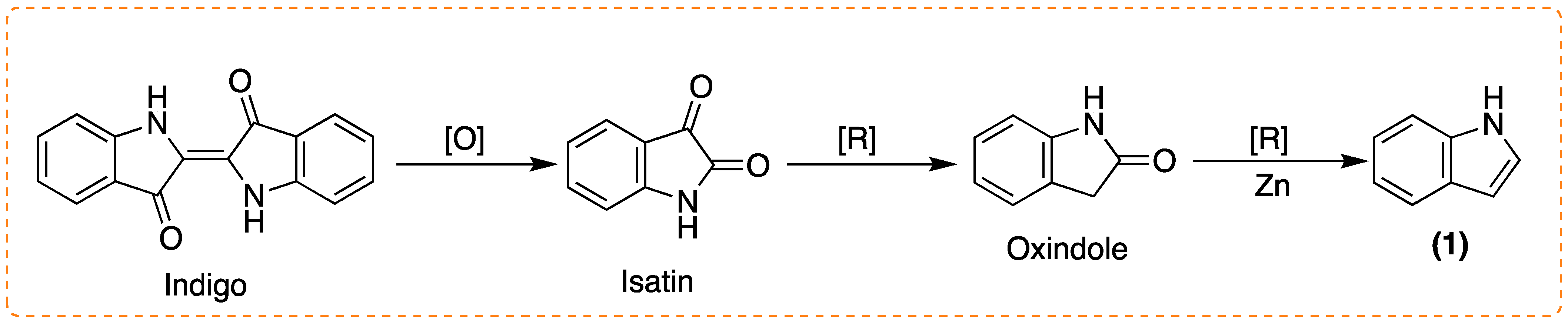
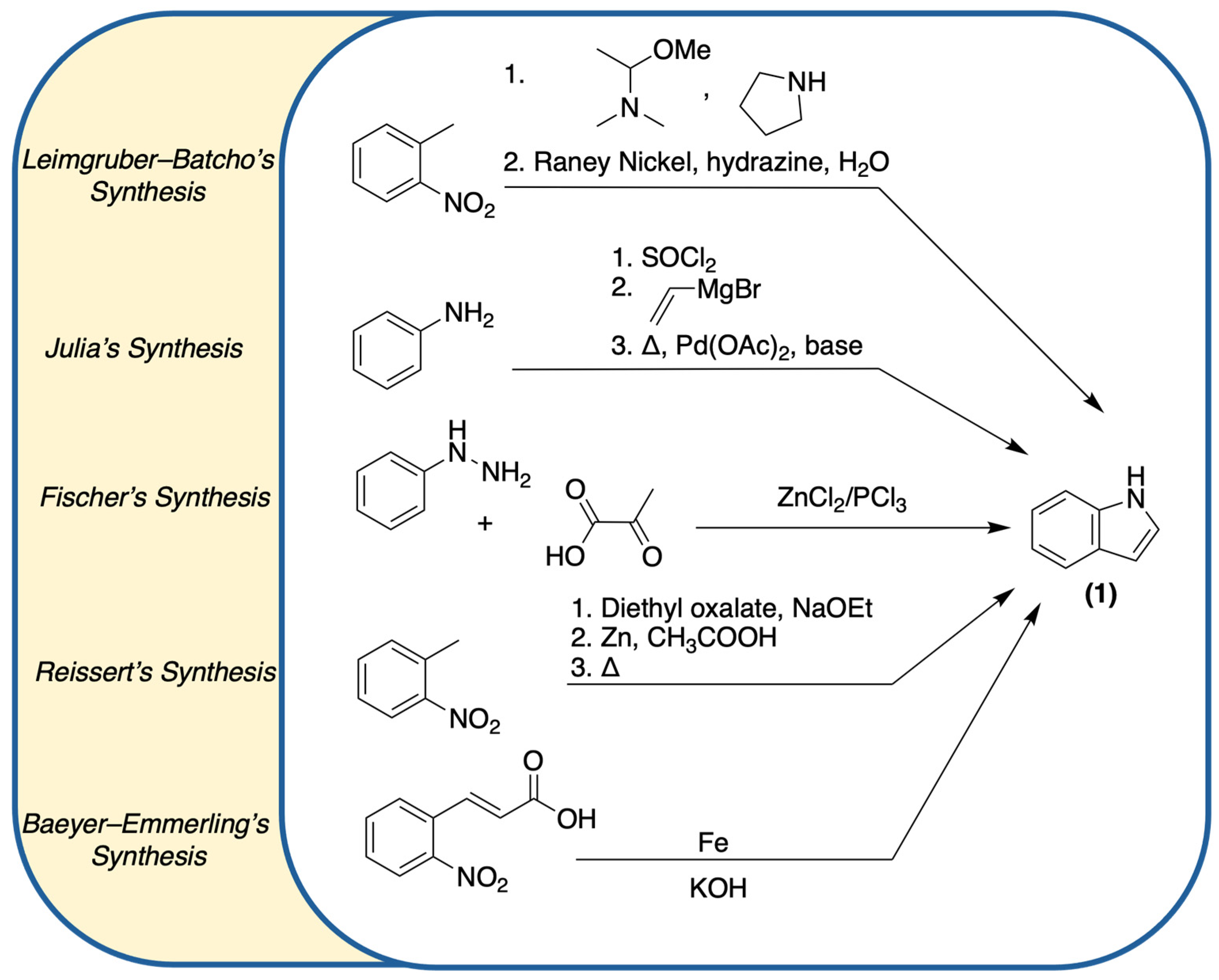
1.1. Accessing (1) and Derivatives: Synthetic Processes

Due to the importance of indole derivatives, several synthetic routes have been developed over the years, focused on (1) or the substituted indole derivatives. The use of transition-metal catalysts is one of the most investigated methods in the literature, since they are crucial for achieving alkyne activation, leading to nucleophilic additions permitting the formation of the indole ring. Palladium, zinc, iron, nickel, rhodium, ruthenium, copper, and silver are some of the transition-metal catalysts reported in the literature [14,15,16]. There are several well-known reaction approaches to access unsubstituted (1), such as Leimgruber–Batcho [17], Julia [18], Fischer [19], Reissert [20], and Baeyer–Emmerling [21] reactions (Scheme 3).
As for the synthesis of substituted indole compounds, a plethora of complex synthetic processes is reported in the literature. Examples described by some research groups are summarized in Scheme 4 [22,23,24].
Non-catalyzed indole synthesis was also highly reported in the literature. Some of the most referenced methods are depicted in Scheme 5, where Madelung [25], Bartoli [26], Larock [27], and Fukuyama [28] reactions can be seen in more detail.

1.2. Biological Profile of the Scaffold

An extensive variety of derivatives could be obtained from (1) (from natural to synthetic and hemisynthetic sources), and they were distinguished for their potential and numerous applications. Sunitinib (8) (anticancer), sumatriptan (9) (antimigraine), perindopril (10) (antihypertension and a heart-failure moderator), and delavirdine (11) (antiviral) are examples of well-known derivatives of (1) used as active pharmaceutical ingredients (APIs), according to the DrugBank database (Figure 3) [5,29,30,31,32].
Other indole derivatives are also used as antitubercular, anticholinergic, antiarrhythmic, anti-asthmatic, antimalarial, antidiabetic, antiplatelet, antidiarrheal, antispasmodic, and antileishmanial agents [33,34]. Several multidisciplinary research groups have settled some structure–activity relationship (SAR) studies, meaning the relevant correlations between the molecular structure of the indole core and its substitution pattern and the corresponding interactions with biological targets. Figure 4 represents some of those established correlations for the development of antimicrobial, antidepressant, and anti-inflammatory indole derivatives [34].

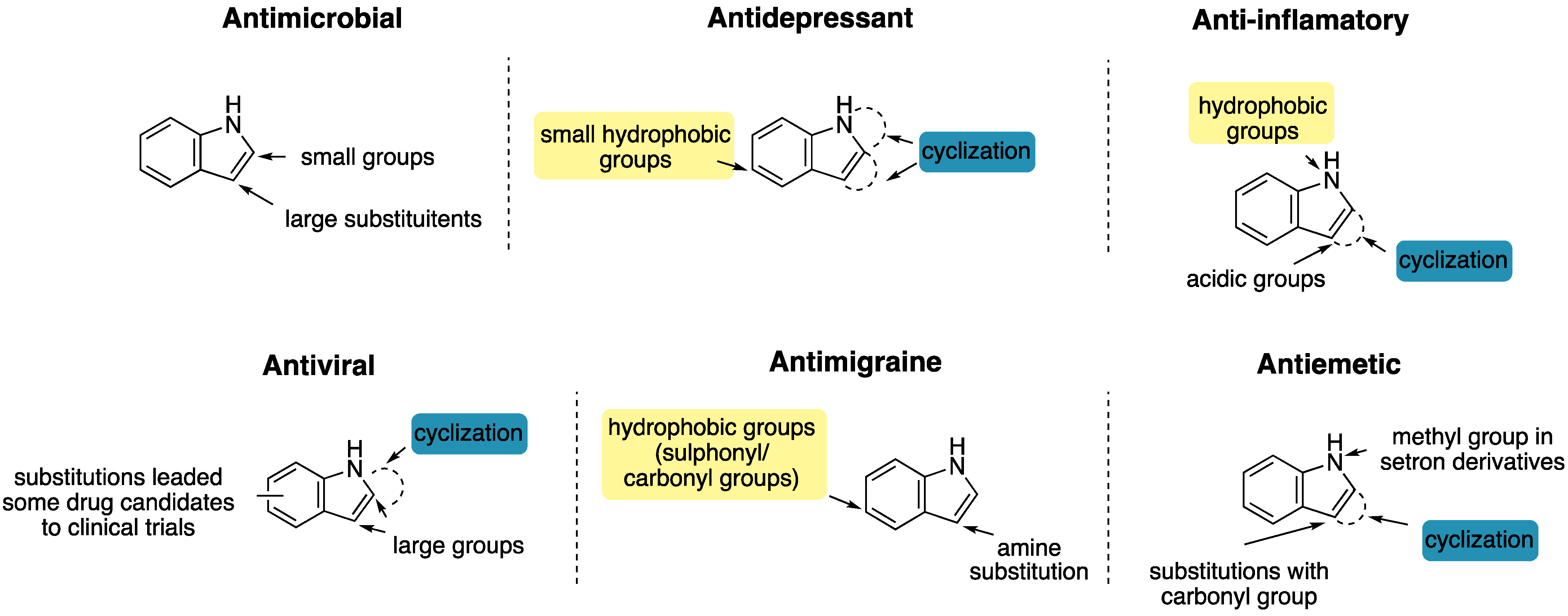
1.2.1. Antimicrobial Activity

From the leaves of Alstonia rupestri were extracted the indole derivatives scholarisins I, II, III, and F (12a-d) (Figure 5). These compounds showed potent antifungal activity against Giberella publicaris and Cercospora nicotianae, exhibiting MIC values of 0.64–0.69 µM, 1.37–1.44 µM, 1.80–1.91 µM, and 1.55–1.71 µM, respectively. Melokhanines B, D, E, and F (13a-d) (Figure 5) were also extracted from natural sources (Melodinus khasianus), and exhibited antibacterial activity against Pseudomonas aeruginosa, presenting MIC values ranging from 2 to 5 µM [33,34]. Regarding synthetic derivatives, compound (14) (Figure 5), synthesized by Choppara and co-workers [35], proved to be active against Gram-positive bacteria, Gram-negative bacteria, and fungi, such as Bacillus subtillis, Klebsiella pneumonia, Pseudomonas aeruginosa, and Aspergillus spp. The group of Mielczarek [36] successfully synthesized the indole derivative (15) (Figure 5), which displayed moderate activity against Bacillus subtillis and Escherichia coli, inferring that substitutions with small groups decrease the activity of the drug candidate [35,36].

1.2.2. Antidepressant Activity

Located on the leaves, mitragynine (16) (Figure 6) is extracted from the Mitragyna speciosa Korth plant. It was tested in mouse models of depression, using the forced-swim test and the tail-suspension test, revealing an abrupt decrease of corticosterone release and allowing a reduction of stress and depressive effects. Moreover, having inhibitory activity against MAO, lyaloside (17) (IC50 of 50.04 µg/mL) and strictosamide (18) (IC50 of 132.5 µg/mL) (Figure 6), extracted from Psychotria suterella and Psychotria laciniate, respectively, induced antidepressant activities in the mitochondrial fractions of rat brains [33]. Synthetic compounds, like indalpine (19) and roxindole (20) (Figure 6), demonstrated activities through 5-HT reuptake inhibition and as a D2R agonist, respectively, with an EC50 of 0.37 nM for (20) [16,37].

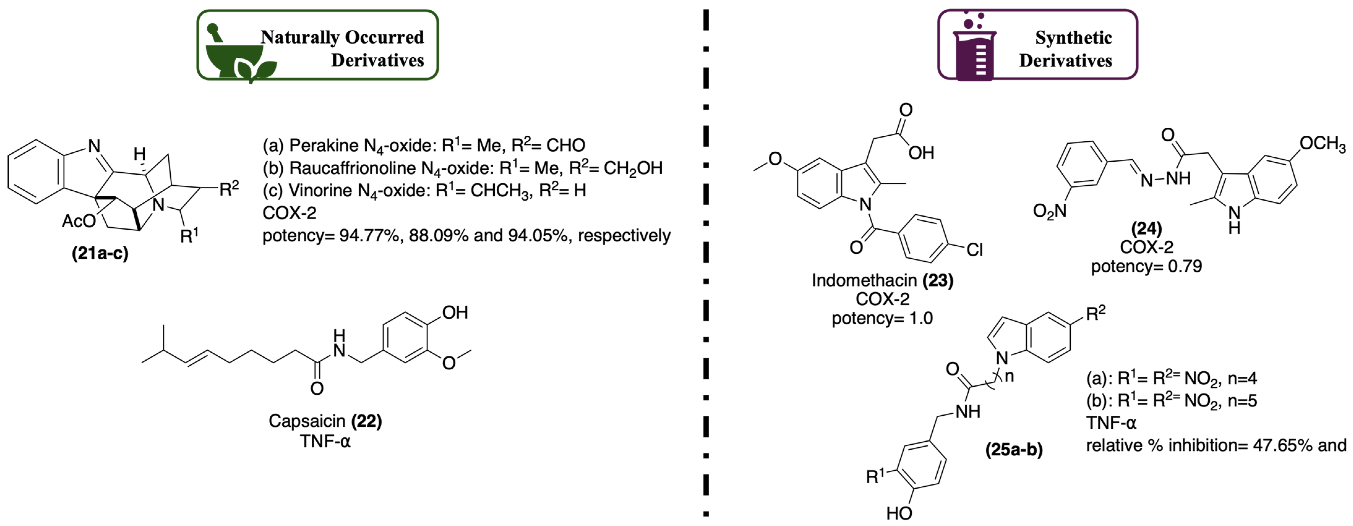
1.2.3. Anti-Inflammatory Activity

From Alstonia yunnanensis, indole derivatives like perakine N4-oxide (21a), raucaffrinoline N4-oxide (21b), and vinorine N4-oxide (21c) (Figure 7) can be found with selective inhibitory activity against COX-2, presenting potency percentages of 94.77%, 88.09%, and 94.05%, respectively. Synthesized by Bhat and co-workers [38], compound (24) (Figure 7) demonstrated a potency of 0.79 against COX-2, which was found to be even more selective when compared to indomethacin (23), (potency of 1.0), a control drug, emphasizing the importance of the presence of the nitrophenol substituent [16,33,38]. Extracted from the seeds of the Capsicum family species, capsaicin (22) (Figure 7) can be found to have inhibitory activity against TNF-α, a proinflammatory kinase. The group of Mukthung [39] synthesized compounds (25a) and (25b) (Figure 7), with different numbers of carbons on the main chain and different substituents in the aromatic rings. It was observed that there was inhibition of TNF-α, suggesting that the presence of nitro groups is crucial for increasing the activity (relative % inhibition of 47.65% and 51.95%, respectively) [16,39]. Curiously, strictosamide (18) (Figure 6), in addition to antidepressant activity, also displayed anti-inflammatory activity, with a relative % inhibition of 28.1%, in an assay with mice with ear-edemas-induced TPA in a dose of 40 mg/kg [33].

1.2.4. Antiviral Activity

From the Alstonia family species, there are 17-nor-excelsinidine (26) and strictamine (27) (Figure 8) compounds, which are extracted from Alstonia scholaris and have antiviral activities against HSV, with EC50 values of 1.09 µg/mL and 0.36 µg/mL, respectively, and ADV, with EC50 values of 0.94 µM and 0.28 µM, respectively [16,33]. From a synthetic origin, oglufanide (28) (Figure 8), an indole derivative capable of inhibiting VEGF and possibly affecting and inhibiting angiogenesis, can also be active against the hepatitis C virus. Atevirdine (29) (Figure 8), acts as a non-nucleoside reverse transcriptase inhibitor (NNRTI). NNRTIs bind to reverse transcriptase (the enzyme responsible for the conversion of RNA to DNA), playing an essential role in stopping virus development [16,33]. Recent in silico studies revealed that indomethacin (23) (Figure 7) could exhibit antiviral activity against SARS-CoV-2 [40].

1.2.5. Antimigraine Activity

Several indole–triptan derivatives were developed regarding antimigraine activity, like nicergoline (30) and rizatriptan (31) (Figure 9), which act as agonists of 5-HT1B and 5-HT1D receptors. From marine sources, in this case from the Caledonian sponge Gellius sp., gelliusine A (32) (Figure 9) showed an analogous behavior to ergot indole derivatives [34]. Ergot is a disease of wild and cultivated wheat, and over the years, many compounds have been isolated from diseased plants and studied regarding their mechanism of action. Likewise, the ergot compounds, derived from ergotamine (33), originated from lysergic acid (34), an ergolinic alkaloid (Figure 10), were classified as another indole class that holds structural similarities to serotonin and dopamine, herein expressing an agonist activity in the same receptors. They have been used as antimigraine agents and for the treatment of diseases implying vasodilatation, such as Parkinson’s disease, given its vasoconstriction effects [8].

1.2.6. Antiemetic Activity

Antiemetic drugs are usually applied to treat nausea and vomiting symptoms, due to their antagonist action on 5-HT3 receptors. Even though there are no natural sources of indole derivatives with antiemetic activity, as far as we know, some synthetic products have been developed to ameliorate the side effects caused by chemotherapy, radiation therapy, and surgery [34]. Some examples of synthetic compounds derived from the indole scaffold with antiemetic activity are ondansetron (35), tropisetron (36), and alosetron (37) (Figure 11) [34].

2. Indole in Cancer

In 2023, the National Cancer Institute (NCI) defined cancer as “a disease in which some of the body’s cells grow uncontrollably and spread to other parts of the body” [41]. Generally, cancer is a multifactorial disease, which can be caused by either internal or external [42] factors, and therefore, there is always a certain degree of unpredictability since it is a consequence of epigenetic DNA modifications, resulting in a genetic mutation that leads to the formation of several defective proteins in the organisms. In fact, there is no conventional explanation for the development of cancer, and, over the years, several approaches/mechanisms of action have been presented regarding tumorigenesis [43]. This malfunction is not so amendable as it may seem to our immune system, considering that once the mutation is present, it starts to develop defensive tools surrounding DNA repair mechanisms, deceiving normal cells and enhancing cancer cells’ growth, usually using the synergy between p53 and MDM2 by inhibition and overexpression, respectively. This event allows for the mutation to grow and accumulate genetic and epigenetic alterations (giving heterogenicity properties), providing the necessary conditions to form carcinogen–DNA adducts that inactivate tumor-suppressor genes and/or activate protooncogenes [43,44,45]. Those effects make tumor cells capable of behaving as self-sufficient, of growing, and of ignoring signals to stop dividing or to die (apoptosis), supplying oxygen by promoting angiogenesis and producing energy from nutrients obtained in different ways, among other features. In tumor cells, the conditions to proliferate from localized tumors to other anatomical locations, is a process called metastasis [44,45,46]. As previously mentioned, mutations can be triggered by damage to DNA sequences, whether caused by DNA methylation, which is essential for the regulation of gene expression. But, when over or under-expressed, it can also develop oncogenic diseases or, by interaction with reactive oxygen species (ROS), where DNA oxidation modifies the nitrogenous bases, induces damage to it, resulting in genomic instability. Damaged DNA produces mutated proteins that will work in the function of cancer survival and not regulate normal cell mechanisms [47,48]. Genetic mutations affect signaling pathways, stimulating the activation of proto-oncogenes, such as GTPases (e.g., Ras, Rab, and Rho), lipid kinases (e.g., PI3K), NRTK (e.g., Abl, Src, and BTK), RTK (e.g., EGFR), amongst others, and inactivation of tumor-suppressor genes, such as p53, PTEN, APC, MLH1, BRCA1 and BRCA2, and CDK (e.g., Akf and Raf), that can work as both an oncogene and TSG. Those proteins, important for normal cell growth and regulation, become overexpressed, and others, responsible for apoptosis, become muted, allowing the tumor to grow, proliferate, and metastasize [49]. In the last decades, indole (1) has been considered a privileged structure in the design of new APIs to treat certain pathologies, especially targeting the proteins involved in cancer treatment. Considering that the indole scaffold can easily undertake chemical modifications, allowing the synthesis of numerous derivatives, over the last few years, many reports regarding the synthesis and bioprofiles of these derivatives have established some trends, with crucial assessments regarding their anticancer activity. It has been observed that the presence of a carbonyl group at position 2 of the indole scaffold (oxindole-type compounds) increased cytotoxicity in cancer cells, as well as the presence of a spiro-ring at position 3 (Figure 12). The same trend was noticed regarding the presence of a methoxyl group at position 3, whereas at position 5 of the indole scaffold, it was observed that there was activity in the upregulation of TSG. The presence of a double bond and an ester group at position 3 also increased cytotoxicity. The introduction of an aryl group, substituted with Br at position 3 or 5, exhibited favorable results regarding TK inhibition (Figure 12). The presence of halogen derivatives (F, Cl, Br) at position 5 or position 7 of the aromatic ring of the indole scaffold also produces an effect on cytotoxicity (Figure 12) [34,50].

2.1. Main Targets of Indole Derivatives in Cancer

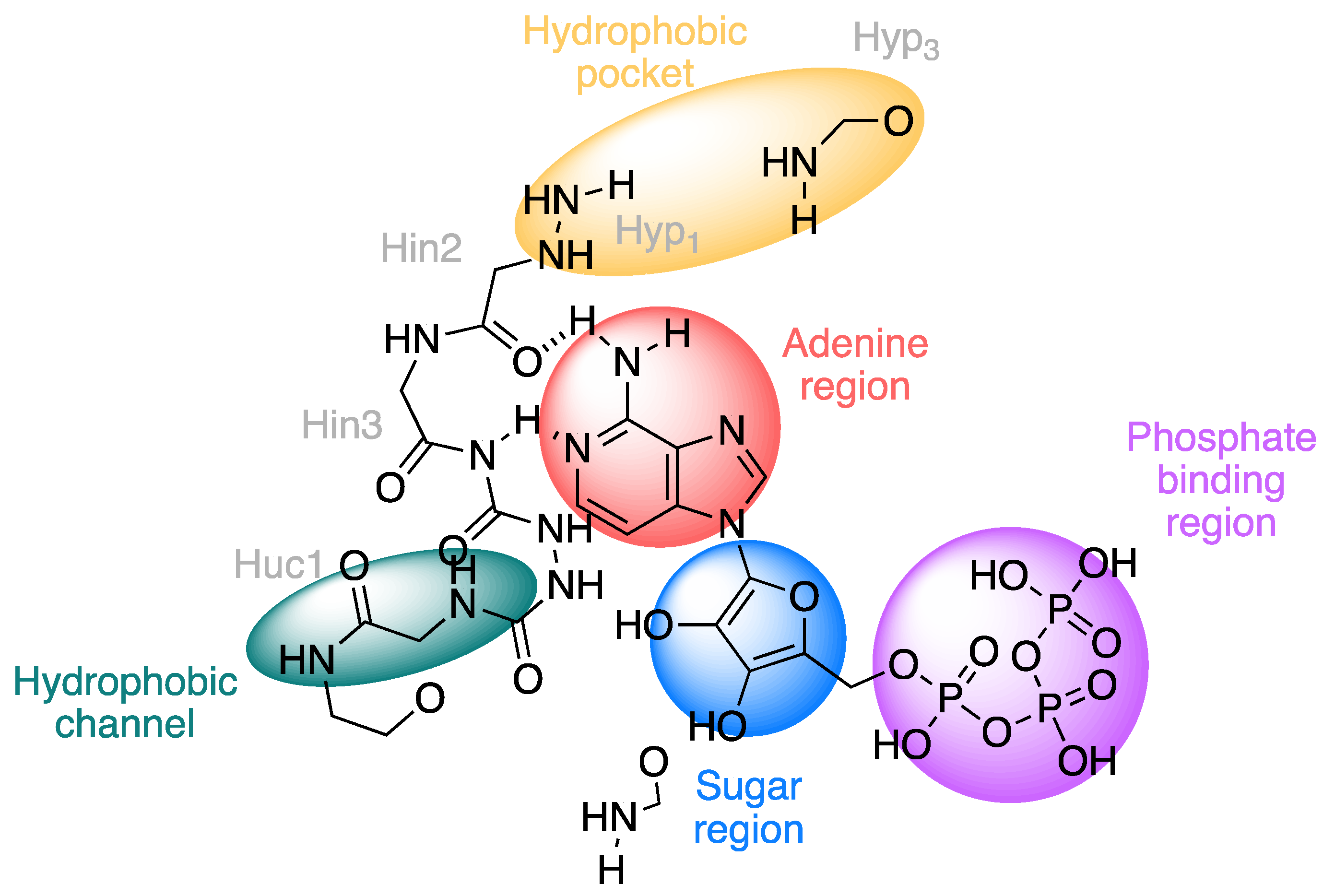
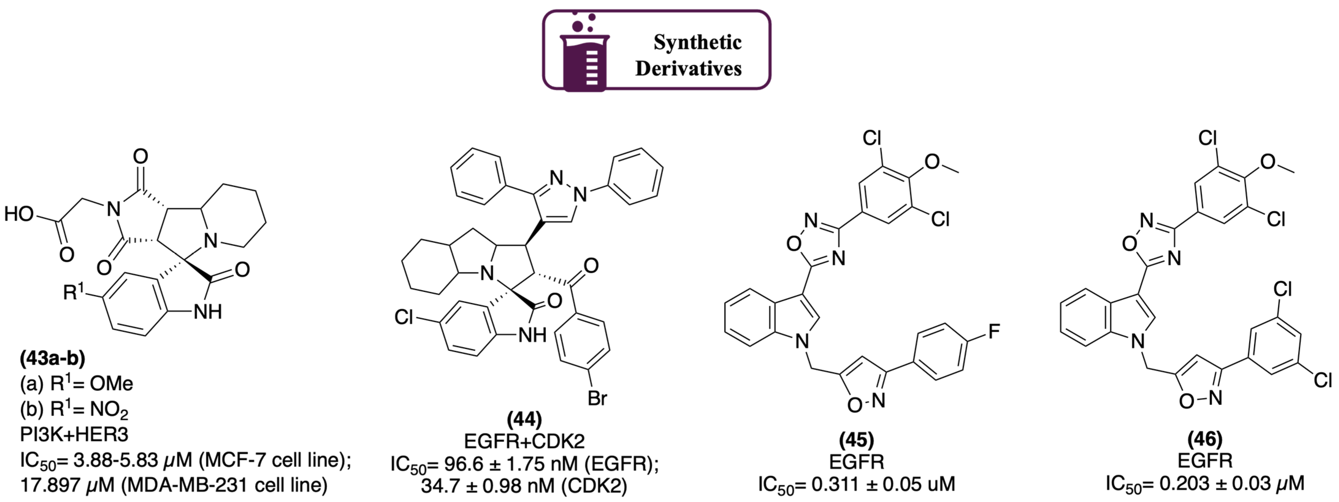
2.1.1. Protein Kinases

Protein kinases (PKs) are capable of catalyzing phosphate-group transfer from adenosine triphosphate (ATP) to the residues of specific proteins. They assist several signaling pathways, expressing activity in cellular processes, such as the regulation of transcription factors, mRNA stability, and protein translation, therefore playing a role in cell growth and development. Given its diversity and multiple roles in several pathways, PKs represent a major influence in cell development, being consequently a target in cancer research. However, the innumerous families of this class of proteins make the design of inhibitors a difficult and challenging task, since the specificity depends deeply on the interactions established by the small molecule with the active site of the PK. Great efforts have been made over the last several years to develop small molecules that can inhibit PK in the active site, whether through competition or conformational alteration [29,51]. Studies developed by Paul and co-workers [52] using a theoretical model of PK ATP-binding site (Figure 13) demonstrated key interactions to consider when designing inhibitors. A general PK active site presents an adenine region that holds the main interactions (through hydrogen bonds) with the hinge region, a sugar interaction region (that contributes to the overall binding), a hydrophobic channel and pocket (without established interactions), and a phosphate-binding region (which anchors ATP to the binding site) [52].
Regarding synthetic indole derivatives, sunitinib (Sutent) (8) (Figure 3 and Figure 14) is a well-known multitarget PTKI that is often used as the first-line treatment for renal cell carcinoma and gastrointestinal stromal tumors, exhibiting antiangiogenetic activity [53,54]. Osimertinib (Tagrisso) (38) (Figure 14) targets the EGFRATP binding site, and it is used as a first-line drug for non-small cell lung cancer and as a potential antiglioblastoma agent [53,55]. Among the indole alkaloids derived from natural sources, midostaurin (39) (Figure 14), an indole alkaloid isolated from Streptomyces staurosporeus, was identified as a multikinase inhibitor due to its activity against PKC-α, VEGFR, KIT, PDGFR, WT, and mutant FLT3, with IC50 values in a sub-micromolar range. It induces apoptosis, being preferably used in the treatment of acute myeloid leukemia and systemic mastocytosis [56,57]. Breifussin C (40) (Figure 14), extracted from the marine hydrozoan Thuiaria breitfussi, targets PKs such as PIM1 and DRAK1. It displays activity in a wide range of cancer cell lines, highlighting the drug-resistant triple-negative breast MDA-MB-468 cell line, with IC50 values between 0.34 and 3.0 µM. It also presented IC50 values below 200 nM of inhibition of PIM1 and DRAK1, suggesting that (40) is a potential candidate to be a selective kinase inhibitor in breast cancer treatment [6,58]. Saccharomonosporine A (41) (Figure 14), extracted from Callyspongia siphonella and targeting PIM1, presents antiproliferative activity in colon (HT29) cancer cells, with an IC50 value of 3.5 µM. Christodoulou and co-workers [59] synthesized a set of derivatives of (41), which presented inhibitory activity against PIM 1, 2, and 3, with IC50 values of 0.22 to 2.46 µM [6,59]. Meridianins B, C, D, and E (42a-d) (Figure 14) from sea squirt Aplidium meridianum displayed activity in CDK, PK, and GSK-β. (42b) and (42c) demonstrated inhibitory activity against PKA and PKG, and (42a) and (42d) were able to inhibit CKA1 and CDK5. Also tested against NT2 cells, (42a) displayed an accumulative effect at the G0/G1 phase, and (42d) displayed cell arrest at the G2/M phase. Such events lead to a cytotoxic effect and, subsequently, to cell apoptosis. Since CDK and GSK play active roles in neural functions, these compounds were designed to treat brain cancer [6,60].
Spirooxindole derivatives were revealed to be very promising indole-derived scaffolds, exhibiting remarkable antiproliferative activity targeting PKs. For example, the group of Barakat61 reported the synthesis of a family of spirooxindoles-based N-alkylated maleimides, with inhibitory activity in HER2 and HER3. The MTT assay in breast cancer cell lines (MCF-7 and MDA-MB-231) showed the potency of compounds (43a) and (43b) (Figure 15), with IC50 values in the range of 3.88 to 5.83 µM in MCF-7 cells and an IC50 of 17.897 µM in MDA-MB-231 cells [61]. The morphological studies revealed that these compounds were able to suppress PI3K activity and, thus, p-Akt, and for that matter the PI3K/Akt signaling pathway. Since this pathway is intrinsically activated by the up-regulation of HER3, then inhibiting the PI3K/Akt signaling pathway leads to the inhibition of HER3. Also, their activity was tested against SKBR-3 cells (HER2-expressed breast cancer cell line), showing their capability of inhibiting HER-2 [61]. Al-Jassas and co-workers62 also synthesized new families of spirooxindole derivatives, having a pyrazole unit targeting CDK2. The cytotoxicity was measured in breast (MCF-7) and liver (Hep-G2) cancer cell lines, where compound (44) (Figure 15) revealed IC50 values of 0.189 µM and 1.04 µM, respectively, for each cell line. Moreover, the best enzymatic inhibition against EGFR and CDK2, displayed IC50 values of 96.6 and 34.7 nM, respectively [62]. Furthermore, compound (44) was able to induce an increase in the cell population at the S-phase of the cell cycle, but a diminishing effect on the cell population at the G1 and G2/M phases, causing cell arrest in the S-phase. Dubba and co-workers [63] reported the synthesis of a group of indole–oxadiazole-coupled isoxazole hybrids with inhibitory activity against EGFR. Preliminary in vitro assays of breast (MCF-7 and MDA-MB-231) cancer cell lines established the higher potency of the derivatives against MCF-7 cells (IC50 values in a range of 2.16 to 21.43 µM) compared to MDA-MB-231 cells (IC50 values in a range of 8.33 to 61.61 µM). The most potent compounds were tested regarding EGFR inhibition. Compounds (45) and (46) (Figure 15) demonstrated great potency, with IC50 values of 0.311 and 0.203 µM, respectively, being considered good drug candidates for breast cancer treatment [63].

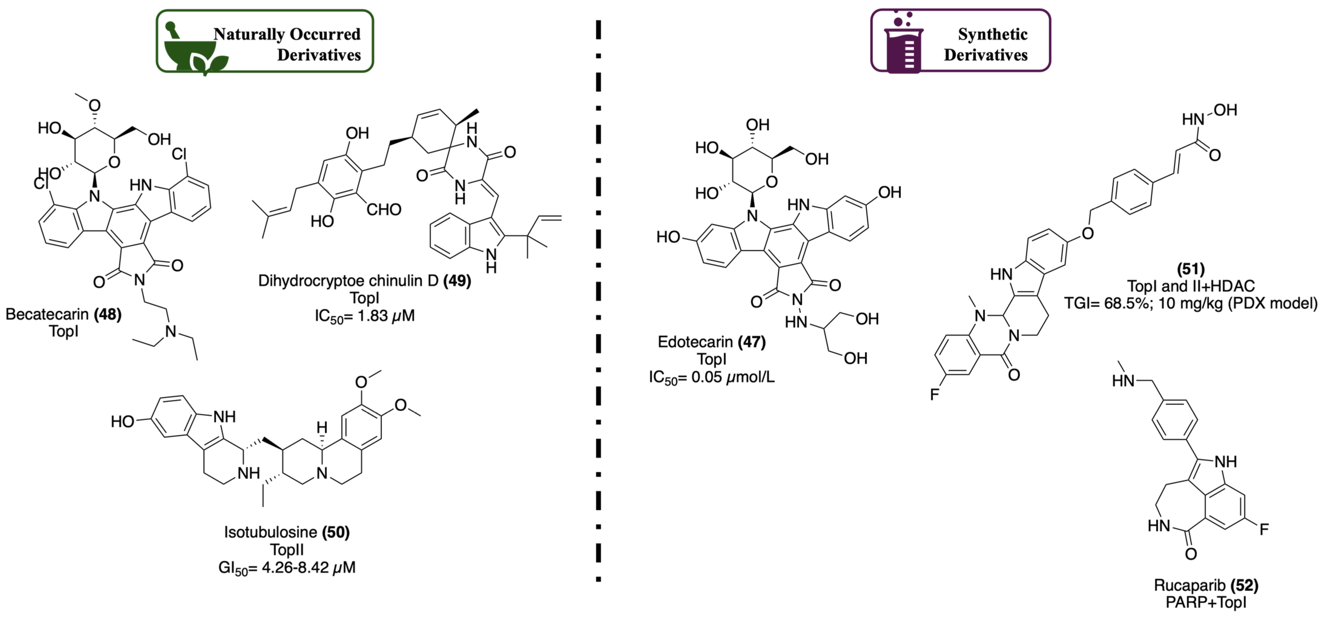
2.1.2. DNA Topoisomerase

Processes like replication, transcription, and recombination require accessing DNA stored information, with separation of the DNA helix double strand leading to a supercoiling tension that can only be relieved by a group of enzymes called topoisomerases. This main function of topoisomerases is essential for the maintenance of genomic stability and proper DNA function [64,65]. There are two isoforms of the topoisomerase enzyme, TopI and TopII. While TopI works with only one of the DNA strands, TopII deals with both [64,65]. However, mutations of these enzymes lead to genomic instability, and since their mechanism of action requires the formation of bonds with DNA, mutations will trigger the uncontrolled functioning of topoisomerases, whether by damaged DNA accumulation, defective chromosomal rearrangements, or even by topoisomerase inhibitors, which due to the lack of specificity for abnormal cells, can down-regulate the enzyme. Such events lead to errors in DNA replication and transcription and, subsequently, to the formation of unreliable cells, enabling mutations and, thus, cancer development [64,65]. The synthetic indole edotecarin (47) (Figure 16) is an inhibitor of TopI (IC50 of 0.05 µmol/L), with potent activity in glioblastoma multiforme and malignant brain tumors, as demonstrated in Phase II clinical trials. Nonetheless, it never moved further into Phase III clinical trials [56]. Becatecarin (48) (Figure 16), a synthetic analog of rebeccamycin isolated from Nocardia sp., is also an inhibitor of TopI. Preliminary studies indicated good potency against Ewing sarcoma, medulloblastoma, neuroblastoma, and rhabdomyosarcoma cell lines, and even better potency against a leukemia cell line when compared with rebeccamycin. However, in Phase II clinical trials, it exhibited myelosuppression as a side effect when administered in children with solid CNS tumors [56]. From Aspergillus effuses H1-1 and H1-2, dihydrocryptoe chinulin D (49) (Figure 16) was isolated, exhibiting activity against TopI with an IC50 value of 1.83 µM, revealing to be a potential anticancer agent in leukemia and lymphoma treatment [6]. Isotubulosine (50) (Figure 16), extracted from the Pogonopus tubulosus trunk, is potent against TopII, with GI50 values between 4.26 and 8.42 µM, and induced cell arrest at the G2/M phase of the cell cycle. The group of Wu [66] developed a class of evodiamine-inspired HDAC dual inhibitors as antitumor agents for leukemia therapy. The hybrids were active against breast (MCF-7), lung (A549), colorectal (HCT116), liver (HepG-2), and blood (K562 and HEL) cancer cells. SAR studies against HDAC exhibited a direct correlation between the activity and the linker in the scaffolds. Also, the assay on the inhibitory activity on TopI and TopII showed that it is preferable to have less activity in HDAC to access a more balanced inhibitory effect in both enzymes, which allowed for highlighting compound (51) (Figure 16) since it presented a tumor growth inhibition of 68.5% in a dose of 10 mg/kg in patient-derived xerographs’ models. Further experiments on compound (51)’s cellular effects, metabolic stability, and antitumor efficacy, made this small molecule a promising lead compound for antitumor treatment. Recently, it was reported that PARP inhibitors had the potential for multi-target therapy, as this enzyme plays a vital role in cell regulation [67]. Considering this observation, the synthetic and already patented drug rucaparib (52) (Figure 16) could be repurposed. This drug exhibits an inhibitory activity in PARP 1, 2, and 3, being active in advanced ovarian cancer but directly affecting TopI, opening an opportunity for new anticancer treatment options [55,68].

2.1.3. Tubulin Polymerization

Microtubules represent a crucial element for cell stability and organization, being responsible for cell division and, therefore, for chromosome segregation, equal distribution of genetic material to daughter cells, and the transport of proteins and organelles. Their structure is comprehended by tubulin proteins, aggregated in such a way that they generate a cylindrical structure that interacts with other proteins, regulating microtubule behavior [69,70,71]. The process of tubulin aggregation is denominated tubulin polymerization and consists of the organization and rearrangement of tubulins grouped in dimers that will assemble, producing layers of each isoform of tubulin, α-tubulin, and β-tubulin [69,70,71]. Tubulin polymerization only happens if GTP binds to tubulin, with the binding to β-tubulin particularly important because it is the only one that takes GTP to hydrolyze to GDP. GDP formation weakens the affinity of tubulin to other molecules, triggering polymerization and ultimately embodying microtubule normal dynamics [69,70,71]. Given the importance of this process, microtubules need their dynamic (in)stability to work properly; otherwise, any deflecting in tubulin regulators will affect microtubules, whether through cell mitosis or cell proliferation [69,70,71]. Vindesine (53) (Figure 17) is a semi-synthetic alkaloid, derived from vinblastine (6) (Figure 2), whose mechanism of action consists of blocking the cell from entering metaphase mitosis over inhibition of tubulin action. Vinorelbine (54) (Figure 17) is also a semi-synthetic indole derivative that expresses an improved inhibitory activity in microtubules due to lower side effects. It revealed excellent behavior at inhibiting cancer cell migration, as well as arresting cells at the G2/M phase and inducing apoptosis [8,72,73]. From synthetic sources, Yan and co-workers [74] synthesized a set of indole–chalcone derivatives with inhibitory activity of tubulin polymerization. All the compounds were tested for their antiproliferative activity towards lung (A549), cervix (HeLa), liver (Bel-7402), breast (MCF-7), ovarian (A2780), and colorectal (HCT-8) cancer cell lines. Most of the compounds were active, particularly compound (55) (Figure 17), with IC50 values ranging from 0.0003 to 0.009 µM [74]. The most active compounds were submitted to a tubulin polymerization activity assay, and compound (55) proved to be the most potent inhibitor, with an IC50 value of 2.68 µM in tubulin polymerization. Further tests showed that the cellular mechanism of this compound involved the arrest of the G2/M phase and induced apoptosis, suggesting its use as a chemotherapeutic agent [74]. To improve the results obtained with compound (55), Romagnoli and co-workers [75] synthesized a new class of potent inhibitors of tubulin polymerization, designated 2-alkoxycarbonyl-3-anilinoindoles (56) (Figure 17). These compounds were tested against cervix (HeLa), colon (HT29), breast (MCF-7), and blood (HL-60) cancer cells, exhibiting, mostly, IC50 values under 2 µM, despite some of them presenting IC50 values above 5 µM and even 10 µM [75]. Compounds (56a) and (56b) (Figure 17) were the most potent ones as tubulin polymerase inhibitors (IC50 values, 0.40 and 0.37 µM, respectively) and had specificity to the colchicine binding site. In addition to that, they also increased the cell population in the G2/M phase and decreased it in the G1 phase of the cell cycle. Given these results, it was noticed that an addition of a methyl acetate group provided an increase in activity, and because of that, compounds (56a) and (56b) were considered as potential new antiproliferative agents that target tubulin at the colchicine site [75].

2.1.4. P-Glycoprotein

Glycoproteins are a subclass of transmembrane proteins responsible for transporting small molecules out of the cell, operating the removal of xenobiotics and the entrance of nutrients into the cell. Among the known glycoproteins, P-glycoprotein (P-gp or ABCB1) has been identified as a key class of proteins in cancer development [76,77]. In addition to their physiological role, they are active in drug transport by regulating the uptake of exogenous small molecules through endothelial cells from the organs, such as kidneys, GI, liver, ovaries, testicles, adrenal and pituitary glands, placenta, choroid plexus, and the capillary cells of the brain. This may result in the access of cytotoxic agents or, if a mutation in P-gp occurs, in a multidrug-resistance effect, which will initiate a cascade of events and eventually result in the development of severe diseases [76,77]. It was observed in numerous studies that P-gp is expressed in different ways, according to the type of cancer where its mutation is present. For example, in acute myeloid leukemia (AML) and lung, ovarian, and renal cancer, P-gp is upregulated, but in colorectal, breast, and prostate cancer, it is down-regulated [76,77]. Voacamine (57) (Figure 18) is an example of a natural bis-indole alkaloid, extracted from Peschiera fuchsiaefolia, that targets P-gp, displaying, in addition, an alteration of microtubules. With activity in the U-2 OS/WT and U-2 OS/DX cell lines, (57) induces autophagy and is used in the treatment of osteosarcoma [58,73]. In 2017, Paterna and co-workers [78] demonstrated the potential of the monoterpene indole alkaloids from Tabernaemontana elegans dregamine (58a) and tabernaemontanine (58b) (Figure 18) as P-gp modulators. The group synthesized a set of those derivatives and tested them against PAR cells and ABCB1-gene transfected mouse T-lymphoma cells (multidrug-resistance cells) regarding their cytotoxicity, verifying that the compounds demonstrated higher toxicity levels than the parent compounds. The ABCB1 modulating ability of the compounds was then tested, and compounds with an imine moiety and extra aromatic ring, such as (59a-c) (Figure 18), and a methoxybenzyloxycarbonyl moiety, such as (59d) (Figure 18), demonstrated the best inhibitory activity against ABCB1. Moreover, assays with doxorubicin showed their good synergy, suggesting their use as multidrug-resistance reversers [78]. Curiously, Raimundo and co-workers [79] recently identified compound (59b) (BBIT20) (Figure 18) as a potential agent against aggressive and resistant cancers, revealing activity in triple-negative breast and advanced ovarian cancer through the disruption of the BRCA1-BARD1 interaction [79]. A few years later, the group of Cardoso [80] achieved equivalent results, synthesizing different derivatives of (58a-b). An MTT assay against sensitive and resistant human colon adenocarcinoma cells (COLO 205, COLO 320), L5178Y parental (5178Y, PAR), and human ABCB1-gene transfected (5178Y) mouse T-lymphoma cells was performed and demonstrated a lack of significant cytotoxicity, neither in sensitive nor resistant cell lines. A FAR assay to access the potential of indole derivatives as P-gp inhibitors was also performed, evidencing that, for COLO 320 cells, the values were too low, disabling their usage. Yet, for 5178Y multi-resistance cells, the obtained values were satisfying, allowing the group to classify the synthesized compounds between active and strong inhibitors of P-gp when applied in a concentration of 20 µM. Furthermore, a drug-combination assay with doxorubicin was made, and the results pointed to compounds (60a-c) (Figure 18) as promising agents, suggesting their potential use in combined therapy [80].

2.1.5. Other Interesting Targets

Evidently, the indole scaffold enables an immeasurable panoply of bioactivities, whether in terms of types of derivatives or in terms of their pharmacological applications. Since indole derivatives demonstrate high potential as anticancer agents for several targets of interest and within the scope of this review, Table 1 displays other targets of interest where this type of compound has some promising activity. In the past few years, it has been discovered that some interesting derivatives exhibited activity against some of these targets. Compounds (61) [81], (62) [81], and (63) [82] (Table 1) also target PKs and their signaling pathways. Yet, in contrast to what was previously explained, this occurs through other mechanisms of action, whether by inhibition of a downstream and direct p53 activator, such as the protein Aurora A, by the inhibition of upstream pathways like the PDK1/Akt one, or by the inhibition of ROS that contribute to the activation of the MAPK signaling pathway [81,82]. Compound (64) [58] also showed an interesting mechanism of action, since it pursues the dysfunction of the topoisomerase by cross-linking to the DNA strand, creating an error in the DNA chain with an exogenous agent, leading to cytotoxicity [58]. Acting through another pathway, compound (65) [83] (Table 1) disrupts the interaction between proteins p53 and MDM2. These two are regulators of one another by inhibition, and once their interaction is inhibited, it allows p53 to stabilize and activate freely the apoptosis system [83,84]. Also, with a role in the functions of the p53 protein, there is SIRT, which when inhibited, promotes the acetylation of p53 and, for that, its activation as well as the apoptosis pathway. Compound (66) [85] is an example of an inhibitor of SIRT [85,86]. Following the same logic, compound (67) [87] (Table 1) inhibits anti-apoptotic proteins, such as Bcl-2, Bcl-xl, and Mcl-1, enabling pro-apoptotic proteins to act [87,88]. As for the σ2 receptors, when activated by agonists, they exhibit antiproliferative and cytotoxic activity in tumor cells, and since they are present in great density, they were used as biomarkers of the tumors’ proliferation [88]. One example of an agonist of these receptors is compound (68) (Table 1) [88]. In the line of the apoptosis inducers, there is also compound (69) [89] (Table 1), which works as an inhibitor of the protein HDAC, promoting the down-regulation of anti-apoptotic genes and the de-upregulation of pro-apoptotic genes, inducing apoptosis [89,90].

3. Conclusions and Future Perspectives

Indole-based molecules can be widely found in nature, and their study and clinical uses have been the scope of many research groups over the last decades. Furthermore, it is indubitable the effort that has been carried out regarding the design and synthesis of new indole derivatives, offering advantageous structural properties for its numerous clinical applications, including in viral, inflammatory, and, in particular, cancer diseases. Cancer is one of the deadliest diseases of this century, and the outlook for the future is not very encouraging. It was observed that the most promising indole derivatives that are currently available on the market came from natural sources and semi-synthetic routes, demonstrating their potential to be considered in future drug design and development. Several examples of these natural or semi-synthetic derivatives are given throughout this review, exhibiting potent activity for several targets of interest in cancer, such as PK, TopI and TopII, and microtubule inhibitors, among others. Similarly, many synthetic indole derivatives were also pointed out as promising candidates for cancer treatment, such as spirooxindole derivatives and others, showing great activity for the same described targets. Despite the knowledge obtained so far, it is imperative to continue developing new drug-design approaches targeting the indole scaffold and subsequent screening technologies to access more potent and highly selective compounds as anticancer agents, to move further the clinical trials pipeline. Either by the modification of natural products or synthesis of lead compounds, advances in the chemical modification of indole frameworks generating active moieties are a crucial approach to generating new molecules with potential for a particular target or multiple targets in future research work.

Funding

This work received financial support from PT national funds (FCT/MCTES, Fundação para a Ciência e Tecnologia and Ministério da Ciência, Tecnologia e Ensino Superior) through the project UIDB/50006/2020|UIDP/50006/2020 and 2022.02910.PTDC. C.S.M. thanks FCT (Fundação para a Ciência e Tecnologia) for funding through program DL 57/2016-Norma transitória (project UIDB/50006/2020|UIDP/50006/2020).

Conflicts of Interest

The authors declare no conflict of interest.

Abbreviations

Ablabelson leukemia gene
AChacetylcholine
AChEacetylcholinesterase
ADVadenovirus
Akfprotein kinase B
AMLacute myeloid leukemia
APCadenomatous polyposis coli
API(s)active pharmaceutical ingredient(s)
ATPadenosine triphosphate
BRCA1associated ring domain 1
BRCA1breast cancer gene 1
BRCA2breast cancer gene 2
CDKcyclin-dependent kinase
COX-2cyclooxygenase-2
DRAK1kinase-related apoptosis-inducing protein kinase 1
EGFRepithelial growth factor receptor
FLT3fms-like tyrosine kinase 3
GSK-βglycogene synthase kinase beta
GTPguanosine triphosphate
HCVhepatitis C virus
HDACtopoisomerase-histone deacetylase
HER2human epidermal growth factor receptor 2
HER3human epidermal growth factor receptor 3
HSVherpes simplex virus
HVBhepatitis B virus
KITproto-oncogene c-kit
L-Glnglutamine
MAOmonoamine oxidase
MDM2mouse double minute 2 homolog
MLH1mutL homolog 1
mRNAmessenger ribonucleic acid
NNRTInonnucleoside reverse transcriptase inhibitor
NRTKnon-receptor tyrosine kinase
Pphosphate
p53tumor protein 53
PARparental chemosensitive cells
PARPpoly ADP-ribose polymerase
PDGFRplatelet-derived growth factor receptor alpha
PI3Kphosphoinositide 3-kinase
PIM1serine/threonine kinase
PK(s)protein kinase(s)
PTENphosphatase and tensin homolog
PTKIprotein tyrosine kinase inhibitor
Rabras-associated binding gene
Rafrapidly accelerated fibrosarcoma gene
Rasrat sarcoma gene
Rhorhodopsin gene
ROSreactive oxygen species
RTKreceptor tyrosine kinase
SARstructure-activity relationship
Srcsarcoma gene
TKtyrosine kinase
TopItopoisomerase I
TopIItopoisomerase II
TPA12-tetradecanoylphorbol-13-acetate
TSGtumor-suppressor gene
VEGRvascular endothelial growth factor
WTWilms’ tumor gene

References

  1. Dhuguru, J.; Skouta, R. Role of Indole Scaffolds as Pharmacophores in the Development of Anti-Lung Cancer Agents. Molecules 2020, 25, 1615. [Google Scholar] [CrossRef] [PubMed]
  2. Mohammadi Ziarani, G.; Moradi, R.; Ahmadi, T.; Lashgari, N. Recent advances in the application of indoles in multicomponent reactions. RSC Adv. 2018, 8, 12069–12103. [Google Scholar] [CrossRef] [PubMed]
  3. Bandini, M. Electrophilicity: The “dark-side” of indole chemistry. Org. Biomol. Chem. 2013, 11, 5206. [Google Scholar] [CrossRef] [PubMed]
  4. Dhiman, A.; Sharma, R.; Singh, R.K. Target-based anticancer indole derivatives and insight into structure–activity relationship: A mechanistic review update (2018–2021). Acta Pharm. Sin. B 2022, 12, 3006–3027. [Google Scholar] [CrossRef] [PubMed]
  5. Bajad, N.G.; Singh, S.K.; Singh, S.K.; Singh, T.D.; Singh, M. Indole: A promising scaffold for the discovery and development of potential anti-tubercular agents. Curr. Res. Pharmacol. Drug Discov. 2022, 3, 100119. [Google Scholar] [CrossRef] [PubMed]
  6. Islam, F.; Dehbia, Z.; Zehravi, M.; Das, R.; Sivakumar, M.; Krishnan, K.; Billah, A.A.M.; Bose, B.; Ghosh, A.; Paul, S.; et al. Indole alkaloids from marine resources: Understandings from therapeutic point of view to treat cancers. Chem. Biol. Interact. 2023, 383, 110682. [Google Scholar] [CrossRef] [PubMed]
  7. Piechowska, P.; Zawirska-Wojtasiak, R.; Mildner-Szkudlarz, S. Bioactive β-Carbolines in Food: A Review. Nutrients 2019, 11, 814. [Google Scholar] [CrossRef]
  8. Dewick, P.M. Medicinal Natural Products: A Biosynthetic Approach, 3rd ed.; John Wiley & Sons Ltd.: Hoboken, NJ, USA, 2009. [Google Scholar]
  9. Politis, M.; Niccolini, F. Serotonin in Parkinson’s disease. Behav. Brain Res. 2015, 277, 136–145. [Google Scholar] [CrossRef]
  10. Tian, J.; Du, E.; Guo, L. Mitochondrial Interaction with Serotonin in Neurobiology and Its Implication in Alzheimer’s Disease. J. Alzheimers Dis. Rep. 2023, 7, 1165–1177. [Google Scholar] [CrossRef]
  11. Szabó, T.; Volk, B.; Milen, M. Recent Advances in the Synthesis of β-Carboline Alkaloids. Molecules 2021, 26, 663. [Google Scholar] [CrossRef]
  12. Paul, A.; Acharya, K.; Chakraborty, N. Biosynthesis, extraction, detection and pharmacological attributes of vinblastine and vincristine, two important chemotherapeutic alkaloids of Catharanthus roseus (L.) G. Don: A review. S. Afr. J. Bot. 2023, 161, 365–376. [Google Scholar] [CrossRef]
  13. Atanasov, A.G.; Zotchev, S.B.; Dirsch, V.M.; Supuran, C.T. Natural products in drug discovery: Advances and opportunities. Nat. Rev. Drug Discov. 2021, 20, 200–216. [Google Scholar] [CrossRef] [PubMed]
  14. Neto, J.S.S.; Zeni, G. Recent advances in the synthesis of indoles from alkynes and nitrogen sources. Org. Chem. Front. 2020, 7, 155–210. [Google Scholar] [CrossRef]
  15. Ayesha; Bilal, M.; Rasool, N.; Khan, S.G.; Rashid, U.; Altaf, H.; Ali, I. Synthesis of Indoles via Intermolecular and Intramolecular Cyclization by Using Palladium-Based Catalysts. Catalysts 2021, 11, 1018. [Google Scholar] [CrossRef]
  16. Kumari, A.; Singh, R.K. Medicinal chemistry of indole derivatives: Current to future therapeutic prospectives. Bioorg. Chem. 2019, 89, 103021. [Google Scholar] [CrossRef] [PubMed]
  17. Leimgruber, B.W. Leimgruber–Batcho Indole Synthesis. In Indole Ring Synthesis; Wiley: New York, NY, USA, 2016; pp. 338–348. [Google Scholar] [CrossRef]
  18. Baudin, J.B.; Julia, S.A. Synthesis of indoles from N-aryl-1-alkenylsulphinamides. Tetrahedron Lett. 1986, 27, 837–840. [Google Scholar] [CrossRef]
  19. Fischer, E.; Jourdan, F. Ueber die Hydrazine der Brenztraubensäure. Berichte Dtsch. Chem. Ges. 1883, 16, 2241–2245. [Google Scholar] [CrossRef]
  20. Reissert, A. Einwirkung von Oxalester und Natriumäthylat auf Nitrotoluole. Synthese nitrirter Phenylbrenztraubensäuren. Berichte Dtsch. Chem. Ges. 1897, 30, 1030–1053. [Google Scholar] [CrossRef]
  21. Baeyer, A.; Emmerling, A. Synthese des Indols. Berichte Dtsch. Chem. Ges. 1869, 2, 679–682. [Google Scholar] [CrossRef]
  22. Sanz, R.; Guilarte, V.; Pérez, A. Straightforward selective preparation of nitro- or amino-indoles from 2-halonitroanilines and alkynes. First synthesis of 7-amino-5-nitroindoles. Tetrahedron Lett. 2009, 50, 4423–4426. [Google Scholar] [CrossRef]
  23. Ponpandian, T.; Muthusubramanian, S. Copper catalysed domino decarboxylative cross coupling-cyclisation reactions: Synthesis of 2-arylindoles. Tetrahedron Lett. 2012, 53, 4248–4252. [Google Scholar] [CrossRef]
  24. Liang, S.; Hammond, L.; Xu, B.; Hammond, G.B. Commercial Supported Gold Nanoparticles Catalyzed Alkyne Hydroamination and Indole Synthesis. Adv. Synth. Catal. 2016, 358, 3313–3318. [Google Scholar] [CrossRef]
  25. Madelung, W. Über eine neue Darstellungsweise für substituierte Indole. I. Berichte Dtsch. Chem. Ges. 1912, 45, 1128–1134. [Google Scholar] [CrossRef]
  26. Bartoli, G.; Palmieri, G.; Bosco, M.; Dalpozzo, R. The reaction of vinyl grignard reagents with 2-substituted nitroarenes: A new approach to the synthesis of 7-substituted indoles. Tetrahedron Lett. 1989, 30, 2129–2132. [Google Scholar] [CrossRef]
  27. Larock, R.C.; Yum, E.K. Synthesis of indoles via palladium-catalyzed heteroannulation of internal alkynes. J. Am. Chem. Soc. 1991, 113, 6689–6690. [Google Scholar] [CrossRef]
  28. Fukuyama, T.; Chen, X.; Peng, G. A Novel Tin-Mediated Indole Synthesis. J. Am. Chem. Soc. 1994, 116, 3127–3128. [Google Scholar] [CrossRef]
  29. Roskoski, R. Properties of FDA-approved small molecule protein kinase inhibitors: A 2023 update. Pharmacol. Res. 2023, 187, 106552. [Google Scholar] [CrossRef]
  30. Sravanthi, T.V.; Manju, S.L. Indoles—A promising scaffold for drug development. Eur. J. Pharm. Sci. 2016, 91, 1–10. [Google Scholar] [CrossRef] [PubMed]
  31. Yu, C.; Gopalakrishnan, G. In vitro pharmacological characterization of SPN-810M (molindone). J. Exp. Pharmacol. 2018, 10, 65–73. [Google Scholar] [CrossRef]
  32. Knox, C.; Wilson, M.; Klinger, C.M.; Franklin, M.; Oler, E.; Wilson, A.; Pon, A.; Cox, J.; Chin, N.E.L.; Strawbridge, S.A.; et al. DrugBank 6.0: The DrugBank Knowledgebase for 2024. Nucleic Acids Res. 2024, 52, D1265–D1275. [Google Scholar] [CrossRef]
  33. Omar, F.; Tareq, A.M.; Alqahtani, A.M.; Dhama, K.; Sayeed, M.A.; Emran, T.B.; Simal-Gandara, J. Plant-Based Indole Alkaloids: A Comprehensive Overview from a Pharmacological Perspective. Molecules 2021, 26, 2297. [Google Scholar] [CrossRef] [PubMed]
  34. Chadha, N.; Silakari, O. Indoles as therapeutics of interest in medicinal chemistry: Bird’s eye view. Eur. J. Med. Chem. 2017, 134, 159–184. [Google Scholar] [CrossRef] [PubMed]
  35. Choppara, P.; Bethu, M.S.; Prasad, Y.V.; Rao, J.V.; Ranjan, T.J.U.; Prasad, G.V.S.; Doradla, R.; Murthy, Y.L.N. Synthesis, characterization and cytotoxic investigations of novel bis(indole) analogues besides antimicrobial study. Arab. J. Chem. 2019, 12, 2721–2731. [Google Scholar] [CrossRef]
  36. Mielczarek, M.; Thomas, R.V.; Ma, C.; Kandemir, H.; Yang, X.; Bhadbhade, M.; Black, D.S.; Griffith, R.; Lewis, P.J.; Kumar, N. Synthesis and biological activity of novel mono-indole and mono-benzofuran inhibitors of bacterial transcription initiation complex formation. Bioorg. Med. Chem. 2015, 23, 1763–1775. [Google Scholar] [CrossRef] [PubMed]
  37. Siramshetty, V.B.; Grishagin, I.; Nguyễn, Ð.T.; Peryea, T.; Skovpen, Y.; Stroganov, O.; Katzel, D.; Sheils, T.; Jadhav, A.; Mathé, E.A.; et al. NCATS Inxight Drugs: A comprehensive and curated portal for translational research. Nucleic Acids Res. 2022, 50, D1307–D1316. [Google Scholar] [CrossRef] [PubMed]
  38. Bhat, M.A.; Al-Omar, M.A.; Raish, M.; Ansari, M.A.; Abuelizz, H.A.; Bakheit, A.H.; Naglah, A.M. Indole Derivatives as Cyclooxygenase Inhibitors: Synthesis, Biological Evaluation and Docking Studies. Molecules 2018, 23, 1250. [Google Scholar] [CrossRef] [PubMed]
  39. Mukthung, C.; Chancharunee, S.; Kielar, F.; Pongcharoen, S.; Wichai, U. Capsaicin Derivatives Containing Indole and Nitroindole for Improved Anti-Inflammatory Activity. Naresuan Univ. J. Sci. Technology. 2018, 26, 157–169. [Google Scholar] [CrossRef]
  40. Girgis, A.S.; Panda, S.S.; Kariuki, B.M.; Bekheit, M.S.; Barghash, R.F.; Aboshouk, D.R. Indole-Based Compounds as Potential Drug Candidates for SARS-CoV-2. Molecules 2023, 28, 6603. [Google Scholar] [CrossRef] [PubMed]
  41. Brown, J.S.; Amend, S.R.; Austin, R.H.; Gatenby, R.A.; Hammarlund, E.U.; Pienta, K.J. Updating the Definition of Cancer. Mol. Cancer Res. 2023, 21, 1142–1147. [Google Scholar] [CrossRef] [PubMed]
  42. de Martel, C.; Georges, D.; Bray, F.; Ferlay, J.; Clifford, G.M. Global burden of cancer attributable to infections in 2018: A worldwide incidence analysis. Lancet Glob. Health 2020, 8, e180–e190. [Google Scholar] [CrossRef]
  43. Hanselmann, R.G.; Welter, C. Origin of Cancer: An Information, Energy, and Matter Disease. Front. Cell Dev. Biol. 2016, 4, 121. [Google Scholar] [CrossRef] [PubMed]
  44. Holland, J.F.; Frei, E.; Kude, D.W. Multistage Carcinogenes. In Cancer Medicine, 6th ed.; Kufe, D.W., Pollock, R.E., Weichselbaum, R.R., Bast, R.C., Gansler, T.S., Holland, J.F., Frei, E., Eds.; BC Decker Inc.: Hamilton, ON, Canada, 2003. [Google Scholar]
  45. Weinberg, R.A. Multi-step Tumorigenesis. In The Biology of CANCER, 2nd ed.; Bochicchio, A., Zayatz, E., Mickey, R.K., Harik, L., Ej Publishing Services, Eds.; Garland Science, Taylor & Francis Group, LLC: Abingdon, UK, 2014. [Google Scholar]
  46. Baba, A.I.; Câtoi, C. Tumor Cell Morphology. In Comparative Oncology; The Publishing House of the Romanian Academy: Bucuresti, Romania, 2007. [Google Scholar]
  47. Lakshminarasimhan, R.; Liang, G. The Role of DNA Methylation in Cancer. In Advances in Experimental Medicine and Biology; Springer New York LLC: New York, NY, USA, 2016; Volume 945, pp. 151–172. [Google Scholar] [CrossRef]
  48. Srinivas, U.S.; Tan, B.W.Q.; Vellayappan, B.A.; Jeyasekharan, A.D. ROS and the DNA damage response in cancer. Redox Biol. 2019, 25, 101084. [Google Scholar] [CrossRef] [PubMed]
  49. Sever, R.; Brugge, J.S. Signal Transduction in Cancer. Cold Spring Harb. Perspect. Med. 2015, 5, a006098. [Google Scholar] [CrossRef] [PubMed]
  50. Khetmalis, Y.M.; Shivani, M.; Murugesan, S.; Chandra Sekhar, K.V.G. Oxindole and its derivatives: A review on recent progress in biological activities. Biomed. Pharmacother. 2021, 141, 111842. [Google Scholar] [CrossRef] [PubMed]
  51. Dar, A.C.; Shokat, K.M. The Evolution of Protein Kinase Inhibitors from Antagonists to Agonists of Cellular Signaling. Annu. Rev. Biochem. 2011, 80, 769–795. [Google Scholar] [CrossRef] [PubMed]
  52. Paul, M.K.; Mukhopadhyay, A.K. Tyrosine kinase—Role and significance in Cancer. Int. J. Med. Sci. 2004, 1, 101–115. [Google Scholar] [CrossRef] [PubMed]
  53. Salerno, S.; Barresi, E.; Baglini, E.; Poggetti, V.; Da Settimo, F.; Taliani, S. Target-Based Anticancer Indole Derivatives for the Development of Anti-Glioblastoma Agents. Molecules 2023, 28, 2587. [Google Scholar] [CrossRef] [PubMed]
  54. Jin, J.; Xie, Y.; Zhang, J.S.; Wang, J.Q.; Dai, S.J.; He, W.F.; Li, S.Y.; Ashby, C.R., Jr.; Chen, Z.S.; He, Q. Sunitinib resistance in renal cell carcinoma: From molecular mechanisms to predictive biomarkers. Drug Resist. Updates 2023, 67, 100929. [Google Scholar] [CrossRef]
  55. Hong, Y.; Zhu, Y.Y.; He, Q.; Gu, S.X. Indole derivatives as tubulin polymerization inhibitors for the development of promising anticancer agents. Bioorg. Med. Chem. 2022, 55, 116597. [Google Scholar] [CrossRef]
  56. Sherer, C.; Snape, T.J. Heterocyclic scaffolds as promising anticancer agents against tumours of the central nervous system: Exploring the scope of indole and carbazole derivatives. Eur. J. Med. Chem. 2015, 97, 552–560. [Google Scholar] [CrossRef]
  57. Gallogly, M.M.; Lazarus, H.M.; Cooper, B.W. Midostaurin: A novel therapeutic agent for patients with FLT3-mutated acute myeloid leukemia and systemic mastocytosis. Ther. Adv. Hematol. 2017, 8, 245–261. [Google Scholar] [CrossRef]
  58. Jia, Y.; Wen, X.; Gong, Y.; Wang, X. Current scenario of indole derivatives with potential anti-drug-resistant cancer activity. Eur. J. Med. Chem. 2020, 200, 112359. [Google Scholar] [CrossRef]
  59. Christodoulou, M.S.; Nicoletti, F.; Mangano, K.; Chiacchio, M.A.; Facchetti, G.; Rimoldi, I.; Beccalli, E.M.; Giofrè, S. Novel 3,3-disubstituted oxindole derivatives. Synthesis and evaluation of the anti-proliferative activity. Bioorg. Med. Chem. Lett. 2020, 30, 126845. [Google Scholar] [CrossRef]
  60. Gompel, M.; Leost, M.; De Kier Joffe, E.B.; Puricelli, L.; Franco, L.H.; Palermo, J.; Meijer, L. Meridianins, a new family of protein kinase inhibitors isolated from the Ascidian Aplidium meridianum. Bioorg. Med. Chem. Lett. 2004, 14, 1703–1707. [Google Scholar] [CrossRef]
  61. Barakat, A.; Abu-Serie, M.M.; Ali, M.; Al-Majid, A.M.; Ashraf, S.; Zia, K.; Ul-Haq, Z.; Al-Dhfyan, A.; Abdel-Aziz, H.A.; El-Faham, A.; et al. Synthesis, In Vitro and in Cell Study of a New Spirooxindoles-Based N-Alkylated Maleimides Targeting HER2/3 Signaling Pathway. Polycycl. Aromat. Compd. 2023, 43, 5251–5275. [Google Scholar] [CrossRef]
  62. Al-Jassas, R.M.; Islam, M.S.; Al-Majid, A.M.; Nafie, M.S.; Haukka, M.; Rahman, A.F.M.M.; Alayyaf, A.M.A.; Barakat, A. Synthesis and SARs study of novel spiro-oxindoles as potent antiproliferative agents with CDK-2 inhibitory activities. Arch. Pharm. 2023, 356, e202300185. [Google Scholar] [CrossRef]
  63. Dubba, A.; Kumar Koppula, S. Synthesis of Indole-Oxadiazole coupled isoxazole hybrids as potent EGFR targeting anticancer agents. Chem. Biol. Lett. 2024, 11, 651. [Google Scholar] [CrossRef]
  64. Bush, N.G.; Evans-Roberts, K.; Maxwell, A. DNA Topoisomerases. EcoSal Plus 2015, 6. [Google Scholar] [CrossRef]
  65. Champoux, J.J. DNA Topoisomerases: Structure, Function, and Mechanism. Annu. Rev. Biochem. 2001, 70, 369–413. [Google Scholar] [CrossRef]
  66. Wu, S.; Huang, Y.; Wang, T.; Li, K.; Lu, J.; Huang, M.; Dong, G.; Sheng, C. Evodiamine-Inspired Topoisomerase-Histone Deacetylase Dual Inhibitors: Novel Orally Active Antitumor Agents for Leukemia Therapy. J. Med. Chem. 2022, 65, 4818–4831. [Google Scholar] [CrossRef]
  67. Zhang, J.; Gao, Y.; Zhang, Z.; Zhao, J.; Jia, W.; Xia, C.; Wang, F.; Liu, T. Multi-therapies Based on PARP Inhibition: Potential Therapeutic Approaches for Cancer Treatment. J. Med. Chem. 2022, 65, 16099–16127. [Google Scholar] [CrossRef]
  68. Dockery, L.; Gunderson, C.; Moore, K. Rucaparib: The past, present, and future of a newly approved PARP inhibitor for ovarian cancer. Onco Targets Ther. 2017, 10, 3029–3037. [Google Scholar] [CrossRef]
  69. Cheng, Z.; Lu, X.; Feng, B. A review of research progress of antitumor drugs based on tubulin targets. Transl. Cancer Res. 2020, 9, 4020–4027. [Google Scholar] [CrossRef]
  70. Parker, A.L.; Kavallaris, M.; McCarroll, J.A. Microtubules and Their Role in Cellular Stress in Cancer. Front. Oncol. 2014, 4, 153. [Google Scholar] [CrossRef]
  71. Sept, D. Microtubule Polymerization: One Step at a Time. Curr. Biol. 2007, 17, R764–R766. [Google Scholar] [CrossRef]
  72. Dhyani, P.; Quispe, C.; Sharma, E.; Bahukhandi, A.; Sati, P.; Attri, D.C.; Szopa, A.; Sharifi-Rad, J.; Docea, A.O.; Mardare, I.; et al. Anticancer potential of alkaloids: A key emphasis to colchicine, vinblastine, vincristine, vindesine, vinorelbine and vincamine. Cancer Cell Int. 2022, 22, 206. [Google Scholar] [CrossRef]
  73. Qin, R.; You, F.M.; Zhao, Q.; Xie, X.; Peng, C.; Zhan, G.; Han, B. Naturally derived indole alkaloids targeting regulated cell death (RCD) for cancer therapy: From molecular mechanisms to potential therapeutic targets. J. Hematol. Oncol. 2022, 15, 133. [Google Scholar] [CrossRef]
  74. Yan, J.; Chen, J.; Zhang, S.; Hu, J.; Huang, L.; Li, X. Synthesis, Evaluation, and Mechanism Study of Novel Indole-Chalcone Derivatives Exerting Effective Antitumor Activity Through Microtubule Destabilization in Vitro and in Vivo. J. Med. Chem. 2016, 59, 5264–5283. [Google Scholar] [CrossRef]
  75. Romagnoli, R.; Prencipe, F.; Oliva, P.; Kimatrai Salvador, M.; Brancale, A.; Ferla, S.; Hamel, E.; Viola, G.; Bortolozzi, R.; Persoons, L.; et al. Design, synthesis and biological evaluation of 2-alkoxycarbonyl-3-anilinoindoles as a new class of potent inhibitors of tubulin polymerization. Bioorg. Chem. 2020, 97, 103665. [Google Scholar] [CrossRef]
  76. Pilotto Heming, C.; Muriithi, W.; Wanjiku Macharia, L.; Niemeyer Filho, P.; Moura-Neto, V.; Aran, V. P-glycoprotein and cancer: What do we currently know? Heliyon 2022, 8, e11171. [Google Scholar] [CrossRef]
  77. Ahmed Juvale, I.I.; Abdul Hamid, A.A.; Abd Halim, K.B.; Che Has, A.T. P-glycoprotein: New insights into structure, physiological function, regulation and alterations in disease. Heliyon 2022, 8, e09777. [Google Scholar] [CrossRef]
  78. Paterna, A.; Kincses, A.; Spengler, G.; Mulhovo, S.; Molnár, J.; Ferreira, M.J.U. Dregamine and tabernaemontanine derivatives as ABCB1 modulators on resistant cancer cells. Eur. J. Med. Chem. 2017, 128, 247–257. [Google Scholar] [CrossRef]
  79. Raimundo, L.; Paterna, A.; Calheiros, J.; Ribeiro, J.; Cardoso, D.S.P.; Piga, I.; Neto, S.J.; Hegan, D.; Glazer, P.M.; Indraccolo, S.; et al. BBIT20 inhibits homologous DNA repair with disruption of the BRCA1–BARD1 interaction in breast and ovarian cancer. Br. J. Pharmacol. 2021, 178, 3627–3647. [Google Scholar] [CrossRef]
  80. Cardoso, D.S.P.; Kincses, A.; Nové, M.; Spengler, G.; Mulhovo, S.; Aires-de-Sousa, J.; Dos Santos, D.J.V.A.; Ferreira, M.U. Alkylated monoterpene indole alkaloid derivatives as potent P-glycoprotein inhibitors in resistant cancer cells. Eur. J. Med. Chem. 2021, 210, 112985. [Google Scholar] [CrossRef]
  81. Dhokne, P.; Sakla, A.P.; Shankaraiah, N. Structural insights of oxindole based kinase inhibitors as anticancer agents: Recent advances. Eur. J. Med. Chem. 2021, 216, 113334. [Google Scholar] [CrossRef] [PubMed]
  82. Mehra, A.; Sharma, V.; Verma, A.; Venugopal, S.; Mittal, A.; Singh, G.; Kaur, B. Indole Derived Anticancer Agents. ChemistrySelect 2022, 7, e202202361. [Google Scholar] [CrossRef]
  83. Yu, B.; Yu, D.Q.; Liu, H.M. Spirooxindoles: Promising scaffolds for anticancer agents. Eur. J. Med. Chem. 2015, 97, 673–698. [Google Scholar] [CrossRef] [PubMed]
  84. Nag, S.; Zhang, X.; Srivenugopal, K.S.; Wang, M.H.; Wang, W.; Zhang, R. Targeting MDM2-p53 Interaction for Cancer Therapy: Are We There Yet? Curr. Med. Chem. 2014, 21, 553–574. [Google Scholar] [CrossRef]
  85. Panathur, N.; Gokhale, N.; Dalimba, U.; Koushik, P.V.; Yogeeswari, P.; Sriram, D. New indole–isoxazolone derivatives: Synthesis, characterisation and in vitro SIRT1 inhibition studies. Bioorg. Med. Chem. Lett. 2015, 25, 2768–2772. [Google Scholar] [CrossRef]
  86. Solomon, J.M.; Pasupuleti, R.; Xu, L.; McDonagh, T.; Curtis, R.; DiStefano, P.S.; Huber, L.J. Inhibition of SIRT1 Catalytic Activity Increases p53 Acetylation but Does Not Alter Cell Survival following DNA Damage. Mol. Cell. Biol. 2006, 26, 28–38. [Google Scholar] [CrossRef]
  87. Friberg, A.; Vigil, D.; Zhao, B.; Daniels, R.N.; Burke, J.P.; Garcia-Barrantes, P.M.; Camper, D.; Chauder, B.A.; Lee, T.; Olejniczak, E.T.; et al. Discovery of Potent Myeloid Cell Leukemia 1 (Mcl-1) Inhibitors Using Fragment-Based Methods and Structure-Based Design. J. Med. Chem. 2013, 56, 15–30. [Google Scholar] [CrossRef] [PubMed]
  88. Niso, M.; Abate, C.; Contino, M.; Ferorelli, S.; Azzariti, A.; Perrone, R.; Colabufo, N.A.; Berardi, F. Sigma-2 Receptor Agonists as Possible Antitumor Agents in Resistant Tumors: Hints for Collateral Sensitivity. ChemMedChem 2013, 8, 2026–2035. [Google Scholar] [CrossRef] [PubMed]
  89. Cai, M.; Hu, J.; Tian, J.L.; Yan, H.; Zheng, C.G.; Hu, W.L. Novel hybrids from N-hydroxyarylamide and indole ring through click chemistry as histone deacetylase inhibitors with potent antitumor activities. Chin. Chem. Lett. 2015, 26, 675–680. [Google Scholar] [CrossRef]
  90. Eckschlager, T.; Plch, J.; Stiborova, M.; Hrabeta, J. Histone Deacetylase Inhibitors as Anticancer Drugs. Int. J. Mol. Sci. 2017, 18, 1414. [Google Scholar] [CrossRef] [PubMed]
Figure 1. Reactivity of indole (1) scaffold.
Ddc 03 00029 g001
Scheme 1. Adolf von Baeyer’s synthetic approach to access indole.
Ddc 03 00029 sch001
Scheme 2. Biosynthetic pathway of L-tryptophan from shikimic acid.
Ddc 03 00029 sch002
Figure 2. Natural derivatives of (1) with relevant biological activities.
Ddc 03 00029 g002
Scheme 3. Transition-metal catalyzed reactions to access (1).
Ddc 03 00029 sch003
Scheme 4. Examples of transition-metal catalyzed reactions to access indole derivatives.
Ddc 03 00029 sch004
Scheme 5. Examples of non-catalyzed reactions to access substituted indole derivatives.
Ddc 03 00029 sch005
Figure 3. Indole derivatives as commercially available APIs.
Ddc 03 00029 g003
Figure 4. Possible substitutions undertaken by (1) for accessing potential active scaffolds.
Ddc 03 00029 g004
Figure 5. Indole derivatives with antimicrobial activity.
Ddc 03 00029 g005
Figure 6. Indole derivatives with antidepressant activity.
Ddc 03 00029 g006
Figure 7. Indole derivatives with anti-inflammatory activity.
Ddc 03 00029 g007
Figure 8. Indole derivatives with antiviral activity.
Ddc 03 00029 g008
Figure 9. Indole derivatives with antimigraine activity.
Ddc 03 00029 g009
Figure 10. Ergotamine and (+)-lysergic acid structures.
Ddc 03 00029 g010
Figure 11. Indole derivatives with antiemetic activity.
Ddc 03 00029 g011
Figure 12. SAR trends for anticancer indole (1) derivatives.
Ddc 03 00029 g012
Figure 13. Interactions of a model of ATP-binding site of PKs.
Ddc 03 00029 g013
Figure 14. Natural and synthetic indole-derived inhibitors of PKs used in cancer treatment.
Ddc 03 00029 g014
Figure 15. Examples of synthesized indole derivatives as promising PK inhibitors.
Ddc 03 00029 g015
Figure 16. Examples of natural and synthesized indole derivatives as DNA TopI and TopII inhibitors.
Ddc 03 00029 g016
Figure 17. Examples of natural and synthetic tubulin polymerization indole derivative inhibitors.
Ddc 03 00029 g017
Figure 18. Examples of natural and synthesized indole derivatives as P-gp inhibitors.
Ddc 03 00029 g018
Table 1. Highlighting indole derivatives with miscellaneous targets aiming at anticancer activity.
Indole DerivativeTargetBioactivityRef.
Ddc 03 00029 i001Aurora A-[81]
Ddc 03 00029 i002PDK1/Akt and Aurora inhibitionIC50 = 416 nM and 35 nM, respectively[81]
Ddc 03 00029 i003Inhibition of ROS-mediated MAPK pathway;
G1/S cycle arrest;
Apoptosis
IC50 = 0.054 µM (PC3) and 1.439 µM (DU-145)[82]
Ddc 03 00029 i004TopI inhibitor;
DNA cross-linking
IC50 = 0.41–3.5 µM[58]
Ddc 03 00029 i005p53-MDM2 interaction disruptionKi = 18 nM[83]
Ddc 03 00029 i006SIRTIC50 = 38 nM[85]
Ddc 03 00029 i007High selectivity over Bcl-2 and Bcl-xl16- and 270-fold[87]
Mcl-1Ki = 55 nM
Ddc 03 00029 i008σ2 receptorKi = 5.34 nM[88]
Ddc 03 00029 i009HDACIC50 = 78–380 nM[89]
Disclaimer/Publisher’s Note: The statements, opinions and data contained in all publications are solely those of the individual author(s) and contributor(s) and not of MDPI and/or the editor(s). MDPI and/or the editor(s) disclaim responsibility for any injury to people or property resulting from any ideas, methods, instructions or products referred to in the content.

