Review

Bradykinin-Mediated Angioedema Induced by Commonly Used Cardiovascular Drugs

1
Department of Otorhinolaryngology, Head and Neck Surgery, Ulm University Hospital, 89075 Ulm, Germany
2
Otorhinolaryngology Department, University Hospital Rechts der Isar, Munich Technical University, 81675 Munich, Germany
3
Institute of Pharmacology, University Hospital, Heinrich Heine University, 40225 Duesseldorf, Germany
*
Author to whom correspondence should be addressed.
Drugs Drug Candidates 2023, 2(3), 708-727; https://doi.org/10.3390/ddc2030036
Submission received: 6 July 2023 / Revised: 3 August 2023 / Accepted: 10 August 2023 / Published: 8 September 2023
(This article belongs to the Special Issue Drugs of the Kallikrein-Kinin System)

Abstract

:
ACE inhibitors, sartans, and sacubitril are among the most important drugs for the prevention of cardiovascular mortality and morbidity. At the same time, they are known to cause non-allergic bradykinin-mediated angioedema, a potentially fatal swelling of the mucosa and/or submucosa and deeper skin without signs of urticaria or pruritus, occurring mainly in the head and neck region. In contrast with hereditary angioedema, which is also mediated by bradykinin, angioedema triggered by these drugs is by far the most common subtype of non-allergic angioedema. The molecular mechanisms underlying this type of angioedema, which are discussed here, are not yet sufficiently understood. There are a number of approved drugs for the prevention and treatment of acute attacks of hereditary angioedema. These include inhibitors of bradykinin synthesis that act as kallkrein inhibitors, such as the parenterally applied plasma pool, and recombinant C1 esterase inhibitor, ecallantide, lanadelumab, and the orally available berotralstat, as well as the bradykinin receptor type 2 antagonist icatibant. In contrast, no diagnostic tools, guidelines, or treatments have yet been approved for the diagnosis and treatment of acute non-allergic drug-induced angioedema, although it is more common and can take life-threatening courses. Approved specific drugs and a structured diagnostic workflow are needed for this emergency diagnosis.

1. Non-Allergic Angioedema

Angioedema is a localized swelling of mucous membranes (mucosa and/or submucosa) or deeper skin layers (dermis and/or subcutis). Angioedema is self-limiting but not harmless and can affect different parts of the body including the head and neck, gastrointestinal tract, genitals, and extremities [1]. In cases of mild swelling, for example on the lips, hands, or tongue, the symptoms are not severe and usually do not last longer than one day [2]. In contrast, swelling of the gastrointestinal tract, in hereditary angioedema (HAE) patients for example, can last for several days and be very painful. In addition, such angioedema are often not recognized, especially at the first manifestation, and are thus incorrectly diagnosed and treated [3]. Swelling in the head and neck area, particularly in the pharynx and larynx, often requires intensive in-patient treatment lasting several days, and it can lead to death by suffocation [4]. Many different forms of angioedema are known. They are each based on a disorder of the endothelial permeability barrier which has different underlying causes. Allergic angioedema and angioedema in acute or chronic urticaria is usually caused by mast cell mediators such as histamine, leukotrienes, or cytokines, whether or not IgE is involved [1,5,6]. They are by far the most common manifestations of angioedema and are often, but not always, characterized by concomitant wheals and itching. Angioedema may also occur in connection with pseudoallergic reactions to drugs [7]. In contrast, the rarer non-allergic forms of angioedema are usually triggered by bradykinin, either due to increased synthesis or decreased degradation of bradykinin. Such angioedema is not associated with wheals and pruritus [8].
The first description of an acute and clearly defined edema, which could be distinguished from urticaria, was made by Heinrich Irenäus Quincke in 1882. The term “angioneurotic edema”, originally used by Quincke to define a circumscribed edema without urticaria and/or pruritus, was changed to the term “Quincke’s edema” [9]. This term is still used today as a synonym for non-allergic angioedema. According to a consensus paper of the “Hereditary Angioedema International Working Group” from 2014, non-allergic angioedema can be divided into different subgroups [1], because both hereditary and acquired forms exist (Table 1). The consensus paper considers the group of non-allergic angioedema induced by angiotensin converting enzyme (ACE) inhibitors as a separate group. In addition, it is well known that also angiotensin II receptor type 1 antagonists (sartans), sacubitril (inhibitor of neutral endopeptidase), and other medications, for example, plasminogen activators such as alteplase, can induce bradykinin-induced angioedema [4,10,11,12].
According to a new, yet unpublished classification by the DANCE Steering Committee, the category “angioedema with normal C1-Esterase-Inhibitor (C1-INH)” was subdivided into the subgroups “contact pathway mutations” and “intrinsic vascular endothelium dysfunction”, as well as the category “drug adverse reactions”. The latter includes various drugs such as ACE inhibitors (unpublished classification, we refer to a lecture by Dr. Avner Reshef, Israel, during the Bradykinin Conference 2022 in Berlin).

2. Epidemiology

Hereditary angioedema caused by increased generation of bradykinin is an orphan disease. The estimated prevalence varies from 1/100,000 to 1/10,000 [1,20]. Other subtypes of HAE are even rarer. In contrast, given the wide use of ACE inhibitors worldwide, angioedema induced by this group of drugs is by far the most common subtype of non-allergic angioedema. Again, various studies have reported fluctuating incidences, which could partly be explained by ethnic differences between patients. Caucasians were described to have an incidence of ACE-inhibitor-induced angioedema of 0.1–0.7% [21,22,23], whereas susceptibility was higher in black patients by a factor of 4.6 (relative risk) [24]. This ethnic group also showed a higher sensitivity to intradermal bradykinin [25]. A later meta-analysis of the side effects of drugs for the treatment of cardiovascular diseases showed a three-fold higher relative risk for the manifestation of ACE-inhibitor-induced angioedema in black patients compared with white patients [26]. In addition, in large clinical trials (HOPE, VALIANT, OCTAVE, and ONTARGET), the frequency of angioedema varied with enalapril (0.7%) or captopril/ramipril (0.3–0.5%), while angioedema induced by telmisartan or valsartan was less frequent overall (0.1–0.25%) [23,27,28,29]. A meta-analysis of 40 studies, in which 74,857 patients were treated with ACE-inhibitors and 35,479 patients with sartans, showed very similar results. In this analysis, the incidence of ACE-inhibitor-induced angioedema was 0.3% and that of sartans was 0.11% [30]. In addition, oral antidiabetic drugs of the gliptin type could significantly increase the risk of ACE inhibitors inducing non-allergic angioedema [31,32]. However, other side effects such as cough (88.2%) or symptomatic hypotension (4.1%) are significantly more likely to lead to ACE-inhibitor intolerance [33]. A frequently discussed problem is the question of whether patients with prior ACE-inhibitor-induced angioedema are prone to develop sartan-induced angioedema when regular sartan treatment is initiated after termination of the ACE inhibitor. Rasmussen et al., performed a nationwide retrospective registry-based cohort study of the Danish population from 1994 to 2016 and found no increase in the incidence of angioedema in patients with sartan and previous ACE-inhibitor-related angioedema when compared with other antihypertensive drugs [34]. Nevertheless, recurrent and sometimes severe non-allergic angioedema has been described to occur after discontinuation of ACE inhibitors and switching to a sartan [35,36]. In addition, a clinical investigation suggests that sartans reduce bradykinin metabolism by ACE, which provides a molecular pathway triggering non-allergic angioedema [37].
A combination of the neprilysin inhibitor sacubitril and valsartan has become available for the treatment of heart failure with reduced ejection fraction [38]. Inhibition of the non-specific protease neprilysin also leads to a reduction in the breakdown of bradykinin and thus to the occurrence of non-allergic angioedema [4]. This was already shown in 2004 in a large clinical study (OCTAVE) on the efficacy and safety of the drug omapatrilat in 25,302 hypertensive patients [39]. Although omapatrilat treatment was more effective than enalapril in lowering blood pressure, it was associated with a significantly higher incidence of angioedema (2.2% vs. 0.7%) and thus did not receive marketing approval [40]. This result seems quite conclusive, as omapatrilat inhibits both neprilysin and ACE. When comparing enalapril and sacubitril/valsartan in 8442 patients with heart failure, the pivotal study (PARADIGM-HF) showed a 0.2% incidence of angioedema with enalapril (10 cases in 4187 patients), while the incidence with sacubitril/valsartan was 0.5% (19 cases in 4212 patients). However, as in the VALIANT trial conducted with captopril and valsartan [28], this difference was not significant due to the rarity of events [38]. At first glance, however, it is not the higher frequency of angioedema in the sacubitril/valsartan arm that is surprising, but the comparatively low incidence of angioedema in the enalapril arm, which might be related to the strict pre-selection of the study population in the PARADIGM-HF study with regard to tolerability.
There are more drugs that can possibly trigger bradykinin-mediated angioedema, but are less known in this regard. The incidence rates vary in the current literature. Examples are tissue plasminogen activators [41,42,43], renin inhibitors [30,44], dipeptidyl peptidase-4 inhibitors [32,45], and mTOR inhibitors such as sirolimus and everolimus [46]. Table 2 summarizes drugs that are currently known to trigger bradykinin-mediated angioedema.

3. Diagnosis

The acute differential diagnosis between drug-induced-angioedema mediated by bradykinin, HAE, and “histaminergic” angioedema such as acute urticaria without wheals or acute allergic angioedema is often challenging. In the emergency situation, there are three important factors: the medical history, the clinical picture, and the treatment response. Laboratory values are relevant in the course of disease, but in most of the cases, not for acute diagnosis. Based on these clinical parameters (age, dyspnea, itching, erythema, response to corticosteroids, and the intake of ACE-inhibitors or angiotensin-receptor blockers), Lenschow et al., proposed a scoring algorithm to identify patients with acute bradykinin-mediated angioedema [47]. In the following, we summarize the most important characteristic parameters for each diagnosis.

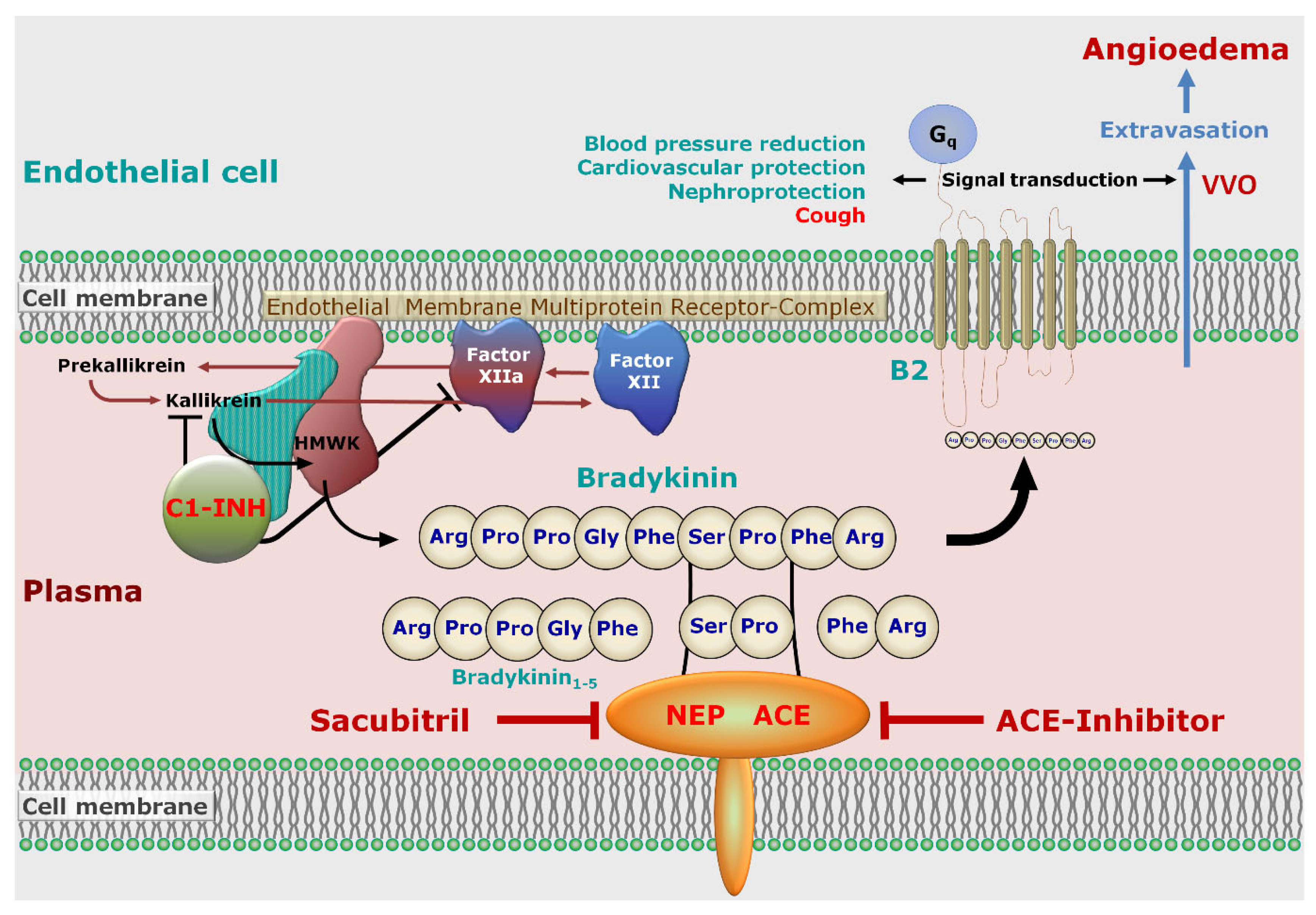
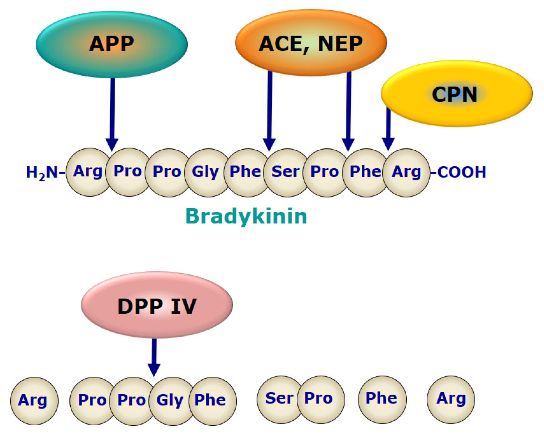
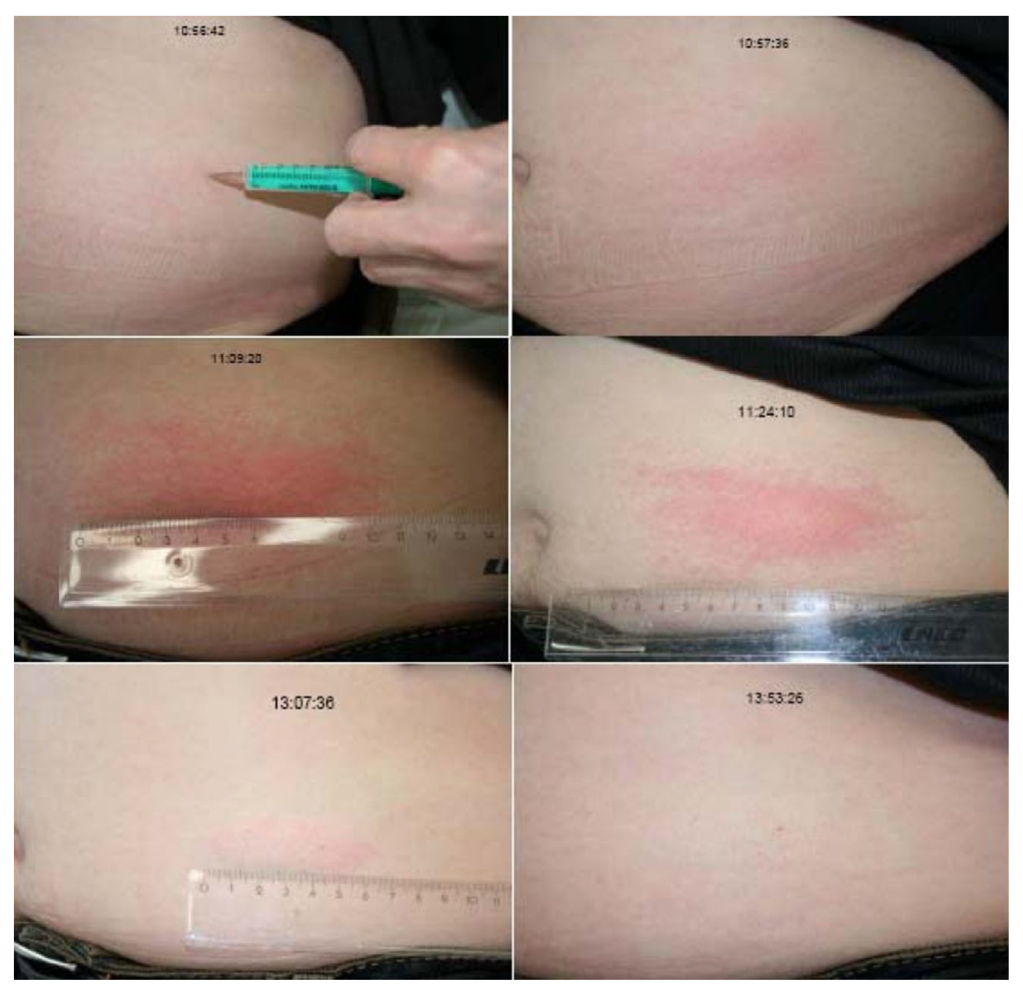
3.1. Drug-Induced-Angioedema Mediated by Bradykinin

In patients with a regular intake of one of the drugs summarized in Table 2 in combination with a non-itching angioedema in the head and neck region without wheals, the diagnosis drug-induced-angioedema mediated by bradykinin is extremely likely. These swellings often do not sufficiently respond to a therapy with corticosteroids or antihistamines [48]. Laboratory markers are not available, but complement testing for C1q and C4 and assessment of the C1-INH concentration and activity as well as the family history is suitable to exclude HAE or acquired angioedema with reduced C1-INH values [49]. An example of a possible step-by-step treatment approach was published elsewhere [50].
In addition, a newly developed LC-MS/MS methodology for the measurement of plasma peptides in the kallikrein-kinin system [51] might be helpful as well. This method has been successfully used to detect increased concentrations of the bradykinin metabolites bradykinin1-5 and bradykinin2-9 in patients with HAE [52] representing uncertain cases of drug-induced-angioedema mediated by bradykinin. Nevertheless, it is not yet established in everyday clinical practice and further research and a structured guideline is necessary.
Table 2. An overview of drugs with influence on bradykinin, possibly leading to bradykinin-mediated angioedema (1 heart failure with reduced ejection fraction, 2 dipeptidyl peptidase 4).
MedicationInfluence on BradykininIndicationsFurther Remarks
ACE-inhibitorsACE is involved in the degradation of bradykinin to bradykinin1–7 and bradykinin1–5Hypertension, heart failure 1, diabetic nephropathy, event prevention in cardiovascular diseaseEvent prevention at high cardiovascular risk only, unless required to control hypertension [53]
AliskirenDirect renin inhibitor; the exact pathophysiology of angioedema is not yet understoodEssential hypertensionNo reduction of cardiovascular or renal endpoints
Angiotensin II receptor blockers (sartans)reduced bradykinin metabolism by ACE and Neprilysin, molecular mechanism remains unclear [37]Hypertension, diabetic nephropathy, heart failure 1, event prevention in cardiovascular diseaseEvent prevention at high cardiovascular risk only, unless required to control hypertension [53]
Direct DPP-4 inhibitors 2DPP-4 further degrades bradykinin after cleavage by aminopeptidase P to the ineffective bradykinin2–9Type 2 diabetes mellitusunlikely leading to bradykinin-mediated angioedema alone, only in combination, e.g., with an ACE inhibitor
m-TOR inhibitorsThe mechanism of angioedema development remains unclear.Prophylaxis of kidney, heart and liver transplant rejectionSignificantly higher incidence of angioedema in combination with an ACE inhibitor
Neprilysin inhibitorsNeprilysin is involved in the degradation of bradykinin.Heart failure 1No combination with ACE-inibitors or sartans
Tissue plasminogen activatorcatalyzes the conversion of plasminogen to plasmin; plasmin has influence on bradykinin productionFibrinolysis in acute myocardial infarction, acute ischemic stroke and pulmonary embolismRelatively high number of potential life-threatening orolingual manifestations reported

3.2. Hereditary Angioedema

Patients with diagnosed HAE usually are equipped with an emergency ID. In not yet diagnosed patients with HAE, patients often report a family history of angioedema. The head and neck region is less frequently affected and wheals and pruritus are absent. Individual prodromi such as an erythema marginatum can appear. Hereditary angioedema shows no treatment response to corticosteroids or antihistamines. With reference to the diagnostic workflow of the hereditary angioedema guideline, C1-INH values and C4 complement should be measured to support the diagnosis. Nevertheless, retrieving the results of the values can last for several days, as not every laboratory can measure them [54]. In addition to this, the rare forms of HAE with normal C1-INH can only be diagnosed via genetic testing or according to the clinical picture and patient’s history (including family members with a history of recurrent angioedema), as it is extremely likely that not all underlying mutations are known [55].

3.3. Acute Urticaria

Urticaria is characterized by the development of wheals (hives), angioedema, or both. Isolated angioedema in acute spontaneous urticaria is not easy to differentiate from bradykinin-mediated edema. According to the current guideline, routine diagnostic tests are only necessary in chronic urticaria (>6 weeks) [56]. The resolution of acute angioedema in urticaria can take up to 72 h and additional itching may occur. In some forms of urticaria, eliciting factors such as pressure are reported.

3.4. Allergic Angioedema

Angioedema is one of the most common symptoms of acute allergic immediate hypersensitivity reactions and anaphylactic reactions. Regarding the medical history, questioning trigger factors, augmentation factors, and known allergies are most important. Concomitant hives and pruritus are frequent. The use of clinical criteria to identify anaphylaxis is suggested according to the current guideline, with blood sampling for the later measurement of tryptase [57].

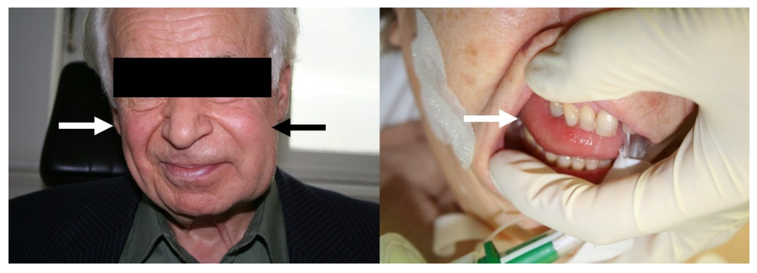
4. Symptoms and Course

In contrast with HAE, which manifests itself most often on the skin of the extremities and the abdomen, and less frequently in the head and neck [1], ACE-inhibitor-induced angioedema occurs mainly in the head and neck region [8]. The development and the severity of such non-allergic angioedema gradually increases within a time-span of several hours. Usually affected are the face, lips, facial soft tissues, eyelids, uvula, and the mucous membranes of the pharynx and larynx [48]. In contrast, gastrointestinal manifestations have only been reported sporadically [3,58]. It must be assumed that swellings in the head and neck region (Figure 1) are often not recognized as ACE-inhibitor-induced angioedema and thus discontinuation of the ACE inhibitor (see Section 6.1) is not considered. Of note, ACE-inhibitor-induced angioedema has also been described to occur in a small child [59]. The clinical course of ACE-inhibitor-induced angioedema cannot be foreseen. It can range from fast dissolving angioedema to a clinical emergency situation, especially in the case of swellings in the pharynx and larynx. In addition, a retrospective study involving 84 patients with ACE-inhibitor-induced angioedema found a nine times higher risk for emergency airway management compared with angioedema due to other reasons [60]. Upper airway obstruction, which occurs in about 10% of cases, can cause acute laryngeal obstruction with stridor and acute shortness of breath, and, in the worst case, can be fatal [61,62]. However, in contrast with HAE [63,64], there are no specific tools for diagnosis. Despite the current short availability and still limited use of sacubitril/valsartan, life-threatening angioedema has already been described for this combination [65].
Another important feature of ACE-inhibitor-induced angioedema is the time of onset after the initial start of ACE-inhibitor therapy. It is generally assumed that ACE-inhibitor-induced angioedema occurs within the first week of treatment. However, this is often not the case. For example, an analysis of data from more than 12,000 patients randomly assigned to enalapril treatment showed that although the incidence of angioedema was highest in the first four weeks, angioedema continued to occur after 12 weeks of therapy, albeit with a significantly lower incidence [23]. A retrospective analysis of the data from the ear nose and throat (ENT) clinic of the University Hospital Düsseldorf showed a significantly higher mean duration of uneventful treatment of about 39 months before an angioedema event [66]. In addition, one review listed a number of case studies in which ACE-inhibitor-induced angioedema occurred with a significant delay; in one case, after 23 years of uncomplicated therapy with enalapril [67].
Emergency admissions due to ACE-inhibitor-induced angioedema account for about one third of all emergency admissions for angioedema [68]. In view of the growing need to treat particular elderly and chronically ill patients with cardiovascular diseases, an increasing number of drug-induced non-allergic angioedema, including potentially life-threatening events, must be expected [69]. Moreover, the increase in the use of ACE-inhibitors is associated with an increase in hospitalization rates due to ACE-inhibitor-induced angioedema [40]. This is also supported by a study investigating the change in mortality rates of angioedema in the U.S. between 1979 and 2010. According to this study, the mortality rate of HAE fell from 0.28 to 0.06 per million, while the mortality rate of other non-allergic angioedema rose from 0.24 to 0.34 per million [70].

5. Pathophysiology

5.1. Endothelial Permeability

Angioedema is generally thought to be caused by a disturbance of endothelial permeability, although an obstruction of the flow of interstitial fluid into the lymphatic vessels is probably also involved (see below) [71]. Endothelial cells lining the inner surface of all types of blood vessels regulate several important physiologic functions, including platelet aggregation, coagulation, hemostasis, adhesion, and migration of immune cells and other inflammatory responses, as well as regional tissue blood flow distribution [72]. Endothelial permeability, which is also known as vascular permeability, is a fundamental regulator of fluid exchange between plasma and tissues [73] and it significantly contributes to immune defense, e.g., by extravasation of immunoglobulins and antibodies [74]. Fluid extravasation occurs in the capillaries and venules of the microvasculature of tissues and ensures the exchange of nutrients such as sugar, fatty acids, and vitamins; small amounts of proteins such as albumin; the supply with oxygen; and the evacuation of cellular waste products. Under physiologic steady state conditions, there is a continuous flow from the plasma through the interstitial fluid to the lymph compartments, which finally drains back into the circulation via the subclavian vein angles and lymph node micro vessels [75].
In general, plasma fluid and proteins can traverse the endothelial barrier by a paracellular and a transcellular route and both routes are involved in physiologic and pathophysiologic extravasation, i.e., the development of edema. Paracellular extravasation in capillaries, which mainly transports fluid and small molecules, occurs through inter-endothelial junctions (continuous capillaries) and/or endothelial cell pores (fenestrated capillaries), and both types of capillaries are present in human skin [76]. In contrast, the transcellular route (transcytosis) accounts for the majority of protein transport across the endothelial barrier, for example the most abundant interstitial protein albumin. This route is characterized by vesicles that were discovered and linked to extravasation by George E. Palade [77,78]. These vesicles can shuttle after endocytosis to the basolateral site of endothelial cells and release their content by exocytosis and/or form large channels through the endothelial cytoplasm, which have been termed vesiculo-vacuolar organelles (VVO) [74,79]. An excellent review on molecular mechanisms regulating these two routes of extravasation has been published recently [80].
Under basal conditions, plasma is permanently filtrated by these pathways. This situation changes dramatically once vascular permeabilizing mediators activate microvascular endothelial cells. Such mediators include histamine, thrombin, platelet activating factor, vascular endothelial growth factor, tumor necrosis factor α, lipopolysaccharide, and bradykinin [81]. In this scenario, the amounts of proteins that traverse from plasma to the interstitial space are greatly increased and include proteins larger than albumin such as antibodies or fibrinogen, which also increases interstitial oncotic pressure and may eventually result in tissue edema. Furthermore, fibrinogen and other proteins of the clotting cascade are part of the exudate and are activated in the interstitial fluid to synthetize fibrin. Fibrin forms a water trapping mesh and likely contributes to the maintenance of edema, even if disruption of the endothelial barrier has ceased [71]. This situation suggests that early treatment of angioedema is of great importance for timely and complete resolution of symptoms (see Section 6.5). Hence, most forms of edema, including ACE-inhibitor-induced angioedema, are the result of over-activation of para- and transcellular extravasation chiefly occurring at post-capillary venules [82].

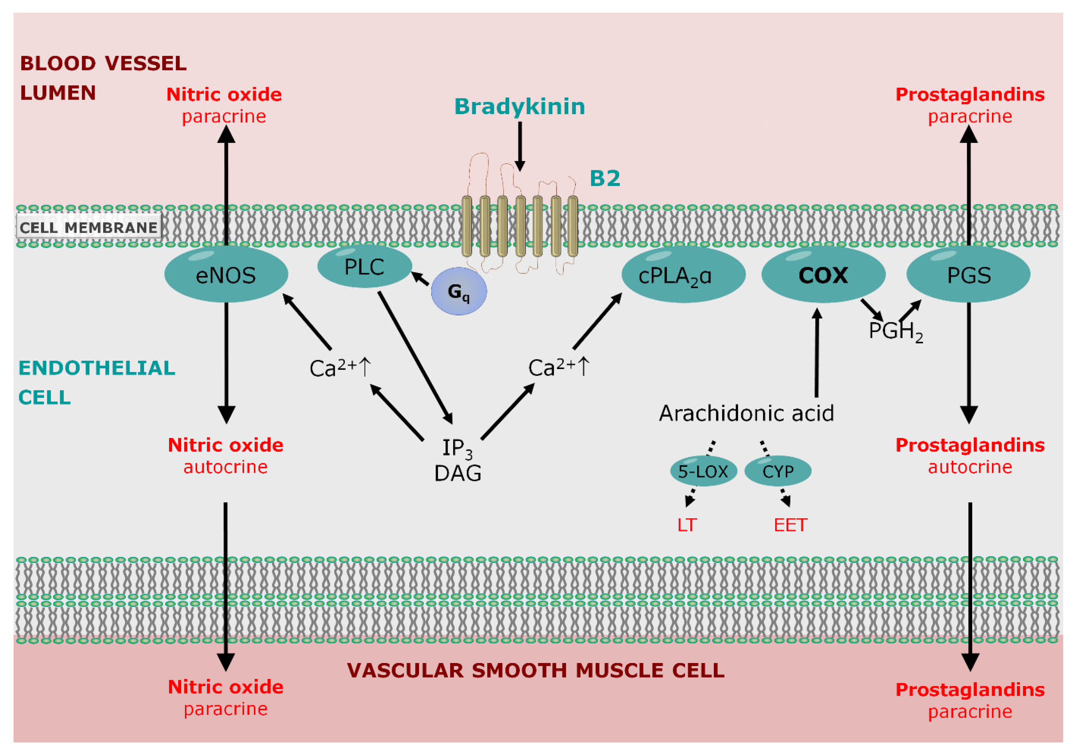
5.2. Role of Bradykinin Signaling

Non-allergic angioedema is almost exclusively caused by a local oversupply of bradykinin, which can be triggered by both increased synthesis and decreased degradation. Kinins are pharmacologically active and only short-lived peptides that are cleaved from kininogens by kallikreins in the plasma and tissues [83]. In the plasma, bradykinin is synthesized by plasma kallikrein from high molecular weight kininogen (HMWK) [84]. Coagulation factor XII (Hageman factor) is also involved in the synthesis of bradykinin, which is effectively inhibited by C1-INH (Figure 2). While HAE is characterized by an increased synthesis of bradykinin, the vast majority of acquired non-allergic angioedema are due to a drug-induced disruption of bradykinin degradation, for the most part by the inhibition of ACE (Figure 3), but also by the inhibition of neprilysin with sacubitril [4]. In addition, kinins can constrict smooth bronchial muscles [85] and sensitize airway afferent C fibers [86], suggesting that dry cough induced by ACE inhibitors is mediated by bradykinin and/or substance P. Moreover, the local accumulation of bradykinin can activate pro-inflammatory peptides and release histamine locally, which may contribute to hypersensitivity of the cough reflex [87].
Beside its well-known role as an inflammatory and pain mediator, bradykinin is involved in the regulation of the vascular tone and capillary fluid turnover. It signals via two distinct receptors, the bradykinin B1 receptor (B1) and B2 [2,91,92]. Among many other tissues, B2 is constitutively expressed in vascular endothelial and smooth muscle cells, while B1 expression is dependent on inflammatory mediators such as cytokines.
A recent study of the Cryo-EM structures of human B2-Gq proteins complexes provided deep insight into the molecular mechanisms underlying ligand binding, receptor activation, and Gq proteins coupling of B2 [93]. These new findings will most likely enable a structure-based drug design of B2 ligands for a variety of indications, including new B2 antagonists for the treatment of bradykinin-induced angioedema. Of note, a genome wide association study (GWAS) revealed a new pharmacogenomic locus within chromosome 14q32.2 baring the non-coding intergenic lead single nucleotide polymorphism (SNP) rs34485356, which is located 60 kb upstream of the B2 gene (BDKRB2) and is significantly associated with ACE-inhibitor-induced angioedema [94]. In addition, the authors found that carriers of the risk allele had significantly lower systolic and diastolic blood pressure, consistent with increased B2 activity. However, the high frequency of this risk allele, ranging from ∼78–90%, results in very low specificity and, hence, in a low positive predictive value that is unsuitable for preventive screening [95].
The binding of bradykinin at the endothelial Gαq/11-coupled B2 results in the activation of three main distinct signaling pathways (Figure 4): activation of phospholipase C-ß and the subsequent IP3-dependent release of intracellular calcium; activation of endothelial NO-synthase (eNOS) and the subsequent generation of nitric oxide (NO); and activation of phospholipase A2 and D and the subsequent generation of prostaglandins, leukotrienes, and epoxyeicosatrienoic acids and, finally, activation of the JAK/Stat-pathway and the subsequent alterations of the cell protein expression profile [2,96]. It is known that the activation of B2 leads to the formation of prostaglandins by cyclooxygenases, which generate prostaglandin H2, the precursor of prostaglandin synthases [97]. Accordingly, a recent study in mice and humans showed that unspecific inhibition of cyclooxygenases by ibuprofen weakened the plasma extravasation induced by the intradermal injection of bradykinin [98]. The results of a subsequent study provide evidence that the activation of B2 in small dermal blood vessels involves cytosolic phospholipase A2α and cyclooxygenase-1-dependent formation of prostacyclin, while the inhibition of 5-lipoxygenase, epoxyeicosatrienoic acid generation, and a variety of other prostaglandin receptor antagonists had no effect [99]. These findings extend our current knowledge on the signaling events following the activation of B2 in small dermal blood vessels and opens the possibility for developing new therapeutic options to treat acute non-allergic angioedema by targeting prostacyclin formation and/or signaling.
The development of C1-INH and B2 transgenic mice has provided important insights into the role of bradykinin in vivo [102,103,104,105], as the effect of bradykinin on vascular permeability in these mice could be specifically interrupted by treatment with C1-INH or the blocking of B2, respectively. In contrast, no effect of B2 on basal endothelial permeability was observed in the small dermal blood vessels of transgenic mice harboring an endothelial-specific overexpression of B2 [105]. However, transgenic rats with a targeted overexpression of B2 in the endothelium developed spontaneous intestinal angioedema [106]. In addition, the use of agonists and antagonists at bradykinin receptors has been shown to dilate peripheral and coronary arteries; lower arterial blood pressure in normotensive animals; and have antithrombogenic, antiproliferative, and antifibrinogenic effects [92]. The B2 antagonist icatibant (formerly HOE140) has been used in a variety of animal studies to evaluate the mostly protective effects of bradykinin; for example, in the cardiovascular system or the kidneys. These studies included subjects and patients considering the involvement of bradykinin in the cardiac and renal effects of ACE inhibitors [107,108]. So far, icatibant is the first and only B2 blocker approved for the drug treatment of acute HAE [109].

6. Therapy

Depending on the degree of severity and especially in laryngeal manifestation, all forms of angioedema may require emergency treatment, including a ventilation tube, tracheotomy, or emergency coniotomy. A more detailed description of emergency measures can be found elsewhere [8]. Usually, acute angioedema is primarily treated as “mastcell-mediated angioedema” with antihistamines and steroids due to their increased incidence based on the current anaphylaxis guidelines. Patients with HAE should be equipped with an emergency card and treated accordingly.
Pharmacotherapy in HAE is based on the inhibition of bradykinin synthesis, either by the substitution of C1-INH or by the inhibition of kallikrein, and by the blockade of B2 [20,110]. This pharmacotherapy is well established and validated by randomized studies. With the exception of the B2 antagonist icatibant, all other approved drugs are kallikrein inhibitors. For acute treatment and short-term prophylaxis, this concerns, for example, the infusion of or subcutaneous application of human plasma-derived C1-INH such as Berinert® or Cynrize®. The latter is also approved for long-term prophylaxis. Another effective principle for acute treatment is the specific blockade of B2 by subcutaneously applied icatibant (Firazyr®), which has been approved for this indication, but not for short-term or long-term prophylaxis. This is most likely a result of the short halflife of icatibant and might be overcome by the novel B2 agonist Deucrictibant [111]. A third possibility is the pharmacological inhibition of kallikrein, which, like C1-INH, leads to an interruption of the synthesis of bradykinin. While the synthetic peptide ecallantide (Kalbitor®) is approved in the USA but not in the EU for acute treatment, the monoclonal antibody Lanadelumab is approved for routine prophylaxis of recurrent HAE attacks but not for acute treatment [112,113,114]. Lanadelumab is a fully human monoclonal antibody that inhibits kallikrein activity. Lanadelumab can be administered subcutaneously and only needs to be given every two weeks. In symptom-free patients, the interval can be extended to every 4 weeks. Self-application is possible after appropriate training. In addition, the oral kallikrein inhibitor Berotralstat (BCX7353) is approved for long-term prophylaxis of acute attacks in HAE based on favorable outcomes of phase II and phase III clinical trials [115,116]. Recently published guidelines provide more detailed recommendations on the pharmacotherapy of HAE [63,64].

6.1. Discontinuation of ACE-Inhibitors

In contrast with the well-evaluated possibilities of pharmacotherapy for HAE, no drug has been approved to date for the treatment of acute ACE-inhibitor-induced angioedema. As ACE-inhibitor-induced angioedema is a drug side effect, the most obvious measure is to discontinue the ACE inhibitor. However, it has never been investigated whether this measure is useful for patients in acute cases in terms of a faster improvement of symptoms, but continuation of ACE-inhibitor therapy despite ACE-inhibitor-induced angioedema is associated with an approximately 10-fold higher rate of recurrence according to an earlier retrospective study in 82 patients [117]. According to a retrospective observational study involving 267,612 patients on ACE inhibitors, up to half of the patients on ACE inhibitors fail to stop after the first ACE-inhibitor-induced angioedema [118]. In these patients, the probability of recurrence of angioedema is more than 100 times higher (21.9–25.2%) than that of a first event (0.16%). Therefore, a “prophylactic” effect of discontinuation of the ACE inhibitor in the sense of absent recurring angioedema should be expected. Again, no prospective data are available.
In contrast, retrospective analyses have surprisingly shown that this is not always the case. For example, in the above-mentioned retrospective observational study with 267,612 patients, approximately 9% of the patients who had stopped taking ACE-inhibitors after the initial angioedema experienced a recurrence [118]. In a population of 54 Italian patients who were switched to either a sartan, a calcium antagonist, or another medication because of ACE-inhibitor-induced angioedema, angioedema continued to occur in eight patients (15%) [119]. Only in two patients were recurrences probably due to a sartan. In another retrospective study by the same group of researchers, the recurrence rate was dramatically higher, at about 50%, in 111 patients after discontinuing the ACE inhibitor [120]. Surprisingly, this rate was independent of whether the patients were treated with sartan, a calcium antagonist, a ß-blocker, or any other medication. About two thirds of these patients had repeated angioedema at a rate of one to five attacks per year. The authors suspect that patients suffering from ACE-inhibitor-induced angioedema already have a disposition to develop angioedema manifested by therapy with ACE inhibitors. This view appears to be supported by a case report on a first manifestation of ACE-inhibitor-induced angioedema four weeks after the discontinuation of Lisinopril due to persistent dry cough, despite months of prior uneventful treatment [121].

6.2. Antifibrinolytics

In some countries, antifibrinolytics (such as tranexamic acid) still play a role in the treatment of attacks in patients with HAE. For on-demand or prophylactic treatment of HAE attacks, the current international guideline advises against their use [56]. It is argued that data for their efficacy are lacking or not beneficial; nevertheless, the safety profile seems good and it must be admitted that in some countries no other solutions exist.

6.3. Ecallantide

Ecallantide (DX-88, Dyax Corp., Cambridge, MA, USA) is a recombinant protein of 60 amino acids that specifically inhibits plasma kallikrein. This suggests that ecallantide approximately mimics the physiological effects of C1-INH (Figure 2). However, there are a number of differences in the biological effects of C1-INH. For example, ecallantide has a much weaker inhibitory effect on C1r and C1s, plasmin, factor XIIa, and factor XIa, although it is a potent plasma kallikrein inhibitor [122]. The drug has been shown to be effective in the treatment of acute HAE attacks [113]. A multi-center, randomized, and comparatively large phase II study evaluated the benefit of ecallantide at three different doses in 79 patients with mild to moderate ACE-inhibitor-induced angioedema [123], but showed no difference from the “standard therapy”, which was optional for the study physicians. The vast majority of patients received glucocorticoids (85%) and/or H1-antihistamines (85%) regardless of the allocation. Similar results were obtained in a second randomized controlled phase II study with a comparable study protocol [124].

6.4. C1-INH

A total of at least 27 case reports and case series have suggested a beneficial effect of C1-INH in the treatment of ACE-inhibitor-induced angioedema [112]. Furthermore, a randomized controlled trial was performed to evaluated the efficacy of human C1-INH concentrate for the treatment of acute ACE-inhibitor-induced angioedema. Thirty patients (16 C1-INH, 14 placebo) were randomized and dosed. Regarding the results with a baseline application of steroids and antihistamines, C1-INH was inferior in the treatment of ACE-induced-angioedema when compared with a placebo with respect to the time to complete resolution of symptoms [125]. As most acute angioedema is often treated like “allergic” angioedema with corticosteroids and antihistamines, it remains unclear whether this treatment has at least a slight effect on acute bradykinin-mediated angioedema as a side effect of drugs such as ACE-inhibitors (see Section 6.5).

6.5. Icatibant

For the treatment of ACE-inhibitor-induced angioedema with icatibant, ten case reports and five case series indicating clinically useful effects have been described [112]. For example, one prospective case series involving eight patients showed that the single subcutaneous administration of 30 mg of icatibant reduced the time to the complete disappearance of symptoms from 33 h to 4.4 h [126]. A retrospective case series on the use of icatibant for non-HAE described 14 patients with ACE-inhibitor-induced angioedema where treatment with icatibant resulted in a median resolution time of 9 h; however, in this study, no comparator population was available [127]. Meanwhile, three randomized studies evaluating the efficacy and safety of icatibant have also been completed and published [48,128,129].
The AMACE (AMelioration of Angiotensin Converting Enzyme Inhibitor Induced Angioedema Study), a randomized double-blind phase II study, included 27 patients with ACE-inhibitor-induced angioedema, of which 13 patients received subcutaneously 30 mg of icatibant and 14 patients in the control group received intravenously 500 mg of prednisolone and 2 mg of clemastine (“standard of care”) [48]. In the patient population, all patients were fair-skinned (white) and two thirds were male, and edema was assessed visually (endoscopically). All patients were treated within approximately 6 h after the onset of symptoms. The primary endpoint of the study was time to complete resolution of symptoms. As a result, treatment with icatibant significantly lowered the primary endpoint (p = 0.002) from 27.1 h to 8.0 h. Thus, this study not only provided the first proof of the efficacy of icatibant in this indication, but also the first clinical evidence that B2 activation is involved in ACE-inhibitor-induced angioedema. Only few side effects were observed. The most common side effect of icatibant is a transient uncomplicated reaction at the injection site, which is probably a pseudoallergic reaction. It is likely caused by the activation of the mast cell receptor “mass-related G-protein coupled receptor member X2” (MRGPRX2) [130]. Icatibant’s binding to this receptor directly activates mast cells and can thus be classified as an “off-target” effect. In the two pivotal studies “For Angioedema Subcutaneous Treatment”(FAST)-1 and FAST-2 [109], icatibant induced a transient uncomplicated reaction at the injection site after subcutaneous application in nearly all patients with HAE (>95%), which we were able to document as well (Figure 5).
However, the favorable clinical results of the AMACE trial were not confirmed in subsequent clinical trials. A second randomized, double-blind study included 31 patients with ACE-inhibitor-induced angioedema, of which 13 patients received 30 mg of icatibant subcutaneously and 18 patients received placebo (isotonic saline solution) [128]. Edema was assessed only on the basis of symptoms and no endoscopy was performed. In addition, patients could also be treated with glucocorticoids and H1 antihistamines, as determined by their physicians. In contrast with AMACE, this “standard of care” therapy was not only given to patients in the control group, but also to about 90% of patients in both study arms. Another difference to AMACE was the significantly longer time interval between the onset of symptoms and treatment with the study medication, which was 10.3 h on average. Furthermore, in this study, two thirds of the patient population were black and two thirds female. The primary endpoints of the study were time to complete disappearance of symptoms and assessment of the severity of symptoms over time. In contrast with AMACE, no significant difference was found between placebo and icatibant in this study.
In a third randomized study with icatibant, 121 patients with ACE-inhibitor-induced angioedema were enrolled, of whom 60 patients received 30 mg of icatibant subcutaneously and 58 patients received placebo (isotonic saline solution) [129]. Again, edema was assessed only on the basis of symptoms and no endoscopy was performed. The median time interval between the onset of symptoms and treatment with the study drug was 7.8 h, which was also significantly longer than in the AMACE study. Two-thirds of the patient population was black, but the sexes were roughly equally distributed. In this study, too, more than 90% of patients in both study arms received the “standard of care” therapy (glucocorticoids and H1 antihistamines). The primary endpoint of the study was time to discharge, which is similar to the endpoint “complete disappearance of symptoms”. In contrast with AMACE, no significant difference was found between the placebo and icatibant in this study either.
In summary, at first glance, the therapeutic efficacy of icatibant in ACE-inhibitor-induced angioedema in the AMACE study may be considered to be a rather random result. On the other hand, if icatibant is ineffective, this would mean that ACE-inhibitor-induced angioedema might not be caused by the over activation of B2. However, icatibant has been approved for the treatment of hereditary angioedema, which is caused by bradykinin [109]. Furthermore, this view is contradicted by the subsequent results of the study of Straka et al. [128] in patients with ACE-inhibitor-induced angioedema [131]. The authors reported that in patients with angioedema, plasma levels of bradykinin and the ratio of plasma levels of bradykinin to the ACE degradation product bradykinin1–5 (Figure 2) were elevated, while plasma levels of bradykinin1-5 itself remained more or less constant. The plasma concentration of the two cleavage products of HMWK, which are formed during the proteolytic cleavage by kallikrein, also remained unchanged. These results not only confirm prior reports suggesting that plasma levels of bradykinin are elevated in ACE-inhibitor-induced angioedema [132], they also suggest that the degradation of bradykinin is reduced, which can be casually attributed to the residual effect of ACE inhibitors at the time of blood collection. Finally, no evidence of increased synthesis of bradykinin from HMWK could be found. Accordingly, the lack of clinical efficacy of icatibant for the treatment of ACE-inhibitor-induced angioedema in the two randomized controlled trials described above [128,129] probably cannot be attributed to a possibly incorrect pathophysiological approach. More likely reasons would be two other important differences between these two studies and AMACE, which are (1) the significantly longer time interval between the onset of symptoms and treatment with the study medication and (2) the treatment with icatibant as an add-on to glucocorticoids and H1 antihistamines.
Clinical experience in the treatment of acute HAE has shown a better and faster response if pharmacotherapy is initiated early after symptom onset [20], and this might be true as well in ACE-inhibitor-induced angioedema. One of the underlying reasons might be the time-dependent formation of an interstitial water trapping fibrin mesh (see Section 5.1). Glucocorticoids and antihistamines have never been tested for their efficacy in non-allergic angioedema and are generally considered to be ineffective [133]. On the other hand, the activation of B2 leads to the formation of prostaglandins by cyclooxygenases, which generate prostaglandin H2, the precursor of prostaglandin synthases [97]. Accordingly, the unspecific inhibition of cyclooxygenases by ibuprofen weakens plasma extravasation induced by the intradermal injection of bradykinin [98], and a further study suggests the involvement of phospholipase A2α and cyclooxygenase-1-dependent formation of prostacyclin [99]. Glucocorticoids can block phospholipase A2 via the induction of annexin A1 and thus the release of arachidonic acid, the starting product of prostaglandin synthesis [134]. It has also been described that glucocorticoids induce the expression of ACE [135] and this effect may also be useful in ACE-inhibitor-induced angioedema. Likewise, a case study of laryngeal cartilage angioedema associated with the use of lisinopril shows complete regression after intravenous therapy with diphenhydramine and methylprednisolone [136]. Finally, some clinical data suggest that ACE-inhibitor-induced angioedema is associated with inflammatory responses. In one study, the plasma concentrations of C-reactive protein were increased by 7.3-fold (43.2 mg/L) in the acute phase of 25 cases compared with 18 cases with angioedema of an unknown origin (5.9 mg/L) [66]. Similarly, another retrospective study published 14 years later reported on abnormally elevated levels of C-reactive protein in 50 out of 73 patients in the acute phase, 18 of which showed levels of >15–60 mg/L and 8 patients showed levels of >60 mg/L [60]. Thus, it is quite possible that the simultaneous administration of glucocorticoids may have reduced the therapeutic effect of icatibant in the two randomized trials described above. Likewise, neither ecallantide nor C1-INH showed a clinically useful effect in patients with ACE-inhibitor-induced angioedema pretreated with glucocorticoids. Taken together, even if one might consider the discordant results of the three randomized clinical with icatibant in ACE-inhibitor-induced angioedema as unfortunate, this could pave the way to better substantiate the “standard of care” emergency treatment of non-allergic angioedema with high-dose glucocorticoids such as 500 mg prednisolone.

7. Conclusions

Bradykinin-mediated angioedema is a comparatively rare, but underestimated symptom. Hereditary angioedema is well understood and an established guideline and diagnostic and therapeutic workflow exist. In striking contrast, bradykinin-mediated angioedema triggered by the intake of drugs such as ACE-inhibitors is still difficult to diagnose and only off-label treatment exists. The current results of randomized controlled trials with icatibant are inconsistent and a final evaluation about the effectiveness of therapeutics is not possible.

Author Contributions

All of the authors made substantial contributions to the conception and writing of the manuscript. G.K. conceived of the presented idea, and supervised and organized the work. The main subject of M.B. was the current trends in diagnostics. The main subject of J.G. and J.H. was the treatment and course of HAE and the main subjects of G.K. were epidemiology, pathophysiology, and a summary of the current study data. All of the authors discussed the data and approved the final manuscript. All authors have read and agreed to the published version of the manuscript.

Funding

This research was funded by the German Research Foundation (DFG) to GK (grant number KO 1557/14-2).

Institutional Review Board Statement

Not applicable.

Informed Consent Statement

Informed consent was obtained from all subjects involved in the study.

Data Availability Statement

Data sharing not applicable.

Conflicts of Interest

J. Hahn has received speaker/consultancy fees from CSL Behring and Takeda. She has also received funding to attend conferences/educational events from CSL Behring and Takeda. She has participated as an investigator in a clinical trial/registry for CSL Behring, BioCryst, Pharvaris, and Takeda. J. Greve has received speaker/consultancy fees from CSL Behring, Takeda, and BioCryst. He has also received funding to attend conferences/educational events from CSL Behring and Takeda. He has participated as an investigator in a clinical trial/registry for CSL Behring, Pharvaris, BioCryst, and Takeda. M. Bas has not had relationships with industry during the past 5 years. G. Kojda: has not had relationships with industry during the past 5 years.

References

  1. Cicardi, M.; Aberer, W.; Banerji, A.; Bas, M.; Bernstein, J.A.; Bork, K.; Caballero, T.; Farkas, H.; Grumach, A.; Kaplan, A.P.; et al. Classification, diagnosis, and approach to treatment for angioedema: Consensus report from the hereditary angioedema international working group. Allergy 2014, 69, 602–616. [Google Scholar] [CrossRef] [PubMed]
  2. Bas, M.; Adams, V.; Suvorava, T.; Niehues, T.; Hoffmann, T.K.; Kojda, G. Non-allergic angioedema. Role of bradykinin. Allergy 2007, 62, 842–856. [Google Scholar] [CrossRef]
  3. Mujer, M.T.P.; Rai, M.P.; Nemakayala, D.R.; Yam, J.L. Angioedema of the small bowel caused by lisinopril. Drug Ther. Bull. 2019, 57, 14–15. [Google Scholar] [CrossRef] [PubMed]
  4. Bas, M.; Greve, J.; Strassen, U.; Khosravani, F.; Hoffmann, T.K.; Kojda, G. Angioedema induced by cardiovascular drugs: New players join old friends. Allergy 2015, 70, 1196–1200. [Google Scholar] [CrossRef] [PubMed]
  5. Buttgereit, T.; Maurer, M. Classification and pathophysiology of angioedema. Hautarzt 2019, 70, 84–91. [Google Scholar] [CrossRef] [PubMed]
  6. Hahn, J.; Hoffmann, T.K.; Bock, B.; Nordmann-Kleiner, M.; Trainotti, S.; Greve, J. Angioedema. Dtsch. Arztebl. Int. 2017, 114, 489–496. [Google Scholar] [CrossRef]
  7. White, A.A.; Stevenson, D.D. Aspirin-exacerbated respiratory disease. N. Engl. J. Med. 2018, 379, 1060–1070. [Google Scholar] [CrossRef]
  8. Bas, M.; Greve, J.; Bier, H.; Knopf, A.; Stark, T.; Schuler, P.; Hoffmann, T.K.; Kojda, G. Notfallsituation akutes angioödem [Emergency management of acute angioedema]. Dtsch. Med. Wochenschr. 2010, 135, 1027–1031. [Google Scholar] [CrossRef]
  9. Goring, H.D. In memory of the 160th birthday and the 80th anniversary of the death of heinrich irenaus quincke, as well as of his description of angioedema 120 years ago. Hautarzt 2002, 53, 822–825. [Google Scholar]
  10. Hofman, Z.; de Maat, S.; Hack, C.E.; Maas, C. Bradykinin: Inflammatory product of the coagulation system. Clin. Rev. Allergy Immunol. 2016, 51, 152–161. [Google Scholar] [CrossRef]
  11. Hill, M.D.; Barber, P.A.; Takahashi, J.; Demchuk, A.M.; Feasby, T.E.; Buchan, A.M. Anaphylactoid reactions and angioedema during alteplase treatment of acute ischemic stroke. CMAJ 2000, 162, 1281–1284. [Google Scholar]
  12. Bozkurt, S.; Arslan, E.D.; Köse, A.; Ayrık, C.; Yılmaz, A.; Dündar, G.A. Lingual angioedema after alteplase treatment in a patient with acute ischemic stroke. World J. Emerg. Med. 2015, 6, 74–76. [Google Scholar] [CrossRef]
  13. Dewald, G.; Bork, K. Missense mutations in the coagulation factor xii (Hageman factor) gene in hereditary angioedema with normal c1 inhibitor. Biochem. Biophys. Res. Commun. 2006, 343, 1286–1289. [Google Scholar] [CrossRef]
  14. Bork, K.; Wulff, K.; Steinmüller-Magin, L.; Braenne, I.; Staubach-Renz, P.; Witzke, G.; Hardt, J. Hereditary angioedema with a mutation in the plasminogen gene. Allergy 2018, 73, 442–450. [Google Scholar] [CrossRef] [PubMed]
  15. Bafunno, V.; Firinu, D.; D’Apolito, M.; Cordisco, G.; Loffredo, S.; Leccese, A.; Bova, M.; Barca, M.P.; Santacroce, R.; Cicardi, M.; et al. Mutation of the angiopoietin-1 gene (ANGPT1) associates with a new type of hereditary angioedema. J. Allergy Clin. Immunol. 2018, 141, 1009–1017. [Google Scholar] [CrossRef] [PubMed]
  16. Bork, K.; Wulff, K.; Rossmann, H.; Steinmüller-Magin, L.; Braenne, I.; Witzke, G.; Hardt, J. Hereditary angioedema cosegregating with a novel kininogen 1 gene mutation changing the n-terminal cleavage site of bradykinin. Allergy 2019, 74, 2479–2481. [Google Scholar] [CrossRef] [PubMed]
  17. Ariano, A.; D’Apolito, M.; Bova, M.; Bellanti, F.; Loffredo, S.; D’Andrea, G.; Intrieri, M.; Petraroli, A.; Maffione, A.B.; Spadaro, G.; et al. A myoferlin gain-of-function variant associates with a new type of hereditary angioedema. Allergy 2020, 75, 2989–2992. [Google Scholar] [CrossRef] [PubMed]
  18. Bork, K.; Wulff, K.; Möhl, B.S.; Steinmüller-Magin, L.; Witzke, G.; Hardt, J.; Meinke, P. Novel hereditary angioedema linked with a heparan sulfate 3-o-sulfotransferase 6 gene mutation. J. Allergy Clin. Immunol. 2021, 148, 1041–1048. [Google Scholar] [CrossRef]
  19. Zuraw, B.L. Hereditary angioedema with normal c1 inhibitor: Four types and counting. J. Allergy Clin. Immunol. 2018, 141, 884–885. [Google Scholar] [CrossRef]
  20. Longhurst, H.; Cicardi, M. Hereditary angio-oedema. Lancet 2012, 379, 474–481. [Google Scholar] [CrossRef]
  21. Bas, M.; Hoffmann, T.K.; Kojda, G. Evaluation and management of angioedema of the head and neck. Curr. Opin. Otolaryngol. Head Neck Surg. 2006, 14, 170–175. [Google Scholar] [CrossRef] [PubMed]
  22. Agostoni, A.; Cicardi, M. Drug-induced angioedema without urticaria. Drug Saf. 2001, 24, 599–606. [Google Scholar] [CrossRef]
  23. Kostis, J.B.; Kim, H.J.; Rusnak, J.; Casale, T.; Kaplan, A.; Corren, J.; Levy, E. Incidence and characteristics of angioedema associated with enalapril. Arch. Intern. Med. 2005, 165, 1637–1642. [Google Scholar] [CrossRef] [PubMed]
  24. Brown, N.J.; Ray, W.A.; Snowden, M.; Griffin, M.R. Black americans have an increased rate of angiotensin converting enzyme inhibitor-associated angioedema. Clin. Pharmacol. Ther. 1996, 60, 8–13. [Google Scholar] [CrossRef]
  25. Gainer, J.V.; Nadeau, J.H.; Ryder, D.; Brown, N.J. Increased sensitivity to bradykinin among african americans. J. Allergy Clin. Immunol. 1996, 98, 283–287. [Google Scholar] [CrossRef] [PubMed]
  26. McDowell, S.E.; Coleman, J.J.; Ferner, R.E. Systematic review and meta-analysis of ethnic differences in risks of adverse reactions to drugs used in cardiovascular medicine. BMJ 2006, 332, 1177–1181. [Google Scholar] [CrossRef] [PubMed]
  27. Yusuf, S.; Sleight, P.; Pogue, J.; Bosch, J.; Davies, R.; Dagenais, G. Effects of an angiotensin-converting-enzyme inhibitor, ramipril, on cardiovascular events in high-risk patients. The heart outcomes prevention evaluation study investigators. N. Engl. J. Med. 2000, 342, 145–153. [Google Scholar] [PubMed]
  28. Pfeffer, M.A.; McMurray, J.J.; Velazquez, E.J.; Rouleau, J.L.; Kober, L.; Maggioni, A.P.; Solomon, S.D.; Swedberg, K.; Van de Werf, F.; White, H.; et al. Valsartan, captopril, or both in myocardial infarction complicated by heart failure, left ventricular dysfunction, or both. N. Engl. J. Med. 2003, 349, 1893–1906. [Google Scholar] [CrossRef]
  29. Yusuf, S.; Teo, K.K.; Pogue, J.; Dyal, L.; Copland, I.; Schumacher, H.; Dagenais, G.; Sleight, P.; Anderson, C. Telmisartan, ramipril, or both in patients at high risk for vascular events. N. Engl. J. Med. 2008, 358, 1547–1559. [Google Scholar]
  30. Makani, H.; Messerli, F.H.; Romero, J.; Wever-Pinzon, O.; Korniyenko, A.; Berrios, R.S.; Bangalore, S. Meta-analysis of randomized trials of angioedema as an adverse event of renin-angiotensin system inhibitors. Am. J. Cardiol. 2012, 110, 383–391. [Google Scholar] [CrossRef]
  31. Brown, N.J.; Byiers, S.; Carr, D.; Maldonado, M.; Warner, B.A. Dipeptidyl peptidase-iv inhibitor use associated with increased risk of ace inhibitor-associated angioedema. Hypertension 2009, 54, 516–523. [Google Scholar] [CrossRef]
  32. Byrd, J.B.; Shreevatsa, A.; Putlur, P.; Foretia, D.; McAlexander, L.; Sinha, T.; Does, M.D.; Brown, N.J. Dipeptidyl peptidase iv deficiency increases susceptibility to angiotensin-converting enzyme inhibitor-induced peritracheal edema. J. Allergy Clin. Immunol. 2007, 120, 403–408. [Google Scholar] [CrossRef]
  33. Yusuf, S.; Teo, K.; Anderson, C.; Pogue, J.; Dyal, L.; Copland, I.; Schumacher, H.; Dagenais, G.; Sleight, P. Effects of the angiotensin-receptor blocker telmisartan on cardiovascular events in high-risk patients intolerant to angiotensin-converting enzyme inhibitors: A randomised controlled trial. Lancet 2008, 372, 1174–1183. [Google Scholar]
  34. Rasmussen, E.R.; Pottegård, A.; Bygum, A.; von Buchwald, C.; Homøe, P.; Hallas, J. Angiotensin ii receptor blockers are safe in patients with prior angioedema related to angiotensin-converting enzyme inhibitors—A nationwide registry-based cohort study. J. Intern. Med. 2019, 285, 553–561. [Google Scholar] [CrossRef] [PubMed]
  35. Hellebrand, M.C.; Kojda, G.; Hoffmann, T.K.; Bas, M. Angioedema due to ace inhibitors and AT1 receptor antagonists. Hautarzt 2005, 57, 808–810. [Google Scholar] [CrossRef] [PubMed]
  36. Warner, K.K.; Visconti, J.A.; Tschampel, M.M. Angiotensin ii receptor blockers in patients with ace inhibitor-induced angioedema. Ann. Pharmacother. 2000, 34, 526–528. [Google Scholar] [CrossRef] [PubMed]
  37. Campbell, D.J.; Krum, H.; Esler, M.D. Losartan increases bradykinin levels in hypertensive humans. Circulation 2005, 111, 315–320. [Google Scholar] [CrossRef]
  38. McMurray, J.J.; Packer, M.; Desai, A.S.; Gong, J.; Lefkowitz, M.P.; Rizkala, A.R.; Rouleau, J.L.; Shi, V.C.; Solomon, S.D.; Swedberg, K.; et al. Angiotensin-neprilysin inhibition versus enalapril in heart failure. N. Engl. J. Med. 2014, 371, 993–1004. [Google Scholar] [CrossRef]
  39. Kostis, J.B.; Packer, M.; Black, H.R.; Schmieder, R.; Henry, D.; Levy, E. Omapatrilat and enalapril in patients with hypertension: The omapatrilat cardiovascular treatment vs. Enalapril (octave) trial. Am. J. Hypertens. 2004, 17, 103–111. [Google Scholar] [CrossRef]
  40. Kostis, W.J.; Shetty, M.; Chowdhury, Y.S.; Kostis, J.B. Ace inhibitor-induced angioedema: A review. Curr. Hypertens. Rep. 2018, 20, 55. [Google Scholar] [CrossRef]
  41. Hurford, R.; Rezvani, S.; Kreimei, M.; Herbert, A.; Vail, A.; Parry-Jones, A.R.; Douglass, C.; Molloy, J.; Alachkar, H.; Tyrrell, P.J.; et al. Incidence, predictors and clinical characteristics of orolingual angio-oedema complicating thrombolysis with tissue plasminogen activator for ischaemic stroke. J. Neurol. Neurosurg. Psychiatry 2015, 86, 520–523. [Google Scholar] [CrossRef] [PubMed]
  42. Lapostolle, A.; Weisenburger-Lile, D.; Yger, M.; Alamowitch, S.; Fain, O. Bradykinin-mediated angioedema following tenecteplase administration in an acute ischemic stroke. Stroke 2022, 53, e446–e447. [Google Scholar] [CrossRef] [PubMed]
  43. Khalid, M.; Kanaa, M.; Alkawaleet, Y.; Ayub, M.T. Angioedema: A life-threatening complication of tissue plasminogen activator. Cureus 2018, 10, e2392. [Google Scholar] [CrossRef]
  44. White, W.B.; Bresalier, R.; Kaplan, A.P.; Palmer, B.F.; Riddell, R.H.; Lesogor, A.; Chang, W.; Keefe, D.L. Safety and tolerability of the direct renin inhibitor aliskiren: A pooled analysis of clinical experience in more than 12,000 patients with hypertension. J. Clin. Hypertens. 2010, 12, 765–775. [Google Scholar] [CrossRef] [PubMed]
  45. Byrd, J.B.; Touzin, K.; Sile, S.; Gainer, J.V.; Yu, C.; Nadeau, J.; Adam, A.; Brown, N.J. Dipeptidyl peptidase iv in angiotensin-converting enzyme inhibitor associated angioedema. Hypertension 2008, 51, 141–147. [Google Scholar] [CrossRef]
  46. Duerr, M.; Glander, P.; Diekmann, F.; Dragun, D.; Neumayer, H.H.; Budde, K. Increased incidence of angioedema with ace inhibitors in combination with mtor inhibitors in kidney transplant recipients. Clin. J. Am. Soc. Nephrol. 2010, 5, 703–708. [Google Scholar] [CrossRef]
  47. Lenschow, M.; Bas, M.; Johnson, F.; Wirth, M.; Strassen, U. A score for the differential diagnosis of bradykinin- and histamine-induced head and neck swellings. Eur. Arch. Otorhinolaryngol. 2018, 275, 1767–1773. [Google Scholar] [CrossRef]
  48. Bas, M.; Greve, J.; Stelter, K.; Havel, M.; Strassen, U.; Rotter, N.; Veit, J.; Schossow, B.; Hapfelmeier, A.; Kehl, V.; et al. A randomized trial of icatibant in ace-inhibitor-induced angioedema. N. Engl. J. Med. 2015, 372, 418–425. [Google Scholar] [CrossRef]
  49. Balla, Z.; Zsilinszky, Z.; Pólai, Z.; Andrási, N.; Kőhalmi, K.V.; Csuka, D.; Varga, L.; Farkas, H. The importance of complement testing in acquired angioedema related to angiotensin-converting enzyme inhibitors. J. Allergy Clin. Immunol. Pract. 2021, 9, 947–955. [Google Scholar] [CrossRef]
  50. Hahn, J.; Bock, B.; Muth, C.M.; Pfaue, A.; Friedrich, D.; Hoffmann, T.K.; Greve, J. The ulm emergency algorithm for the acute treatment of drug-induced, bradykinin-mediated angioedema. Med. Klin. Intensivmed. Notfmed. 2019, 114, 708–716. [Google Scholar] [CrossRef]
  51. Gangnus, T.; Burckhardt, B.B. Targeted lc-ms/ms platform for the comprehensive determination of peptides in the kallikrein-kinin system. Anal. Bioanal. Chem. 2021, 413, 2971–2984. [Google Scholar] [CrossRef]
  52. Marceau, F.; Rivard, G.E.; Hébert, J.; Gauthier, J.; Bachelard, H.; Gangnus, T.; Burckhardt, B.B. Picomolar sensitivity analysis of multiple bradykinin-related peptides in the blood plasma of patients with hereditary angioedema in remission: A pilot study. Front. Allergy 2022, 3, 837463. [Google Scholar] [CrossRef]
  53. Knuuti, J.; Wijns, W.; Saraste, A.; Capodanno, D.; Barbato, E.; Funck-Brentano, C.; Prescott, E.; Storey, R.F.; Deaton, C.; Cuisset, T.; et al. 2019 esc guidelines for the diagnosis and management of chronic coronary syndromes. Eur. Heart J. 2020, 41, 407–477. [Google Scholar] [CrossRef]
  54. Maurer, M.; Magerl, M.; Betschel, S.; Aberer, W.; Ansotegui, I.J.; Aygören-Pürsün, E.; Banerji, A.; Bara, N.A.; Boccon-Gibod, I.; Bork, K.; et al. The international wao/eaaci guideline for the management of hereditary angioedema-the 2021 revision and update. Allergy 2022, 77, 1961–1990. [Google Scholar] [CrossRef]
  55. Bork, K.; Wulff, K.; Witzke, G.; Staubach, P.; Hardt, J.; Meinke, P. Gene mutations linked to hereditary angioedema in solitary angioedema patients with normal c1 inhibitor. J. Allergy Clin. Immunol. Pract. 2023, 11, 2441–2449. [Google Scholar] [CrossRef] [PubMed]
  56. Zuberbier, T.; Abdul Latiff, A.H.; Abuzakouk, M.; Aquilina, S.; Asero, R.; Baker, D.; Ballmer-Weber, B.; Bangert, C.; Ben-Shoshan, M.; Bernstein, J.A.; et al. The international EAACI/GA2LEN/EuroGuiDerm/APAAACI guideline for the definition, classification, diagnosis, and management of urticaria. Allergy 2022, 77, 734–766. [Google Scholar] [CrossRef] [PubMed]
  57. Muraro, A.; Worm, M.; Alviani, C.; Cardona, V.; DunnGalvin, A.; Garvey, L.H.; Riggioni, C.; de Silva, D.; Angier, E.; Arasi, S.; et al. Eaaci guidelines: Anaphylaxis (2021 update). Allergy 2022, 77, 357–377. [Google Scholar] [CrossRef] [PubMed]
  58. Palmquist, S.; Mathews, B. Isolated intestinal type angioedema due to ace-inhibitor therapy. Clin. Case Rep. 2017, 5, 707–710. [Google Scholar] [CrossRef]
  59. Ichikawa, K.; Matsuoka, D.; Murase, T.; Nakazawa, Y. Angiotensin-converting enzyme inhibitor-induced angioedema following posterior reversible encephalopathy syndrome in a child: A case report. Cureus 2022, 14, e31568. [Google Scholar] [CrossRef]
  60. Pfaue, A.; Schuler, P.J.; Mayer, B.; Hoffmann, T.K.; Greve, J.; Hahn, J. Clinical features of angioedema induced by renin-angiotensin-aldosterone system inhibition: A retrospective analysis of 84 patients. J. Community Hosp. Intern. Med. Perspect. 2019, 9, 453–459. [Google Scholar] [CrossRef]
  61. Dean, D.E.; Schultz, D.L.; Powers, R.H. Asphyxia due to angiotensin converting enzyme (ACE) inhibitor mediated angioedema of the tongue during the treatment of hypertensive heart disease. J. Forensic Sci. 2001, 46, 1239–1243. [Google Scholar] [CrossRef]
  62. Atalay, E.; Ozdemir, M.T.; Cigsar, G.; Omurca, F.; Aslan, N.; Yildiz, M.; Gey, Z.B. Angiotensin converting enzyme inhibitor-related angioedema: A case of an unexpected death. Iran. J. Allergy Asthma Immunol. 2015, 14, 642–645. [Google Scholar] [PubMed]
  63. Betschel, S.; Badiou, J.; Binkley, K.; Borici-Mazi, R.; Hebert, J.; Kanani, A.; Keith, P.; Lacuesta, G.; Waserman, S.; Yang, B.; et al. The international/canadian hereditary angioedema guideline. Allergy Asthma Clin. Immunol. 2019, 15, 72. [Google Scholar] [CrossRef] [PubMed]
  64. Maurer, M.; Magerl, M.; Ansotegui, I.; Aygoren-Pursun, E.; Betschel, S.; Bork, K.; Bowen, T.; Balle Boysen, H.; Farkas, H.; Grumach, A.S.; et al. The international wao/eaaci guideline for the management of hereditary angioedema-the 2017 revision and update. Allergy 2018, 73, 1575–1596. [Google Scholar] [CrossRef]
  65. Raheja, H.; Kumar, V.; Kamholz, S.; Hollander, G.; Shani, J. Life threatening angioedema due to valsartan/sacubitril with previously well-tolerated ace inhibitor. Am. J. Ther. 2017, 10, e508–e509. [Google Scholar] [CrossRef] [PubMed]
  66. Bas, M.; Hoffmann, T.K.; Bier, H.; Kojda, G. Increased c-reactive protein in ace-inhibitor-induced angioedema. Br. J. Clin. Pharmacol. 2005, 59, 233–238. [Google Scholar] [CrossRef] [PubMed]
  67. Brown, T.; Gonzalez, J.; Monteleone, C. Angiotensin-converting enzyme inhibitor-induced angioedema: A review of the literature. J. Clin. Hypertens 2017, 19, 1377–1382. [Google Scholar] [CrossRef]
  68. Banerji, A.; Clark, S.; Blanda, M.; LoVecchio, F.; Snyder, B.; Camargo, C.A., Jr. Multicenter study of patients with angiotensin-converting enzyme inhibitor-induced angioedema who present to the emergency department. Ann. Allergy Asthma Immunol. 2008, 100, 327–332. [Google Scholar] [CrossRef]
  69. Sondhi, D.; Lippmann, M.; Murali, G. Airway compromise due to angiotensin-converting enzyme inhibitor-induced angioedema: Clinical experience at a large community teaching hospital. Chest 2004, 126, 400–404. [Google Scholar] [CrossRef]
  70. Kim, S.J.; Brooks, J.C.; Sheikh, J.; Kaplan, M.S.; Goldberg, B.J. Angioedema deaths in the united states, 1979–2010. Ann. Allergy Asthma Immunol. 2014, 113, 630–634. [Google Scholar] [CrossRef]
  71. Dvorak, H.F.; Quay, S.C.; Orenstein, N.S.; Dvorak, A.M.; Hahn, P.; Bitzer, A.M.; Carvalho, A.C. Tumor shedding and coagulation. Science 1981, 212, 923–924. [Google Scholar] [CrossRef] [PubMed]
  72. Gewaltig, M.T.; Kojda, G. Vasoprotection by nitric oxide: Mechanisms and therapeutic potential. Cardiovasc. Res. 2002, 55, 250–260. [Google Scholar] [CrossRef] [PubMed]
  73. Curry, F.R.; Adamson, R.H. Vascular permeability modulation at the cell, microvessel, or whole organ level: Towards closing gaps in our knowledge. Cardiovasc. Res. 2010, 87, 218–229. [Google Scholar] [CrossRef] [PubMed]
  74. Nagy, J.A.; Benjamin, L.; Zeng, H.; Dvorak, A.M.; Dvorak, H.F. Vascular permeability, vascular hyperpermeability and angiogenesis. Angiogenesis 2008, 11, 109–119. [Google Scholar] [CrossRef]
  75. Breslin, J.W. Mechanical forces and lymphatic transport. Microvasc. Res. 2014, 96, 46–54. [Google Scholar] [CrossRef]
  76. Takada, M.; Hattori, S. Presence of fenestrated capillaries in the skin. Anat. Rec. 1972, 173, 213–219. [Google Scholar] [CrossRef]
  77. Weibel, E.R.; Palade, G.E. New cytoplasmatic components in arterial endothelia. J. Cell Biol. 1964, 23, 101–112. [Google Scholar] [CrossRef]
  78. Majno, G.; Palade, G.E. Studies on inflammation. I. The effect of histamine and serotonin on vascular permeability: An electron microscopic study. J. Biophys. Biochem. Cytol. 1961, 11, 571–605. [Google Scholar] [CrossRef]
  79. Kohn, S.; Nagy, J.A.; Dvorak, H.F.; Dvorak, A.M. Pathways of macromolecular tracer transport across venules and small veins. Structural basis for the hyperpermeability of tumor blood vessels. Lab. Investig. 1992, 67, 596–607. [Google Scholar]
  80. Wettschureck, N.; Strilic, B.; Offermanns, S. Passing the vascular barrier: Endothelial signaling processes controlling extravasation. Physiol. Rev. 2019, 99, 1467–1525. [Google Scholar] [CrossRef]
  81. Mehta, D.; Malik, A.B. Signaling mechanisms regulating endothelial permeability. Physiol. Rev. 2006, 86, 279–367. [Google Scholar] [CrossRef] [PubMed]
  82. Majno, G.; Palade, G.E.; Schoefl, G.I. Studies on inflammation. II. The site of action of histamine and serotonin along the vascular tree: A topographic study. J. Biophys. Biochem. Cytol. 1961, 11, 607–626. [Google Scholar] [CrossRef] [PubMed]
  83. Borgono, C.A.; Diamandis, E.P. The emerging roles of human tissue kallikreins in cancer. Nat. Rev. Cancer 2004, 4, 876–890. [Google Scholar] [CrossRef] [PubMed]
  84. Kaplan, A.P.; Joseph, K. Pathogenic mechanisms of bradykinin mediated diseases: Dysregulation of an innate inflammatory pathway. Adv. Immunol. 2014, 121, 41–89. [Google Scholar]
  85. Fuller, R.W.; Dixon, C.M.; Cuss, F.M.; Barnes, P.J. Bradykinin-induced bronchoconstriction in humans. Mode of action. Am. Rev. Respir. Dis. 1987, 135, 176–180. [Google Scholar]
  86. Fox, A.J.; Lalloo, U.G.; Belvisi, M.G.; Bernareggi, M.; Chung, K.F.; Barnes, P.J. Bradykinin-evoked sensitization of airway sensory nerves: A mechanism for ace-inhibitor cough. Nat. Med. 1996, 2, 814–817. [Google Scholar] [CrossRef]
  87. Hulsmann, A.R.; Raatgeep, H.R.; Saxena, P.R.; Kerrebijn, K.F.; de Jongste, J.C. Bradykinin-induced contraction of human peripheral airways mediated by both bradykinin beta 2 and thromboxane prostanoid receptors. Am. J. Respir. Crit. Care Med. 1994, 150, 1012–1018. [Google Scholar] [CrossRef]
  88. Jaspard, E.; Wei, L.; Alhenc-Gelas, F. Differences in the properties and enzymatic specificities of the two active sites of angiotensin i-converting enzyme (kininase II). Studies with bradykinin and other natural peptides. J. Biol. Chem. 1993, 268, 9496–9503. [Google Scholar] [CrossRef]
  89. Qadri, F.; Bader, M. Kinin b1 receptors as a therapeutic target for inflammation. Expert Opin. Ther. Targets 2018, 22, 31–44. [Google Scholar] [CrossRef]
  90. Kato, T.; Nagatsu, T.; Fukasawa, K.; Harada, M.; Nagatsu, I.; Sakakibara, S. Successive cleavage of n-terminal arg1-pro2 and lys3-pro4 from substance p but no release of arg1-pro2 from bradykinin, by X-pro dipeptidyl-aminopeptidase. Biochim. Biophys. Acta 1978, 525, 417–422. [Google Scholar] [CrossRef]
  91. Blaukat, A. Structure and signalling pathways of kinin receptors. Andrologia 2003, 35, 17–23. [Google Scholar] [CrossRef] [PubMed]
  92. Leeb-Lundberg, L.M.; Marceau, F.; Muller-Esterl, W.; Pettibone, D.J.; Zuraw, B.L. International union of pharmacology. XLV. Classification of the kinin receptor family: From molecular mechanisms to pathophysiological consequences. Pharmacol. Rev. 2005, 57, 27–77. [Google Scholar] [CrossRef] [PubMed]
  93. Shen, J.; Zhang, D.; Fu, Y.; Chen, A.; Yang, X.; Zhang, H. Cryo-EM structures of human bradykinin receptor-Gq proteins complexes. Nat. Commun. 2022, 13, 714. [Google Scholar] [CrossRef]
  94. Ghouse, J.; Ahlberg, G.; Andreasen, L.; Banasik, K.; Brunak, S.; Schwinn, M.; Larsen, I.H.; Petersen, O.; Sørensen, E.; Ullum, H.; et al. Association of variants near the bradykinin receptor B2 gene with angioedema in patients taking ace inhibitors. J. Am. Coll. Cardiol. 2021, 78, 696–709. [Google Scholar] [CrossRef] [PubMed]
  95. Scott, S.A.; Nicoletti, P. Novel pharmacogenomic locus implicated in angiotensin-converting enzyme inhibitor-induced angioedema. J. Am. Coll. Cardiol. 2021, 78, 710–712. [Google Scholar] [CrossRef]
  96. Fisslthaler, B.; Popp, R.; Kiss, L.; Potente, M.; Harder, D.R.; Fleming, I.; Busse, R. Cytochrome P450 2C is an EDHF synthase in coronary arteries. Nature 1999, 401, 493–497. [Google Scholar] [CrossRef]
  97. Busse, R.; Fleming, I. Regulation of endothelium-derived vasoactive autacoid production by hemodynamic forces. Trends Pharmacol. Sci. 2003, 24, 24–29. [Google Scholar] [CrossRef]
  98. Gholamreza-Fahimi, E.; Bisha, M.; Hahn, J.; Strassen, U.; Krybus, M.; Khosravani, F.; Hoffmann, T.K.; Hohlfeld, T.; Greve, J.; Bas, M.; et al. Cyclooxygenase activity in bradykinin-induced dermal extravasation. A study in mice and humans. Biomed. Pharmacother. 2020, 123, 109797. [Google Scholar] [CrossRef]
  99. Krybus, M.; Sieradzki, M.; Fahimi, E.; Metry, S.; Nüsing, R.; Geisslinger, G.; Steiner, I.; Daldrup, T.; Lehr, M.; Kojda, G. Contribution of cyclooxygenase-1-dependent prostacyclin synthesis to bradykinin-induced dermal extravasation. Biomed. Pharmacother. 2022, 148, 112786. [Google Scholar] [CrossRef]
  100. Suvorava, T.; Metry, S.; Pick, S.; Kojda, G. Alterations in endothelial nitric oxide synthase activity and their relevance to blood pressure. Biochem. Pharmacol. 2022, 205, 115256. [Google Scholar] [CrossRef]
  101. Dennis, E.A.; Cao, J.; Hsu, Y.H.; Magrioti, V.; Kokotos, G. Phospholipase A2 enzymes: Physical structure, biological function, disease implication, chemical inhibition, and therapeutic intervention. Chem. Rev. 2011, 111, 6130–6185. [Google Scholar] [CrossRef]
  102. Han, E.D.; MacFarlane, R.C.; Mulligan, A.N.; Scafidi, J.; Davis, A.E., III. Increased vascular permeability in c1 inhibitor-deficient mice mediated by the bradykinin type 2 receptor. J. Clin. Investig. 2002, 109, 1057–1063. [Google Scholar] [CrossRef] [PubMed]
  103. Madeddu, P.; Emanueli, C.; Gaspa, L.; Salis, B.; Milia, A.F.; Chao, L.; Chao, J. Role of the bradykinin b2 receptor in the maturation of blood pressure phenotype: Lesson from transgenic and knockout mice. Immunopharmacology 1999, 44, 9–13. [Google Scholar] [CrossRef] [PubMed]
  104. Oschatz, C.; Maas, C.; Lecher, B.; Jansen, T.; Bjorkqvist, J.; Tradler, T.; Sedlmeier, R.; Burfeind, P.; Cichon, S.; Hammerschmidt, S.; et al. Mast cells increase vascular permeability by heparin-initiated bradykinin formation in vivo. Immunity 2011, 34, 258–268. [Google Scholar] [CrossRef]
  105. Bisha, M.; Dao, V.T.; Gholamreza-Fahimi, E.; Vogt, M.; van Zandvoort, M.; Weber, S.; Bas, M.; Khosravani, F.; Kojda, G.; Suvorava, T. The role of bradykinin receptor type 2 in spontaneous extravasation in mice skin: Implications for non-allergic angio-oedema. Br. J. Pharmacol. 2018, 175, 1607–1620. [Google Scholar] [CrossRef] [PubMed]
  106. Veronez, C.L.; Maghsodi, S.; Todiras, M.; Popova, E.; Rodrigues, A.F.; Qadri, F.; Pesquero, J.B.; Bader, M. Endothelial B2-receptor overexpression as an alternative animal model for hereditary angioedema. Allergy 2019, 74, 1998–2002. [Google Scholar] [CrossRef]
  107. Hornig, B.; Kohler, C.; Drexler, H. Role of bradykinin in mediating vascular effects of angiotensin-converting enzyme inhibitors in humans. Circulation 1997, 95, 1115–1118. [Google Scholar] [CrossRef]
  108. Gainer, J.V.; Morrow, J.D.; Loveland, A.; King, D.J.; Brown, N.J. Effect of bradykinin-receptor blockade on the response to angiotensin-converting-enzyme inhibitor in normotensive and hypertensive subjects. N. Engl. J. Med. 1998, 339, 1285–1292. [Google Scholar] [CrossRef]
  109. Cicardi, M.; Banerji, A.; Bracho, F.; Malbran, A.; Rosenkranz, B.; Riedl, M.; Bork, K.; Lumry, W.; Aberer, W.; Bier, H.; et al. Icatibant, a new bradykinin-receptor antagonist, in hereditary angioedema. N. Engl. J. Med. 2010, 363, 532–541. [Google Scholar] [CrossRef]
  110. Zuraw, B.L. Clinical practice. Hereditary angioedema. N. Engl. J. Med. 2008, 359, 1027–1036. [Google Scholar] [CrossRef]
  111. Lesage, A.; Marceau, F.; Gibson, C.; Loenders, B.; Katzer, W.; Ambrosi, H.D.; Saupe, J.; Faussner, A.; Pardali, E.; Knolle, J. In vitro pharmacological profile of PHA-022121, a small molecule bradykinin B2 receptor antagonist in clinical development. Int. Immunopharmacol. 2022, 105, 108523. [Google Scholar] [CrossRef] [PubMed]
  112. Riha, H.M.; Summers, B.B.; Rivera, J.V.; Van Berkel, M.A. Novel therapies for angiotensin-converting enzyme inhibitor-induced angioedema: A systematic review of current evidence. J. Emerg. Med. 2017, 53, 662–679. [Google Scholar] [CrossRef] [PubMed]
  113. Cicardi, M.; Levy, R.J.; McNeil, D.L.; Li, H.H.; Sheffer, A.L.; Campion, M.; Horn, P.T.; Pullman, W.E. Ecallantide for the treatment of acute attacks in hereditary angioedema. N. Engl. J. Med. 2010, 363, 523–531. [Google Scholar] [CrossRef]
  114. Banerji, A.; Busse, P.; Shennak, M.; Lumry, W.; Davis-Lorton, M.; Wedner, H.J.; Jacobs, J.; Baker, J.; Bernstein, J.A.; Lockey, R.; et al. Inhibiting plasma kallikrein for hereditary angioedema prophylaxis. N. Engl. J. Med. 2017, 376, 717–728. [Google Scholar] [CrossRef] [PubMed]
  115. Aygoren-Pursun, E.; Bygum, A.; Grivcheva-Panovska, V.; Magerl, M.; Graff, J.; Steiner, U.C.; Fain, O.; Huissoon, A.; Kinaciyan, T.; Farkas, H.; et al. Oral plasma kallikrein inhibitor for prophylaxis in hereditary angioedema. N. Engl. J. Med. 2018, 379, 352–362. [Google Scholar] [CrossRef]
  116. Zuraw, B.; Lumry, W.R.; Johnston, D.T.; Aygören-Pürsün, E.; Banerji, A.; Bernstein, J.A.; Christiansen, S.C.; Jacobs, J.S.; Sitz, K.V.; Gower, R.G.; et al. Oral once-daily berotralstat for the prevention of hereditary angioedema attacks: A randomized, double-blind, placebo-controlled phase 3 trial. J. Allergy Clin. Immunol. 2021, 148, 164–172.e9. [Google Scholar] [CrossRef]
  117. Brown, N.J.; Snowden, M.; Griffin, M.R. Recurrent angiotensin-converting enzyme inhibitor—Associated angioedema. JAMA 1997, 278, 232–233. [Google Scholar] [CrossRef]
  118. Mahmoudpour, S.H.; Asselbergs, F.W.; Terreehorst, I.; Souverein, P.C.; de Boer, A.; der Zee, A.H.M.-V. Continuation of angiotensin converting enzyme inhibitor therapy, in spite of occurrence of angioedema. Int. J. Cardiol. 2015, 201, 644–645. [Google Scholar] [CrossRef]
  119. Cicardi, M.; Zingale, L.C.; Bergamaschini, L.; Agostoni, A. Angioedema associated with angiotensin-converting enzyme inhibitor use: Outcome after switching to a different treatment. Arch. Intern. Med. 2004, 164, 910–913. [Google Scholar] [CrossRef]
  120. Beltrami, L.; Zanichelli, A.; Zingale, L.; Vacchini, R.; Carugo, S.; Cicardi, M. Long-term follow-up of 111 patients with angiotensin-converting enzyme inhibitor-related angioedema. J. Hypertens. 2011, 29, 2273–2277. [Google Scholar] [CrossRef]
  121. Kainat, A.; Phang, C.R.; Ain, N.U.; Agarwal, B. The first occurrence of angioedema after discontinuation of angiotensin-converting enzyme inhibitor. Cureus 2021, 13, e19553. [Google Scholar] [CrossRef] [PubMed]
  122. Levy, J.H.; O’Donnell, P.S. The therapeutic potential of a kallikrein inhibitor for treating hereditary angioedema. Expert. Opin. Investig. Drugs 2006, 15, 1077–1090. [Google Scholar] [CrossRef] [PubMed]
  123. Lewis, L.M.; Graffeo, C.; Crosley, P.; Klausner, H.A.; Clark, C.L.; Frank, A.; Miner, J.; Iarrobino, R.; Chyung, Y. Ecallantide for the acute treatment of angiotensin-converting enzyme inhibitor-induced angioedema: A multicenter, randomized, controlled trial. Ann. Emerg. Med. 2015, 65, 204–213. [Google Scholar] [CrossRef] [PubMed]
  124. Bernstein, J.A.; Moellman, J.J.; Collins, S.P.; Hart, K.W.; Lindsell, C.J. Effectiveness of ecallantide in treating angiotensin-converting enzyme inhibitor-induced angioedema in the emergency department. Ann. Allergy Asthma Immunol. 2015, 114, 245–249. [Google Scholar] [CrossRef]
  125. Strassen, U.; Bas, M.; Wirth, M.; Wirth, M.; Gröger, M.; Stelter, K.; Volkenstein, S.; Kehl, V.; Kojda, G.; Hoffmann, T.K.; et al. Efficacy of human c1 esterase inhibitor concentrate for treatment of ace-inhibitor induced angioedema. Am. J. Emerg. Med. 2023, 64, 121–128. [Google Scholar] [CrossRef]
  126. Bas, M.; Greve, J.; Stelter, K.; Bier, H.; Stark, T.; Hoffmann, T.K.; Kojda, G. Therapeutic efficacy of icatibant in angioedema induced by angiotensin-converting enzyme inhibitors: A case series. Ann. Emerg. Med. 2010, 56, 278–282. [Google Scholar] [CrossRef]
  127. Le, T.A.; Smith, W.; Hissaria, P. Real-world off-label use of icatibant for acute management of non-hereditary angioedema. Intern. Med. J. 2021, 51, 419–423. [Google Scholar] [CrossRef]
  128. Straka, B.T.; Ramirez, C.E.; Byrd, J.B.; Stone, E.; Woodard-Grice, A.; Nian, H.; Yu, C.; Banerji, A.; Brown, N.J. Effect of bradykinin receptor antagonism on ace inhibitor-associated angioedema. J. Allergy Clin. Immunol. 2017, 140, 242–248.e2. [Google Scholar] [CrossRef]
  129. Sinert, R.; Levy, P.; Bernstein, J.A.; Body, R.; Sivilotti, M.L.A.; Moellman, J.; Schranz, J.; Baptista, J.; Kimura, A.; Nothaft, W. Randomized trial of icatibant for angiotensin-converting enzyme inhibitor-induced upper airway angioedema. J. Allergy Clin. Immunol. Pract. 2017, 5, 1402–1409.e3. [Google Scholar] [CrossRef]
  130. McNeil, B.D.; Pundir, P.; Meeker, S.; Han, L.; Undem, B.J.; Kulka, M.; Dong, X. Identification of a mast-cell-specific receptor crucial for pseudo-allergic drug reactions. Nature 2015, 519, 237–241. [Google Scholar] [CrossRef]
  131. Hubers, S.A.; Kohm, K.; Wei, S.; Yu, C.; Nian, H.; Grabert, R.; Sexton, D.J.; Brown, N.J. Endogenous bradykinin and b1-b5 during angiotensin-converting enzyme inhibitor-associated angioedema. J. Allergy Clin. Immunol. 2018, 142, 1636–1639.e5. [Google Scholar] [CrossRef] [PubMed]
  132. Nussberger, J.; Cugno, M.; Cicardi, M. Bradykinin-mediated angioedema. N. Engl. J. Med. 2002, 347, 621–622. [Google Scholar] [CrossRef] [PubMed]
  133. Agostoni, A.; Cicardi, M.; Cugno, M.; Zingale, L.C.; Gioffre, D.; Nussberger, J. Angioedema due to angiotensin-converting enzyme inhibitors. Immunopharmacology 1999, 44, 21–25. [Google Scholar] [CrossRef] [PubMed]
  134. Yang, Y.H.; Morand, E.; Leech, M. Annexin a1: Potential for glucocorticoid sparing in RA. Nat. Rev. Rheumatol. 2013, 9, 595–603. [Google Scholar] [CrossRef]
  135. Fishel, R.S.; Eisenberg, S.; Shai, S.Y.; Redden, R.A.; Bernstein, K.E.; Berk, B.C. Glucocorticoids induce angiotensin-converting enzyme expression in vascular smooth muscle. Hypertension 1995, 25, 343–349. [Google Scholar] [CrossRef]
  136. Shiber, J.R. Angioedema of the arytenoids. N. Engl. J. Med. 2005, 353, e15. [Google Scholar] [CrossRef]
Figure 1. Different manifestations of angiotensin converting enzyme (ACE)-inhibitor-induced angioedema (see arrows). (Left) Mildly pronounced angioedema in the cheek area; (Right) Severe case of ACE-inhibitor-induced angioedema with tongue base involvement, shortness of breath, intubation, and emergency admission in a female senior.
Ddc 02 00036 g001
Figure 2. Schematic representation of bradykinin synthesis, metabolism, and effects. Pre-kallikrein and high molecular weight kininogen (HMWK) circulate in plasma as a bi-molecular complex and bind to an endothelial membrane-multiprotein receptor complex (Endothelial membrane-multiprotein receptor complex: C1q Globular Head Receptor-Cytokeratin1 Complex, Cytokeratin1, and Urokinase-Plasminogen Activator Receptor-Cytokeratin1 Complex; for details see [84].) on the surface of endothelial cells. After the initial activation of prekallikrein to kallikrein, for example by inflammatory conditions, an amplification loop with a strong formation of bradykinin develops, because kallikrein activates coagulation factor XII, which then further activates kallikrein (red arrows). However, this is slowed down by the serine protease inhibitor C1-INH, which inhibits kallikrein and factor XIIa (not shown is the inhibition by C1-INH of the complement proteins C1r and C1s, plasmin and factor XIa). Therefore, a deficiency in C1-INH leads to a destabilization of the system and the clinical picture of hereditary angioedema. Once synthesized, bradykinin activates the constitutively expressed G-protein coupled (Gq) bradykinin receptor type 2 (B2), which mediates the cellular effects of bradykinin. Many effects of bradykinin are currently understood to be mediated mainly by B2 activation on endothelial cells, resulting in the release of NO, prostacyclin (PGI2), and endothelial hyperpolarization factor (EDHF). These mediators not only contribute to the therapeutic value of ACE inhibitors (shown in green), but also to their side effects such as the commonly occurring cough or the rare development of non-allergic angioedema (shown in red). Little is known about the signal transduction, which triggers non-allergic angioedema via the formation of vesiculo-vacuolar organelles (VVO).
Ddc 02 00036 g002
Figure 3. Schematic representation of the proteolysis of bradykinin. The angiotensin converting enzyme (ACE) and the neutral endopeptidase (NEP, also neprilysin) are the most important bradykinin-degrading proteases. The affinity of ACE to bradykinin is higher than that to angiotensin I [88]. In addition, other proteases can cleave bradykinin. These include aminopeptidase P (APP) and carboxypeptidase N (CPN) [89]. In contrast, because of its binding properties to the substrate amino acids, dipeptidyl peptidase IV (DPP IV) cannot hydrolyze bradykinin directly, but only the no longer active residual peptide bradykinin2–9 after cleavage of the N-terminal Arg [90].
Ddc 02 00036 g003
Figure 4. Simplified scheme of signal transduction events following the stimulation of bradykinin receptors type 2 (B2). Upon the binding of bradykinin to B2 the G-protein, Gαq/11 (Gq) activates membrane bound phospholipase C-β (PLC), which hydrolyses phosphatidylinositol 4,5-bisphosphate and thereby generates inositol 1,4,5-trisphosphate (IP3) and diacylglycerol (DAG). The lipophilic membrane bound DAG activates protein kinase C (not shown) and the water soluble IP3 stimulates the release of Ca2+ from the sarcoplasmic reticulum. It activates the IP3-receptor, a ligand-gated Ca2+ channel located in the SR membrane (not shown). The elevation of cytosolic Ca2+ levels initiates the activation of calmodulin, which is an essential co-factor for eNOS and activates the enzyme to produce nitric oxide [100]. At the same time, cytosolic Ca2+ binds to the C2 domain of cytosolic phospholipase 2a (cPLA2a) and initiates translocation of the enzyme from the cytosol to the cell membrane, where it releases arachidonic acid from the cell membrane [101]. Subsequently, arachidonic acid is mainly metabolized to prostaglandins by prostaglandin synthases (PGS) following the generation of prostaglandin H2 (PGH2) by cyclooxygenases (COX), to leukotrienes (LT) synthetized by lipoxygenases, and/or to epoxyeicosatrienoic acids (EET) synthetized by cytochrome P450 2C [96]. Prostaglandins, nitric oxide, and LT can act in an autocrine manner, i.e., in the same cell, and in a paracrine manner, i.e., in adjacent cells, but EETs act in paracrine manner only. Depending on the type of endothelial cells, these mediators might be synthesized in different quantities. In small dermal blood vessels, nitric oxide does not mediate extravasation [98], while prostacyclin mainly generated by COX-1 appears to be an important mediator [99].
Ddc 02 00036 g004
Figure 5. Example of a transient uncomplicated reaction at the injection site after the subcutaneous administration of icatibant into the abdominal subcutis in a patient with hereditary angioedema. The images were taken by Prof. Murat Bas, MD, at the local ENT clinic and the imprinted times of the day specifies the time order of the photos. They show the course of the reaction over time (see time display). The reaction reached a maximum after about 13 min and subsided completely after about 3 h. It is most likely a pseudoallergic reaction after activation of the mast cell receptor MRGPRX2 [130].
Ddc 02 00036 g005
Table 1. Subtypes of non-allergic Angioedema listed according to Cicardi et al. [1], a new yet unpublished classification by the DANCE Steering Committee was presented during the Bradykinin Conference 2022 in Berlin (see Text).
Subtypes of non-allergic Angioedema
Hereditary Angioedema (HAE)
Increased generation of bradykinin caused by mutations of the C1-Esterase-Inhibitor (C1-INH) gene SERPING1 inducing a loss of C1-INH (HAE type 1, 85% of the cases), or dysfunction of C1-INH (HAE type 2)
Increased generation of bradykinin despite normal C1-INH caused by missense mutations in the Factor 12 gene [13], the plasminogen gene [14], the angiopoietin-1 gene [15], the kininogen 1 gene [16], the myoferlin gene [17], the HS3ST6 gene [18] or of unknown cause [19]
Acquired Angioedema
Decreased degradation of bradykinin caused by drugs such as ACE-inhibitors, sartans #, plasminogen activators, or the Neprilysin inhibitor sacubitril
Increased generation of bradykinin caused by a loss of C1-INH due to autoantibodies and/or underlying (malignant) conditions
Angioedema of an unknown cause not responding to antihistamines
# angiotensin II receptor type 1 antagonists.
Disclaimer/Publisher’s Note: The statements, opinions and data contained in all publications are solely those of the individual author(s) and contributor(s) and not of MDPI and/or the editor(s). MDPI and/or the editor(s) disclaim responsibility for any injury to people or property resulting from any ideas, methods, instructions or products referred to in the content.

