Review

The Central Role of Immune Checkpoint Receptors in Genitourinary Tumor Immunotherapy: Mechanisms, Biomarkers, and Therapeutic Landscape


1
Facultad de Medicina, Universidad del Norte, Asunción 1218, Paraguay
2
ChauxLab Institute, Asunción 1230, Paraguay
Receptors 2026, 5(2), 18; https://doi.org/10.3390/receptors5020018
Submission received: 29 May 2025 / Revised: 31 March 2026 / Accepted: 8 May 2026 / Published: 29 May 2026

Abstract

Immune checkpoint receptors (ICRs) play a pivotal role in modulating antitumor immunity and have become central targets in the immunotherapy of genitourinary (GU) malignancies. This review provides a comprehensive overview of the fundamental mechanisms of ICR signaling, the expression and pathophysiological roles of these receptors in GU cancers (kidney, bladder, prostate, testicular, and penile), and the evolving therapeutic landscape. Key ICRs, including PD-1, CTLA-4, LAG-3, TIM-3, and TIGIT, orchestrate complex signaling cascades that can lead to T-cell exhaustion and tumor immune evasion. Their expression varies significantly across GU cancer types, histological subtypes, and tumor stages, influencing prognosis and therapeutic response. Immune checkpoint inhibitors (ICIs) reinvigorate antitumor immunity by disrupting these inhibitory pathways and remodeling the tumor microenvironment (TME); however, resistance mechanisms (primary, adaptive, and acquired) and immune-related adverse events (irAEs) pose significant clinical challenges. Established biomarkers such as PD-L1 expression, tumor mutational burden (TMB), and microsatellite instability (MSI)/deficient mismatch repair (dMMR) status guide ICI use, but their predictive power has limitations. Consequently, emerging tissue-based (e.g., immune cell signatures, multiplex IHC/IF, spatial transcriptomics), liquid biopsy-based (e.g., ctDNA, CTCs, exosomes), and imaging-based (radiomics, AI-driven analysis) biomarkers are under active investigation to refine patient selection and monitor treatment efficacy. The therapeutic armamentarium is rapidly expanding with novel ICIs targeting new receptors, bispecific antibodies, and innovative combination strategies involving ICIs with chemotherapy, targeted therapies, radiotherapy, and other immunotherapies. Furthermore, ICIs are increasingly explored in neoadjuvant, adjuvant, and maintenance settings. This review highlights the dynamic progress in understanding ICR biology and its clinical translation, emphasizing the ongoing efforts to develop more personalized and effective immunotherapeutic strategies for patients with genitourinary tumors.

1. Introduction

The intricate interplay between the immune system and cancer cells is substantially modulated by immune checkpoint receptors (ICRs), which are critical regulators of immune homeostasis. In the context of malignancy, these receptors and their signaling pathways are frequently exploited by tumors to evade immune surveillance and destruction, primarily by inducing a state of T-cell dysfunction, such as exhaustion or anergy [1]. A comprehensive understanding of the ligands, downstream signaling cascades, structural interactions, and regulatory mechanisms governing ICR expression is fundamental to advancing cancer immunotherapy.

1.1. Key Immune Checkpoint Signaling Across Cell Populations

1.1.1. T-Cell Signaling and Regulation

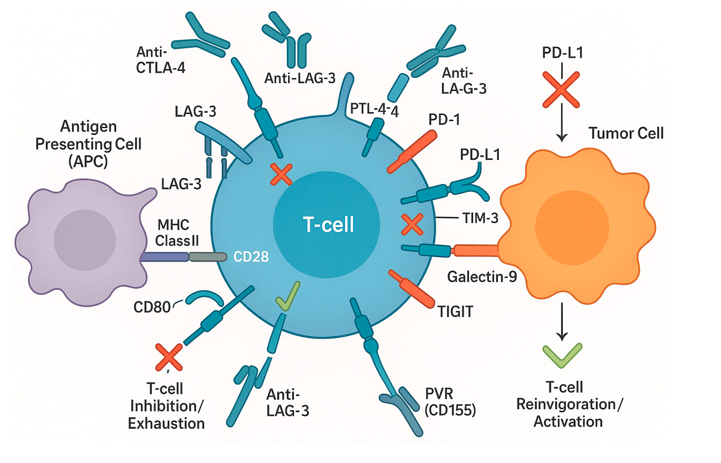
Several key ICRs, including programmed cell death protein 1 (PD-1), cytotoxic T-lymphocyte-associated protein 4 (CTLA-4), lymphocyte activation gene 3 (LAG-3), T-cell immunoglobulin and mucin domain 3 (TIM-3), and T-cell immunoreceptor with Ig and ITIM domains (TIGIT), play pivotal roles in dampening antitumor immune responses (Figure 1).
PD-1, prominently expressed on activated T cells, interacts with its ligands PD-L1 and PD-L2. This engagement recruits the phosphatase SHP-2 to cytoplasmic immunoreceptor tyrosine-based switch motifs (ITSMs) within PD-1, leading to the dephosphorylation of crucial signaling molecules like ZAP-70 and the attenuation of PI3K/AKT pathways, thereby suppressing T-cell activation [2,3,4,5]. Notably, PD-L1 can also directly promote tumor invasiveness by activating signaling pathways such as β-catenin and WIP [5,6].
CTLA-4 primarily functions during the initial T-cell activation phase by competing with the co-stimulatory receptor CD28 for binding to CD80/CD86 on antigen-presenting cells. This interaction diminishes T-cell survival and proliferation [1,7]. Inhibition of CTLA-4 can enhance T-cell activity, evidenced by increased secretion of cytokines like IL-2 and IFN-γ [1].
LAG-3 negatively regulates T-cell proliferation and function by binding to MHC class II molecules, inhibiting T-cell receptor (TCR) signaling and downregulating CD40L expression [8]. LAG-3 often acts synergistically with PD-1, amplifying immunosuppression within the tumor microenvironment (TME), suggesting that co-blockade strategies may be particularly effective [1,9].
TIM-3 contributes to T-cell exhaustion through interaction with ligands such as galectin-9, which can induce T-cell apoptosis or skew T-cells towards a regulatory phenotype [10]. TIM-3 expression is frequently upregulated in chronic infections and tumors, indicative of its role in maintaining immune tolerance and T-cell anergy [8,11].
TIGIT suppresses the activation of both CD8+ T cells and Natural Killer (NK) cells. Its ligand, PVR (Poliovirus receptor), is frequently expressed on tumor cells, providing a direct mechanism for immune inhibition [1,12]. Blockade of TIGIT has shown promise in rejuvenating exhausted T cells [13,14].
The concerted action of these ICRs, often involving overlapping downstream pathways like PI3K/AKT [15], and the chronic upregulation of immunosuppressive cytokines such as IL-10 and TGF-β, solidify T-cell exhaustion within the TME [16,17].

1.1.2. Signaling in NK, B, and Myeloid Cells

While T-cells are primary targets, ICR signaling also profoundly impacts other immune cell populations, contributing to tumor immune evasion. NK cells, crucial for innate antitumor defense, can be inhibited by receptors like NKG2A, which binds to HLA-E on tumor cells [18,19]. PD-1 is also expressed on NK cells, and its engagement with PD-L1 diminishes NK cell cytotoxicity and cytokine secretion [20,21]. Similarly, CTLA-4 blockade can augment NK cell effector functions [22].
B-cells, increasingly recognized for their role in tumor immunity, express ICRs like PD-1 and CTLA-4. PD-1 expression on activated B-cells limits their proliferation and antibody production in the TME [23,24]. Soluble forms of ICRs, such as soluble PD-1, can also inhibit B-cell activities [25], highlighting the complexity of checkpoint-mediated B-cell regulation [26,27]. Myeloid-Derived Suppressor Cells (MDSCs) promote tumor growth by suppressing antitumor immunity, and their expansion and function are enhanced by ICR expression, including PD-1 and CTLA-4 [28,29]. PD-1 blockade can decrease MDSC accumulation, thereby improving T-cell responses [28]. Dendritic Cells (DCs), essential for antigen presentation, can also be functionally impaired by ICR signaling. PD-1 and CTLA-4 engagement can hinder DC maturation and their T-cell activating capacity [21,24,29]. Modulating ICR signals in DCs, potentially in combination with DC vaccines, is an emerging therapeutic strategy [30,31].
The collective signaling of these diverse immune populations creates a synergistic suppressive network. While PD-1 and CTLA-4 primarily drive T-cell dysfunction, their expression on NK and B cells further dampens innate and humoral surveillance. Simultaneously, ICR signaling in MDSCs and DCs reinforces an environment where antigen presentation is impaired and metabolic competition is skewed, effectively orchestrating a multi-layered defense for the tumor against the host’s immune system.

1.2. Structural Insights into Immune Checkpoint Receptor–Ligand Interactions

Advanced structural biology techniques, particularly X-ray crystallography and cryoelectronic microscopy (cryo-EM), have provided high-resolution insights into ICR-ligand interactions, informing the development of novel therapeutics. Crystallographic studies of the PD-1/PD-L1 complex have detailed the binding interface, revealing key residues and conformational changes crucial for inhibitory signaling [32,33]. This structural knowledge underpins the design of small molecule inhibitors targeting this interaction [33].
Cryo-EM offers complementary insights, particularly regarding the multimeric states and dynamic interactions of ICRs like PD-1/PD-L1 within their physiological membrane environment [34,35]. Similar structural characterizations of CTLA-4, LAG-3, and TIM-3 have elucidated their binding mechanisms with respective ligands (e.g., CD80/CD86 for CTLA-4, MHC class II for LAG-3, galectin-9 for TIM-3), thereby guiding the development of specific blocking antibodies and small molecule inhibitors [32,36].
The integration of these structural data with computational methods, such as molecular docking and time-resolved crystallography, accelerates the discovery and optimization of therapeutic compounds [37,38,39]. Future advancements, including serial femtosecond crystallography (SFX) and high-throughput crystallography, promise to further refine our understanding of these dynamic molecular interactions and uncover new therapeutic targets [40,41,42].

1.3. Impact of Antigen Exposure Chronicity on Immune Checkpoint Receptor Function

The expression and function of ICRs are dynamically regulated by the nature of antigen exposure. During acute antigen exposure, T-cells mount robust responses with typically low ICR expression. However, chronic antigen exposure, characteristic of the TME or chronic infections, leads to sustained ICR upregulation (e.g., PD-1, CTLA-4, TIM-3) and progressive T-cell exhaustion, characterized by diminished effector functions and proliferative capacity [43,44,45,46]. This physiological response, aimed at preventing immunopathology, is co-opted by tumors for immune evasion [47,48,49].
The state of T-cell exhaustion driven by chronic antigen stimulation, often involving underlying epigenetic modifications, poses a significant challenge for immunotherapy [50,51]. While checkpoint blockade can be effective, particularly in inflamed TMEs, deeply exhausted T-cells may require combination therapies that address multiple inhibitory pathways or T-cell metabolic reprogramming to restore function [52,53,54,55].

1.4. Epigenetic and Transcriptional Regulation of Immune Checkpoint Receptor Expression

The expression of ICRs on both tumor and immune cells is tightly controlled by a complex network of epigenetic and transcriptional mechanisms. DNA methylation plays a crucial role; for instance, hypermethylation of the PDCD1 (PD-1) gene promoter can correlate with reduced PD-1 expression and potentially improved antitumor immunity, while hypomethylation of CD274 (PD-L1) may predict better responses to PD-1/PD-L1 blockade [56,57,58].
Transcriptional regulation is heavily influenced by the TME. Hypoxia, a common feature, can drive PD-L1 expression via hypoxia-inducible factors (HIFs) like HIF1α [59]. Pro-inflammatory cytokines, such as IL-6, can also enhance PD-1 and PD-L1 expression through pathways like JAK/STAT, contributing to a chronic inflammatory state conducive to T-cell exhaustion [60].
Non-coding RNAs, including long noncoding RNAs (lncRNAs) and microRNAs (miRNAs), are emerging as significant regulators. Specific lncRNAs can modulate ICR gene expression, while certain miRNAs can target ICR mRNAs (e.g., PD-L1 mRNA) for degradation, thereby influencing protein expression and immune cell activity [61]. These regulatory layers offer potential therapeutic targets, with epigenetic modifying drugs being explored in combination with immunotherapy to resensitize tumors [62].

2. Expression and Pathophysiological Roles

The expression patterns and pathophysiological significance of ICRs exhibit considerable heterogeneity across different genitourinary (GU) malignancies, including kidney, bladder, prostate, testicular, and penile cancers. These variations, influenced by histological subtype, tumor stage, and the unique TME, have profound implications for disease progression, prognosis, and the efficacy of immunotherapeutic interventions.

2.1. Comparative Expression Patterns of Immune Checkpoint Receptors Across Genitourinary Cancers

Recent analyses reveal distinct ICR expression profiles in GU cancers. In renal cell carcinoma (RCC), particularly clear cell RCC (ccRCC), PD-L1 expression is often elevated and correlates with higher tumor burden and advanced stage [63,64]. Conversely, CTLA-4 expression in papillary RCC (pRCC) does not appear to vary significantly with tumor subtype or stage [65]. Non-clear cell RCC types may show common elevation of PD-1 and PD-L1, while other checkpoints like LAG-3 are variably expressed [63].
In bladder cancer, specifically urothelial carcinoma, PD-L1 expression is significantly higher in muscle-invasive bladder cancer (MIBC) compared to non-muscle invasive bladder cancer (NMIBC), especially in basal subtypes [66]. Higher PD-L1 expression generally correlates with advanced stages and an immune-infiltrated TME [66,67].
Prostate cancer typically exhibits lower PD-L1 expression compared to other GU malignancies. However, metastatic castration-resistant prostate cancer (mCRPC) with high microsatellite instability or mismatch repair deficiencies can respond to immunotherapy [68]. While CTLA-4 and PD-1 expression may vary with tumor aggressiveness, their overall levels remain relatively low [69].
Testicular cancer typically shows lower ICR expression, though some non-seminomatous germ cell tumors may exhibit increased levels [63]. Penile cancer research is less extensive, but initial studies suggest high PD-L1 expression may correlate with advanced disease [69].
These expression patterns are further nuanced by histological subtypes, such as the basal versus luminal subtypes in bladder cancer showing varied PD-L1 expression and distinct TMEs [64,66]. Tumor stage also plays a critical role, with progressive ICR expression often observed with advancing disease across various GU cancers [63,67].

2.2. Immune Checkpoint Receptor Expression, Localization, and Clinical Outcomes

The expression levels and localization of ICRs within the TME of GU cancers are increasingly linked to patient prognosis and immunotherapy response. In MIBC, high PD-L1 expression is associated with better outcomes following treatment with PD-1/PD-L1 inhibitors like pembrolizumab and atezolizumab [70]. Higher densities of PD-L1-expressing cells often correlate with increased CD8+ T-cell infiltration, a hallmark of an active antitumor immune response [70].
In RCC, high PD-L1 expression can be a negative prognostic marker, correlating with advanced tumor stage [71]. However, it can also predict response to combination immunotherapies [72]. For prostate cancer, CTLA-4 expression on infiltrating T-cells is associated with T-cell dysfunction and poor prognosis, suggesting CTLA-4 as a therapeutic target in mCRPC [72,73]. In urothelial carcinoma, elevated LAG-3 expression, frequently co-expressed with PD-L1, indicates an additional layer of immune suppression and may predict benefit from dual checkpoint blockade [74]. Similarly, high T-cell immunoglobulin and mucin domain 3 (TIM-3) expression in bladder cancer correlates with a suppressive TME and poor prognosis, marking TIM-3 as a prognostic factor and therapeutic target [75]. These findings underscore the potential of ICR expression and localization as biomarkers to guide personalized therapy, potentially complemented by radiographic assessments of the TME [76].

2.3. Interplay of the Tumor Microenvironment with Immune Checkpoint Receptor Function

The unique cellular and cytokine milieu of the TME in GU cancers critically influences ICR engagement and the functional consequences of their blockade. In MIBC, a high density of tumor-infiltrating lymphocytes (TILs) expressing PD-1 and T-cell immunoreceptor with Ig and ITIM domains (TIGIT) on CD8+ T-cells correlates with immune exhaustion and poor prognosis [77]. Inflammatory cytokines like IL-6 and TGF-β within the bladder cancer TME further promote immune suppression by enhancing checkpoint expression [77].
The TME is no longer viewed as an isolated entity but as a dynamic system that reflects broader systemic changes. Recent conceptual frameworks suggest a “breaking of boundaries” in immune microenvironments, where chronic inflammatory states and metabolic dysregulation in non-malignant tissues prime the systemic landscape for tumor-supportive niches [78]. In GU cancers, this systemic-to-local crosstalk is crucial for understanding why certain patients exhibit primary resistance to ICIs.
In RCC, factors like vascular endothelial growth factor (VEGF) within the TME can increase PD-L1 expression on tumor cells, impairing T-cell function [79]. This provides a rationale for combining immunotherapy with anti-angiogenic agents [80]. The prostate cancer TME is often characterized by elevated IL-10 and TGF-β, which upregulate PD-1 and CTLA-4 on T-cells, fostering a suppressive environment [81,82]. Furthermore, chronic exposure to pro-inflammatory cytokines such as TNF-α plays a dual role; while initially anti-tumoral, its persistence within the TME can induce the upregulation of PD-L1 and other inhibitory ligands on both tumor and stromal cells, thereby driving immune evasion and apoptosis resistance [78,83].
Blockade of PD-1 combined with therapies targeting these cytokines shows promise in treatment-resistant models [84]. Even in testicular cancer, which generally has a favorable immune landscape, alterations in local cytokines can lead to adaptive resistance by modulating PD-L1 and TIM-3 expression [79,85]. Engagement of ICRs like PD-1 and LAG-3 on TILs leads to an exhausted phenotype with diminished effector functions [86,87]. Conversely, checkpoint blockade, particularly dual blockade (e.g., PD-1/CTLA-4), can revert these impairments and enhance antitumor immunity [88,89].

2.4. Novel and Less-Studied Immune Checkpoint Receptors in Genitourinary Malignancies

Beyond the well-established ICRs, research is uncovering the roles of novel or less-studied checkpoints in GU cancer pathogenesis. Molecules like CD24 and CD200 are overexpressed in various GU tumors, contributing to immune evasion by inhibiting T-cell activation and promoting a suppressive TME [26]. Telomerase, specifically human telomerase reverse transcriptase (hTERT), has been identified as a potential checkpoint, as its activity can modulate immune responses by inhibiting cytotoxic T-lymphocyte activation in RCC and bladder cancer [90].
Checkpoints on NK cells, such as KIR, NKG2A, and TIM-3, also regulate antitumor immunity [22]. For instance, elevated NKG2A expression in prostate cancer is linked to reduced NK cell activity. Glyco-immune checkpoints, involving cell-surface glycan interactions with lectin-like receptors, represent another emerging evasion mechanism, with antibody-lectin chimeras (AbLecs) being explored as therapeutics [91]. Phagocytosis checkpoints, notably CD47 (the “don’t eat me” signal), are expressed in GU cancers and inhibit macrophage-mediated tumor cell clearance; targeting CD47 is a promising strategy [92]. These novel targets offer potential for combinatorial approaches, possibly with epigenetic modulators, to overcome complex immune evasion mechanisms [91,92].

2.5. Differential Immune Checkpoint Receptor Expression in Primary Versus Metastatic Genitourinary Cancers

Significant differences in ICR expression exist between primary GU tumors and their metastatic sites, impacting treatment strategies. In RCC, the checkpoint CD73, which suppresses T-cell responses, shows notably higher expression in metastatic lesions compared to primary tumors, suggesting a mechanism for enhanced immune evasion at distant sites [93]. This highlights CD73 as a potential target in metastatic RCC [94].
In bladder cancer, primary tumors often exhibit higher TIL infiltration and ICR expression (e.g., PD-L1) than their metastases. Metastatic lesions may feature altered cytokine profiles, such as increased IL-8, associated with immunosuppression and reduced response to PD-1/PD-L1 blockade [95]. Similarly, metastatic castration-resistant prostate cancer frequently displays elevated PD-L1 levels in metastases compared to primary tumors, contributing to a more immunosuppressive microenvironment [68,96].
These disparities necessitate tailored therapeutic approaches. For instance, the upregulation of specific checkpoints like CD73 in metastases could guide the use of targeted therapies with conventional ICIs. The heterogeneous immune infiltration at different metastatic sites, such as brain metastases regularly having fewer TILs, also influences ICI efficacy [97]. Furthermore, immune evasion mechanisms involving factors like DUX4, an embryonic transcription factor suppressing IFN-γ signaling, are prevalent in metastatic GU cancers and correlate with poor immunotherapy responses, marking DUX4 as another potential therapeutic target [98].

2.6. Tumor-Type-Specific Expression Patterns

The expression and functional relevance of immune checkpoint receptors varies substantially across genitourinary malignancies, reflecting distinct tumor microenvironmental compositions and immunological contexts. In renal cell carcinoma (RCC), PD-L1 expression on tumor cells and immune infiltrates is widely documented and serves as a prognostic marker, while CTLA-4 expression on tumor-infiltrating T cells correlates with response durability to anti-PD-1 therapy.
Urothelial carcinoma demonstrates characteristic high PD-L1 expression and robust CD8+ T cell infiltration, particularly in high-grade tumors, making it exceptionally responsive to PD-1/PD-L1 blockade. In contrast, prostate cancer frequently exhibits modest PD-L1 expression and variable T cell infiltration, with emerging evidence suggesting that PD-1+ T cells in this context may have undergone terminal differentiation, limiting their functional recovery with checkpoint blockade alone.
Testicular cancer, despite being an immunogenic malignancy, shows heterogeneous PD-L1 expression patterns that may explain variable responses to ICI in treatment-resistant settings. LAG-3 expression on tumor-infiltrating lymphocytes shows differential prevalence across these tumor types, with higher frequency observed in urothelial and renal cancers compared to prostate cancer. These tumor-specific immunological landscapes underscore the critical importance of tailoring therapeutic strategies and biomarker-driven selection approaches to the distinct biological context of each genitourinary malignancy.

3. Mechanistic Insights into Immune Checkpoint Blockade in Genitourinary Tumor Immunotherapy

ICIs have revolutionized the treatment landscape for various malignancies, including GU cancers. Their efficacy hinges on reinvigorating the host’s antitumor immune responses by disrupting inhibitory signaling pathways. However, the mechanisms underlying their action, the dynamic remodeling of the TME, the emergence of resistance, the influence of systemic factors like the gut microbiome, and the occurrence of immune-related adverse events (irAEs) are complex and multifaceted. Understanding these mechanistic insights is crucial for optimizing ICI therapy in GU cancers.

3.1. Molecular and Cellular Mechanisms of Immune Reinvigoration by ICIs

Recent research has identified several key mechanisms by which ICIs specifically reinvigorate antitumor immunity in GU cancers, considering their distinct immune landscapes. Beyond direct T-cell derepression, ICIs induce broader changes within the TME. Reprogramming of myeloid cells is one such mechanism; for instance, JAK inhibition with ruxolitinib can modify the myeloid cell compartment, enhancing T-cell responses and improving therapeutic outcomes when combined with PD-1/CTLA-4 blockade [99]. This suggests that targeting myeloid subsets can create a more permissive environment for T-cell engagement.
The regulation of immune checkpoint gene expression itself is also critical. The innate immune-modulating gene OAS1 has been shown to influence the expression of various checkpoint genes, thereby directly affecting antitumor immune responses and potentially serving as a prognostic biomarker [100]. Furthermore, emerging checkpoint molecules like CD24 and CD200, which can regulate immune cell interactions and promote immune evasion, represent novel targets whose inhibition may restore T-cell functionality and augment the efficacy of existing ICIs [26].
Activation of innate immune pathways can synergize with ICIs. Toll-like receptor (TLR) agonists, such as TLR5 agonists, can shift macrophage polarization from a pro-tumor (M2) to an antitumor (M1) state and promote CD8+ T-cell priming, thereby enhancing the efficacy of anti-PD-1 therapy [101]. Moreover, inducing specific forms of immunogenic cell death, like pyroptosis with agents such as nigericin, can bolster antitumor immunity and increase the effectiveness of PD-1 inhibitors [102]. These findings highlight the importance of modulating both innate and adaptive immune responses, as well as the TME itself (e.g., counteracting immunosuppressive cytokines like TGF-β and IL-10), to maximize ICI efficacy [101,103].

3.2. Dynamic Remodeling of the Tumor Microenvironment by ICI Therapy

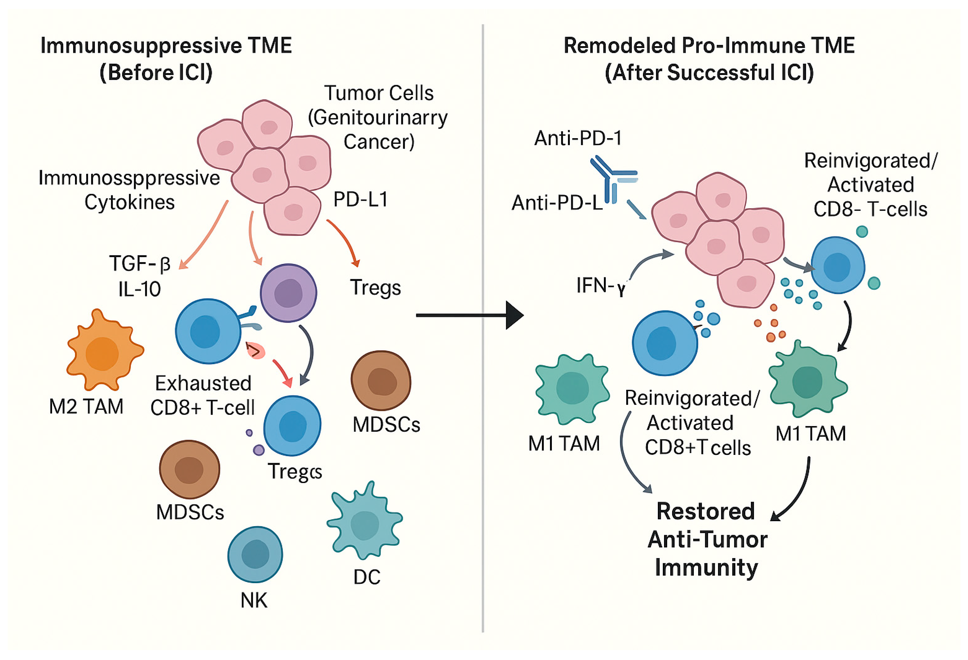
ICI therapy induces significant dynamic remodeling of the TME, which differs between responsive and non-responsive GU cancer patients. These changes involve alterations in immune cell infiltrates, cytokine profiles, and tumor cell characteristics (Figure 2).
In responsive bladder cancer patients, ICIs enhance the infiltration of TILs, particularly CD8+ cytotoxic T-cells, and increase pro-inflammatory cytokines like IFN-γ [104]. Conversely, non-responders often show reduced TIL functionality and elevated levels of immunosuppressive cells like Tregs [105]. Similar patterns are seen in renal cell carcinoma (RCC), where increased densities of effector T-cells and NK cells in the TME correlate with improved progression-free survival following PD-1 blockade [106,107].
Cytokine networks within the TME are also profoundly affected. In prostate cancer, elevated IL-6 and IL-2 levels are linked to T-cell activation post-PD-1/PD-L1 blockade [108]. Responders generally exhibit increased anti-inflammatory cytokines, promoting T-cell effector functions [109], while non-responders typically maintain high levels of immunosuppressive cytokines like TGF-β and IL-10 [110].
Tumor cells themselves adapt to ICI therapy. In bladder cancer, resistant tumor cells frequently upregulate PD-L1 expression, an adaptive resistance mechanism [104]. ICIs can also modify the expression of chemokines and their receptors; for example, upregulation of CXCL8 post-ICI treatment can recruit additional immune cells to the TME, potentially enhancing therapeutic responses [111]. These dynamic changes underscore the need for personalized approaches, potentially combining ICIs with agents targeting immunosuppressive cytokines or informed by tumor mutational burden (TMB) and cytokine profiles [112,113].

3.3. Mechanisms of Resistance to Immune Checkpoint Blockade

Resistance to ICIs, whether primary, adaptive, or acquired, remains a significant clinical challenge in GU cancers. Primary resistance can be driven by intrinsic tumor characteristics. Genetic alterations, such as mutations in the B2M gene (essential for MHC class I antigen presentation), impair tumor antigen display and T-cell recognition in bladder and renal cancer [114]. Downregulation of MHC molecules further contributes to this antigen presentation deficiency [115].
Adaptive resistance involves changes within the TME that allow tumors to evade ICI effects. The TIGIT/CD155 axis, for example, mediates immune suppression; elevated CD155 levels can inhibit T-cell activity via TIGIT engagement [116]. Immunosuppressive cytokines like IL-10 and TGF-β also promote Treg activity and inhibit effector T-cells, facilitating escape from ICI efficacy [115].
Acquired resistance develops as tumors evolve under therapeutic pressure. ICIs can lead to the upregulation of alternative immune checkpoints (e.g., CTLA-4, LAG-3) on TILs, diminishing therapeutic effects over time [117]. Activation of oncogenic signaling pathways, such as Wnt/β-catenin, can decrease T-cell infiltration [118]. Moreover, the mammalian target of rapamycin (mTOR) signaling pathway serves as a central nutrient-sensitive regulator of cellular metabolism and proliferation. In the TME of genitourinary cancers, aberrant mTOR activation facilitates immune escape by promoting tumor cell growth and directly impairing T-cell effector functions [119,120]. Given its role in orchestrating these resistance nodes, pharmacological inhibition of mTOR is emerging as a strategic approach to reverse ICI resistance and resensitize the immune landscape. Additionally, increased recruitment of immunosuppressive myeloid cells and enhanced PD-L1 expression on resistant tumor cells contribute to acquired resistance [117,121]. Addressing these multifaceted resistance mechanisms necessitates combination therapies targeting multiple pathways, personalized approaches based on tumor and TME profiling, and strategies to remodel the TME, such as using nanoparticles or oncolytic viruses with ICIs [122,123,124,125].

3.4. Influence of the Gut Microbiome on ICI Efficacy

The gut microbiome has emerged as a critical modulator of ICI efficacy in GU cancers. Microbial diversity is positively associated with favorable responses to PD-1 blockade, potentially by enhancing T-cell priming [126]. Specific bacterial species, such as certain strains of Bifidobacterium and Lactobacillus, correlate with increased CD8+ T-cell infiltration and activation, leading to better treatment outcomes [127].
Mechanistically, the gut microbiome influences systemic immunity by modulating cytokine production. Responders to ICIs often exhibit upregulated pro-inflammatory cytokines like IL-12 and TNF-α, while dysbiosis can promote immunosuppressive cytokines like IL-10 [127]. Microbial metabolites, such as short-chain fatty acids (SCFAs), can enhance T-cell activation and cytokine production [128]. The microbiome can also shape antigen presentation machinery and even upregulate PD-L1 expression on tumor cells [129,130].
Conversely, certain microbial species can contribute to ICI resistance. Bacteroides fragilis has been associated with increased Treg levels in the TME [131]. Imbalanced microbiomes may also lead to nutrient competition, impairing T-cell function [132]. These insights are paving the way for personalized microbiome-based therapies, such as fecal microbiota transplantation (FMT) or specific pre/probiotics, frequently in combination with ICIs, to improve clinical outcomes [127,133].

3.5. Off-Target Effects and Mechanisms of Immune-Related Adverse Events

While ICIs unleash antitumor immunity, they can also trigger irAEs by disrupting self-tolerance. Blockade of PD-1, PD-L1, and CTLA-4 can lead to immune activation against normal tissues, causing conditions like dermatitis, colitis, hepatotoxicity, and endocrinopathies [134,135,136]. For example, renal tubular cells in RCC patients can express PD-L1, potentially leading to immune-mediated nephritis upon PD-1 blockade [134].
The concept of shared antigens between tumors and normal tissues is a leading hypothesis for irAEs; T-cells activated against tumor antigens may cross-react with similar antigens on healthy tissues [137,138]. Pre-existing autoantibodies and specific immune profiles (e.g., Treg frequency, cytokine ratios) may predict irAE risk [139,140].
The TME’s cytokine milieu also influences irAE severity. Elevated pro-inflammatory cytokines like IL-6 and TNF-α are associated with increased irAE incidence [141]. Furthermore, the phenotype of TILs post-ICI therapy can impact irAE risk; dysfunctional T-cell phenotypes in RCC have been linked to a higher likelihood of irAEs [142]. Managing irAEs involves identifying predictive biomarkers, developing preventive strategies, and exploring combination therapies that mitigate toxicity while preserving antitumor efficacy [139,143,144].

4. Biomarkers to Guide Immune Checkpoint Inhibitor Therapy

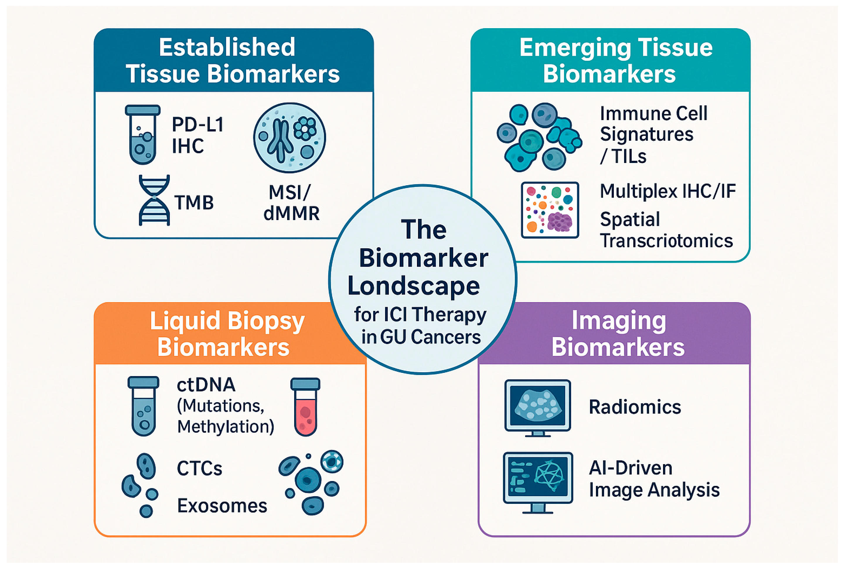
The advent of ICIs has significantly advanced the treatment of GU malignancies. However, patient responses to ICIs are variable, underscoring the critical need for robust biomarkers to predict efficacy, monitor response, and guide personalized therapeutic strategies. This section reviews the current clinical utility and limitations of established biomarkers, as well as promising emerging tissue-based, liquid biopsy-based, and imaging-based biomarkers in GU oncology (Figure 3).
The future of personalized immunotherapy in GU cancers lies in the transition from single-analyte biomarkers to integrative multiomic signatures. Recent bibliometric and systematic analyses highlight that the integration of genomics, transcriptomics, and proteomics through machine learning is now the primary driver for discovering novel diagnostic and prognostic targets [145]. For instance, comprehensive multiomics integration has recently identified cell-specific factors, such as B-cell-derived regulators, that could serve as high-precision biomarkers in complex inflammatory and malignant states [146].
In parallel, network pharmacology is revolutionizing drug discovery by shifting the “one drug, one target” paradigm toward a more systemic “multi-target, multi-pathway” approach. By mapping the complex interactions between ICIs and the host’s immune network, these computational models can predict synergistic drug combinations and identify potential resistance nodes before clinical implementation [146,147]. This approach is particularly relevant for GU malignancies, where the interplay between the TME and systemic immunity requires a holistic therapeutic perspective.

4.1. Established Biomarkers: Clinical Utility and Limitations

Several biomarkers are currently utilized or investigated for their predictive value in ICI therapy for GU cancers, including PD-L1 expression by immunohistochemistry (IHC), TMB, and MSI or deficient mismatch repair (dMMR) status.
PD-L1 expression by IHC is a widely adopted biomarker, particularly in bladder cancer and RCC. Higher PD-L1 expression levels generally correlate with improved responses and survival outcomes in patients treated with PD-1/PD-L1 inhibitors [145]. However, its utility is hampered by variability in assay platforms (e.g., 22C3 vs. SP142), scoring algorithms, and cutoff values, necessitating standardization [146]. Furthermore, the spatiotemporal heterogeneity of PD-L1 expression within tumors and between primary and metastatic sites can limit the predictive accuracy of a single biopsy [147,148].
TMB, reflecting the number of somatic mutations per megabase of DNA, has emerged as a predictor of ICI response. A higher TMB is hypothesized to generate more neoantigens, thereby enhancing immunogenicity. In RCC, high TMB has been associated with greater benefit from ICI therapy [145]. Nevertheless, the lack of a standardized definition for “high TMB” across studies poses a challenge [149]. Moreover, TMB does not always correlate with immune infiltration or response, as other factors within the TME or defects in antigen presentation machinery can override the potential benefit of a high neoantigen load [148].
MSI and dMMR status are established pan-cancer biomarkers for ICI response. Tumors with MSI-high (MSI-H) or dMMR status often exhibit a hypermutated phenotype and respond favorably to PD-1 blockade [150]. However, the prevalence of MSI-H/dMMR is relatively low in most GU cancers compared to malignancies like colorectal cancer, limiting its broad applicability [145]. The limitations of these individual biomarkers highlight the need for integrated, multi-biomarker approaches to improve predictive accuracy [148,150].

4.2. Emerging Tissue-Based Biomarkers

Beyond established markers, several promising tissue-based biomarkers are under investigation. Specific immune cell signatures, derived from detailed immunoprofiling of TILs, are gaining traction. Higher densities of CD8+ cytotoxic T-lymphocytes (CTLs) within the TME, particularly those expressing PD-1, correlate with better ICI responsiveness in bladder cancer and RCC [151,152]. Multiomics approaches integrating genomic, transcriptomic, and immunological data are being used to define comprehensive immune signatures predictive of ICI benefit [153].
Multiplex IHC and immunofluorescence (IF) techniques allow for the simultaneous visualization and spatial characterization of multiple immune checkpoint proteins and immune cell types within the tumor tissue. This provides crucial insights into the immune contexture and cellular interactions within the TME [154]. In bladder cancer, for instance, specific immune cell compositions (e.g., high CD8, CD4, and PD-L1-expressing cells) identified by multiplex IHC have shown a significant correlation with improved responses to PD-1 inhibitors [155].
Spatial transcriptomics represents a cutting-edge approach that maps gene expression patterns within the spatial context of the tissue. This technology can reveal distinct immune microenvironments and cellular interactions associated with treatment response [156]. In bladder tumors, spatial transcriptomics has identified regions with active T-cell responses that correlate with higher ICR expression and improved ICI efficacy [157].
The validation of these emerging tissue-based biomarkers is ongoing through clinical trials and the development of multi-biomarker platforms, often integrated with genomic profiling to enhance predictive power [158,159,160].

4.3. Liquid Biopsy-Based Biomarkers

Liquid biopsies, analyzing analytes such as circulating tumor DNA (ctDNA), circulating tumor cells (CTCs), and exosomes from bodily fluids, offer a non-invasive means to predict and monitor ICI efficacy.
ctDNA analysis can reveal tumor-specific genetic alterations and TMB. High TMB detected in ctDNA has been validated as a favorable biomarker for ICI response in RCC and bladder cancer [161,162]. Dynamic changes in ctDNA levels during therapy can also serve as a real-time indicator of treatment response, with decreasing levels associated with better clinical outcomes [163,164]. Furthermore, ctDNA methylation patterns are emerging as epigenetic biomarkers, with specific methylation signatures correlating with ICI response [163,164].
CTCs, though less established than ctDNA, hold potential for monitoring disease progression and treatment response. Changes in CTC counts and characteristics during ICI therapy may provide prognostic insights [165,166].
Exosomes, nano-sized vesicles released by cells, carry molecular cargo (proteins, RNAs, metabolites) that can reflect tumor biology and immune status. Profiling exosomal contents, such as immune-related microRNAs or proteins like TGF-β, may predict ICI efficacy and reveal resistance mechanisms [167,168,169].
Analysis of circulating immune cells using advanced techniques like flow cytometry and single-cell sequencing can reveal dynamic changes in immune cell populations (e.g., activated T-cells, Tregs) that correlate with ICI response [170,171]. While promising, the clinical implementation of liquid biopsy biomarkers faces challenges in standardization, reproducibility, and the integration of multiomic data [169,171,172,173,174,175].

4.4. Radiomics and AI-Driven Analysis of Medical Imaging

Radiomics, the high-throughput extraction of quantitative features from medical images (CT, MRI, PET), coupled with artificial intelligence (AI)-driven analysis, is emerging as a powerful non-invasive tool for predicting ICI response. Machine learning models can correlate radiomic features reflecting tumor phenotype, burden, and heterogeneity with treatment outcomes [176,177,178].
Deep learning algorithms enhance the ability to identify unique imaging biomarkers and monitor temporal changes in tumor characteristics, facilitating early assessment of ICI efficacy [179,180,181,182]. The integration of radiomics with liquid biopsy data, such as ctDNA profiles, offers a synergistic approach to comprehensively assess tumor burden and response [183].
Challenges in radiomics include the need for standardization of imaging protocols and feature extraction methods, as well as validation in large, multi-institutional cohorts to ensure generalizability [184,185,186]. Future directions involve advanced computational techniques, multi-modal data integration (e.g., imaging with spatial transcriptomics), and the application of these tools in personalized medicine [187,188,189].

5. Therapeutic Landscape and Evolving Strategies

The therapeutic landscape for GU cancers has been substantially reshaped by the advent of ICIs targeting receptors such as PD-1, PD-L1, and CTLA-4. More recently, novel ICIs targeting emerging receptors like LAG-3, TIM-3, and TIGIT, as well as innovative bispecific antibodies and combination strategies, are further expanding the therapeutic armamentarium. This section reviews pivotal clinical trial results, evolving combination strategies, long-term outcomes, and the impact of ICIs in neoadjuvant, adjuvant, and maintenance settings for GU malignancies.

5.1. Novel Immune Checkpoint Inhibitors and Bispecific Antibodies

Recent clinical development has focused on ICIs targeting novel co-inhibitory receptors to overcome resistance to established PD-1/PD-L1 or CTLA-4 blockade and to enhance antitumor immunity.
  • LAG-3 Inhibition: The combination of anti-LAG-3 antibodies, such as relatlimab, with PD-1 inhibitors like nivolumab has shown promising efficacy. A Phase II trial in advanced urothelial carcinoma reported an objective response rate (ORR) of 30% with this combination, notably higher than historical controls for anti-PD-1 monotherapy [190]. Mechanistically, LAG-3 blockade is thought to reinvigorate exhausted T-cells and enhance immune activation, particularly in tumors with high PD-L1 expression and dense TIL infiltration [191].
  • TIM-3 Inhibition: Targeting TIM-3 is another strategy under investigation. Early trial data in metastatic RCC suggest that TIM-3 monoclonal antibodies, especially in combination with PD-1 or CTLA-4 inhibitors, can enhance antitumor activity compared to monotherapies [192]. TIM-3 blockade appears to foster a more inflammatory T-cell environment within tumors, restoring T-cell functionality [193].
  • TIGIT Inhibition: TIGIT has emerged as a significant target, particularly for patients who have developed resistance to prior ICIs. Preliminary Phase II trial results of a TIGIT-targeting agent, alone or with PD-1 blockade, demonstrated an ORR of approximately 40% in previously treated metastatic bladder cancer patients [194]. TIGIT blockade can remodel the TME by increasing CD8+ T-cell infiltration and pro-inflammatory cytokine levels [195].
  • Bispecific Antibodies: Bispecific antibodies, engineered to engage multiple immune targets simultaneously (e.g., PD-L1 and TIGIT), represent an innovative therapeutic class. Preclinical models and early clinical trials in GU malignancies have shown that these agents can improve T-cell activation and reduce tumor growth, offering potential for enhanced efficacy, especially in resistant settings [196,197].

5.2. Combination Strategies Involving ICIs

Combining ICIs with other therapeutic modalities is a key strategy to improve efficacy and overcome resistance in GU cancers.
  • ICIs with Chemotherapy: The combination of ICIs with chemotherapy has demonstrated synergistic effects. For instance, pembrolizumab plus chemotherapy improved overall survival (OS) and progression-free survival (PFS) in metastatic urothelial carcinoma compared to chemotherapy alone [198]. Chemotherapy can increase tumor antigenicity and enhance TIL infiltration, thereby priming the TME for ICI activity [199,200,201].
  • ICIs with Targeted Therapies: Combining ICIs with targeted agents, such as multi-kinase inhibitors, is showing considerable promise. The combination of pembrolizumab with lenvatinib has improved efficacy in advanced RCC and is under evaluation in bladder cancer [202,203]. Similarly, combining ICIs with drugs targeting the VEGF pathway aims to create a less immunosuppressive TME [204].
  • ICIs with Radiotherapy: Radiotherapy can induce immunogenic cell death and release tumor antigens, potentially synergizing with ICIs to elicit systemic antitumor immunity (the abscopal effect). Combining pembrolizumab with localized radiotherapy has shown improved response rates in some GU cancer settings [205,206,207].
  • Novel Immunotherapy Combinations: The integration of ICIs with other immunotherapies, such as Chimeric Antigen Receptor (CAR) T-cell therapy or cancer vaccines, is being explored. CAR-T cells engineered to express PD-1 inhibitors or personalized cancer vaccines combined with PD-1 blockade aim to boost antitumor T-cell responses and overcome resistance [202,208].
  • Microbiome Modulation: Given the influence of the gut microbiome on ICI efficacy, strategies like fecal microbiota transplantation (FMT) from responders to non-responders are being investigated to enhance treatment outcomes [209].

5.3. Biomarker-Guided Patient Selection and Outcome Optimization

Translating ICI efficacy into improved patient outcomes requires rational biomarker-guided selection strategies that identify patients most likely to respond while sparing those at risk for treatment failure and unnecessary toxicity. PD-L1 expression status, assessed by immunohistochemistry on tumor and immune infiltrates, demonstrates prognostic and predictive value in urothelial carcinoma and emerging utility in RCC, with high PD-L1 expression associated with response rates of 40–55% versus 10–20% in PD-L1-negative populations. However, PD-L1 status is imperfect as a predictor, with substantial heterogeneity in responses within both PD-L1-positive and -negative populations.
Tumor mutational burden (TMB), quantified by next-generation sequencing, shows emerging prognostic value and is particularly useful in prostate cancer where mismatch repair deficiency and hypermutated phenotypes enrich for ICI responders (response rates 40–50% versus 10–15% in MMR-proficient disease). Immunological biomarkers—including T cell receptor (TCR) clonality, circulating immune profiling, and immune infiltrate composition—show mechanistic relevance but lack standardized clinical validation and remain primarily research tools.
Radiomics-based signatures derived from baseline imaging show promise in some studies but require prospective validation and standardization before clinical implementation. The field currently lacks a unified, validated biomarker panel capable of accurately predicting ICI response across tumor types, representing a critical limitation that hampers optimization of patient selection and rational therapeutic strategy.

5.4. Durability of Response and Long-Term Survival Outcomes

Long-term follow-up from pivotal ICI trials provides crucial insights into the durability of responses and survival benefits. In advanced RCC, nivolumab demonstrated sustained OS benefit over everolimus in previously treated patients [210]. Pooled analyses of ipilimumab trials in melanoma showed a plateau in OS rates around three years, suggesting long-term survival for a subset of patients [211], a trend also observed in GU cancers treated with ICIs [212]. The KEYNOTE-001 trial of pembrolizumab in advanced melanoma reported durable response rates, with similar trends emerging in GU cancer cohorts [213].
Combination therapies, such as avelumab plus axitinib in treatment-naïve advanced RCC, have also shown significant improvements in long-term PFS and OS compared to sunitinib monotherapy [214]. Durable responses are often associated with robust immune activation, increased TILs, and favorable cytokine profiles (e.g., high IFN-γ, low IL-10/TGF-β) [215].

5.5. ICIs in Neoadjuvant, Adjuvant, and Maintenance Settings

The role of ICIs is expanding beyond metastatic disease into earlier stages of GU cancer treatment.
  • Neoadjuvant ICI Therapy: Trials like the SWOG S1314 (atezolizumab in MIBC) and CheckMate 016 (nivolumab plus ipilimumab in patients with metastatic RCC) are evaluating the potential of neoadjuvant ICIs to improve pathological complete response (pCR) rates and surgical outcomes [216,217].
  • Adjuvant ICI Therapy: In the adjuvant setting, ICIs aim to reduce recurrence rates and improve survival post-definitive local treatment. Trials such as NCT03187305 (pembrolizumab in locally advanced penile cancer) and NCT03249700 (pembrolizumab plus chemotherapy post-cystectomy in MIBC) are exploring this approach [2,4].
  • Maintenance ICI Therapy: In the therapeutic landscape of urothelial carcinoma, the standard of care has been redefined. While the KEYNOTE-045 trial established the role of pembrolizumab in the salvage setting for patients progressing after platinum-based chemotherapy [218], the JAVELIN Bladder 100 trial revolutionized the sequence of care. This study demonstrated that avelumab maintenance therapy significantly extends overall survival in patients with locally advanced or metastatic UC whose disease had not progressed on first-line platinum-based induction chemotherapy [219].

5.6. Tumor-Specific Therapeutic Outcomes and Strategic Considerations

The clinical translation of immune checkpoint blockade has demonstrated markedly heterogeneous outcomes across genitourinary malignancies, reflecting their underlying biological distinctiveness. Renal cell carcinoma has emerged as a primary success story for ICIs, with first-line anti-PD-1-based combinations (nivolumab + ipilimumab) demonstrating superior progression-free and overall survival compared to standard tyrosine kinase inhibitors in advanced disease, establishing ICIs as standard-of-care in this setting.
Urothelial carcinoma similarly shows exceptional responsiveness to PD-1/PD-L1 blockade, particularly in platinum-ineligible patients and those with high PD-L1 expression, though durability of response remains variable and mechanisms of primary and acquired resistance are incompletely understood. Prostate cancer, conversely, has demonstrated more modest benefit from ICI monotherapy. Most clinical benefit had been observed in specific molecular subsets (deficient mismatch repair, high tumor mutational burden) rather than unselected populations. Combination approaches integrating ICIs with androgen deprivation therapy or conventional chemotherapy are under active investigation, but results have been mixed.
Testicular cancer, while historically chemotherapy-responsive, has shown encouraging early results with ICIs in cisplatin-refractory settings, though the optimal combination strategies and patient selection criteria remain to be defined. These differential outcomes underscore the necessity of tumor-specific therapeutic strategies, refined patient selection criteria, and continued investigation into the mechanistic drivers of response and resistance in each genitourinary malignancy.

6. Future Perspectives and Conclusions

The advent of immune checkpoint receptor-targeted immunotherapy has undeniably transformed the therapeutic paradigm for a spectrum of genitourinary malignancies. This review has underscored the central and multifaceted roles of immune checkpoint receptors—including established players such as PD-1, CTLA-4, LAG-3, TIM-3, and TIGIT, as well as emerging novel checkpoints—in dictating the intricate balance between antitumor immunity and immune evasion. A fundamental understanding of their signaling pathways, which extend beyond T cells to modulate the functions of natural killer cells, B cells, myeloid cells, and dendritic cells, is paramount to advancing the field. Structural biology insights continue to refine our comprehension of receptor-ligand interactions, informing the development of next-generation therapeutics. Epigenetic and transcriptional mechanisms reveal complex layers of ICR regulation influenced by factors such as chronic antigen exposure and the evolving tumor microenvironment.
Mechanistically, immune checkpoint inhibitors reinvigorate antitumor immune responses not only through direct T cell derepression but also through broader tumor microenvironmental remodeling, including myeloid cell reprogramming, modulation of cytokine profiles, and even influencing tumor cell characteristics themselves. However, the clinical success of ICIs is often challenged by primary, adaptive, and acquired resistance mechanisms. These involve genetic alterations in tumor cells, defects in antigen presentation machinery, dynamic changes in the tumor microenvironment (such as the upregulation of alternative checkpoints or the influx of immunosuppressive cells), and the influence of systemic factors like the gut microbiome. Furthermore, the very mechanism of immune reactivation can lead to immune-related adverse events (irAEs), driven by off-target effects and shared antigen recognition between tumor and normal tissues, necessitating careful clinical management and the development of predictive strategies.

6.1. Tumor-Specific Perspectives and Current State of the Field

The impact of immune checkpoint receptor-targeted immunotherapy varies considerably across genitourinary malignancies, reflecting distinct biological landscapes and therapeutic responses. Renal cell carcinoma has become the flagship indication for ICIs, with combination anti-PD-1/CTLA-4 therapy now representing the standard-of-care in advanced disease and demonstrating durable responses in a substantial proportion of patients. The mechanistic insights into RCC’s immunogenicity—driven by high mutational burden, characteristic hypoxia-driven microenvironment, and robust T cell infiltration—have directly enabled successful ICI translation. Urothelial carcinoma similarly stands as a major success story, with first-line and second-line PD-1/PD-L1 blockade offering meaningful survival benefits and establishing ICIs as standard options across disease stages. The immunologically “hot” tumor microenvironment of urothelial cancer, characterized by high PD-L1 expression and substantial CD8+ T cell infiltration, has proven conducive to checkpoint blockade efficacy.
In contrast, prostate cancer represents a more cautious landscape for ICIs. While select molecular subsets—particularly those with mismatch repair deficiency or elevated tumor mutational burden—show ICI responsiveness, the majority of unselected prostate cancer populations demonstrate limited benefit from checkpoint blockade monotherapy. The immunologically “cold” phenotype of many prostate cancers, with heterogeneous T cell infiltration and variable checkpoint receptor expression, has necessitated more sophisticated combination approaches. Recent evidence suggests that combining ICIs with androgen deprivation therapy may overcome some of these barriers, though outcomes remain inferior to those observed in RCC or urothelial cancer. Testicular cancer, despite its exceptional chemotherapy responsiveness and inherent immunogenicity, has been understudied in the ICI era; preliminary data suggest potential benefit in cisplatin-refractory disease, but this remains an emerging indication requiring further mechanistic investigation and clinical validation.
This heterogeneity across tumor types underscores a critical principle: the expression and pathophysiological significance of ICRs are markedly heterogeneous across different genitourinary cancers (kidney, bladder, prostate, testicular, and penile), varying significantly with histological subtype, tumor stage, and the specific characteristics of the tumor microenvironment. This heterogeneity, observed even between primary tumors and metastatic sites, directly impacts prognosis and the predictive value of ICR expression for therapeutic response. The unique cellular and cytokine milieu within each GU cancer tumor microenvironment further dictates the functional consequences of ICR engagement and blockade. This highlights the necessity of tailoring both therapeutic strategy and biomarker-directed patient selection to the distinct biological context of each genitourinary malignancy. Future clinical development must embrace this tumor-specific paradigm rather than pursuing monolithic approaches across all genitourinary cancers.

6.2. Biomarkers to Guide Therapy: Current State and Emerging Landscape

The quest for robust biomarkers to guide ICI therapy is critical to optimizing treatment selection and patient outcomes (Table 1). While established biomarkers such as PD-L1 immunohistochemistry, tumor mutational burden, and microsatellite instability/deficient mismatch repair status offer clinical utility, their limitations—including assay variability, tumor heterogeneity, and low prevalence in some genitourinary cancers—are apparent.
The clinical efficacy of anti-PD-1-based combinations in renal cell carcinoma exemplifies the transformative potential of ICR-targeted immunotherapy. Nivolumab plus ipilimumab achieved objective response rates of 41.6% in CheckMate 214, with median progression-free survival of 15.3 months and median overall survival of 75.6 months—a substantial improvement over tyrosine kinase inhibitor monotherapy previously considered standard-of-care. Similarly, pembrolizumab combined with axitinib (KEYNOTE-426) demonstrated an objective response rate of 55.1%, establishing anti-PD-1/VEGF combinations as an alternative standard-of-care approach.
In urothelial carcinoma, pembrolizumab (KEYNOTE-045) achieved objective response rates of 21.1% with median overall survival of 10.3 months in platinum-refractory disease—a population historically managed with best supportive care. Atezolizumab monotherapy (IMvigor211) similarly offered meaningful clinical benefit, and dual checkpoint blockade with durvalumab plus tremelimumab (DANUBE trial) achieved response rates of 27.2%, suggesting additive value of multi-agent approaches in this setting. These clinical outcomes, though variable across tumor types and patient populations, provide concrete evidence of ICI efficacy and establish benchmarks against which emerging therapies and biomarker-guided strategies should be compared.
Consequently, the field is rapidly advancing toward emerging tissue-based biomarkers (including specific immune cell signatures, multiplex immunohistochemistry and immunofluorescence, and spatial transcriptomics), liquid biopsy-based analytes (circulating tumor DNA mutations and methylation patterns, circulating immune cells, exosomes), and non-invasive imaging biomarkers (radiomics and artificial intelligence-driven image analysis).
The integration of these multiomic data streams holds immense promise for developing more accurate predictive and monitoring models. However, it is essential to acknowledge that promising preclinical findings and early-phase biomarker discoveries have frequently failed to demonstrate equivalent clinical utility in prospective, adequately powered patient cohorts. While spatial transcriptomics and radiomics show intriguing mechanistic insights into immune microenvironment composition and tumor heterogeneity, these technologies remain primarily investigational tools without standardized clinical protocols, validated cutoff values, or routine integration into treatment decisions.

6.3. Clinical Translation and Real-World Outcomes

The clinical translation of mechanistic insights into immune checkpoint receptor biology has achieved remarkable success in select patient populations yet remains incomplete across the broader spectrum of genitourinary malignancies. Renal cell carcinoma has achieved the most comprehensive clinical benefit, with anti-PD-1/CTLA-4 combinations now demonstrating durable response rates exceeding 40% and median overall survival extending beyond 75 months in first-line treatment. Urothelial carcinoma similarly shows substantial clinical benefit from PD-1/PD-L1 blockade, with response rates of 20–55% depending on PD-L1 expression status and treatment line, and median overall survival gains of 10–15 months observed in the metastatic setting. However, this success in renal and bladder cancers contrasts sharply with more modest clinical outcomes in other settings.
Prostate cancer demonstrates limited response to ICI monotherapy in most unselected populations, with meaningful benefit confined to molecularly selected subsets (mismatch repair deficiency, high tumor mutational burden) representing <10% of the patient population; even combination approaches with androgen deprivation therapy have shown modest benefit compared to RCC or urothelial cancer outcomes. Testicular cancer remains understudied, but preliminary data suggest potential for cisplatin-refractory disease, though response rates (60–70%) and durability remain to be established in larger prospective trials. These disparities highlight that while the mechanistic framework supporting ICR-targeted immunotherapy is robust, clinical translation has been differential and incomplete, with several tumors requiring substantially refined therapeutic approaches and enhanced patient selection strategies.

6.4. Clinical Implementation Barriers and Practical Considerations

While immune checkpoint receptor-targeted immunotherapy represents a genuine breakthrough in genitourinary oncology, its broad clinical implementation faces substantial real-world barriers that warrant explicit acknowledgment. First, the translational gap between mechanistic insights and clinical efficacy remains significant. Many promising preclinical findings and early-phase biomarker discoveries have failed to demonstrate equivalent clinical utility in prospective, adequately powered patient cohorts. For example, while spatial transcriptomics and radiomics show intriguing mechanistic insights into immune microenvironment composition and tumor heterogeneity, these technologies remain primarily investigational tools without standardized clinical protocols, validated cutoff values, or routine integration into treatment decisions.
Second, cost and accessibility represent critical barriers to equitable ICI deployment. Advanced biomarker platforms—including deep tumor sequencing for mutational burden assessment, comprehensive immune profiling, T cell receptor repertoire analysis, and radiomics-based imaging assessment—remain expensive and available only at specialized centers. This disparity creates a two-tiered oncology landscape where access to optimized immunotherapy selection is confined to patients at major academic institutions, while the majority of genitourinary cancer patients receive less personalized, population-level ICI strategies. Similarly, combination ICI regimens incur substantial cost, limiting their accessibility in resource-limited healthcare systems.
Third, cumulative toxicity from multi-agent approaches remains incompletely characterized. While immune-related adverse events (irAEs) from single-agent ICIs are well-documented, the safety profile of dual anti-PD-1/CTLA-4 combinations, triple combinations incorporating emerging targets (LAG-3, TIM-3), and ICIs combined with conventional therapies (chemotherapy, radiotherapy, androgen deprivation therapy) requires continued scrutiny. Some patients experience severe, treatment-limiting toxicities that may offset oncologic benefit, particularly in settings with modest efficacy gains.
Fourth, several genitourinary malignancies demonstrate modest absolute benefit from ICI monotherapy. Prostate cancer remains the clearest example: the majority of patients with metastatic hormone-sensitive or castration-resistant prostate cancer derive limited benefit from checkpoint blockade alone, with meaningful responses restricted to molecular subsets representing a minority of the population. Combination approaches are under investigation, but clinical outcomes remain inferior to those in RCC or urothelial carcinoma, requiring honest appraisal of where ICI-based strategies have truly transformed outcomes versus where they represent incremental advances or remain investigational.
Finally, mechanisms of primary and acquired resistance remain incompletely understood for most genitourinary malignancies, limiting the development of rational strategies to overcome treatment failure. While emerging mechanistic insights implicate altered antigen presentation, tumor microenvironmental remodeling, and T cell exhaustion, translation of these insights into actionable clinical strategies has been slow. The field lacks validated approaches to predict ICI resistance preemptively or to rescue ICI-refractory patients with alternative immunotherapeutic strategies.
In aggregate, while ICIs have genuinely transformed outcomes for select patient populations with RCC and urothelial carcinoma, enthusiasm for this therapeutic class must be tempered by acknowledgment of these implementation barriers, incomplete efficacy in several genitourinary malignancies, and knowledge gaps regarding optimal patient selection and combination strategies. Addressing these limitations will be essential for the field to realize the full potential of immune checkpoint-based immunotherapy.

6.5. Evolving Therapeutic Landscape

The therapeutic landscape for genitourinary tumors continues to evolve at a rapid pace. Pivotal clinical trials are evaluating novel ICRs targeting LAG-3, TIM-3, and TIGIT, as well as innovative bispecific antibodies designed to enhance immune cell engagement and anti-tumor efficacy. Combination strategies—pairing ICIs with chemotherapy, targeted therapies, radiotherapy, and other immunotherapies such as chimeric antigen receptor-engineered T cells or cancer vaccines—are demonstrating enhanced efficacy and are actively being investigated to overcome primary and acquired resistance. Importantly, the role of ICIs is expanding beyond advanced disease into neoadjuvant, adjuvant, and maintenance settings, with the potential to significantly improve long-term survival outcomes and alter standard treatment paradigms across multiple genitourinary malignancies.

6.6. Unresolved Challenges and Critical Research Gaps

Despite significant advances in understanding immune checkpoint receptor biology and considerable clinical success in select populations, major knowledge gaps and unresolved challenges continue to limit the field’s progress toward truly personalized immunotherapy. First, mechanisms underlying differential ICI efficacy across tumor types remain incompletely understood. Why renal cell carcinoma demonstrates robust response to anti-PD-1/CTLA-4 combinations while prostate cancer shows limited benefit despite similar checkpoint receptor expression patterns is not definitively answered. The relative contributions of baseline immune infiltration, tumor immunogenicity (mutational burden, neoantigen load), microenvironmental composition, and intrinsic tumor cell properties to ICI responsiveness have not been systematically deconvoluted in prospective analyses.
Second, predictive biomarkers for ICI response remain inadequate. Current standards—PD-L1 expression and tumor mutational burden—demonstrate only modest positive predictive value, with substantial proportions of PD-L1-positive or TMB-high patients failing to respond, while some PD-L1-negative, TMB-low patients achieve responses. Emerging biomarkers, including T cell receptor clonality, immune infiltrate composition, spatial transcriptomics signatures, and radiomics-derived features show mechanistic promise but lack standardized assessment, validated cutoff values, and prospective clinical validation. A unified, multi-parameter biomarker panel with high predictive accuracy remains elusive.
Third, mechanisms of primary and acquired resistance are incompletely characterized. While emerging evidence implicates tumor microenvironmental remodeling (loss of T cell infiltration, fibroblast expansion), altered antigen presentation, T cell exhaustion, and alternative checkpoint pathway upregulation, these mechanisms have not been systematically studied in patient-derived samples, limiting opportunities for rational therapeutic intervention. Prospective, mechanism-driven studies of ICI-resistant tumors remain limited in scope and sample size.
Fourth, optimal combination strategies for maximizing therapeutic benefit while minimizing cumulative toxicity remain undefined. The relative merits of dual checkpoint blockade (anti-PD-1/CTLA-4), emerging triple combinations incorporating LAG-3 or TIM-3 antagonists, ICIs combined with conventional chemotherapy, ICIs combined with targeted therapies (tyrosine kinase inhibitors, PARP inhibitors), and ICIs combined with radiotherapy or androgen deprivation therapy have not been comprehensively compared in head-to-head trials. Consequently, optimal sequencing and combination selection remain largely empirical.
Fifth, the impact of host factors on ICI efficacy has been incompletely investigated. While gut microbiota composition shows associations with ICI response in some studies, these findings have not been consistently replicated, and interventions targeting microbiota to enhance ICI efficacy remain investigational. Similarly, the influence of age, sex, germline genetic variation, and organ-specific comorbidities on ICI response and toxicity warrants more systematic investigation.
Finally, translational platforms capable of prospectively identifying ICI-refractory patients and defining mechanism-guided salvage strategies are lacking. While preclinical tumor models and patient-derived systems offer promise, none have achieved clinical utility sufficient to guide treatment decisions for ICI-refractory disease. The development of such platforms represents a critical priority for the field.

6.7. Tumor-Type-Specific Recommendations for Future Research and Clinical Practice

Advancing immune checkpoint receptor-targeted immunotherapy requires a tumor-specific research agenda that acknowledges where the field has matured and where fundamental questions remain unresolved.

6.7.1. Renal Cell Carcinoma

Renal cell carcinoma has achieved the most substantial clinical benefit from ICI-based combinations, with anti-PD-1/CTLA-4 and anti-PD-1/VEGF approaches demonstrating durable responses and survival benefits. Future research should prioritize mechanisms underlying RCC’s exceptional immunogenicity and ICI responsiveness, to inform therapeutic development in less immunogenic genitourinary malignancies. Additional priorities include optimization of ICI timing, sequencing, and combination with targeted therapies in first-line, adjuvant, and neoadjuvant settings; prospective biomarker-driven studies to identify biomarkers predictive of durable response versus inevitable progression; head-to-head comparison of anti-PD-1/CTLA-4 versus anti-PD-1/VEGF combinations to optimize therapeutic selection; and mechanistic investigation of ICI resistance to enable rational salvage strategies for progressive disease.

6.7.2. Urothelial Carcinoma

Urothelial carcinoma similarly demonstrates robust ICI responsiveness in platinum-ineligible and progressive disease settings. Priority research areas include optimization of PD-L1 expression assessment and identification of complementary biomarkers to enhance patient selection. Equally important are investigations of ICI efficacy and optimal timing in neoadjuvant and adjuvant settings, where data remain limited compared to first-line therapy. The field must also pursue characterization of mechanisms driving heterogeneous and limited response in PD-L1-positive populations. Evaluation of dual checkpoint blockade in first-line and salvage settings, and systematic studies of durability of response, post-progression survival, and long-term clinical outcomes beyond currently available follow-up periods.

6.7.3. Prostate Cancer

Prostate cancer represents the most challenging genitourinary malignancy for ICI development, with limited benefit from checkpoint blockade in unselected populations. The field must prioritize molecular characterization and prospective validation of ICI-responsive subsets (mismatch repair deficiency, BRCA1/2 loss, high TMB). Additional critical research areas include investigation of combination approaches integrating ICIs with androgen deprivation therapy, conventional chemotherapy, PARP inhibitors, and radiotherapy to overcome primary resistance. Mechanistic studies are needed to explain the immunologically “cold” phenotype of most prostate cancers and develop rational strategies to convert cold to hot tumors. The field requires prospective biomarker-driven trials selecting for molecularly defined responsive populations rather than unselected populations, and development of translational models enabling mechanism-guided therapeutic selection.

6.7.4. Testicular Cancer

Testicular cancer remains understudied in the ICI era despite its exceptional chemotherapy responsiveness and inherent immunogenicity. Research priorities include characterization of immune infiltration and checkpoint receptor expression patterns in treatment-naive, chemotherapy-responsive, and cisplatin-refractory disease. Prospective clinical trials of ICI monotherapy and combinations in cisplatin-refractory populations are critically needed. The field must investigate the apparent paradox of excellent chemotherapy responsiveness coexisting with limited data on ICI efficacy, pursue mechanistic studies to understand whether testicular cancer’s immunogenicity renders it particularly responsive to immune modulation. It must also conduct biomarker-driven studies to optimize patient selection for ICI therapy.

6.7.5. Cross-Tumor Research Priorities

Across all genitourinary malignancies, future research must address standardization and clinical validation of emerging biomarker platforms (spatial transcriptomics, radiomics, immune profiling, T cell receptor sequencing) through prospective, adequately powered trials. Equally essential are prospective comparison of combination ICI strategies to identify optimal approaches, mechanistic investigation of acquired resistance through longitudinal biomarker assessment, systematic evaluation of ICI efficacy across clinically defined subgroups (age, sex, performance status, comorbidities, prior treatment history), and development of translational platforms enabling mechanism-guided therapeutic selection in ICI-refractory disease.

6.8. Toward Precision-Based Genitourinary Oncology

The future of personalized immunotherapy in GU cancers is being reshaped by a move away from traditional, cost-intensive drug discovery toward high-efficiency integrative models. Patient-derived organoids (PDOs), utilizing 3D culture and microfluidic platforms, now allow for the precise identification of driver genes and the ex vivo screening of therapeutic sensitivity [220,221]. When integrated with network pharmacology and multi-omics—specifically genomics, proteomics, and metabolomics—these models facilitate a “multi-target, multi-pathway” approach that significantly shortens research timelines [223]. Current global trends, bolstered by artificial intelligence, emphasize the use of these integrated data streams to predict resistance and tailor ICI combinations to the individual molecular landscape, marking a transition toward truly precision-based GU oncology [222,223].

7. Concluding Remarks

In conclusion, immune checkpoint receptor-targeted immunotherapy represents a cornerstone in the management of genitourinary cancers, having achieved transformative outcomes in select populations while simultaneously confronting substantial implementation barriers and knowledge gaps. Future progress will undoubtedly rely on a deeper mechanistic understanding of ICR biology, the complex tumor-immune interplay within the diverse genitourinary cancer tumor microenvironments, and the sophisticated integration of multi-modal biomarkers.
These advancements will be instrumental in refining patient selection, overcoming resistance, minimizing toxicities, and ultimately realizing the full potential of personalized and more effective immunotherapeutic strategies for patients with genitourinary malignancies. The field must embrace a tumor-specific paradigm that acknowledges both the genuine breakthroughs achieved in renal and urothelial cancers and the substantial challenges that persist in other genitourinary malignancies. This provides clinicians, researchers, and patients with evidence-based guidance for optimal therapeutic decision-making.

Funding

This research received no external funding.

Institutional Review Board Statement

Not applicable.

Informed Consent Statement

Not applicable.

Data Availability Statement

No new data were created or analyzed in this study.

Conflicts of Interest

The author declares no conflicts of interest.

References

  1. Naimi, A.; Mohammed, R.N.; Raji, A.; Chupradit, S.; Yumashev, A.V.; Suksatan, W.; Shalaby, M.N.; Thangavelu, L.; Kamrava, S.; Shomali, N.; et al. Tumor Immunotherapies by Immune Checkpoint Inhibitors (ICIs); the Pros and Cons. Cell Commun. Signal. 2022, 20, 44. [Google Scholar] [CrossRef]
  2. Li, N.; Li, Z.; Fu, L.; Yan, M.; Wang, Y.; Yu, J.; Wu, J. PD-1 Suppresses the Osteogenic and Odontogenic Differentiation of Stem Cells from Dental Apical Papilla via Targeting SHP2/NF-κB Axis. Stem Cells 2022, 40, 763–777. [Google Scholar] [CrossRef]
  3. Patsoukis, N.; Wang, Q.; Strauss, L.; Boussiotis, V.A. Revisiting the PD-1 Pathway. Sci. Adv. 2020, 6, eabd2712. [Google Scholar] [CrossRef]
  4. Yang, R.; Pei, T.; Huang, R.; Xiao, Y.; Yan, J.; Zhu, J.; Zheng, C.; Xiao, W.; Huang, C. Platycodon Grandiflorum Triggers Antitumor Immunity by Restricting PD-1 Expression of CD8+ T Cells in Local Tumor Microenvironment. Front. Pharmacol. 2022, 13, 774440. [Google Scholar] [CrossRef]
  5. Yu, W.; Hua, Y.; Qiu, H.; Hao, J.; Zou, K.; Li, Z.; Hu, S.; Guo, P.; Chen, M.; Sui, S.; et al. PD-L1 Promotes Tumor Growth and Progression by Activating WIP and β-Catenin Signaling Pathways and Predicts Poor Prognosis in Lung Cancer. Cell Death Dis. 2020, 11, 506. [Google Scholar] [CrossRef]
  6. Zhang, T.; Zheng, S.; Liu, Y.; Li, X.; Wu, J.; Sun, Y.; Liu, G. DNA Damage Response and PD-1/PD-L1 Pathway in Ovarian Cancer. DNA Repair 2021, 102, 103112. [Google Scholar] [CrossRef] [PubMed]
  7. Chen, Y.; Hu, H.; Yuan, X.; Fan, X.; Zhang, C. Advances in Immune Checkpoint Inhibitors for Advanced Hepatocellular Carcinoma. Front. Immunol. 2022, 13, 896752. [Google Scholar] [CrossRef] [PubMed]
  8. Li, W.-X.; Xu, X.-H.; Jin, L.-P. Regulation of the Innate Immune Cells during Pregnancy: An Immune Checkpoint Perspective. J. Cell. Mol. Med. 2021, 25, 10362–10375. [Google Scholar] [CrossRef]
  9. Kondo, Y.; Suzuki, S.; Ono, S.; Goto, M.; Miyabe, S.; Ogawa, T.; Tsuchida, H.; Ito, H.; Takahara, T.; Satou, A.; et al. In Situ PD-L1 Expression in Oral Squamous Cell Carcinoma Is Induced by Heterogeneous Mechanisms among Patients. Int. J. Mol. Sci. 2022, 23, 4077. [Google Scholar] [CrossRef] [PubMed]
  10. Moore, E.K.; Strazza, M.; Mor, A. Combination Approaches to Target PD-1 Signaling in Cancer. Front. Immunol. 2022, 13, 927265. [Google Scholar] [CrossRef] [PubMed]
  11. Ye, H. Immune Escape Mechanism of PD-1/PD-L1 in Non-Small-Cell Lung cancer(NSCLC) and Its Related Drug Treatment. Theor. Nat. Sci. 2023, 6, 104–107. [Google Scholar] [CrossRef]
  12. Chen, S.; Zhang, Z.; Zheng, X.; Tao, H.; Zhang, S.; Ma, J.; Liu, Z.; Wang, J.; Qian, Y.; Cui, P.; et al. Response Efficacy of PD-1 and PD-L1 Inhibitors in Clinical Trials: A Systematic Review and Meta-Analysis. Front. Oncol. 2021, 11, 562315. [Google Scholar] [CrossRef]
  13. Bialek, J.; Yankulov, S.; Kawan, F.; Fornara, P.; Theil, G. Role of Nivolumab in the Modulation of PD-1 and PD-L1 Expression in Papillary and Clear Cell Renal Carcinoma (RCC). Biomedicines 2022, 10, 3244. [Google Scholar] [CrossRef] [PubMed]
  14. Takehara, T.; Wakamatsu, E.; Machiyama, H.; Nishi, W.; Emoto, K.; Azuma, M.; Soejima, K.; Fukunaga, K.; Yokosuka, T. PD-L2 Suppresses T Cell Signaling via Coinhibitory Microcluster Formation and SHP2 Phosphatase Recruitment. Commun. Biol. 2021, 4, 581. [Google Scholar] [CrossRef] [PubMed]
  15. Caforio, M.; de Billy, E.; De Angelis, B.; Iacovelli, S.; Quintarelli, C.; Paganelli, V.; Folgiero, V. PI3K/Akt Pathway: The Indestructible Role of a Vintage Target as a Support to the Most Recent Immunotherapeutic Approaches. Cancers 2021, 13, 4040. [Google Scholar] [CrossRef]
  16. Han, P.; Yu, T.; Hou, Y.; Zhao, Y.; Liu, Y.; Sun, Y.; Wang, H.; Xu, P.; Li, G.; Sun, T.; et al. Low-Dose Decitabine Inhibits Cytotoxic T Lymphocytes-Mediated Platelet Destruction via Modulating PD-1 Methylation in Immune Thrombocytopenia. Front. Immunol. 2021, 12, 630693. [Google Scholar] [CrossRef]
  17. Kendirci, R.; Katirci, K.A.E. İmmün Toleransın Temel Düzenleyicisi: PD-1/PD-L1 Sinyal Yolağının Fizyolojik ve Patolojik Gebelikteki Rolü. Turk. J. Health Sport 2024, 5, 1–6. [Google Scholar] [CrossRef]
  18. André, P.; Denis, C.; Soulas, C.; Bourbon-Caillet, C.; Lopez, J.; Arnoux, T.; Bléry, M.; Bonnafous, C.; Gauthier, L.; Morel, A.; et al. Anti-NKG2A mAb Is a Checkpoint Inhibitor That Promotes Anti-Tumor Immunity by Unleashing Both T and NK Cells. Cell 2018, 175, 1731–1743.e13. [Google Scholar] [CrossRef]
  19. Sun, H.; Sun, C. The Rise of NK Cell Checkpoints as Promising Therapeutic Targets in Cancer Immunotherapy. Front. Immunol. 2019, 10, 2354. [Google Scholar] [CrossRef]
  20. Fang, Y. Unleashing Anti-Tumor Activity of Natural Killer Cells via Modulation of Immune Checkpoints Receptors and Molecules. Highlights Sci. Eng. Technol. 2022, 8, 463–471. [Google Scholar] [CrossRef]
  21. Park, S.; Vinod, N.; Kim, D.I.; Lim, I. Feasibility Study of Non-Invasive Evaluation of Human PD-1 Expression Using 125I-Nivolumab in a Mouse Tumor Model. Res. Sq. 2022. [Google Scholar] [CrossRef]
  22. Kim, N.; Kim, H.S. Targeting Checkpoint Receptors and Molecules for Therapeutic Modulation of Natural Killer Cells. Front. Immunol. 2018, 9, 2041. [Google Scholar] [CrossRef] [PubMed]
  23. Ntsethe, A.; Mkhwanazi, Z.A.; Dludla, P.V.; Nkambule, B.B. B Cell Subsets and Immune Checkpoint Expression in Patients with Chronic Lymphocytic Leukemia. Curr. Issues Mol. Biol. 2024, 46, 1731–1740. [Google Scholar] [CrossRef]
  24. Schroeder, J.C.; Puntigam, L.; Hofmann, L.; Jeske, S.S.; Beccard, I.J.; Doescher, J.; Laban, S.; Hoffmann, T.K.; Brunner, C.; Theodoraki, M.-N.; et al. Circulating Exosomes Inhibit B Cell Proliferation and Activity. Cancers 2020, 12, 2110. [Google Scholar] [CrossRef]
  25. Dong, M.P.; Enomoto, M.; Thuy, L.T.T.; Hai, H.; Hieu, V.N.; Hoang, D.V.; Iida-Ueno, A.; Odagiri, N.; Amano-Teranishi, Y.; Hagihara, A.; et al. Clinical Significance of Circulating Soluble Immune Checkpoint Proteins in Sorafenib-Treated Patients with Advanced Hepatocellular Carcinoma. Sci. Rep. 2020, 10, 3392. [Google Scholar] [CrossRef]
  26. Moon, S.Y.; Han, M.; Ryu, G.; Shin, S.-A.; Lee, J.H.; Lee, C.S. Emerging Immune Checkpoint Molecules on Cancer Cells: CD24 and CD200. Int. J. Mol. Sci. 2023, 24, 15072. [Google Scholar] [CrossRef] [PubMed]
  27. Sedano, R.; Cabrera, D.; Jiménez, A.; Ma, C.; Jairath, V.; Arrese, M.; Arab, J.P. Immunotherapy for Cancer: Common Gastrointestinal, Liver, and Pancreatic Side Effects and Their Management. Am. J. Gastroenterol. 2022, 117, 1917–1932. [Google Scholar] [CrossRef]
  28. Feng, M.; Jiang, W.; Kim, B.Y.S.; Zhang, C.C.; Fu, Y.-X.; Weissman, I.L. Phagocytosis Checkpoints as New Targets for Cancer Immunotherapy. Nat. Rev. Cancer 2019, 19, 568–586. [Google Scholar] [CrossRef]
  29. Hui, E. Immune Checkpoint Inhibitors. J. Cell Biol. 2019, 218, 740–741. [Google Scholar] [CrossRef]
  30. Alavi, S.; Stewart, A.J.; Kefford, R.F.; Lim, S.Y.; Shklovskaya, E.; Rizos, H. Interferon Signaling Is Frequently Downregulated in Melanoma. Front. Immunol. 2018, 9, 1414. [Google Scholar] [CrossRef] [PubMed]
  31. Gray, M.; Stanczak, M.A.; Xiao, H.; Pijnenborg, J.F.A.; Mantuano, N.R.; Malaker, S.A.; Weidenbacher, P.A.; Miller, C.L.; Tanzo, J.T.; Ahn, G.; et al. Targeted Desialylation Overcomes Glyco-Immune Checkpoints and Potentiates the Anticancer Immune Response In Vivo. ChemRxiv 2019. [Google Scholar] [CrossRef]
  32. Shiravand, Y.; Khodadadi, F.; Kashani, S.M.A.; Hosseini-Fard, S.R.; Hosseini, S.; Sadeghirad, H.; Ladwa, R.; O’Byrne, K.; Kulasinghe, A. Immune Checkpoint Inhibitors in Cancer Therapy. Curr. Oncol. 2022, 29, 3044–3060. [Google Scholar] [CrossRef]
  33. Yin, H.; Zhou, X.; Huang, Y.-H.; King, G.J.; Collins, B.M.; Gao, Y.; Craik, D.J.; Wang, C.K. Rational Design of Potent Peptide Inhibitors of the PD-1:PD-L1 Interaction for Cancer Immunotherapy. J. Am. Chem. Soc. 2021, 143, 18536–18547. [Google Scholar] [CrossRef]
  34. García-Nafría, J.; Tate, C.G. Structure Determination of GPCRs: Cryo-EM Compared with X-Ray Crystallography. Biochem. Soc. Trans. 2021, 49, 2345–2355. [Google Scholar] [CrossRef] [PubMed]
  35. Nygaard, R.; Kim, J.; Mancia, F. Cryo-Electron Microscopy Analysis of Small Membrane Proteins. Curr. Opin. Struct. Biol. 2020, 64, 26–33. [Google Scholar] [CrossRef]
  36. Shi, X.; Yang, Y.; Zhang, W.; Wang, J.; Xiao, D.; Ren, H.; Wang, T.; Gao, F.; Liu, Z.; Zhou, K.; et al. FLASH X-Ray Spares Intestinal Crypts from Pyroptosis Initiated by cGAS-STING Activation upon Radioimmunotherapy. Proc. Natl. Acad. Sci. USA 2022, 119, e2208506119. [Google Scholar] [CrossRef]
  37. Doukov, T.; Herschlag, D.; Yabukarski, F. Instrumentation and Experimental Procedures for Robust Collection of X-Ray Diffraction Data from Protein Crystals across Physiological Temperatures. J. Appl. Crystallogr. 2020, 53, 1493–1501. [Google Scholar] [CrossRef]
  38. Manna, A.; Sonker, M.; Koh, D.; Steiger, M.; Ansari, A.; Hu, H.; Quereda-Moraleda, I.; Grieco, A.; Doppler, D.; de Sanctis, D.; et al. Cyclic Olefin Copolymer-Based Fixed-Target Sample Delivery Device for Protein X-Ray Crystallography. Anal. Chem. 2024, 96, 20371–20381. [Google Scholar] [CrossRef] [PubMed]
  39. Martin-Garcia, J.M. Protein Dynamics and Time Resolved Protein Crystallography at Synchrotron Radiation Sources: Past, Present and Future. Crystals 2021, 11, 521. [Google Scholar] [CrossRef]
  40. Hoshina, D.; Hotta, M. Intravenous Immunoglobulin for Pembrolizumab-Induced Bullous Pemphigoid-like Eruption: A Case Report. Dermatol. Ther. 2022, 35, e15948. [Google Scholar] [CrossRef]
  41. Kirkwood, H.J.; de Wijn, R.; Mills, G.; Letrun, R.; Kloos, M.; Vakili, M.; Karnevskiy, M.; Ahmed, K.; Bean, R.J.; Bielecki, J.; et al. A Multi-Million Image Serial Femtosecond Crystallography Dataset Collected at the European XFEL. Sci. Data 2022, 9, 161. [Google Scholar] [CrossRef]
  42. Nam, K.H. Serial X-Ray Crystallography. Crystals 2022, 12, 99. [Google Scholar] [CrossRef]
  43. Arthofer, E.; Chakraborty, K.; Viney, L.; Johnson, M.J.; Webber, B.R.; Moriarity, B.S.; Lou, E.; Choudhry, M.; Klebanoff, C.A.; Henley, T. Genetic Editing of CISH Enhances T Cell Effector Programs Independently of Immune Checkpoint Cell Surface Ligand Expression. bioRxiv 2021. [Google Scholar] [CrossRef]
  44. Carrio, R.; Cucchetti, M.; Devonish, M.; Poisson, L.; Babiceanu, M.; Kettring, A.; Liu, Y.; Jackson, D.; Gomes, E.; Baudhuin, J.; et al. Abstract 6378: An In Vitro Human CD8 T Cell Exhaustion Model for the Functional Screening of Immune Checkpoint Inhibitors. Cancer Res. 2023, 83, 6378. [Google Scholar] [CrossRef]
  45. Dumolard, L.; Aspord, C.; Marche, P.N.; Macek Jilkova, Z. Immune Checkpoints on T and NK Cells in the Context of HBV Infection: Landscape, Pathophysiology and Therapeutic Exploitation. Front. Immunol. 2023, 14, 1148111. [Google Scholar] [CrossRef]
  46. Eslami, M.; Arjmand, N.; Mahmoudian, F.; Babaeizad, A.; Tahmasebi, H.; Fattahi, F.; Oksenych, V. Deciphering Host-Virus Interactions and Advancing Therapeutics for Chronic Viral Infection. Viruses 2025, 17, 390. [Google Scholar] [CrossRef]
  47. Globig, A.-M.; Zhao, S.; Roginsky, J.; Avina-Ochoa, N.; Heeg, M.; Chaudhary, O.; Hoffmann, F.A.; Chen, D.; O’Connor, C.; Emu, B.; et al. Adrenergic Receptors Regulate T Cell Differentiation in Viral Infection and Cancer. J. Immunol. 2023, 210, 59.13. [Google Scholar] [CrossRef]
  48. Joosse, M.E.; Nederlof, I.; Walker, L.S.K.; Samsom, J.N. Tipping the Balance: Inhibitory Checkpoints in Intestinal Homeostasis. Mucosal Immunol. 2019, 12, 21–35. [Google Scholar] [CrossRef]
  49. Kamali, A.N.; Bautista, J.M.; Eisenhut, M.; Hamedifar, H. Immune Checkpoints and Cancer Immunotherapies: Insights into Newly Potential Receptors and Ligands. Ther. Adv. Vaccines Immunother. 2023, 11, 25151355231192043. [Google Scholar] [CrossRef]
  50. Selck, C.; Jhala, G.; De George, D.J.; Kwong, C.-T.J.; Christensen, M.K.; Pappas, E.G.; Liu, X.; Ge, T.; Trivedi, P.; Kallies, A.; et al. Extraislet Expression of Islet Antigen Boosts T Cell Exhaustion to Partially Prevent Autoimmune Diabetes. Proc. Natl. Acad. Sci. USA 2024, 121, e2315419121. [Google Scholar] [CrossRef] [PubMed]
  51. Zhang, Y.; Tanno, T.; Kanellopoulou, C. Cancer Therapeutic Implications of microRNAs in the Regulation of Immune Checkpoint Blockade. ExRNA 2019, 1, 19. [Google Scholar] [CrossRef]
  52. Hou, D.; Castro, B.; Dapash, M.; Zolp, A.; Katz, J.; Arrieta, V.; Biermann, J.; Melms, J.; Kueckelhaus, J.; Benotmane, J.; et al. B-Cells Drive Response to PD-1 Blockade in Glioblastoma upon Neutralization of TGFβ-Mediated Immunosuppression. Res. Sq. 2023. [Google Scholar] [CrossRef]
  53. Mejía-Guarnizo, L.V.; Monroy-Camacho, P.S.; Turizo-Smith, A.D.; Rodríguez-García, J.A. The Role of Immune Checkpoints in Antitumor Response: A Potential Antitumor Immunotherapy. Front. Immunol. 2023, 14, 1298571. [Google Scholar] [CrossRef]
  54. Raposo, C.J.; Yan, P.K.; Chen, A.Y.; Majidi, S.; Hiam-Galvez, K.J.; Satpathy, A.T. Functional Memory T Cells Are Derived from Exhausted Clones and Expanded by Checkpoint Blockade. bioRxiv 2025. [Google Scholar] [CrossRef]
  55. Sacchetti, B.; Botticelli, A.; Pierelli, L.; Nuti, M.; Alimandi, M. CAR-T with License to Kill Solid Tumors in Search of a Winning Strategy. Int. J. Mol. Sci. 2019, 20, 1903. [Google Scholar] [CrossRef]
  56. Hoffmann, F.; Fröhlich, A.; Sirokay, J.; de Vos, L.; Zarbl, R.; Dietrich, J.; Strieth, S.; Landsberg, J.; Dietrich, D. DNA Methylation of GITR, OX40, 4-1BB, CD27, and CD40 Correlates with BAP1 Aberrancy and Prognosis in Uveal Melanoma. Melanoma Res. 2023, 33, 116–125. [Google Scholar] [CrossRef]
  57. Ralser, D.J.; Klümper, N.; Gevensleben, H.; Zarbl, R.; Kaiser, C.; Landsberg, J.; Hölzel, M.; Strieth, S.; Faridi, A.; Abramian, A.; et al. Molecular and Immune Correlates of PDCD1 (PD-1), PD-L1 (CD274), and PD-L2 (PDCD1LG2) DNA Methylation in Triple Negative Breast Cancer. J. Immunother. 2021, 44, 319–324. [Google Scholar] [CrossRef]
  58. de Vos, L.; Carrillo Cano, T.M.; Zarbl, R.; Klümper, N.; Ralser, D.J.; Franzen, A.; Herr, E.; Gabrielpillai, J.; Vogt, T.J.; Dietrich, J.; et al. CTLA4, PD-1, PD-L1, PD-L2, TIM-3, TIGIT, and LAG3 DNA Methylation Is Associated with BAP1-Aberrancy, Transcriptional Activity, and Overall Survival in Uveal Melanoma. J. Immunother. 2022, 45, 324–334. [Google Scholar] [CrossRef]
  59. Zhang, B.; Tang, B.; Gao, J.; Li, J.; Kong, L.; Qin, L. A Hypoxia-Related Signature for Clinically Predicting Diagnosis, Prognosis and Immune Microenvironment of Hepatocellular Carcinoma Patients. J. Transl. Med. 2020, 18, 342. [Google Scholar] [CrossRef] [PubMed]
  60. Hu, J.; Yu, A.; Othmane, B.; Qiu, D.; Li, H.; Li, C.; Liu, P.; Ren, W.; Chen, M.; Gong, G.; et al. Siglec15 Shapes a Non-Inflamed Tumor Microenvironment and Predicts the Molecular Subtype in Bladder Cancer. Theranostics 2021, 11, 3089–3108. [Google Scholar] [CrossRef] [PubMed]
  61. Xu, S.; Wang, Q.; Kang, Y.; Liu, J.; Yin, Y.; Liu, L.; Wu, H.; Li, S.; Sui, S.; Shen, M.; et al. Long Noncoding RNAs Control the Modulation of Immune Checkpoint Molecules in Cancer. Cancer Immunol. Res. 2020, 8, 937–951. [Google Scholar] [CrossRef]
  62. Suzuki, Y.; Ichinohe, K.; Sugawara, A.; Kida, S.; Murase, S.; Zhang, J.; Yamada, O.; Hattori, T.; Oshima, Y.; Kikuchi, H. Development of Indole Alkaloid-Type Dual Immune Checkpoint Inhibitors against CTLA-4 and PD-L1 Based on Diversity-Enhanced Extracts. Front. Chem. 2021, 9, 766107. [Google Scholar] [CrossRef] [PubMed]
  63. Gandhy, S.U.; Madan, R.A.; Aragon-Ching, J.B. The Immunotherapy Revolution in Genitourinary Malignancies. Immunotherapy 2020, 12, 819–831. [Google Scholar] [CrossRef]
  64. Schmidt, A.L.; Siefker-Radtke, A.; McConkey, D.; McGregor, B. Renal Cell and Urothelial Carcinoma: Biomarkers for New Treatments. Am. Soc. Clin. Oncol. Educ. Book 2020, 40, e197–e206. [Google Scholar] [CrossRef] [PubMed]
  65. Sangundo, M.F.; Soerohardjo, I.; Heriyanto, D.S. Cytotoxic T Lymphocyte Associated Antigen-4 (CTLA4) Expression with Renal Cell Carcinoma Subtype and Staging. Indones. J. Biomed. Clin. Sci. 2024, 56. [Google Scholar] [CrossRef]
  66. Li, H.; Zhang, Q.; Shuman, L.; Kaag, M.; Raman, J.D.; Merrill, S.; DeGraff, D.J.; Warrick, J.I.; Chen, G. Evaluation of PD-L1 and Other Immune Markers in Bladder Urothelial Carcinoma Stratified by Histologic Variants and Molecular Subtypes. Sci. Rep. 2020, 10, 1439. [Google Scholar] [CrossRef]
  67. Wu, Y.; Huang, W.; Xie, Y.; Wang, C.; Luo, N.; Chen, Y.; Wang, L.; Cheng, Z.; Gao, Z.; Liu, S. Siglec-9, a Putative Immune Checkpoint Marker for Cancer Progression across Multiple Cancer Types. Front. Mol. Biosci. 2022, 9, 743515. [Google Scholar] [CrossRef]
  68. Pavlov, A.Y.; Dzidzaria, A.G.; Gafanov, R.A.; Samusevich, V.A.; Mirzoev, E.E.; Kortyshkova, A.O.; Aivazov, M.T.; Mirzoev, F.E. Metastatic Castration-Resistant Prostate Cancer and Immune Checkpoint Inhibitors. Cancer Urol. 2024, 20, 153–163. [Google Scholar] [CrossRef]
  69. Bahlinger, V.; Hartmann, A.; Eckstein, M. Immunotherapy in Genitourinary Cancers: Role of Surgical Pathologist for Detection of Immunooncologic Predictive Factors. Adv. Anat. Pathol. 2023, 30, 203–210. [Google Scholar] [CrossRef]
  70. Peng, M. Immune Landscape of Distinct Subtypes in Urothelial Carcinoma Based on Immune Gene Profile. Front. Immunol. 2022, 13, 970885. [Google Scholar] [CrossRef]
  71. Ivanova, E.; Asadullina, D.; Gilyazova, G.; Rakhimov, R.; Izmailov, A.; Pavlov, V.; Khusnutdinova, E.; Gilyazova, I. Exosomal MicroRNA Levels Associated with Immune Checkpoint Inhibitor Therapy in Clear Cell Renal Cell Carcinoma. Biomedicines 2023, 11, 801. [Google Scholar] [CrossRef]
  72. Zhang, X.; Wang, Y.; A, G.; Qu, C.; Chen, J. Pan-Cancer Analysis of PARP1 Alterations as Biomarkers in the Prediction of Immunotherapeutic Effects and the Association of Its Expression Levels and Immunotherapy Signatures. Front. Immunol. 2021, 12, 721030. [Google Scholar] [CrossRef]
  73. Li, J.; Fang, Y.; Zhang, Y.; Wang, H.; Yang, Z.; Ding, D. Supramolecular Self-Assembly-Facilitated Aggregation of Tumor-Specific Transmembrane Receptors for Signaling Activation and Converting Immunologically Cold to Hot Tumors. Adv. Mater. 2021, 33, e2008518. [Google Scholar] [CrossRef] [PubMed]
  74. Liu, W.; Xia, L.; Xia, Z.; Chen, L. Comprehensive Analysis of Innate Immunophenotyping Based on Immune Score Predicting Immune Alterations and Prognosis in Breast Cancer Patients. Genes 2021, 13, 88. [Google Scholar] [CrossRef] [PubMed]
  75. Tao, Y.; Yuan, D.; Shi, Y.; Cao, Y.; Liu, M. Integrative Analysis the Characterization of LAMBs in Predicting the Prognosis and the Immunotherapy Response of Cancers. Res. Sq. 2022. [Google Scholar] [CrossRef]
  76. Trebeschi, S.; Drago, S.G.; Birkbak, N.J.; Kurilova, I.; Cǎlin, A.M.; Delli Pizzi, A.; Lalezari, F.; Lambregts, D.M.J.; Rohaan, M.W.; Parmar, C.; et al. Predicting Response to Cancer Immunotherapy Using Noninvasive Radiomic Biomarkers. Ann. Oncol. 2019, 30, 998–1004. [Google Scholar] [CrossRef] [PubMed]
  77. Liu, Z.; Zhou, Q.; Wang, Z.; Zhang, H.; Zeng, H.; Huang, Q.; Chen, Y.; Jiang, W.; Lin, Z.; Qu, Y.; et al. Intratumoral TIGIT+ CD8+ T-Cell Infiltration Determines Poor Prognosis and Immune Evasion in Patients with Muscle-Invasive Bladder Cancer. J. Immunother. Cancer 2020, 8, e000978. [Google Scholar] [CrossRef]
  78. Li, G.; Wan, Y.; Jiao, A.; Jiang, K.; Cui, G.; Tang, J.; Yu, S.; Hu, Z.; Zhao, S.; Yi, Z.; et al. Breaking Boundaries: Chronic Diseases and the Frontiers of Immune Microenvironments. Med. Res. 2025, 1, 62–102. [Google Scholar] [CrossRef]
  79. Zapała, Ł.; Kunc, M.; Sharma, S.; Pęksa, R.; Popęda, M.; Biernat, W.; Radziszewski, P. Immune Checkpoint Receptor VISTA on Immune Cells Is Associated with Expression of T-Cell Exhaustion Marker TOX and Worse Prognosis in Renal Cell Carcinoma with Venous Tumor Thrombus. J. Cancer Res. Clin. Oncol. 2023, 149, 4131–4139. [Google Scholar] [CrossRef]
  80. Smok-Kalwat, J.; Mertowska, P.; Mertowski, S.; Smolak, K.; Kozińska, A.; Koszałka, F.; Kwaśniewski, W.; Grywalska, E.; Góźdź, S. The Importance of the Immune System and Molecular Cell Signaling Pathways in the Pathogenesis and Progression of Lung Cancer. Int. J. Mol. Sci. 2023, 24, 1506. [Google Scholar] [CrossRef]
  81. Elia, A.R.; Caputo, S.; Bellone, M. Immune Checkpoint-Mediated Interactions between Cancer and Immune Cells in Prostate Adenocarcinoma and Melanoma. Front. Immunol. 2018, 9, 1786. [Google Scholar] [CrossRef] [PubMed]
  82. Moldoveanu, D.; Ramsay, L.; Lajoie, M.; Anderson-Trocme, L.; Lingrand, M.; Berry, D.; Perus, L.J.M.; Wei, Y.; Moraes, C.; Alkallas, R.; et al. Spatially Mapping the Immune Landscape of Melanoma Using Imaging Mass Cytometry. Sci. Immunol. 2022, 7, eabi5072. [Google Scholar] [CrossRef] [PubMed]
  83. Karati, D.; Kumar, D. Molecular Insight into the Apoptotic Mechanism of Cancer Cells: An Explicative Review. Curr. Mol. Pharmacol. 2024, 17, e18761429273223. [Google Scholar] [CrossRef]
  84. Xu, P.; Wasielewski, L.J.; Yang, J.C.; Cai, D.; Evans, C.P.; Murphy, W.J.; Liu, C. The Immunotherapy and Immunosuppressive Signaling in Therapy-Resistant Prostate Cancer. Biomedicines 2022, 10, 1778. [Google Scholar] [CrossRef]
  85. Tran, N.L.; Lee, I.K.; Choi, J.; Kim, S.-H.; Oh, S.J. Acetate Decreases PVR/CD155 Expression via PI3K/AKT Pathway in Cancer Cells. BMB Rep. 2021, 54, 431–436. [Google Scholar] [CrossRef]
  86. Rezouki, I.; Zohair, B.; Ssi, S.A.; Karkouri, M.; Razzouki, I.; Elkarroumi, M.; Badou, A. High VISTA Expression Is Linked to a Potent Epithelial-Mesenchymal Transition and Is Positively Correlated with PD1 in Breast Cancer. Front. Oncol. 2023, 13, 1154631. [Google Scholar] [CrossRef]
  87. Valentini, A.M.; Di Pinto, F.; Coletta, S.; Guerra, V.; Armentano, R.; Caruso, M.L. Tumor Microenvironment Immune Types in Gastric Cancer Are Associated with Mismatch Repair However, Not HER2 Status. Oncol. Lett. 2019, 18, 1775–1785. [Google Scholar] [CrossRef] [PubMed]
  88. Marin-Acevedo, J.A.; Kimbrough, E.O.; Lou, Y. Next Generation of Immune Checkpoint Inhibitors and beyond. J. Hematol. Oncol. 2021, 14, 45. [Google Scholar] [CrossRef]
  89. Xie, G.; Cheng, T.; Lin, J.; Zhang, L.; Zheng, J.; Liu, Y.; Xie, G.; Wang, B.; Yuan, Y. Local Angiotensin II Contributes to Tumor Resistance to Checkpoint Immunotherapy. J. Immunother. Cancer 2018, 6, 88. [Google Scholar] [CrossRef]
  90. Carrozza, F.; Santoni, M.; Piva, F.; Cheng, L.; Lopez-Beltran, A.; Scarpelli, M.; Montironi, R.; Battelli, N.; Tamberi, S. Emerging Immunotherapeutic Strategies Targeting Telomerases in Genitourinary Tumors. Crit. Rev. Oncol. Hematol. 2018, 131, 1–6. [Google Scholar] [CrossRef]
  91. Stark, J.C.; Gray, M.A.; Wisnovsky, S.; Ibarlucea-Benitez, I.; Riley, N.M.; Ribi, M.K.; Lustig, M.; Errington, W.J.; Bruncsics, B.; Sarkar, C.A.; et al. Antibody-Lectin Chimeras for Glyco-Immune Checkpoint Blockade. bioRxiv 2022. [Google Scholar] [CrossRef]
  92. Liu, Y.; Wang, Y.; Yang, Y.; Weng, L.; Wu, Q.; Zhang, J.; Zhao, P.; Fang, L.; Shi, Y.; Wang, P. Emerging Phagocytosis Checkpoints in Cancer Immunotherapy. Signal Transduct. Target. Ther. 2023, 8, 104. [Google Scholar] [CrossRef]
  93. Tripathi, A.; Johnston, S.E.; Zhao, Y.D.; Hassan, O.; Thompson, L.F.; Lalani, A.-K.A.; Wei, X.X.; Giannakis, M.; Signoretti, S.; Choueiri, T.K.; et al. CD73 Expression in Primary and Metastatic Renal Cell Carcinoma (RCC). J. Clin. Oncol. 2018, 36, 643. [Google Scholar] [CrossRef]
  94. Jang, A.; Adler, D.M.; Rauterkus, G.P.; Bilen, M.A.; Barata, P.C. Immunotherapies in Genitourinary Oncology: Where Are We Now? Where Are We Going? Cancers 2021, 13, 5065. [Google Scholar] [CrossRef]
  95. Chhibber, A.; Braun, K.V.N.; Han, C.; Chang, H.; Roberts, M.; Li, B. Abstract PD6-05: Characterization of the Immune Landscape of Primary vs. Metastatic Breast Cancer in a Real-World Data Cohort. Cancer Res. 2022, 82, PD6-05. [Google Scholar] [CrossRef]
  96. Lanka, S.M.; Zorko, N.A.; Antonarakis, E.S.; Barata, P.C. Metastatic Castration-Resistant Prostate Cancer, Immune Checkpoint Inhibitors, and beyond. Curr. Oncol. 2023, 30, 4246–4256. [Google Scholar] [CrossRef]
  97. Restle, D.; Dux, J.; Li, X.; Byun, A.J.; Choe, J.K.; Li, Y.; Vaghjiani, R.G.; Thomas, C.; Misawa, K.; Tan, K.S.; et al. Organ-Specific Heterogeneity in Tumor-Infiltrating Immune Cells and Cancer Antigen Expression in Primary and Autologous Metastatic Lung Adenocarcinoma. J. Immunother. Cancer 2023, 11, e006609. [Google Scholar] [CrossRef] [PubMed]
  98. Pineda, J.M.B.; Bradley, R.K. DUX4 Is a Common Driver of Immune Evasion and Immunotherapy Failure in Metastatic Cancers. Elife 2024, 12, 89017. [Google Scholar] [CrossRef] [PubMed]
  99. Zak, J.; Pratumchai, I.; Marro, B.S.; Marquardt, K.L.; Zavareh, R.B.; Lairson, L.L.; Oldstone, M.B.A.; Varner, J.A.; Bachanova, V.; Teijaro, J.R. Myeloid Reprogramming by JAK Inhibition Enhances Checkpoint Blockade Therapy. bioRxiv 2022. [Google Scholar] [CrossRef]
  100. Jiang, S.; Deng, X.; Luo, M.; Zhou, L.; Chai, J.; Tian, C.; Yan, Y.; Luo, Z. Pan-Cancer Analysis Identified OAS1 as a Potential Prognostic Biomarker for Multiple Tumor Types. Front. Oncol. 2023, 13, 1207081. [Google Scholar] [CrossRef]
  101. Lee, J.; Im, K.-I.; Gil, S.; Na, H.; Min, G.-J.; Kim, N.; Cho, S.-G. TLR5 Agonist in Combination with Anti-PD-1 Treatment Enhances Anti-Tumor Effect through M1/M2 Macrophage Polarization Shift and CD8+ T Cell Priming. Cancer Immunol. Immunother. 2024, 73, 102. [Google Scholar] [CrossRef] [PubMed]
  102. Wu, L.; Bai, S.; Huang, J.; Cui, G.; Li, Q.; Wang, J.; Du, X.; Fu, W.; Li, C.; Wei, W.; et al. Nigericin Boosts Anti-Tumor Immune Response via Inducing Pyroptosis in Triple-Negative Breast Cancer. Cancers 2023, 15, 3221. [Google Scholar] [CrossRef]
  103. Peng, L.; Fang, H.; Yang, X.; Zeng, X. Analysis of Combination Therapy of Immune Checkpoint Inhibitors in Osteosarcoma. Front. Chem. 2022, 10, 847621. [Google Scholar] [CrossRef] [PubMed]
  104. Yu, A.; Hu, J.; Fu, L.; Huang, G.; Deng, D.; Zhang, M.; Wang, Y.; Shu, G.; Jing, L.; Li, H.; et al. Bladder Cancer Intrinsic LRFN2 Drives Anticancer Immunotherapy Resistance by Attenuating CD8+ T Cell Infiltration and Functional Transition. J. Immunother. Cancer 2023, 11, e007230. [Google Scholar] [CrossRef]
  105. Boudin, L.; de Nonneville, A.; Finetti, P.; Mescam, L.; Le Cesne, A.; Italiano, A.; Blay, J.-Y.; Birnbaum, D.; Mamessier, E.; Bertucci, F. CSPG4 Expression in Soft Tissue Sarcomas Is Associated with Poor Prognosis and Low Cytotoxic Immune Response. J. Transl. Med. 2022, 20, 464. [Google Scholar] [CrossRef]
  106. Motzer, R.J.; Robbins, P.B.; Powles, T.; Albiges, L.; Haanen, J.B.; Larkin, J.; Mu, X.J.; Ching, K.A.; Uemura, M.; Pal, S.K.; et al. Avelumab plus Axitinib versus Sunitinib in Advanced Renal Cell Carcinoma: Biomarker Analysis of the Phase 3 JAVELIN Renal 101 Trial. Nat. Med. 2020, 26, 1733–1741. [Google Scholar] [CrossRef]
  107. Saddawi-Konefka, R.; O’Farrell, A.; Faraji, F.; Clubb, L.; Allevato, M.M.; Anang, N.-A.A.S.; Jensen, S.M.; Wang, Z.; Wu, V.H.; Yung, B.S.; et al. Lymphatic-Preserving Treatment Sequencing with Immune Checkpoint Inhibition Unleashes cDC1-Dependent Antitumor Immunity in HNSCC. bioRxiv 2022. [Google Scholar] [CrossRef]
  108. Nishida, N. Role of Oncogenic Pathways on the Cancer Immunosuppressive Microenvironment and Its Clinical Implications in Hepatocellular Carcinoma. Cancers 2021, 13, 3666. [Google Scholar] [CrossRef]
  109. Russell, L.; Peng, K.W.; Russell, S.J.; Diaz, R.M. Oncolytic Viruses: Priming Time for Cancer Immunotherapy. BioDrugs 2019, 33, 485–501. [Google Scholar] [CrossRef]
  110. Keenan, T.E.; Guerriero, J.L.; Barroso-Sousa, R.; Li, T.; O’Meara, T.; Giobbie-Hurder, A.; Tayob, N.; Hu, J.; Severgnini, M.; Agudo, J.; et al. Molecular Correlates of Response to Eribulin and Pembrolizumab in Hormone Receptor-Positive Metastatic Breast Cancer. Nat. Commun. 2021, 12, 5563. [Google Scholar] [CrossRef]
  111. Fang, S.; Cheng, X.; Shen, T.; Dong, J.; Li, Y.; Li, Z.; Tian, L.; Zhang, Y.; Pan, X.; Yin, Z.; et al. CXCL8 up-Regulated LSECtin through AKT Signal and Correlates with the Immune Microenvironment Modulation in Colon Cancer. Cancers 2022, 14, 5300. [Google Scholar] [CrossRef]
  112. Skoulidis, F.; Goldberg, M.E.; Greenawalt, D.M.; Hellmann, M.D.; Awad, M.M.; Gainor, J.F.; Schrock, A.B.; Hartmaier, R.J.; Trabucco, S.E.; Gay, L.; et al. STK11/LKB1 Mutations and PD-1 Inhibitor Resistance in KRAS-Mutant Lung Adenocarcinoma. Cancer Discov. 2018, 8, 822–835. [Google Scholar] [CrossRef] [PubMed]
  113. Yumoto, S.; Horiguchi, H.; Kadomatsu, T.; Horino, T.; Sato, M.; Terada, K.; Miyata, K.; Moroishi, T.; Baba, H.; Oike, Y. Host ANGPTL2 Establishes an Immunosuppressive Tumor Microenvironment and Resistance to Immune Checkpoint Therapy. Cancer Sci. 2024, 115, 3846–3858. [Google Scholar] [CrossRef] [PubMed]
  114. Lakshmipathi, J.; Santha, S.; Li, M.; Qian, Y.; Roy, S.F.; Luheshi, N.; Politi, K.; Bosenberg, M.; Eyles, J.; Muthusamy, V. Intratumoral IL12 mRNA Administration Activates Innate and Adaptive Pathways in Checkpoint Inhibitor Resistant Tumors Resulting in Complete Responses. Res. Sq. 2025, 74, 250. [Google Scholar] [CrossRef]
  115. Wang, Z.; Zhang, Q.; Qi, C.; Bai, Y.; Zhao, F.; Chen, H.; Li, Z.; Wang, X.; Chen, M.; Gong, J.; et al. Combination of AKT1 and CDH1 Mutations Predicts Primary Resistance to Immunotherapy in dMMR/MSI-H Gastrointestinal Cancer. J. Immunother. Cancer 2022, 10, e004703. [Google Scholar] [CrossRef]
  116. Kawashima, S.; Inozume, T.; Kawazu, M.; Ueno, T.; Nagasaki, J.; Tanji, E.; Honobe, A.; Ohnuma, T.; Kawamura, T.; Umeda, Y.; et al. TIGIT/CD155 Axis Mediates Resistance to Immunotherapy in Patients with Melanoma with the Inflamed Tumor Microenvironment. J. Immunother. Cancer 2021, 9, e003134. [Google Scholar] [CrossRef]
  117. Kwon, M.; An, M.; Klempner, S.J.; Lee, H.; Kim, K.-M.; Sa, J.K.; Cho, H.J.; Hong, J.Y.; Lee, T.; Min, Y.W.; et al. Determinants of Response and Intrinsic Resistance to PD-1 Blockade in Microsatellite Instability-High Gastric Cancer. Cancer Discov. 2021, 11, 2168–2185. [Google Scholar] [CrossRef]
  118. Zhou, T.; Damsky, W.; Weizman, O.-E.; McGeary, M.K.; Hartmann, K.P.; Rosen, C.E.; Fischer, S.; Jackson, R.; Flavell, R.A.; Wang, J.; et al. IL-18BP Is a Secreted Immune Checkpoint and Barrier to IL-18 Immunotherapy. Nature 2020, 583, 609–614. [Google Scholar] [CrossRef] [PubMed]
  119. Basnet, R.; Bahadur Basnet, B.; Gupta, R.; Basnet, T.; Khadka, S.; Shan Alam, M. Mammalian Target of Rapamycin (mTOR) Signalling Pathway-A Potential Target for Cancer Intervention: A Short Overview. Curr. Mol. Pharmacol. 2024, 17, e310323215268. [Google Scholar] [CrossRef]
  120. Mafi, S.; Mansoori, B.; Taeb, S.; Sadeghi, H.; Abbasi, R.; Cho, W.C.; Rostamzadeh, D. MTOR-Mediated Regulation of Immune Responses in Cancer and Tumor Microenvironment. Front. Immunol. 2021, 12, 774103. [Google Scholar] [CrossRef]
  121. Dixon, K.O.; Schorer, M.; Nevin, J.; Etminan, Y.; Amoozgar, Z.; Kondo, T.; Kurtulus, S.; Kassam, N.; Sobel, R.A.; Fukumura, D.; et al. Functional Anti-TIGIT Antibodies Regulate Development of Autoimmunity and Antitumor Immunity. J. Immunol. 2018, 200, 3000–3007. [Google Scholar] [CrossRef] [PubMed]
  122. Johansen, A.Z.; Carretta, M.; Thorseth, M.-L.; Khan, S.; Fjæstad, K.Y.; Brøchner, C.B.; Linder, H.; Ankjærgaard, C.; Donia, M.; Chen, I.; et al. Chitooligosaccharides Improve the Efficacy of Checkpoint Inhibitors in a Mouse Model of Lung Cancer. Pharmaceutics 2022, 14, 1046. [Google Scholar] [CrossRef] [PubMed]
  123. Rayford, A.; Gärtner, F.; Ramnefjell, M.P.; Lorens, J.B.; Micklem, D.R.; Aanerud, M.; Engelsen, A.S.T. AXL Expression Reflects Tumor-Immune Cell Dynamics Impacting Outcome in Non-Small Cell Lung Cancer Patients Treated with Immune Checkpoint Inhibitor Monotherapy. Front. Immunol. 2024, 15, 1444007. [Google Scholar] [CrossRef]
  124. Rugo, H.S.; Delord, J.-P.; Im, S.-A.; Ott, P.A.; Piha-Paul, S.A.; Bedard, P.L.; Sachdev, J.; Le Tourneau, C.; van Brummelen, E.M.J.; Varga, A.; et al. Safety and Antitumor Activity of Pembrolizumab in Patients with Estrogen Receptor-Positive/human Epidermal Growth Factor Receptor 2-Negative Advanced Breast Cancer. Clin. Cancer Res. 2018, 24, 2804–2811. [Google Scholar] [CrossRef]
  125. Tang, L.; Wang, D.; Hu, T.; Lin, X.; Wu, S. Current Applications of Tumor Local Ablation (TLA) Combined with Immune Checkpoint Inhibitors in Breast Cancer Treatment. Canc. Drug Resist. 2024, 7, 33. [Google Scholar] [CrossRef]
  126. Gopalakrishnan, V.; Spencer, C.N.; Nezi, L.; Reuben, A.; Andrews, M.C.; Karpinets, T.V.; Prieto, P.A.; Vicente, D.; Hoffman, K.; Wei, S.C.; et al. Gut Microbiome Modulates Response to Anti-PD-1 Immunotherapy in Melanoma Patients. Science 2018, 359, 97–103. [Google Scholar] [CrossRef]
  127. Li, X.; Zhang, S.; Guo, G.; Han, J.; Yu, J. Gut Microbiome in Modulating Immune Checkpoint Inhibitors. EBioMedicine 2022, 82, 104163. [Google Scholar] [CrossRef] [PubMed]
  128. Nomura, M.; Nagatomo, R.; Doi, K.; Shimizu, J.; Baba, K.; Saito, T.; Matsumoto, S.; Inoue, K.; Muto, M. Association of Short-Chain Fatty Acids in the Gut Microbiome with Clinical Response to Treatment with Nivolumab or Pembrolizumab in Patients with Solid Cancer Tumors. JAMA Netw. Open 2020, 3, e202895. [Google Scholar] [CrossRef]
  129. He, Y.; Huang, J.; Li, Q.; Xia, W.; Zhang, C.; Liu, Z.; Xiao, J.; Yi, Z.; Deng, H.; Xiao, Z.; et al. Gut Microbiota and Tumor Immune Escape: A New Perspective for Improving Tumor Immunotherapy. Cancers 2022, 14, 5317. [Google Scholar] [CrossRef] [PubMed]
  130. Szallasi, Z.; Prosz, A.; Sztupinszki, Z.; Moldvay, J. Are Tumor-Associated Carbohydrates the Missing Link between the Gut Microbiome and Response to Immune Checkpoint Inhibitor Treatment in Cancer? Oncoimmunology 2024, 13, 2324493. [Google Scholar] [CrossRef]
  131. Shaikh, F.Y.; Gills, J.J.; Sears, C.L. Impact of the Microbiome on Checkpoint Inhibitor Treatment in Patients with Non-Small Cell Lung Cancer and Melanoma. EBioMedicine 2019, 48, 642–647. [Google Scholar] [CrossRef]
  132. Gao, X.; Jiang, J. Exploring the Regulatory Mechanism of Intestinal Flora Based on PD-1 Receptor/ligand Targeted Cancer Immunotherapy. Front. Immunol. 2024, 15, 1359029. [Google Scholar] [CrossRef]
  133. Baruch, E.N.; Youngster, I.; Ben-Betzalel, G.; Ortenberg, R.; Lahat, A.; Katz, L.; Adler, K.; Dick-Necula, D.; Raskin, S.; Bloch, N.; et al. Fecal Microbiota Transplant Promotes Response in Immunotherapy-Refractory Melanoma Patients. Science 2021, 371, 602–609. [Google Scholar] [CrossRef]
  134. Brahmer, J.R.; Lacchetti, C.; Schneider, B.J.; Atkins, M.B.; Brassil, K.J.; Caterino, J.M.; Chau, I.; Ernstoff, M.S.; Gardner, J.M.; Ginex, P.; et al. Management of Immune-Related Adverse Events in Patients Treated with Immune Checkpoint Inhibitor Therapy: American Society of Clinical Oncology Clinical Practice Guideline. J. Clin. Oncol. 2018, 36, 1714–1768. [Google Scholar] [CrossRef]
  135. Chang, L.-S.; Barroso-Sousa, R.; Tolaney, S.M.; Hodi, F.S.; Kaiser, U.B.; Min, L. Endocrine Toxicity of Cancer Immunotherapy Targeting Immune Checkpoints. Endocr. Rev. 2019, 40, 17–65. [Google Scholar] [CrossRef]
  136. Martins, F.; Sofiya, L.; Sykiotis, G.P.; Lamine, F.; Maillard, M.; Fraga, M.; Shabafrouz, K.; Ribi, C.; Cairoli, A.; Guex-Crosier, Y.; et al. Adverse Effects of Immune-Checkpoint Inhibitors: Epidemiology, Management and Surveillance. Nat. Rev. Clin. Oncol. 2019, 16, 563–580. [Google Scholar] [CrossRef] [PubMed]
  137. Das, S.; Johnson, D.B. Immune-Related Adverse Events and Anti-Tumor Efficacy of Immune Checkpoint Inhibitors. J. Immunother. Cancer 2019, 7, 306. [Google Scholar] [CrossRef]
  138. Fukushima, T.; Morimoto, M.; Kobayashi, S.; Ueno, M.; Uojima, H.; Hidaka, H.; Kusano, C.; Chuma, M.; Numata, K.; Tsuruya, K.; et al. Association between Immune-Related Adverse Events and Survival in Patients with Hepatocellular Carcinoma Treated with Atezolizumab plus Bevacizumab. Oncologist 2023, 28, e526–e533. [Google Scholar] [CrossRef] [PubMed]
  139. Hommes, J.W.; Verheijden, R.J.; Suijkerbuijk, K.P.M.; Hamann, D. Biomarkers of Checkpoint Inhibitor Induced Immune-Related Adverse Events—A Comprehensive Review. Front. Oncol. 2020, 10, 585311. [Google Scholar] [CrossRef] [PubMed]
  140. Marie, M.A.; McCallen, J.D.; Hamedi, Z.S.; Naqash, A.R.; Hoffman, A.; Atwell, D.; Amara, S.; Muzaffar, M.; Walker, P.R.; Yang, L.V. Case Report: Peripheral Blood T Cells and Inflammatory Molecules in Lung Cancer Patients with Immune Checkpoint Inhibitor-Induced Thyroid Dysfunction: Case Studies and Literature Review. Front. Oncol. 2022, 12, 1023545. [Google Scholar] [CrossRef]
  141. Darvin, P.; Toor, S.M.; Sasidharan Nair, V.; Elkord, E. Immune Checkpoint Inhibitors: Recent Progress and Potential Biomarkers. Exp. Mol. Med. 2018, 50, 1–11. [Google Scholar] [CrossRef]
  142. Müller, B.; Bärenwaldt, A.; Herzig, P.; Zippelius, A.; Maul, L.V.; Hess, V.; König, D.; Läubli, H. Changes of Peripheral T Cell Subsets in Melanoma Patients with Immune-Related Adverse Events. Front. Immunol. 2023, 14, 1125111. [Google Scholar] [CrossRef]
  143. McCormick, B.J.; Zieman, D.; West-Santos, C.; Phillips, M.B. Immune Checkpoint Inhibitor Lichenoid Eruption due to Pembrolizumab. BMJ Case Rep. 2023, 16, e254692. [Google Scholar] [CrossRef]
  144. Yamaguchi, A.; Saito, Y.; Okamoto, K.; Furugen, A.; Narumi, K.; Takekuma, Y.; Sakakibara-Konishi, J.; Shimizu, Y.; Kinoshita, I.; Sugawara, M.; et al. Association between Multisystem Immune-Related Adverse Events and Progression-Free Survivals in PD-1/PD-L1 Inhibitor Monotherapy. Vivo 2024, 38, 2886–2896. [Google Scholar] [CrossRef]
  145. Zhu, J.; Armstrong, A.J.; Friedlander, T.W.; Kim, W.; Pal, S.K.; George, D.J.; Zhang, T. Biomarkers of Immunotherapy in Urothelial and Renal Cell Carcinoma: PD-L1, Tumor Mutational Burden, and beyond. J. Immunother. Cancer 2018, 6, 4. [Google Scholar] [CrossRef]
  146. Sigurjonsdottir, G.; De Marchi, T.; Ehinger, A.; Hartman, J.; Bosch, A.; Staaf, J.; Killander, F.; Niméus, E. Comparison of SP142 and 22C3 PD-L1 Assays in a Population-Based Cohort of Triple-Negative Breast Cancer Patients in the Context of Their Clinically Established Scoring Algorithms. Breast Cancer Res. 2023, 25, 123. [Google Scholar] [CrossRef] [PubMed]
  147. Rotman, J.; den Otter, L.A.S.; Bleeker, M.C.G.; Samuels, S.S.; Heeren, A.M.; Roemer, M.G.M.; Kenter, G.G.; Zijlmans, H.J.M.A.A.; van Trommel, N.E.; de Gruijl, T.D.; et al. PD-L1 and PD-L2 Expression in Cervical Cancer: Regulation and Biomarker Potential. Front. Immunol. 2020, 11, 596825. [Google Scholar] [CrossRef] [PubMed]
  148. Yang, J.; Qiu, L.; Wang, X.; Chen, X.; Cao, P.; Yang, Z.; Wen, Q. Liquid Biopsy Biomarkers to Guide Immunotherapy in Breast Cancer. Front. Immunol. 2023, 14, 1303491. [Google Scholar] [CrossRef]
  149. Ferrata, M.; Schad, A.; Zimmer, S.; Musholt, T.J.; Bahr, K.; Kuenzel, J.; Becker, S.; Springer, E.; Roth, W.; Weber, M.M.; et al. PD-L1 Expression and Immune Cell Infiltration in Gastroenteropancreatic (GEP) and Non-GEP Neuroendocrine Neoplasms with High Proliferative Activity. Front. Oncol. 2019, 9, 343. [Google Scholar] [CrossRef]
  150. Kelly, A.D.; Murugesan, K.; Kuang, Z.; Montesion, M.; Ross, J.S.; Albacker, L.A.; Huang, R.S.P.; Lin, D.I.; Demirci, U.; Creeden, J. Pan-Cancer Landscape of CD274 (PD-L1) Rearrangements in 283,050 Patient Samples, Its Correlation with PD-L1 Protein Expression, and Immunotherapy Response. J. Immunother. Cancer 2021, 9, e003550. [Google Scholar] [CrossRef] [PubMed]
  151. Sammons, S.; Elliott, A.; Barroso-Sousa, R.; Chumsri, S.; Tan, A.R.; Sledge, G.W., Jr.; Tolaney, S.M.; Torres, E.T.R. Concurrent Predictors of an Immune Responsive Tumor Microenvironment within Tumor Mutational Burden-High Breast Cancer. Front. Oncol. 2023, 13, 1235902. [Google Scholar] [CrossRef]
  152. Wang, S.; He, Z.; Wang, X.; Li, H.; Liu, X.-S. Antigen Presentation and Tumor Immunogenicity in Cancer Immunotherapy Response Prediction. eLife 2019, 8, e49020. [Google Scholar] [CrossRef]
  153. Jiang, P.; Gu, S.; Pan, D.; Fu, J.; Sahu, A.; Hu, X.; Li, Z.; Traugh, N.; Bu, X.; Li, B.; et al. Signatures of T Cell Dysfunction and Exclusion Predict Cancer Immunotherapy Response. Nat. Med. 2018, 24, 1550–1558. [Google Scholar] [CrossRef] [PubMed]
  154. Kim, J.; Yong, S.H.; Jang, G.; Kim, Y.; Park, R.; Koh, H.-H.; Kim, S.; Oh, C.-M.; Lee, S.H. Spatial Profiling of Non-Small Cell Lung Cancer Provides Insights into Tumorigenesis and Immunotherapy Response. Commun. Biol. 2024, 7, 930. [Google Scholar] [CrossRef] [PubMed]
  155. Vos, J.L.; Traets, J.J.; Qiao, X.; Seignette, I.M.; Peters, D.; Wouters, M.W.; Hooijberg, E.; Broeks, A.; van der Wal, J.E.; Karakullukcu, M.B.; et al. Diversity of the Immune Microenvironment and Response to Checkpoint Inhibitor Immunotherapy in Mucosal Melanoma. J. Clin. Insight 2024, 9, 179982. [Google Scholar] [CrossRef]
  156. Lyu, H.; Li, M.; Jiang, Z.; Liu, Z.; Wang, X. Correlate the TP53 Mutation and the HRAS Mutation with Immune Signatures in Head and Neck Squamous Cell Cancer. Comput. Struct. Biotechnol. J. 2019, 17, 1020–1030. [Google Scholar] [CrossRef]
  157. Poddubskaya, E.; Suntsova, M.; Lyadova, M.; Luppov, D.; Guryanova, A.; Lyadov, V.; Garazha, A.; Sorokin, M.; Semenova, A.; Shatalov, V.; et al. Biomarkers of Success of Anti-PD-(L)1 Immunotherapy for Non-Small Cell Lung Cancer Derived from RNA- and Whole-Exome Sequencing: Results of a Prospective Observational Study on a Cohort of 85 Patients. Front. Immunol. 2024, 15, 1493877. [Google Scholar] [CrossRef] [PubMed]
  158. Bertucci, F.; Boudin, L.; Finetti, P.; Van Berckelaer, C.; Van Dam, P.; Dirix, L.; Viens, P.; Gonçalves, A.; Ueno, N.T.; Van Laere, S.; et al. Immune Landscape of Inflammatory Breast Cancer Suggests Vulnerability to Immune Checkpoint Inhibitors. Oncoimmunology 2021, 10, 1929724. [Google Scholar] [CrossRef]
  159. Saux, O.L.; Sabatier, R.; Treilleux, I.; Renaud, L.-I.; Brachet, P.-E.; Martinez, A.; Frénel, J.-S.; Abdeddaim, C.; Berthet, J.; Barrin, S.; et al. Immune Landscape and TAM Density in Endometrial Cancer: Implications for Immune Checkpoint Inhibitors Efficacy. Res. Sq. 2024. [Google Scholar] [CrossRef]
  160. Zhou, J.; Donaubauer, A.-J.; Frey, B.; Becker, I.; Rutzner, S.; Eckstein, M.; Sun, R.; Ma, H.; Schubert, P.; Schweizer, C.; et al. Prospective Development and Validation of a Liquid Immune Profile-Based Signature (LIPS) to Predict Response of Metastatic Cancer Patients to Immune Checkpoint Inhibitors. bioRxiv 2020. [Google Scholar] [CrossRef]
  161. Gao, M.; Wu, X.; Jiao, X.; Hu, Y.; Wang, Y.; Zhuo, N.; Dong, F.; Wang, Y.; Wang, F.; Cao, Y.; et al. Prognostic and Predictive Value of Angiogenesis-Associated Serum Proteins for Immunotherapy in Esophageal Cancer. J. Immunother. Cancer 2024, 12, e006616. [Google Scholar] [CrossRef]
  162. Moeckel, C.; Bakhl, K.; Georgakopoulos-Soares, I.; Zaravinos, A. The Efficacy of Tumor Mutation Burden as a Biomarker of Response to Immune Checkpoint Inhibitors. Int. J. Mol. Sci. 2023, 24, 6710. [Google Scholar] [CrossRef]
  163. Li, Y.; Ma, Y.; Wu, Z.; Zeng, F.; Song, B.; Zhang, Y.; Li, J.; Lui, S.; Wu, M. Tumor Mutational Burden Predicting the Efficacy of Immune Checkpoint Inhibitors in Colorectal Cancer: A Systematic Review and Meta-Analysis. Front. Immunol. 2021, 12, 751407. [Google Scholar] [CrossRef]
  164. Malone, E.R.; Oliva, M.; Sabatini, P.J.B.; Stockley, T.L.; Siu, L.L. Molecular Profiling for Precision Cancer Therapies. Genome Med. 2020, 12, 8. [Google Scholar] [CrossRef]
  165. Mondelo-Macía, P.; García-González, J.; León-Mateos, L.; Anido, U.; Aguín, S.; Abdulkader, I.; Sánchez-Ares, M.; Abalo, A.; Rodríguez-Casanova, A.; Díaz-Lagares, Á.; et al. Clinical Potential of Circulating Free DNA and Circulating Tumour Cells in Patients with Metastatic Non-Small-Cell Lung Cancer Treated with Pembrolizumab. Mol. Oncol. 2021, 15, 2923–2940. [Google Scholar] [CrossRef] [PubMed]
  166. Vacante, M.; Ciuni, R.; Basile, F.; Biondi, A. The Liquid Biopsy in the Management of Colorectal Cancer: An Overview. Biomedicines 2020, 8, 308. [Google Scholar] [CrossRef]
  167. Fejza, A.; Carobolante, G.; Poletto, E.; Camicia, L.; Schinello, G.; Di Siena, E.; Ricci, G.; Mongiat, M.; Andreuzzi, E. The Entanglement of Extracellular Matrix Molecules and Immune Checkpoint Inhibitors in Cancer: A Systematic Review of the Literature. Front. Immunol. 2023, 14, 1270981. [Google Scholar] [CrossRef] [PubMed]
  168. Liu, S.; Wang, J. Current and Future Perspectives of Cell-Free DNA in Liquid Biopsy. Curr. Issues Mol. Biol. 2022, 44, 2695–2709. [Google Scholar] [CrossRef]
  169. Main, S.C.; Cescon, D.W.; Bratman, S.V. Liquid Biopsies to Predict CDK4/6 Inhibitor Efficacy and Resistance in Breast Cancer. Canc. Drug Resist. 2022, 5, 727–748. [Google Scholar] [CrossRef]
  170. Ao, H.; Xin, Z.; Jian, Z. Liquid Biopsy to Identify Biomarkers for Immunotherapy in Hepatocellular Carcinoma. Biomark. Res. 2021, 9, 91. [Google Scholar] [CrossRef] [PubMed]
  171. Huang, J.; Huang, D.; Ruan, X.; Zhan, Y.; Chun, S.T.-T.; Ng, A.T.-L.; Na, R. Clinical Translational Research of Liquid Biopsy Applications in Prostate Cancer and Other Urological Cancers. Camb. Prism. Precis. Med. 2023, 1, e33. [Google Scholar] [CrossRef]
  172. He, K.; Baniasad, M.; Kwon, H.; Caval, T.; Xu, G.; Lebrilla, C.; Hommes, D.W.; Bertozzi, C. Decoding the Glycoproteome: A New Frontier for Biomarker Discovery in Cancer. J. Hematol. Oncol. 2024, 17, 12. [Google Scholar] [CrossRef]
  173. Ou, X.; Chen, P.; Liu, B.-F. Liquid Biopsy on Microfluidics: From Existing Endogenous to Emerging Exogenous Biomarkers Analysis. Anal. Chem. 2025, 97, 8625–8640. [Google Scholar] [CrossRef] [PubMed]
  174. Zhang, Y.; Wang, Y.; Su, X.; Wang, P.; Lin, W. The Value of Circulating Circular RNA in Cancer Diagnosis, Monitoring, Prognosis, and Guiding Treatment. Front. Oncol. 2021, 11, 736546. [Google Scholar] [CrossRef] [PubMed]
  175. Zhu, L.; Sun, H.-T.; Wang, S.; Huang, S.-L.; Zheng, Y.; Wang, C.-Q.; Hu, B.-Y.; Qin, W.; Zou, T.-T.; Fu, Y.; et al. Isolation and Characterization of Exosomes for Cancer Research. J. Hematol. Oncol. 2020, 13, 152. [Google Scholar] [CrossRef]
  176. Colen, R.R.; Rolfo, C.; Ak, M.; Ayoub, M.; Ahmed, S.; Elshafeey, N.; Mamindla, P.; Zinn, P.O.; Ng, C.; Vikram, R.; et al. Radiomics Analysis for Predicting Pembrolizumab Response in Patients with Advanced Rare Cancers. J. Immunother. Cancer 2021, 9, e001752. [Google Scholar] [CrossRef]
  177. Deantonio, L.; Garo, M.L.; Paone, G.; Valli, M.C.; Cappio, S.; La Regina, D.; Cefali, M.; Palmarocchi, M.C.; Vannelli, A.; De Dosso, S. 18F-FDG PET Radiomics as Predictor of Treatment Response in Oesophageal Cancer: A Systematic Review and Meta-Analysis. Front. Oncol. 2022, 12, 861638. [Google Scholar] [CrossRef] [PubMed]
  178. Wesdorp, N.J.; Hellingman, T.; Jansma, E.P.; van Waesberghe, J.-H.T.M.; Boellaard, R.; Punt, C.J.A.; Huiskens, J.; Kazemier, G. Advanced Analytics and Artificial Intelligence in Gastrointestinal Cancer: A Systematic Review of Radiomics Predicting Response to Treatment. Eur. J. Nucl. Med. Mol. Imaging 2021, 48, 1785–1794. [Google Scholar] [CrossRef]
  179. Jones, M.A.; Islam, W.; Faiz, R.; Chen, X.; Zheng, B. Applying Artificial Intelligence Technology to Assist with Breast Cancer Diagnosis and Prognosis Prediction. Front. Oncol. 2022, 12, 980793. [Google Scholar] [CrossRef]
  180. Koh, S.M.; Hong Kei, I.L.; Nguyen, T.M. Radiomics-Based Lung Nodule Classification with Stacking Ensembles. medRxiv 2025. [Google Scholar] [CrossRef]
  181. Pesapane, F.; Rotili, A.; Botta, F.; Raimondi, S.; Bianchini, L.; Corso, F.; Ferrari, F.; Penco, S.; Nicosia, L.; Bozzini, A.; et al. Radiomics of MRI for the Prediction of the Pathological Response to Neoadjuvant Chemotherapy in Breast Cancer Patients: A Single Referral Centre Analysis. Cancers 2021, 13, 4271. [Google Scholar] [CrossRef]
  182. Rocca, A.; Brunese, M.C.; Santone, A.; Avella, P.; Bianco, P.; Scacchi, A.; Scaglione, M.; Bellifemine, F.; Danzi, R.; Varriano, G.; et al. Early Diagnosis of Liver Metastases from Colorectal Cancer through CT Radiomics and Formal Methods: A Pilot Study. J. Clin. Med. 2021, 11, 31. [Google Scholar] [CrossRef] [PubMed]
  183. Orzan, F.; Iancu, Ş.D.; Dioşan, L.; Bálint, Z. Textural Analysis and Artificial Intelligence as Decision Support Tools in the Diagnosis of Multiple Sclerosis—A Systematic Review. Front. Neurosci. 2024, 18, 1457420. [Google Scholar] [CrossRef]
  184. Mireștean, C.C.; Iancu, R.I.; Iancu, D.P.T. Simultaneous Integrated Boost (SIB) vs. Sequential Boost in Head and Neck Cancer (HNC) Radiotherapy: A Radiomics-Based Decision Proof of Concept. J. Clin. Med. 2023, 12, 2413. [Google Scholar] [CrossRef] [PubMed]
  185. Qin, Y.; Zhu, L.-H.; Zhao, W.; Wang, J.-J.; Wang, H. Review of Radiomics- and Dosiomics-Based Predicting Models for Rectal Cancer. Front. Oncol. 2022, 12, 913683. [Google Scholar] [CrossRef]
  186. van Timmeren, J.E.; Cester, D.; Tanadini-Lang, S.; Alkadhi, H.; Baessler, B. Radiomics in Medical Imaging-“How-to” Guide and Critical Reflection. Insights Imaging 2020, 11, 91. [Google Scholar] [CrossRef]
  187. Arezzo, F.; Loizzi, V.; La Forgia, D.; Moschetta, M.; Tagliafico, A.S.; Cataldo, V.; Kawosha, A.A.; Venerito, V.; Cazzato, G.; Ingravallo, G.; et al. Radiomics Analysis in Ovarian Cancer: A Narrative Review. Appl. Sci. 2021, 11, 7833. [Google Scholar] [CrossRef]
  188. Bouchareb, Y.; Moradi Khaniabadi, P.; Al Kindi, F.; Al Dhuhli, H.; Shiri, I.; Zaidi, H.; Rahmim, A. Artificial Intelligence-Driven Assessment of Radiological Images for COVID-19. Comput. Biol. Med. 2021, 136, 104665. [Google Scholar] [CrossRef] [PubMed]
  189. Qin, Y.; Deng, Y.; Jiang, H.; Hu, N.; Song, B. Artificial Intelligence in the Imaging of Gastric Cancer: Current Applications and Future Direction. Front. Oncol. 2021, 11, 631686. [Google Scholar] [CrossRef] [PubMed]
  190. Li, Y.-Q.; Chen, X.-M.; Si, G.-F.; Yuan, X.-M. Progress of Lymphocyte Activation Gene 3 and Programmed Cell Death Protein 1 Antibodies for Cancer Treatment: A Review. Biomol. Biomed. 2024, 24, 764–774. [Google Scholar] [CrossRef]
  191. Roy, D.; Gilmour, C.; Patnaik, S.; Wang, L.L. Combinatorial Blockade for Cancer Immunotherapy: Targeting Emerging Immune Checkpoint Receptors. Front. Immunol. 2023, 14, 1264327. [Google Scholar] [CrossRef]
  192. Hays, P. Personalized Medicine in Oncology: Small Molecule Inhibitors, Biologics and Immunotherapies. Open Access Gov. 2025, 46, 120–121. [Google Scholar] [CrossRef]
  193. Jo, W.; Won, T.; Daoud, A.; Čiháková, D. Immune Checkpoint Inhibitors Associated Cardiovascular Immune-Related Adverse Events. Front. Immunol. 2024, 15, 1340373. [Google Scholar] [CrossRef]
  194. Yin, N.; Li, X.; Zhang, X.; Xue, S.; Cao, Y.; Niedermann, G.; Lu, Y.; Xue, J. Development of Pharmacological Immunoregulatory Anti-Cancer Therapeutics: Current Mechanistic Studies and Clinical Opportunities. Signal Transduct. Target. Ther. 2024, 9, 126. [Google Scholar] [CrossRef] [PubMed]
  195. Dey, S.; Devender, M.; Tiwari, S.; Khokhar, M.; Pandey, R.K. Revolutionizing Cancer Therapy: A Comprehensive Review of Immune Checkpoint Inhibitors. Preprints 2024. [Google Scholar] [CrossRef]
  196. Compagno, S.; Casadio, C.; Galvani, L.; Rosellini, M.; Marchetti, A.; Tassinari, E.; Piazza, P.; Mottaran, A.; Santoni, M.; Schiavina, R.; et al. Novel Immune Checkpoint Inhibitor Targets in Advanced or Metastatic Renal Cell Carcinoma: State of the Art and Future Perspectives. J. Clin. Med. 2024, 13, 5738. [Google Scholar] [CrossRef] [PubMed]
  197. Yu, Z. Immunotherapy Breakthrough: Immune Checkpoint Inhibitors in Cancer Treatment. Highlights Sci. Eng. Technol. 2024, 123, 523–526. [Google Scholar] [CrossRef]
  198. Pfail, J.; Drobner, J.; Doppalapudi, K.; Saraiya, B.; Packiam, V.; Ghodoussipour, S. The Role of Tumor and Host Microbiome on Immunotherapy Response in Urologic Cancers. J. Cancer Immunol. 2024, 6, 1–13. [Google Scholar] [CrossRef]
  199. Mei, J.; Tang, Y.-H.; Wei, W.; Shi, M.; Zheng, L.; Li, S.-H.; Guo, R.-P. Hepatic Arterial Infusion Chemotherapy Combined with PD-1 Inhibitors plus Lenvatinib versus PD-1 Inhibitors plus Lenvatinib for Advanced Hepatocellular Carcinoma. Front. Oncol. 2021, 11, 618206. [Google Scholar] [CrossRef]
  200. Zhang, Y.; Kadasah, S.; Xie, J.; Gu, D. Head and Neck Squamous Cell Carcinoma: NT5E Could Be a Prognostic Biomarker. Appl. Bionics Biomech. 2022, 2022, 3051907. [Google Scholar] [CrossRef]
  201. Zhuang, J.; Qin, B.; Yu, Y.; Kou, Z.; Sun, C.; Meng, F.; Zhang, Y.; Li, L. Case Report: Response with Chemotherapy and Olaparib in a Patient with Advanced Cervical Adenocarcinoma. Res. Sq. 2023. [Google Scholar] [CrossRef]
  202. de Almeida, D.V.P.; Fong, L.; Rettig, M.B.; Autio, K.A. Immune Checkpoint Blockade for Prostate Cancer: Niche Role or next Breakthrough? Am. Soc. Clin. Oncol. Educ. Book 2020, 40, e89–e106. [Google Scholar] [CrossRef]
  203. Wang, D.; Huang, C.; Wang, D.; Chang, D. Tislelizumab plus Chemotherapy in Metastatic Extramammary Paget Disease after Surgery: A Case Report. Front. Oncol. 2024, 14, 1402490. [Google Scholar] [CrossRef]
  204. Yang, M.; Cui, M.; Sun, Y.; Liu, S.; Jiang, W. Mechanisms, Combination Therapy, and Biomarkers in Cancer Immunotherapy Resistance. Cell Commun. Signal. 2024, 22, 338. [Google Scholar] [CrossRef] [PubMed]
  205. Morita, Y.; Saijo, A.; Nokihara, H.; Mitsuhashi, A.; Yoneda, H.; Otsuka, K.; Ogino, H.; Bando, Y.; Nishioka, Y. Radiation Therapy Induces an Abscopal Effect and Upregulates Programmed Death-Ligand 1 Expression in a Patient with Non-Small Cell Lung Cancer. Thorac. Cancer 2022, 13, 1079–1082. [Google Scholar] [CrossRef]
  206. Yano, M.; Aso, S.; Sato, M.; Aoyagi, Y.; Matsumoto, H.; Nasu, K. Pembrolizumab and Radiotherapy for Platinum-Refractory Recurrent Uterine Carcinosarcoma with an Abscopal Effect: A Case Report. Anticancer Res. 2020, 40, 4131–4135. [Google Scholar] [CrossRef] [PubMed]
  207. Yuan, J.; Khilnani, A.; Brody, J.; Andtbacka, R.H.I.; Hu-Lieskovan, S.; Luke, J.J.; Diab, A.; Marabelle, A.; Snyder, A.; Cao, Z.A.; et al. Current Strategies for Intratumoural Immunotherapy—Beyond Immune Checkpoint Inhibition. Eur. J. Cancer 2021, 157, 493–510. [Google Scholar] [CrossRef]
  208. Santiago-Sánchez, G.S.; Fabian, K.P.; Hodge, J.W. A Landscape of Checkpoint Blockade Resistance in Cancer: Underlying Mechanisms and Current Strategies to Overcome Resistance. Cancer Biol. Ther. 2024, 25, 2308097. [Google Scholar] [CrossRef] [PubMed]
  209. Qian, F.-F.; Han, B.-H. Mechanisms of Resistance to Immune Checkpoint Inhibitors and Strategies to Reverse Drug Resistance in Lung Cancer. Chin. Med. J. 2020, 133, 2444–2455. [Google Scholar] [CrossRef]
  210. Motzer, R.J.; Escudier, B.; McDermott, D.F.; George, S.; Hammers, H.J.; Srinivas, S.; Tykodi, S.S.; Sosman, J.A.; Procopio, G.; Plimack, E.R.; et al. Nivolumab versus Everolimus in Advanced Renal-Cell Carcinoma. N. Engl. J. Med. 2015, 373, 1803–1813. [Google Scholar] [CrossRef]
  211. Schadendorf, D.; Hodi, F.S.; Robert, C.; Weber, J.S.; Margolin, K.; Hamid, O.; Patt, D.; Chen, T.-T.; Berman, D.M.; Wolchok, J.D. Pooled Analysis of Long-Term Survival Data from Phase II and Phase III Trials of Ipilimumab in Unresectable or Metastatic Melanoma. J. Clin. Oncol. 2015, 33, 1889–1894. [Google Scholar] [CrossRef]
  212. Larkin, J.; Chiarion-Sileni, V.; Gonzalez, R.; Grob, J.-J.; Rutkowski, P.; Lao, C.D.; Cowey, C.L.; Schadendorf, D.; Wagstaff, J.; Dummer, R.; et al. Five-Year Survival with Combined Nivolumab and Ipilimumab in Advanced Melanoma. N. Engl. J. Med. 2019, 381, 1535–1546. [Google Scholar] [CrossRef]
  213. Hamid, O.; Robert, C.; Daud, A.; Hodi, F.S.; Hwu, W.J.; Kefford, R.; Wolchok, J.D.; Hersey, P.; Joseph, R.; Weber, J.S.; et al. Five-Year Survival Outcomes for Patients with Advanced Melanoma Treated with Pembrolizumab in KEYNOTE-001. Ann. Oncol. 2019, 30, 582–588. [Google Scholar] [CrossRef]
  214. Motzer, R.J.; Penkov, K.; Haanen, J.; Rini, B.; Albiges, L.; Campbell, M.T.; Venugopal, B.; Kollmannsberger, C.; Negrier, S.; Uemura, M.; et al. Avelumab plus Axitinib versus Sunitinib for Advanced Renal-Cell Carcinoma. N. Engl. J. Med. 2019, 380, 1103–1115. [Google Scholar] [CrossRef]
  215. Joseph, A.; Simonaggio, A.; Stoclin, A.; Vieillard-Baron, A.; Geri, G.; Oudard, S.; Michot, J.-M.; Lambotte, O.; Azoulay, E.; Lemiale, V. Immune-Related Adverse Events: A Retrospective Look into the Future of Oncology in the Intensive Care Unit. Ann. Intensive Care 2020, 10, 143. [Google Scholar] [CrossRef] [PubMed]
  216. Boxley, C.; Dixit, R.; Adams, K.; Anderson, R.; Ratwani, R.M.; Booker, E. The Impact of COVID-19 on Primary Care Accessibility and the Role of Telehealth for Patients with Chronic Conditions. Health Policy Technol. 2023, 12, 100772. [Google Scholar] [CrossRef] [PubMed]
  217. Motzer, R.J.; Tannir, N.M.; McDermott, D.F.; Arén Frontera, O.; Melichar, B.; Choueiri, T.K.; Plimack, E.R.; Barthélémy, P.; Porta, C.; George, S.; et al. Nivolumab plus Ipilimumab versus Sunitinib in Advanced Renal-Cell Carcinoma. N. Engl. J. Med. 2018, 378, 1277–1290. [Google Scholar] [CrossRef] [PubMed]
  218. Fradet, Y.; Bellmunt, J.; Vaughn, D.J.; Lee, J.L.; Fong, L.; Vogelzang, N.J.; Climent, M.A.; Petrylak, D.P.; Choueiri, T.K.; Necchi, A.; et al. Randomized Phase III KEYNOTE-045 Trial of Pembrolizumab versus Paclitaxel, Docetaxel, or Vinflunine in Recurrent Advanced Urothelial Cancer: Results of >2 Years of Follow-Up. Ann. Oncol. 2019, 30, 970–976. [Google Scholar] [CrossRef]
  219. Powles, T.; Park, S.H.; Voog, E.; Caserta, C.; Valderrama, B.P.; Gurney, H.; Kalofonos, H.; Radulović, S.; Demey, W.; Ullén, A.; et al. Avelumab Maintenance Therapy for Advanced or Metastatic Urothelial Carcinoma. N. Engl. J. Med. 2020, 383, 1218–1230. [Google Scholar] [CrossRef]
  220. Qiang, Y.; Yao, N.; Zuo, F.; Qiu, S.; Cao, X.; Zheng, W. Tumor Organoid Model and Its Pharmacological Applications in Tumorigenesis Prevention. Curr. Mol. Pharmacol. 2023, 16, 435–447. [Google Scholar] [CrossRef]
  221. Jose, A.; Kulkarni, P.; Thilakan, J.; Munisamy, M.; Malhotra, A.G.; Singh, J.; Kumar, A.; Rangnekar, V.M.; Arya, N.; Rao, M. Integration of Pan-Omics Technologies and Three-Dimensional in Vitro Tumor Models: An Approach toward Drug Discovery and Precision Medicine. Mol. Cancer 2024, 23, 50. [Google Scholar] [CrossRef] [PubMed]
  222. Wang, Z.; Zhao, Y.; Zhang, L. Emerging Trends and Hot Topics in the Application of Multi-Omics in Drug Discovery: A Bibliometric and Visualized Study. Curr. Pharm. Anal. 2024, 21, 20–32. [Google Scholar] [CrossRef]
  223. Liu, Y.; Zhang, S.; Liu, K.; Hu, X.; Gu, X. Advances in Drug Discovery Based on Network Pharmacology and Omics Technology. Curr. Pharm. Anal. 2024, 21, 33–43. [Google Scholar] [CrossRef]
Figure 1. Overview of Key Immune Checkpoint Receptors and Mechanisms of Action of Immune Checkpoint Inhibitors (ICIs). The diagram illustrates the primary co-inhibitory receptors (PD-1, CTLA-4, LAG-3, TIM-3, and TIGIT) and their respective ligands (PD-L1/L2, CD80/86, MHC II, Galectin-9, and PVR/CD155). Engagement of these pathways leads to T-cell dysfunction through SHP-2 phosphatase recruitment to cytoplasmic ITSMs, subsequently dephosphorylating ZAP-70 and attenuating the PI3K/AKT/mTOR signaling axis. ICIs (e.g., anti-PD-1, anti-CTLA-4) disrupt these inhibitory cascades, restoring T-cell activation and promoting anti-tumor immunity while preserving co-stimulatory signals from CD28. This orchestrated reinvigoration extends to NK and B cells, which express similar checkpoint profiles. Symbols: ✔ indicates activation/restoration of immune responses; ✖ indicates inhibition or blockade of signaling pathways.
Receptors 05 00018 g001
Figure 2. The Genitourinary Tumor Microenvironment (TME) and Modulation by Immune Checkpoint Inhibitors (ICIs). This diagram depicts the spatiotemporal remodeling of the GU cancer TME before and after successful therapy. The left panel shows an immunosuppressive landscape dominated by exhausted CD8+ T-cells, regulatory T cells (Tregs), myeloid-derived suppressor cells (MDSCs), and M2-polarized tumor-associated macrophages (TAMs). These cells secrete inhibitory cytokines (e.g., TGF-β, IL-10) and express PD-L1 to facilitate immune evasion. The right panel illustrates the shift toward a pro-immune environment post-ICI treatment, characterized by M1 macrophage polarization, increased secretion of pro-inflammatory cytokines such as IFN-γ, and the metabolic and functional reinvigoration of cytotoxic effector cells.
Receptors 05 00018 g002
Figure 3. The Evolving Biomarker Landscape for Immune Checkpoint Inhibitor (ICI) Therapy in Genitourinary Cancers. This infographic outlines the four pillars of modern GU oncology biomarkers, including established tissue biomarkers such as: PD-L1 IHC, TMB, and MSI/dMMR status; emerging tissue-based modalities such as spatial transcriptomics, multiplex IHC/IF, and patient-derived organoid (PDO) screening; liquid biopsy analytes, including real-time monitoring of ctDNA, CTCs, and exosomal cargo; and imaging biomarkers based on radiomics and AI-driven image analysis to assess tumor heterogeneity non-invasively. The integration of these multiomic data streams through network pharmacology aims to deliver personalized immunotherapeutic strategies.
Receptors 05 00018 g003
Table 1. Landmark Clinical Trials of ICIs in Genitourinary Malignancies.
MalignancyAgent(s)TrialNORR (%)Median PFS *Median OS *Ref.
RCCNivolumab + IpilimumabCheckMate 214109641.615.375.6[217]
RCCPembrolizumab + AxitinibKEYNOTE-42691555.115.1NR at interim[214]
UrothelialAtezolizumabIMvigor211931232.115.0[219]
UrothelialPembrolizumabKEYNOTE-04554221.13.310.3[218]
UrothelialDurvalumab + TremelimumabDANUBE103227.24.2NR[219]
Prostate (mCRPC)Pembrolizumab + OlaparibKEYNOTE-92176045 (HRD+)8.3 (HRD+)NR[220]
Prostate (mHSPC)Nivolumab + Ipilimumab + ADTMAGNITUDE134180 (composite)18.5NR at interim[221]
Testicular (cisplatin-refractory)NivolumabPilot studies<10060–70 *VariesNot mature[215]
TesticularPembrolizumab + ChemotherapyEmerging trials<10050–60 **VariesNot mature[222]
* In months. ** Testicular cancer data limited due to small patient populations and early trial phase. NR = Not reached; HRD+ = Homologous recombination deficient; mCRPC = metastatic castration-resistant prostate cancer; mHSPC = metastatic hormone-sensitive prostate cancer; ADT = androgen deprivation therapy.
Disclaimer/Publisher’s Note: The statements, opinions and data contained in all publications are solely those of the individual author(s) and contributor(s) and not of MDPI and/or the editor(s). MDPI and/or the editor(s) disclaim responsibility for any injury to people or property resulting from any ideas, methods, instructions or products referred to in the content.