Review

Odorant Receptors and Cancer

by
Rafaella G. Naressi
1,
Bettina Malnic
1 and
Luciana M. Gutiyama
2,*
1
Laboratório de Neurociência Molecular, Departamento de Bioquímica, Instituto de Química, Universidade de São Paulo, Sao Paulo 05508-000, Brazil
2
Laboratório de Biologia Molecular, Divisão de Laboratórios Especializados, Instituto Nacional de Câncer, Rio de Janeiro 20230-130, Brazil
*
Author to whom correspondence should be addressed.
Receptors 2025, 4(2), 11; https://doi.org/10.3390/receptors4020011
Submission received: 10 March 2025 / Revised: 16 April 2025 / Accepted: 30 May 2025 / Published: 9 June 2025

Abstract

:
Odorant receptors (ORs) constitute the largest family of G protein-coupled receptors (GPCRs), with nearly 400 receptors identified in humans. The “omics” era has revealed an unexpected expression of ORs beyond olfactory tissues. For many decades these receptors were neglected from cancer research, largely due to the assumption that their expression in cancerous tissues was a background leakage, unrelated to conventional cancer pathways such as cell replication, differentiation, or DNA damage response. The Cancer Genome Atlas (TCGA) data shows, however, that OR expression profiles are specific to each tumor type. This evidence supports that ORs may be related to tumor malignancy. In this review, we explore the extranasal expression of ORs in cancer and discuss the potential implications of their presence in cancerous tissues.

1. Introduction

Emerging evidence has highlighted the expression and function of extranasal odorant receptors (ORs) in non-olfactory tissues, indicating that they may have different functions in these tissues. Although ORs outside the nose were first identified a few years after their discovery [1,2], their role in non-olfactory tissues was initially overlooked due to lack of indications for functional roles of these receptors outside the nasal cavity. Most extranasal ORs were unexpectedly discovered during investigations of differentially expressed genes across various non-olfactory tissues, including multiple types of cancer and their corresponding healthy tissue [3,4,5].
These extranasal ORs have been shown to play a significant role in several physiological processes, such as regulating blood pressure through renin secretion [6,7]; glucose and lipid metabolism regulation through a variety of metabolic events including insulin and glucagon secretion [8] and fatty acid oxidation [9]; maintenance of the oxygen homeostasis of breathing in the carotid body [10,11]; sperm chemotaxis [12,13]; and many others, emphasizing the need for further research in this area. Additionally, studies have revealed the involvement of ORs in tumorigenesis and the progression of various cancers, including breast [4,14], skin [3], prostate [15,16], lung [17], and colon [18] cancers, among others. As a result, ORs have gained attention as potential diagnostic biomarkers and, more importantly, as promising drug targets [19].

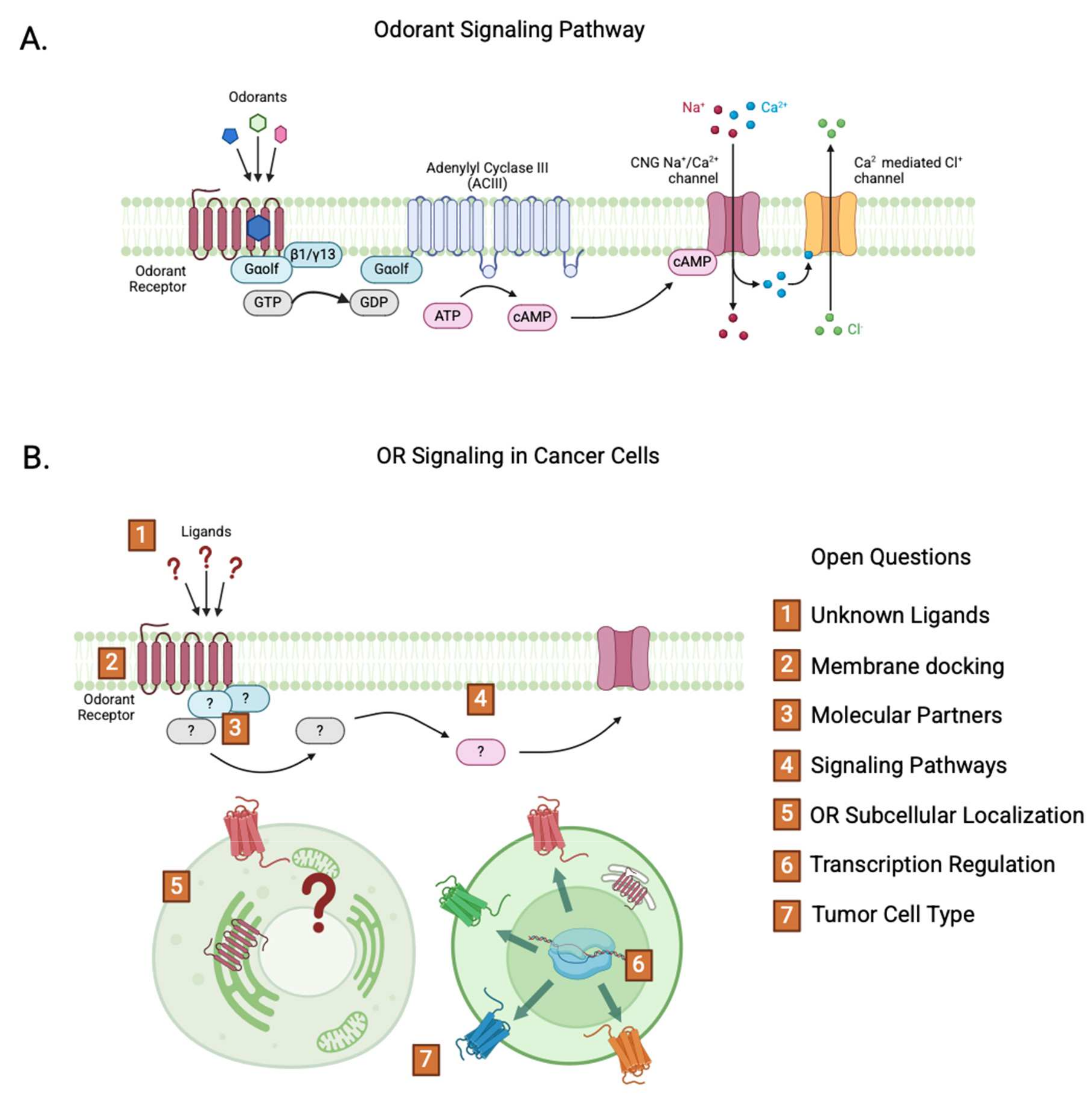
2. Odorant Receptors

In 1991, Buck and Axel [20] identified the OR gene family in rats, which contains approximately 1000 genes in mice and 400 genes in humans [21,22]. They were awarded the Nobel Prize in Physiology or Medicine in 2004 for uncovering the molecular basis of smell.
The ORs are G-protein-coupled receptors (GPCRs) and are located in the cilia of olfactory sensory neurons (OSNs) within the olfactory mucosa. Only one OR gene type is expressed per olfactory neuron, following the “one neuron-one receptor” rule [23,24]. In OSNs, binding of an odorant to its corresponding OR occurs via specific interactions within the receptor’s ligand-binding pocket, which is formed within the transmembrane helixes characteristic of Class A GPCRs. This ligand-receptor interaction induces a conformational change in the OR, activating the associated G protein (Gαolf, in this case), which then triggers the canonical cAMP-mediated signaling cascade leading to neuronal depolarization (Figure 1A).
Odorants are small (200–300 Da) organic volatile molecules which exhibit diverse chemical structures. Despite the reduced number of functional ORs when compared to other mammals, it is estimated that humans can detect thousands of odorants. This is possible because one OR can recognize multiple odorants and one odorant is recognized by multiple ORs in a combinatorial code [25,26]. However, the majority of ORs are still orphan receptors whose ligand(s) are unknown. This is mostly due to the fact that functional assays are difficult to perform with these receptors. While the first OR activation assays were performed in vivo, via calcium imaging and single-cell RT PCR of OSNs of mice exposed to certain odorants [26], today the main activation assays are conducted using heterologous systems. In these experiments, cell lines such as HEK293T are transfected to express ORs which are then activated by odorants. The cells are co-transfected with Gαolf, as well as some type of reporter gene which generally relies on the increase in AMPc caused by the OR activation. However, OR expression in this system has proven itself challenging, as ORs are not always trafficked to the plasmatic membrane and can frequently be trapped in the endoplasmic reticulum [27]. The addition of chaperones, such as RTP1S, can increase the functional expression of some ORs, although other ORs have an increased expression in the absence of RTP1S [28,29]. A tag of rhodopsin, consisting of the first 20 amino acids of the protein, can also be inserted at the amino-terminal end of the OR to increase its trafficking to the membrane without compromising its function [30]. To date, only 83 ORs are fully deorphanized, with effective dose–response curves traced in different concentrations of odorants, as gathered in M2OR, a database for odorant and OR pairs that includes the results from most OR activation assays [31]. Another 253 ORs have been activated by specific odorants in primary and secondary screenings, although these results could not be reproduced in the attempts of generating a dose–response curve [31].
Recently, it was stated that extranasal ORs can be activated not only by a variety of odorant like molecules, such as short-chain fatty acids (SCFAs) like acetate and propionate [32], but also by different types of molecules like the peptide hormones asprosin and insulin peptide [33,34]. Asprosin has specifically activated the mouse OR olfr734 in a heterologous cell activation model based on HEK293T cells, as shown by measuring the activity of CRE-Luc, a reporter for evaluating GPCR-mediated cAMP signaling [33]. This unexpected activation of ORs by a peptide suggests that extranasal ORs may also be activated by ligands that are structurally different from odorants, and probably through a different mechanism than the one that requires the binding of the ligand to the odorant transmembrane binding pocket [35].
GPCRs are the largest family of drug targets in clinical trials, representing 34% of FDA-approved drugs [36]. There are ~800 human GPCRs, and half of them are ORs. The consistent expression of ORs in various types of cancer, detected through diverse OMICs approaches, has highlighted their potential as novel therapeutic targets, as indicated by the increasing number of publications on the subject in recent years (Figure 2). In this review, we focus on the available data regarding ORs and their potential involvement in cancer.

2.1. ORs in Cancer

Curiously, although many cancer cell transcriptomes have ORs being expressed, they seldom express the same specific OR—on the contrary, it appears that each cancer has its own predominantly expressed OR. For example, a study investigated the expression of 301 OR genes in 968 cancer cell lines, and found that 332 of the tested cell lines expressed only one of the OR genes at levels considered to be above the background [37]. While some ORs had expression in cell lines of more than one type of cancer, such as OR4A467, OR1D5 and OR4C46, other ORs were expressed in a single tumor type, such as OR13A1, expressed in B cell malignancies and OR2C3, expressed in melanoma [37]. Most of the cell lines also expressed G protein alpha subunits and adenylyl cyclase III, indicating these receptors could in fact initiate a signaling pathway if present in the cell membrane [37].
The expression of ORs in cancer seems to surpass the “one neuron-one receptor” rule. Several breast cancer cell lines have 3 ORs being expressed at the same time, with HCC1396 having 112 OR genes expressed [4]. In the prostate, OR51E1 and OR51E2 are co-expressed in cell lines as well as patients, being overexpressed in about ⅔ of tumor samples [38]. The same pattern can be seen in Acute Myeloid Leukemia (AML) patients, with nine ORs expressed in a single patient [39]. Although the high number of ORs expressed could be due to different mutations leading to different profiles of cancerous cells in the same patient, the high percentage of cells expressing different ORs highly suggests that they are co-expressed in the same cell. This can be seen in AML, for example, with OR52H1 and OR52B6 being expressed in nearly 100% of The Cancer Genome Atlas (TCGA) patients’ samples [39]. Altogether, that reinforces the possibility of a different mechanism of expression regulation in cancer cells, in comparison to the canonical monogenic expression of ORs in OSNs.
The OR gene family constitutes approximately 3% of the human genome, with around 400 functional and 600 OR pseudogenes [22,40,41]. Recent studies have highlighted the potential role of pseudogenes in various cellular processes, including tumorigenesis [17,42]. In our investigation on OR expression in AML [39], the most differentially expressed pseudogenes were OR7E128P and OR7E126P, both from the OR7E family. These findings align with the results of Flegel and colleagues who investigated the expression profile of ORs among 16 human tissues and found that 58% of the 62 ORs pseudogenes found expressed belong to the OR7E subfamily [43].
Both OR7E126P and OR7E128P genes are located within the 11q13.4 breakpoint interval on chromosome 11, home to the largest OR gene cluster, containing 43% of the OR genes in the human genome [22,44,45]. Chromosomal rearrangements involving chromosome 11, particularly in the KMT2A gene, are common in acute leukemias [46]. While OR genes have been implicated in certain known translocations, such as t (4;11) [47], a recent study suggests that, despite their co-localization, ORs do not directly mediate structural rearrangements of chromosome 11. Instead, it is proposed that these alterations may arise from distinct DNA motifs and recombination mechanisms, whether based on homology or non-homology, which drive the chromosomal instability [48]. Other studies have identified the involvement of OR pseudogenes in lung squamous cell carcinoma and glioblastoma [17,49].
Current data suggest that ORs may play a role in four of the eight hallmarks of cancer [50]: proliferation; angiogenesis; invasion and metastasis; and cell death. Depending on the cancer type and context, ORs can either positively or negatively regulate these pathways. The signaling pathways activated by ORs in cancer differ from traditional olfactory signaling depending on the cell type. The most-associated pathways include MAPK/ERK, PLC-IP3/Ca2+; PKA-CREB-HES1; PI3K-AKT; Gβ-γ-PI3Kγ-ARF1; MAPK p38; and SAPK/JNK [15,38,51,52,53,54,55].
In the next section, we will describe some ORs that were identified in different cancer types (Table 1). Some ORs exhibited a significant correlation between their expression and patient survival rates five years later, as shown in the overall survival rate (OS) column of Table 1. This association suggests that OR expression may have potential as a prognostic factor. It is worth noting that some ORs, such as OR7A5, are associated with either lower or higher OS rates, depending on the cancer type. The factors influencing the impact of ORs on OS remain unclear; however, these associations were never contradictory within the same cancer type, suggesting that the role of a given OR is likely shaped by the specific cellular and molecular context. Further studies are needed to clarify how ORs influence OS across different cancer types.
There are still many open questions regarding the role of ORs in cancer (Figure 1B). The downstream signaling pathways of most ORs remain poorly characterized in cancer cells, and evidence suggests that the same receptor can activate distinct pathways depending on the cellular context—leading to entirely different effects across tumor types. Additionally, key aspects such as ligand identity, membrane localization, transcriptional regulation, subcellular distribution, and the presence of specific molecular partners are still largely unknown. These variables highlight the complexity and context-dependency of OR function in cancer. Whenever available, these aspects will be further discussed in the sections dedicated to each cancer type.

2.1.1. Prostate Cancer

Prostate cancer was one of the first to have an OR identified as up-regulated [70]. Initially thought to be a different type of GPCR, OR51E2 is also known as PSGR (prostate-specific G protein coupled receptor). Besides OR51E2, another OR, the OR51E1, is also highly expressed in prostate cancer. Both ORs have an at-least 10-times-higher expression in two out of three prostate cancer tumors in comparison to a healthy prostate [38]. However, the overexpression of OR51E1 alone leads to ERK1/2 phosphorylation and ultimately to cell cycle arrest via activation of p53 [38].
While OR51E1 is activated by longer aliphatic acids (3C–6C), OR51E2 is activated by shorter fatty acids, such as acetate and propionate [38]. OR51E2 is also activated by beta-ionone as well as by steroids such as ADT and 19OH AD, and inhibited by alpha-ionone [71]. Its activation was shown to induce the phosphorylation of MAPK p38 and SAPK/JNK, ultimately leading to a 50% reduction in cell proliferation [71,72]. Additionally, it has been shown in two prostate cancer cell lines, DU145 and LNCaP, that the activation of OR51E2 by beta-ionone was also able to lead to the activation of ERK1/2, via the Gβγ and PI3Kγ signaling pathway. The pharmacological inhibition of Gβγ and PI3Kγ signaling reduced metastasis, even with the activation of OR51E2, suggesting an important role of Gβγ and PI3Kγ in mediating OR51E2 function in prostate cancer cells [16].

2.1.2. Breast Cancer

In breast cancer, different odorants (citral, cyclovertal and citrathal R), both individually and in a mixture, were able to reduce the expression of the Ki67 proliferation marker in three cell lines, in addition to inducing the cells to undergo apoptosis through a higher expression of p53 [73]. Citral and cyclovertal were also able to activate signaling pathways related to cell growth and survival, by decreasing the expression of ERK1 and 2, and p38-MAPK [73].
Although 352 ORs had expressions of more than a two-fold-change in comparison to normal tissues, three ORs were highlighted for their higher expression among others, as well as for having been associated with different subtypes of breast cancer [4,57,73]. OR2W3 is correlated with stage IV breast cancer, which is slightly more prevalent in the triple negative subtype and in the basal-like subtype, known to be more aggressive. This receptor was highly correlated with genes of tumor invasion, such as CTSV and MMP11 [4]. Moreover, OR2W3 has been associated with shorter survival, of only 35% after 150 months against 85% after 150 months in patients with a normal expression of the OR. Therefore, this OR could be used as a prognostic marker for the subtypes where it is present [4].
OR5B21, on the other hand, is highly expressed in breast cancer metastasis, especially in the brain but also in bones and lungs [57]. In assays where OR5B21 was knocked down, the cells showed less invasion and migration capacity, through the inhibition of STAT3 and NF-kB p65 phosphorylation. Although proliferation was not altered, there was a reduction in metastasis and an overall better survival rate [57]. Finally, OR2B6 has been correlated to the breast cancer of subtype Luminal A, a less aggressive subtype [4]. Still, it is correlated to cell proliferation markers, such as MKI67, MYBL2 and CCNB1 [4].
Lastly, OR6M1, expressed in MCF-7 cells, a human cancer cell line derived from estrogen receptor (ER)-positive breast cancer, was shown to be activated by antroquinone in concentrations of 50 and 100µM, ultimately leading to a decrease in cell viability as well as cell death [55].

2.1.3. Leukemia

Different types of leukemia have exhibited altered expression of ORs. In chronic myeloid leukemia (CML), OR2AT4 was found to be highly expressed in the K562 cell line, and its activation by sandalore was able to induce apoptosis and differentiation, as well as leading to a reduction in proliferation [52]. Another study with the same cell line showed that the same OR can be activated by (−)-epigallocatechin gallate (EGCG), leading to a reduction in cell viability as well as extrinsic apoptosis via Caspase-3 and 8 [60]. A decrease in phosphorylation of proteins related to cell proliferation was also shown following the activation, especially in AKT, p38-MAPK and STAT5. The same assay was conducted in a cell line derived from an acute lymphoblastic leukemia (ALL) patient, MOLT-4, which does not express OR2AT4, and none of the previous mentioned results were obtained, indicating these results are OR dependent [60]. Other non-olfactory GPCRs have been described as being correlated to subtypes of AML, and it is possible that some ORs could also be associated with different AML subtypes, which were probably filtered out from this study due to their relatively low expression levels [74].
Analysis of the TCGA cohort brought up 19 ORs enriched in AML in comparison to healthy blood samples, with 16 of them being validated in an independent AML cohort, the BEAT AML cohort [39]. Of these, OR52B6, OR52H1, OR2AK2, OR13D1 and OR52K2 were found in 90% of the samples. Given the different subtypes of AML, these ORs could be valuable markers or even drug targets for the disease, independently of the subtype, since these ORs also did not show a considerable expression in any of the healthy tissue samples available on Genotype-Tissue Expression Project (GTEx). Moreover, these ORs seem to be AML specific, since they do not show significant expression in other TCGA cancer samples. In addition, 3 of the 16 identified ORs (OR2G2, OR2AE1 and OR10A2) were correlated with worse survival rates [39].

2.1.4. Melanoma

In melanoma cell lines, OR2C3 stands out by having a higher expression among all other ORs in 8 out of 52 of the tested cell lines [37]. The same OR can be found expressed in melanoma samples from patients derived from TCGA, although it is expressed in only 14 out of the 474 samples. Still, none of the healthy tissues available on GTEX had such high expression of this gene [37].
Another OR, OR51E2, was upregulated four-fold in metastatic melanoma cells, but not on other dermal tissue cells around the cancer [3]. In vitro assays showed an OR51E2-depentent increase in intracellular Ca2+ in melanoma primary cell cultures in the presence of β-ionone. This led to a dose-dependent decrease in cell number through apoptosis, in addition to the inhibition of cell migration [3].

2.1.5. Gliomas

In glioblastoma (GBM), more than nine ORs have been reported as having differential expression when compared to healthy tissues [49]. ORs such as OR2B6 and OR51E1 had a higher expression in GBM samples when compared to low-grade glioma (LGG) [49]. OR7D2 was expressed in all malignant stages of GBM, regardless of sample heterogeneity. OR4N2 was more frequent in malign cells, and is likely involved in the tumoral plasticity mechanism, promoting GBM adaptation and resistance. OR51E1 was highly expressed in pericytes in the tumor microenvironment, likely related to angiogenesis and contributing to the tumor microenvironment (TME) remodeling. OR2B11 was also found expressed in the TME, but specifically associated with TME macrophages, as well as in the mesenchymal subtype of GBM. It is believed that OR2B11 has a role in the modulation of an immunosuppressant TME, via NF-kB/TGF-B signaling, which could lead to immunotherapy resistance. Finally, OR2L13 was found expressed predominantly in oligodendrocytes and neurons, and could contribute to tumor recurrence and resistance. Nonetheless, two OR pseudogenes also had significant expression in GBM. OR7E156P had been associated with miR-143/HIF1A, known to promote tumor growth and invasion, thus favoring GBM progression. OR7E14P, on the other hand, was enriched in MES-like GBM states and could be related to the subtype aggressiveness as well as tumor resistance [49].
In another study, a few more ORs were found to be upregulated both in LGG and GBM, including OR2A9P, another pseudogene [62]. OR7A5 was linked to a worse prognostic and lower overall survival in five years, being considered as an oncogene and involved in the initiation and progression of the disease with an increase in in vitro cell proliferation depending on the expression of the receptor. The OR was found to modulate lipid metabolism, possibly through pathways such as cAMP/HSL, cAMP/CREB and cAMP/MAPK, which could affect the malignant progression of the tumor [62].

2.1.6. Gastric Cancer

Gastric cancer is the third most frequent cancer to lead to the death of patients, because its diagnosis often happens in advanced stages of the disease, leading to poor prognosis [61]. Although not functionally expressed, the pseudogene OR3A4 was found to affect gastric cancer [61]. OR3A4P was identified through a microarray platform specifically set for lncRNAs related to metastasis, and was highly expressed in primary tissues as well as metastasis tissues and in the blood of patients of gastric cancer. With its high expression, OR3A4P was shown to promote high expression of other genes, such as PCNA, responsible for increased processivity of DNA replication, VEGF-C, which promotes angiogenesis, Ki-67, associated with cellular proliferation, among others [61]. Its presence in the circulating blood poses the OR3A4P as an interesting potential biomarker to the disease, with its detection reaching 86.94% sensitivity and 91.27% specificity in patients, which could enhance early diagnosis of the cancer and, thus, better prognosis. High levels of OR3A4P were related to shorter OS, shorter interval before relapse and, most importantly, highly associated with both metastasis and angiogenesis. In vitro, OR3A4P overexpression led to a significant increase in both proliferation and cell invasion and migration, while its silencing reduced all of them [61].
Curiously, the OR3A4P is expressed in other types of cancer, such as in breast cancer, ovarian cancer, hepatocellular carcinoma, colorectal cancer, non-small-cell lung cancer, osteosarcoma and in diffuse large B-cell lymphoma (DLBCL) [56,59,61,63,65,66]. In DLBCL patients, the FOXM1 induced the upregulation of OR3A4P and patients with high OR3A4P expression presented poor prognosis. Moreover, the same study showed that the knockdown of OR3A4P suppressed cell proliferation and promoted cell apoptosis in DLBCL by inactivating the Wnt/β-catenin signaling pathway [56].

2.2. ORs and Tumor Microenvironment

OR2B11, OR52K1 and OR3A2 were found not only in GBM but in its TME, with a possible role in the promotion of tumorigenicity [49]. Furthermore, OR2B11 was also found in macrophages of the TME associated with the tumor, and could have an important role in the modulation of the TME to sustain tumor growth. OR2B11 has also been related to having a high expression in macrophages in bone marrow and microglia. In the mesenchymal subtype of GBM, it is believed that OR2B11 could promote mesenchymal transition, contributing to an aggressive TME and therefore associated with a lower survival rate and decreased response to immunotherapy [49].
In mice, Olfr78 was expressed in macrophages derived from the blood marrow [75]. Curiously, this OR could be activated by the lactate found in TME because of the Warburg effect. Tumor associated macrophages (TAMs) can be categorized in two subtypes: M1, antitumoral and M2, tumoral. The activation of Olfr78 by lactate seems to induce the transformation of TAM into an M2 phenotype. In the absence of Olfr78, this effect was not seen in the presence of lactate. This could explain why lower expression of the human Olfr78 counterpart OR51E2, in cancers such as breast, glioblastoma and lung adenocarcinoma, is correlated with longer survival rates [75].

2.3. ORs and Angiogenesis

Angiogenesis is essential, especially to solid tumors, for the oxygen transportation during cancer development. OR51E1 has been found in endothelial cells as well as cells in the pericyte, in addition to being correlated with genes of vascular remodeling and angiogenesis, and could have a vascular function [49]. In gastric cancer, OR3A4P was found to modulate the expression of pro-angiogenesis factors such as VEGF-C and MMP9, with its overexpression significantly increasing the expression of both genes, while its silencing led to a reduction in their expression. Also, the same OR had the capacity of regulating the activity of other genes involved in angiogenesis, such as NTN4 [61].

3. Conclusions and Perspectives

Here, we highlighted the growing evidence of OR expression in diverse cancer types. These extranasal ORs are emerging as possible novel players in tumorigenesis, with implications for cell proliferation, migration, invasion, apoptosis, and metabolic regulation. Their apparent context-specific nature also underscores the complexity of their roles within the tumor. As mentioned across this review, this represents a largely unexplored field of cancer biology demanding continued interdisciplinary efforts at the intersection of cell and molecular biology, pharmacology, and oncology.
Despite the potential of ORs as therapeutic targets for various cancers, several challenges explain why, although 40% of all drugs target GPCRs, none currently target ORs (Figure 1B and Table 2).
A primary challenge is the lack of specific agonists for most ORs, which remain “orphan” receptors. Outside the olfactory system, endogenous ligands with different structures, when compared to that of odorants, may also bind to and activate the extranasal ORs. As described above, hormone peptides, which are larger in size than odorants, have also been described as able to activate ORs [33,34]. However, the previously mentioned difficulty of achieving heterologous expression of these receptors limits the identification of potential ligands. Additionally, for a ligand to be used as an activator for an OR in cancer, it would have to be specific to that given OR. In the olfactory system, odorants are known for being able to activate more than one OR [26]. Given the expression of several ORs throughout the human body, a non-specific ligand could trigger unforeseen effects outside the cancer cells.
While ORs are expressed in certain cancers, the absence of viable antibodies complicates the validation of their functional presence in the membrane—in OSNs and in in vitro heterologous systems, this process is dependent on specific chaperone proteins. Still, it is uncertain whether the functional expression is necessary for ORs to have a role in cancer, as shown from the example described in the gastric cancer session of this review, where the overexpression of the pseudogene OR3A4 leads to an increase in cell proliferation, migration and invasion. Additionally, certain OR pseudogenes may play a role in lung cancer and other malignancies [17,42].
Despite these hurdles, the abnormal and tumor-specific expression of extranasal ORs offers a valuable tool for precise cancer diagnosis, prognosis, and patient stratification. Targeted expression of these receptors could also assist in measuring minimal residual disease (MRD) in patients undergoing treatment, since many cancers have no molecular marker to perform the molecular follow up. Furthermore, this specificity has been exploited for therapeutic purposes, such as using chimeric antigen receptor (CAR)-expressing T cells to target OR2H1-expressing tumor cells both in vitro and in vivo in mice [76]. While CAR T cell therapy has shown relative success in treating hematological malignancies, it has not yet proven effective for solid tumors. The recombinant OR2H1 IgG generated in this study specifically detected the OR2H1 protein in 60 human lung cancers, 40 ovarian carcinomas, and 73 cholangiocarcinomas, offering a promising new therapeutic approach for epithelial cancers [76].
In conclusion, advancing our understanding of extranasal ORs in both healthy tissues and tumors is crucial for enhancing cancer treatment strategies and modulating cellular processes to better control cancer progression.

Author Contributions

Conceptualization, R.G.N., B.M. and L.M.G.; data curation, R.G.N.; figures and table, R.G.N.; writing—original draft preparation, R.G.N. and L.M.G.; writing—review and editing, B.M. All authors have read and agreed to the published version of the manuscript.

Funding

RGN and BM are supported by FAPESP (#2024/02568-9 and #2022/12481-2).

Institutional Review Board Statement

Not applicable.

Informed Consent Statement

Not applicable.

Data Availability Statement

No new data were created or analyzed in this study. Data sharing is not applicable to this article.

Conflicts of Interest

The authors declare no conflicts of interest.

Abbreviations

The following abbreviations are used in this manuscript:
OROdorant receptor
GPCRG protein coupled receptor
OSNOlfactory sensory neuron
AMLAcute Myeloid Leukemia
TMETumor microenvironment
GBMGlioblastoma
DLBCLDiffuse large B-cell lymphoma
TAMTumor associated macrophages
LGGLow-grade glioma
OSOverall survival

References

  1. Parmentier, M.; Libert, F.; Schurmans, S.; Schiffmann, S.; Lefort, A.; Eggerickx, D.; Ledent, C.; Mollereau, C.; Gerard, C.; Perret, J.; et al. Expression of members of the putative olfactory receptor gene family in mammalian germ cells. Nature 1992, 355, 453–455. [Google Scholar] [CrossRef] [PubMed]
  2. Spehr, M.; Gisselmann, G.; Poplawski, A.; Riffell, J.A.; Wetzel, C.H.; Zimmer, R.K.; Hatt, H. Identification of a testicular odorant receptor mediating human sperm chemotaxis. Science 2003, 299, 2054–2058. [Google Scholar] [CrossRef]
  3. Gelis, L.; Jovancevic, N.; Bechara, F.G.; Neuhaus, E.M.; Hatt, H. Functional expression of olfactory receptors in human primary melanoma and melanoma metastasis. Exp. Dermatol. 2017, 26, 569–576. [Google Scholar] [CrossRef] [PubMed]
  4. Masjedi, S.; Zwiebel, L.J.; Giorgio, T.D. Olfactory receptor gene abundance in invasive breast carcinoma. Sci. Rep. 2019, 9, 13736. [Google Scholar] [CrossRef]
  5. Orecchioni, M.; Kobiyama, K.; Winkels, H.; Ghosheh, Y.; McArdle, S.; Mikulski, Z.; Kiosses, W.B.; Fan, Z.; Wen, L.; Jung, Y.; et al. Olfactory receptor 2 in vascular macrophages drives atherosclerosis by NLRP3-dependent IL-1 production. Science 2022, 375, 214–221. [Google Scholar] [CrossRef] [PubMed]
  6. Poll, B.G.; Xu, J.; Gupta, K.; Shubitowski, T.B.; Pluznick, J.L. Olfactory receptor 78 modulates renin but not baseline blood pressure. Physiol. Rep. 2021, 9, e15017. [Google Scholar] [CrossRef]
  7. Xu, J.; Choi, R.; Gupta, K.; Warren, H.R.; Santhanam, L.; Pluznick, J.L. An evolutionarily conserved olfactory receptor is required for sex differences in blood pressure. Sci. Adv. 2024, 10, eadk1487. [Google Scholar] [CrossRef]
  8. Yang, Z.; Cheng, J.; Shang, P.; Sun, J.P.; Yu, X. Emerging roles of olfactory receptors in glucose metabolism. Trends Cell Biol. 2023, 33, 463–476. [Google Scholar] [CrossRef]
  9. Tong, T.; Ryu, S.E.; Min, Y.; de March, C.A.; Bushdid, C.; Golebiowski, J.; Moon, C.; Park, T. Olfactory receptor 10J5 responding to alpha-cedrene regulates hepatic steatosis via the cAMP-PKA pathway. Sci. Rep. 2017, 7, 9471. [Google Scholar] [CrossRef]
  10. Chang, A.J.; Ortega, F.E.; Riegler, J.; Madison, D.V.; Krasnow, M.A. Oxygen regulation of breathing through an olfactory receptor activated by lactate. Nature 2015, 527, 240–244. [Google Scholar] [CrossRef]
  11. Colinas, O.; Mombaerts, P.; Lopez-Barneo, J.; Ortega-Saenz, P. Carotid Body Function in Tyrosine Hydroxylase Conditional Olfr78 Knockout Mice. Function 2024, 5, zqae010. [Google Scholar] [CrossRef] [PubMed]
  12. Fukuda, N.; Yomogida, K.; Okabe, M.; Touhara, K. Functional characterization of a mouse testicular olfactory receptor and its role in chemosensing and in regulation of sperm motility. J. Cell Sci. 2004, 117, 5835–5845. [Google Scholar] [CrossRef] [PubMed]
  13. Flegel, C.; Vogel, F.; Hofreuter, A.; Schreiner, B.S.; Osthold, S.; Veitinger, S.; Becker, C.; Brockmeyer, N.H.; Muschol, M.; Wennemuth, G.; et al. Characterization of the Olfactory Receptors Expressed in Human Spermatozoa. Front. Mol. Biosci. 2015, 2, 73. [Google Scholar] [CrossRef] [PubMed]
  14. Li, M.; Wang, X.; Ma, R.R.; Shi, D.B.; Wang, Y.W.; Li, X.M.; He, J.Y.; Wang, J.; Gao, P. The Olfactory Receptor Family 2, Subfamily T, Member 6 (OR2T6) Is Involved in Breast Cancer Progression via Initiating Epithelial-Mesenchymal Transition and MAPK/ERK Pathway. Front. Oncol. 2019, 9, 1210. [Google Scholar] [CrossRef]
  15. Rodriguez, M.; Siwko, S.; Zeng, L.; Li, J.; Yi, Z.; Liu, M. Prostate-specific G-protein-coupled receptor collaborates with loss of PTEN to promote prostate cancer progression. Oncogene 2016, 35, 1153–1162. [Google Scholar] [CrossRef]
  16. Xu, X.; Khater, M.; Wu, G. The olfactory receptor OR51E2 activates ERK1/2 through the Golgi-localized Gbetagamma-PI3Kgamma-ARF1 pathway in prostate cancer cells. Front. Pharmacol. 2022, 13, 1009380. [Google Scholar] [CrossRef]
  17. Zhao, Y.Q.; Zhang, H.H.; Wu, J.; Li, L.; Li, J.; Zhong, H.; Jin, Y.; Lei, T.Y.; Zhao, X.Y.; Xu, B.; et al. Prediction of Tumor Microenvironment Characteristics and Treatment Response in Lung Squamous Cell Carcinoma by Pseudogene OR7E47P-related Immune Genes. Curr. Med. Sci. 2023, 43, 1133–1150. [Google Scholar] [CrossRef]
  18. Weber, L.; Al-Refae, K.; Ebbert, J.; Jagers, P.; Altmuller, J.; Becker, C.; Hahn, S.; Gisselmann, G.; Hatt, H. Activation of odorant receptor in colorectal cancer cells leads to inhibition of cell proliferation and apoptosis. PLoS ONE 2017, 12, e0172491. [Google Scholar] [CrossRef]
  19. Naressi, R.G.; Schechtman, D.; Malnic, B. Odorant receptors as potential drug targets. Trends Pharmacol. Sci. 2023, 44, 11–14. [Google Scholar] [CrossRef]
  20. Buck, L.; Axel, R. A novel multigene family may encode odorant receptors: A molecular basis for odor recognition. Cell 1991, 65, 175–187. [Google Scholar] [CrossRef]
  21. Zhang, X.; Firestein, S. The olfactory receptor gene superfamily of the mouse. Nat. Neurosci. 2002, 5, 124–133. [Google Scholar] [CrossRef] [PubMed]
  22. Malnic, B.; Godfrey, P.A.; Buck, L.B. The human olfactory receptor gene family. Proc. Natl. Acad. Sci. USA 2004, 101, 2584–2589. [Google Scholar] [CrossRef] [PubMed]
  23. Monahan, K.; Lomvardas, S. Monoallelic expression of olfactory receptors. Annu. Rev. Cell Dev. Biol. 2015, 31, 721–740. [Google Scholar] [CrossRef] [PubMed]
  24. Nagai, M.H.; Armelin-Correa, L.M.; Malnic, B. Monogenic and Monoallelic Expression of Odorant Receptors. Mol. Pharmacol. 2016, 90, 633–639. [Google Scholar] [CrossRef]
  25. Kurian, S.M.; Naressi, R.G.; Manoel, D.; Barwich, A.S.; Malnic, B.; Saraiva, L.R. Odor coding in the mammalian olfactory epithelium. Cell Tissue Res. 2021, 383, 445–456. [Google Scholar] [CrossRef]
  26. Malnic, B.; Hirono, J.; Sato, T.; Buck, L.B. Combinatorial receptor codes for odors. Cell 1999, 96, 713–723. [Google Scholar] [CrossRef]
  27. Gimelbrant, A.A.; Haley, S.L.; McClintock, T.S. Olfactory receptor trafficking involves conserved regulatory steps. J. Biol. Chem. 2001, 276, 7285–7290. [Google Scholar] [CrossRef]
  28. Saito, H.; Kubota, M.; Roberts, R.W.; Chi, Q.; Matsunami, H. RTP family members induce functional expression of mammalian odorant receptors. Cell 2004, 119, 679–691. [Google Scholar] [CrossRef]
  29. Sharma, R.; Ishimaru, Y.; Davison, I.; Ikegami, K.; Chien, M.S.; You, H.; Chi, Q.; Kubota, M.; Yohda, M.; Ehlers, M.; et al. Olfactory receptor accessory proteins play crucial roles in receptor function and gene choice. Elife 2017, 6, e21895. [Google Scholar] [CrossRef]
  30. Zhuang, H.; Matsunami, H. Synergism of accessory factors in functional expression of mammalian odorant receptors. J. Biol. Chem. 2007, 282, 15284–15293. [Google Scholar] [CrossRef]
  31. Lalis, M.; Hladis, M.; Khalil, S.A.; Briand, L.; Fiorucci, S.; Topin, J. M2OR: A database of olfactory receptor-odorant pairs for understanding the molecular mechanisms of olfaction. Nucleic Acids Res. 2024, 52, D1370–D1379. [Google Scholar] [CrossRef] [PubMed]
  32. Fleischer, J.; Bumbalo, R.; Bautze, V.; Strotmann, J.; Breer, H. Expression of odorant receptor Olfr78 in enteroendocrine cells of the colon. Cell Tissue Res. 2015, 361, 697–710. [Google Scholar] [CrossRef] [PubMed]
  33. Li, E.; Shan, H.; Chen, L.; Long, A.; Zhang, Y.; Liu, Y.; Jia, L.; Wei, F.; Han, J.; Li, T.; et al. OLFR734 Mediates Glucose Metabolism as a Receptor of Asprosin. Cell Metab. 2019, 30, 319–328.e318. [Google Scholar] [CrossRef]
  34. Cheng, J.; Yang, Z.; Ge, X.Y.; Gao, M.X.; Meng, R.; Xu, X.; Zhang, Y.Q.; Li, R.Z.; Lin, J.Y.; Tian, Z.M.; et al. Autonomous sensing of the insulin peptide by an olfactory G protein-coupled receptor modulates glucose metabolism. Cell Metab. 2022, 34, 240–255.e210. [Google Scholar] [CrossRef] [PubMed]
  35. Billesbolle, C.B.; de March, C.A.; van der Velden, W.J.C.; Ma, N.; Tewari, J.; Del Torrent, C.L.; Li, L.; Faust, B.; Vaidehi, N.; Matsunami, H.; et al. Structural basis of odorant recognition by a human odorant receptor. Nature 2023, 615, 742–749. [Google Scholar] [CrossRef]
  36. Hauser, A.S.; Attwood, M.M.; Rask-Andersen, M.; Schioth, H.B.; Gloriam, D.E. Trends in GPCR drug discovery: New agents, targets and indications. Nat. Rev. Drug Discov. 2017, 16, 829–842. [Google Scholar] [CrossRef]
  37. Ranzani, M.; Iyer, V.; Ibarra-Soria, X.; Del Castillo Velasco-Herrera, M.; Garnett, M.; Logan, D.; Adams, D.J. Revisiting olfactory receptors as putative drivers of cancer. Wellcome Open Res. 2017, 2, 9. [Google Scholar] [CrossRef]
  38. Pronin, A.; Slepak, V. Ectopically expressed olfactory receptors OR51E1 and OR51E2 suppress proliferation and promote cell death in a prostate cancer cell line. J. Biol. Chem. 2021, 296, 100475. [Google Scholar] [CrossRef]
  39. Guardia, G.D.A.; Naressi, R.G.; Buzzato, V.C.; da Costa, J.B.; Zalcberg, I.; Ramires, J.; Malnic, B.; Gutiyama, L.M.; Galante, P.A.F. Acute Myeloid Leukemia Expresses a Specific Group of Olfactory Receptors. Cancers 2023, 15, 3073. [Google Scholar] [CrossRef]
  40. Glusman, G.; Yanai, I.; Rubin, I.; Lancet, D. The complete human olfactory subgenome. Genome Res. 2001, 11, 685–702. [Google Scholar] [CrossRef]
  41. Niimura, Y.; Nei, M. Evolution of olfactory receptor genes in the human genome. Proc. Natl. Acad. Sci. USA 2003, 100, 12235–12240. [Google Scholar] [CrossRef]
  42. Nakamura-Garcia, A.K.; Espinal-Enriquez, J. Pseudogenes in Cancer: State of the Art. Cancers 2023, 15, 4024. [Google Scholar] [CrossRef]
  43. Flegel, C.; Manteniotis, S.; Osthold, S.; Hatt, H.; Gisselmann, G. Expression profile of ectopic olfactory receptors determined by deep sequencing. PLoS ONE 2013, 8, e55368. [Google Scholar] [CrossRef]
  44. Buettner, J.A.; Glusman, G.; Ben-Arie, N.; Ramos, P.; Lancet, D.; Evans, G.A. Organization and evolution of olfactory receptor genes on human chromosome 11. Genomics 1998, 53, 56–68. [Google Scholar] [CrossRef]
  45. Olender, T.; Feldmesser, E.; Atarot, T.; Eisenstein, M.; Lancet, D. The olfactory receptor universe--from whole genome analysis to structure and evolution. Genet. Mol. Res. 2004, 3, 545–553. [Google Scholar]
  46. Meyer, C.; Larghero, P.; Almeida Lopes, B.; Burmeister, T.; Groger, D.; Sutton, R.; Venn, N.C.; Cazzaniga, G.; Corral Abascal, L.; Tsaur, G.; et al. The KMT2A recombinome of acute leukemias in 2023. Leukemia 2023, 37, 988–1005. [Google Scholar] [CrossRef]
  47. Ou, Z.; Stankiewicz, P.; Xia, Z.; Breman, A.M.; Dawson, B.; Wiszniewska, J.; Szafranski, P.; Cooper, M.L.; Rao, M.; Shao, L.; et al. Observation and prediction of recurrent human translocations mediated by NAHR between nonhomologous chromosomes. Genome Res. 2011, 21, 33–46. [Google Scholar] [CrossRef]
  48. Redaelli, S.; Grati, F.R.; Tritto, V.; Giannuzzi, G.; Recalcati, M.P.; Sala, E.; Villa, N.; Crosti, F.; Roversi, G.; Malvestiti, F.; et al. Olfactory receptor genes and chromosome 11 structural aberrations: Players or spectators? Hum. Genet. Genom. Adv. 2024, 5, 100261. [Google Scholar] [CrossRef]
  49. Cho, H.J.; Yeo, D.J.; Yang, H.; Koo, J. Comprehensive Transcriptomic Analysis Reveals Cell-Type-Specific Roles of Human Odorant Receptors in Glioblastoma and the Tumor Microenvironment. Int J Mol Sci 2024, 25, 13382. [Google Scholar] [CrossRef]
  50. Hanahan, D. Hallmarks of Cancer: New Dimensions. Cancer Discov. 2022, 12, 31–46. [Google Scholar] [CrossRef]
  51. Massberg, D.; Simon, A.; Haussinger, D.; Keitel, V.; Gisselmann, G.; Conrad, H.; Hatt, H. Monoterpene (-)-citronellal affects hepatocarcinoma cell signaling via an olfactory receptor. Arch. Biochem. Biophys. 2015, 566, 100–109. [Google Scholar] [CrossRef]
  52. Manteniotis, S.; Wojcik, S.; Brauhoff, P.; Mollmann, M.; Petersen, L.; Gothert, J.R.; Schmiegel, W.; Duhrsen, U.; Gisselmann, G.; Hatt, H. Functional characterization of the ectopically expressed olfactory receptor 2AT4 in human myelogenous leukemia. Cell Death Discov. 2016, 2, 15070. [Google Scholar] [CrossRef]
  53. Massberg, D.; Jovancevic, N.; Offermann, A.; Simon, A.; Baniahmad, A.; Perner, S.; Pungsrinont, T.; Luko, K.; Philippou, S.; Ubrig, B.; et al. The activation of OR51E1 causes growth suppression of human prostate cancer cells. Oncotarget 2016, 7, 48231–48249. [Google Scholar] [CrossRef]
  54. Kalbe, B.; Schulz, V.M.; Schlimm, M.; Philippou, S.; Jovancevic, N.; Jansen, F.; Scholz, P.; Lubbert, H.; Jarocki, M.; Faissner, A.; et al. Helional-induced activation of human olfactory receptor 2J3 promotes apoptosis and inhibits proliferation in a signanon-small-cell lung cancer cell line. Eur. J. Cell Biol. 2017, 96, 34–46. [Google Scholar] [CrossRef]
  55. Choi, Y.R.; Shim, J.; Park, J.H.; Kim, Y.S.; Kim, M.J. Discovery of Orphan Olfactory Receptor 6M1 as a New Anticancer Target in MCF-7 Cells by a Combination of Surface Plasmon Resonance-Based and Cell-Based Systems. Sensors 2021, 21, 3468. [Google Scholar] [CrossRef]
  56. Bao, Y.; Tang, Z.; Chen, R.; Yu, X.; Qi, X. Pan-cancer analysis identifies olfactory receptor family 7 subfamily A member 5 as a potential biomarker for glioma. PeerJ 2024, 12, e17631. [Google Scholar] [CrossRef]
  57. Asadi, M.; Ahmadi, N.; Ahmadvand, S.; Jafari, A.A.; Safaei, A.; Erfani, N.; Ramezani, A. Investigation of olfactory receptor family 51 subfamily j member 1 (OR51J1) gene susceptibility as a potential breast cancer-associated biomarker. PLoS ONE 2021, 16, e0246752. [Google Scholar] [CrossRef]
  58. Liu, G.; Hu, X.; Zhou, G. Long non-coding RNA OR3A4 promotes proliferation and migration in breast cancer. Biomed. Pharmacother. 2017, 96, 426–433. [Google Scholar] [CrossRef]
  59. Choi, Y.R.; Na, H.J.; Lee, J.A.; Kim, Y.; Kim, Y.S.; Kim, M.J. Discovery of (-)-epigallocatechin gallate, a novel olfactory receptor 2AT4 agonist that regulates proliferation and apoptosis in leukemia cells. Heliyon 2024, 10, e30298. [Google Scholar] [CrossRef]
  60. Meng, H.; Zhao, B.; Wang, Y. FOXM1-induced upregulation of lncRNA OR3A4 promotes the progression of diffuse large B-cell lymphoma via Wnt/beta-catenin signaling pathway. Exp. Mol. Pathol. 2020, 115, 104451. [Google Scholar] [CrossRef]
  61. Guo, X.; Yang, Z.; Zhi, Q.; Wang, D.; Guo, L.; Li, G.; Miao, R.; Shi, Y.; Kuang, Y. Long noncoding RNA OR3A4 promotes metastasis and tumorigenicity in gastric cancer. Oncotarget 2016, 7, 30276–30294. [Google Scholar] [CrossRef]
  62. Li, W.; Fu, Q.; Man, W.; Guo, H.; Yang, P. LncRNA OR3A4 participates in the angiogenesis of hepatocellular carcinoma through modulating AGGF1/akt/mTOR pathway. Eur. J. Pharmacol. 2019, 849, 106–114. [Google Scholar] [CrossRef]
  63. Giandomenico, V.; Cui, T.; Grimelius, L.; Oberg, K.; Pelosi, G.; Tsolakis, A.V. Olfactory receptor 51E1 as a novel target for diagnosis in somatostatin receptor-negative lung carcinoids. J. Mol. Endocrinol. 2013, 51, 277–286. [Google Scholar] [CrossRef]
  64. Wang, X.; Chen, K.; Zhao, Z. LncRNA OR3A4 Regulated the Growth of Osteosarcoma Cells by Modulating the miR-1207-5p/G6PD Signaling. Onco Targets Ther. 2020, 13, 3117–3128. [Google Scholar] [CrossRef]
  65. Guo, F.; Du, J.; Liu, L.; Gou, Y.; Zhang, M.; Sun, W.; Yu, H.; Fu, X. lncRNA OR3A4 Promotes the Proliferation and Metastasis of Ovarian Cancer Through KLF6 Pathway. Front. Pharmacol. 2021, 12, 727876. [Google Scholar] [CrossRef]
  66. Wei, P.; Tang, H.; Li, D. Insights into pancreatic cancer etiology from pathway analysis of genome-wide association study data. PLoS ONE 2012, 7, e46887. [Google Scholar] [CrossRef]
  67. Cui, T.; Tsolakis, A.V.; Li, S.C.; Cunningham, J.L.; Lind, T.; Oberg, K.; Giandomenico, V. Olfactory receptor 51E1 protein as a potential novel tissue biomarker for small intestine neuroendocrine carcinomas. Eur. J. Endocrinol. 2013, 168, 253–261. [Google Scholar] [CrossRef]
  68. Weber, L.; Schulz, W.A.; Philippou, S.; Eckardt, J.; Ubrig, B.; Hoffmann, M.J.; Tannapfel, A.; Kalbe, B.; Gisselmann, G.; Hatt, H. Characterization of the Olfactory Receptor OR10H1 in Human Urinary Bladder Cancer. Front. Physiol. 2018, 9, 456. [Google Scholar] [CrossRef]
  69. Xu, L.L.; Stackhouse, B.G.; Florence, K.; Zhang, W.; Shanmugam, N.; Sesterhenn, I.A.; Zou, Z.; Srikantan, V.; Augustus, M.; Roschke, V.; et al. PSGR, a novel prostate-specific gene with homology to a G protein-coupled receptor, is overexpressed in prostate cancer. Cancer Res. 2000, 60, 6568–6572. [Google Scholar]
  70. Neuhaus, E.M.; Zhang, W.; Gelis, L.; Deng, Y.; Noldus, J.; Hatt, H. Activation of an olfactory receptor inhibits proliferation of prostate cancer cells. J. Biol. Chem. 2009, 284, 16218–16225. [Google Scholar] [CrossRef]
  71. Abaffy, T.; Bain, J.R.; Muehlbauer, M.J.; Spasojevic, I.; Lodha, S.; Bruguera, E.; O’Neal, S.K.; Kim, S.Y.; Matsunami, H. A Testosterone Metabolite 19-Hydroxyandrostenedione Induces Neuroendocrine Trans-Differentiation of Prostate Cancer Cells via an Ectopic Olfactory Receptor. Front. Oncol. 2018, 8, 162. [Google Scholar] [CrossRef]
  72. Klauser, A.L.; Hirschfeld, M.; Ritter, A.; Rucker, G.; Jager, M.; Gundarova, J.; Weiss, D.; Juhasz-Boss, I.; Berner, K.; Erbes, T.; et al. Anticarcinogenic Effects of Odorant Substances Citral, Citrathal R and Cyclovertal on Breast Cancer in vitro. Breast Cancer Dove Med Press. 2021, 13, 659–673. [Google Scholar] [CrossRef]
  73. Li, M.; Schweiger, M.W.; Ryan, D.J.; Nakano, I.; Carvalho, L.A.; Tannous, B.A. Olfactory receptor 5B21 drives breast cancer metastasis. iScience 2021, 24, 103519. [Google Scholar] [CrossRef]
  74. Maiga, A.; Lemieux, S.; Pabst, C.; Lavallee, V.P.; Bouvier, M.; Sauvageau, G.; Hebert, J. Transcriptome analysis of G protein-coupled receptors in distinct genetic subgroups of acute myeloid leukemia: Identification of potential disease-specific targets. Blood Cancer J. 2016, 6, e431. [Google Scholar] [CrossRef]
  75. Vadevoo, S.M.P.; Gunassekaran, G.R.; Lee, C.; Lee, N.; Lee, J.; Chae, S.; Park, J.Y.; Koo, J.; Lee, B. The macrophage odorant receptor Olfr78 mediates the lactate-induced M2 phenotype of tumor-associated macrophages. Proc. Natl. Acad. Sci. USA 2021, 118, e2102434118. [Google Scholar] [CrossRef]
  76. Martin, A.L.; Anadon, C.M.; Biswas, S.; Mine, J.A.; Handley, K.F.; Payne, K.K.; Mandal, G.; Chaurio, R.A.; Powers, J.J.; Sprenger, K.B.; et al. Olfactory Receptor OR2H1 Is an Effective Target for CAR T Cells in Human Epithelial Tumors. Mol. Cancer Ther. 2022, 21, 1184–1194. [Google Scholar] [CrossRef]
Figure 1. Schematic comparison between the OR signaling pathway in olfactory sensory neurons and in cancer cells. (A) Odorant binding induces a conformational change in the OR structure, leading to the dissociation of GDP from the Gαolf subunit and subsequent GTP binding to Gαolf. Gαolf-GTP activates adenylyl cyclase III (AC3), which increases the intracellular cAMP concentration. Consequently, cyclic nucleotide-gated (CNG) channels are opened and cation concentrations, including calcium ions (Ca2+), transiently increase within the OSN. This influx of cations depolarizes the ciliary membrane and activates calcium-activated chloride channels, enabling chloride ions to efflux from the cell and further amplify the membrane depolarization, thus enhancing the odorant signaling. (B) Open questions regarding OR signaling in cancer cells: (1) while ORs in OSNs can be activated by a wide range of odorants with diverse chemical structures, the endogenous activators of ORs in cancer cells remain unidentified; (2) it is not clear if ORs are targeted to the cell membrane in cancer cells; (3) the specific G protein subunits involved in OR signaling in cancer cells remain unknown; (4) although in vitro studies suggest that ORs activate pathways such as ERK, MAPK, AKT, and PI3K, their precise signaling mechanisms in cancer cells and tissues in vivo are not understood.; (5) it is unclear whether ORs have specific intracellular functions or are localized within particular cellular organelles; (6) multiple ORs have been found to be co-expressed in vitro in cancer cell lines. Although this may suggest that this co-expression occurs within the same individual cancer cells, in contrast to the one neuron-one receptor rule in the olfactory system, this remains uncertain due to the lack of data from single-cell RNA sequencing or single-molecule RNA-FISH. Therefore, the mechanisms governing OR gene transcriptional regulation in cancer cells need to be determined at single-cell resolution; (7) some tumors exhibit significant cellular heterogeneity, but the expression patterns of ORs across different tumor cell subtypes are unclear. Most studies analyze bulk tumor samples, highlighting the need for single-cell approaches to resolve this complexity.
Receptors 04 00011 g001
Figure 2. Emerging focus on olfactory receptors in cancer. The number of publications retrieved using the search query [“Olfactory receptor” + “cancer”] on the PubMed website as of 14 February, 2025.
Receptors 04 00011 g002
Table 1. List of cancer associated ORs.
CancerOROS *Ref.
Acute Myeloid Leukemia (AML)OR2AE1, OR10A2, OR2G2Lower[39]
OR5C1, OR1L6, OR10A4, OR13F1Higher[39]
OR52B6, OR2L3, OR52H1, OR2L5, OR2AK2, OR13D1, OR9A4, OR52K2, OR52K1, OR2G3, OR2B2, OR10A5-[39]
Adrenocortical carcinoma (ACC)OR7A5Higher[56]
Breast cancer (BRCA)OR7A5Higher[56]
OR2W3, OR2B6Lower[4]
OR5B21Higher[57]
OR6M1-[55]
OR2T6-[14]
OR51J1-[58]
OR3A4P-[59]
Chronic Myeloid Leukemia (CML)OR2AT4-[52,60]
Colorectal Cancer (CRC)OR3A4P-[61]
Diffuse Large B-Cell Lymphoma (DLBCL)OR3A4P-[56]
Esophageal Cancer (EC)OR3A4P-[61]
Gallbladder Cancer (GBC)OR3A4P-[61]
Gastric Cancer (GC)OR3A4P-[61]
Glioblastoma multiforme (GBM)OR7A5Lower[62]
OR7E156P-[49]
OR7D2, OR7E14P, OR4N2-[49]
Hepatocellular carcinomaOR3A4P-[63]
Kidney renal clear cell carcinoma (KIRC)OR7A5Lower[62]
Kidney renal papillary cell carcinoma (KIRP)OR7A5Lower[62]
Liver hepatocellular carcinoma (HCC)OR7A5Higher[62]
OR1A2-[51]
Low-grade glioma (LGG)OR7A5Lower[62]
Lung adenocarcinoma (LUAD)OR7A5Lower[62]
Lung squamous cell carcinoma (LUSC)OR7A5Higher[62]
OR51E1-[64]
Malignant Melanoma (MM)OR2C3-[37]
OR51E2-[3]
Non-small-cell lung cancer (NSCLC)OR2J3-[54]
OsteosarcomaOR3A4P-[65]
Ovarian CancerOR3A4P-[66]
Ovarian serous cystadenocarcinoma (OSA)OR7A5Lower[62]
Pancreas (PDAC)OR13C4-[67]
OR3A4P-[61]
Prostate (PCA)OR51E1-[38,53]
OR51E2-[38]
Small intestine neuroendocrine carcinomas (SI-NEC)OR51E1-[68]
Thymoma (THYM)OR7A5Higher[62]
Thyroid cancer (THCA)OR7A5Higher[62]
Urinary bladder cancer (BLCA)OR10H1-[69]
Uterine corpus endometrial carcinoma (UCEC)OR7A5Lower[62]
* OS = Overall survival in five years.
Table 2. Therapeutic potential of ORs in cancer and their challenges.
Therapeutic PotentialChallenges
Therapeutic activation by specific ligands
  • Most ORs remain orphans
  • Ligands often activate multiple ORs;
  • Functional expression is uncertain due to lack of chaperones and membrane targeting validation.
Biomarkers for diagnosis, prognosis, or minimum residual disease
  • Limited availability of efficient and specific validated antibodies;
  • Lack of evidence about the OR expression across cancer subtypes and disease stages.
Therapeutic targets for CAR-T or monoclonal antibodies
  • Limited availability of efficient and specific validated antibodies;
  • Lack of evidence about the OR expression across cancer subtypes, disease stages and across the intratumoral intra-heterogeneity;
  • CAR-T therapies have shown limited efficacy in solid tumors;
  • Potential off-target effects in healthy tissues.
Regulatory function as RNA
  • Poorly understood molecular mechanisms;
  • Limited number of functional studies available.
Disclaimer/Publisher’s Note: The statements, opinions and data contained in all publications are solely those of the individual author(s) and contributor(s) and not of MDPI and/or the editor(s). MDPI and/or the editor(s) disclaim responsibility for any injury to people or property resulting from any ideas, methods, instructions or products referred to in the content.

